Sacroiliitis
Joint Diseases
Spondylitis, Ankylosing
Low Back Pain
Pelvic Bones
Joints
Sacrum
Nerve Block
Back Pain
Pelvic Girdle Pain
Manipulation, Orthopedic
Pain, Referred
Zygapophyseal Joint
Arthritis, Infectious
Sciatica
Arthritis
Lumbosacral Plexus
Anesthetics, Local
HLA-B27 Antigen
Pain Measurement
Lumbar Vertebrae
Anesthesia, Local
Pain Management
Clinical, radiographic and HLA associations as markers for different patterns of psoriatic arthritis. (1/231)
OBJECTIVE: The aim of this study was to examine whether the five clinical forms of psoriatic arthritis (PsA) identified by Moll and Wright (Semin Arthritis Rheum 1973;3:55-78) could be clearly distinguished, especially as the disease evolved over time, to analyse whether radiographic features or HLA associations could define subsets with greater precision and to identify predictors of disease outcome. METHODS: Seventy-three patients (37 males and 36 females) were followed for a median time of 8 yr (range 1-16 yr). A standard clinical protocol was used to assess patients at each visit and two clinical scores. based on the joint areas involved, were defined to evaluate the mode of onset and the evolution of arthritis. X-ray films of the hands, feet and sacroiliac joints were taken and the patients were divided into two categories according to the presence or absence of erosions and an X-ray erosion score was also used. Three classification methods were used to define the different clinical subsets. HLA-A, B and DR antigens were tested by standard microlymphocytotoxicity assays. A multiple linear regression model was used in the statistical analysis. RESULTS: The five classical clinical subsets defined by Moll and Wright did not remain since distinct peripheral arthritis patterns tended to evolve over time. Only two discrete groups were identified, axial disease (AD) (sacroilitis with or without peripheral arthritis) in 29% of cases and peripheral disease (PD) without sacroilitis in 71%. AD was positively associated with the duration of arthritis (P < 0.04), presence of mutilation (P < 0.02) and the joint area score over disease evolution (JASE) (P < 0.02). There were erosions in 71% of the patients. Erosions correlated with the presence of mutilation (P < 0.007) and with the JASE (P < 0.0005). HLA-B27 was found in 43% of patients with AD, but only in 11% of PD patients (P < 0.01). No other clear HLA correlations were found. CONCLUSIONS: Despite the relatively small number of patients, this longitudinal study suggests that only two clinical subsets can be clearly defined in PsA, AD and PD; these are primarily determined on clinical grounds although HLA-B27 is strongly associated with AD. The evolution of PD pattern with time means that narrower peripheral arthritis subsets are of little clinical use. (+info)Three pathways between the sacroiliac joint and neural structures. (2/231)
BACKGROUND AND PURPOSE: Despite ongoing clinical suspicion regarding the relationship between sacroiliac joint (SIJ) dysfunction and lower extremity symptoms, there is a paucity of scientific literature addressing this topic. The purpose of this study was to describe patterns of contrast extravasation during SIJ arthrography and postarthrography CT in patients with lower back pain and to determine whether there are pathways of communication between the SIJ and nearby neural structures. METHODS: Fluoroscopically guided SIJ arthrography was performed on 76 SIJs. After the injection of contrast medium, anteroposterior, lateral, and oblique radiographs as well as 5-mm contiguous axial and direct coronal CT images were obtained. Contrast extravasation patterns were recorded for each joint. These observations included a search for contrast extravasation from the SIJ that contacted nearby lumbosacral nerve roots or structures of the plexus. RESULTS: Sixty-one percent of all joints studied revealed one of five contrast extravasation patterns. Three of these observed patterns show a pathway of communication between the SIJ and nearby neural structures. These included posterior extravasation into the dorsal sacral foramina, superior recess extravasation at the sacral alar level to the fifth lumbar epiradicular sheath, and ventral extravasation to the lumbosacral plexus. CONCLUSION: Three pathways between the SIJ and neural structures exist. (+info)Studying patients with inflammatory back pain and arthritis of the lower limbs clinically and by magnetic resonance imaging: many, but not all patients with sacroiliitis have spondyloarthropathy. (3/231)
OBJECTIVE: Clinical and magnetic resonance imaging (MRI) data of 170 consecutive patients with inflammatory back pain (IBP) and/or oligoarthritis of the lower limbs were evaluated in a retrospective study. The aim was to determine the frequency of sacroiliitis and spondyloarthropathy (SpA) in this population, and to assess the significance of HLA B27 measurements for diagnosis in early disease. METHODS: Pelvic X-rays were performed in all IBP patients and dynamic MRI of the sacroiliac joints in patients with IBP who had indefinite results on sacroiliac X-rays (n = 32). RESULTS: European Spondyloarthropathy Study Group criteria for SpA were fulfilled by 106/170 patients (62.4%); eight additional patients had symptoms suggestive of SpA (4.7%). The most frequent SpA subset was undifferentiated SpA (uSpA), diagnosed in 46/106 patients (43.4%). Sacroiliitis was detected by MRI in 21/32 patients with IBP and unclear X-rays (65.6%). Of those, 14 were diagnosed as SpA and seven females with moderate unilateral sacroiliitis, but no features of SpA, also not on follow-up (at least 1 yr), were classified as undifferentiated sacroiliitis (US). Ten of the 14 SpA (71.4%) and none of the seven US patients were HLA B27 positive. CONCLUSION: HLA B27 positivity in IBP patients with MRI-proven sacroiliitis positively predicts SpA. uSpA is a frequent SpA subset. There are HLA B27-negative non-SpA patients with moderate unilateral sacroiliitis whom we propose to be classified as US. (+info)Four clinical tests of sacroiliac joint dysfunction: the association of test results with innominate torsion among patients with and without low back pain. (4/231)
BACKGROUND AND PURPOSE: The purpose of this study was to assess the association between innominate torsion (asymmetric anteroposterior positioning of the pelvic innominates) and Gillet, standing forward flexion, sitting forward flexion, and supine-to-sit tests. SUBJECTS: A sample of 21- to 50-year-old patients with low back pain (n=150) and a comparison group of patients with upper-extremity impairments (n=138) were recruited from outpatient physical therapy facilities. METHODS: The association of single and combined test results with innominate torsion (calculated from pelvic landmark data) and with presence or absence of low back pain were estimated via odds ratios, sensitivities, specificities, and predictive values. RESULTS: Individual test sensitivities were low (8%-44%), as were negative predictive values (28%-38%), for identifying the presence of innominate torsion. Combining tests and controlling for sex, age group, leg-length difference, or iliac crest level did not improve performance characteristics. The associations of test results with low back pain were weak, with the exception of the Gillet test (odds ratio=4.57). CONCLUSION AND DISCUSSION: The data do not support the value of these tests in identifying innominate torsion, although the use of these tests for identifying other phenomena (eg, sacroiliac joint hypomobility) cannot be ruled out. Further exploration of the association of Gillet test results with low back pain is warranted. (+info)Measurement of sacroiliac joint dysfunction: a multicenter intertester reliability study. (5/231)
BACKGROUND AND PURPOSE: Previous research suggests that visual estimates of sacroiliac joint (SIJ) alignment are unreliable. The purpose of this study was to determine whether handheld calipers and an inclinometer could be used to obtain reliable measurements of SIJ alignment in subjects suspected of having SIJ dysfunction. SUBJECTS: Seventy-three subjects, evaluated at 1 of 5 outpatient clinics, participated in the study. METHODS: A total of 23 therapists, randomly paired for each subject, served as examiners. The angle of inclination of each innominate was measured while the subject was standing. The position of the innominates relative to each other was then derived. An intraclass correlation coefficient (ICC), the standard error of measurement (SEM), and a kappa coefficient were calculated to examine the reliability of the derived measurements. RESULTS: The ICC was .27, the SEM was 5.4 degrees, and the kappa value was .18. CONCLUSION AND DISCUSSION: Measurements of SIJ alignment were unreliable. Therapists should consider procedures other than those that assess SIJ alignment when evaluating the SIJ. (+info)Evolution of chronic recurrent multifocal osteitis toward spondylarthropathy over the long term. (6/231)
OBJECTIVE: To retrospectively assess, with a sufficiently long followup (mean 11.6 years; median 9 years), the long-term outcome of chronic recurrent multifocal osteitis (CRMO), a multifocal, inflammatory bone disease. METHODS: Patients included were 8 children/adolescents and 7 adults with no family history of rheumatic disease who had been diagnosed as having CRMO between 1979 and 1995. Ten patients had undergone at least 1 bone biopsy of the lesions, with histologic examination and multiple cultures. In 1996, in addition to an in-depth interview, 12 patients underwent an extensive physical examination, laboratory evaluation, HLA-A, B, C, and DR typing, bone radiography and scintigraphy, and computed tomography scan of the sternoclavicular and sacroiliac joints. RESULTS: Remission was observed in 3 patients. The other 12 patients developed various associations of vertebral (n = 10), sacroiliac (n = 6), anterior thoracic (n = 7), peripheral articular (n = 2), enthesopathic (n = 4), or dermatologic (palmoplantar pustulosis in 3 cases and psoriasis in 2) involvements. Spine involvement was the most common and occurred the earliest (median time to appearance after the onset of osteitis 5.63 years). Clinical sacroiliitis was always unilateral. No patients carried the HLA-B27 haplotype. CRMO responded well to nonsteroidal antiinflammatory drugs. Twelve patients met the European Spondylarthropathy Study Group criteria for spondylarthopathy. CONCLUSION: After 10 years, CRMO had usually evolved to spondylarthropathy, but with certain features not usually seen in the latter: predominantly, unilateral sacroiliitis, no familial form, and no link with HLA-B27. (+info)The active straight leg raising test and mobility of the pelvic joints. (7/231)
Objective signs to assess impairment in patients who are disabled by peripartum pelvic girdle pain hardly exist. The purpose of this study was to develop a clinical test to quantify and qualify disability in these patients. The study examined the relationship between impaired active straight leg raising (ASLR) and mobility of pelvic joints in patients with peripartum pelvic girdle pain, focusing on (1) the reduction of impairment of ASLR when the patient was wearing a pelvic belt, and (2) motions between the pubic bones measured by X-ray examination when the patient was standing on one leg, alternating left and right. Twenty-one non-pregnant patients with peripartum pelvic girdle pain in whom pain and impairment of ASLR were mainly located on one side were selected. ASLR was performed in the supine position, first without a pelvic belt and then with a belt. The influence of the belt on the ability to actively raise the leg was assessed by the patient. Mobility of the pelvic joints was radiographically visualized by means of the Chamberlain method. Assessment was blinded. Ability to perform ASLR was improved by a pelvic belt in 20 of the 21 patients (binomial two-tailed P = 0.0000). When the patient was standing on one leg, alternating the symptomatic side and the reference side, a significant difference between the two sides was observed with respect to the size of the radiographically visualized steps between the pubic bones (binomial two-tailed P = 0.01). The step at the symptomatic side was on average larger when the leg at that side was hanging down than when the patient was standing on the leg at that side. Impairment of ASLR correlates strongly with mobility of the pelvic joints in patients with peripartum pelvic girdle pain. The ASLR test could be a suitable instrument to quantify and qualify disability in diseases related to mobility of the pelvic joints. Further studies are needed to assess the relationship with clinical parameters, sensitivity, specificity and responsiveness in various categories of patients. In contrast with the opinion of Chamberlain, that a radiographically visualized step between the pubic bones is caused by cranial shift of the pubic bone at the side of the standing leg, it is concluded that the step is caused by caudal shift of the pubic bone at the side of the leg hanging down. The caudal shift is caused by an anterior rotation of the hip bone about a horizontal axis near the sacroiliac joint. (+info)Quantitative analyses of sacroiliac biopsies in spondyloarthropathies: T cells and macrophages predominate in early and active sacroiliitis- cellularity correlates with the degree of enhancement detected by magnetic resonance imaging. (8/231)
OBJECTIVE: Sacroiliitis is a hallmark of the spondyloarthropathies (SpA). The degree of inflammation can be quantified by magnetic resonance imaging (MRI). The aim of this study was to further elucidate the pathogenesis of SpA by quantitative cellular analysis of immunostained sacroiliac biopsy specimens and to compare these findings with the degree of enhancement in the sacroiliac joints (SJ) as detected by dynamic MRI. METHODS: The degree of acute sacroiliitis detected by MRI after intravenous administration of gadolinium-DTPA was quantitatively assessed by calculating the enhancement observed in the SJ and chronic changes were graded as described in 32 patients with ankylosing spondylitis (n=18), undifferentiated SpA (n=12) and psoriatic arthritis (n=2). Back pain was graded on a visual analogue scale (VAS, 0-10) and disease duration (DD) was assessed. Shortly after MRI, SJ of patients with VAS > 5 were biopsied guided by computed tomography. Immunohistological examination was performed using the APAAP technique; only whole sections > 3 mm were counted. RESULTS: By MRI, chronic changes II in 13 patients (group II, DD 7.3 (SD 4.8) years), while enhancement < 70% was found in eight (group A, DD 5.6 (SD 3.3) years) and > 70% in 12 patients (group B, DD 4.7 (SD 5.8) years). The relative percentage of cartilage (78-93%), bone (7-18%) and proliferating connective tissue (1-4%) was comparable between the groups (range). There were more inflammatory cells in group I compared with group II (mean (SD) 26.7(20.1) versus 5.3 (5. 2), p=0.04) and group A compared with B (21.8 (17.3) versus 6.0 (5. 6), p=0.05) cells/10 mm(2)), T cells (10.9 (8.5)) being slightly more frequent than macrophages (9.6 (16.8/10 mm(2))). Clusters of proliferating fibroblasts were seen in three and new vessel formation in seven cases. CONCLUSION: This study shows that T cells and macrophages are the most frequent cells in early and active sacroiliitis in SpA. The correlation of cellularity and MRI enhancement provides further evidence for the role of dynamic MRI to detect early sacroiliitis. (+info)The sacroiliac (SI) joint is the joint that connects the iliac bone (part of the pelvis) and the sacrum (the triangular bone at the base of the spine). There are two sacroiliac joints, one on each side of the spine. The primary function of these joints is to absorb shock between the upper body and lower body and distribute the weight of the upper body to the lower body. They also provide a small amount of movement to allow for flexibility when walking or running. The SI joints are supported and stabilized by strong ligaments, muscles, and bones.
Sacroiliitis is a medical condition characterized by inflammation of one or both of the sacroiliac joints, which connect the spine's sacrum to the hip bones (ilium). This inflammation can cause pain in the lower back, hips, and legs, and may be accompanied by stiffness and difficulty walking. Sacroiliitis can be caused by various factors, including mechanical stress, trauma, infectious diseases, or underlying inflammatory conditions such as ankylosing spondylitis. The diagnosis of sacroiliitis typically involves a combination of physical examination, medical history, imaging studies, and laboratory tests to determine the underlying cause and appropriate treatment.
Spondylarthritis is a term used to describe a group of interrelated inflammatory diseases that primarily affect the spine and sacroiliac joints (where the spine connects to the pelvis), but can also involve other joints, ligaments, tendons, and entheses (sites where tendons or ligaments attach to bones). These conditions share common genetic, clinical, and imaging features.
The most common forms of spondylarthritis include:
1. Ankylosing spondylitis - a chronic inflammatory disease that primarily affects the spine and sacroiliac joints, causing pain and stiffness. In some cases, it can lead to fusion of the spine's vertebrae.
2. Psoriatic arthritis - a form of arthritis that occurs in people with psoriasis, an autoimmune skin condition. It can cause inflammation in the joints, tendons, and entheses.
3. Reactive arthritis - a type of arthritis that develops as a reaction to an infection in another part of the body, often the urinary or gastrointestinal tract.
4. Enteropathic arthritis - a form of arthritis associated with inflammatory bowel diseases like Crohn's disease and ulcerative colitis.
5. Undifferentiated spondylarthritis - when a patient presents with features of spondylarthritis but does not meet the criteria for any specific subtype.
Common symptoms of spondylarthritis include:
- Back pain and stiffness, often worse in the morning or after periods of inactivity
- Peripheral joint pain and swelling
- Enthesitis (inflammation at tendon or ligament insertion points)
- Dactylitis (swelling of an entire finger or toe)
- Fatigue
- Uveitis (inflammation of the eye)
- Skin rashes, such as psoriasis
- Inflammatory bowel disease symptoms
Diagnosis typically involves a combination of medical history, physical examination, laboratory tests, and imaging studies. Treatment often includes nonsteroidal anti-inflammatory drugs (NSAIDs), disease-modifying antirheumatic drugs (DMARDs), biologic agents, and lifestyle modifications to manage symptoms and prevent joint damage.
Joint diseases is a broad term that refers to various conditions affecting the joints, including but not limited to:
1. Osteoarthritis (OA): A degenerative joint disease characterized by the breakdown of cartilage and underlying bone, leading to pain, stiffness, and potential loss of function.
2. Rheumatoid Arthritis (RA): An autoimmune disorder causing inflammation in the synovial membrane lining the joints, resulting in swelling, pain, and joint damage if left untreated.
3. Infectious Arthritis: Joint inflammation caused by bacterial, viral, or fungal infections that spread through the bloodstream or directly enter the joint space.
4. Gout: A type of arthritis resulting from the buildup of uric acid crystals in the joints, typically affecting the big toe and characterized by sudden attacks of severe pain, redness, and swelling.
5. Psoriatic Arthritis (PsA): An inflammatory joint disease associated with psoriasis, causing symptoms such as pain, stiffness, and swelling in the joints and surrounding tissues.
6. Juvenile Idiopathic Arthritis (JIA): A group of chronic arthritis conditions affecting children, characterized by joint inflammation, pain, and stiffness.
7. Ankylosing Spondylitis: A form of arthritis primarily affecting the spine, causing inflammation, pain, and potential fusion of spinal vertebrae.
8. Bursitis: Inflammation of the fluid-filled sacs (bursae) that cushion joints, leading to pain and swelling.
9. Tendinitis: Inflammation or degeneration of tendons, which connect muscles to bones, often resulting in pain and stiffness near joints.
These conditions can impact the function and mobility of affected joints, causing discomfort and limiting daily activities. Proper diagnosis and treatment are essential for managing joint diseases and preserving joint health.
Ankylosing spondylitis is a type of inflammatory arthritis that primarily affects the spine, although other joints can also be involved. It causes swelling in the spinal joints (vertebrae) that can lead to stiffness and pain. Over time, some of these joints may grow together, causing new bone formation and resulting in a rigid spine. This fusion of the spine is called ankylosis.
The condition typically begins in the sacroiliac joints, where the spine connects to the pelvis. From there, it can spread up the spine and potentially involve other areas of the body such as the eyes, heart, lungs, and gastrointestinal system.
Ankylosing spondylitis has a strong genetic link, with most people carrying the HLA-B27 gene. However, not everyone with this gene will develop the condition. It primarily affects males more often than females and tends to start in early adulthood.
Treatment usually involves a combination of medication, physical therapy, and exercise to help manage pain, maintain mobility, and prevent deformity. In severe cases, surgery may be considered.
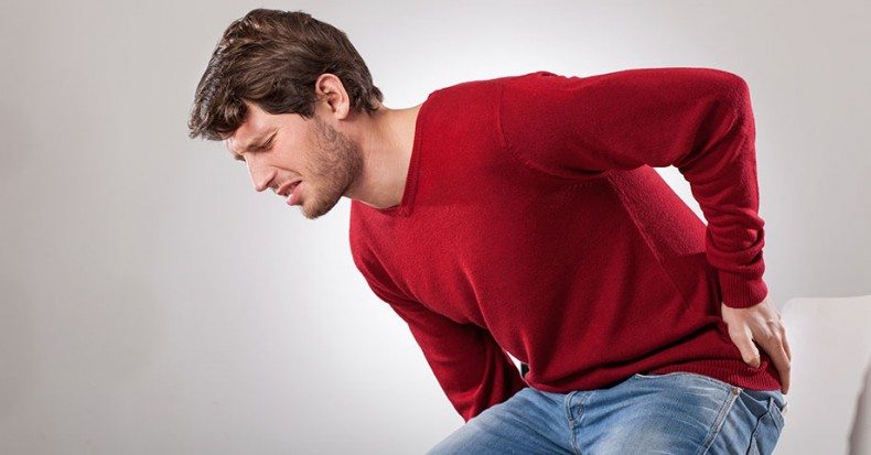
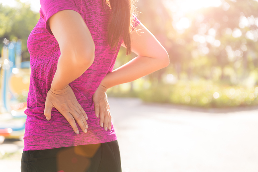
Low back pain is a common musculoskeletal disorder characterized by discomfort or pain in the lower part of the back, typically between the costal margin (bottom of the ribcage) and the gluteal folds (buttocks). It can be caused by several factors including strain or sprain of the muscles or ligaments, disc herniation, spinal stenosis, osteoarthritis, or other degenerative conditions affecting the spine. The pain can range from a dull ache to a sharp stabbing sensation and may be accompanied by stiffness, limited mobility, and radiating pain down the legs in some cases. Low back pain is often described as acute (lasting less than 6 weeks), subacute (lasting between 6-12 weeks), or chronic (lasting more than 12 weeks).
Intra-articular injections refer to the administration of medication directly into a joint space. This route of administration is used for treating various joint conditions such as inflammation, pain, and arthritis. Commonly injected medications include corticosteroids, local anesthetics, and viscosupplementation agents. The procedure is usually performed using imaging guidance, like ultrasound or fluoroscopy, to ensure accurate placement of the medication within the joint.
The pelvic bones, also known as the hip bones, are a set of three irregularly shaped bones that connect to form the pelvic girdle in the lower part of the human body. They play a crucial role in supporting the spine and protecting the abdominal and pelvic organs.
The pelvic bones consist of three bones:
1. The ilium: This is the largest and uppermost bone, forming the majority of the hip bone and the broad, flaring part of the pelvis known as the wing of the ilium or the iliac crest, which can be felt on the side of the body.
2. The ischium: This is the lower and back portion of the pelvic bone that forms part of the sitting surface or the "sit bones."
3. The pubis: This is the front part of the pelvic bone, which connects to the other side at the pubic symphysis in the midline of the body.
The pelvic bones are joined together at the acetabulum, a cup-shaped socket that forms the hip joint and articulates with the head of the femur (thigh bone). The pelvic bones also have several openings for the passage of blood vessels, nerves, and reproductive and excretory organs.
The shape and size of the pelvic bones differ between males and females due to their different roles in childbirth and locomotion. Females typically have a wider and shallower pelvis than males to accommodate childbirth, while males usually have a narrower and deeper pelvis that is better suited for weight-bearing and movement.
Osteoarticular tuberculosis is a form of extrapulmonary tuberculosis (TB) that involves the bones and joints. It is caused by the bacterium Mycobacterium tuberculosis. The infection can spread to the bones and joints through the bloodstream or from nearby infected organs, such as the lungs.
The most commonly affected sites are the spine (Pott's disease), hip, knee, wrist, and small bones of the hands and feet. Symptoms may include pain, swelling, stiffness, and decreased range of motion in the affected joint or bone. In some cases, the infection can lead to deformity, chronic disability, or even death if left untreated.
Diagnosis typically involves a combination of medical history, physical examination, imaging studies (such as X-rays, CT scans, or MRI), and laboratory tests (such as blood tests, sputum cultures, or biopsy). Treatment usually consists of a long course of antibiotics (usually for at least six months) to kill the bacteria. Surgery may also be necessary in some cases to remove infected tissue or stabilize damaged joints.
A joint is the location at which two or more bones make contact. They are constructed to allow movement and provide support and stability to the body during motion. Joints can be classified in several ways, including structure, function, and the type of tissue that forms them. The three main types of joints based on structure are fibrous (or fixed), cartilaginous, and synovial (or diarthrosis). Fibrous joints do not have a cavity and have limited movement, while cartilaginous joints allow for some movement and are connected by cartilage. Synovial joints, the most common and most movable type, have a space between the articular surfaces containing synovial fluid, which reduces friction and wear. Examples of synovial joints include hinge, pivot, ball-and-socket, saddle, and condyloid joints.
The sacrum is a triangular-shaped bone in the lower portion of the human vertebral column, located between the lumbar spine and the coccyx (tailbone). It forms through the fusion of several vertebrae during fetal development. The sacrum's base articulates with the fifth lumbar vertebra, while its apex connects with the coccyx.
The sacrum plays an essential role in supporting the spine and transmitting weight from the upper body to the pelvis and lower limbs. It also serves as an attachment site for various muscles and ligaments. The sacral region is often a focus in medical and chiropractic treatments due to its importance in spinal stability, posture, and overall health.
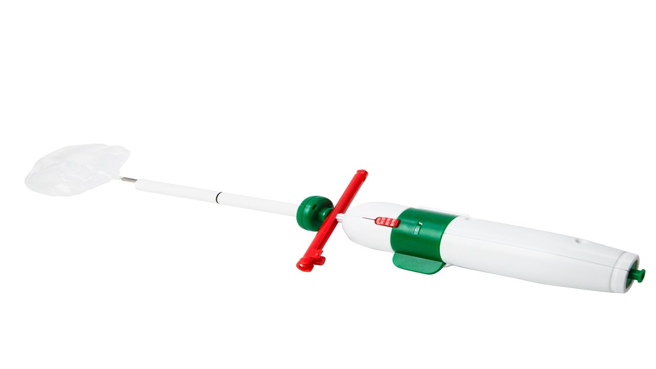
A nerve block is a medical procedure in which an anesthetic or neurolytic agent is injected near a specific nerve or bundle of nerves to block the transmission of pain signals from that area to the brain. This technique can be used for both diagnostic and therapeutic purposes, such as identifying the source of pain, providing temporary or prolonged relief, or facilitating surgical procedures in the affected region.
The injection typically contains a local anesthetic like lidocaine or bupivacaine, which numbs the nerve, preventing it from transmitting pain signals. In some cases, steroids may also be added to reduce inflammation and provide longer-lasting relief. Depending on the type of nerve block and its intended use, the injection might be administered close to the spine (neuraxial blocks), at peripheral nerves (peripheral nerve blocks), or around the sympathetic nervous system (sympathetic nerve blocks).
While nerve blocks are generally safe, they can have side effects such as infection, bleeding, nerve damage, or in rare cases, systemic toxicity from the anesthetic agent. It is essential to consult with a qualified medical professional before undergoing this procedure to ensure proper evaluation, technique, and post-procedure care.
Back pain is a common symptom characterized by discomfort or soreness in the back, often occurring in the lower region of the back (lumbago). It can range from a mild ache to a sharp stabbing or shooting pain, and it may be accompanied by stiffness, restricted mobility, and difficulty performing daily activities. Back pain is typically caused by strain or sprain to the muscles, ligaments, or spinal joints, but it can also result from degenerative conditions, disc herniation, spinal stenosis, osteoarthritis, or other medical issues affecting the spine. The severity and duration of back pain can vary widely, with some cases resolving on their own within a few days or weeks, while others may require medical treatment and rehabilitation.
The ilium is the largest and broadest of the three parts that make up the hip bone or coxal bone. It is the uppermost portion of the pelvis and forms the side of the waist. The ilium has a curved, fan-like shape and articulates with the sacrum at the back to form the sacroiliac joint. The large, concave surface on the top of the ilium is called the iliac crest, which can be felt as a prominent ridge extending from the front of the hip to the lower back. This region is significant in orthopedics and physical examinations for its use in assessing various medical conditions and performing certain maneuvers during the physical examination.
Pelvic girdle pain (PGP) is a condition characterized by pain in the pelvic joints, muscles, and ligaments during pregnancy or after childbirth. It can also affect people who have had trauma to the pelvis or have certain medical conditions that affect the joints. The pain may be localized to one side of the body or spread across both sides of the pelvis.
PGP is caused by increased laxity in the pelvic joints, which can result from hormonal changes during pregnancy or from trauma to the area. This increased laxity can cause the joints to move unevenly, leading to pain and inflammation. In some cases, the pain may be accompanied by stiffness, clicking or grinding sounds in the pelvic area, and difficulty walking or standing for long periods of time.
PGP is typically diagnosed based on a physical examination and medical history. Treatment may include physical therapy, pain management techniques such as heat or cold therapy, and in some cases, bracing or surgery. It's important to seek medical attention if you experience pelvic pain, as early intervention can help prevent long-term complications and improve outcomes.
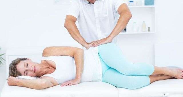
Orthopedic manipulation is a hands-on technique that is used by healthcare professionals, such as orthopedic doctors, chiropractors, and physical therapists, to diagnose and treat muscle and joint disorders. This manual procedure involves moving the joints or soft tissues in a specific direction and amplitude with the aim of improving joint mobility, reducing pain, relieving muscle tension, and enhancing overall function.
Orthopedic manipulation can be performed on various parts of the body, including the spine, extremities, and cranial structures. It is often used as a complementary treatment alongside other therapeutic interventions, such as exercise, medication, or surgery, to manage a wide range of musculoskeletal conditions, including but not limited to:
* Back pain and stiffness
* Neck pain and stiffness
* Joint pain and inflammation
* Muscle spasms and tension
* Headaches and migraines
* Disc disorders
* Sprains and strains
* Postural dysfunctions
It is important to note that orthopedic manipulation should only be performed by trained and licensed healthcare professionals, as improper techniques can lead to injury or further damage. Patients should consult with their healthcare provider to determine if orthopedic manipulation is an appropriate treatment option for their specific condition.
The pubic bone, also known as the pubis or pubic symphysis, is a part of the pelvis - the complex ring-like structure that forms the lower part of the trunk and supports the weight of the upper body. The pubic bone is the anterior (front) portion of the pelvic girdle, located at the bottom of the abdomen, and it connects to the other side at the pubic symphysis, a cartilaginous joint.
The pubic bone plays an essential role in supporting the lower limbs and providing attachment for various muscles involved in movements like walking, running, and jumping. It also protects some abdominal organs and contributes to the structure of the pelvic outlet, which is crucial during childbirth.
Referred pain is a type of pain that is felt in a part of the body other than its actual source. This occurs because the brain incorrectly interprets nerve signals from damaged tissues or organs. In the case of referred pain, the brain misinterprets the location of the pain signal and attributes it to a different area of the body.
Referred pain is often described as a dull, aching sensation rather than a sharp, stabbing pain. It can be difficult to diagnose because the source of the pain may not be immediately apparent. Common examples of referred pain include:
* Heart attack pain that is felt in the left arm or jaw
* Gallbladder pain that is felt in the right shoulder blade
* Kidney stones that cause pain in the lower back and abdomen
* Appendicitis that causes pain in the lower right quadrant of the abdomen, but can sometimes be referred to the lower left quadrant in pregnant women or those with a longer colon.
Referred pain is thought to occur because the nerves carrying pain signals from different parts of the body converge on the same neurons in the spinal cord before traveling to the brain. If these neurons are stimulated by pain signals from multiple sources, the brain may have difficulty distinguishing between them and may interpret the pain as coming from a single location.
Spondylitis is a term used to describe inflammation in the spinal vertebrae, often leading to stiffness and pain. The most common form is Ankylosing Spondylitis, which is a chronic autoimmune disease where the body's immune system mistakenly attacks the joints in the spine. This can cause the bones in the spine to grow together, resulting in a rigid and inflexible spine. Other forms of spondylitis include reactive spondylitis, infectious spondylitis, and seronegative spondyloarthropathies. Symptoms may also include pain and stiffness in the neck, lower back, hips, and small joints of the body.
A zygapophyseal joint, also known as a facet joint, is a type of synovial joint that connects the articulating processes of adjacent vertebrae in the spine. These joints are formed by the superior and inferior articular processes of the vertebral bodies and are covered with hyaline cartilage. They allow for smooth movement between the vertebrae, providing stability and limiting excessive motion while allowing flexibility in the spine. The zygapophyseal joints are supported by a capsule and ligaments that help to maintain their alignment and restrict abnormal movements. These joints can become sources of pain and discomfort when they become inflamed or damaged due to conditions such as arthritis, degenerative disc disease, or injury.
Infectious arthritis, also known as septic arthritis, is a type of joint inflammation that is caused by a bacterial or fungal infection. The infection can enter the joint through the bloodstream or directly into the synovial fluid of the joint, often as a result of a traumatic injury, surgery, or an underlying condition such as diabetes or a weakened immune system.
The most common symptoms of infectious arthritis include sudden onset of severe pain and swelling in the affected joint, fever, chills, and difficulty moving the joint. If left untreated, infectious arthritis can lead to serious complications such as joint damage or destruction, sepsis, and even death. Treatment typically involves antibiotics or antifungal medications to eliminate the infection, along with rest, immobilization, and sometimes surgery to drain the infected synovial fluid.
It is important to seek medical attention promptly if you experience symptoms of infectious arthritis, as early diagnosis and treatment can help prevent long-term complications and improve outcomes.
Sciatica is not a medical condition itself but rather a symptom of an underlying medical problem. It's typically described as pain that radiates along the sciatic nerve, which runs from your lower back through your hips and buttocks and down each leg.
The pain can vary widely, from a mild ache to a sharp, burning sensation or excruciating discomfort. Sometimes, the pain is severe enough to make moving difficult. Sciatica most commonly occurs when a herniated disk, bone spur on the spine, or narrowing of the spine (spinal stenosis) compresses part of the nerve.
While sciatica can be quite painful, it's not typically a sign of permanent nerve damage and can often be relieved with non-surgical treatments. However, if the pain is severe or persists for a long period, it's essential to seek medical attention as it could indicate a more serious underlying condition.
Arthritis is a medical condition characterized by inflammation in one or more joints, leading to symptoms such as pain, stiffness, swelling, and reduced range of motion. There are many different types of arthritis, including osteoarthritis, rheumatoid arthritis, psoriatic arthritis, gout, and lupus, among others.
Osteoarthritis is the most common form of arthritis and is caused by wear and tear on the joints over time. Rheumatoid arthritis, on the other hand, is an autoimmune disorder in which the body's immune system mistakenly attacks the joint lining, causing inflammation and damage.
Arthritis can affect people of all ages, including children, although it is more common in older adults. Treatment for arthritis may include medications to manage pain and reduce inflammation, physical therapy, exercise, and in some cases, surgery.
The lumbosacral plexus is a complex network of nerves that arises from the lower part of the spinal cord, specifically the lumbar (L1-L5) and sacral (S1-S4) roots. This plexus is responsible for providing innervation to the lower extremities, including the legs, feet, and some parts of the abdomen and pelvis.
The lumbosacral plexus can be divided into several major branches:
1. The femoral nerve: It arises from the L2-L4 roots and supplies motor innervation to the muscles in the anterior compartment of the thigh, as well as sensation to the anterior and medial aspects of the leg and thigh.
2. The obturator nerve: It originates from the L2-L4 roots and provides motor innervation to the adductor muscles of the thigh and sensation to the inner aspect of the thigh.
3. The sciatic nerve: This is the largest nerve in the body, formed by the union of the tibial and common fibular (peroneal) nerves. It arises from the L4-S3 roots and supplies motor innervation to the muscles of the lower leg and foot, as well as sensation to the posterior aspect of the leg and foot.
4. The pudendal nerve: It originates from the S2-S4 roots and is responsible for providing motor innervation to the pelvic floor muscles and sensory innervation to the genital region.
5. Other smaller nerves, such as the ilioinguinal, iliohypogastric, and genitofemoral nerves, also arise from the lumbosacral plexus and supply sensation to various regions in the lower abdomen and pelvis.
Damage or injury to the lumbosacral plexus can result in significant neurological deficits, including muscle weakness, numbness, and pain in the lower extremities.
The spine, also known as the vertebral column, is a complex structure in the human body that is part of the axial skeleton. It is composed of 33 individual vertebrae (except in some people where there are fewer due to fusion of certain vertebrae), intervertebral discs, facet joints, ligaments, muscles, and nerves.
The spine has several important functions:
1. Protection: The spine protects the spinal cord, which is a major component of the nervous system, by enclosing it within a bony canal.
2. Support: The spine supports the head and upper body, allowing us to maintain an upright posture and facilitating movement of the trunk and head.
3. Movement: The spine enables various movements such as flexion (bending forward), extension (bending backward), lateral flexion (bending sideways), and rotation (twisting).
4. Weight-bearing: The spine helps distribute weight and pressure evenly across the body, reducing stress on individual vertebrae and other structures.
5. Blood vessel and nerve protection: The spine protects vital blood vessels and nerves that pass through it, including the aorta, vena cava, and spinal nerves.
The spine is divided into five regions: cervical (7 vertebrae), thoracic (12 vertebrae), lumbar (5 vertebrae), sacrum (5 fused vertebrae), and coccyx (4 fused vertebrae, also known as the tailbone). Each region has unique characteristics that allow for specific functions and adaptations to the body's needs.
Local anesthetics are a type of medication that is used to block the sensation of pain in a specific area of the body. They work by temporarily numbing the nerves in that area, preventing them from transmitting pain signals to the brain. Local anesthetics can be administered through various routes, including topical application (such as creams or gels), injection (such as into the skin or tissues), or regional nerve blocks (such as epidural or spinal anesthesia).
Some common examples of local anesthetics include lidocaine, prilocaine, bupivacaine, and ropivacaine. These medications can be used for a variety of medical procedures, ranging from minor surgeries (such as dental work or skin biopsies) to more major surgeries (such as joint replacements or hernia repairs).
Local anesthetics are generally considered safe when used appropriately, but they can have side effects and potential complications. These may include allergic reactions, toxicity (if too much is administered), and nerve damage (if the medication is injected into a nerve). It's important to follow your healthcare provider's instructions carefully when using local anesthetics, and to report any unusual symptoms or side effects promptly.
HLA-B27 antigen is a type of human leukocyte antigen (HLA) found on the surface of white blood cells. HLAs are proteins that help the body's immune system distinguish its own cells from foreign substances such as viruses and bacteria.
HLA-B27 is a specific type of HLA-B antigen, which is part of the major histocompatibility complex (MHC) class I molecules. The presence of HLA-B27 antigen can be inherited from parents to their offspring.
While most people with the HLA-B27 antigen do not develop any health problems, this antigen is associated with an increased risk of developing certain inflammatory diseases, particularly spondyloarthritis, a group of disorders that affect the joints and spine. Examples of these conditions include ankylosing spondylitis, reactive arthritis, psoriatic arthritis, and enteropathic arthritis associated with inflammatory bowel disease. However, not everyone with HLA-B27 will develop these diseases, and many people without the antigen can still develop spondyloarthritis.
Osteitis is a medical term that refers to the inflammation of bone tissue. It can occur as a result of various conditions, such as infection (osteomyelitis), trauma, or autoimmune disorders. The symptoms of osteitis may include pain, swelling, warmth, and redness in the affected area, as well as fever and general malaise. Treatment typically involves addressing the underlying cause of the inflammation, which may involve antibiotics for infection or anti-inflammatory medications for other causes. In some cases, surgery may be necessary to remove infected or damaged bone tissue.
The knee joint, also known as the tibiofemoral joint, is the largest and one of the most complex joints in the human body. It is a synovial joint that connects the thighbone (femur) to the shinbone (tibia). The patella (kneecap), which is a sesamoid bone, is located in front of the knee joint and helps in the extension of the leg.
The knee joint is made up of three articulations: the femorotibial joint between the femur and tibia, the femoropatellar joint between the femur and patella, and the tibiofibular joint between the tibia and fibula. These articulations are surrounded by a fibrous capsule that encloses the synovial membrane, which secretes synovial fluid to lubricate the joint.
The knee joint is stabilized by several ligaments, including the medial and lateral collateral ligaments, which provide stability to the sides of the joint, and the anterior and posterior cruciate ligaments, which prevent excessive forward and backward movement of the tibia relative to the femur. The menisci, which are C-shaped fibrocartilaginous structures located between the femoral condyles and tibial plateaus, also help to stabilize the joint by absorbing shock and distributing weight evenly across the articular surfaces.
The knee joint allows for flexion, extension, and a small amount of rotation, making it essential for activities such as walking, running, jumping, and sitting.
Pain measurement, in a medical context, refers to the quantification or evaluation of the intensity and/or unpleasantness of a patient's subjective pain experience. This is typically accomplished through the use of standardized self-report measures such as numerical rating scales (NRS), visual analog scales (VAS), or categorical scales (mild, moderate, severe). In some cases, physiological measures like heart rate, blood pressure, and facial expressions may also be used to supplement self-reported pain ratings. The goal of pain measurement is to help healthcare providers better understand the nature and severity of a patient's pain in order to develop an effective treatment plan.
The lumbar vertebrae are the five largest and strongest vertebrae in the human spine, located in the lower back region. They are responsible for bearing most of the body's weight and providing stability during movement. The lumbar vertebrae have a characteristic shape, with a large body in the front, which serves as the main weight-bearing structure, and a bony ring in the back, formed by the pedicles, laminae, and processes. This ring encloses and protects the spinal cord and nerves. The lumbar vertebrae are numbered L1 to L5, starting from the uppermost one. They allow for flexion, extension, lateral bending, and rotation movements of the trunk.
Local anesthesia is a type of anesthesia that numbs a specific area of the body, blocking pain signals from that particular region while allowing the person to remain conscious and alert. It is typically achieved through the injection or application of a local anesthetic drug, which works by temporarily inhibiting the function of nerve fibers carrying pain sensations. Common examples of local anesthetics include lidocaine, prilocaine, and bupivacaine.
Local anesthesia is commonly used for minor surgical procedures, dental work, or other medical interventions where only a small area needs to be numbed. It can also be employed as part of a combined anesthetic technique, such as in conjunction with sedation or regional anesthesia, to provide additional pain relief and increase patient comfort during more extensive surgeries.
The duration of local anesthesia varies depending on the type and dosage of the anesthetic agent used; some last for just a few hours, while others may provide numbness for up to several days. Overall, local anesthesia is considered a safe and effective method for managing pain during various medical procedures.
Pain management is a branch of medicine that focuses on the diagnosis and treatment of pain and improvement in the quality of life of patients with chronic pain. The goal of pain management is to reduce pain levels, improve physical functioning, and help patients cope mentally and emotionally with their pain. This may involve the use of medications, interventional procedures, physical therapy, psychological therapy, or a combination of these approaches.
The definition of pain management can vary depending on the medical context, but it generally refers to a multidisciplinary approach that addresses the complex interactions between biological, psychological, and social factors that contribute to the experience of pain. Pain management specialists may include physicians, nurses, physical therapists, psychologists, and other healthcare professionals who work together to provide comprehensive care for patients with chronic pain.
Biomechanics is the application of mechanical laws to living structures and systems, particularly in the field of medicine and healthcare. A biomechanical phenomenon refers to a observable event or occurrence that involves the interaction of biological tissues or systems with mechanical forces. These phenomena can be studied at various levels, from the molecular and cellular level to the tissue, organ, and whole-body level.
Examples of biomechanical phenomena include:
1. The way that bones and muscles work together to produce movement (known as joint kinematics).
2. The mechanical behavior of biological tissues such as bone, cartilage, tendons, and ligaments under various loads and stresses.
3. The response of cells and tissues to mechanical stimuli, such as the way that bone tissue adapts to changes in loading conditions (known as Wolff's law).
4. The biomechanics of injury and disease processes, such as the mechanisms of joint injury or the development of osteoarthritis.
5. The use of mechanical devices and interventions to treat medical conditions, such as orthopedic implants or assistive devices for mobility impairments.
Understanding biomechanical phenomena is essential for developing effective treatments and prevention strategies for a wide range of medical conditions, from musculoskeletal injuries to neurological disorders.
 Sacroiliac joint
Sacroiliac joint dysfunction
Sacroiliac joint pain
Surgery for the dysfunctional sacroiliac joint
Interosseous sacroiliac ligament
Renal osteodystrophy
Psoriatic arthritis
Protocetidae
Ulcerative colitis
Sacroiliitis
Spinal manipulation
Agnes Zawadzki
Ankylosing spondylitis
Septic arthritis
Joint manipulation
Ambulocetus
Edgar Ferdinand Cyriax
Yeoman's test
Diffuse idiopathic skeletal hyperostosis
Joseph Capuron
Hip bone
Failed back syndrome
Psoriasis
Gene therapy for osteoarthritis
Saddle
Pelvis
SAPHO syndrome
Pelvic girdle pain
Curtis Dickman
Pubic symphysis diastasis
Sacroiliac joint - Wikipedia
 Sacroiliac joint pain - aftercare: MedlinePlus Medical Encyclopedia
 Sacroiliac Joint Injury: Background, Epidemiology, Functional Anatomy
 Article - Billing and Coding: Sacroiliac Joint Injections and Procedures (A59233)
 Slideshow: Best Sacroiliac Joint Pain Exercises | Spine-health
 SI Joint Stretches: 7 Moves to Help Ease Sacroiliac Joint Pain
 Six Effective Exercises for Sacroiliac Joint Injuries
 Sacroiliitis is characterized by tenderness to palpation of the sacroiliac joint
 Sacroiliac (SI) Joint Pain | Orthopedic Blog | OrthoCarolina
 Jennifer's story, Sacroiliac joint fusion| Mayfield Brain & Spine
 Sacroiliac Joint Dysfunction Exercises for Pain Relief
 LOINC 36501-5 CT Sacroiliac Joint WO contrast
 Review of Current Evidence for Minimally Invasive Posterior Sacroiliac Joint Fusion. - International Association for the Study...
 Sacroiliac joint Archives - The Akasha Center for Integrative Medicine
 World's First Device to Target the Sacroiliac Joint, Bax-si, Revolutionizes Non-Invasive Pain Relief
 Sacro-Iliac Joint Injection | UCI Health | Orange County, CA
 Sacroiliac Joint Pain - The Valley Patriot
 Sacroiliac Joint Injection Treatment in Marietta, GA - Non-Surgical Orthopaedics
 sacroiliac joints
 Sacroiliac Joint Pain: Causes, Symptoms, Treatment | UPMC HealthBeat
 How Stretches & Exercise Can Ease Sacroiliac Joint Pain?
Sacroiliac Joint Injection
Sacroiliac Joint Injection
 Sacroiliac Joint Prolotherapy - WikiMSK
Sacroliitis/Sacroiliac joint pain
Sacroiliac Joint | Profiles RNS
 Sacroiliac Joint Injections - SpineINA
Sacroiliac Joint Biacuplasty | paindoctor
 Sacroiliac joint disorders
 sacroiliac joint Archives - Dr Lumbago
Dysfunction47
- SI joint dysfunction can be difficult to pinpoint without proper medical diagnosis, but chief symptoms can include low back pain (below L5), pelvis and buttock pain, hip/groin/thigh pain, sensation of lower extremity pain or numbness, sitting issues, pain changing positions or transitional motions, poor sleep related to pain and a feeling of the leg giving way or buckling. (orthocarolina.com)
- Sacroiliac joint dysfunction exercises are designed to improve joint function and general mobility. (gshs.org)
- Below you'll find some stretches and exercises designed to alleviate joint dysfunction and pain. (gshs.org)
- If you experience a roughly 75 or 80 percent reduction in pain after the sacroiliac joint injection, then it's possible your low back pain was the result of sacroiliac dysfunction. (lowbackpain.com)
- The pain at the SI joints can be caused by difference in leg length, pain or dysfunction of the ankles, feet, knees, hips and dropped arches. (chiropractorfreehold.com)
- Of all the people who do suffer from this debilitating pain of lower back pain, between 15 and 30% of them are sufferers of sacroiliac dysfunction. (healthwebmagazine.com)
- It is considered an essential part of the treatment plan for sacroiliac joint pain and dysfunction. (healthwebmagazine.com)
- Sacroiliitis joint dysfunction is the inflammation of one or both of your sacroiliac joints, which are situated where the lower spine and pelvis connect. (surgerycenterofnewengland.com)
- SIJ dysfunction can occur with injury, such as when a person falls and lands on 1 side of the body and alters the position of the joint, or when an athlete overtrains. (floreatchiropractic.com)
- Sacroiliac joint dysfunction does have a tendency to return. (floreatchiropractic.com)
- Back pain often results from dysfunction of the sacroiliac (SI) joints. (shirleyrdchiro.com.au)
- Another option in non-surgical treatment of the sacroiliac joint dysfunction is radiofrequency rhizotomy. (princetonsjc.com)
- These joints support the entire weight of the upper body, and any kind of dysfunction in them typically leads to a terrible pain. (shop-orthopedics.com)
- How common is sacroiliac joint dysfunction? (purephysiotherapy.co.uk)
- Sacroiliac joint dysfunction is a common source of lower back pain. (purephysiotherapy.co.uk)
- Sacroiliac joint dysfunction responds very well to physiotherapy treatment and exercise. (purephysiotherapy.co.uk)
- Who is most likely to suffer form sacroiliac joint dysfunction? (purephysiotherapy.co.uk)
- These factors could increase the likelihood of someone developing sacroiliac joint dysfunction. (purephysiotherapy.co.uk)
- In the general population, sacroiliac joint dysfunction affects between 1% to 3% of people. (purephysiotherapy.co.uk)
- The iFuse Implant System® is intended for sacroiliac fusion for conditions including sacroiliac joint dysfunction that is a direct result of sacroiliac joint disruption and degenerative sacroiliitis. (orthounitedohio.com)
- This fusion reduces the pain and instability of sacroiliac joint dysfunction or inflammation of the sacroiliac joint (sacroiliitis). (longhornbrainandspine.com)
- Dysfunction is believed to occur due to a limitation in its normal motion patterns and/or misalignment of the joint. (thepaincenterinc.com)
- If the patient experiences 75-80% pain relief for the normal duration of the anesthetic, a tentative diagnosis of SI joint dysfunction is made. (thepaincenterinc.com)
- Not only can sacroiliac joint dysfunction be experienced by the patient as low back pain but it can also cause pain in the groin, and according to a 2017 study, up to 60% of SIJ patients report pain that radiates into the leg! (drjosesaldivarblog.com)
- Another study, also published in 2018, found that among a group of 1,500 pregnant women, 80% had sacroiliac dysfunction. (drjosesaldivarblog.com)
- The dysfunction of the Sacro-iliac joint results in inflammation, numbness and tingling sensation. (drvikram.com)
- Sacroiliac joint dysfunction is the condition which results from flawed movement of either one or both of two small firm joints at the base of the spine. (drvikram.com)
- The sacro-iliac joint dysfunction is also called by various names such as : Sacro-iliac joint disease, Sacroiliac joint syndrome, Sacro-iliac joint strain and Sacro-iliac joint inflammation etc. (drvikram.com)
- The inflammation of the joint results in sacroiliitis and may result in Sacroiliac joint dysfunction, misalignment of joint and degeneration of sacroiliac joint. (drvikram.com)
- In Ayurveda Sacro-iliac joint dysfunction is related to Tridosaj vyadhi in which all three dosha are aggravated. (drvikram.com)
- Can sacroiliac joint dysfunction be cured? (orthoindy.com)
- Sacroiliac (SI) joint dysfunction occurs in your lower back, hips and thighs when one or both of your SI joints become inflamed. (orthoindy.com)
- OrthoIndy non-operative spine physician, Dr. Nicholas Jasper , discusses what causes SI joint dysfunction, common symptoms and various treatment options. (orthoindy.com)
- SI joint dysfunction is the universal term for pain in your SI joint. (orthoindy.com)
- Dr. Jasper discusses several causes of SI joint dysfunction. (orthoindy.com)
- If you have pain in your knee, hip, ankle or foot, it could be contributing to your SI joint dysfunction. (orthoindy.com)
- If this problem is addressed and treated, your SI joint dysfunction will likely be resolved. (orthoindy.com)
- People who put a lot of strain on their lower back, whether intentionally or not, are most at risk to develop SI joint dysfunction. (orthoindy.com)
- Symptoms for SI joint dysfunction can be felt in your lower back, hips and legs. (orthoindy.com)
- To determine whether you have SI joint dysfunction, your physician will ask you for a complete medical history, have you describe your symptoms and conduct a physical examination. (orthoindy.com)
- Treatment for SI joint dysfunction is non-operative with very few exceptions. (orthoindy.com)
- Is SI joint dysfunction permanent? (orthoindy.com)
- A sacroiliac (SI) joint injection-also called a sacroiliac joint block-is primarily used either to diagnose or treat low back pain and/or sciatica symptoms associated with sacroiliac joint dysfunction. (iscoreinc.com)
- Joint inflammation and/or dysfunction in this area can cause pain. (iscoreinc.com)
- A diagnostic SI joint injection is used to confirm a suspected diagnosis of sacroiliac joint dysfunction. (iscoreinc.com)
- A lateral branch block might be performed to determine if a patient is a candidate for a radiofrequency nerve ablation to provide longer lasting relief of the pain associated with SI joint dysfunction. (iscoreinc.com)
- The replication degree and antagonistic effects regarding Sacroiliac Joint Dysfunction(SJD)treatment were basic. (bvsalud.org)
Ligaments29
- The sacroiliac joint or SI joint (SIJ) is the joint between the sacrum and the ilium bones of the pelvis, which are connected by strong ligaments. (wikipedia.org)
- The ridge and corresponding depression, along with the very strong ligaments, increase the sacroiliac joints' stability and makes dislocations very rare. (wikipedia.org)
- The ligaments of the sacroiliac joint include the following: Anterior sacroiliac ligament Interosseous sacroiliac ligament Posterior sacroiliac ligament Sacrotuberous ligament Sacrospinous ligament The anterior ligament is not much of a ligament at all and in most cases is just a slight thickening of the anterior joint capsule. (wikipedia.org)
- The anterior ligament is thin and not as well defined as the posterior sacroiliac ligaments. (wikipedia.org)
- The posterior sacroiliac (SI) ligaments can be further divided into short (intrinsic) and long (extrinsic). (wikipedia.org)
- The dorsal sacroiliac ligaments include both long and short ligaments. (wikipedia.org)
- The long dorsal sacroiliac joint ligaments run in an oblique vertical direction while the short (interosseous) runs perpendicular from just behind the articular surfaces of the sacrum to the ilium and functions to keep the sacroiliac joint from distracting or opening. (wikipedia.org)
- The sacrotuberous and sacrospinous ligaments (also known as the extrinsic sacroiliac joint ligaments) limit the amount the sacrum flexes. (wikipedia.org)
- Stability is provided by the ridges present in the joint and by the presence of generously sized ligaments. (medscape.com)
- Your SI joint is supported by various muscles and ligaments that let your body transfer energy from your legs to your body when you walk, run, or move around. (healthline.com)
- Injury of the sacroiliac joint can be caused by severe impact, loosening ligaments caused by hormonal changes during pregnancy, and wear and tear associated with aging. (gshs.org)
- The sacroiliac joint (SIJ) is a large, irregularly shaped, serpentine joint structure bordered anteriorly and posteriorly by the sacroiliac ligaments. (iasp-pain.org)
- The sacro-iliac joint is a joint in the pelvis, which is connected by ligaments. (ucihealth.org)
- This joint is reinforced by strong surrounding ligaments. (valleypatriot.com)
- Pregnancy can be another cause of SI joint pain because the body is producing hormones that cause ligament laxity, therefore allowing excessive SI joint movement which strains the supporting muscles, tendons and ligaments. (chiropractorfreehold.com)
- In addition to the back muscles, ligaments - which are strong bands of tissue - hold the bones of the SI joint in place. (upmc.com)
- These ligaments add stability to the pelvis and allow the joints to move as they should. (upmc.com)
- SI joint pain exercises will relax tense muscles and ligaments and restore the natural movements of the joints. (healthwebmagazine.com)
- These will help to condition the surrounding muscles and ligaments so they can better support the joint. (healthwebmagazine.com)
- The sacroiliac is a joint that is highly regulated in its ability to move by various incredibly strong ligaments. (sacroiliac-joint-pain.org)
- The best nonsurgical paths of curative treatment include exercises and stretches that will firm up the ligaments and muscles in the joint region, prolotherapy and pharmaceutical injections designed to tighten the joint. (sacroiliac-joint-pain.org)
- These joints are tucked away deep inside the pelvis and are held in place by strong ligaments. (shop-orthopedics.com)
- One way to help prevent the pain caused by bones and ligaments from rubbing together in the sacroiliac joints is to wear a sturdy back brace. (shop-orthopedics.com)
- The sacroiliac joint usually comprises a synovial joint and the sacroiliac ligaments. (longhornbrainandspine.com)
- The femur can attach to the pelvis The sacroiliac joint is small and strong and also has strong ligaments attached, it does not move independently and transmits forces of the upper body to hips and legs and acts as a shock absorber. (drvikram.com)
- The sacro-iliac joint has a small range of motion, providing stability .The sacro-iliac joint connects the pelvis to the spine by a strong set of the ligaments. (drvikram.com)
- Distraction can be performed on the anterior sacroiliac ligaments by applying pressure to anterior superior iliac spine. (drvikram.com)
- The most common cause is the ligaments and cartilage loosening or tightening because of increased pressure or activity in the joint. (orthoindy.com)
- Several authors have recommended operative stabilization when the pubic diastasis is greater than 2.5 cm, based on experimental evidence demonstrating that pubic bone displacement greater than 2.5 cm implies rupture of the anterior sacroiliac, the sacrospinous, and the sacrotuberous ligaments, rendering the pelvis rotationally unstable. (medscape.com)
Spine35
- citation needed] Like most lower extremity joints, one of the SI joints' functions is shock absorption (depending on the amount of available motion at the sacroiliac joint) for the spine, along with the job of torque conversion allowing the transverse rotations that take place in the lower extremity to be transmitted up the spine. (wikipedia.org)
- You have one SI joint on each side of your lower spine. (healthline.com)
- More specifically, these joints are found where the flat bone at the base of your spine (known as your sacrum) meets your ilium or hip bone. (healthline.com)
- The message meant her insurance would cover sacroiliac joint fusion surgery by neurosurgeon Dr. Brad Curt at the Mayfield Spine Surgery Center . (mayfieldclinic.com)
- The sacroiliac joints connect the base of the spine (sacrum) to the hip bones (ilium). (mayfieldclinic.com)
- This joint connects the left or right iliac bone to the spine. (gshs.org)
- The joint helps support the spine. (ucihealth.org)
- Although it may be hard to tell when looking at pelvic bone structure, there is actual a joint between the sacrum (base of your spine) and ilium (hip bone). (lowbackpain.com)
- Often this pain is caused by injury or damage to the joint between the hip and the spine. (healthwebmagazine.com)
- Sacroiliac Joint Injection is an injection of an anesthetic with a long lasting steroid mixture into the sacroiliac joints, which are located in the back where the lumbosacral spine joins the pelvis. (mvhsc.com)
- Your SI joints are next to your spine. (spineina.com)
- The sacroiliac joint is located where the sacrum (the triangular bone at the base of the spine) meets the pelvis and can be a source of chronic lower back and/or hip pain. (interventionpainclinic.com)
- Alignment of the spine is dependent on the function of the SI joints. (floreatchiropractic.com)
- They may also help to improve movement in the spine and lower extremities and help decrease stress at the sacroiliac joint during daily activities. (floreatchiropractic.com)
- These are the joints that are found on each side of the pelvis connecting the sacrum (large triangular bone at the base of the spine) to each hip bone (ilium). (shirleyrdchiro.com.au)
- The sacrum (triangular bone at the base of the spine) articulates on each side with the pelvic bones at the sacroiliac joints. (shirleyrdchiro.com.au)
- The joint surfaces typically only move against one another in small amounts and are therefore not subjected to the types of degeneration found in far more mobile joints, like those of the knee, shoulder and spine. (sacroiliac-joint-pain.org)
- Now, what that is is that technique itself has been researched a lot and mostly has been researched in treatment for the facet joints - the hinge-like joints in the spine - and that really applies to the whole spine (the lumbar, thoracic, and cervical spine). (princetonsjc.com)
- The sacroiliac joints connect the iliac bones (os ilium, plural os ilii) of the pelvis to the sacrum (os sacrum) of the spine. (orthopaedie-schmerztherapie.ch)
- This refers to pain originating from the sacroiliac joint (a joint at the base of your back whereby the spine joins to the pelvis) commonly caused by abnormal motion (mainly stiffness) in the area. (purephysiotherapy.co.uk)
- The sacroiliac joint (SIJ) is a joint at the base of your back whereby the spine joins to the pelvis. (purephysiotherapy.co.uk)
- Sometimes, X-rays, CT-scan or MRI may be helpful in the diagnosis of SI joint-related problems because they can rule out other common sources of pain-such as your lumbar spine or hip joints. (orthounitedohio.com)
- This injection procedure is performed to relieve pain caused by arthritis in the sacroiliac joint where the spine and hip bone meet. (thepaincenterinc.com)
- The sacroiliac joint connects the sacrum to the pelvis on both sides of the lower end of the spine. (thepaincenterinc.com)
- The sacroiliac joints (SIJ) sit between the sacrum (tailbone) and ilium (pelvis), which serve to connect the spine and pelvis and facilitate load transfer from the low back to the lower extremities. (drjosesaldivarblog.com)
- Each sacro-iliac joint links an ilium bone to the sacrum, which mainly consists of the five vertebrae that are fused together at the base of the spine. (drvikram.com)
- It is a chronic inflammatory disease which causes the inflammation between the vertebrae and facet joints between pelvis and spine.This may lead to the Sacro-iliac joints fusing, resulting in reduced flexibility. (drvikram.com)
- Osteoarthritis can occur in sacroiliac joints, as can ankylosing spondylitis- a type of inflammatory arthritis which affects the spine. (drvikram.com)
- The sacroiliac joint, or SI, is located at the bottom of the spine, just above the tailbone and below the lumbar spine. (gotpainarizona.com)
- There are two joints that connect your pelvis or hips to your spine. (orthoindy.com)
- This is the largest joint in the spine and is central to our gait pattern," Dr. Jasper said. (orthoindy.com)
- The sacroiliac joints lie next to the spine and connect the sacrum with the hip on both sides. (iscoreinc.com)
- The Sacroiliac Joints are right next to the spine - They connect the sacrum to the hips on both sides. (fsapcare.com)
- Overview of Spondyloarthritis Spondyloarthritis (also called spondyloarthropathy or spondyloarthritides) is a term used to describe a group of diseases that cause prominent joint inflammation, affect the spine and other. (msdmanuals.com)
- Ankylosing Spondylitis Ankylosing spondylitis is a spondyloarthritis characterized by inflammation of the spine (spondylitis), large joints, and fingers and toes, resulting in stiffness and pain. (msdmanuals.com)
Pelvis15
- The joint locks (or rather becomes close packed) on one side as weight is transferred from one leg to the other, and through the pelvis the body weight is transmitted from the sacrum to the hip bone. (wikipedia.org)
- The SIJ is a true diarthrodial joint that joins the sacrum to the pelvis. (medscape.com)
- There are several muscles that help support the pelvis and sacroiliac (SI) joint , including those in the groin, thighs, abdomen, and lower back. (spine-health.com)
- Strengthening exercises can help condition these muscles and support the SI joint and pelvis, minimizing your pain. (spine-health.com)
- This hormone makes your joints more elastic to allow your pelvis to widen during childbirth. (healthline.com)
- SI joints are true synovial joints between the sacrum and ilium of the pelvis. (kevinmd.com)
- By: Dr. Rami Rustum - January, 2016 The sacroiliac (SI) joint is a strong weight bearing joint in the pelvis that connects the sacrum and pelvis. (valleypatriot.com)
- The Sacroiliac joints are formed by a few different parts, including the sacrum and iliac bones (together the left and right iliac bones help form the pelvis). (shop-orthopedics.com)
- The sacro-iliac joints are large joints in the back of your pelvis. (langdonspine.com)
- There is a sacroiliac joint on each side of the sacrum where it joins onto the ilium (pelvis bone). (purephysiotherapy.co.uk)
- The sacroiliac joint (SI joint) usually located in the pelvic cavity and connects the iliac bones (pelvis) to either side of the sacrum. (longhornbrainandspine.com)
- The pain of Sacro-iliac joint can be triggered when one leg is longer or weaker than the other one, which results in uneven movement and stress in the pelvis. (drvikram.com)
- It is the joint that connects the sacrum and pelvis. (gotpainarizona.com)
- There are multiple ligamentous structures in our pelvis that help keep the SI joint in place. (orthoindy.com)
- Since there is an SI joint on each side of your lower back that connects to your pelvis, symptoms sometimes only affect one side of your body. (orthoindy.com)
Sacroiliitis6
- This patient most likely has sacroiliitis, or inflammation of the sacroiliac (SI) joints. (kevinmd.com)
- Therapy for sacroiliitis is similar to that for other joint pain, including rest, anti-inflammatory medications, and possibly physical therapy. (kevinmd.com)
- Trochanteric bursitis can be differentiated from hip joint pain based on its characteristic location relative to the pain associated with sacroiliitis. (kevinmd.com)
- Sacroiliitis is characterized by tenderness to palpation of the sacroiliac joint, pain that is reproduced with the FABER (Flexion, ABduction, External Rotation) test, and no pain with passive range of motion of the hips. (kevinmd.com)
- Sacroiliitis specifically is inflammation of one or both of the sacroiliac joints. (lowbackpain.com)
- Older adults pose a higher risk of experiencing the painful inflammation of the sacroiliitis joint due to age-related cartilage degeneration. (surgerycenterofnewengland.com)
Diagnosis6
- Sacroiliac joint pain: a comprehensive review of epidemiology, diagnosis and treatment. (medlineplus.gov)
- This article discusses the diagnosis, management, and rehabilitation of sacroiliac injuries and pain. (medscape.com)
- Seated pain, given a sacroiliac diagnosis, is not typical or expected. (sacroiliac-joint-pain.org)
- Seated pain group one consists of misdiagnosed patients who are suffering from specific health issues that might increase pain while seated, with whatever sacroiliac joint diagnosis they have received being nothing more than a scapegoat. (sacroiliac-joint-pain.org)
- Curing sacroiliac joint laxity is dependent on achieving a correct diagnosis . (sacroiliac-joint-pain.org)
- For this reason, SI joint disorders should always be considered in lower back, hip, and pelvic pain diagnosis. (orthounitedohio.com)
Ilium4
- Sacroiliac joints are paired C-shaped or L-shaped joints capable of a small amount of movement (2-18 degrees, which is debatable at this time) that are formed between the auricular surfaces of the sacrum and the ilium bones. (wikipedia.org)
- These joints are between your lower back and your hip bones, where your sacrum meets your ilium. (upmc.com)
- The immovable joint formed by the lateral surfaces of the SACRUM and ILIUM. (rush.edu)
- The sacroiliac joint is a joint between the sacrum and the ilium, or pelvic bone. (floreatchiropractic.com)
Injections11
- How frequently you require sacroiliac joint injections depends on the root cause and severity of your pain. (lowbackpain.com)
- Many patients receive up to three sacroiliac joint injections over the course of a year. (lowbackpain.com)
- Sacroiliac joint injections might be just one part of your recovery program. (lowbackpain.com)
- Some people may benefit from pain-relieving injections into the SI joint. (upmc.com)
- A few recommended procedures include: joint injections, radiofrequency denervation, physical therapy and yoga. (surgerycenterofnewengland.com)
- SI joint injections are also known as sacroiliac joint blockers. (spineina.com)
- If your doctor decides that SI joint injections might benefit you, you'll be given the injection under conscious sedation. (spineina.com)
- Sacro-iliac joint injections are carried out as a day case procedure in an operating theatre. (langdonspine.com)
- Use of cortisone injections in the treatment of muscle and joint inflammatory reactions is becoming increasingly popular. (medscape.com)
- Joint injections, while technically more difficult to perform, also can be of great benefit in the patient's recovery. (medscape.com)
- The purpose of this article is to introduce the basic principles of muscle and joint injections. (medscape.com)
Ease Sacroiliac Joint Pain1
- How Stretches & Exercise Can Ease Sacroiliac Joint Pain? (healthwebmagazine.com)
Symptoms14
- To appropriately diagnose a dysfunctional sacroiliac (SI) joint and prescribe the right treatment, your doctor will need your health history, including symptoms, a physical exam including the SI joint provocative maneuvers, and imaging studies. (orthocarolina.com)
- In 2020, she finally was referred by a friend to Dr. Curt, a Mayfield neurosurgeon in the West Chester office, who evaluated Jennifer's symptoms and recommended the SI joint surgery. (mayfieldclinic.com)
- When I talked with her about living with the symptoms or electing a fusion of the right SI joint, she made the decision to proceed with the surgery. (mayfieldclinic.com)
- However, sitting still might exacerbate some pain syndromes, including rare instances of sacroiliac symptoms. (sacroiliac-joint-pain.org)
- This position exponentially decreases the load on the SI joints and should improve, not exacerbate, symptoms. (sacroiliac-joint-pain.org)
- It is always optimal to resolve the cause of sacroiliac pain and allow the symptoms to end naturally, compared to continuously treating pain like it were a separate entity and allowing the source to persist. (sacroiliac-joint-pain.org)
- This is logical and easily understood when explained, but many patients do not keep this important truth in mind when selecting treatment for their SI joint symptoms . (sacroiliac-joint-pain.org)
- If you experience the symptoms of sacroiliac joint pain then you should see your physiotherapist take a complete background history and offer an examination. (purephysiotherapy.co.uk)
- A variety of tests performed during physical examination may help reveal the SI joint as the cause of your symptoms. (orthounitedohio.com)
- The most relied upon method to accurately determine whether the SI joint is the cause of your lower back pain symptoms is to inject the SI joint with a local anesthetic. (orthounitedohio.com)
- If your symptoms decrease by at least 50%, it can be concluded that the SI joint is either the source of or a major contributor to your lower back, hip, or pelvic pain. (orthounitedohio.com)
- Once the SI joint is confirmed as the cause of your symptoms, treatment can begin. (orthounitedohio.com)
- Neurosurgeons recommend a patient to undergo sacroiliac joint fusion surgery when the symptoms persist for several weeks or months and do not respond to nonsurgical treatment. (longhornbrainandspine.com)
- The characteristic symptoms of temporomandibular dysfunctions (TMD) are: muscular and/or joint pain on touch, mandibular function impairment, and joint noises, the overall prevalence of these symptoms affecting over 75% of the population and this condition is not limited to adults 1 . (bvsalud.org)
Patients with sacroiliac joint2
- The effect of two manipulative therapy techniques and their outcome in patients with sacroiliac joint syndrome. (chiropractorfreehold.com)
- Functional outcome from sacroiliac joint prolotherapy in patients with sacroiliac joint instability. (wikimsk.org)
Anesthetic7
- The procedure involves injecting anesthetic and a steroid medication into the joint. (ucihealth.org)
- The needle, which might be guided using fluoroscopy, will be carefully inserted into the sacroiliac joint and the mix of anesthetic and steroid will be injected to bathe the areas responsible for your pain. (lowbackpain.com)
- A local anesthetic numbs the skin and all the tissue down to the surface of the sacroiliac joint. (thepaincenterinc.com)
- This is done by numbing the sacroiliac joint with local anesthetic (e.g. lidocaine). (iscoreinc.com)
- If this second diagnostic injection also provides 75-80% pain relief for the duration of the anesthetic, there is a reasonable degree of medical certainty the sacroiliac joint is the source of the patient's pain. (iscoreinc.com)
- In order to accomplish the first two tasks - Locating the origin of pain and determining if the medication will work - He must first inject a temporary anesthetic into the Sacroiliac Joints. (fsapcare.com)
- In this instance, the injection includes both the anesthetic (lidocaine) and a steroid (cortizone) to reduce inflammation in the joint. (fsapcare.com)
Ligament3
- The long dorsal sacroiliac ligament can become stretched in periods of reduced lumbar lordosis (eg, pregnancy ). (medscape.com)
- Strengthening exercises Strengthening helps to improve the stability of the sacroiliac and spinal joints, which helps to reduce ligament strain and pain. (floreatchiropractic.com)
- The sacroiliac joint is heavily reliant on strong ligament structures to help support it. (purephysiotherapy.co.uk)
Pregnancy4
- SI joint pain is also common during pregnancy because your body produces more of a specific hormone called relaxin. (healthline.com)
- Also, it is not uncommon for women to have SI joint pain during and after pregnancy. (upmc.com)
- It is commonly caused by a traumatic injury damaging sacroiliac joints, arthritis, pregnancy or, in rare cases, infection of the sacroiliac joint. (surgerycenterofnewengland.com)
- Sometimes these joints become inflamed due to deterioration with age, trauma to the joints, pregnancy, or from repetitive use injuries. (spineina.com)
Instability3
- Being that hypermobility is deemed to be the problem causing joint pain and instability , it is natural to pursue interventions that will tighten the joint and lead to symptomatic resolution. (sacroiliac-joint-pain.org)
- This condition leads to instability and too much movement in Sacro-iliac joints. (drvikram.com)
- There is hope for Sacroiliac (SI) Instability and its associated pathology. (si-instability.com)
Arthritis6
- As with any joint in the human body, the sacroiliac joint can be affected by arthritis or be the source of pain and inflammation. (lowbackpain.com)
- Sacroiliac pain is also related to some types of arthritis, such as ankylosing spondylitis, an inflammatory process most often affecting the lower back, which may cause the vertebrae to fuse. (floreatchiropractic.com)
- This occurs due to too much motion, arthritis or inflammation of the joint, traumatic injury , infection, etc. (longhornbrainandspine.com)
- Reactive arthritis is so called because the joint inflammation seems to be a reaction to an infection originating in the digestive (gastrointestinal) tract or the genitals or urinary (genitourinary) organs. (msdmanuals.com)
- In reactive arthritis, joint pain and inflammation may be mild or severe, but joint damage is rare. (msdmanuals.com)
- Rheumatoid Arthritis (RA) Rheumatoid arthritis is an inflammatory arthritis in which joints, usually including those of the hands and feet, are inflamed, resulting in swelling, pain, and often destruction of joints. (msdmanuals.com)
Steroid4
- There steroid mixture reduces inflammation in the joint space. (mvhsc.com)
- A randomized controlled trial of intra-articular prolotherapy versus steroid injection for sacroiliac joint pain. (wikimsk.org)
- A sacro-iliac joint injection is an injection of a mixture of local anaesthetic and hydrocortisone (steroid) into the sacro-iliac joint. (langdonspine.com)
- The steroid medication can reduce swelling and inflammation in the joint. (thepaincenterinc.com)
Osteoarthritis2
- Hip joint osteoarthritis causes pain directly in the hip joint that frequently radiates to the groin. (kevinmd.com)
- Osteoarthritis , which wears down the cartilage in your joints. (upmc.com)
Prevalence2
- Schwarzer et al remarked that "the prevalence of sacroiliac pain would appear to be at least 13% and perhaps as high a 30%" in patients with low back and buttock pain. (medscape.com)
- Radiographic Prevalence of Sacroiliac Joint Abnormalities and Clinical Outcomes in Patients With Femoroacetabular Impingement Syndrome. (rush.edu)
Sacro-iliac12
- When this happens, your physician may recommend a sacro-iliac joint injection as part of your treatment for pain. (ucihealth.org)
- What is the goal of sacro-iliac joint injection? (ucihealth.org)
- Are there any side effects caused by sacro-iliac joint injection? (ucihealth.org)
- Are there any restrictions after sacro-iliac joint injection? (ucihealth.org)
- What are the medications used for sacro-iliac joint injection? (ucihealth.org)
- What is a Sacro-Iliac Joint Injection? (langdonspine.com)
- Sometimes a sacro-iliac joint can look inflamed on a scan. (langdonspine.com)
- A sacro-iliac joint injection is a diagnostic injection but will also hopefully also provide you with a period of symptom relief. (langdonspine.com)
- Approximately 65% of patients have a good response following a sacro-iliac joint injection. (langdonspine.com)
- If you have a had a good period of relief which subsequently wears off, then you may be a suitable candidate for a further procedure called sacro-iliac joint Rhizolysis or Denervation. (langdonspine.com)
- The sacro-iliac joint injection is a diagnostic procedure to see if you are a suitable candidate for this further procedure. (langdonspine.com)
- The sacro-iliac joint may bear additional stress in cases where a spinal fusion was performed down to the sacrum, which leads to the irritation of the Sacro-iliac joint. (drvikram.com)
Lumbar4
- For support during activity, you can use a sacroiliac belt or lumbar brace. (medlineplus.gov)
- Although still somewhat controversial, the sacroiliac joint (SIJ) is generally accepted as an anatomic structure within the lumbar complex that if injured can be a cause of lower back pain. (medscape.com)
- Interpreting the MINT Randomized Trials Evaluating Radiofrequency Ablation for Lumbar Facet and Sacroiliac Joint Pain: A Call From ASRA for Better Education, Study Design, and Performance. (rush.edu)
- 1-4 In addition, the SI joint is a pain generator in up to 43% of patients with continued or new onset lower back pain after a lumbar fusion. (orthounitedohio.com)
Posterior5
- As a true joint, the SIJ is a pain-sensitive structure richly innervated by a combination of unmyelinated free nerve endings and the posterior primary rami of L2-S3. (medscape.com)
- Review of Current Evidence for Minimally Invasive Posterior Sacroiliac Joint Fusion. (iasp-pain.org)
- More recent SIJ fusion systems have proposed not only a posterior approach but one that stabilizes the joint space by placing an allograft within the SIJ. (iasp-pain.org)
- Six Month Interim Outcomes from SECURE: A Single arm, Multicenter, Prospective, Clinical Study on a Novel Minimally Invasive Posterior Sacroiliac Fusion Device. (rush.edu)
- The test applies anterior posterior shear stress on sacro-iliac joints. (drvikram.com)
Iliac bones2
- The sacroiliac joint (SIJ) is a term used to describe the place where the sacrum and the iliac bones join. (medlineplus.gov)
- Your SI joint connects the left and right iliac bones (the bones on each side of your hip) to your sacrum, which is why it's called your sacroiliac joint. (orthoindy.com)
Fusion13
- Medicare Reimbursement Rates and Utilization Trends in Sacroiliac Joint Fusion. (rush.edu)
- SI joint treatment using the patented triangular design of the iFuse implant has been clinically evaluated more than any other SI joint fusion procedure. (orthounitedohio.com)
- The iFuse implant is the only SI joint fusion device with multiple prospective clinical studies, including two randomized controlled trials 6,7 , demonstrating that treatment improved pain, patient function, and quality of life. (orthounitedohio.com)
- What's the patient conditions for a Sacroiliac Joint Fusion? (longhornbrainandspine.com)
- Patients will discuss with their neurosurgeon about their qualifications for the spinal fusion joint surgical procedure. (longhornbrainandspine.com)
- What's the risks associated with a Sacroiliac Joint Fusion? (longhornbrainandspine.com)
- How should a Patient prepare for a Sacroiliac Joint Fusion Procedure? (longhornbrainandspine.com)
- Preparation of sacroiliac joint fusion surgery requires trimming the hair of the patients over the surgical site and cleaning the area with antiseptics. (longhornbrainandspine.com)
- How does a Spinal Surgeon perform Sacroiliac Joint Fusion? (longhornbrainandspine.com)
- A sacroiliac joint fusion surgery usually grafts one or both sides of the sacrum to the ileum to promote bone growth across the joint. (longhornbrainandspine.com)
- Neurosurgeons usually perform the minimally invasive procedure as the common method of sacroiliac joint fusion. (longhornbrainandspine.com)
- How long does a Sacroiliac Joint Fusion procedure typically take? (longhornbrainandspine.com)
- Does Level of Response to SI Joint Block Predict Response to SI Joint Fusion? (ijssurgery.com)
Infection3
- causing inflammation of the joints and tendon attachments at the joints, often related to an infection. (msdmanuals.com)
- Joint pain and inflammation can occur in response to an infection, usually of the genitourinary or gastrointestinal tract. (msdmanuals.com)
- There is some evidence that the chlamydia bacteria and possibly other bacteria actually spread to the joints, but the roles of the infection and the immune reaction to it are not clear. (msdmanuals.com)
Bone7
- In the sacroiliac joint , that's not so because there is a very, very complicated contour to the bone. (princetonsjc.com)
- During this surgery, neurosurgeons use a bone graft to encourage bone growth over the sacroiliac joint. (longhornbrainandspine.com)
- This bone graft helps to support these bones and encourage bone growth across the joint. (longhornbrainandspine.com)
- The sacroiliac joint is located on the sacrum (tailbone) and the iliac portion of pelvic bone. (drvikram.com)
- Pelvic bone connects to hip joint so that the upper portion of the leg bone ie. (drvikram.com)
- 11-15-13 - This is my HyProCure Surgery story to reduce the chronic subluxing Talus bone, realign my Subtalar joint and stabilize my rear foot. (si-instability.com)
- Most commonly associated with joint surfaces, it usually comprises a thin, compact bone shell with a large amount of bony struts (trabecular bone) for support of the cortical shell. (medscape.com)
Occur3
- However most agree that there are only slight movements occur on these joints, with only 3 degrees range of motion during flexion-extension, followed by 1.5 degrees axial rotation, and 0.8 degrees lateral bending. (wikipedia.org)
- Biomechanical factors that predispose to SI joint injury include repetitive torsional forces or unidirectional pelvic shear forces, as might occur with stepping off of a curb. (kevinmd.com)
- In the sacroiliac joints pain and functional disorders often occur. (orthopaedie-schmerztherapie.ch)
Exercises9
- Let's take a closer look at 5 stretches and 2 gentle exercises you can do at home to help ease SI joint pain. (healthline.com)
- Certain types of exercise, such as multi-joint exercises can aggravate pain in these joints, while other types of low-impact exercises can work to improve flexibility, essentially helping to prevent injury in the future. (gshs.org)
- Now, let's learn what type of exercises you can do to keep these important joints in perfect shape. (gshs.org)
- But for some, certain exercises can be helpful in relieving joint pain, preventing joint pain from reoccurring, and even eliminating lower back pain. (gshs.org)
- Many of these joint pain exercises have their roots in either yoga or pilates . (gshs.org)
- Some exercises can also help with fibromyalgia and joint pain , in mild to moderate cases. (gshs.org)
- A physical therapist can guide you through exercises to stretch and strengthen muscles around the SI joint. (upmc.com)
- Should I Do Exercises And Stretching For SI Joint Pain? (healthwebmagazine.com)
- A whole range of muscles support the SI joint, so SI joint pain exercises like aerobics will help to improve circulation and deliver oxygen and nutrients throughout the body. (healthwebmagazine.com)
Synovial3
- It is a synovial plane joint with irregular elevations and depressions that produce interlocking of the two bones. (wikipedia.org)
- The sacroiliac joints, which are "synovial plane" joints, support the weight of the upper body. (lowbackpain.com)
- A synovial plane joint doesn't bend like a ball and socket, rather it allows very slight tilting, rotation or sliding movements - usually a matter of millimeters or degrees. (lowbackpain.com)
Ankylosing spondylitis1
- The SI joint may be involved as part of a systemic inflammatory syndrome such as spondyloarthritis, particularly ankylosing spondylitis, but may also be involved as an isolated musculoskeletal condition. (kevinmd.com)
Injury4
- Prone Leg Lift - This is an exercise designed to help strengthen the area around the joint, to build it up, and help prevent future injury. (losethebackpain.com)
- Because you use your SI joint in many day-to-day movements, it's vulnerable to injury. (upmc.com)
- Sacroiliac joint pain typically is felt when sitting to standing, is located on one side and is often caused by a twisting injury. (shirleyrdchiro.com.au)
- Researchers have also found that leg length discrepancy (LLD) can place uneven loads on the sacroiliac joints, which can increase the risk for injury. (drjosesaldivarblog.com)
Treatment3
- Effect of Sacroiliac Joint Pain on Outcomes in Patients Undergoing Hip Arthroscopy for the Treatment of Femoroacetabular Impingement Syndrome: A Matched Comparative Cohort Analysis at Minimum 2-Year Follow-up. (rush.edu)
- Biacuplasty is considered to be a safe and effective treatment option for chronic pain in the sacroiliac joint. (interventionpainclinic.com)
- Your healthcare provider may recommend other treatment options such as physical therapy, medications, or sacroiliac joint injection if appropriate. (interventionpainclinic.com)
Loosen2
- Stretching the muscles around your SI joint can potentially help you loosen up tight areas. (healthline.com)
- Sacroiliac joints must loosen and stretch to accommodate childbirth. (drvikram.com)
Laxity3
- Sacroiliac joint laxity describes abnormally excessive movement in the SI joint. (sacroiliac-joint-pain.org)
- Since the SI joint demonstrates a highly variable degree of movement from individual to individual, it is really difficult to diagnose laxity objectively. (sacroiliac-joint-pain.org)
- This report explores sacroiliac laxity and puts many facts in perspective for readers to understand. (sacroiliac-joint-pain.org)
Degenerative1
- Hypomobility is also called pathological hypomobility which leads to a very little mobility of sacroiliac joints and is often due to the degenerative changes occurring in the joints. (drvikram.com)
Groin3
- Patients with Sacroiliac pain can experience pain in the glutes, thighs, groin, and even sometimes the upper back. (bradwhisnant.com)
- It's common for pain from the SI joint to feel like disc or lower back pain, or sometimes hip or groin pain. (orthounitedohio.com)
- groin-to-knee, R. lower quadrant and lowback/sacroiliac joint pain. (who.int)
Injuries1
- Unfortunately, not all cases of sacroiliac injuries are treatable. (gshs.org)
Chronic5
- Studies show that the sacroiliac (SI) joint is a source of pain in 15-30% of patients with chronic low back pain. (orthocarolina.com)
- Biacuplasty is a minimally invasive procedure that uses radiofrequency (RF) energy to treat chronic pain in the sacroiliac (SI) joint. (interventionpainclinic.com)
- Clinical publications have identified the SI joint as a pain generator in 15-30% of chronic lower back pain patients. (orthounitedohio.com)
- Research measures the association of about 15-30% chronic low back pain with sacroiliac joint. (longhornbrainandspine.com)
- If these joints become inflamed, or are not working properly, then acute pain or chronic pain may be the result. (fsapcare.com)
Procedure3
- During the procedure, a small needle is inserted into the skin near the SI joint. (interventionpainclinic.com)
- In the recent developments, there is a new technique and a new way to do this procedure and that rhizotomy is performed by placing a probe along the path that separates the actual foramena, where the nerves exit, and the actual joint and, by creating this barrier, we can actually eliminate those little branches. (princetonsjc.com)
- The iFuse procedure involves inserting typically three triangular-shaped titanium implants across the sacroiliac joint to maximize SI joint stability, reduce pain, and improve function. (orthounitedohio.com)
Intra-articular2
- By intra-articular (into the joint) injection of a local anaesthetic and a water-soluble corticoid, the pain receptors in the joint and joint capsule of the sacroiliac joint are temporarily blocked. (orthopaedie-schmerztherapie.ch)
- The intra-articular injection of the sacroiliac joint (blue cannula in the illustration) is performed in prone position under C-arm control and application of a contrast medium. (orthopaedie-schmerztherapie.ch)
Diagnose4
- If the sacroiliac joint injection is being used to diagnose the source of your low back pain your orthopaedist may ask you to perform some of the movements that cause your pain after you've received your injection. (lowbackpain.com)
- If we can definitively diagnose sacroiliac joint as being the cause of the pain, what we can do is eliminate the small branches that feed that information from the joint and, thereby, reducing or eliminating the pain. (princetonsjc.com)
- The purpose of a sacroiliac joint injection is two-fold: to diagnose the source of a patient's pain, and to provide therapeutic pain relief. (iscoreinc.com)
- Lateral branch blocks may be performed to diagnose SI joint pain. (iscoreinc.com)
Sides1
- The 2 sides of the sacroiliac joint normally work together. (floreatchiropractic.com)
Alleviate2
- In some cases, stretching may help alleviate SI joint pain by loosening tight muscles that put extra stress on these joints. (healthline.com)
- A sacroiliac joint injection is administered to alleviate sacroiliac joint pain. (lowbackpain.com)
Treatments3
- It's important to note that Biacuplasty is not the only option for sacroiliac joint pain, and it should be considered as a last resort if other treatments have failed. (interventionpainclinic.com)
- Treatments that can change the joint structure to decrease movement, increase stability and end pain are called curative modalities of care. (sacroiliac-joint-pain.org)
- That nerve - that little branch - can take a variety of paths, so what we used to do is actually create several points of treatments that would have to be very precisely placed, pretty much hoping that we would actually get the nerve as it exits the foramen before it gets to the joint. (princetonsjc.com)
Surgery2
- It is always best to exhaust conservative means of care before even considering any type of sacroiliac surgery . (sacroiliac-joint-pain.org)
- Neurosurgeons use fluoroscopic imaging to guide and implant instruments into the sacroiliac joint throughout the surgery. (longhornbrainandspine.com)
Syndrome1
- Learn more about SI Joint Syndrome from Dr. Chapman. (orthocarolina.com)
Bones3
- as we get older, the movement reduces to a point where the bones begin to fuse together and therefore no further movement comes from the joint. (purephysiotherapy.co.uk)
- Several joints are usually affected at once-especially the knees, toe joints, and areas where tendons are attached to bones, such as at the heels. (msdmanuals.com)
- The epiphysis also serves as an attachment region in many bones, allowing joint capsular attachments, many ligamentous attachments, and some tendinous attachments as well. (medscape.com)
Thigh1
- Tight muscles around your back, hips, buttocks, thigh, and core can all potentially contribute to SI joint discomfort. (healthline.com)
Back15
- It's estimated that roughly 25 percent of lower back pain cases are caused by irritation to the sacroiliac (SI) joint. (healthline.com)
- The double knee-to-chest stretch may help ease SI joint pain by reducing tension in your lower back, hamstrings, and hips. (healthline.com)
- The pain management injection specialists at Non-Surgical Orthopaedics have extensive experience diagnosing and treating all types of joint, back and leg pain. (lowbackpain.com)
- Sacroiliac (SI) joint pain usually hurts in your low back and buttocks. (upmc.com)
- If your low back hurts after prolonged sitting, it could be your SI joint. (upmc.com)
- Some people say that massage therapy helps relieve low back pain, including SI joint pain. (upmc.com)
- When you experience sacroiliac joint pain , you feel it in your lower back and your buttocks. (healthwebmagazine.com)
- Yes, this means that 1 out of 4 patients that come to see you with low back need you to treat their sacroiliac joint. (bradwhisnant.com)
- The sacroiliac joint appears to be the single greatest cause of back pain. (shirleyrdchiro.com.au)
- The range of motion is small and difficult to describe but, when normal joint play is lost, agonising pain can be precipitated… (the sacroiliac joints) are complex and not fully understood, but it is clear to the authors that they can have a profound effect on body mechanics… anyone who still holds the view that these joints are immobile can never hope to achieve control of common back pain. (shirleyrdchiro.com.au)
- The right back brace will help support some of the weight that was originally transferred to the sacroiliac joint, in turn decreasing pain and discomfort. (shop-orthopedics.com)
- It has been estimated that the sacroiliac joint can be responsible for 15% to 30% of low back pain cases (1) . (purephysiotherapy.co.uk)
- The relationship between the sacroiliac joint and low back pain has been a topic of debate, with some believing that the sacroiliac joint does not contribute towards low back pain (3) . (purephysiotherapy.co.uk)
- The sacroiliac joint (SI joint) can be a significant cause of lower back pain. (orthounitedohio.com)
- The final step of confirmation usually done by injecting local anesthetics into the joint space through the low back, whether it reduces the pain. (longhornbrainandspine.com)
Relieve3
- In this article, we'll walk you through 7 different moves that may help relieve tension in the muscles that support your SI joints. (healthline.com)
- These include manipulation, exercise, and physical therapy such as stretching to help relieve the sacroiliac joint pain . (healthwebmagazine.com)
- Sacroiliac pain sitting is one of the last common symptomatic expressions, since sitting tends to relieve most patients' pain. (sacroiliac-joint-pain.org)
Belt2
- Your doctor may also recommend joint pain supplements such as Joint Advance , or a sacroiliac belt for pain relief. (gshs.org)
- Bracing Your Chiropractor may also recommend wearing a sacroiliac belt, designed to provide support to the sacroiliac joints. (floreatchiropractic.com)
Stretches1
- If you're dealing with ongoing SI joint pain that doesn't seem to improve with gentle stretches, you may want to visit a physical therapist. (healthline.com)
Radiofrequency1
- A special probe is then passed through the needle, which delivers radiofrequency energy to the joint. (interventionpainclinic.com)
Physical3
- However, the amount of motion is small, making assessment of sacroiliac motion during physical examination quite difficult. (medscape.com)
- If the patient begins to experiences extended pain relief after a therapeutic sacroiliac joint injection, he or she can begin a physical therapy or rehabilitation program to further reduce pain and return the patient to normal activity levels. (thepaincenterinc.com)
- Once a Sacroiliac Joint Injection is successful, Dr. Henriquez may prescribe physical therapy to help a patient return to normal activities and keep the body strong and maintained physically. (fsapcare.com)
Prolotherapy2
- Sacroiliac joint prolotherapy is sometimes used to treat sacroiliac joint pain . (wikimsk.org)
- The use of prolotherapy in the sacroiliac joint. (wikimsk.org)