Thoracic Vertebrae
Fracture Healing
Hip Fractures
Fracture Fixation, Internal
Spinal Cord
Fractures, Spontaneous
Lumbar Vertebrae
Spinal Cord Injuries
Orthopedic Fixation Devices
Fracture Fixation
Spinal Fusion
Osteoporotic Fractures
Radius Fractures
Back Pain
Fractures, Stress
Femoral Neck Fractures
Cervical Vertebrae
Fracture Fixation, Intramedullary
Rib Fractures
Skull Fractures
Spondylitis, Ankylosing
Treatment Outcome
Tomography, X-Ray Computed
Retrospective Studies
Pain Measurement
Fractures, Compression
Injections, Spinal
Osteoporosis
Bone Plates
Bone Nails
Orbital Fractures
Spinal Nerves
Magnetic Resonance Imaging
Spinal Cord Diseases
Colles' Fracture
Spinal Cord Compression
Spinal Neoplasms
Bony Callus
Spinal Nerve Roots
Spinal Diseases
Spinal Cord Neoplasms
Bone Density
Periprosthetic Fractures
Casts, Surgical
Bone Wires
External Fixators
Osteoporosis, Postmenopausal
Recovery of Function
Bone Density Conservation Agents
Muscular Atrophy, Spinal
Clavicle
Pelvic Bones
Traction
Internal Fixators
Dislocations
Spinal Cord Ischemia
Ganglia, Spinal
Calcaneus
Fibula
Range of Motion, Articular
Orthopedic Procedures
Follow-Up Studies
Postoperative Complications
Vertebroplasty
Odontoid Process
Spinal Curvatures
Paraplegia
Biomechanical Phenomena
Laminectomy
Carpal Bones
Tuberculosis, Spinal
Injury Severity Score
Prospective Studies
Absorptiometry, Photon
Risk Factors
Diphosphonates
Pain
Hematoma, Epidural, Spinal
Kyphosis
Posterior Horn Cells
Tarsal Bones
Bone Cements
Incidence
Surgical Procedures, Minimally Invasive
Association of polymorphism at the type I collagen (COL1A1) locus with reduced bone mineral density, increased fracture risk, and increased collagen turnover. (1/1243)
OBJECTIVE: To examine the relationship between a common polymorphism within intron 1 of the COL1A1 gene and osteoporosis in a nested case-control study. METHODS: We studied 185 healthy women (mean +/- SD age 54.3+/-4.6 years). Bone mineral density (BMD) was measured using dual x-ray absorptiometry, and fractures were determined radiographically. The COL1A1 genotype was assessed using the polymerase chain reaction and Bal I endonuclease digestion. RESULTS: Genotype frequencies were similar to those previously observed and in Hardy-Weinberg equilibrium: SS 61.1%, Ss 36.2%, and ss 2.7%. Carriage of at least one copy of the "s" allele was associated with a significant reduction in lumbar spine BMD (P = 0.02) and an increased risk of total fracture (P = 0.04). Urinary pyridinoline levels were significantly elevated in those with the risk allele (P < 0.05). CONCLUSION: These data support the findings that the COL1A1 gene polymorphism is associated with low BMD and fracture risk, and suggest a possible physiologic effect on total body turnover of type I collagen. (+info)Intraoperative ultrasonography evaluation of posterior vertebral wall displacement in thoracolumbar fractures. (2/1243)
Intraoperative ultrasonography (IOUS) was used to evaluate the location and compressive effects of intraspinal fragments in thoracolumbar fractures and the efficacy of reduction maneuvers in patients operated on for isolated or attached intraspinal fragments or for global posterior wall disruption. Dynamic IOUS was used to evaluate the effects of traction and lordosis. Fifty-eight patients were evaluated using a 7.5 MHz ultrasound probe, including 27 treated by impaction, 19 by removal of apparently isolated fragments, and 12 by traction followed by lordosis for global posterior wall disruption. IOUS had limitations and problems caused by split fragments and residual pedicular attachments that can compromise intraoperative maneuvers. The risk of secondary displacement of isolated fragments treated by impaction was very high. In particular, the pinching effect produced by T-shaped fractures was commonly responsible for secondary displacement. IOUS evaluation of canal clearance after fragment removal was satisfactory, but did not provide quantitative data. IOUS was easier to perform and apparently more reliable than intraoperative myelography. The dynamic IOUS data suggest that, except for severely tilted fragments that are completely free or remain attached to a pedicle, residual discal attachments significantly influence the likelihood of successful reduction. (+info)Pathological fracture of a lumbar vertebra caused by rheumatoid arthritis--a case report. (3/1243)
We describe a case of rheumatoid arthritis (RA) with collapse of the L3 lumbar vertebra for which surgery was performed. The pathogenesis of lumbar lesions affected by RA is discussed and the literature reviewed. (+info)Exposure of medical personnel to methylmethacrylate vapor during percutaneous vertebroplasty. (4/1243)
The occupational exposure to methylmethacrylate (MMA) vapor during percutaneous vertebroplasty was determined. During five vertebroplasty procedures, air-sampling pumps were attached to medical personnel. MMA vapor levels in the samples were then quantified using gas chromatography. The samples collected yielded MMA vapor levels of less than five parts per million (ppm). The MMA vapor concentrations measured were well below the recommended maximum exposure of 100 ppm over the course of an 8-hour workday. (+info)A high incidence of vertebral fracture in women with breast cancer. (5/1243)
Because treatment for breast cancer may adversely affect skeletal metabolism, we investigated vertebral fracture risk in women with non-metastatic breast cancer. The prevalence of vertebral fracture was similar in women at the time of first diagnosis to that in an age-matched sample of the general population. The incidence of vertebral fracture, however, was nearly five times greater than normal in women from the time of first diagnosis [odds ratio (OR), 4.7; 95% confidence interval (95% CI), 2.3-9.9], and 20-fold higher in women with soft-tissue metastases without evidence of skeletal metastases (OR, 22.7; 95% CI, 9.1-57.1). We conclude that vertebral fracture risk is markedly increased in women with breast cancer. (+info)Effect of calcitonin on vertebral and other fractures. (6/1243)
We examined the incidence of vertebral and non-vertebral fractures in published randomised clinical trials using calcitonin by parenteral injection or intranasal spray. Trials were reviewed that compared calcitonin with placebo, no therapy, or calcium with or without vitamin D, and that mentioned fracture as an outcome. Studies that compared the effect of calcitonin with other active treatments were excluded. Fourteen trials with 1309 men and women were identified. In the calcitonin and the control groups, vertebral and non-vertebral fractures were summed and divided by the number of individuals originally allocated to the treatment groups. The relative risk of any fracture for individuals taking calcitonin versus those not taking calcitonin was 0.43 (95% CI 0.38-0.50). The effect was apparent for both vertebral fracture (RR 0.45; 95% CI 0.39-0.53) and non-vertebral fractures (RR 0.34; 95% CI 0.17-0.68). When studies identifying patients with fracture, rather than numbers of fractures were pooled, the magnitude of effect was less (RR 0.74; 95% CI 0.60-0.93), and the separate effects on vertebral and non-vertebral fractures was of borderline significance. We conclude that, within the limitations of this study, treatment with calcitonin is associated with a significant decrease in the number of vertebral and non-vertebral fractures. (+info)Reducing the cervical flexion tear-drop fracture with a posterior approach and plating technique: an original method. (7/1243)
Flexion tear-drop fractures (FTDF) in the cervical spine constitute a highly unstable condition with a high incidence of neurological complications due to posterior displacement of the fractured vertebra in the spinal canal. The widely accepted surgical management for this condition includes complete excision and grafting of the vertebral body through an anterior approach. Thorough radiological and CT analysis of FTDF shows that the vertebral body is often separated into two parts by a sagittal plane fracture, but remains continuous through the pedicle and anterior arch of the vertebral foramen with the lateral mass and the articular processes. We therefore hypothesized that reduction would be possible by acting on the articular process through a posterior approach with a particular plating technique. Eight patients with FTDF were operated on with the technique we describe. Three had complete tetraplegia, four had incomplete tetraplegia and one was normal. A preoperative CT scan was made in all patients. Local kyphosis, posterior displacement of the vertebral body, and general lordosis in the cervical spine were recorded. In all cases, a satisfactory reduction was achieved on the postoperative radiographs and at the mean follow-up of 18.6+/-12.1 months, with residual posterior displacement being less than 1 mm. No complication occurred. Out of seven neurologically impaired patients, five showed some motor recovery at the latest follow-up. The posterior technique is described, and the rationale and pros and cons are discussed. The study showed that posterior reduction and fixation of flexion tear-drop fracture is not only possible, but permits an accurate restoration of the anatomy of the fractured cervical spine. (+info)MR imaging for early complications of transpedicular screw fixation. (8/1243)
This series comprises ten patients treated with transpedicular screw fixation, who suffered early postoperative problems such as radicular pain or motor weakness. Besides plain radiographs, all patients were also evaluated with MR imaging. Three patients were reoperated for either repositioning or removal of the screws. MR images, especially T1-weighted ones, were very helpful for visualizing the problem and verifying the positions of the screws. In cases of wide areas of signal void around the screws, the neighboring axial MR images at either side, which have fewer artifacts, gave more information about the screws and the vertebrae. (+info)A spinal fracture, also known as a vertebral compression fracture, is a break in one or more bones (vertebrae) of the spine. This type of fracture often occurs due to weakened bones caused by osteoporosis, but it can also result from trauma such as a car accident or a fall.
In a spinal fracture, the front part of the vertebra collapses, causing the height of the vertebra to decrease, while the back part of the vertebra remains intact. This results in a wedge-shaped deformity of the vertebra. Multiple fractures can lead to a hunched forward posture known as kyphosis or dowager's hump.
Spinal fractures can cause pain, numbness, tingling, or weakness in the back, legs, or arms, depending on the location and severity of the fracture. In some cases, spinal cord compression may occur, leading to more severe symptoms such as paralysis or loss of bladder and bowel control.
A bone fracture is a medical condition in which there is a partial or complete break in the continuity of a bone due to external or internal forces. Fractures can occur in any bone in the body and can vary in severity from a small crack to a shattered bone. The symptoms of a bone fracture typically include pain, swelling, bruising, deformity, and difficulty moving the affected limb. Treatment for a bone fracture may involve immobilization with a cast or splint, surgery to realign and stabilize the bone, or medication to manage pain and prevent infection. The specific treatment approach will depend on the location, type, and severity of the fracture.
Spinal injuries refer to damages or traumas that occur to the vertebral column, which houses and protects the spinal cord. These injuries can be caused by various factors such as trauma from accidents (motor vehicle, sports-related, falls, etc.), violence, or degenerative conditions like arthritis, disc herniation, or spinal stenosis.
Spinal injuries can result in bruising, fractures, dislocations, or compression of the vertebrae, which may then cause damage to the spinal cord and its surrounding tissues, nerves, and blood vessels. The severity of a spinal injury can range from mild, with temporary symptoms, to severe, resulting in permanent impairment or paralysis below the level of injury.
Symptoms of spinal injuries may include:
- Pain or stiffness in the neck or back
- Numbness, tingling, or weakness in the limbs
- Loss of bladder or bowel control
- Difficulty walking or maintaining balance
- Paralysis or loss of sensation below the level of injury
- In severe cases, respiratory problems and difficulty in breathing
Immediate medical attention is crucial for spinal injuries to prevent further damage and ensure proper treatment. Treatment options may include immobilization, surgery, medication, rehabilitation, and physical therapy.
The thoracic vertebrae are the 12 vertebrae in the thoracic region of the spine, which is the portion between the cervical and lumbar regions. These vertebrae are numbered T1 to T12, with T1 being closest to the skull and T12 connecting to the lumbar region.
The main function of the thoracic vertebrae is to provide stability and support for the chest region, including protection for the vital organs within, such as the heart and lungs. Each thoracic vertebra has costal facets on its sides, which articulate with the heads of the ribs, forming the costovertebral joints. This connection between the spine and the ribcage allows for a range of movements while maintaining stability.
The thoracic vertebrae have a unique structure compared to other regions of the spine. They are characterized by having long, narrow bodies, small bony processes, and prominent spinous processes that point downwards. This particular shape and orientation of the thoracic vertebrae contribute to their role in limiting excessive spinal movement and providing overall trunk stability.
Fracture healing is the natural process by which a broken bone repairs itself. When a fracture occurs, the body responds by initiating a series of biological and cellular events aimed at restoring the structural integrity of the bone. This process involves the formation of a hematoma (a collection of blood) around the fracture site, followed by the activation of inflammatory cells that help to clean up debris and prepare the area for repair.
Over time, specialized cells called osteoblasts begin to lay down new bone matrix, or osteoid, along the edges of the broken bone ends. This osteoid eventually hardens into new bone tissue, forming a bridge between the fracture fragments. As this process continues, the callus (a mass of newly formed bone and connective tissue) gradually becomes stronger and more compact, eventually remodeling itself into a solid, unbroken bone.
The entire process of fracture healing can take several weeks to several months, depending on factors such as the severity of the injury, the patient's age and overall health, and the location of the fracture. In some cases, medical intervention may be necessary to help promote healing or ensure proper alignment of the bone fragments. This may include the use of casts, braces, or surgical implants such as plates, screws, or rods.
A hip fracture is a medical condition referring to a break in the upper part of the femur (thigh) bone, which forms the hip joint. The majority of hip fractures occur due to falls or direct trauma to the area. They are more common in older adults, particularly those with osteoporosis, a condition that weakens bones and makes them more prone to breaking. Hip fractures can significantly impact mobility and quality of life, often requiring surgical intervention and rehabilitation.
Fracture fixation, internal, is a surgical procedure where a fractured bone is fixed using metal devices such as plates, screws, or rods that are implanted inside the body. This technique helps to maintain the alignment and stability of the broken bone while it heals. The implants may be temporarily or permanently left inside the body, depending on the nature and severity of the fracture. Internal fixation allows for early mobilization and rehabilitation, which can result in a faster recovery and improved functional outcome.
The spinal cord is a major part of the nervous system, extending from the brainstem and continuing down to the lower back. It is a slender, tubular bundle of nerve fibers (axons) and support cells (glial cells) that carries signals between the brain and the rest of the body. The spinal cord primarily serves as a conduit for motor information, which travels from the brain to the muscles, and sensory information, which travels from the body to the brain. It also contains neurons that can independently process and respond to information within the spinal cord without direct input from the brain.
The spinal cord is protected by the bony vertebral column (spine) and is divided into 31 segments: 8 cervical, 12 thoracic, 5 lumbar, 5 sacral, and 1 coccygeal. Each segment corresponds to a specific region of the body and gives rise to pairs of spinal nerves that exit through the intervertebral foramina at each level.
The spinal cord is responsible for several vital functions, including:
1. Reflexes: Simple reflex actions, such as the withdrawal reflex when touching a hot surface, are mediated by the spinal cord without involving the brain.
2. Muscle control: The spinal cord carries motor signals from the brain to the muscles, enabling voluntary movement and muscle tone regulation.
3. Sensory perception: The spinal cord transmits sensory information, such as touch, temperature, pain, and vibration, from the body to the brain for processing and awareness.
4. Autonomic functions: The sympathetic and parasympathetic divisions of the autonomic nervous system originate in the thoracolumbar and sacral regions of the spinal cord, respectively, controlling involuntary physiological responses like heart rate, blood pressure, digestion, and respiration.
Damage to the spinal cord can result in various degrees of paralysis or loss of sensation below the level of injury, depending on the severity and location of the damage.
Spontaneous fractures are bone breaks that occur without any identifiable trauma or injury. They are typically caused by underlying medical conditions that weaken the bones, making them more susceptible to breaking under normal stress or weight. The most common cause of spontaneous fractures is osteoporosis, a condition characterized by weak and brittle bones. Other potential causes include various bone diseases, certain cancers, long-term use of corticosteroids, and genetic disorders affecting bone strength.
It's important to note that while the term "spontaneous" implies that the fracture occurred without any apparent cause, it is usually the result of an underlying medical condition. Therefore, if you experience a spontaneous fracture, seeking medical attention is crucial to diagnose and manage the underlying cause to prevent future fractures and related complications.
The lumbar vertebrae are the five largest and strongest vertebrae in the human spine, located in the lower back region. They are responsible for bearing most of the body's weight and providing stability during movement. The lumbar vertebrae have a characteristic shape, with a large body in the front, which serves as the main weight-bearing structure, and a bony ring in the back, formed by the pedicles, laminae, and processes. This ring encloses and protects the spinal cord and nerves. The lumbar vertebrae are numbered L1 to L5, starting from the uppermost one. They allow for flexion, extension, lateral bending, and rotation movements of the trunk.
A femoral fracture is a medical term that refers to a break in the thigh bone, which is the longest and strongest bone in the human body. The femur extends from the hip joint to the knee joint and is responsible for supporting the weight of the upper body and allowing movement of the lower extremity. Femoral fractures can occur due to various reasons such as high-energy trauma, low-energy trauma in individuals with weak bones (osteoporosis), or as a result of a direct blow to the thigh.
Femoral fractures can be classified into different types based on their location, pattern, and severity. Some common types of femoral fractures include:
1. Transverse fracture: A break that occurs straight across the bone.
2. Oblique fracture: A break that occurs at an angle across the bone.
3. Spiral fracture: A break that occurs in a helical pattern around the bone.
4. Comminuted fracture: A break that results in multiple fragments of the bone.
5. Open or compound fracture: A break in which the bone pierces through the skin.
6. Closed or simple fracture: A break in which the bone does not pierce through the skin.
Femoral fractures can cause severe pain, swelling, bruising, and difficulty walking or bearing weight on the affected leg. Diagnosis typically involves a physical examination, medical history, and imaging tests such as X-rays or CT scans. Treatment may involve surgical intervention, including the use of metal rods, plates, or screws to stabilize the bone, followed by rehabilitation and physical therapy to restore mobility and strength.
Spinal cord injuries (SCI) refer to damage to the spinal cord that results in a loss of function, such as mobility or feeling. This injury can be caused by direct trauma to the spine or by indirect damage resulting from disease or degeneration of surrounding bones, tissues, or blood vessels. The location and severity of the injury on the spinal cord will determine which parts of the body are affected and to what extent.
The effects of SCI can range from mild sensory changes to severe paralysis, including loss of motor function, autonomic dysfunction, and possible changes in sensation, strength, and reflexes below the level of injury. These injuries are typically classified as complete or incomplete, depending on whether there is any remaining function below the level of injury.
Immediate medical attention is crucial for spinal cord injuries to prevent further damage and improve the chances of recovery. Treatment usually involves immobilization of the spine, medications to reduce swelling and pressure, surgery to stabilize the spine, and rehabilitation to help regain lost function. Despite advances in treatment, SCI can have a significant impact on a person's quality of life and ability to perform daily activities.
Orthopedic fixation devices are medical implants used in orthopedic surgery to provide stability and promote the healing of fractured or broken bones, as well as joints or spinal segments. These devices can be internal or external and include a variety of products such as:
1. Intramedullary nails: Long rods that are inserted into the center of a bone to stabilize fractures in long bones like the femur or tibia.
2. Plates and screws: Metal plates are attached to the surface of a bone with screws to hold the fragments together while they heal.
3. Screws: Used alone or in combination with other devices, they can be used to stabilize small fractures or to fix implants like total joint replacements.
4. Wires: Used to hold bone fragments together, often in conjunction with other devices.
5. External fixators: A external frame attached to the bones using pins or wires that is placed outside the skin to provide stability and alignment of fractured bones.
6. Spinal fixation devices: These include pedicle screws, rods, hooks, and plates used to stabilize spinal fractures or deformities.
7. Orthopedic staples: Small metal staples used to stabilize small bone fragments or for joint fusion.
The choice of orthopedic fixation device depends on the location and severity of the injury or condition being treated. The primary goal of these devices is to provide stability, promote healing, and restore function.
Fracture fixation is a surgical procedure in orthopedic trauma surgery where a fractured bone is stabilized using various devices and techniques to promote proper healing and alignment. The goal of fracture fixation is to maintain the broken bone ends in correct anatomical position and length, allowing for adequate stability during the healing process.
There are two main types of fracture fixation:
1. Internal fixation: In this method, metal implants like plates, screws, or intramedullary rods are inserted directly into the bone to hold the fragments in place. These implants can be either removed or left in the body once healing is complete, depending on the type and location of the fracture.
2. External fixation: This technique involves placing pins or screws through the skin and into the bone above and below the fracture site. These pins are then connected to an external frame that maintains alignment and stability. External fixators are typically used when there is significant soft tissue damage, infection, or when internal fixation is not possible due to the complexity of the fracture.
The choice between internal and external fixation depends on various factors such as the type and location of the fracture, patient's age and overall health, surgeon's preference, and potential complications. Both methods aim to provide a stable environment for bone healing while minimizing the risk of malunion, nonunion, or deformity.
A comminuted fracture is a type of bone break where the bone is shattered into three or more pieces. This type of fracture typically occurs after high-energy trauma, such as a car accident or a fall from a great height. Commminuted fractures can also occur in bones that are weakened by conditions like osteoporosis or cancer. Because of the severity and complexity of comminuted fractures, they often require extensive treatment, which may include surgery to realign and stabilize the bone fragments using metal screws, plates, or rods.
Spinal fusion is a surgical procedure where two or more vertebrae in the spine are fused together to create a solid bone. The purpose of this procedure is to restrict movement between the fused vertebrae, which can help reduce pain and stabilize the spine. This is typically done using bone grafts or bone graft substitutes, along with hardware such as rods, screws, or cages to hold the vertebrae in place while they heal together. The procedure may be recommended for various spinal conditions, including degenerative disc disease, spinal stenosis, spondylolisthesis, scoliosis, or fractures.
Osteoporotic fractures are breaks or cracks in bones that occur as a result of osteoporosis, a condition characterized by weak and brittle bones. Osteoporosis causes bones to lose density and strength, making them more susceptible to fractures, even from minor injuries or falls.
The most common types of osteoporotic fractures are:
1. Hip fractures: These occur when the upper part of the thigh bone (femur) breaks, often due to a fall. Hip fractures can be serious and may require surgery and hospitalization.
2. Vertebral compression fractures: These occur when the bones in the spine (vertebrae) collapse, causing height loss, back pain, and deformity. They are often caused by everyday activities, such as bending or lifting.
3. Wrist fractures: These occur when the bones in the wrist break, often due to a fall. Wrist fractures are common in older adults with osteoporosis.
4. Other fractures: Osteoporotic fractures can also occur in other bones, such as the pelvis, ribs, and humerus (upper arm bone).
Prevention is key in managing osteoporosis and reducing the risk of osteoporotic fractures. This includes getting enough calcium and vitamin D, engaging in regular weight-bearing exercise, avoiding smoking and excessive alcohol consumption, and taking medications as prescribed by a healthcare provider.
A radius fracture is a break in the bone that runs from the wrist to the elbow, located on the thumb side of the forearm. Radius fractures can occur as a result of a fall, direct blow to the forearm, or a high-energy collision such as a car accident. There are various types of radius fractures, including:
1. Distal radius fracture: A break at the end of the radius bone, near the wrist joint, which is the most common type of radius fracture.
2. Radial shaft fracture: A break in the middle portion of the radius bone.
3. Radial head and neck fractures: Breaks in the upper part of the radius bone, near the elbow joint.
4. Comminuted fracture: A complex radius fracture where the bone is broken into multiple pieces.
5. Open (compound) fracture: A radius fracture with a wound or laceration in the skin, allowing for communication between the outside environment and the fractured bone.
6. Intra-articular fracture: A radius fracture that extends into the wrist joint or elbow joint.
7. Torus (buckle) fracture: A stable fracture where one side of the bone is compressed, causing it to buckle or bend, but not break completely through.
Symptoms of a radius fracture may include pain, swelling, tenderness, bruising, deformity, limited mobility, and in some cases, numbness or tingling in the fingers. Treatment options depend on the type and severity of the fracture but can range from casting to surgical intervention with implant fixation.
Back pain is a common symptom characterized by discomfort or soreness in the back, often occurring in the lower region of the back (lumbago). It can range from a mild ache to a sharp stabbing or shooting pain, and it may be accompanied by stiffness, restricted mobility, and difficulty performing daily activities. Back pain is typically caused by strain or sprain to the muscles, ligaments, or spinal joints, but it can also result from degenerative conditions, disc herniation, spinal stenosis, osteoarthritis, or other medical issues affecting the spine. The severity and duration of back pain can vary widely, with some cases resolving on their own within a few days or weeks, while others may require medical treatment and rehabilitation.
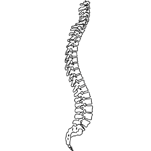
The spine, also known as the vertebral column, is a complex structure in the human body that is part of the axial skeleton. It is composed of 33 individual vertebrae (except in some people where there are fewer due to fusion of certain vertebrae), intervertebral discs, facet joints, ligaments, muscles, and nerves.
The spine has several important functions:
1. Protection: The spine protects the spinal cord, which is a major component of the nervous system, by enclosing it within a bony canal.
2. Support: The spine supports the head and upper body, allowing us to maintain an upright posture and facilitating movement of the trunk and head.
3. Movement: The spine enables various movements such as flexion (bending forward), extension (bending backward), lateral flexion (bending sideways), and rotation (twisting).
4. Weight-bearing: The spine helps distribute weight and pressure evenly across the body, reducing stress on individual vertebrae and other structures.
5. Blood vessel and nerve protection: The spine protects vital blood vessels and nerves that pass through it, including the aorta, vena cava, and spinal nerves.
The spine is divided into five regions: cervical (7 vertebrae), thoracic (12 vertebrae), lumbar (5 vertebrae), sacrum (5 fused vertebrae), and coccyx (4 fused vertebrae, also known as the tailbone). Each region has unique characteristics that allow for specific functions and adaptations to the body's needs.
Stress fractures are defined as small cracks or severe bruising in bones that occur from repetitive stress or overuse. They most commonly occur in weight-bearing bones, such as the legs and feet, but can also occur in the arms, hips, and back. Stress fractures differ from regular fractures because they typically do not result from a single, traumatic event. Instead, they are caused by repeated stress on the bone that results in microscopic damage over time. Athletes, military personnel, and individuals who engage in high-impact activities or have weak bones (osteoporosis) are at increased risk of developing stress fractures. Symptoms may include pain, swelling, tenderness, and difficulty walking or bearing weight on the affected bone.
A femoral neck fracture is a type of hip fracture that occurs in the narrow, vertical section of bone just below the ball of the femur (thigh bone) that connects to the hip socket. This area is called the femoral neck. Femoral neck fractures can be categorized into different types based on their location and the direction of the fractured bone.
These fractures are typically caused by high-energy trauma, such as car accidents or falls from significant heights, in younger individuals. However, in older adults, particularly those with osteoporosis, femoral neck fractures can also result from low-energy trauma, like a simple fall from standing height.
Femoral neck fractures are often serious and require prompt medical attention. Treatment usually involves surgery to realign and stabilize the broken bone fragments, followed by rehabilitation to help regain mobility and strength. Potential complications of femoral neck fractures include avascular necrosis (loss of blood flow to the femoral head), nonunion or malunion (improper healing), and osteoarthritis in the hip joint.
An ulna fracture is a break in the ulna bone, which is one of the two long bones in the forearm. The ulna is located on the pinky finger side of the forearm and functions to support the elbow joint and assist in rotation and movement of the forearm. Ulna fractures can occur at various points along the bone, including the shaft, near the wrist, or at the elbow end of the bone. Symptoms may include pain, swelling, bruising, tenderness, deformity, limited mobility, and in some cases, numbness or tingling in the fingers. Treatment typically involves immobilization with a cast or splint, followed by rehabilitation exercises to restore strength and range of motion. In severe cases, surgery may be required to realign and stabilize the fractured bone.
The cervical vertebrae are the seven vertebrae that make up the upper part of the spine, also known as the neck region. They are labeled C1 to C7, with C1 being closest to the skull and C7 connecting to the thoracic vertebrae in the chest region. The cervical vertebrae have unique structures to allow for a wide range of motion in the neck while also protecting the spinal cord and providing attachment points for muscles and ligaments.
Intramedullary fracture fixation is a surgical technique used to stabilize and align bone fractures. In this procedure, a metal rod or nail is inserted into the marrow cavity (intramedullary canal) of the affected bone, spanning the length of the fracture. The rod is then secured to the bone using screws or other fixation devices on either side of the fracture. This provides stability and helps maintain proper alignment during the healing process.
The benefits of intramedullary fixation include:
1. Load sharing: The intramedullary rod shares some of the load bearing capacity with the bone, which can help reduce stress on the healing bone.
2. Minimal soft tissue dissection: Since the implant is inserted through the medullary canal, there is less disruption to the surrounding muscles, tendons, and ligaments compared to other fixation methods.
3. Biomechanical stability: Intramedullary fixation provides rotational and bending stiffness, which helps maintain proper alignment of the fracture fragments during healing.
4. Early mobilization: Patients with intramedullary fixation can often begin weight bearing and rehabilitation exercises earlier than those with other types of fixation, leading to faster recovery times.
Common indications for intramedullary fracture fixation include long bone fractures in the femur, tibia, humerus, and fibula, as well as certain pelvic and spinal fractures. However, the choice of fixation method depends on various factors such as patient age, fracture pattern, location, and associated injuries.
Rib fractures are breaks or cracks in the bones that make up the rib cage, which is the protective structure around the lungs and heart. Rib fractures can result from direct trauma to the chest, such as from a fall, motor vehicle accident, or physical assault. They can also occur from indirect forces, such as during coughing fits in people with weakened bones (osteoporosis).
Rib fractures are painful and can make breathing difficult, particularly when taking deep breaths or coughing. In some cases, rib fractures may lead to complications like punctured lungs (pneumothorax) or collapsed lungs (atelectasis), especially if multiple ribs are broken in several places.
It is essential to seek medical attention for suspected rib fractures, as proper diagnosis and management can help prevent further complications and promote healing. Treatment typically involves pain management, breathing exercises, and, in some cases, immobilization or surgery.
A skull fracture is a break in one or more of the bones that form the skull. It can occur from a direct blow to the head, penetrating injuries like gunshot wounds, or from strong rotational forces during an accident. There are several types of skull fractures, including:
1. Linear Skull Fracture: This is the most common type, where there's a simple break in the bone without any splintering, depression, or displacement. It often doesn't require treatment unless it's near a sensitive area like an eye or ear.
2. Depressed Skull Fracture: In this type, a piece of the skull is pushed inward toward the brain. Surgery may be needed to relieve pressure on the brain and repair the fracture.
3. Diastatic Skull Fracture: This occurs along the suture lines (the fibrous joints between the skull bones) that haven't fused yet, often seen in infants and young children.
4. Basilar Skull Fracture: This involves fractures at the base of the skull. It can be serious due to potential injury to the cranial nerves and blood vessels located in this area.
5. Comminuted Skull Fracture: In this severe type, the bone is shattered into many pieces. These fractures usually require extensive surgical repair.
Symptoms of a skull fracture can include pain, swelling, bruising, bleeding (if there's an open wound), and in some cases, clear fluid draining from the ears or nose (cerebrospinal fluid leak). Severe fractures may cause brain injury, leading to symptoms like confusion, loss of consciousness, seizures, or neurological deficits. Immediate medical attention is necessary for any suspected skull fracture.
A mandibular fracture is a break or crack in the lower jaw (mandible) bone. It can occur at any point along the mandible, but common sites include the condyle (the rounded end near the ear), the angle (the curved part of the jaw), and the symphysis (the area where the two halves of the jaw meet in the front). Mandibular fractures are typically caused by trauma, such as a direct blow to the face or a fall. Symptoms may include pain, swelling, bruising, difficulty chewing or speaking, and malocclusion (misalignment) of the teeth. Treatment usually involves immobilization with wires or screws to allow the bone to heal properly.
Ankylosing spondylitis is a type of inflammatory arthritis that primarily affects the spine, although other joints can also be involved. It causes swelling in the spinal joints (vertebrae) that can lead to stiffness and pain. Over time, some of these joints may grow together, causing new bone formation and resulting in a rigid spine. This fusion of the spine is called ankylosis.
The condition typically begins in the sacroiliac joints, where the spine connects to the pelvis. From there, it can spread up the spine and potentially involve other areas of the body such as the eyes, heart, lungs, and gastrointestinal system.
Ankylosing spondylitis has a strong genetic link, with most people carrying the HLA-B27 gene. However, not everyone with this gene will develop the condition. It primarily affects males more often than females and tends to start in early adulthood.
Treatment usually involves a combination of medication, physical therapy, and exercise to help manage pain, maintain mobility, and prevent deformity. In severe cases, surgery may be considered.
Treatment outcome is a term used to describe the result or effect of medical treatment on a patient's health status. It can be measured in various ways, such as through symptoms improvement, disease remission, reduced disability, improved quality of life, or survival rates. The treatment outcome helps healthcare providers evaluate the effectiveness of a particular treatment plan and make informed decisions about future care. It is also used in clinical research to compare the efficacy of different treatments and improve patient care.
A tooth fracture is a dental health condition characterized by a break or crack in the tooth structure. It can occur in different parts of the tooth, including the crown (the visible part), root, or filling. Tooth fractures can result from various factors such as trauma, biting or chewing on hard objects, grinding or clenching teeth, and having large, old amalgam fillings that weaken the tooth structure over time. Depending on the severity and location of the fracture, it may cause pain, sensitivity, or affect the tooth's functionality and appearance. Treatment options for tooth fractures vary from simple bonding to root canal treatment or even extraction in severe cases. Regular dental check-ups are essential for early detection and management of tooth fractures.
X-ray computed tomography (CT or CAT scan) is a medical imaging method that uses computer-processed combinations of many X-ray images taken from different angles to produce cross-sectional (tomographic) images (virtual "slices") of the body. These cross-sectional images can then be used to display detailed internal views of organs, bones, and soft tissues in the body.
The term "computed tomography" is used instead of "CT scan" or "CAT scan" because the machines take a series of X-ray measurements from different angles around the body and then use a computer to process these data to create detailed images of internal structures within the body.
CT scanning is a noninvasive, painless medical test that helps physicians diagnose and treat medical conditions. CT imaging provides detailed information about many types of tissue including lung, bone, soft tissue and blood vessels. CT examinations can be performed on every part of the body for a variety of reasons including diagnosis, surgical planning, and monitoring of therapeutic responses.
In computed tomography (CT), an X-ray source and detector rotate around the patient, measuring the X-ray attenuation at many different angles. A computer uses this data to construct a cross-sectional image by the process of reconstruction. This technique is called "tomography". The term "computed" refers to the use of a computer to reconstruct the images.
CT has become an important tool in medical imaging and diagnosis, allowing radiologists and other physicians to view detailed internal images of the body. It can help identify many different medical conditions including cancer, heart disease, lung nodules, liver tumors, and internal injuries from trauma. CT is also commonly used for guiding biopsies and other minimally invasive procedures.
In summary, X-ray computed tomography (CT or CAT scan) is a medical imaging technique that uses computer-processed combinations of many X-ray images taken from different angles to produce cross-sectional images of the body. It provides detailed internal views of organs, bones, and soft tissues in the body, allowing physicians to diagnose and treat medical conditions.
Retrospective studies, also known as retrospective research or looking back studies, are a type of observational study that examines data from the past to draw conclusions about possible causal relationships between risk factors and outcomes. In these studies, researchers analyze existing records, medical charts, or previously collected data to test a hypothesis or answer a specific research question.
Retrospective studies can be useful for generating hypotheses and identifying trends, but they have limitations compared to prospective studies, which follow participants forward in time from exposure to outcome. Retrospective studies are subject to biases such as recall bias, selection bias, and information bias, which can affect the validity of the results. Therefore, retrospective studies should be interpreted with caution and used primarily to generate hypotheses for further testing in prospective studies.
Pain measurement, in a medical context, refers to the quantification or evaluation of the intensity and/or unpleasantness of a patient's subjective pain experience. This is typically accomplished through the use of standardized self-report measures such as numerical rating scales (NRS), visual analog scales (VAS), or categorical scales (mild, moderate, severe). In some cases, physiological measures like heart rate, blood pressure, and facial expressions may also be used to supplement self-reported pain ratings. The goal of pain measurement is to help healthcare providers better understand the nature and severity of a patient's pain in order to develop an effective treatment plan.
A compression fracture is a type of bone fracture that occurs when there is a collapse of a vertebra in the spine. This type of fracture is most commonly seen in the thoracic and lumbar regions of the spine. Compression fractures are often caused by weakened bones due to osteoporosis, but they can also result from trauma or tumors that weaken the bone.
In a compression fracture, the front part (anterior) of the vertebra collapses, while the back part (posterior) remains intact, causing the height of the vertebra to decrease. This can lead to pain, deformity, and decreased mobility. In severe cases, multiple compression fractures can result in a condition called kyphosis, which is an abnormal curvature of the spine that leads to a hunchback appearance.
Compression fractures are typically diagnosed through imaging tests such as X-rays, CT scans, or MRI scans. Treatment may include pain medication, bracing, physical therapy, or in some cases, surgery. Preventive measures such as maintaining a healthy diet, getting regular exercise, and taking medications to prevent or treat osteoporosis can help reduce the risk of compression fractures.
An intra-articular fracture is a type of fracture that involves the joint surface or articular cartilage of a bone. These types of fractures can occur in any joint, but they are most commonly seen in the weight-bearing joints such as the knee, ankle, and wrist.
Intra-articular fractures can be caused by high-energy trauma, such as motor vehicle accidents or falls from significant heights, or by low-energy trauma, such as a simple fall in older adults with osteoporosis.
These types of fractures are often complex and may involve displacement or depression of the joint surface, which can increase the risk of developing post-traumatic arthritis. Therefore, prompt diagnosis and appropriate treatment are essential to ensure optimal outcomes and minimize long-term complications. Treatment options for intra-articular fractures may include surgical fixation with plates, screws, or pins, as well as joint replacement in some cases.
Spinal injections, also known as epidural injections or intrathecal injections, are medical procedures involving the injection of medications directly into the spinal canal. The medication is usually delivered into the space surrounding the spinal cord (the epidural space) or into the cerebrospinal fluid that surrounds and protects the spinal cord (the subarachnoid space).
The medications used in spinal injections can include local anesthetics, steroids, opioids, or a combination of these. The purpose of spinal injections is to provide diagnostic information, therapeutic relief, or both. They are commonly used to treat various conditions affecting the spine, such as radicular pain (pain that radiates down the arms or legs), disc herniation, spinal stenosis, and degenerative disc disease.
Spinal injections can be administered using different techniques, including fluoroscopy-guided injections, computed tomography (CT) scan-guided injections, or with the help of a nerve stimulator. These techniques ensure accurate placement of the medication and minimize the risk of complications.
It is essential to consult a healthcare professional for specific information regarding spinal injections and their potential benefits and risks.
Osteoporosis is a systemic skeletal disease characterized by low bone mass, deterioration of bone tissue, and disruption of bone architecture, leading to increased risk of fractures, particularly in the spine, wrist, and hip. It mainly affects older people, especially postmenopausal women, due to hormonal changes that reduce bone density. Osteoporosis can also be caused by certain medications, medical conditions, or lifestyle factors such as smoking, alcohol abuse, and a lack of calcium and vitamin D in the diet. The diagnosis is often made using bone mineral density testing, and treatment may include medication to slow bone loss, promote bone formation, and prevent fractures.
Bone plates are medical devices used in orthopedic surgery to stabilize and hold together fractured or broken bones during the healing process. They are typically made of surgical-grade stainless steel, titanium, or other biocompatible materials. The plate is shaped to fit the contour of the bone and is held in place with screws that are inserted through the plate and into the bone on either side of the fracture. This provides stability and alignment to the broken bones, allowing them to heal properly. Bone plates can be used to treat a variety of fractures, including those that are complex or unstable. After healing is complete, the bone plate may be left in place or removed, depending on the individual's needs and the surgeon's recommendation.
I believe you are referring to "bone pins" or "bone nails" rather than "bone nails." These terms are used in the medical field to describe surgical implants made of metal or biocompatible materials that are used to stabilize and hold together fractured bones during the healing process. They can also be used in spinal fusion surgery to provide stability and promote bone growth between vertebrae.
Bone pins or nails typically have a threaded or smooth shaft, with a small diameter that allows them to be inserted into the medullary canal of long bones such as the femur or tibia. They may also have a head or eyelet on one end that allows for attachment to external fixation devices or other surgical instruments.
The use of bone pins and nails has revolutionized orthopedic surgery, allowing for faster healing times, improved stability, and better functional outcomes for patients with fractures or spinal deformities.
Orbital fractures refer to breaks in the bones that make up the eye socket, also known as the orbit. These bones include the maxilla, zygoma, frontal bone, and palatine bone. Orbital fractures can occur due to trauma, such as a blunt force injury or a penetrating wound.
There are several types of orbital fractures, including:
1. Blowout fracture: This occurs when the thin bone of the orbital floor is broken, often due to a direct blow to the eye. The force of the impact can cause the eyeball to move backward, breaking the bone and sometimes trapping the muscle that moves the eye (the inferior rectus).
2. Blow-in fracture: This type of fracture involves the breakage of the orbital roof, which is the bone that forms the upper boundary of the orbit. It typically occurs due to high-impact trauma, such as a car accident or a fall from a significant height.
3. Direct fracture: A direct fracture happens when there is a break in one or more of the bones that form the walls of the orbit. This type of fracture can result from a variety of traumas, including motor vehicle accidents, sports injuries, and assaults.
4. Indirect fracture: An indirect fracture occurs when the force of an injury is transmitted to the orbit through tissues surrounding it, causing the bone to break. The most common type of indirect orbital fracture is a blowout fracture.
Orbital fractures can cause various symptoms, including pain, swelling, bruising, and double vision. In some cases, the fracture may also lead to enophthalmos (sinking of the eye into the orbit) or telecanthus (increased distance between the inner corners of the eyes). Imaging tests, such as CT scans, are often used to diagnose orbital fractures and determine the best course of treatment. Treatment may include observation, pain management, and in some cases, surgery to repair the fracture and restore normal function.
Spinal nerves are the bundles of nerve fibers that transmit signals between the spinal cord and the rest of the body. There are 31 pairs of spinal nerves in the human body, which can be divided into five regions: 8 cervical, 12 thoracic, 5 lumbar, 5 sacral, and 1 coccygeal. Each spinal nerve carries both sensory information (such as touch, temperature, and pain) from the periphery to the spinal cord, and motor information (such as muscle control) from the spinal cord to the muscles and other structures in the body. Spinal nerves also contain autonomic fibers that regulate involuntary functions such as heart rate, digestion, and blood pressure.
Medical Definition:
Magnetic Resonance Imaging (MRI) is a non-invasive diagnostic imaging technique that uses a strong magnetic field and radio waves to create detailed cross-sectional or three-dimensional images of the internal structures of the body. The patient lies within a large, cylindrical magnet, and the scanner detects changes in the direction of the magnetic field caused by protons in the body. These changes are then converted into detailed images that help medical professionals to diagnose and monitor various medical conditions, such as tumors, injuries, or diseases affecting the brain, spinal cord, heart, blood vessels, joints, and other internal organs. MRI does not use radiation like computed tomography (CT) scans.
Spinal cord diseases refer to a group of conditions that affect the spinal cord, which is a part of the central nervous system responsible for transmitting messages between the brain and the rest of the body. These diseases can cause damage to the spinal cord, leading to various symptoms such as muscle weakness, numbness, pain, bladder and bowel dysfunction, and difficulty with movement and coordination.
Spinal cord diseases can be congenital or acquired, and they can result from a variety of causes, including infections, injuries, tumors, degenerative conditions, autoimmune disorders, and genetic factors. Some examples of spinal cord diseases include multiple sclerosis, spina bifida, spinal cord injury, herniated discs, spinal stenosis, and motor neuron diseases such as amyotrophic lateral sclerosis (ALS).
The treatment for spinal cord diseases varies depending on the underlying cause and severity of the condition. Treatment options may include medication, physical therapy, surgery, and rehabilitation. In some cases, the damage to the spinal cord may be irreversible, leading to permanent disability or paralysis.
Spinal anesthesia is a type of regional anesthesia that involves injecting local anesthetic medication into the cerebrospinal fluid in the subarachnoid space, which is the space surrounding the spinal cord. This procedure is typically performed by introducing a needle into the lower back, between the vertebrae, to reach the subarachnoid space.
Once the local anesthetic is introduced into this space, it spreads to block nerve impulses from the corresponding levels of the spine, resulting in numbness and loss of sensation in specific areas of the body below the injection site. The extent and level of anesthesia depend on the amount and type of medication used, as well as the patient's individual response.
Spinal anesthesia is often used for surgeries involving the lower abdomen, pelvis, or lower extremities, such as cesarean sections, hernia repairs, hip replacements, and knee arthroscopies. It can also be utilized for procedures like epidural steroid injections to manage chronic pain conditions affecting the spine and lower limbs.
While spinal anesthesia provides effective pain relief during and after surgery, it may cause side effects such as low blood pressure, headache, or difficulty urinating. These potential complications should be discussed with the healthcare provider before deciding on this type of anesthesia.
A Colles' fracture is a specific type of fracture in the distal end of the radius bone in the forearm, which is the larger of the two bones in the lower arm. This type of fracture occurs when the wrist is forcefully bent backward (dorsiflexion), often as a result of falling onto an outstretched hand.
In a Colles' fracture, the distal end of the radius bone breaks and is displaced downward and angulated backward, resulting in a characteristic "dinner fork" deformity. This type of fracture is more common in older individuals, particularly women with osteoporosis, but can also occur in younger people as a result of high-energy trauma.
Colles' fractures are typically treated with immobilization using a cast or splint to hold the bones in proper alignment while they heal. In some cases, surgery may be necessary to realign and stabilize the fracture, particularly if there is significant displacement or instability of the bone fragments.
The spinal canal is the bony, protective channel within the vertebral column that contains and houses the spinal cord. It extends from the foramen magnum at the base of the skull to the sacrum, where the spinal cord ends and forms the cauda equina. The spinal canal is formed by a series of vertebral bodies stacked on top of each other, intervertebral discs in between them, and the laminae and spinous processes that form the posterior elements of the vertebrae. The spinal canal provides protection to the spinal cord from external trauma and contains cerebrospinal fluid (CSF) that circulates around the cord, providing nutrients and cushioning. Any narrowing or compression of the spinal canal, known as spinal stenosis, can cause various neurological symptoms due to pressure on the spinal cord or nerve roots.
Spinal cord compression is a medical condition that refers to the narrowing of the spinal canal, which puts pressure on the spinal cord and the nerves that branch out from it. This can occur due to various reasons such as degenerative changes in the spine, herniated discs, bone spurs, tumors, or fractures. The compression can lead to a range of symptoms including pain, numbness, tingling, weakness, or loss of bladder and bowel control. In severe cases, it can cause paralysis. Treatment options depend on the underlying cause and may include physical therapy, medication, surgery, or radiation therapy.
Spinal neoplasms refer to abnormal growths or tumors found within the spinal column, which can be benign (non-cancerous) or malignant (cancerous). These tumors can originate in the spine itself, called primary spinal neoplasms, or they can spread to the spine from other parts of the body, known as secondary or metastatic spinal neoplasms. Spinal neoplasms can cause various symptoms, such as back pain, neurological deficits, and even paralysis, depending on their location and size. Early diagnosis and treatment are crucial to prevent or minimize long-term complications and improve the patient's prognosis.
Bony callus is a medical term that refers to the specialized tissue that forms in response to a bone fracture. It is a crucial part of the natural healing process, as it helps to stabilize and protect the broken bone while it mends.
When a bone is fractured, the body responds by initiating an inflammatory response, which triggers the production of various cells and signaling molecules that promote healing. As part of this process, specialized cells called osteoblasts begin to produce new bone tissue at the site of the fracture. This tissue is initially soft and pliable, allowing it to bridge the gap between the broken ends of the bone.
Over time, this soft callus gradually hardens and calcifies, forming a bony callus that helps to stabilize the fracture and provide additional support as the bone heals. The bony callus is typically composed of a mixture of woven bone (which is less organized than normal bone) and more structured lamellar bone (which is similar in structure to normal bone).
As the bone continues to heal, the bony callus may be gradually remodeled and reshaped by osteoclasts, which are specialized cells that break down and remove excess or unwanted bone tissue. This process helps to restore the bone's original shape and strength, allowing it to function normally again.
It is worth noting that excessive bony callus formation can sometimes lead to complications, such as stiffness, pain, or decreased range of motion in the affected limb. In some cases, surgical intervention may be necessary to remove or reduce the size of the bony callus and promote proper healing.
Spinal nerve roots are the initial parts of spinal nerves that emerge from the spinal cord through the intervertebral foramen, which are small openings between each vertebra in the spine. These nerve roots carry motor, sensory, and autonomic fibers to and from specific regions of the body. There are 31 pairs of spinal nerve roots in total, with 8 cervical, 12 thoracic, 5 lumbar, 5 sacral, and 1 coccygeal pair. Each root has a dorsal (posterior) and ventral (anterior) ramus that branch off to form the peripheral nervous system. Irritation or compression of these nerve roots can result in pain, numbness, weakness, or loss of reflexes in the affected area.
Spinal diseases refer to a range of medical conditions that affect the spinal column, which is made up of vertebrae (bones), intervertebral discs, facet joints, nerves, ligaments, and muscles. These diseases can cause pain, discomfort, stiffness, numbness, weakness, or even paralysis, depending on the severity and location of the condition. Here are some examples of spinal diseases:
1. Degenerative disc disease: This is a condition where the intervertebral discs lose their elasticity and height, leading to stiffness, pain, and decreased mobility.
2. Herniated disc: This occurs when the inner material of the intervertebral disc bulges or herniates out through a tear in the outer layer, causing pressure on the spinal nerves and resulting in pain, numbness, tingling, or weakness in the affected area.
3. Spinal stenosis: This is a narrowing of the spinal canal or the neural foramen (the openings where the spinal nerves exit the spinal column), which can cause pressure on the spinal cord or nerves and result in pain, numbness, tingling, or weakness.
4. Scoliosis: This is a curvature of the spine that can occur in children or adults, leading to an abnormal posture, back pain, and decreased lung function.
5. Osteoarthritis: This is a degenerative joint disease that affects the facet joints in the spine, causing pain, stiffness, and decreased mobility.
6. Ankylosing spondylitis: This is a chronic inflammatory disease that affects the spine and sacroiliac joints, leading to pain, stiffness, and fusion of the vertebrae.
7. Spinal tumors: These are abnormal growths that can occur in the spinal column, which can be benign or malignant, causing pain, neurological symptoms, or even paralysis.
8. Infections: Bacterial or viral infections can affect the spine, leading to pain, fever, and other systemic symptoms.
9. Trauma: Fractures, dislocations, or sprains of the spine can occur due to accidents, falls, or sports injuries, causing pain, neurological deficits, or even paralysis.
Spinal cord neoplasms refer to abnormal growths or tumors within the spinal cord. These can be benign (non-cancerous) or malignant (cancerous). They originate from the cells within the spinal cord itself (primary tumors), or they may spread to the spinal cord from other parts of the body (metastatic tumors). Spinal cord neoplasms can cause various symptoms depending on their location and size, including back pain, neurological deficits, and even paralysis. Treatment options include surgery, radiation therapy, and chemotherapy.
Bone screws are medical devices used in orthopedic and trauma surgery to affix bone fracture fragments or to attach bones to other bones or to metal implants such as plates, rods, or artificial joints. They are typically made of stainless steel or titanium alloys and have a threaded shaft that allows for purchase in the bone when tightened. The head of the screw may have a hexagonal or star-shaped design to allow for precise tightening with a screwdriver. Bone screws come in various shapes, sizes, and designs, including fully threaded, partially threaded, cannulated (hollow), and headless types, depending on their intended use and location in the body.
Bone density refers to the amount of bone mineral content (usually measured in grams) in a given volume of bone (usually measured in cubic centimeters). It is often used as an indicator of bone strength and fracture risk. Bone density is typically measured using dual-energy X-ray absorptiometry (DXA) scans, which provide a T-score that compares the patient's bone density to that of a young adult reference population. A T-score of -1 or above is considered normal, while a T-score between -1 and -2.5 indicates osteopenia (low bone mass), and a T-score below -2.5 indicates osteoporosis (porous bones). Regular exercise, adequate calcium and vitamin D intake, and medication (if necessary) can help maintain or improve bone density and prevent fractures.
Periprosthetic fractures are defined as fractures that occur in close proximity to a prosthetic joint, such as those found in total hip or knee replacements. These types of fractures typically occur as a result of low-energy trauma, and can be caused by a variety of factors including osteoporosis, bone weakness, or loosening of the prosthetic implant.
Periprosthetic fractures are classified based on the location of the fracture in relation to the prosthesis, as well as the stability of the implant. Treatment options for periprosthetic fractures may include non-surgical management, such as immobilization with a brace or cast, or surgical intervention, such as open reduction and internal fixation (ORIF) or revision arthroplasty.
The management of periprosthetic fractures can be complex and requires careful consideration of various factors, including the patient's age, overall health status, bone quality, and functional needs. As such, these types of fractures are typically managed by orthopedic surgeons with experience in joint replacement surgery and fracture care.
Spinal stenosis is a narrowing of the spinal canal or the neural foramina (the openings through which nerves exit the spinal column), typically in the lower back (lumbar) or neck (cervical) regions. This can put pressure on the spinal cord and/or nerve roots, causing pain, numbness, tingling, or weakness in the affected areas, often in the legs, arms, or hands. It's most commonly caused by age-related wear and tear, but can also be due to degenerative changes, herniated discs, tumors, or spinal injuries.
Wrist injuries refer to damages or traumas affecting the structures of the wrist, including bones, ligaments, tendons, muscles, and cartilage. These injuries can occur due to various reasons such as falls, accidents, sports-related impacts, or repetitive stress. Common types of wrist injuries include fractures (such as scaphoid fracture), sprains (like ligament tears), strains (involving muscles or tendons), dislocations, and carpal tunnel syndrome. Symptoms may include pain, swelling, tenderness, bruising, limited mobility, and in severe cases, deformity or numbness. Immediate medical attention is necessary for proper diagnosis and treatment to ensure optimal recovery and prevent long-term complications.
Surgical casts are medical devices used to immobilize and protect injured body parts, typically fractured or broken bones, during the healing process. They are usually made of plaster or fiberglass materials that harden when wet and conform to the shape of the affected area once applied. The purpose of a surgical cast is to restrict movement and provide stability to the injured site, allowing for proper alignment and healing of the bones.
The casting process involves first aligning the broken bone fragments into their correct positions, often through manual manipulation or surgical intervention. Once aligned, the cast material is applied in layers, with each layer being allowed to dry before adding the next. This creates a rigid structure that encases and supports the injured area. The cast must be kept dry during the healing process to prevent it from becoming weakened or damaged.
Surgical casts come in various shapes and sizes depending on the location and severity of the injury. They may also include additional components such as padding, Velcro straps, or window openings to allow for regular monitoring of the skin and underlying tissue. In some cases, removable splints or functional braces may be used instead of traditional casts, providing similar support while allowing for limited movement and easier adjustments.
It is essential to follow proper care instructions when wearing a surgical cast, including elevating the injured limb, avoiding excessive weight-bearing, and monitoring for signs of complications such as swelling, numbness, or infection. Regular check-ups with a healthcare provider are necessary to ensure proper healing and adjust the cast if needed.
I'm not aware of a medical term called "bone wires." The term "wiring" is used in orthopedic surgery to describe the use of metal wire to hold bones or fractures in place during healing. However, I couldn't find any specific medical definition or term related to "bone wires." It may be a colloquialism, a term used in a specific context, or a term from science fiction. If you could provide more context about where you encountered this term, I might be able to give a more accurate answer.
Maxillary fractures, also known as Le Fort fractures, are complex fractures that involve the upper jaw or maxilla. Named after the French surgeon René Le Fort who first described them in 1901, these fractures are categorized into three types (Le Fort I, II, III) based on the pattern and level of bone involvement.
1. Le Fort I fracture: This type of maxillary fracture involves a horizontal separation through the lower part of the maxilla, just above the teeth's roots. It often results from direct blows to the lower face or chin.
2. Le Fort II fracture: A Le Fort II fracture is characterized by a pyramidal-shaped fracture pattern that extends from the nasal bridge through the inferior orbital rim and maxilla, ending at the pterygoid plates. This type of fracture usually results from forceful impacts to the midface or nose.
3. Le Fort III fracture: A Le Fort III fracture is a severe craniofacial injury that involves both the upper and lower parts of the face. It is also known as a "craniofacial dysjunction" because it separates the facial bones from the skull base. The fracture line extends through the nasal bridge, orbital rims, zygomatic arches, and maxilla, ending at the pterygoid plates. Le Fort III fractures typically result from high-impact trauma to the face, such as car accidents or assaults.
These fractures often require surgical intervention for proper alignment and stabilization of the facial bones.
An external fixator is a type of orthopedic device used in the treatment of severe fractures or deformities of bones. It consists of an external frame that is attached to the bone with pins or wires that pass through the skin and into the bone. This provides stability to the injured area while allowing for alignment and adjustment of the bone during the healing process.
External fixators are typically used in cases where traditional casting or internal fixation methods are not feasible, such as when there is extensive soft tissue damage, infection, or when a limb needs to be gradually stretched or shortened. They can also be used in reconstructive surgery for bone defects or deformities.
The external frame of the fixator is made up of bars and clamps that are adjustable, allowing for precise positioning and alignment of the bones. The pins or wires that attach to the bone are carefully inserted through small incisions in the skin, and are held in place by the clamps on the frame.
External fixators can be used for a period of several weeks to several months, depending on the severity of the injury and the individual's healing process. During this time, the patient may require regular adjustments and monitoring by an orthopedic surgeon or other medical professional. Once the bone has healed sufficiently, the external fixator can be removed in a follow-up procedure.
An accidental fall is an unplanned, unexpected event in which a person suddenly and involuntarily comes to rest on the ground or other lower level, excluding intentional changes in position (e.g., jumping to catch a ball) and landings that are part of a planned activity (e.g., diving into a pool). Accidental falls can occur for various reasons, such as environmental hazards, muscle weakness, balance problems, visual impairment, or certain medical conditions. They are a significant health concern, particularly among older adults, as they can lead to serious injuries, loss of independence, reduced quality of life, and increased mortality.
Zygomatic fractures, also known as "tripod fractures" or "malar fractures," refer to breaks in the zygomatic bone, which is the cheekbone. This type of facial fracture typically occurs due to significant trauma, such as a forceful blow to the face during sports injuries, traffic accidents, or physical assaults.
In zygomatic fractures, the bone can be displaced or depressed, leading to various symptoms, including:
* Facial asymmetry or flattening of the cheek area
* Bruising and swelling around the eyes (periorbital ecchymosis) and cheeks
* Diplopia (double vision) due to muscle entrapment or trauma to the eye muscles
* Subconjunctival hemorrhage (bleeding in the white part of the eye)
* Trismus (difficulty opening the mouth) due to muscle spasms or injury to the temporomandibular joint
* Numbness or altered sensation in the upper lip, cheek, or side of the nose
Diagnosis is usually made through a combination of physical examination and imaging techniques like X-rays, CT scans, or MRI. Treatment typically involves closed reduction (manipulation without surgery) or open reduction with internal fixation (surgical reconstruction using plates and screws). The primary goal of treatment is to restore the facial structure's integrity, symmetry, and function while minimizing complications and promoting optimal healing.
Postmenopausal osteoporosis is a specific type of osteoporosis that occurs in women after they have gone through menopause. It is defined as a skeletal disorder characterized by compromised bone strength, leading to an increased risk of fractures. In this condition, the decline in estrogen levels that occurs during menopause accelerates bone loss, resulting in a decrease in bone density and quality, which can lead to fragility fractures, particularly in the hips, wrists, and spine.
It's important to note that while postmenopausal osteoporosis is more common in women, men can also develop osteoporosis due to other factors such as aging, lifestyle choices, and medical conditions.
Forearm injuries refer to damages or traumas that affect the anatomy and function of the forearm, which is the area between the elbow and wrist. This region consists of two long bones (the radius and ulna) and several muscles, tendons, ligaments, nerves, and blood vessels that enable movements such as flexion, extension, pronation, and supination of the hand and wrist.
Common forearm injuries include:
1. Fractures: Breaks in the radius or ulna bones can occur due to high-energy trauma, falls, or sports accidents. These fractures may be simple (stable) or compound (displaced), and might require immobilization, casting, or surgical intervention depending on their severity and location.
2. Sprains and Strains: Overstretching or tearing of the ligaments connecting the bones in the forearm or the muscles and tendons responsible for movement can lead to sprains and strains. These injuries often cause pain, swelling, bruising, and limited mobility.
3. Dislocations: In some cases, forceful trauma might result in the dislocation of the radioulnar joint, where the ends of the radius and ulna meet. This injury can be extremely painful and may necessitate immediate medical attention to realign the bones and stabilize the joint.
4. Tendonitis: Repetitive motions or overuse can cause inflammation and irritation of the tendons in the forearm, resulting in a condition known as tendonitis. This injury typically presents with localized pain, swelling, and stiffness that worsen with activity.
5. Nerve Injuries: Direct trauma, compression, or stretching can damage nerves in the forearm, leading to numbness, tingling, weakness, or paralysis in the hand and fingers. Common nerve injuries include radial nerve neuropathy and ulnar nerve entrapment.
6. Compartment Syndrome: Forearm compartment syndrome occurs when increased pressure within one of the forearm's fascial compartments restricts blood flow to the muscles, nerves, and tissues inside. This condition can result from trauma, bleeding, or swelling and requires immediate medical intervention to prevent permanent damage.
Accurate diagnosis and appropriate treatment are crucial for managing forearm injuries and ensuring optimal recovery. Patients should consult with a healthcare professional if they experience persistent pain, swelling, stiffness, weakness, or numbness in their forearms or hands.
"Recovery of function" is a term used in medical rehabilitation to describe the process in which an individual regains the ability to perform activities or tasks that were previously difficult or impossible due to injury, illness, or disability. This can involve both physical and cognitive functions. The goal of recovery of function is to help the person return to their prior level of independence and participation in daily activities, work, and social roles as much as possible.
Recovery of function may be achieved through various interventions such as physical therapy, occupational therapy, speech-language therapy, and other rehabilitation strategies. The specific approach used will depend on the individual's needs and the nature of their impairment. Recovery of function can occur spontaneously as the body heals, or it may require targeted interventions to help facilitate the process.
It is important to note that recovery of function does not always mean a full return to pre-injury or pre-illness levels of ability. Instead, it often refers to the person's ability to adapt and compensate for any remaining impairments, allowing them to achieve their maximum level of functional independence and quality of life.
Bone density conservation agents, also known as anti-resorptive agents or bone-sparing drugs, are a class of medications that help to prevent the loss of bone mass and reduce the risk of fractures. They work by inhibiting the activity of osteoclasts, the cells responsible for breaking down and reabsorbing bone tissue during the natural remodeling process.
Examples of bone density conservation agents include:
1. Bisphosphonates (e.g., alendronate, risedronate, ibandronate, zoledronic acid) - These are the most commonly prescribed class of bone density conservation agents. They bind to hydroxyapatite crystals in bone tissue and inhibit osteoclast activity, thereby reducing bone resorption.
2. Denosumab (Prolia) - This is a monoclonal antibody that targets RANKL (Receptor Activator of Nuclear Factor-κB Ligand), a key signaling molecule involved in osteoclast differentiation and activation. By inhibiting RANKL, denosumab reduces osteoclast activity and bone resorption.
3. Selective estrogen receptor modulators (SERMs) (e.g., raloxifene) - These medications act as estrogen agonists or antagonists in different tissues. In bone tissue, SERMs mimic the bone-preserving effects of estrogen by inhibiting osteoclast activity and reducing bone resorption.
4. Hormone replacement therapy (HRT) - Estrogen hormone replacement therapy has been shown to preserve bone density in postmenopausal women; however, its use is limited due to increased risks of breast cancer, cardiovascular disease, and thromboembolic events.
5. Calcitonin - This hormone, secreted by the thyroid gland, inhibits osteoclast activity and reduces bone resorption. However, it has largely been replaced by other more effective bone density conservation agents.
These medications are often prescribed for individuals at high risk of fractures due to conditions such as osteoporosis or metabolic disorders that affect bone health. It is essential to follow the recommended dosage and administration guidelines to maximize their benefits while minimizing potential side effects. Regular monitoring of bone density, blood calcium levels, and other relevant parameters is also necessary during treatment with these medications.
Spinal muscular atrophy (SMA) is a genetic disorder that affects the motor neurons in the spinal cord, leading to muscle weakness and atrophy. It is caused by a mutation in the survival motor neuron 1 (SMN1) gene, which results in a deficiency of SMN protein necessary for the survival of motor neurons.
There are several types of SMA, classified based on the age of onset and severity of symptoms. The most common type is type 1, also known as Werdnig-Hoffmann disease, which presents in infancy and is characterized by severe muscle weakness, hypotonia, and feeding difficulties. Other types include type 2 (intermediate SMA), type 3 (Kugelberg-Welander disease), and type 4 (adult-onset SMA).
The symptoms of SMA may include muscle wasting, fasciculations, weakness, hypotonia, respiratory difficulties, and mobility impairment. The diagnosis of SMA typically involves genetic testing to confirm the presence of a mutation in the SMN1 gene. Treatment options for SMA may include medications, physical therapy, assistive devices, and respiratory support.
The clavicle, also known as the collarbone, is a long, slender bone that lies horizontally between the breastbone (sternum) and the shoulder blade (scapula). It is part of the shoulder girdle and plays a crucial role in supporting the upper limb. The clavicle has two ends: the medial end, which articulates with the sternum, and the lateral end, which articulates with the acromion process of the scapula. It is a common site of fracture due to its superficial location and susceptibility to direct trauma.
The pelvic bones, also known as the hip bones, are a set of three irregularly shaped bones that connect to form the pelvic girdle in the lower part of the human body. They play a crucial role in supporting the spine and protecting the abdominal and pelvic organs.
The pelvic bones consist of three bones:
1. The ilium: This is the largest and uppermost bone, forming the majority of the hip bone and the broad, flaring part of the pelvis known as the wing of the ilium or the iliac crest, which can be felt on the side of the body.
2. The ischium: This is the lower and back portion of the pelvic bone that forms part of the sitting surface or the "sit bones."
3. The pubis: This is the front part of the pelvic bone, which connects to the other side at the pubic symphysis in the midline of the body.
The pelvic bones are joined together at the acetabulum, a cup-shaped socket that forms the hip joint and articulates with the head of the femur (thigh bone). The pelvic bones also have several openings for the passage of blood vessels, nerves, and reproductive and excretory organs.
The shape and size of the pelvic bones differ between males and females due to their different roles in childbirth and locomotion. Females typically have a wider and shallower pelvis than males to accommodate childbirth, while males usually have a narrower and deeper pelvis that is better suited for weight-bearing and movement.
Traction, in medical terms, refers to the application of a pulling force to distract or align parts of the body, particularly bones, joints, or muscles, with the aim of immobilizing, reducing displacement, or realigning them. This is often achieved through the use of various devices such as tongs, pulleys, weights, or specialized traction tables. Traction may be applied manually or mechanically and can be continuous or intermittent, depending on the specific medical condition being treated. Common indications for traction include fractures, dislocations, spinal cord injuries, and certain neurological conditions.
Ankle injuries refer to damages or traumas that occur in the ankle joint and its surrounding structures, including bones, ligaments, tendons, and muscles. The ankle joint is a complex structure composed of three bones: the tibia (shinbone), fibula (lower leg bone), and talus (a bone in the foot). These bones are held together by various strong ligaments that provide stability and enable proper movement.
There are several types of ankle injuries, with the most common being sprains, strains, and fractures:
1. Ankle Sprain: A sprain occurs when the ligaments surrounding the ankle joint get stretched or torn due to sudden twisting, rolling, or forced movements. The severity of a sprain can range from mild (grade 1) to severe (grade 3), with partial or complete tearing of the ligament(s).
2. Ankle Strain: A strain is an injury to the muscles or tendons surrounding the ankle joint, often caused by overuse, excessive force, or awkward positioning. This results in pain, swelling, and difficulty moving the ankle.
3. Ankle Fracture: A fracture occurs when one or more bones in the ankle joint break due to high-impact trauma, such as a fall, sports injury, or vehicle accident. Fractures can vary in severity, from small cracks to complete breaks that may require surgery and immobilization for proper healing.
Symptoms of ankle injuries typically include pain, swelling, bruising, tenderness, and difficulty walking or bearing weight on the affected ankle. Immediate medical attention is necessary for severe injuries, such as fractures, dislocations, or significant ligament tears, to ensure appropriate diagnosis and treatment. Treatment options may include rest, ice, compression, elevation (RICE), immobilization with a brace or cast, physical therapy, medication, or surgery, depending on the type and severity of the injury.
Internal fixators are medical devices that are implanted into the body through surgery to stabilize and hold broken or fractured bones in the correct position while they heal. These devices can be made from various materials, such as metal (stainless steel or titanium) or bioabsorbable materials. Internal fixators can take many forms, including plates, screws, rods, nails, wires, or cages, depending on the type and location of the fracture.
The main goal of using internal fixators is to promote bone healing by maintaining accurate reduction and alignment of the fractured bones, allowing for early mobilization and rehabilitation. This can help reduce the risk of complications such as malunion, nonunion, or deformity. Internal fixators are typically removed once the bone has healed, although some bioabsorbable devices may not require a second surgery for removal.
It is important to note that while internal fixators provide stability and support for fractured bones, they do not replace the need for proper immobilization, protection, or rehabilitation during the healing process. Close follow-up with an orthopedic surgeon is essential to ensure appropriate healing and address any potential complications.
A dislocation is a condition in which a bone slips out of its normal position in a joint. This can happen as a result of trauma or injury, such as a fall or direct blow to the body. Dislocations can cause pain, swelling, and limited mobility in the affected area. In some cases, a dislocation may also damage surrounding tissues, such as ligaments, tendons, and nerves.
Dislocations are typically treated by reducing the dislocation, which means putting the bone back into its normal position. This is usually done with the help of medication to relieve pain and relaxation techniques to help the person stay still during the reduction. In some cases, surgery may be necessary to repair damaged tissues or if the dislocation cannot be reduced through other methods. After the dislocation has been reduced, the joint may be immobilized with a splint or sling to allow it to heal properly.
It is important to seek medical attention promptly if you suspect that you have a dislocation. If left untreated, a dislocation can lead to further complications, such as joint instability and chronic pain.
Spinal cord ischemia refers to a reduction or interruption of blood flow to the spinal cord, leading to insufficient oxygen and nutrient supply. This condition can cause damage to the spinal cord tissue, potentially resulting in neurological deficits, such as muscle weakness, sensory loss, or autonomic dysfunction. Spinal cord ischemia may be caused by various factors, including atherosclerosis, embolism, spinal artery stenosis, or complications during surgery. The severity and extent of the neurological impairment depend on the duration and location of the ischemic event in the spinal cord.
Multiple trauma, also known as polytrauma, is a medical term used to describe severe injuries to the body that are sustained in more than one place or region. It often involves damage to multiple organ systems and can be caused by various incidents such as traffic accidents, falls from significant heights, high-energy collisions, or violent acts.
The injuries sustained in multiple trauma may include fractures, head injuries, internal bleeding, chest and abdominal injuries, and soft tissue injuries. These injuries can lead to a complex medical situation requiring immediate and ongoing care from a multidisciplinary team of healthcare professionals, including emergency physicians, trauma surgeons, critical care specialists, nurses, rehabilitation therapists, and mental health providers.
Multiple trauma is a serious condition that can result in long-term disability or even death if not treated promptly and effectively.
Spinal ganglia, also known as dorsal root ganglia, are clusters of nerve cell bodies located in the peripheral nervous system. They are situated along the length of the spinal cord and are responsible for transmitting sensory information from the body to the brain. Each spinal ganglion contains numerous neurons, or nerve cells, with long processes called axons that extend into the periphery and innervate various tissues and organs. The cell bodies within the spinal ganglia receive sensory input from these axons and transmit this information to the central nervous system via the dorsal roots of the spinal nerves. This allows the brain to interpret and respond to a wide range of sensory stimuli, including touch, temperature, pain, and proprioception (the sense of the position and movement of one's body).
The calcaneus is the largest tarsal bone in the human foot, and it is commonly known as the heel bone. It articulates with the cuboid bone anteriorly, the talus bone superiorly, and several tendons and ligaments that help to form the posterior portion of the foot's skeletal structure. The calcaneus plays a crucial role in weight-bearing and movement, as it forms the lower part of the leg's ankle joint and helps to absorb shock during walking or running.
The fibula is a slender bone located in the lower leg of humans and other vertebrates. It runs parallel to the larger and more robust tibia, and together they are known as the bones of the leg or the anterior tibial segment. The fibula is the lateral bone in the leg, positioned on the outside of the tibia.
In humans, the fibula extends from the knee joint proximally to the ankle joint distally. Its proximal end, called the head of the fibula, articulates with the lateral condyle of the tibia and forms part of the inferior aspect of the knee joint. The narrowed portion below the head is known as the neck of the fibula.
The shaft of the fibula, also called the body of the fibula, is a long, thin structure that descends from the neck and serves primarily for muscle attachment rather than weight-bearing functions. The distal end of the fibula widens to form the lateral malleolus, which is an important bony landmark in the ankle region. The lateral malleolus articulates with the talus bone of the foot and forms part of the ankle joint.
The primary functions of the fibula include providing attachment sites for muscles that act on the lower leg, ankle, and foot, as well as contributing to the stability of the ankle joint through its articulation with the talus bone. Fractures of the fibula can occur due to various injuries, such as twisting or rotational forces applied to the ankle or direct trauma to the lateral aspect of the lower leg.
Articular Range of Motion (AROM) is a term used in physiotherapy and orthopedics to describe the amount of movement available in a joint, measured in degrees of a circle. It refers to the range through which synovial joints can actively move without causing pain or injury. AROM is assessed by measuring the degree of motion achieved by active muscle contraction, as opposed to passive range of motion (PROM), where the movement is generated by an external force.
Assessment of AROM is important in evaluating a patient's functional ability and progress, planning treatment interventions, and determining return to normal activities or sports participation. It is also used to identify any restrictions in joint mobility that may be due to injury, disease, or surgery, and to monitor the effectiveness of rehabilitation programs.
The diaphysis refers to the shaft or middle portion of a long bone in the body. It is the part that is typically cylindrical in shape and contains the medullary cavity, which is filled with yellow marrow. The diaphysis is primarily composed of compact bone tissue, which provides strength and support for weight-bearing and movement.
In contrast to the diaphysis, the ends of long bones are called epiphyses, and they are covered with articular cartilage and contain spongy bone tissue filled with red marrow, which is responsible for producing blood cells. The area where the diaphysis meets the epiphysis is known as the metaphysis, and it contains growth plates that are responsible for the longitudinal growth of bones during development.
Orthopedic procedures are surgical or nonsurgical methods used to treat musculoskeletal conditions, including injuries, deformities, or diseases of the bones, joints, muscles, ligaments, and tendons. These procedures can range from simple splinting or casting to complex surgeries such as joint replacements, spinal fusions, or osteotomies (cutting and repositioning bones). The primary goal of orthopedic procedures is to restore function, reduce pain, and improve the quality of life for patients.
Follow-up studies are a type of longitudinal research that involve repeated observations or measurements of the same variables over a period of time, in order to understand their long-term effects or outcomes. In medical context, follow-up studies are often used to evaluate the safety and efficacy of medical treatments, interventions, or procedures.
In a typical follow-up study, a group of individuals (called a cohort) who have received a particular treatment or intervention are identified and then followed over time through periodic assessments or data collection. The data collected may include information on clinical outcomes, adverse events, changes in symptoms or functional status, and other relevant measures.
The results of follow-up studies can provide important insights into the long-term benefits and risks of medical interventions, as well as help to identify factors that may influence treatment effectiveness or patient outcomes. However, it is important to note that follow-up studies can be subject to various biases and limitations, such as loss to follow-up, recall bias, and changes in clinical practice over time, which must be carefully considered when interpreting the results.
Postoperative complications refer to any unfavorable condition or event that occurs during the recovery period after a surgical procedure. These complications can vary in severity and may include, but are not limited to:
1. Infection: This can occur at the site of the incision or inside the body, such as pneumonia or urinary tract infection.
2. Bleeding: Excessive bleeding (hemorrhage) can lead to a drop in blood pressure and may require further surgical intervention.
3. Blood clots: These can form in the deep veins of the legs (deep vein thrombosis) and can potentially travel to the lungs (pulmonary embolism).
4. Wound dehiscence: This is when the surgical wound opens up, which can lead to infection and further complications.
5. Pulmonary issues: These include atelectasis (collapsed lung), pneumonia, or respiratory failure.
6. Cardiovascular problems: These include abnormal heart rhythms (arrhythmias), heart attack, or stroke.
7. Renal failure: This can occur due to various reasons such as dehydration, blood loss, or the use of certain medications.
8. Pain management issues: Inadequate pain control can lead to increased stress, anxiety, and decreased mobility.
9. Nausea and vomiting: These can be caused by anesthesia, opioid pain medication, or other factors.
10. Delirium: This is a state of confusion and disorientation that can occur in the elderly or those with certain medical conditions.
Prompt identification and management of these complications are crucial to ensure the best possible outcome for the patient.
Vertebroplasty is a medical procedure used to treat spinal fractures, particularly those resulting from osteoporosis or cancer. The procedure involves injecting a type of bone cement called polymethylmethacrylate (PMMA) into the damaged vertebra. This helps to stabilize the bone, reduce pain, and improve function.
During the procedure, a small incision is made in the skin, and a hollow needle is guided using fluoroscopy (a type of X-ray guidance) into the fractured vertebra. Once in place, the PMMA cement is injected into the bone, where it hardens quickly, providing stability to the fractured vertebra.
It's important to note that while vertebroplasty can be an effective treatment for some patients with spinal fractures, it's not always necessary or appropriate. The decision to undergo this procedure should be made in consultation with a healthcare provider and based on a thorough evaluation of the patient's individual needs and circumstances.
The odontoid process, also known as the dens, is a tooth-like projection from the second cervical vertebra (axis). It fits into a ring formed by the first vertebra (atlas), allowing for movement between these two vertebrae. The odontoid process helps to support the head and facilitates movements such as nodding and shaking. It is an essential structure in maintaining stability and mobility of the upper spine.
Spinal curvatures refer to the normal or abnormal curvature patterns of the spine as viewed from the side. The human spine has four distinct curves that form an "S" shape when viewed from the side: cervical, thoracic, lumbar, and sacral. These natural curves provide strength, flexibility, and balance to the spine, allowing us to stand upright, maintain proper posture, and absorb shock during movement.
Abnormal spinal curvatures are often referred to as spinal deformities and can be classified into two main categories: hyperkyphosis (increased kyphosis) and hyperlordosis (increased lordosis). Examples of such conditions include:
1. Kyphosis: An excessive curvature in the thoracic or sacral regions, leading to a hunchback or rounded appearance. Mild kyphosis is common and usually not problematic, but severe cases can cause pain, breathing difficulties, and neurological issues.
2. Lordosis: An abnormal increase in the curvature of the lumbar or cervical spine, resulting in an exaggerated swayback posture. This can lead to lower back pain, muscle strain, and difficulty maintaining proper balance.
3. Scoliosis: A lateral (side-to-side) spinal curvature that causes the spine to twist and rotate, forming a C or S shape when viewed from behind. Most scoliosis cases are idiopathic (of unknown cause), but they can also be congenital (present at birth) or secondary to other medical conditions.
These abnormal spinal curvatures may require medical intervention, such as physical therapy, bracing, or surgery, depending on the severity and progression of the condition.
Paraplegia is a medical condition characterized by partial or complete loss of motor function and sensation in the lower extremities, typically affecting both legs. This results from damage to the spinal cord, often due to trauma such as accidents, falls, or gunshot wounds, or from diseases like spina bifida, polio, or tumors. The specific area and extent of the injury on the spinal cord determine the severity and location of paralysis. Individuals with paraplegia may require assistive devices for mobility, such as wheelchairs, and may face various health challenges, including pressure sores, urinary tract infections, and chronic pain.
Motor neurons are specialized nerve cells in the brain and spinal cord that play a crucial role in controlling voluntary muscle movements. They transmit electrical signals from the brain to the muscles, enabling us to perform actions such as walking, talking, and swallowing. There are two types of motor neurons: upper motor neurons, which originate in the brain's motor cortex and travel down to the brainstem and spinal cord; and lower motor neurons, which extend from the brainstem and spinal cord to the muscles. Damage or degeneration of these motor neurons can lead to various neurological disorders, such as amyotrophic lateral sclerosis (ALS) and spinal muscular atrophy (SMA).
In the field of medicine, "time factors" refer to the duration of symptoms or time elapsed since the onset of a medical condition, which can have significant implications for diagnosis and treatment. Understanding time factors is crucial in determining the progression of a disease, evaluating the effectiveness of treatments, and making critical decisions regarding patient care.
For example, in stroke management, "time is brain," meaning that rapid intervention within a specific time frame (usually within 4.5 hours) is essential to administering tissue plasminogen activator (tPA), a clot-busting drug that can minimize brain damage and improve patient outcomes. Similarly, in trauma care, the "golden hour" concept emphasizes the importance of providing definitive care within the first 60 minutes after injury to increase survival rates and reduce morbidity.
Time factors also play a role in monitoring the progression of chronic conditions like diabetes or heart disease, where regular follow-ups and assessments help determine appropriate treatment adjustments and prevent complications. In infectious diseases, time factors are crucial for initiating antibiotic therapy and identifying potential outbreaks to control their spread.
Overall, "time factors" encompass the significance of recognizing and acting promptly in various medical scenarios to optimize patient outcomes and provide effective care.
Biomechanics is the application of mechanical laws to living structures and systems, particularly in the field of medicine and healthcare. A biomechanical phenomenon refers to a observable event or occurrence that involves the interaction of biological tissues or systems with mechanical forces. These phenomena can be studied at various levels, from the molecular and cellular level to the tissue, organ, and whole-body level.
Examples of biomechanical phenomena include:
1. The way that bones and muscles work together to produce movement (known as joint kinematics).
2. The mechanical behavior of biological tissues such as bone, cartilage, tendons, and ligaments under various loads and stresses.
3. The response of cells and tissues to mechanical stimuli, such as the way that bone tissue adapts to changes in loading conditions (known as Wolff's law).
4. The biomechanics of injury and disease processes, such as the mechanisms of joint injury or the development of osteoarthritis.
5. The use of mechanical devices and interventions to treat medical conditions, such as orthopedic implants or assistive devices for mobility impairments.
Understanding biomechanical phenomena is essential for developing effective treatments and prevention strategies for a wide range of medical conditions, from musculoskeletal injuries to neurological disorders.
A laminectomy is a surgical procedure that involves the removal of the lamina, which is the back part of the vertebra that covers the spinal canal. This procedure is often performed to relieve pressure on the spinal cord or nerves caused by conditions such as herniated discs, spinal stenosis, or tumors. By removing the lamina, the surgeon can access the affected area and alleviate the compression on the spinal cord or nerves, thereby reducing pain, numbness, or weakness in the back, legs, or arms.
Laminectomy may be performed as a standalone procedure or in combination with other surgical techniques such as discectomy, foraminotomy, or spinal fusion. The specific approach and extent of the surgery will depend on the patient's individual condition and symptoms.
Carpal bones are the eight small bones that make up the wrist joint in humans and other primates. These bones are arranged in two rows, with four bones in each row. The proximal row includes the scaphoid, lunate, triquetral, and pisiform bones, while the distal row includes the trapezium, trapezoid, capitate, and hamate bones.
The carpal bones play an essential role in the function of the wrist joint by providing stability, support, and mobility. They allow for a wide range of movements, including flexion, extension, radial deviation, ulnar deviation, and circumduction. The complex structure of the carpal bones also helps to absorb shock and distribute forces evenly across the wrist during activities such as gripping or lifting objects.
Injuries to the carpal bones, such as fractures or dislocations, can be painful and may require medical treatment to ensure proper healing and prevent long-term complications. Additionally, degenerative conditions such as arthritis can affect the carpal bones, leading to pain, stiffness, and decreased mobility in the wrist joint.
Tuberculosis (TB) of the spine, also known as Pott's disease, is a specific form of extrapulmonary tuberculosis that involves the vertebral column. It is caused by the Mycobacterium tuberculosis bacterium, which primarily affects the lungs but can spread through the bloodstream to other parts of the body, including the spine.
In Pott's disease, the infection leads to the destruction of the spongy bone (vertebral body) and the intervertebral disc space, resulting in vertebral collapse, kyphosis (hunchback deformity), and potential neurological complications due to spinal cord compression. Common symptoms include back pain, stiffness, fever, night sweats, and weight loss. Early diagnosis and treatment with a multidrug antibiotic regimen are crucial to prevent long-term disability and further spread of the infection.
The Injury Severity Score (ISS) is a medical scoring system used to assess the severity of trauma in patients with multiple injuries. It's based on the Abbreviated Injury Scale (AIS), which classifies each injury by body region on a scale from 1 (minor) to 6 (maximum severity).
The ISS is calculated by summing the squares of the highest AIS score in each of the three most severely injured body regions. The possible ISS ranges from 0 to 75, with higher scores indicating more severe injuries. An ISS over 15 is generally considered a significant injury, and an ISS over 25 is associated with a high risk of mortality. It's important to note that the ISS has limitations, as it doesn't consider the number or type of injuries within each body region, only the most severe one.
Prospective studies, also known as longitudinal studies, are a type of cohort study in which data is collected forward in time, following a group of individuals who share a common characteristic or exposure over a period of time. The researchers clearly define the study population and exposure of interest at the beginning of the study and follow up with the participants to determine the outcomes that develop over time. This type of study design allows for the investigation of causal relationships between exposures and outcomes, as well as the identification of risk factors and the estimation of disease incidence rates. Prospective studies are particularly useful in epidemiology and medical research when studying diseases with long latency periods or rare outcomes.
Photon Absorptiometry is a medical technique used to measure the absorption of photons (light particles) by tissues or materials. In clinical practice, it is often used as a non-invasive method for measuring bone mineral density (BMD). This technique uses a low-energy X-ray beam or gamma ray to penetrate the tissue and then measures the amount of radiation absorbed by the bone. The amount of absorption is related to the density and thickness of the bone, allowing for an assessment of BMD. It can be used to diagnose osteoporosis and monitor treatment response in patients with bone diseases. There are two types of photon absorptiometry: single-photon absorptiometry (SPA) and dual-photon absorptiometry (DPA). SPA uses one energy level, while DPA uses two different energy levels to measure BMD, providing more precise measurements.
The femur is the medical term for the thigh bone, which is the longest and strongest bone in the human body. It connects the hip bone to the knee joint and plays a crucial role in supporting the weight of the body and allowing movement during activities such as walking, running, and jumping. The femur is composed of a rounded head, a long shaft, and two condyles at the lower end that articulate with the tibia and patella to form the knee joint.
Medical Definition:
"Risk factors" are any attribute, characteristic or exposure of an individual that increases the likelihood of developing a disease or injury. They can be divided into modifiable and non-modifiable risk factors. Modifiable risk factors are those that can be changed through lifestyle choices or medical treatment, while non-modifiable risk factors are inherent traits such as age, gender, or genetic predisposition. Examples of modifiable risk factors include smoking, alcohol consumption, physical inactivity, and unhealthy diet, while non-modifiable risk factors include age, sex, and family history. It is important to note that having a risk factor does not guarantee that a person will develop the disease, but rather indicates an increased susceptibility.
Diphosphonates are a class of medications that are used to treat bone diseases, such as osteoporosis and Paget's disease. They work by binding to the surface of bones and inhibiting the activity of bone-resorbing cells called osteoclasts. This helps to slow down the breakdown and loss of bone tissue, which can help to reduce the risk of fractures.
Diphosphonates are typically taken orally in the form of tablets, but some forms may be given by injection. Commonly prescribed diphosphonates include alendronate (Fosamax), risedronate (Actonel), and ibandronate (Boniva). Side effects of diphosphonates can include gastrointestinal symptoms such as nausea, heartburn, and abdominal pain. In rare cases, they may also cause esophageal ulcers or osteonecrosis of the jaw.
It is important to follow the instructions for taking diphosphonates carefully, as they must be taken on an empty stomach with a full glass of water and the patient must remain upright for at least 30 minutes after taking the medication to reduce the risk of esophageal irritation. Regular monitoring of bone density and kidney function is also recommended while taking these medications.
The "femur neck" is the narrow, upper part of the femur (thigh bone) where it connects to the pelvis. It is the region through which the femoral head articulates with the acetabulum to form the hip joint. The femur neck is a common site for fractures, especially in older adults with osteoporosis.
The lumbosacral region is the lower part of the back where the lumbar spine (five vertebrae in the lower back) connects with the sacrum (a triangular bone at the base of the spine). This region is subject to various conditions such as sprains, strains, herniated discs, and degenerative disorders that can cause pain and discomfort. It's also a common site for surgical intervention when non-surgical treatments fail to provide relief.
Pain is an unpleasant sensory and emotional experience associated with actual or potential tissue damage, or described in terms of such damage. It is a complex phenomenon that can result from various stimuli, such as thermal, mechanical, or chemical irritation, and it can be acute or chronic. The perception of pain involves the activation of specialized nerve cells called nociceptors, which transmit signals to the brain via the spinal cord. These signals are then processed in different regions of the brain, leading to the conscious experience of pain. It's important to note that pain is a highly individual and subjective experience, and its perception can vary widely among individuals.
The elbow joint, also known as the cubitus joint, is a hinge joint that connects the humerus bone of the upper arm to the radius and ulna bones of the forearm. It allows for flexion and extension movements of the forearm, as well as some degree of rotation. The main articulation occurs between the trochlea of the humerus and the trochlear notch of the ulna, while the radial head of the radius also contributes to the joint's stability and motion. Ligaments, muscles, and tendons surround and support the elbow joint, providing strength and protection during movement.
The scaphoid bone is one of the eight carpal bones located in the wrist, which connect the forearm bones (radius and ulna) to the hand bones (metacarpals). It is situated on the thumb side of the wrist and has a unique shape that resembles a boat or a small cashew nut. The scaphoid bone plays a crucial role in the mobility and stability of the wrist joint. Injuries to this bone, such as fractures or dislocations, are common in sports activities, falls, or accidents and may require medical attention for proper diagnosis and treatment.
An epidural spinal hematoma is a rare but potentially serious medical condition characterized by the accumulation of blood in the epidural space of the spinal canal. The epidural space is the outermost layer of the spinal canal and it contains fat, blood vessels, and nerve roots.
In an epidural spinal hematoma, blood collects in this space, often as a result of trauma or injury to the spine, or due to complications from medical procedures such as spinal taps or epidural anesthesia. The buildup of blood can put pressure on the spinal cord and nerves, leading to symptoms such as back pain, muscle weakness, numbness, or paralysis below the level of the hematoma.
Epidural spinal hematomas require immediate medical attention and may necessitate surgical intervention to relieve the pressure on the spinal cord and prevent further nerve damage. Risk factors for developing an epidural spinal hematoma include bleeding disorders, anticoagulant medication use, and spinal trauma or surgery.
Kyphosis is a medical term used to describe an excessive curvature of the spine in the sagittal plane, leading to a rounded or humped back appearance. This condition often affects the thoracic region of the spine and can result from various factors such as age-related degenerative changes, congenital disorders, Scheuermann's disease, osteoporosis, or traumatic injuries. Mild kyphosis may not cause any significant symptoms; however, severe cases can lead to pain, respiratory difficulties, and decreased quality of life. Treatment options typically include physical therapy, bracing, and, in some cases, surgical intervention.
Posterior horn cells refer to the neurons located in the posterior (or dorsal) horn of the gray matter in the spinal cord. These cells are primarily responsible for receiving and processing sensory information from peripheral nerves, particularly related to touch, pressure, pain, and temperature. The axons of these cells form the ascending tracts that carry this information to the brain for further processing. It's worth noting that damage to posterior horn cells can result in various sensory deficits, such as those seen in certain neurological conditions.
The tarsal bones are a group of seven articulating bones in the foot that make up the posterior portion of the foot, located between the talus bone of the leg and the metatarsal bones of the forefoot. They play a crucial role in supporting the body's weight and facilitating movement.
There are three categories of tarsal bones:
1. Proximal row: This includes the talus, calcaneus (heel bone), and navicular bones. The talus articulates with the tibia and fibula to form the ankle joint, while the calcaneus is the largest tarsal bone and forms the heel. The navicular bone is located between the talus and the cuneiform bones.
2. Intermediate row: This includes the cuboid bone, which is located laterally (on the outside) to the navicular bone and articulates with the calcaneus, fourth and fifth metatarsals, and the cuneiform bones.
3. Distal row: This includes three cuneiform bones - the medial, intermediate, and lateral cuneiforms - which are located between the navicular bone proximally and the first, second, and third metatarsal bones distally. The medial cuneiform is the largest of the three and articulates with the navicular bone, first metatarsal, and the intermediate cuneiform. The intermediate cuneiform articulates with the medial and lateral cuneiforms and the second metatarsal. The lateral cuneiform articulates with the intermediate cuneiform, cuboid, and fourth metatarsal.
Together, these bones form a complex network of joints that allow for movement and stability in the foot. Injuries or disorders affecting the tarsal bones can result in pain, stiffness, and difficulty walking.
Bone cements are medical-grade materials used in orthopedic and trauma surgery to fill gaps between bone surfaces and implants, such as artificial joints or screws. They serve to mechanically stabilize the implant and provide a smooth, load-bearing surface. The two most common types of bone cement are:
1. Polymethylmethacrylate (PMMA) cement: This is a two-component system consisting of powdered PMMA and liquid methyl methacrylate monomer. When mixed together, they form a dough-like consistency that hardens upon exposure to air. PMMA cement has been widely used for decades in joint replacement surgeries, such as hip or knee replacements.
2. Calcium phosphate (CP) cement: This is a two-component system consisting of a powdered CP compound and an aqueous solution. When mixed together, they form a paste that hardens through a chemical reaction at body temperature. CP cement has lower mechanical strength compared to PMMA but demonstrates better biocompatibility, bioactivity, and the ability to resorb over time.
Both types of bone cements have advantages and disadvantages, and their use depends on the specific surgical indication and patient factors.
In epidemiology, the incidence of a disease is defined as the number of new cases of that disease within a specific population over a certain period of time. It is typically expressed as a rate, with the number of new cases in the numerator and the size of the population at risk in the denominator. Incidence provides information about the risk of developing a disease during a given time period and can be used to compare disease rates between different populations or to monitor trends in disease occurrence over time.
A jaw fracture, also known as a mandibular fracture, is a break in the lower jawbone. It can occur at any point along the bone, from the condyle (the rounded end that articulates with the skull) to the symphysis (the area where the two halves of the jaw meet in the front).
Jaw fractures are typically caused by trauma, such as a direct blow to the face during sports injuries, traffic accidents, or physical assaults. They can also result from falls, particularly in older adults with osteoporosis.
Symptoms of jaw fractures may include pain, swelling, bruising, difficulty speaking, chewing, or opening the mouth wide, and malocclusion (the teeth do not fit together properly when biting down). In some cases, there may be visible deformity or mobility in the jaw.
Diagnosis of jaw fractures typically involves a thorough physical examination, dental X-rays, CT scans, or other imaging studies to assess the location and severity of the fracture. Treatment may involve immobilization with wires or braces, pain management, antibiotics to prevent infection, and in some cases, surgery to realign and stabilize the bone fragments.
Minimally invasive surgical procedures are a type of surgery that is performed with the assistance of specialized equipment and techniques to minimize trauma to the patient's body. This approach aims to reduce blood loss, pain, and recovery time as compared to traditional open surgeries. The most common minimally invasive surgical procedure is laparoscopy, which involves making small incisions (usually 0.5-1 cm) in the abdomen or chest and inserting a thin tube with a camera (laparoscope) to visualize the internal organs.
The surgeon then uses long, slender instruments inserted through separate incisions to perform the necessary surgical procedures, such as cutting, coagulation, or suturing. Other types of minimally invasive surgical procedures include arthroscopy (for joint surgery), thoracoscopy (for chest surgery), and hysteroscopy (for uterine surgery). The benefits of minimally invasive surgical procedures include reduced postoperative pain, shorter hospital stays, quicker return to normal activities, and improved cosmetic results. However, not all surgeries can be performed using minimally invasive techniques, and the suitability of a particular procedure depends on various factors, including the patient's overall health, the nature and extent of the surgical problem, and the surgeon's expertise.
Alendronate is a medication that falls under the class of bisphosphonates. It is commonly used in the treatment and prevention of osteoporosis in postmenopausal women and men, as well as in the management of glucocorticoid-induced osteoporosis and Paget's disease of bone.
Alendronate works by inhibiting the activity of osteoclasts, which are cells responsible for breaking down and reabsorbing bone tissue. By reducing the activity of osteoclasts, alendronate helps to slow down bone loss and increase bone density, thereby reducing the risk of fractures.
The medication is available in several forms, including tablets and oral solutions, and is typically taken once a week for osteoporosis prevention and treatment. It is important to follow the dosing instructions carefully, as improper administration can reduce the drug's effectiveness or increase the risk of side effects. Common side effects of alendronate include gastrointestinal symptoms such as heartburn, stomach pain, and nausea.
 Spinal fracture
San Francisco Zoo tiger attacks
Hangman's fracture
Motorcycle fatality rate in U.S. by year
Bone fracture
Novomessor ensifer
Yoo Juhyun
Joshua A. Hirsch
Vertebral augmentation
Senile osteoporosis
Spinal stenosis
Dario Franchitti
Burst fracture
Nanfang'ao Bridge
Catastrophic injury
DFINE, Inc.
Targeted radiofrequency ablation
Sophia Flörsch
Cervical fracture
Claire Lomas
Hip fracture
Andrea Petkovic
2019 Spa-Francorchamps Formula 2 round
Victor Feguer
Carlos de Castro
Georgian Airways Flight 834
1913 Auckland Rugby League season
Bad Ragaz Ring Method
Lung cancer
Acromesomelic dysplasia
Spinal fracture - Wikipedia
 Spinal Trauma & Fractures | Mercy
 Back Strengthening Exercise To Prevent Spinal Fractures- Better Bones
 Single Bone Mineral Density Test can predict 'Silent' Spinal Fractures years later - Health Jockey
 Paula Badosa (spinal fracture) pulls out of French Open
 Freeze fracture abnormalities in the spinal roots of dystrophic mice
 Spinal fractures associated with ejection from jet aircraft: two case reports and a review. | Emergency Medicine Journal
 Medical Science Monitor | Percutaneous Kyphoplasty for Osteoporotic Vertebral Compression Fractures Associated with Spinal...
 Spinal anesthesia for hip fracture surgery associated with increased pain and analgesic use - Physician's Weekly
PS14 A Systematic Review of Evidence on Malignant Spinal Metastases: Technologies for Identifying Patients at High Risk of...
Urge urinary incontinence was associated with increased risk of falls and non-spinal, non-traumatic fractures in older women |...
Journal of Rehabilitation Medicine - Abstract - FRACTURE RISK IN PATIENTS WITH MUSCULAR DYSTROPHY AND SPINAL MUSCULAR ATROPHY
 How can spinal fractures be treated? | Top Doctors
 Sports Participation by Individuals With Spinal Cord Injury: Overview, History, Commonly Participated Sports
 Predicting risk of spinal fracture using bioengineering | Pursuit by The University of Melbourne
 Osteoporosis: Effects of Bone Fractures
 Spinal Fractures | Norelle Health
 Spinal Compression Fractures - VIRchicago
 Osteoporosis & spinal fractures - MSKpractitioner.com
 View of Traumatic spinal fractures: presentation and prognosis
 Spinal Fracture Causes, Symptoms & Treatments
SPINAL FRACTURES: Growing Problem & Treatment - Pilates Plus for Boomers
 Spinal Fracture
Are Bone Fractures Preventable?: South Texas Spinal Clinic: Orthopedic Surgeons
 Spinal Fractures Arkansas | Degenerative Disc Disease Arkansas | Spinal Stenosis AR
Spinal Fractures Treatment Waxahachie TX | Vertebral Compression Fracture Mansfield TX
 Vertebroplasty helps reduce acute pain among patients with spinal fractures
 "Spinal fracture treated" by The Morning Call
 Compression fractures of the back: MedlinePlus Medical Encyclopedia
 The treatment of symptomatic osteoporotic spinal compression fractures<...
Osteoporosis31
- Patients with prevalent vertebral fractures defined at the time of surgery, or patients with secondary causes of osteoporosis were excluded. (medscape.com)
- We may stabilize vertebral fractures, such as those caused by osteoporosis or spinal tumors, using vertebroplasty or kyphoplasty: the injection of bone cement into the fractured vertebrae to provide stability as it hardens. (nyp.org)
- Patients with spinal fractures caused by tumors or osteoporosis usually undergo a procedure called kyphoplasty, where the fracture is filled with surgical cement. (medicalxpress.com)
- Osteoporosis is the most common cause of this type of fracture. (medlineplus.gov)
- Compression fractures due to osteoporosis may cause no symptoms at first. (medlineplus.gov)
- Most compression fractures are seen in older people with osteoporosis. (medlineplus.gov)
- Fractures due to osteoporosis often become less painful with rest and pain medicines. (medlineplus.gov)
- Medicines to treat osteoporosis can help prevent future fractures. (medlineplus.gov)
- Taking steps to prevent and treat osteoporosis is the most effective way to prevent compression or insufficiency fractures. (medlineplus.gov)
- A common cause of compression fracture in the spine is osteoporosis. (mispineandjointcare.com)
- If a bone scan shows older fractures that have healed, it indicates the possibility of osteoporosis. (mispineandjointcare.com)
- If you have been diagnosed with osteoporosis or low bone density, you could be suffering from a spinal compression fracture. (neurosurgerycnj.com)
- Osteoporosis is the most common cause of spinal fractures and can also flatten the vertebrae, rounding the spine and increasing pressure. (neurosurgerycnj.com)
- At the Neurological Associates-The Interventional Group , we offer comprehensive treatment options for compression fractures of the spine caused by osteoporosis, tumors, or traumatic injury. (neurologysantamonica.com)
- Kyphoplasty is a minimally invasive surgery that is performed to treat painful compression fractures of the spine resulting from osteoporosis or a tumor. (neurologysantamonica.com)
- This percutaneous procedure is also used to reduce pain and functional disability due to vertebral fractures occurred by osteoporosis. (neurologysantamonica.com)
- Vertebroplasty is a minimally invasive surgical procedure that stabilizes spinal fractures caused by vertebral compression from osteoporosis or a traumatic injury. (neurologysantamonica.com)
- For these reasons we have included a focused evaluation of spinal fractures in the deformity chapter including (both high and low energy fractures) although these injuries are discussed in part in Chapters 5 and 6, Osteoporosis and Musculoskeletal Injuries respectively. (southfloridabackspineandscoliosis.com)
- Vertebral compression fractures (VCF) are low energy injuries which occur in patients with underlying osteoporosis and affect 700, 000-1,000,000 in the US annually and 25% of women in their lifetime(2-3). (southfloridabackspineandscoliosis.com)
- Osteoporosis is characterized by low bone mass and increased risk of fractures. (ce4rt.com)
- X-ray techs and DXA operators are in a unique position to recognize osteoporosis fracture risk. (ce4rt.com)
- Fractures in these regions can occur due to injury from falls, motor vehicle accidents, violent acts and sports accidents, and also from the degeneration of bones due to old age and disease (osteoporosis and tumors). (dallasspinesurgery.com)
- Osteoporosis is a condition of low bone density and structural deterioration of bone tissue that causes an increased risk of fractures. (spine-health.com)
- Osteoporosis Osteoporosis is a condition in which a decrease in the density of bones weakens the bones, making breaks (fractures) likely. (msdmanuals.com)
- In most people, compression fractures due to osteoporosis do not cause any symptoms, but when pain occurs, walking, standing, or sitting for a long time worsens the pain. (msdmanuals.com)
- About two thirds of the people with compression fractures due to osteoporosis do not have any symptoms. (msdmanuals.com)
- In only 37.8% of the radiographs with fractures was a vertebral fracture highlighted in the radiologist's report, and only 13.2% of the women with vertebral fractures were on antiresorptive therapy for osteoporosis. (who.int)
- Osteoporosis is a silent skeletal disease which decreases the bone quantity and quality causing fragility fractures. (who.int)
- Melton reported that 30% of postmenopausal white women in the USA have osteoporosis, of whom 25% have a vertebral fracture [3]. (who.int)
- Spinal cord injury is linked with a peril of occurrence of secondary conditions, which can be incapacitating and sometimes cost a person's life, such as urinary tract infections, deep vein thrombosis, muscle spasms, pressure ulcers, osteoporosis, chronic pain, and respiratory complications. (ipsnews.net)
- Osteoporosis is a disease in which bones become fragile and more likely to break (fracture). (nih.gov)
Vertebrae27
- Acute vertebral fractures were determined in 20 vertebrae in 14 (24%) of the 59 female patients, whereas 1 male patient (2%) had 1 vertebral fracture during the follow-up period. (medscape.com)
- A spinal fracture, also called a vertebral fracture or a broken back, is a fracture affecting the vertebrae of the spinal column. (wikipedia.org)
- Vertebral fractures of the thoracic vertebrae, lumbar vertebrae or sacrum are usually associated with major trauma and can cause spinal cord injury that results in a neurological deficit. (wikipedia.org)
- The thoracolumbar injury classification and severity score (TLICS) is a scoring system to determine the need to surgically treat a spinal fracture of thoracic or lumbar vertebrae. (wikipedia.org)
- A spinal fracture is a dislocation or fracture of the vertebrae that make up the spine, and can occur anywhere along the spinal column. (hoag.org)
- If a vertebral compression fracture is suspected, the doctor will test for tenderness and sensitivity near specific vertebrae along the spine. (hoag.org)
- We use surgery to stabilize fractured vertebrae, release pressure, and treat other injuries in the body. (nyp.org)
- Using a pig model to study the magnetically-guided drug delivery system, Denyer and colleagues were successfully able to steer magnetic nanoparticles to the magnetic cement in the animal's spinal vertebrae. (medicalxpress.com)
- Compression fractures of the back are broken vertebrae. (medlineplus.gov)
- Having many fractures of the vertebrae can lead to kyphosis . (medlineplus.gov)
- A compression fracture of the vertebra occurs when the bones of the spine (vertebrae) collapse. (mispineandjointcare.com)
- A compression fracture occurs when the vertebrae in the spine are pushed together and collapse. (neurosurgerycnj.com)
- These fractures can cause the main portion of the vertebrae to collapse, causing immense pain and even changing the shape of the spine. (neurosurgerycnj.com)
- Inflating the balloon elevates the fracture which brings the pieces into a normal position, and also makes the soft inner bone dense, which creates a cavity inside the vertebrae. (neurologysantamonica.com)
- The doctors in the Stanford Medicine Spine Center have the experience needed to precisely diagnose and effectively treat a spinal fracture, the condition where one or more of the bones (vertebrae) in the spine has broken. (stanfordhealthcare.org)
- A spine fracture is a break in one of the bones in your spine, also known as vertebrae. (stanfordhealthcare.org)
- Flexion: this fracture occurs when the back part of the vertebrae is stationary while the front part has moved out of place. (dominguezchiropractic.com)
- Kyphoplasty, also known as balloon kyphoplasty, is a minor procedure which restores a fractured vertebrae to a normal height to relieve a patient's pain and to gain mobility. (coastalspineandpaincenter.com)
- X-rays are taken of the spinal fracture and the physician assesses the fractured vertebrae. (coastalspineandpaincenter.com)
- A medical balloon is inserted into the tube and then into the vertebrae where it is then inflated in order to raise the fractured vertebrae to its previous height. (coastalspineandpaincenter.com)
- If the fracture has resulted in multiple bone fragments that have compressed the adjacent spinal cord, your surgeon will carefully remove the fragments to decompress the spinal cord and replace the fractured vertebra with bone graft, allowing it to fuse with the adjacent vertebrae to form one single bone. (dallasspinesurgery.com)
- In a compression fracture of the spine, the drum-shaped part (body) of one or more back bones (vertebrae) collapses into itself and becomes squashed (compressed) into a wedge shape. (msdmanuals.com)
- Injuries of the Spinal Cord and Vertebrae A spinal cord injury is damage to the bundle of cells and nerves that carry incoming and outgoing messages between the brain and the rest of the body. (msdmanuals.com)
- SCI can be caused by direct injury to the spinal cord itself or from damage to the tissue and bones (vertebrae) that surround the spinal cord. (nih.gov)
- The spinal cord extends from the brain to the lower back through a canal in the center of the vertebrae. (nih.gov)
- The spinal column, which surrounds and protects the spinal cord, is made up of 33 rings of bone (vertebrae), pads of cartilage (discs), and narrow spaces called foramen that act as passages for spinal nerves to travel to and from the rest of the body. (nih.gov)
- Spinal fusion is surgery in which one or more of the vertebrae of the spine are united together ("fused") so that movement between them can no longer occur. (smartdraw.com)
Lumbar13
- To assess the long-term prevalence of vertebral fractures after lumbar spinal fusion with instrumentation. (medscape.com)
- The incidence of the adjacent and the nonadjacent, remote level subsequent vertebral fractures after lumbar spinal fusion is not well described in the literature. (medscape.com)
- Postmenopausal female patients who underwent lumbar spinal instrumentation surgery were susceptible to develop subsequent vertebral fractures within 2 years after surgery. (medscape.com)
- On the basis of a MEDLINE search using key words "adjacent," "transition zone," or "postoperative complication" combined with "spinal fusion," for the years between 1966 and 2002, Park et al reviewed 56 articles regarding adjacent segment diseases after lumbar or lumbosacral fusion. (medscape.com)
- There are nonsurgical treatments that include pain medication, the use of ice or heat, wearing a back brace, doing lumbar compression fracture exercises, trying to alleviate the pain through rest, and following injury prevention strategies. (neurosurgerycnj.com)
- Spinal fractures occur most commonly in the thoracic (upper back) and lumbar (lower back) regions, and at the thoracolumbar junction. (dallasspinesurgery.com)
- Management of the patient with a Chance fracture of the lumbar spine and concomitant subluxation. (bradentonchiro.com)
- Lumbar spinal nerves (known L1 to L5) in the low back control signals to the lower parts of the abdomen and the back, the buttocks, some parts of the external genital organs, and parts of the leg. (nih.gov)
- 1. Spinal subdural haematoma: a rare complication of lumbar puncture. (nih.gov)
- 2. Spinal subdural hematoma: a rare complication of lumbar puncture. (nih.gov)
- 4. Spinal subdural haematoma: an unusual complication of lumbar puncture. (nih.gov)
- 14. Acute spinal subdural hematoma following lumbar puncture. (nih.gov)
- 18. [Spinal cord hemorrhage complicating diagnostic lumbar puncture]. (nih.gov)
Thoracic5
- The sensitivities of 3D-CT scans for demonstration of the following problems were similar to that of MR imaging and were better than that of conventional radiographs: tearing of the posterior longitudinal ligament, the thoracic spinous process fracture, and the facet fracture. (orthobullets.com)
- Most commonly, these fractures occur in the thoracic or the middle portion of the spine. (mispineandjointcare.com)
- Thoracic spine fracture repair surgery is a treatment option to repair spinal fractures. (dallasspinesurgery.com)
- Following thoracic spine fracture surgery, you will be advised early mobilization and rehabilitation to lessen pain and restore mobility and function. (dallasspinesurgery.com)
- Thoracic spinal nerves (known as T1 to T12) in the upper mid-back control signals to the chest muscles, some muscles of the back, and many organ systems. (nih.gov)
Tumors8
- While kyphoplasty can stabilize the bone, cancer patients are still often left with spinal column tumors that are very hard to reach with conventional chemotherapy, which has to cross the blood-brain barrier when delivered intravenously. (medicalxpress.com)
- This is a very promising technology as it has the potential to become a surgical option for patients with primary spinal column tumors or tumors that metastasize to the spinal column," said Steven Denyer, a third-year medical student in the UIC College of Medicine and a co-lead author on the paper. (medicalxpress.com)
- In future studies to test the efficacy of this technique on treating spinal column tumors, the magnetic nanoparticles would be bound to tiny amounts of chemotherapy drugs. (medicalxpress.com)
- For compression fractures caused by tumors, the outcome depends on the type of tumor involved. (medlineplus.gov)
- Open surgery is rarely required for spinal compression fractures and is usually reserved for patients with severe traumatic injury or large tumors. (weillcornell.org)
- Radiology and Imaging Sciences Depart- detect tumors in the spinal canal. (nih.gov)
- CAD) of tumors, doctors at the NIH are here decided that identifying these masses from the Clinical Center able to find and prevent small masses from before they became symptomatic would and nearly 500 from compressing the spinal cord, which can be a constructive topic of research. (nih.gov)
- Our neurosurgeons offer minimally invasive spinal surgery and treat adult cranial and spinal disorders, primary and metastatic tumors of the brain, skull base tumors, stereotactic surgery and spinal trauma. (sentara.com)
Deformity9
- [ 1 ] The rational for spinal fusion includes prevention of painful motion and correction of deformity. (medscape.com)
- This may lead to a reduction of the spinal deformity caused by the fracture as well as reduction of symptoms such as pain or numbness, thus increasing the quality of life of patients. (neurologysantamonica.com)
- Height restoration is better achieved when the procedure is carried out within eight weeks of the occurrence of the fracture, and delay may cause further deformity of the spine and reduce the chances of a successful kyphoplasty procedure. (neurologysantamonica.com)
- The procedure is designed to stop or reduce the pain caused by the fracture, increase mobility and function and prevent progressive spinal deformity or damage. (neurologysantamonica.com)
- Yet, even 40% of patients with VCF go on to develop chronic disabling pain and deformity while traumatic fractures often require complex spinal reconstruction(1). (southfloridabackspineandscoliosis.com)
- Figure 3 Compression Fx L1 following kyphoplasty with 50% correction of kyphotic deformity. (southfloridabackspineandscoliosis.com)
- The spinal fusion procedure is usually done for the treatment of spinal disorders, such as degenerative, deformity, traumatic, and tumor, among others. (ipsnews.net)
- Increase in the number of births with abnormalities causing modification or misalignment in the spinal column and rising elderly population are predicted to drive the complicated deformity sector. (medgadget.com)
- Certain types of spinal deformity, such as scoliosis, Actual or potential instability. (smartdraw.com)
Types of spinal fractures3
- Nationally recognized expertise in treating all types of spinal fractures, no matter how complex. (stanfordhealthcare.org)
- There are three different types of spinal fractures. (dominguezchiropractic.com)
- Occasionally, compression or other types of spinal fractures result from great force, as may occur in a car crash, a fall from a height, or a gunshot wound. (msdmanuals.com)
Trauma11
- After the immediate trauma, there is a risk of spinal cord injury (or worsening of an already injured spine) if the fracture is unstable, that is, likely to change alignment without internal or external fixation. (wikipedia.org)
- Osteoporotic vertebral body compression fractures might occur even after minor trauma or while twisting, bending or coughing. (wikipedia.org)
- Most spinal fractures are caused from injury or trauma from car accidents, falls, sports or some sort of high velocity impact. (hoag.org)
- Our trauma team quickly stabilizes people with spinal injuries to avoid further damage. (nyp.org)
- Abdominal aortic injuries (particularly dissection) have been known to occur in paediatric trauma patients with Chance fractures. (thebluntdissection.org)
- Blunt abdominal aortic trauma in association with thoracolumbar spine fractures. (thebluntdissection.org)
- Falls or Trauma - If the spine has already been injured or weakened, there is a greater chance that fracture will occur, although this is more uncommon. (neurosurgerycnj.com)
- The authors of both studies warn that extreme caution should be observed when working with severe spinal trauma. (bradentonchiro.com)
- This guideline covers the assessment and early management of spinal column and spinal cord injury in pre-hospital settings (including ambulance services), emergency departments and major trauma centres. (rcplondon.ac.uk)
- The CRG intends to consider the NICE guidelines on major trauma, complex fractures, spinal injury and major trauma: service delivery in future updates to the service specification which are planned for 2017. (rcplondon.ac.uk)
- The guideline should be read alongside the NICE guidelines on major trauma , complex fractures , fractures and major trauma: service delivery . (rcplondon.ac.uk)
Occur13
- Compression fractures can occur suddenly. (medlineplus.gov)
- These fractures rarely occur in the upper back, but they can technically occur anywhere in the spine. (neurosurgerycnj.com)
- Despite their name, since compression fractures occur gradually, they often only cause mild back pain, making them hard to diagnose early. (neurosurgerycnj.com)
- In rare instances, spinal cord injury or nerve damage may occur due to the penetration of a needle in the patient's back. (neurologysantamonica.com)
- Traumatic fractures are far less frequent (27, 100), predominantly affecting males (69%), and more likely to occur from the ages of 18-44. (southfloridabackspineandscoliosis.com)
- Osteoporotic fracture can occur anywhere in the spine, but just to illustrate, I'll use this. (spine-health.com)
- These fractures usually occur in the middle or lower back. (msdmanuals.com)
- Sometimes these fractures occur in people who have cancer that has spread to the spine and weakened it (called pathologic fractures). (msdmanuals.com)
- When bone has been weakened, compression fractures can result from very slight force, as may occur when people lift an object, bend forward, get out of bed, or stumble. (msdmanuals.com)
- Fractures of the Heel Bone Fractures occur in the heel bone (calcaneus), located at the back of the foot. (msdmanuals.com)
- It is estimated that yearly over 250 000 hip fractures, an equal number of wrist fractures and more than twice that number of vertebral fractures occur in the United States of America (USA) alone [1,2]. (who.int)
- Fatalities resulting from catastrophic brain and spinal cord injuries occur infrequently among high school and college football players. (cdc.gov)
- The first thing we need to do is really understand how these fractures occur. (bu.edu)
Stenosis2
- Much of the increase in use has been seen in older adults, in association with laminectomy for spinal stenosis. (medscape.com)
- It is also appropriate for those who have a herniated disk with nerve or spinal cord compression or spinal stenosis. (coastalspineandpaincenter.com)
Traumatic11
- Most spinal fractures are the result of a severe traumatic event such as a car accident or serious collision of some sort such as a sports injury high-velocity collision. (focusphysiotherapy.com)
- When dealing with serious traumatic injuries such as spinal fractures, our team first determines the level of stability and safety. (focusphysiotherapy.com)
- Traumatic injury to the spine such as from a fall or motor vehicle accident can also cause fractures. (mispineandjointcare.com)
- Traumatic spine fractures are usually high energy injuries which typically involve young, male patients. (southfloridabackspineandscoliosis.com)
- During 2005-2014, a total of 28 traumatic brain and spinal cord injury deaths in high school and college football were identified (2.8 deaths per year). (cdc.gov)
- This report updates the incidence and characteristics of deaths caused by traumatic brain injury and spinal cord injury ( 4 ) in high school and college football and presents illustrative case descriptions. (cdc.gov)
- During 2005-2014, a total of 28 deaths (2.8 deaths per year) from traumatic brain and spinal cord injuries occurred among high school (24 deaths) and college football players (four deaths) combined. (cdc.gov)
- The events included in this study were defined as fatal traumatic brain and spinal cord injuries that occurred during a scheduled team activity (game, practice, or conditioning session) and were directly related to football-specific activities (e.g., tackling or being tackled). (cdc.gov)
- Spinal injury: assessment and initial management covers traumatic injuries to the spine but does not cover spinal injury caused by a disease.It aims to reduce death and disability by improving the quality of emergency and urgent care. (rcplondon.ac.uk)
- We aimed to evaluate the 1-month and 6-month VTE readmission rates in non-operatively managed traumatic spinal fractures. (arizona.edu)
- Conclusions: VTE risk and associated mortality remains high for 6-months after non-operatively managed traumatic spinal fracture. (arizona.edu)
Kyphosis3
- When spinal fractures are severe enough, they can cause a condition known as kyphosis. (neurosurgerycnj.com)
- Figure 1 L1 osteoporotic fracture with 22 degrees of focal kyphosis. (southfloridabackspineandscoliosis.com)
- People may become shorter and the back may become rounded (called kyphosis or sometimes a dowager's hump) when several backbones fracture. (msdmanuals.com)
Kyphoplasty8
- By modifying the kyphoplasty bone cement, we can both stabilize the spinal column and provide a targeted drug delivery system. (medicalxpress.com)
- Steven Denyer et al, Magnetic kyphoplasty: A novel drug delivery system for the spinal column, PLOS ONE (2018). (medicalxpress.com)
- Vertebroplasty and kyphoplasty are the minimally invasive procedures performed to treat compression fractures. (mispineandjointcare.com)
- When necessary, your doctor may recommend a vertebroplasty or kyphoplasty, where a special cement is injected into the fracture. (stanfordhealthcare.org)
- Patients who are suffering from fractured spinal injuries are candidates for the kyphoplasty. (coastalspineandpaincenter.com)
- For best success, kyphoplasty should be performed within 8 weeks of injury/fracture. (coastalspineandpaincenter.com)
- Despite the literature supporting the efficacy of kyphoplasty for treatment of osteoporotic vertebral compression fractures in multiple myeloma, few reports exist documenting its use in the treatment of malignant vertebral compression fractures (MVCF) caused by metastases. (londonspine.com)
- We performed a retrospective review of clinical outcome data for 48 patients with multiple spinal metastases treated with kyphoplasty. (londonspine.com)
Severe5
- However, severe fractures may require surgery to realign the bones. (nyp.org)
- The symptoms of a compression fracture include severe pain in the back, arms, and legs. (mispineandjointcare.com)
- Open spinal surgery is considered as an option in severe compression fractures where more than half of the vertebral body height is lost. (mispineandjointcare.com)
- The periosteum in these injuries becomes damaged due to the forceful contraction of the anterior shin muscles and when severe can lead to the development of stress fractures in the shin bone. (spinalandsportscare.com.au)
- The main reasons for completed, but invalid, spine scans were an insufficient scan area or partial scan, degenerative disease/severe scoliosis, and sclerotic spine/spinal fusion/laminectomy. (cdc.gov)
Recovery from spinal4
- When your quality of life is potentially on the line, you deserve the best team, committed to help you find a lasting recovery from spinal fracture. (hoag.org)
- Rehabilitation and recovery from spinal fractures are what we do. (focusphysiotherapy.com)
- The systems neuroscience of neural plasticity and functional recovery from spinal cord injury. (ualberta.ca)
- Most people make a good recovery from spinal fusion. (nuffieldhealth.com)
Surgery24
- Eighteen of the 21 fractures occurred within 2 years of the spinal instrumentation surgery. (medscape.com)
- Regarding time to fracture occurrence after surgery, adjacent level fractures occurred within 8 months, and remote level fractures occurred between 8 and 22 months after surgery. (medscape.com)
- The greater the number of spinal segments between the fracture and the instrumentation was, the longer the time after surgery. (medscape.com)
- The use of spinal fusion surgery is rapidly increasing. (medscape.com)
- It is not surprising that fusion is associated with more complications than those with other types of spinal surgery. (medscape.com)
- Hoag was the first hospital on the West Coast to offer the Mazor X Stealth™ advanced robotic navigation platform for spinal surgery. (hoag.org)
- Our experts also have extensive training and experience with minimally invasive spinal surgery and state-of-the-art technologies to treat spinal cord injuries. (nyp.org)
- If you have a more serious fracture, we can perform surgery to repair it. (nyp.org)
- Other surgery may be done to remove bone if the fracture is due to a tumor. (medlineplus.gov)
- Surgery becomes necessary to prevent bone from impinging on to the spinal nerves. (mispineandjointcare.com)
- If symptoms continue to worsen, or surgery is suggested to help with spinal instability, two common surgical options are offered. (neurosurgerycnj.com)
- See Surgery for Spinal Compression Fractures for more details about these surgical procedures. (weillcornell.org)
- Colombian mum Liz Valentina Parra Rodriguez with her newborn Maryam Violet, who underwent an in-utero surgery for a spinal defect. (gulfnews.com)
- The primary aim of surgery is to realign the fracture, stabilize the spine, and allow early movement. (dallasspinesurgery.com)
- Awartani suffered a broken clavicle, fractured thumb, and had "incomplete spinal surgery. (thedailybeast.com)
- Clinical effectiveness and safety of spinal anaesthesia compared with general anaesthesia in patients undergoing hip fracture surgery using a consensus-based core outcome set and patient-and public-informed outcomes: a systematic review and meta-analysis of randomised controlled trials. (bvsalud.org)
- We conducted a systematic review and meta-analysis of contemporary RCTs to determine the clinical effectiveness of spinal vs general anaesthesia (SA vs GA) in patients undergoing hip fracture surgery using a consensus -based core outcome set, and outcomes defined as important by patient and public involvement (PPI) initiatives. (bvsalud.org)
- Except for acute kidney injury , there were no differences between SA and GA in hip fracture surgery when using a consensus -based core outcome set and patient and public involvement-defined outcomes. (bvsalud.org)
- As per industry analysis by Fact.MR, a market research and competitive intelligence provider, the global spinal surgery products market is projected to expand at a CAGR of 4.3% and reach a market value of US$ 15.5 billion by the end of 2030. (medgadget.com)
- Which Regions Should Spinal Surgery Product Manufacturers Focus On? (medgadget.com)
- The global spinal surgery products market is set to reach a valuation of US$ 15.5 billion by 2030. (medgadget.com)
- Demand for spinal surgery products is expected to rise at a CAGR of 4.3% from 2022 to 2030. (medgadget.com)
- Due to the rising number of Americans experiencing lower back pain and other ailments related to the spine each year, the United States currently holds a dominant position in the North America market for spinal surgery devices. (medgadget.com)
- 15. Spinal subdural haematoma as a complication of cranial surgery. (nih.gov)
Heal10
- Most fractures heal with pain medication, reduction in activity, medications to stabilize bone density and bracing to minimize motion during the healing process. (hoag.org)
- Many spinal fractures heal with conservative treatment. (nyp.org)
- Now, researchers at the University of Illinois at Chicago report in the journal PLOS ONE , that by adding magnetic particles to surgical cement used to heal spinal fractures , they could guide magnetic nanoparticles directly to lesions near the fractures. (medicalxpress.com)
- Most compression fractures due to injury heal in 8 to 10 weeks with rest, wearing of a brace, and pain medicines. (medlineplus.gov)
- Compression fractures can heal on their own within two to three months. (neurosurgerycnj.com)
- It is not used for those fractures that can heal on their own. (neurologysantamonica.com)
- Specialists in our physical therapy program use the latest techniques and technologies to help your spinal fracture heal. (stanfordhealthcare.org)
- Non-steroidal medications to manage pain (although these medications do nothing to heal the fracture. (weillcornell.org)
- Depending on the type of fracture, your surgeon will use internal fixation such as metal screws and rods to hold the fractured bones in place as the bones heal. (dallasspinesurgery.com)
- Traditionally, these fractures tend to heal themselves and pain tends to be short lived. (spine-health.com)
Vertebra5
- A distraction injury on the posterior side of a vertebra can lead to a compression fracture on its anterior side. (wikipedia.org)
- Once the cement-like material is inserted into the fractured vertebra, it becomes hard making the broken bone stable. (neurologysantamonica.com)
- Using imaging guidance a needle is injected into the fractured bone and a low viscosity medical-grade cement mixture is slowly injected into the vertebra. (neurologysantamonica.com)
- In compression fractures, the drum-shaped part (body) of a back bone (vertebra) collapses into itself and becomes squashed (compressed) into a wedge shape. (msdmanuals.com)
- About one half of all women over the age of 50 will have a fracture of the hip, wrist, or vertebra (bones of the spine) during their lifetime. (nih.gov)
Type of fracture1
- You can visualize a whiplash-like injury to the back with this type of fracture. (dominguezchiropractic.com)
Vertebral compression1
- Vertebral compression fractures were noted in only one article. (medscape.com)
Called a vertebral fracture1
- A low-radiation spine x-ray called a vertebral fracture assessment (VFA) can be done with a DEXA to better identify fractures that do not have any symptoms. (nih.gov)
Prevalence of vertebral fractures1
- ABSTRACT The study aimed to determine hospital-based prevalence of vertebral fractures in postmenopausal Saudi Arabian women. (who.int)
Diagnosis5
- Based on the patient's history and physical exam, if a vertebral fracture is suspected, an X-ray will be ordered to confirm the diagnosis. (hoag.org)
- If a nasoorbitoethmoid fracture is suspected, facial CT scanning confirms the diagnosis. (medscape.com)
- The good news is that most of the long-term effects of spinal compression fractures can be prevented with early diagnosis and treatment. (neurosurgerycnj.com)
- Doctors use the standard ASIA (American Spinal Injury Association) Impairment Scale for this diagnosis. (nih.gov)
- Adults (≥18 years) with a primary diagnosis of spinal fracture who were managed non-operatively were included. (arizona.edu)
Decompress the spinal cord1
- C5 Fracture dislocation treated with anterior decompression (C5 corpectomy) to decompress the spinal cord and maximize neurologic recovery and stabilization with fibular strut graft and plate and screw fixation to facilitate mobilization and rehabilitation. (southfloridabackspineandscoliosis.com)
Patients with spinal4
- With the capability to show lesions in the posterior column, MR imaging can serve to evaluate AS patients with spinal fracture for the possibility of 3-column involvement. (orthobullets.com)
- In addition, Stanford Medicine Spine Center patients with spinal fractures may have opportunities to participate in research studies of new treatment approaches not yet available anywhere else. (stanfordhealthcare.org)
- Low bone mineral density and fractures are highly prevalent in pediatric patients with spinal muscular atrophy regardless of disease severity. (nih.gov)
- Patients with Spinal Muscular Atrophy (SMA) are at risk for poor bone health. (nih.gov)
Hardens and stabilizes the fracture2
- The cement quickly hardens and stabilizes the fracture using mechanical support. (neurosurgerycnj.com)
- This outpatient procedure generally takes one to two hours and the cement itself hardens and stabilizes the fracture within 15 minutes. (neurologysantamonica.com)
Radiographs8
- Acute vertebral fractures were determined by magnetic resonance imaging and lateral spine radiographs. (medscape.com)
- MR imaging also showed avascular necrosis and occult fractures better than conventional radiographs or CT scans. (orthobullets.com)
- Herniation of the orbital contents into the maxillary sinus, observed as clouding of the maxillary sinuses on plain radiographs, suggests an orbital floor fracture. (medscape.com)
- Nasal fractures: Radiographs are not usually necessary to diagnose this injury. (medscape.com)
- Plain radiographs, moreover, are often not helpful in diagnosing nasal fractures in children since the nasal bones of children are poorly visualized on plain radiograph because they are not fused and are composed primarily of cartilage. (medscape.com)
- Simple radiographs of the mandible are less sensitive for detecting fractures when compared to panoramic radiographs and can miss condylar fractures. (medscape.com)
- An orthopaedic surgeon then evaluated the radiographs and diagnosed a 'nondisplaced fracture of L3 confined to the posterior fourth of the vertebral body. (bradentonchiro.com)
- Of 785 radiographs analysed 159 (20.3%) patients had 198 vertebral fractures. (who.int)
Patient with a Chance fracture1
- CT scan is the preferred initial diagnostic modality of choice in the haemodynamically stable patient with a Chance fracture. (thebluntdissection.org)
Occurs6
- Hollow viscus injury occurs in ~22% of Chance fractures. (thebluntdissection.org)
- Neurologic injury occurs in 16% overall but in as many as 40% of cervical fractures. (southfloridabackspineandscoliosis.com)
- In fact, the condition is typically diagnosed when a significant medical event such as a fragility fracture occurs. (ce4rt.com)
- They feel no pain when the fracture occurs. (msdmanuals.com)
- The higher up on the spinal cord an injury occurs, more of the body is affected. (nih.gov)
- An injury that occurs lower down the spinal cord may only affect a person's lower body and legs (paraplegia). (nih.gov)
Symptoms7
- Since they can be confused for general back pain or symptoms of a normal part of aging, many compression fractures go untreated or misdiagnosed. (neurosurgerycnj.com)
- We help relieve the symptoms of a spinal fracture, which may include back or neck pain made worse by movement. (stanfordhealthcare.org)
- When the spinal cord is involved, symptoms also may include numbness, tingling, weakness, and bowel or bladder problems. (stanfordhealthcare.org)
- Certain steps you can take to protect your overall health also can help relieve symptoms and speed healing of your fracture. (stanfordhealthcare.org)
- If you have experienced any of these symptoms, it is critical to have an examination with a Doraville spinal fracture chiropractor immediately following an auto accident so that they can diagnose you accurately. (dominguezchiropractic.com)
- Those who opt for this procedure shortly after a fracture can experience pain-free symptoms and go back to normal activities without the need for as many pain medications. (coastalspineandpaincenter.com)
- The symptoms of spinal cord injuries depend on the extent and location of the injury. (nih.gov)
Injuries and Fractures1
- The traumas & fractures market is expected to grow significantly between 2022 and 2030 due to the rise in spinal injuries and fractures, attractive reimbursement scenario, and the need for less invasive procedures. (medgadget.com)
Spinous2
- It is unusual, in that the fracture line extends through the spinous process, pedicle and into the vertebral body. (thebluntdissection.org)
- Multiple fractures of the spinous processes of T4-T10 are sometimes seen in horses that have reared and fallen over backward. (merckvetmanual.com)
Posterior3
- Frontal sinus fractures: Plain posteroanterior, lateral, and Waters radiographic projections demonstrate the fracture, whereas a CT scan with a thin 2-mm cut through the sinuses demonstrates the anatomy, the integrity of the posterior wall, and any pneumocephali that are pathognomonic for a posterior wall fracture. (medscape.com)
- It represents failure of both the posterior and middle spinal columns under tension forces generated by flexion and distraction (from a fulcrum focus anterior to the vertebral body). (thebluntdissection.org)
- Plain films demonstrated a Chance fracture of L3, extending from both laminae through the pedicles and transverse processes and continuing into the posterior-inferior portion of the vertebral body of L3, passing through the inferior end-plate. (bradentonchiro.com)
Sacral3
- The first 1 describes the case of a 49-year-old man who, after a fall on his buttocks, developed an 'oblique (zone III) fracture through the fifth sacral segment with slight anterior displacement of the distal fragment. (bradentonchiro.com)
- Spinal manipulation in a case of sacral fracture: presentation in a chiropractic office. (bradentonchiro.com)
- Sacral spinal nerves (known S1 to S5) in the low back control signals to the thighs and lower parts of the legs, the feet, most of the external genital organs, and the area around the anus. (nih.gov)
Osteoporotic fracture2
- Osteoporotic fracture in the spine usually refers to a fracture of the vertebral body. (spine-health.com)
- Now, osteoporotic fracture that causes pain traditionally was thought to cause pain through movement - micro movement to be precise. (spine-health.com)
Complications2
- Complications from the condition, including vertebral fractures , may result in back pain and limited mobility. (spine-health.com)
- These fractures decrease quality of life, adding to age-related complications with increased morbidity and mortality [4,5]. (who.int)
Instability1
- The way we treat spinal injuries depends on the type of injury and the degree of instability. (nyp.org)
Surgical5
- Researchers embed magnetic particles into surgical cement to help guide magnetic nanoparticles to the spinal column. (medicalxpress.com)
- Compression fractures can be treated using both conservative treatment methods as well as surgical correction. (mispineandjointcare.com)
- Unstable fractures and those with neurologic impairment may require surgical treatment, extensive rehabilitative services, and often develop long term disability. (southfloridabackspineandscoliosis.com)
- Unstable fractures and those with neurologic compression may require surgical treatment which typically involves stabilization with fusion and instrumentation ( PLF , anterior discectomy fusion and instrumentation, ALIF, DLIF ) of the involved segments and decompression ( laminectomy , ADFI, corpectomy , vertebrectomy ) of any compressive neurologic lesions. (southfloridabackspineandscoliosis.com)
- There are several methods and surgical approaches used during spinal fusion. (nuffieldhealth.com)
Injury to the spinal cord1
- These fractures often do not cause injury to the spinal cord. (medlineplus.gov)
Numbness1
- If the spinal nerves have been injured, there may be associated with numbness and a feeling of weakness. (mispineandjointcare.com)
Bones10
- Generally, computed tomography (CT) scanning utilizing fine cuts and both coronal and sagittal reconstructions is the study of choice when evaluating facial fractures because visualization of fractures among the complex curves of facial bones is best achieved using this modality. (medscape.com)
- Metastatic spread of cancer to the bones of the spine is another cause of vertebral fractures. (mispineandjointcare.com)
- No matter what type of spinal fracture injury you have, it is vital to have a professional provide the proper x-rays and treatment so you do not further damage the ligaments and bones. (dominguezchiropractic.com)
- they can show which bones have fractured. (weillcornell.org)
- A CT shows more detail than an X-ray, and can identify the bones in greater detail, and show the nerves, spinal cord,and any possible damage to them. (weillcornell.org)
- If several back bones fracture, the back may become rounded and bent. (msdmanuals.com)
- These may show fracture or damage to the bones of the spine. (nih.gov)
- Fractures of the bones of the spine can cause pain almost anywhere in the spine. (nih.gov)
- A simple spine or hip x-ray may show fracture or collapse of the spinal bones. (nih.gov)
- Spinal fusion is an operation to weld or fuse together the small bones in your back eliminating motion that is causing back pain. (nuffieldhealth.com)
Ankylosing spondylitis2
- Imaging manifestations of spinal fractures in ankylosing spondylitis. (orthobullets.com)
- Spinal fractures in ankylosing spondylitis (AS) were difficult to diagnose before CT and MR imaging were available. (orthobullets.com)
Bone fractures3
Nerves4
- Spinal fractures can be serious, because dislodged bone fragments can pinch and damage the spinal nerves or spinal cord, which provides protection to the spinal cord and nerve roots. (hoag.org)
- The spinal cord is a bundle of nerves through which information about movement and sensation travels between the brain and body. (focusphysiotherapy.com)
- A spinal cord injury (SCI) is damage to the bundle of nerves and nerve fibers that sends and receives signals from the brain. (nih.gov)
- Cervical spinal nerves (known as C1 to C7) in the neck control signals to the back of the head, the neck and shoulders, the arms and hands, and the diaphragm. (nih.gov)
Damage to the spinal cord1
- Car accidents can cause damage to the spinal cord, a nerve-filled tubelike structure that runs from the brain to the lower back. (johnfoy.com)
Cervical fractures1
- The AO Foundation has developed a descriptive system for cervical fractures, the AOSpine subaxial cervical spine fracture classification system. (wikipedia.org)