Mice, Inbred BALB C
Mice, Inbred C57BL
Wandering Spleen
Mice, Inbred Strains
T-Lymphocytes
Lymph Nodes
Spleen Focus-Forming Viruses
Liver
Thymus Gland
Lymphocytes
Antibody Formation
B-Lymphocytes
Lymphoid Tissue
Tissue Distribution
Hemolytic Plaque Technique
Lymphocyte Activation
Antibody-Producing Cells
Mice, Inbred C3H
Bone Marrow
Mice, Inbred CBA
Concanavalin A
Macrophages
Splenic Rupture
Immunity, Cellular
Immunization, Passive
Leukemia, Experimental
Organ Specificity
Cells, Cultured
Mice, Inbred DBA
Bone Marrow Cells
Immune Tolerance
Friend murine leukemia virus
Erythrocytes
Lung
Immunosuppression
Immunization
Disease Models, Animal
Mice, Knockout
Interferon-gamma
Cytotoxicity, Immunologic
Hematopoiesis, Extramedullary
Hypersensitivity, Delayed
Flow Cytometry
Immunoglobulin G
Molecular Sequence Data
Graft vs Host Reaction
Cytokines
Dose-Response Relationship, Immunologic
Mice, Inbred AKR
Kidney
Killer Cells, Natural
Immunoglobulin M
Injections, Intraperitoneal
Mice, Transgenic
T-Lymphocytes, Regulatory
Cytotoxicity Tests, Immunologic
Mitogens
RNA, Messenger
Gaucher Disease
Radiation Chimera
Base Sequence
Antigens, Surface
Mononuclear Phagocyte System
Leukocyte Count
Immune Sera
Neoplasms, Experimental
Splenic Artery
Dendritic Cells
Lipopolysaccharides
CD4-Positive T-Lymphocytes
Immunoglobulins
Immunity
Hematopoiesis
Cell Differentiation
Amino Acid Sequence
Lymphocyte Culture Test, Mixed
Intensive investigation in management of Hodgkin's disease. (1/20402)
Ninety-eight patients with clinically localised Hodgkin's disease underwent laparotomy and splenectomy to determine the extent of microscopic spread. In 68 patients the procedure was carried out for untreated disease apparently confined above the diaphragm. Abdominal disease cannot be confidently excluded on the basis of non-invasive investigation at presentation. Clinical assessment of splenic disease was unreliable unless gross splenomegaly was present. Pedal lymphography was accurate in assessing para-aortic and iliac disease but of no value in assessing other intra-abdominal lymph node involvement, including that of the mesenteric lymph node. Trephine bone marrow biopsy findings were normal in all patients before surgery, and only one patient was found to have diseased bone marrow by Stryker-saw biopsy at operation. Liver disease was identified at operation in nine patients, some of whom were asymptomatic with clinically undetectable splenic and nodal disease. Detailed clinical staging failed to detect disease in one-third of patients who underwent laparotomy. These studies show that if radiotherapy is to remain the treatment of choice for disease truly localised to lymph nodes a detailed staging procedure, including laparotomy and splenectomy, remains essential. The value of this potentially curative treatment is considerably diminished in the patient who has been inadequately staged. (+info)Mercury and Mink. II. Experimental methyl mercury intoxication. (2/20402)
Adult female mink were fed rations containing 1.1, 1.8, 4.8, 8.3 and 15.0 ppm mercury as methyl mercury chloride over a 93 day period. Histopathological evidence of injury was present in all groups. Mink fed rations containing 1.8 to 15.0 ppm mercury developed clinical intoxication within the experimental period. The rapidity of onset of clinical intoxication was directly related to the mercury content of the ration. Mercury concentration in tissue of mink which died were similar, despite differences in mercury content of the diets and time of death. The average mercury concentration in the brain of mink which died was 11.9 ppm. The lesions of methyl mercury poisoning are described and criteria for diagnosis are discussed. (+info)Pathological changes in chickens, ducks and turkeys fed high levels of rapeseed oil. (3/20402)
Rations containing 25% of either regular rapeseed oil (36% erucic acid), Oro rapeseed oil (1.9% erucic acid), soybean oil or a mixture of lard and corn oil were fed to chickens, ducks and turkeys. The regular rapeseed oil ration caused growth depression, increased feed conversion and anemia in all species. All the ducks and some of the chickens fed the regular rapeseed oil ration died. These dead birds were affected with hydropericardium and ascites. No deaths in the turkeys could be attributed to the regular rapeseed oil ration but some turkeys fed this ration had degenerative foci characterized by infiltrations of histiocytic and giant cells in the myocardium. Severe fatty change in the heart, skeletal muscles, spleen and kidney was found at an early age in all birds fed the regular rapeseed oil ration. Less severe fatty change but no other lesions were found in birds fed the Oro rapeseed oil and soybean oil rations. (+info)Decreased liver and lung drug-metabolizing activity in mice treated with Corynebacterium parvum. (4/20402)
Injections of killed suspensions of Corynebacterium parvum (i.p.) in young male mice were followed by time- and dose-dependent decreases in the drug-metabolizing activity of liver microsomes and lung homogenates. In vitro assays with model substrates [aminopyrine, aniline, p-nitroanisole, and benzo(a)pyrene] were used to quantitate drug-metabolizing activity. It is likely that such decreases in mixed function oxidases activity will act to significantly alter the pharmacokinetics of concurrently or subsequently administered drugs. The results provide a possible mechanism to explain several previously reported immunochemotherapeutic interactions. (+info)Suppression of Moloney sarcoma virus immunity following sensitization with attenuated virus. (5/20402)
Murine sarcoma virus (Moloney strain) (MSV-M)-induced tumors are unusual in that they regularly appear less than 2 weeks after virus inoculation, progress for 1 to 2 weeks, and are rejected by normal adult BALB/c mice. Rejectio leaves the animals immune to tumor induction. In the present study, presensitization of normal adult BALB/c mice with attenuated MSV-M resulted in an altered pattern of tumor immunity. Injection of active MSV-M into the presensitized animals resulted in tumor induction and rejection similar to that observed in normal animals, but rejection failed to produce protection against the secondary inoculation with MSV-M. After the second inoculation with active MSV-M, tumors appeared and progressed but ultimately were rejected. Over 80% of the mice died, 25% after the primary challenge and the remainder after the secondary challenge. At death, all mice had histological evidence of leukemia which was the probable cause of death. The animals that died following the secondary challenge also had evidence of disseminated MSV-M. Solid tumor nodules were found in skeletal muscle distant from the original site of inoculation, and active MSV-M was isolated from spleen and lungs. The possibility that the results were produced by specific suppression of MSV-Moloney leukemia virus immunity is discussed. (+info)Effect of portal-systemic anastomosis on renal haemodynamics in cirrhosis. (6/20402)
In 12 patients with portal hypertension and repeated bleedings from oesophageal varices the central haemodynamics, portal pressure, and mean renal blood flow (RBF) were investigated immediately before and two to seven months after portal-systemic shunt. Cardiac output increased significantly, whereas arterial pressure was unchanged after operation. RBF, which was initially less than in controls, did not change. As portal pressure decreased significantly, a direct portal-renal, neural, or humoral reflex mechanism does not explain the subnormal RBF in cirrhosis. As plasma volume was large and unchanged after operation a "diminished circulating plasma volume" is an unlikely explanation. Therefore, on the basis of the present observations, previously postulated causes of renal hypoperfusion in cirrhosis need revision. (+info)Prevention of collagen-induced arthritis by gene delivery of soluble p75 tumour necrosis factor receptor. (7/20402)
Collagen type II-induced arthritis (CIA) in DBA/1 mice can be passively transferred to SCID mice with spleen B- and T-lymphocytes. In the present study, we show that infection ex vivo of splenocytes from arthritic DBA/1 mice with a retroviral vector, containing cDNA for the soluble form of human p75 receptor of tumour necrosis factor (TNF-R) before transfer, prevents the development of arthritis, bone erosion and joint inflammation in the SCID recipients. Assessment of IgG subclass levels and studies of synovial histology suggest that down-regulating the effector functions of T helper-type 1 (Th1) cells may, at least in part, explain the inhibition of arthritis in the SCID recipients. In contrast, the transfer of splenocytes infected with mouse TNF-alpha gene construct resulted in exacerbated arthritis and enhancement of IgG2a antibody levels. Intriguingly, infection of splenocytes from arthritic DBA/1 mice with a construct for mouse IL-10 had no modulating effect on the transfer of arthritis. The data suggest that manipulation of the immune system with cytokines, or cytokine inhibitors using gene transfer protocols can be an effective approach to ameliorate arthritis. (+info)Systemic administration of rIL-12 synergistically enhances the therapeutic effect of a TNF gene-transduced cancer vaccine. (8/20402)
Interleukin-12 (IL-12) is a potent antitumor cytokine, which induces and enhances the activity of natural killer (NK) cells, lymphokine activated killer (LAK) cells and cytotoxic T lymphocytes (CTL). IL-12 also stimulates IFN-gamma production from both T cells and NK cells. In this study, we transfected methylcholanthrene-induced fibrosarcoma (MCA-D) with TNF gene and investigated the therapeutic effect of TNF gene-transduced cancer vaccine and whether the vaccination effect is enhanced by systemic administration of recombinant IL-12 (rIL-12), in a murine model. TNF gene-transduced cancer vaccine or systemic administration of rIL-12 showed slight or moderate inhibition of pre-established tumor. However, simultaneous application of the vaccine and rIL-12 resulted in complete eradication. The cytotoxicity of CTL against parental tumor cells was enhanced with the combination of the vaccine and rIL-12, and IFN-gamma production from spleen cells also increased synergistically. Our findings show that synergistic enhancement of CTL activity and IFN-gamma production could play an important role in the antitumor effect of combination therapy using TNF gene-transduced cancer vaccine and rIL-12. (+info)The spleen is an organ in the upper left side of the abdomen, next to the stomach and behind the ribs. It plays multiple supporting roles in the body:
1. It fights infection by acting as a filter for the blood. Old red blood cells are recycled in the spleen, and platelets and white blood cells are stored there.
2. The spleen also helps to control the amount of blood in the body by removing excess red blood cells and storing platelets.
3. It has an important role in immune function, producing antibodies and removing microorganisms and damaged red blood cells from the bloodstream.
The spleen can be removed without causing any significant problems, as other organs take over its functions. This is known as a splenectomy and may be necessary if the spleen is damaged or diseased.
A splenectomy is a surgical procedure in which the spleen is removed from the body. The spleen is an organ located in the upper left quadrant of the abdomen, near the stomach and behind the ribs. It plays several important roles in the body, including fighting certain types of infections, removing old or damaged red blood cells from the circulation, and storing platelets and white blood cells.
There are several reasons why a splenectomy may be necessary, including:
* Trauma to the spleen that cannot be repaired
* Certain types of cancer, such as Hodgkin's lymphoma or non-Hodgkin's lymphoma
* Sickle cell disease, which can cause the spleen to enlarge and become damaged
* A ruptured spleen, which can be life-threatening if not treated promptly
* Certain blood disorders, such as idiopathic thrombocytopenic purpura (ITP) or hemolytic anemia
A splenectomy is typically performed under general anesthesia and may be done using open surgery or laparoscopically. After the spleen is removed, the incision(s) are closed with sutures or staples. Recovery time varies depending on the individual and the type of surgery performed, but most people are able to return to their normal activities within a few weeks.
It's important to note that following a splenectomy, individuals may be at increased risk for certain types of infections, so it's recommended that they receive vaccinations to help protect against these infections. They should also seek medical attention promptly if they develop fever, chills, or other signs of infection.
BALB/c is an inbred strain of laboratory mouse that is widely used in biomedical research. The strain was developed at the Institute of Cancer Research in London by Henry Baldwin and his colleagues in the 1920s, and it has since become one of the most commonly used inbred strains in the world.
BALB/c mice are characterized by their black coat color, which is determined by a recessive allele at the tyrosinase locus. They are also known for their docile and friendly temperament, making them easy to handle and work with in the laboratory.
One of the key features of BALB/c mice that makes them useful for research is their susceptibility to certain types of tumors and immune responses. For example, they are highly susceptible to developing mammary tumors, which can be induced by chemical carcinogens or viral infection. They also have a strong Th2-biased immune response, which makes them useful models for studying allergic diseases and asthma.
BALB/c mice are also commonly used in studies of genetics, neuroscience, behavior, and infectious diseases. Because they are an inbred strain, they have a uniform genetic background, which makes it easier to control for genetic factors in experiments. Additionally, because they have been bred in the laboratory for many generations, they are highly standardized and reproducible, making them ideal subjects for scientific research.
C57BL/6 (C57 Black 6) is an inbred strain of laboratory mouse that is widely used in biomedical research. The term "inbred" refers to a strain of animals where matings have been carried out between siblings or other closely related individuals for many generations, resulting in a population that is highly homozygous at most genetic loci.
The C57BL/6 strain was established in 1920 by crossing a female mouse from the dilute brown (DBA) strain with a male mouse from the black strain. The resulting offspring were then interbred for many generations to create the inbred C57BL/6 strain.
C57BL/6 mice are known for their robust health, longevity, and ease of handling, making them a popular choice for researchers. They have been used in a wide range of biomedical research areas, including studies of cancer, immunology, neuroscience, cardiovascular disease, and metabolism.
One of the most notable features of the C57BL/6 strain is its sensitivity to certain genetic modifications, such as the introduction of mutations that lead to obesity or impaired glucose tolerance. This has made it a valuable tool for studying the genetic basis of complex diseases and traits.
Overall, the C57BL/6 inbred mouse strain is an important model organism in biomedical research, providing a valuable resource for understanding the genetic and molecular mechanisms underlying human health and disease.
Wandering spleen, also known as "splenoptosis," is a rare condition where the spleen is not fixed in its normal location in the left upper quadrant of the abdomen. Instead, it moves freely within the abdominal cavity due to the absence or laxity of its supporting ligaments. This can lead to twisting of the splenic vessels (splenic torsion), which can result in decreased blood flow to the spleen and subsequent infarction (tissue death). Symptoms may include abdominal pain, nausea, vomiting, and a palpable mass in the abdomen. In some cases, wandering spleen may be asymptomatic and discovered incidentally during imaging studies. Treatment typically involves surgical fixation of the spleen to prevent torsion or, if necessary, splenectomy (removal of the spleen).
Splenomegaly is a medical term that refers to an enlargement or expansion of the spleen beyond its normal size. The spleen is a vital organ located in the upper left quadrant of the abdomen, behind the stomach and below the diaphragm. It plays a crucial role in filtering the blood, fighting infections, and storing red and white blood cells and platelets.
Splenomegaly can occur due to various underlying medical conditions, including infections, liver diseases, blood disorders, cancer, and inflammatory diseases. The enlarged spleen may put pressure on surrounding organs, causing discomfort or pain in the abdomen, and it may also lead to a decrease in red and white blood cells and platelets, increasing the risk of anemia, infections, and bleeding.
The diagnosis of splenomegaly typically involves a physical examination, medical history, and imaging tests such as ultrasound, CT scan, or MRI. Treatment depends on the underlying cause and may include medications, surgery, or other interventions to manage the underlying condition.
Inbred strains of mice are defined as lines of mice that have been brother-sister mated for at least 20 consecutive generations. This results in a high degree of homozygosity, where the mice of an inbred strain are genetically identical to one another, with the exception of spontaneous mutations.
Inbred strains of mice are widely used in biomedical research due to their genetic uniformity and stability, which makes them useful for studying the genetic basis of various traits, diseases, and biological processes. They also provide a consistent and reproducible experimental system, as compared to outbred or genetically heterogeneous populations.
Some commonly used inbred strains of mice include C57BL/6J, BALB/cByJ, DBA/2J, and 129SvEv. Each strain has its own unique genetic background and phenotypic characteristics, which can influence the results of experiments. Therefore, it is important to choose the appropriate inbred strain for a given research question.
Splenic neoplasms refer to abnormal growths or tumors in the spleen, which can be benign (non-cancerous) or malignant (cancerous). These growths can arise from various cell types present within the spleen, including hematopoietic cells (red and white blood cells, platelets), stromal cells (supporting tissue), or lymphoid cells (part of the immune system).
There are several types of splenic neoplasms:
1. Hematologic malignancies: These are cancers that affect the blood and bone marrow, such as leukemias, lymphomas, and multiple myeloma. They often involve the spleen, causing enlargement (splenomegaly) and neoplastic infiltration of splenic tissue.
2. Primary splenic tumors: These are rare and include benign lesions like hemangiomas, lymphangiomas, and hamartomas, as well as malignant tumors such as angiosarcoma, littoral cell angiosarcoma, and primary splenic lymphoma.
3. Metastatic splenic tumors: These occur when cancer cells from other primary sites spread (metastasize) to the spleen. Common sources of metastasis include lung, breast, colon, and ovarian cancers, as well as melanomas and sarcomas.
Symptoms of splenic neoplasms may vary depending on the type and extent of the disease but often include abdominal pain or discomfort, fatigue, weight loss, and anemia. Diagnosis typically involves imaging studies (such as ultrasound, CT, or MRI scans) and sometimes requires a biopsy for confirmation. Treatment options depend on the type of neoplasm and may include surgery, chemotherapy, radiation therapy, targeted therapy, or immunotherapy.
T-lymphocytes, also known as T-cells, are a type of white blood cell that plays a key role in the adaptive immune system's response to infection. They are produced in the bone marrow and mature in the thymus gland. There are several different types of T-cells, including CD4+ helper T-cells, CD8+ cytotoxic T-cells, and regulatory T-cells (Tregs).
CD4+ helper T-cells assist in activating other immune cells, such as B-lymphocytes and macrophages. They also produce cytokines, which are signaling molecules that help coordinate the immune response. CD8+ cytotoxic T-cells directly kill infected cells by releasing toxic substances. Regulatory T-cells help maintain immune tolerance and prevent autoimmune diseases by suppressing the activity of other immune cells.
T-lymphocytes are important in the immune response to viral infections, cancer, and other diseases. Dysfunction or depletion of T-cells can lead to immunodeficiency and increased susceptibility to infections. On the other hand, an overactive T-cell response can contribute to autoimmune diseases and chronic inflammation.
Lymph nodes are small, bean-shaped organs that are part of the immune system. They are found throughout the body, especially in the neck, armpits, groin, and abdomen. Lymph nodes filter lymph fluid, which carries waste and unwanted substances such as bacteria, viruses, and cancer cells. They contain white blood cells called lymphocytes that help fight infections and diseases by attacking and destroying the harmful substances found in the lymph fluid. When an infection or disease is present, lymph nodes may swell due to the increased number of immune cells and fluid accumulation as they work to fight off the invaders.
"Spleen Focus-Forming Virus" (SFFV) is not a widely used medical term, but it is a term from the field of virology. SFFV is a type of retrovirus that primarily infects mice and causes erythroleukemia, a cancer of the blood-forming organs. The virus is called "Spleen Focus-Forming" because when it infects mice, it initially replicates in the spleen and forms distinct foci or clusters of infected cells.
The virus contains an oncogene called v-abl, which is a cancer-causing gene that contributes to the development of leukemia in infected animals. SFFV is closely related to another retrovirus called Friend Virus (FV), and together they are referred to as the FV complex. These viruses have been extensively studied as models for retroviral-induced leukemogenesis and have provided valuable insights into the mechanisms of cancer development.
The liver is a large, solid organ located in the upper right portion of the abdomen, beneath the diaphragm and above the stomach. It plays a vital role in several bodily functions, including:
1. Metabolism: The liver helps to metabolize carbohydrates, fats, and proteins from the food we eat into energy and nutrients that our bodies can use.
2. Detoxification: The liver detoxifies harmful substances in the body by breaking them down into less toxic forms or excreting them through bile.
3. Synthesis: The liver synthesizes important proteins, such as albumin and clotting factors, that are necessary for proper bodily function.
4. Storage: The liver stores glucose, vitamins, and minerals that can be released when the body needs them.
5. Bile production: The liver produces bile, a digestive juice that helps to break down fats in the small intestine.
6. Immune function: The liver plays a role in the immune system by filtering out bacteria and other harmful substances from the blood.
Overall, the liver is an essential organ that plays a critical role in maintaining overall health and well-being.
The thymus gland is an essential organ of the immune system, located in the upper chest, behind the sternum and surrounding the heart. It's primarily active until puberty and begins to shrink in size and activity thereafter. The main function of the thymus gland is the production and maturation of T-lymphocytes (T-cells), which are crucial for cell-mediated immunity, helping to protect the body from infection and cancer.
The thymus gland provides a protected environment where immune cells called pre-T cells develop into mature T cells. During this process, they learn to recognize and respond appropriately to foreign substances while remaining tolerant to self-tissues, which is crucial for preventing autoimmune diseases.
Additionally, the thymus gland produces hormones like thymosin that regulate immune cell activities and contribute to the overall immune response.
Lymphocytes are a type of white blood cell that is an essential part of the immune system. They are responsible for recognizing and responding to potentially harmful substances such as viruses, bacteria, and other foreign invaders. There are two main types of lymphocytes: B-lymphocytes (B-cells) and T-lymphocytes (T-cells).
B-lymphocytes produce antibodies, which are proteins that help to neutralize or destroy foreign substances. When a B-cell encounters a foreign substance, it becomes activated and begins to divide and differentiate into plasma cells, which produce and secrete large amounts of antibodies. These antibodies bind to the foreign substance, marking it for destruction by other immune cells.
T-lymphocytes, on the other hand, are involved in cell-mediated immunity. They directly attack and destroy infected cells or cancerous cells. T-cells can also help to regulate the immune response by producing chemical signals that activate or inhibit other immune cells.
Lymphocytes are produced in the bone marrow and mature in either the bone marrow (B-cells) or the thymus gland (T-cells). They circulate throughout the body in the blood and lymphatic system, where they can be found in high concentrations in lymph nodes, the spleen, and other lymphoid organs.
Abnormalities in the number or function of lymphocytes can lead to a variety of immune-related disorders, including immunodeficiency diseases, autoimmune disorders, and cancer.
Antibody formation, also known as humoral immune response, is the process by which the immune system produces proteins called antibodies in response to the presence of a foreign substance (antigen) in the body. This process involves several steps:
1. Recognition: The antigen is recognized and bound by a type of white blood cell called a B lymphocyte or B cell, which then becomes activated.
2. Differentiation: The activated B cell undergoes differentiation to become a plasma cell, which is a type of cell that produces and secretes large amounts of antibodies.
3. Antibody production: The plasma cells produce and release antibodies, which are proteins made up of four polypeptide chains (two heavy chains and two light chains) arranged in a Y-shape. Each antibody has two binding sites that can recognize and bind to specific regions on the antigen called epitopes.
4. Neutralization or elimination: The antibodies bind to the antigens, neutralizing them or marking them for destruction by other immune cells. This helps to prevent the spread of infection and protect the body from harmful substances.
Antibody formation is an important part of the adaptive immune response, which allows the body to specifically recognize and respond to a wide variety of pathogens and foreign substances.
B-lymphocytes, also known as B-cells, are a type of white blood cell that plays a key role in the immune system's response to infection. They are responsible for producing antibodies, which are proteins that help to neutralize or destroy pathogens such as bacteria and viruses.
When a B-lymphocyte encounters a pathogen, it becomes activated and begins to divide and differentiate into plasma cells, which produce and secrete large amounts of antibodies specific to the antigens on the surface of the pathogen. These antibodies bind to the pathogen, marking it for destruction by other immune cells such as neutrophils and macrophages.
B-lymphocytes also have a role in presenting antigens to T-lymphocytes, another type of white blood cell involved in the immune response. This helps to stimulate the activation and proliferation of T-lymphocytes, which can then go on to destroy infected cells or help to coordinate the overall immune response.
Overall, B-lymphocytes are an essential part of the adaptive immune system, providing long-lasting immunity to previously encountered pathogens and helping to protect against future infections.
Lymphoid tissue is a specialized type of connective tissue that is involved in the immune function of the body. It is composed of lymphocytes (a type of white blood cell), which are responsible for producing antibodies and destroying infected or cancerous cells. Lymphoid tissue can be found throughout the body, but it is particularly concentrated in certain areas such as the lymph nodes, spleen, tonsils, and Peyer's patches in the small intestine.
Lymphoid tissue provides a site for the activation, proliferation, and differentiation of lymphocytes, which are critical components of the adaptive immune response. It also serves as a filter for foreign particles, such as bacteria and viruses, that may enter the body through various routes. The lymphatic system, which includes lymphoid tissue, helps to maintain the health and integrity of the body by protecting it from infection and disease.
Tissue distribution, in the context of pharmacology and toxicology, refers to the way that a drug or xenobiotic (a chemical substance found within an organism that is not naturally produced by or expected to be present within that organism) is distributed throughout the body's tissues after administration. It describes how much of the drug or xenobiotic can be found in various tissues and organs, and is influenced by factors such as blood flow, lipid solubility, protein binding, and the permeability of cell membranes. Understanding tissue distribution is important for predicting the potential effects of a drug or toxin on different parts of the body, and for designing drugs with improved safety and efficacy profiles.
I'm sorry for any confusion, but "Hemolytic Plaque Technique" is not a widely recognized or established medical term. It seems like it might be a combination of two different concepts in medical and scientific research: the Hemolytic Assay and the Plaque Assay technique.
A Hemolytic Assay is a method used to measure the amount of hemolysis, or the rupturing of red blood cells, caused by a substance such as a toxin or an antibody. This assay can help determine the concentration of the hemolysin in a sample.
On the other hand, the Plaque Assay Technique is a method used to measure the number of infectious virus particles in a sample. It involves adding a layer of cells (like bacteria) that the virus can infect and then covering it with a nutrient agar overlay. After a period of incubation, clear areas or "plaques" appear in the agar where the viruses have infected and lysed the cells. By counting these plaques, researchers can estimate the number of infectious virus particles present in the original sample.
Therefore, if you're looking for a definition of a Hemolytic Plaque Technique, it might refer to a research method that combines both concepts, possibly measuring the amount of a substance (like an antibody) that causes hemolysis in red blood cells and correlating it with the number of infectious virus particles present. However, I would recommend consulting the original source or author for clarification on their intended meaning.
Lymphocyte activation is the process by which B-cells and T-cells (types of lymphocytes) become activated to perform effector functions in an immune response. This process involves the recognition of specific antigens presented on the surface of antigen-presenting cells, such as dendritic cells or macrophages.
The activation of B-cells leads to their differentiation into plasma cells that produce antibodies, while the activation of T-cells results in the production of cytotoxic T-cells (CD8+ T-cells) that can directly kill infected cells or helper T-cells (CD4+ T-cells) that assist other immune cells.
Lymphocyte activation involves a series of intracellular signaling events, including the binding of co-stimulatory molecules and the release of cytokines, which ultimately result in the expression of genes involved in cell proliferation, differentiation, and effector functions. The activation process is tightly regulated to prevent excessive or inappropriate immune responses that can lead to autoimmunity or chronic inflammation.
Antibody-producing cells, also known as plasma cells, are a type of white blood cell that is responsible for producing and secreting antibodies in response to a foreign substance or antigen. These cells are derived from B lymphocytes, which become activated upon encountering an antigen and differentiate into plasma cells.
Once activated, plasma cells can produce large amounts of specific antibodies that bind to the antigen, marking it for destruction by other immune cells. Antibody-producing cells play a crucial role in the body's humoral immune response, which helps protect against infection and disease.
'C3H' is the name of an inbred strain of laboratory mice that was developed at the Jackson Laboratory in Bar Harbor, Maine. The mice are characterized by their uniform genetic background and have been widely used in biomedical research for many decades.
The C3H strain is particularly notable for its susceptibility to certain types of cancer, including mammary tumors and lymphomas. It also has a high incidence of age-related macular degeneration and other eye diseases. The strain is often used in studies of immunology, genetics, and carcinogenesis.
Like all inbred strains, the C3H mice are the result of many generations of brother-sister matings, which leads to a high degree of genetic uniformity within the strain. This makes them useful for studying the effects of specific genes or environmental factors on disease susceptibility and other traits. However, it also means that they may not always be representative of the genetic diversity found in outbred populations, including humans.
Bone marrow is the spongy tissue found inside certain bones in the body, such as the hips, thighs, and vertebrae. It is responsible for producing blood-forming cells, including red blood cells, white blood cells, and platelets. There are two types of bone marrow: red marrow, which is involved in blood cell production, and yellow marrow, which contains fatty tissue.
Red bone marrow contains hematopoietic stem cells, which can differentiate into various types of blood cells. These stem cells continuously divide and mature to produce new blood cells that are released into the circulation. Red blood cells carry oxygen throughout the body, white blood cells help fight infections, and platelets play a crucial role in blood clotting.
Bone marrow also serves as a site for immune cell development and maturation. It contains various types of immune cells, such as lymphocytes, macrophages, and dendritic cells, which help protect the body against infections and diseases.
Abnormalities in bone marrow function can lead to several medical conditions, including anemia, leukopenia, thrombocytopenia, and various types of cancer, such as leukemia and multiple myeloma. Bone marrow aspiration and biopsy are common diagnostic procedures used to evaluate bone marrow health and function.
"CBA" is an abbreviation for a specific strain of inbred mice that were developed at the Cancer Research Institute in London. The "Inbred CBA" mice are genetically identical individuals within the same strain, due to many generations of brother-sister matings. This results in a homozygous population, making them valuable tools for research because they reduce variability and increase reproducibility in experimental outcomes.
The CBA strain is known for its susceptibility to certain diseases, such as autoimmune disorders and cancer, which makes it a popular choice for researchers studying those conditions. Additionally, the CBA strain has been widely used in studies related to transplantation immunology, infectious diseases, and genetic research.
It's important to note that while "Inbred CBA" mice are a well-established and useful tool in biomedical research, they represent only one of many inbred strains available for scientific investigation. Each strain has its own unique characteristics and advantages, depending on the specific research question being asked.
Concanavalin A (Con A) is a type of protein known as a lectin, which is found in the seeds of the plant Canavalia ensiformis, also known as jack bean. It is often used in laboratory settings as a tool to study various biological processes, such as cell division and the immune response, due to its ability to bind specifically to certain sugars on the surface of cells. Con A has been extensively studied for its potential applications in medicine, including as a possible treatment for cancer and viral infections. However, more research is needed before these potential uses can be realized.
Macrophages are a type of white blood cell that are an essential part of the immune system. They are large, specialized cells that engulf and destroy foreign substances, such as bacteria, viruses, parasites, and fungi, as well as damaged or dead cells. Macrophages are found throughout the body, including in the bloodstream, lymph nodes, spleen, liver, lungs, and connective tissues. They play a critical role in inflammation, immune response, and tissue repair and remodeling.
Macrophages originate from monocytes, which are a type of white blood cell produced in the bone marrow. When monocytes enter the tissues, they differentiate into macrophages, which have a larger size and more specialized functions than monocytes. Macrophages can change their shape and move through tissues to reach sites of infection or injury. They also produce cytokines, chemokines, and other signaling molecules that help coordinate the immune response and recruit other immune cells to the site of infection or injury.
Macrophages have a variety of surface receptors that allow them to recognize and respond to different types of foreign substances and signals from other cells. They can engulf and digest foreign particles, bacteria, and viruses through a process called phagocytosis. Macrophages also play a role in presenting antigens to T cells, which are another type of immune cell that helps coordinate the immune response.
Overall, macrophages are crucial for maintaining tissue homeostasis, defending against infection, and promoting wound healing and tissue repair. Dysregulation of macrophage function has been implicated in a variety of diseases, including cancer, autoimmune disorders, and chronic inflammatory conditions.
A splenic rupture is a medical condition characterized by a tear or complete breakage in the spleen, leading to the release of blood into the abdominal cavity. The spleen is a soft, fist-shaped organ located in the upper left part of the abdomen, which plays an essential role in filtering the blood and fighting infections.
Splenic rupture can occur as a result of trauma, such as a car accident or a direct blow to the abdomen, or it may develop spontaneously due to underlying medical conditions, such as cancer, infection, or inflammatory diseases. The severity of the rupture can vary from a small tear to a complete shattering of the spleen, leading to significant bleeding and potentially life-threatening complications.
Symptoms of splenic rupture may include sudden, severe pain in the left upper abdomen or shoulder, lightheadedness, dizziness, shortness of breath, rapid heartbeat, and decreased blood pressure. If left untreated, a splenic rupture can lead to shock, organ failure, and even death. Treatment typically involves surgery to remove the spleen (splenectomy) or repair the damage, followed by close monitoring and supportive care to manage any complications.
Cellular immunity, also known as cell-mediated immunity, is a type of immune response that involves the activation of immune cells, such as T lymphocytes (T cells), to protect the body against infected or damaged cells. This form of immunity is important for fighting off infections caused by viruses and intracellular bacteria, as well as for recognizing and destroying cancer cells.
Cellular immunity involves a complex series of interactions between various immune cells and molecules. When a pathogen infects a cell, the infected cell displays pieces of the pathogen on its surface in a process called antigen presentation. This attracts T cells, which recognize the antigens and become activated. Activated T cells then release cytokines, chemicals that help coordinate the immune response, and can directly attack and kill infected cells or help activate other immune cells to do so.
Cellular immunity is an important component of the adaptive immune system, which is able to learn and remember specific pathogens in order to mount a faster and more effective response upon subsequent exposure. This form of immunity is also critical for the rejection of transplanted organs, as the immune system recognizes the transplanted tissue as foreign and attacks it.
Passive immunization is a type of temporary immunity that is transferred to an individual through the injection of antibodies produced outside of the body, rather than through the active production of antibodies in the body in response to vaccination or infection. This can be done through the administration of preformed antibodies, such as immune globulins, which contain a mixture of antibodies that provide immediate protection against specific diseases.
Passive immunization is often used in situations where individuals have been exposed to a disease and do not have time to develop their own active immune response, or in cases where individuals are unable to produce an adequate immune response due to certain medical conditions. It can also be used as a short-term measure to provide protection until an individual can receive a vaccination that will confer long-term immunity.
Passive immunization provides immediate protection against disease, but the protection is typically short-lived, lasting only a few weeks or months. This is because the transferred antibodies are gradually broken down and eliminated by the body over time. In contrast, active immunization confers long-term immunity through the production of memory cells that can mount a rapid and effective immune response upon re-exposure to the same pathogen in the future.
Experimental leukemia refers to the stage of research or clinical trials where new therapies, treatments, or diagnostic methods are being studied for leukemia. Leukemia is a type of cancer that affects the blood and bone marrow, leading to an overproduction of abnormal white blood cells.
In the experimental stage, researchers investigate various aspects of leukemia, such as its causes, progression, and potential treatments. They may conduct laboratory studies using cell cultures or animal models to understand the disease better and test new therapeutic approaches. Additionally, clinical trials may be conducted to evaluate the safety and efficacy of novel treatments in human patients with leukemia.
Experimental research in leukemia is crucial for advancing our understanding of the disease and developing more effective treatment strategies. It involves a rigorous and systematic process that adheres to ethical guidelines and scientific standards to ensure the validity and reliability of the findings.
Organ specificity, in the context of immunology and toxicology, refers to the phenomenon where a substance (such as a drug or toxin) or an immune response primarily affects certain organs or tissues in the body. This can occur due to various reasons such as:
1. The presence of specific targets (like antigens in the case of an immune response or receptors in the case of drugs) that are more abundant in these organs.
2. The unique properties of certain cells or tissues that make them more susceptible to damage.
3. The way a substance is metabolized or cleared from the body, which can concentrate it in specific organs.
For example, in autoimmune diseases, organ specificity describes immune responses that are directed against antigens found only in certain organs, such as the thyroid gland in Hashimoto's disease. Similarly, some toxins or drugs may have a particular affinity for liver cells, leading to liver damage or specific drug interactions.
An antigen is a substance (usually a protein) that is recognized as foreign by the immune system and stimulates an immune response, leading to the production of antibodies or activation of T-cells. Antigens can be derived from various sources, including bacteria, viruses, fungi, parasites, and tumor cells. They can also come from non-living substances such as pollen, dust mites, or chemicals.
Antigens contain epitopes, which are specific regions on the antigen molecule that are recognized by the immune system. The immune system's response to an antigen depends on several factors, including the type of antigen, its size, and its location in the body.
In general, antigens can be classified into two main categories:
1. T-dependent antigens: These require the help of T-cells to stimulate an immune response. They are typically larger, more complex molecules that contain multiple epitopes capable of binding to both MHC class II molecules on antigen-presenting cells and T-cell receptors on CD4+ T-cells.
2. T-independent antigens: These do not require the help of T-cells to stimulate an immune response. They are usually smaller, simpler molecules that contain repetitive epitopes capable of cross-linking B-cell receptors and activating them directly.
Understanding antigens and their properties is crucial for developing vaccines, diagnostic tests, and immunotherapies.
"Cells, cultured" is a medical term that refers to cells that have been removed from an organism and grown in controlled laboratory conditions outside of the body. This process is called cell culture and it allows scientists to study cells in a more controlled and accessible environment than they would have inside the body. Cultured cells can be derived from a variety of sources, including tissues, organs, or fluids from humans, animals, or cell lines that have been previously established in the laboratory.
Cell culture involves several steps, including isolation of the cells from the tissue, purification and characterization of the cells, and maintenance of the cells in appropriate growth conditions. The cells are typically grown in specialized media that contain nutrients, growth factors, and other components necessary for their survival and proliferation. Cultured cells can be used for a variety of purposes, including basic research, drug development and testing, and production of biological products such as vaccines and gene therapies.
It is important to note that cultured cells may behave differently than they do in the body, and results obtained from cell culture studies may not always translate directly to human physiology or disease. Therefore, it is essential to validate findings from cell culture experiments using additional models and ultimately in clinical trials involving human subjects.
'DBA' is an abbreviation for 'Database of Genotypes and Phenotypes,' but in the context of "Inbred DBA mice," it refers to a specific strain of laboratory mice that have been inbred for many generations. The DBA strain is one of the oldest inbred strains, and it was established in 1909 by C.C. Little at the Bussey Institute of Harvard University.
The "Inbred DBA" mice are genetically identical mice that have been produced by brother-sister matings for more than 20 generations. This extensive inbreeding results in a homozygous population, where all members of the strain have the same genetic makeup. The DBA strain is further divided into several sub-strains, including DBA/1, DBA/2, and DBA/J, among others.
DBA mice are known for their black coat color, which can fade to gray with age, and they exhibit a range of phenotypic traits that make them useful for research purposes. For example, DBA mice have a high incidence of retinal degeneration, making them a valuable model for studying eye diseases. They also show differences in behavior, immune response, and susceptibility to various diseases compared to other inbred strains.
In summary, "Inbred DBA" mice are a specific strain of laboratory mice that have been inbred for many generations, resulting in a genetically identical population with distinct phenotypic traits. They are widely used in biomedical research to study various diseases and biological processes.
Bone marrow cells are the types of cells found within the bone marrow, which is the spongy tissue inside certain bones in the body. The main function of bone marrow is to produce blood cells. There are two types of bone marrow: red and yellow. Red bone marrow is where most blood cell production takes place, while yellow bone marrow serves as a fat storage site.
The three main types of bone marrow cells are:
1. Hematopoietic stem cells (HSCs): These are immature cells that can differentiate into any type of blood cell, including red blood cells, white blood cells, and platelets. They have the ability to self-renew, meaning they can divide and create more hematopoietic stem cells.
2. Red blood cell progenitors: These are immature cells that will develop into mature red blood cells, also known as erythrocytes. Red blood cells carry oxygen from the lungs to the body's tissues and carbon dioxide back to the lungs.
3. Myeloid and lymphoid white blood cell progenitors: These are immature cells that will develop into various types of white blood cells, which play a crucial role in the body's immune system by fighting infections and diseases. Myeloid progenitors give rise to granulocytes (neutrophils, eosinophils, and basophils), monocytes, and megakaryocytes (which eventually become platelets). Lymphoid progenitors differentiate into B cells, T cells, and natural killer (NK) cells.
Bone marrow cells are essential for maintaining a healthy blood cell count and immune system function. Abnormalities in bone marrow cells can lead to various medical conditions, such as anemia, leukopenia, leukocytosis, thrombocytopenia, or thrombocytosis, depending on the specific type of blood cell affected. Additionally, bone marrow cells are often used in transplantation procedures to treat patients with certain types of cancer, such as leukemia and lymphoma, or other hematologic disorders.
Immune tolerance, also known as immunological tolerance or specific immune tolerance, is a state of unresponsiveness or non-reactivity of the immune system towards a particular substance (antigen) that has the potential to elicit an immune response. This occurs when the immune system learns to distinguish "self" from "non-self" and does not attack the body's own cells, tissues, and organs.
In the context of transplantation, immune tolerance refers to the absence of a destructive immune response towards the transplanted organ or tissue, allowing for long-term graft survival without the need for immunosuppressive therapy. Immune tolerance can be achieved through various strategies, including hematopoietic stem cell transplantation, costimulation blockade, and regulatory T cell induction.
In summary, immune tolerance is a critical mechanism that prevents the immune system from attacking the body's own structures while maintaining the ability to respond appropriately to foreign pathogens and antigens.
Organ size refers to the volume or physical measurement of an organ in the body of an individual. It can be described in terms of length, width, and height or by using specialized techniques such as imaging studies (like CT scans or MRIs) to determine the volume. The size of an organ can vary depending on factors such as age, sex, body size, and overall health status. Changes in organ size may indicate various medical conditions, including growths, inflammation, or atrophy.
Friend murine leukemia virus (F-MuLV) is a type of retrovirus that specifically infects mice. It was first discovered by Charlotte Friend in the 1950s and has since been widely used as a model system to study retroviral pathogenesis, oncogenesis, and immune responses.
F-MuLV is a complex retrovirus that contains several accessory genes, including gag, pol, env, and others. The virus can cause leukemia and other malignancies in susceptible mice, particularly when it is transmitted from mother to offspring through the milk.
The virus is also known to induce immunosuppression, which makes infected mice more susceptible to other infections and diseases. F-MuLV has been used extensively in laboratory research to investigate various aspects of retroviral biology, including viral entry, replication, gene expression, and host immune responses.
It is important to note that Friend murine leukemia virus only infects mice and is not known to cause any disease in humans or other animals.
Erythrocytes, also known as red blood cells (RBCs), are the most common type of blood cell in circulating blood in mammals. They are responsible for transporting oxygen from the lungs to the body's tissues and carbon dioxide from the tissues to the lungs.
Erythrocytes are formed in the bone marrow and have a biconcave shape, which allows them to fold and bend easily as they pass through narrow blood vessels. They do not have a nucleus or mitochondria, which makes them more flexible but also limits their ability to reproduce or repair themselves.
In humans, erythrocytes are typically disc-shaped and measure about 7 micrometers in diameter. They contain the protein hemoglobin, which binds to oxygen and gives blood its red color. The lifespan of an erythrocyte is approximately 120 days, after which it is broken down in the liver and spleen.
Abnormalities in erythrocyte count or function can lead to various medical conditions, such as anemia, polycythemia, and sickle cell disease.
A lung is a pair of spongy, elastic organs in the chest that work together to enable breathing. They are responsible for taking in oxygen and expelling carbon dioxide through the process of respiration. The left lung has two lobes, while the right lung has three lobes. The lungs are protected by the ribcage and are covered by a double-layered membrane called the pleura. The trachea divides into two bronchi, which further divide into smaller bronchioles, leading to millions of tiny air sacs called alveoli, where the exchange of gases occurs.
Immunosuppression is a state in which the immune system's ability to mount an immune response is reduced, compromised or inhibited. This can be caused by certain medications (such as those used to prevent rejection of transplanted organs), diseases (like HIV/AIDS), or genetic disorders. As a result, the body becomes more susceptible to infections and cancer development. It's important to note that immunosuppression should not be confused with immunity, which refers to the body's ability to resist and fight off infections and diseases.
Immunization is defined medically as the process where an individual is made immune or resistant to an infectious disease, typically through the administration of a vaccine. The vaccine stimulates the body's own immune system to recognize and fight off the specific disease-causing organism, thereby preventing or reducing the severity of future infections with that organism.
Immunization can be achieved actively, where the person is given a vaccine to trigger an immune response, or passively, where antibodies are transferred to the person through immunoglobulin therapy. Immunizations are an important part of preventive healthcare and have been successful in controlling and eliminating many infectious diseases worldwide.
Animal disease models are specialized animals, typically rodents such as mice or rats, that have been genetically engineered or exposed to certain conditions to develop symptoms and physiological changes similar to those seen in human diseases. These models are used in medical research to study the pathophysiology of diseases, identify potential therapeutic targets, test drug efficacy and safety, and understand disease mechanisms.
The genetic modifications can include knockout or knock-in mutations, transgenic expression of specific genes, or RNA interference techniques. The animals may also be exposed to environmental factors such as chemicals, radiation, or infectious agents to induce the disease state.
Examples of animal disease models include:
1. Mouse models of cancer: Genetically engineered mice that develop various types of tumors, allowing researchers to study cancer initiation, progression, and metastasis.
2. Alzheimer's disease models: Transgenic mice expressing mutant human genes associated with Alzheimer's disease, which exhibit amyloid plaque formation and cognitive decline.
3. Diabetes models: Obese and diabetic mouse strains like the NOD (non-obese diabetic) or db/db mice, used to study the development of type 1 and type 2 diabetes, respectively.
4. Cardiovascular disease models: Atherosclerosis-prone mice, such as ApoE-deficient or LDLR-deficient mice, that develop plaque buildup in their arteries when fed a high-fat diet.
5. Inflammatory bowel disease models: Mice with genetic mutations affecting intestinal barrier function and immune response, such as IL-10 knockout or SAMP1/YitFc mice, which develop colitis.
Animal disease models are essential tools in preclinical research, but it is important to recognize their limitations. Differences between species can affect the translatability of results from animal studies to human patients. Therefore, researchers must carefully consider the choice of model and interpret findings cautiously when applying them to human diseases.
A "knockout" mouse is a genetically engineered mouse in which one or more genes have been deleted or "knocked out" using molecular biology techniques. This allows researchers to study the function of specific genes and their role in various biological processes, as well as potential associations with human diseases. The mice are generated by introducing targeted DNA modifications into embryonic stem cells, which are then used to create a live animal. Knockout mice have been widely used in biomedical research to investigate gene function, disease mechanisms, and potential therapeutic targets.
Interferon-gamma (IFN-γ) is a soluble cytokine that is primarily produced by the activation of natural killer (NK) cells and T lymphocytes, especially CD4+ Th1 cells and CD8+ cytotoxic T cells. It plays a crucial role in the regulation of the immune response against viral and intracellular bacterial infections, as well as tumor cells. IFN-γ has several functions, including activating macrophages to enhance their microbicidal activity, increasing the presentation of major histocompatibility complex (MHC) class I and II molecules on antigen-presenting cells, stimulating the proliferation and differentiation of T cells and NK cells, and inducing the production of other cytokines and chemokines. Additionally, IFN-γ has direct antiproliferative effects on certain types of tumor cells and can enhance the cytotoxic activity of immune cells against infected or malignant cells.
Monoclonal antibodies are a type of antibody that are identical because they are produced by a single clone of cells. They are laboratory-produced molecules that act like human antibodies in the immune system. They can be designed to attach to specific proteins found on the surface of cancer cells, making them useful for targeting and treating cancer. Monoclonal antibodies can also be used as a therapy for other diseases, such as autoimmune disorders and inflammatory conditions.
Monoclonal antibodies are produced by fusing a single type of immune cell, called a B cell, with a tumor cell to create a hybrid cell, or hybridoma. This hybrid cell is then able to replicate indefinitely, producing a large number of identical copies of the original antibody. These antibodies can be further modified and engineered to enhance their ability to bind to specific targets, increase their stability, and improve their effectiveness as therapeutic agents.
Monoclonal antibodies have several mechanisms of action in cancer therapy. They can directly kill cancer cells by binding to them and triggering an immune response. They can also block the signals that promote cancer growth and survival. Additionally, monoclonal antibodies can be used to deliver drugs or radiation directly to cancer cells, increasing the effectiveness of these treatments while minimizing their side effects on healthy tissues.
Monoclonal antibodies have become an important tool in modern medicine, with several approved for use in cancer therapy and other diseases. They are continuing to be studied and developed as a promising approach to treating a wide range of medical conditions.
Immunologic cytotoxicity refers to the damage or destruction of cells that occurs as a result of an immune response. This process involves the activation of immune cells, such as cytotoxic T cells and natural killer (NK) cells, which release toxic substances, such as perforins and granzymes, that can kill target cells.
In addition, antibodies produced by B cells can also contribute to immunologic cytotoxicity by binding to antigens on the surface of target cells and triggering complement-mediated lysis or antibody-dependent cellular cytotoxicity (ADCC) by activating immune effector cells.
Immunologic cytotoxicity plays an important role in the body's defense against viral infections, cancer cells, and other foreign substances. However, it can also contribute to tissue damage and autoimmune diseases if the immune system mistakenly targets healthy cells or tissues.
Thymectomy is a surgical procedure that involves the removal of the thymus gland. The thymus gland is a part of the immune system located in the upper chest, behind the sternum (breastbone), and above the heart. It is responsible for producing white blood cells called T-lymphocytes, which help fight infections.
Thymectomy is often performed as a treatment option for patients with certain medical conditions, such as:
* Myasthenia gravis: an autoimmune disorder that causes muscle weakness and fatigue. In some cases, the thymus gland may contain abnormal cells that contribute to the development of myasthenia gravis. Removing the thymus gland can help improve symptoms in some patients with this condition.
* Thymomas: tumors that develop in the thymus gland. While most thymomas are benign (non-cancerous), some can be malignant (cancerous) and may require surgical removal.
* Myasthenic syndrome: a group of disorders characterized by muscle weakness and fatigue, similar to myasthenia gravis. In some cases, the thymus gland may be abnormal and contribute to the development of these conditions. Removing the thymus gland can help improve symptoms in some patients.
Thymectomy can be performed using various surgical approaches, including open surgery (through a large incision in the chest), video-assisted thoracoscopic surgery (VATS, using small incisions and a camera to guide the procedure), or robotic-assisted surgery (using a robot to perform the procedure through small incisions). The choice of surgical approach depends on several factors, including the size and location of the thymus gland, the patient's overall health, and the surgeon's expertise.
In the field of medicine, "time factors" refer to the duration of symptoms or time elapsed since the onset of a medical condition, which can have significant implications for diagnosis and treatment. Understanding time factors is crucial in determining the progression of a disease, evaluating the effectiveness of treatments, and making critical decisions regarding patient care.
For example, in stroke management, "time is brain," meaning that rapid intervention within a specific time frame (usually within 4.5 hours) is essential to administering tissue plasminogen activator (tPA), a clot-busting drug that can minimize brain damage and improve patient outcomes. Similarly, in trauma care, the "golden hour" concept emphasizes the importance of providing definitive care within the first 60 minutes after injury to increase survival rates and reduce morbidity.
Time factors also play a role in monitoring the progression of chronic conditions like diabetes or heart disease, where regular follow-ups and assessments help determine appropriate treatment adjustments and prevent complications. In infectious diseases, time factors are crucial for initiating antibiotic therapy and identifying potential outbreaks to control their spread.
Overall, "time factors" encompass the significance of recognizing and acting promptly in various medical scenarios to optimize patient outcomes and provide effective care.
Extramedullary hematopoiesis (EMH) is defined as the production of blood cells outside of the bone marrow in adults. In normal physiological conditions, hematopoiesis occurs within the bone marrow cavities of flat bones such as the pelvis, ribs, skull, and vertebrae. However, certain disease states or conditions can cause EMH to occur in various organs such as the liver, spleen, lymph nodes, and peripheral blood.
EMH can be seen in several pathological conditions, including hematologic disorders such as myeloproliferative neoplasms (e.g., polycythemia vera, essential thrombocytopenia), myelodysplastic syndromes, and leukemias. It can also occur in response to bone marrow failure or infiltration by malignant cells, as well as in some non-hematologic disorders such as fibrocystic disease of the breast and congenital hemolytic anemias.
EMH may lead to organ enlargement, dysfunction, and clinical symptoms depending on the site and extent of involvement. Treatment of EMH is generally directed at managing the underlying condition causing it.
Delayed hypersensitivity, also known as type IV hypersensitivity, is a type of immune response that takes place several hours to days after exposure to an antigen. It is characterized by the activation of T cells (a type of white blood cell) and the release of various chemical mediators, leading to inflammation and tissue damage. This reaction is typically associated with chronic inflammatory diseases, such as contact dermatitis, granulomatous disorders (e.g. tuberculosis), and certain autoimmune diseases.
The reaction process involves the following steps:
1. Sensitization: The first time an individual is exposed to an antigen, T cells are activated and become sensitized to it. This process can take several days.
2. Memory: Some of the activated T cells differentiate into memory T cells, which remain in the body and are ready to respond quickly if the same antigen is encountered again.
3. Effector phase: Upon subsequent exposure to the antigen, the memory T cells become activated and release cytokines, which recruit other immune cells (e.g. macrophages) to the site of inflammation. These cells cause tissue damage through various mechanisms, such as phagocytosis, degranulation, and the release of reactive oxygen species.
4. Chronic inflammation: The ongoing immune response can lead to chronic inflammation, which may result in tissue destruction and fibrosis (scarring).
Examples of conditions associated with delayed hypersensitivity include:
* Contact dermatitis (e.g. poison ivy, nickel allergy)
* Tuberculosis
* Leprosy
* Sarcoidosis
* Rheumatoid arthritis
* Type 1 diabetes mellitus
* Multiple sclerosis
* Inflammatory bowel disease (e.g. Crohn's disease, ulcerative colitis)
Flow cytometry is a medical and research technique used to measure physical and chemical characteristics of cells or particles, one cell at a time, as they flow in a fluid stream through a beam of light. The properties measured include:
* Cell size (light scatter)
* Cell internal complexity (granularity, also light scatter)
* Presence or absence of specific proteins or other molecules on the cell surface or inside the cell (using fluorescent antibodies or other fluorescent probes)
The technique is widely used in cell counting, cell sorting, protein engineering, biomarker discovery and monitoring disease progression, particularly in hematology, immunology, and cancer research.
Immunoglobulin G (IgG) is a type of antibody, which is a protective protein produced by the immune system in response to foreign substances like bacteria or viruses. IgG is the most abundant type of antibody in human blood, making up about 75-80% of all antibodies. It is found in all body fluids and plays a crucial role in fighting infections caused by bacteria, viruses, and toxins.
IgG has several important functions:
1. Neutralization: IgG can bind to the surface of bacteria or viruses, preventing them from attaching to and infecting human cells.
2. Opsonization: IgG coats the surface of pathogens, making them more recognizable and easier for immune cells like neutrophils and macrophages to phagocytose (engulf and destroy) them.
3. Complement activation: IgG can activate the complement system, a group of proteins that work together to help eliminate pathogens from the body. Activation of the complement system leads to the formation of the membrane attack complex, which creates holes in the cell membranes of bacteria, leading to their lysis (destruction).
4. Antibody-dependent cellular cytotoxicity (ADCC): IgG can bind to immune cells like natural killer (NK) cells and trigger them to release substances that cause target cells (such as virus-infected or cancerous cells) to undergo apoptosis (programmed cell death).
5. Immune complex formation: IgG can form immune complexes with antigens, which can then be removed from the body through various mechanisms, such as phagocytosis by immune cells or excretion in urine.
IgG is a critical component of adaptive immunity and provides long-lasting protection against reinfection with many pathogens. It has four subclasses (IgG1, IgG2, IgG3, and IgG4) that differ in their structure, function, and distribution in the body.
Molecular sequence data refers to the specific arrangement of molecules, most commonly nucleotides in DNA or RNA, or amino acids in proteins, that make up a biological macromolecule. This data is generated through laboratory techniques such as sequencing, and provides information about the exact order of the constituent molecules. This data is crucial in various fields of biology, including genetics, evolution, and molecular biology, allowing for comparisons between different organisms, identification of genetic variations, and studies of gene function and regulation.
A "Graft versus Host Reaction" (GVHR) is a condition that can occur after an organ or bone marrow transplant, where the immune cells in the graft (transplanted tissue) recognize and attack the recipient's (host's) tissues as foreign. This reaction occurs because the donor's immune cells (graft) are able to recognize the host's cells as different from their own due to differences in proteins called human leukocyte antigens (HLAs).
The GVHR can affect various organs, including the skin, liver, gastrointestinal tract, and lungs. Symptoms may include rash, diarrhea, jaundice, and respiratory distress. The severity of the reaction can vary widely, from mild to life-threatening.
To prevent or reduce the risk of GVHR, immunosuppressive drugs are often given to the recipient before and after transplantation to suppress their immune system and prevent it from attacking the graft. Despite these measures, GVHR can still occur in some cases, particularly when there is a significant mismatch between the donor and recipient HLAs.
Peyer's patches are specialized lymphoid nodules found in the mucosa of the ileum, a part of the small intestine. They are a component of the immune system and play a crucial role in monitoring and defending against harmful pathogens that are ingested with food and drink. Peyer's patches contain large numbers of B-lymphocytes, T-lymphocytes, and macrophages, which work together to identify and eliminate potential threats. They also have a unique structure that allows them to sample and analyze the contents of the intestinal lumen, providing an early warning system for the immune system.
Cytokines are a broad and diverse category of small signaling proteins that are secreted by various cells, including immune cells, in response to different stimuli. They play crucial roles in regulating the immune response, inflammation, hematopoiesis, and cellular communication.
Cytokines mediate their effects by binding to specific receptors on the surface of target cells, which triggers intracellular signaling pathways that ultimately result in changes in gene expression, cell behavior, and function. Some key functions of cytokines include:
1. Regulating the activation, differentiation, and proliferation of immune cells such as T cells, B cells, natural killer (NK) cells, and macrophages.
2. Coordinating the inflammatory response by recruiting immune cells to sites of infection or tissue damage and modulating their effector functions.
3. Regulating hematopoiesis, the process of blood cell formation in the bone marrow, by controlling the proliferation, differentiation, and survival of hematopoietic stem and progenitor cells.
4. Modulating the development and function of the nervous system, including neuroinflammation, neuroprotection, and neuroregeneration.
Cytokines can be classified into several categories based on their structure, function, or cellular origin. Some common types of cytokines include interleukins (ILs), interferons (IFNs), tumor necrosis factors (TNFs), chemokines, colony-stimulating factors (CSFs), and transforming growth factors (TGFs). Dysregulation of cytokine production and signaling has been implicated in various pathological conditions, such as autoimmune diseases, chronic inflammation, cancer, and neurodegenerative disorders.
A dose-response relationship in immunology refers to the quantitative relationship between the dose or amount of an antigen (a substance that triggers an immune response) and the magnitude or strength of the resulting immune response. Generally, as the dose of an antigen increases, the intensity and/or duration of the immune response also increase, up to a certain point. This relationship helps in determining the optimal dosage for vaccines and immunotherapies, ensuring sufficient immune activation while minimizing potential adverse effects.
'Inbred AKR mice' is a strain of laboratory mice used in biomedical research. The 'AKR' designation stands for "Akita Radioactive," referring to the location where this strain was first developed in Akita, Japan. These mice are inbred, meaning that they have been produced by many generations of brother-sister matings, resulting in a genetically homogeneous population with minimal genetic variation.
Inbred AKR mice are known for their susceptibility to certain types of leukemia and lymphoma, making them valuable models for studying these diseases and testing potential therapies. They also develop age-related cataracts and have a higher incidence of diabetes than some other strains.
It is important to note that while inbred AKR mice are widely used in research, their genetic uniformity may limit the applicability of findings to more genetically diverse human populations.
A kidney, in medical terms, is one of two bean-shaped organs located in the lower back region of the body. They are essential for maintaining homeostasis within the body by performing several crucial functions such as:
1. Regulation of water and electrolyte balance: Kidneys help regulate the amount of water and various electrolytes like sodium, potassium, and calcium in the bloodstream to maintain a stable internal environment.
2. Excretion of waste products: They filter waste products from the blood, including urea (a byproduct of protein metabolism), creatinine (a breakdown product of muscle tissue), and other harmful substances that result from normal cellular functions or external sources like medications and toxins.
3. Endocrine function: Kidneys produce several hormones with important roles in the body, such as erythropoietin (stimulates red blood cell production), renin (regulates blood pressure), and calcitriol (activated form of vitamin D that helps regulate calcium homeostasis).
4. pH balance regulation: Kidneys maintain the proper acid-base balance in the body by excreting either hydrogen ions or bicarbonate ions, depending on whether the blood is too acidic or too alkaline.
5. Blood pressure control: The kidneys play a significant role in regulating blood pressure through the renin-angiotensin-aldosterone system (RAAS), which constricts blood vessels and promotes sodium and water retention to increase blood volume and, consequently, blood pressure.
Anatomically, each kidney is approximately 10-12 cm long, 5-7 cm wide, and 3 cm thick, with a weight of about 120-170 grams. They are surrounded by a protective layer of fat and connected to the urinary system through the renal pelvis, ureters, bladder, and urethra.
Natural Killer (NK) cells are a type of lymphocyte, which are large granular innate immune cells that play a crucial role in the host's defense against viral infections and malignant transformations. They do not require prior sensitization to target and destroy abnormal cells, such as virus-infected cells or tumor cells. NK cells recognize their targets through an array of germline-encoded activating and inhibitory receptors that detect the alterations in the cell surface molecules of potential targets. Upon activation, NK cells release cytotoxic granules containing perforins and granzymes to induce target cell apoptosis, and they also produce a variety of cytokines and chemokines to modulate immune responses. Overall, natural killer cells serve as a critical component of the innate immune system, providing rapid and effective responses against infected or malignant cells.
Immunoglobulin M (IgM) is a type of antibody that is primarily found in the blood and lymph fluid. It is the first antibody to be produced in response to an initial exposure to an antigen, making it an important part of the body's primary immune response. IgM antibodies are large molecules that are composed of five basic units, giving them a pentameric structure. They are primarily found on the surface of B cells as membrane-bound immunoglobulins (mlgM), where they function as receptors for antigens. Once an mlgM receptor binds to an antigen, it triggers the activation and differentiation of the B cell into a plasma cell that produces and secretes large amounts of soluble IgM antibodies.
IgM antibodies are particularly effective at agglutination (clumping) and complement activation, which makes them important in the early stages of an immune response to help clear pathogens from the bloodstream. However, they are not as stable or long-lived as other types of antibodies, such as IgG, and their levels tend to decline after the initial immune response has occurred.
In summary, Immunoglobulin M (IgM) is a type of antibody that plays a crucial role in the primary immune response to antigens by agglutination and complement activation. It is primarily found in the blood and lymph fluid, and it is produced by B cells after they are activated by an antigen.
"Intraperitoneal injection" is a medical term that refers to the administration of a substance or medication directly into the peritoneal cavity, which is the space between the lining of the abdominal wall and the organs contained within it. This type of injection is typically used in clinical settings for various purposes, such as delivering chemotherapy drugs, anesthetics, or other medications directly to the abdominal organs.
The procedure involves inserting a needle through the abdominal wall and into the peritoneal cavity, taking care to avoid any vital structures such as blood vessels or nerves. Once the needle is properly positioned, the medication can be injected slowly and carefully to ensure even distribution throughout the cavity.
It's important to note that intraperitoneal injections are typically reserved for situations where other routes of administration are not feasible or effective, as they carry a higher risk of complications such as infection, bleeding, or injury to surrounding organs. As with any medical procedure, it should only be performed by trained healthcare professionals under appropriate clinical circumstances.
Transgenic mice are genetically modified rodents that have incorporated foreign DNA (exogenous DNA) into their own genome. This is typically done through the use of recombinant DNA technology, where a specific gene or genetic sequence of interest is isolated and then introduced into the mouse embryo. The resulting transgenic mice can then express the protein encoded by the foreign gene, allowing researchers to study its function in a living organism.
The process of creating transgenic mice usually involves microinjecting the exogenous DNA into the pronucleus of a fertilized egg, which is then implanted into a surrogate mother. The offspring that result from this procedure are screened for the presence of the foreign DNA, and those that carry the desired genetic modification are used to establish a transgenic mouse line.
Transgenic mice have been widely used in biomedical research to model human diseases, study gene function, and test new therapies. They provide a valuable tool for understanding complex biological processes and developing new treatments for a variety of medical conditions.
Regulatory T-lymphocytes (Tregs), also known as suppressor T cells, are a subpopulation of T-cells that play a critical role in maintaining immune tolerance and preventing autoimmune diseases. They function to suppress the activation and proliferation of other immune cells, thereby regulating the immune response and preventing it from attacking the body's own tissues.
Tregs constitutively express the surface markers CD4 and CD25, as well as the transcription factor Foxp3, which is essential for their development and function. They can be further divided into subsets based on their expression of other markers, such as CD127 and CD45RA.
Tregs are critical for maintaining self-tolerance by suppressing the activation of self-reactive T cells that have escaped negative selection in the thymus. They also play a role in regulating immune responses to foreign antigens, such as those encountered during infection or cancer, and can contribute to the immunosuppressive microenvironment found in tumors.
Dysregulation of Tregs has been implicated in various autoimmune diseases, including type 1 diabetes, rheumatoid arthritis, and multiple sclerosis, as well as in cancer and infectious diseases. Therefore, understanding the mechanisms that regulate Treg function is an important area of research with potential therapeutic implications.
Cytotoxicity tests, immunologic are a group of laboratory assays used to measure the immune-mediated damage or destruction (cytotoxicity) of cells. These tests are often used in medical research and clinical settings to evaluate the potential toxicity of drugs, biological agents, or environmental factors on specific types of cells.
Immunologic cytotoxicity tests typically involve the use of immune effector cells, such as cytotoxic T lymphocytes (CTLs) or natural killer (NK) cells, which can recognize and kill target cells that express specific antigens on their surface. The tests may also involve the use of antibodies or other immune molecules that can bind to target cells and trigger complement-mediated cytotoxicity.
There are several types of immunologic cytotoxicity tests, including:
1. Cytotoxic T lymphocyte (CTL) assays: These tests measure the ability of CTLs to recognize and kill target cells that express specific antigens. The test involves incubating target cells with CTLs and then measuring the amount of cell death or damage.
2. Natural killer (NK) cell assays: These tests measure the ability of NK cells to recognize and kill target cells that lack self-antigens or express stress-induced antigens. The test involves incubating target cells with NK cells and then measuring the amount of cell death or damage.
3. Antibody-dependent cellular cytotoxicity (ADCC) assays: These tests measure the ability of antibodies to bind to target cells and recruit immune effector cells, such as NK cells or macrophages, to mediate cell lysis. The test involves incubating target cells with antibodies and then measuring the amount of cell death or damage.
4. Complement-dependent cytotoxicity (CDC) assays: These tests measure the ability of complement proteins to bind to target cells and form a membrane attack complex that leads to cell lysis. The test involves incubating target cells with complement proteins and then measuring the amount of cell death or damage.
Immunologic cytotoxicity tests are important tools in immunology, cancer research, and drug development. They can help researchers understand how immune cells recognize and kill infected or damaged cells, as well as how to develop new therapies that enhance or inhibit these processes.
Mitogens are substances that stimulate mitosis, or cell division, in particular, the proliferation of cells derived from the immune system. They are often proteins or glycoproteins found on the surface of certain bacteria, viruses, and other cells, which can bind to receptors on the surface of immune cells and trigger a signal transduction pathway that leads to cell division.
Mitogens are commonly used in laboratory research to study the growth and behavior of immune cells, as well as to assess the function of the immune system. For example, mitogens can be added to cultures of lymphocytes (a type of white blood cell) to stimulate their proliferation and measure their response to various stimuli.
Examples of mitogens include phytohemagglutinin (PHA), concanavalin A (ConA), and pokeweed mitogen (PWM). It's important to note that while mitogens can be useful tools in research, they can also have harmful effects if they are introduced into the body in large quantities or inappropriately, as they can stimulate an overactive immune response.
Trinitrobenzenes are not typically referred to in the context of medical definitions, as they are chemical compounds that do not have direct relevance to medical fields. However, I can provide you with some general information about trinitrobenzenes.
Trinitrobenzenes are a group of chemical compounds consisting of a benzene ring (a cyclic aromatic hydrocarbon) with three nitro groups (-NO2) attached to it. The most common and well-known compound in this group is 1,3,5-trinitrobenzene (TNB). These compounds are highly explosive and have been used in military and industrial applications for their energetic properties.
In a medical or clinical context, trinitrobenzenes may be encountered only if someone is exposed to these chemicals in an occupational or accidental setting. Exposure can lead to local skin irritation, eye damage, or respiratory issues. Chronic exposure or high-dose acute exposure might cause more severe health problems, including damage to the liver and kidneys. However, trinitrobenzenes are not used as therapeutic agents or diagnostic tools in medicine.
Hematopoietic stem cells (HSCs) are immature, self-renewing cells that give rise to all the mature blood and immune cells in the body. They are capable of both producing more hematopoietic stem cells (self-renewal) and differentiating into early progenitor cells that eventually develop into red blood cells, white blood cells, and platelets. HSCs are found in the bone marrow, umbilical cord blood, and peripheral blood. They have the ability to repair damaged tissues and offer significant therapeutic potential for treating various diseases, including hematological disorders, genetic diseases, and cancer.
Messenger RNA (mRNA) is a type of RNA (ribonucleic acid) that carries genetic information copied from DNA in the form of a series of three-base code "words," each of which specifies a particular amino acid. This information is used by the cell's machinery to construct proteins, a process known as translation. After being transcribed from DNA, mRNA travels out of the nucleus to the ribosomes in the cytoplasm where protein synthesis occurs. Once the protein has been synthesized, the mRNA may be degraded and recycled. Post-transcriptional modifications can also occur to mRNA, such as alternative splicing and addition of a 5' cap and a poly(A) tail, which can affect its stability, localization, and translation efficiency.
Gaucher disease is an inherited metabolic disorder caused by the deficiency of the enzyme glucocerebrosidase. This enzyme is responsible for breaking down a complex fatty substance called glucocerebroside, found in the cells of various tissues throughout the body. When the enzyme is not present in sufficient quantities or is entirely absent, glucocerebroside accumulates inside the lysosomes (cellular organelles responsible for waste material breakdown) of certain cell types, particularly within white blood cells called macrophages. This buildup of lipids leads to the formation of characteristic lipid-laden cells known as Gaucher cells.
There are three main types of Gaucher disease, classified based on the absence or presence and severity of neurological symptoms:
1. Type 1 (non-neuronopathic) - This is the most common form of Gaucher disease, accounting for approximately 95% of cases. It primarily affects the spleen, liver, and bone marrow but does not typically involve the central nervous system. Symptoms may include an enlarged spleen and/or liver, low red blood cell counts (anemia), low platelet counts (thrombocytopenia), bone pain and fractures, and fatigue.
2. Type 2 (acute neuronopathic) - This rare and severe form of Gaucher disease affects both visceral organs and the central nervous system. Symptoms usually appear within the first six months of life and progress rapidly, often leading to death before two years of age due to neurological complications.
3. Type 3 (subacute neuronopathic) - This form of Gaucher disease affects both visceral organs and the central nervous system but has a slower progression compared to type 2. Symptoms may include those seen in type 1, as well as neurological issues such as seizures, eye movement abnormalities, and cognitive decline.
Gaucher disease is inherited in an autosomal recessive manner, meaning that an individual must inherit two defective copies of the gene (one from each parent) to develop the condition. Treatment options for Gaucher disease include enzyme replacement therapy (ERT), substrate reduction therapy (SRT), and chaperone therapy, depending on the type and severity of the disease.
A radiation chimera is not a widely used or recognized medical term. However, in the field of genetics and radiation biology, a "chimera" refers to an individual that contains cells with different genetic backgrounds. A radiation chimera, therefore, could refer to an organism that has become a chimera as a result of exposure to radiation, which can cause mutations and changes in the genetic makeup of cells.
Ionizing radiation, such as that used in cancer treatments or nuclear accidents, can cause DNA damage and mutations in cells. If an organism is exposed to radiation and some of its cells undergo mutations while others do not, this could result in a chimera with genetically distinct populations of cells.
However, it's important to note that the term "radiation chimera" is not commonly used in medical literature or clinical settings. If you encounter this term in a different context, I would recommend seeking clarification from the source to ensure a proper understanding.
A base sequence in the context of molecular biology refers to the specific order of nucleotides in a DNA or RNA molecule. In DNA, these nucleotides are adenine (A), guanine (G), cytosine (C), and thymine (T). In RNA, uracil (U) takes the place of thymine. The base sequence contains genetic information that is transcribed into RNA and ultimately translated into proteins. It is the exact order of these bases that determines the genetic code and thus the function of the DNA or RNA molecule.
Surface antigens are molecules found on the surface of cells that can be recognized by the immune system as being foreign or different from the host's own cells. Antigens are typically proteins or polysaccharides that are capable of stimulating an immune response, leading to the production of antibodies and activation of immune cells such as T-cells.
Surface antigens are important in the context of infectious diseases because they allow the immune system to identify and target infected cells for destruction. For example, viruses and bacteria often display surface antigens that are distinct from those found on host cells, allowing the immune system to recognize and attack them. In some cases, these surface antigens can also be used as targets for vaccines or other immunotherapies.
In addition to their role in infectious diseases, surface antigens are also important in the context of cancer. Tumor cells often display abnormal surface antigens that differ from those found on normal cells, allowing the immune system to potentially recognize and attack them. However, tumors can also develop mechanisms to evade the immune system, making it difficult to mount an effective response.
Overall, understanding the properties and behavior of surface antigens is crucial for developing effective immunotherapies and vaccines against infectious diseases and cancer.
The Mononuclear Phagocyte System (MPS) is a network of specialized immune cells distributed throughout the body, primarily consisting of monocytes, macrophages, and dendritic cells. These cells share a common bone marrow-derived precursor and play crucial roles in innate and adaptive immunity. They are involved in various functions such as:
1. Phagocytosis: engulfing and destroying foreign particles, microbes, and cellular debris.
2. Antigen presentation: processing and presenting antigens to T-cells to initiate an adaptive immune response.
3. Cytokine production: releasing pro- and anti-inflammatory cytokines to regulate immune responses and maintain tissue homeostasis.
4. Immune regulation: modulating the activity of other immune cells, including T-cells, B-cells, and natural killer (NK) cells.
The MPS is essential for maintaining tissue integrity, fighting infections, and orchestrating immune responses. Its components are found in various tissues, including the liver (Kupffer cells), spleen, lymph nodes, bone marrow, and connective tissues.
A leukocyte count, also known as a white blood cell (WBC) count, is a laboratory test that measures the number of leukocytes in a sample of blood. Leukocytes are a vital part of the body's immune system and help fight infection and inflammation. A high or low leukocyte count may indicate an underlying medical condition, such as an infection, inflammation, or a bone marrow disorder. The normal range for a leukocyte count in adults is typically between 4,500 and 11,000 cells per microliter (mcL) of blood. However, the normal range can vary slightly depending on the laboratory and the individual's age and sex.
'Immune sera' refers to the serum fraction of blood that contains antibodies produced in response to an antigenic stimulus, such as a vaccine or an infection. These antibodies are proteins known as immunoglobulins, which are secreted by B cells (a type of white blood cell) and can recognize and bind to specific antigens. Immune sera can be collected from an immunized individual and used as a source of passive immunity to protect against infection or disease. It is often used in research and diagnostic settings to identify or measure the presence of specific antigens or antibodies.
Experimental neoplasms refer to abnormal growths or tumors that are induced and studied in a controlled laboratory setting, typically in animals or cell cultures. These studies are conducted to understand the fundamental mechanisms of cancer development, progression, and potential treatment strategies. By manipulating various factors such as genetic mutations, environmental exposures, and pharmacological interventions, researchers can gain valuable insights into the complex processes underlying neoplasm formation and identify novel targets for cancer therapy. It is important to note that experimental neoplasms may not always accurately represent human cancers, and further research is needed to translate these findings into clinically relevant applications.
The splenic artery is the largest branch of the celiac trunk, which arises from the abdominal aorta. It supplies blood to the spleen and several other organs in the upper left part of the abdomen. The splenic artery divides into several branches that ultimately form a network of capillaries within the spleen. These capillaries converge to form the main venous outflow, the splenic vein, which drains into the hepatic portal vein.
The splenic artery is a vital structure in the human body, and any damage or blockage can lead to serious complications, including splenic infarction (reduced blood flow to the spleen) or splenic rupture (a surgical emergency that can be life-threatening).
Dendritic cells (DCs) are a type of immune cell that play a critical role in the body's defense against infection and cancer. They are named for their dendrite-like projections, which they use to interact with and sample their environment. DCs are responsible for processing antigens (foreign substances that trigger an immune response) and presenting them to T cells, a type of white blood cell that plays a central role in the immune system's response to infection and cancer.
DCs can be found throughout the body, including in the skin, mucous membranes, and lymphoid organs. They are able to recognize and respond to a wide variety of antigens, including those from bacteria, viruses, fungi, and parasites. Once they have processed an antigen, DCs migrate to the lymph nodes, where they present the antigen to T cells. This interaction activates the T cells, which then go on to mount a targeted immune response against the invading pathogen or cancerous cells.
DCs are a diverse group of cells that can be divided into several subsets based on their surface markers and function. Some DCs, such as Langerhans cells and dermal DCs, are found in the skin and mucous membranes, where they serve as sentinels for invading pathogens. Other DCs, such as plasmacytoid DCs and conventional DCs, are found in the lymphoid organs, where they play a role in activating T cells and initiating an immune response.
Overall, dendritic cells are essential for the proper functioning of the immune system, and dysregulation of these cells has been implicated in a variety of diseases, including autoimmune disorders and cancer.
Lipopolysaccharides (LPS) are large molecules found in the outer membrane of Gram-negative bacteria. They consist of a hydrophilic polysaccharide called the O-antigen, a core oligosaccharide, and a lipid portion known as Lipid A. The Lipid A component is responsible for the endotoxic activity of LPS, which can trigger a powerful immune response in animals, including humans. This response can lead to symptoms such as fever, inflammation, and septic shock, especially when large amounts of LPS are introduced into the bloodstream.
CD4-positive T-lymphocytes, also known as CD4+ T cells or helper T cells, are a type of white blood cell that plays a crucial role in the immune response. They express the CD4 receptor on their surface and help coordinate the immune system's response to infectious agents such as viruses and bacteria.
CD4+ T cells recognize and bind to specific antigens presented by antigen-presenting cells, such as dendritic cells or macrophages. Once activated, they can differentiate into various subsets of effector cells, including Th1, Th2, Th17, and Treg cells, each with distinct functions in the immune response.
CD4+ T cells are particularly important in the immune response to HIV (human immunodeficiency virus), which targets and destroys these cells, leading to a weakened immune system and increased susceptibility to opportunistic infections. The number of CD4+ T cells is often used as a marker of disease progression in HIV infection, with lower counts indicating more advanced disease.
Immunoglobulins (Igs), also known as antibodies, are glycoprotein molecules produced by the immune system's B cells in response to the presence of foreign substances, such as bacteria, viruses, and toxins. These Y-shaped proteins play a crucial role in identifying and neutralizing pathogens and other antigens, thereby protecting the body against infection and disease.
Immunoglobulins are composed of four polypeptide chains: two identical heavy chains and two identical light chains, held together by disulfide bonds. The variable regions of these chains form the antigen-binding sites, which recognize and bind to specific epitopes on antigens. Based on their heavy chain type, immunoglobulins are classified into five main isotypes or classes: IgA, IgD, IgE, IgG, and IgM. Each class has distinct functions in the immune response, such as providing protection in different body fluids and tissues, mediating hypersensitivity reactions, and aiding in the development of immunological memory.
In medical settings, immunoglobulins can be administered therapeutically to provide passive immunity against certain diseases or to treat immune deficiencies, autoimmune disorders, and other conditions that may benefit from immunomodulation.
Immunity, in medical terms, refers to the body's ability to resist or fight against harmful foreign substances or organisms such as bacteria, viruses, parasites, and fungi. This resistance is achieved through various mechanisms, including the production of antibodies, the activation of immune cells like T-cells and B-cells, and the release of cytokines and other chemical messengers that help coordinate the immune response.
There are two main types of immunity: innate immunity and adaptive immunity. Innate immunity is the body's first line of defense against infection and involves nonspecific mechanisms such as physical barriers (e.g., skin and mucous membranes), chemical barriers (e.g., stomach acid and enzymes), and inflammatory responses. Adaptive immunity, on the other hand, is specific to particular pathogens and involves the activation of T-cells and B-cells, which recognize and remember specific antigens (foreign substances that trigger an immune response). This allows the body to mount a more rapid and effective response to subsequent exposures to the same pathogen.
Immunity can be acquired through natural means, such as when a person recovers from an infection and develops immunity to that particular pathogen, or artificially, through vaccination. Vaccines contain weakened or inactivated forms of a pathogen or its components, which stimulate the immune system to produce a response without causing the disease. This response provides protection against future infections with that same pathogen.
Hematopoiesis is the process of forming and developing blood cells. It occurs in the bone marrow and includes the production of red blood cells (erythropoiesis), white blood cells (leukopoiesis), and platelets (thrombopoiesis). This process is regulated by various growth factors, hormones, and cytokines. Hematopoiesis begins early in fetal development and continues throughout a person's life. Disorders of hematopoiesis can result in conditions such as anemia, leukopenia, leukocytosis, thrombocytopenia, or thrombocytosis.
Cell differentiation is the process by which a less specialized cell, or stem cell, becomes a more specialized cell type with specific functions and structures. This process involves changes in gene expression, which are regulated by various intracellular signaling pathways and transcription factors. Differentiation results in the development of distinct cell types that make up tissues and organs in multicellular organisms. It is a crucial aspect of embryonic development, tissue repair, and maintenance of homeostasis in the body.
An amino acid sequence is the specific order of amino acids in a protein or peptide molecule, formed by the linking of the amino group (-NH2) of one amino acid to the carboxyl group (-COOH) of another amino acid through a peptide bond. The sequence is determined by the genetic code and is unique to each type of protein or peptide. It plays a crucial role in determining the three-dimensional structure and function of proteins.
A Lymphocyte Culture Test, Mixed (LCTM) is not a standardized medical test with a universally accepted definition. However, in some contexts, it may refer to a laboratory procedure where both T-lymphocytes and B-lymphocytes are cultured together from a sample of peripheral blood or other tissues. This test is sometimes used in research or specialized diagnostic settings to evaluate the immune function or to study the interactions between T-cells and B-cells in response to various stimuli, such as antigens or mitogens.
The test typically involves isolating lymphocytes from a sample, adding them to a culture medium along with appropriate stimulants, and then incubating the mixture for a period of time. The resulting responses, such as proliferation, differentiation, or production of cytokines, can be measured and analyzed to gain insights into the immune function or dysfunction.
It's important to note that LCTM is not a routine diagnostic test and its use and interpretation may vary depending on the specific laboratory or research setting.
Antilymphocyte serum (ALS) is a type of immune serum that contains antibodies against human lymphocytes. It is produced by immunizing animals, such as horses or rabbits, with human lymphocytes to stimulate an immune response and the production of anti-lymphocyte antibodies. The resulting serum is then collected and can be used as a therapeutic agent to suppress the activity of the immune system in certain medical conditions.
ALS is primarily used in the treatment of transplant rejection, particularly in organ transplantation, where it helps to prevent the recipient's immune system from attacking and rejecting the transplanted organ. It can also be used in the management of autoimmune diseases, such as rheumatoid arthritis and lupus, to suppress the overactive immune response that contributes to these conditions.
It is important to note that the use of ALS carries a risk of side effects, including allergic reactions, fever, and decreased white blood cell counts. Close monitoring and appropriate management of these potential adverse events are essential during treatment with ALS.
 Spleen
Spleen United
Rick Spleen
Spleen exonuclease
Wandering spleen
Spleen pain
Spleen transplantation
Accessory spleen
Spleen (disambiguation)
Château Chasse-Spleen
Spleen (Chinese medicine)
Trabeculae of spleen
Spleen and Ideal
Le Spleen de Paris
Lethe
Geniposide
Dina Salústio
Lorette (prostitution)
Timeline of human prenatal development
Brewster Rockit: Space Guy!
Jesper Juul (game researcher)
Orang-Outang, sive Homo Sylvestris
Norman Green
Louis Favre (painter)
Minnesota North Stars
Unhinged (Magic: The Gathering)
List of defunct and relocated National Hockey League teams
The Grim Adventures of Billy & Mandy
Second wine
COVID-19
Spleen - Wikipedia
 Spleen removal: MedlinePlus Medical Encyclopedia
 spleen - Definition - NIDDK
 Spleen
 Spherocytosis And Spleen - 114 Words | Bartleby
 Stomach, Pancreas & Spleen Diagram & Function | Body Maps
 Spleen </span...
 Spleen Archives - Turkish Weekly
spleen cancer Disease Ontology Browser - DOID:672
DEAD CAN DANCE Spleen and Ideal reviews
 SPLEEN - ABC Homeopathy Forum
 Enlarged Spleen in Cats | PetMD
 Spleen - Congestion - Nonneoplastic Lesion Atlas
 A Spleen Palaver | ASM.org
 Spleen by James Dillon - Piano Solo - Sheet Music | Sheet Music Plus
Spleen-preserving Laparoscopic Extended (subtotal) Distal Pancreatectomy - SAGES Abstract Archives
ruptured spleen
JCI - Testicular differentiation factor SF-1 is required for human spleen development
 Le spleen de Paris - lechoixdeslibraires.com
 Human Spleen Frozen Sections | HF-701 | ZYAGEN
 Regulation of Leukocyte Recruitment to the Spleen and Peritoneal Cavity during Pristane-Induced Inflammation
 Liver and Spleen Scan - WellSpan Health Library
 We Spleen: Pat Robertson's Marriage Advice - Ms. Magazine
 Shamans, Software, and Spleens: Law and the Construction of the Information ... - James Boyle - Google Books
 BET Inhibitor Combo Yields Robust Spleen Volume Reduction, Acceptable Safety
 Explore the Wine range of Chateau Chasse-Spleen - Berry Bros. & Rudd
 Spleen Histology - Spleen - histology slide
Spleen Abscess as Malaria Complication - Volume 12, Number 3-March 2006 - Emerging Infectious Diseases journal - CDC
 Blood Flow to the Spleen is Altered in a Mouse Model of Spinal Muscular Atrophy
Enlarged liver and spleen2
- The primary cause of enlarged liver and spleen in dogs is usually some other underlying medical condition that has developed substantially. (pethealthandcare.com)
- Cancer is, for instance, probably the most common cause of an enlarged liver and spleen in puppies and dogs and studies have shown that it accounts for almost 7% of all cases of the condition all over the world. (pethealthandcare.com)
Splenectomy6
- The surgical process to remove the spleen is known as a splenectomy. (wikipedia.org)
- If the damage is severe enough, the spleen may need to be surgically removed through a procedure called splenectomy . (healthline.com)
- In severe cases, removal of the spleen (splenectomy) may be recommended. (petmd.com)
- The ability of an asplenic patient to mount an adequate protective antibody response may relate more to the indication for or age at splenectomy and to the presence of underlying immune suppression than to the absence of the spleen. (bmj.com)
- If your spleen was ruptured in a car accident and had to be removed in a splenectomy you might be wondering how much compensation to which you may be entitled. (millerandzois.com)
- Your immunology, acupuncture, splenectomy or mono pals will freak over these sweet little spleens -- they look adorbs on a backpack, scrubs or badge reel. (iheartguts.com)
Remove the spleen3
- The surgeon uses the scope and other instruments to remove the spleen. (medlineplus.gov)
- It is of utmost importance to involve a surgeon to remove the spleen or attempt a spleen preserving surgery. (nethealthbook.com)
- Frequently the splenic tissue is so fragile that the only surgical option is to remove the spleen. (nethealthbook.com)
Related to the spleen itself1
- While these are some of the most common causes, the medical causes for an enlarged spleen are not directly related to the spleen itself, but rather a symptom of another disease or condition. (petmd.com)
Liver and the spleen1
- Scans of the liver and the spleen are done at the same time. (wellspan.org)
Organs5
- While most of the gut organs are endodermally derived, the spleen is derived from mesenchymal tissue. (wikipedia.org)
- Humans can live without a spleen because other organs - such as the liver - can take over the spleen's function. (healthline.com)
- A liver and spleen scan is a nuclear scan that is done to look at these organs for disease. (wellspan.org)
- The spleen is kind of delicate compared to other internal organs in the body. (millerandzois.com)
- Community participation and involvement was mobilized that integrated non-governmental organizations, Organs rich in lymphoid tissue such as the liver, spleen, thymus, religious and faith-based entities and school children. (who.int)
Organ7
- The spleen is an organ found in almost all vertebrates. (wikipedia.org)
- In addition, the abnormal shaped red blood cells cause damage to the spleen, an organ that helps fight against infection (4). (bartleby.com)
- The spleen is a fist-sized organ of the lymphatic system that operates as filter for blood. (healthline.com)
- The spleen is a small organ about the size of a baseball that sits under the left side of your rib cage. (millerandzois.com)
- The spleen is an organ that primarily filters red blood cells, holds a reserve of white blood cells known as monocytes, and plays an important role in the immune system. (pethealthandcare.com)
- The spleen is a very important organ in the body because it is vital for the filtering of red blood cells as well as holding a reserve of white blood cells in case the immune system needs to call on them to fight some kind of infection. (pethealthandcare.com)
- The spleen is an organ shaped like a shoe that lies relative to the 9th and 11th ribs and is located in the left hypochondrium and partly in the epigastrium. (medscape.com)
Pathology2
Thymus2
- Like the thymus, the spleen possesses only efferent lymphatic vessels. (wikipedia.org)
- 1990. Spleen, lymph nodes, and thymus. (nih.gov)
Physiology1
- The Bajau having larger spleens than their non-diving neighbors suggested that their diving culture had shaped their physiology. (valuewalk.com)
Abdomen7
- In humans, the spleen is purple in color and is in the left upper quadrant of the abdomen. (wikipedia.org)
- Upon examination, a prominent spleen or a protruding abdomen may be noticed. (petmd.com)
- Any sort of forceful impact or external blow to the left side of your abdomen can potentially rupture the spleen. (millerandzois.com)
- The shoulder part of a driver's seatbelt crosses directly above left abdomen where the spleen is. (millerandzois.com)
- Deployed airbags, steering wheels, collapsed doors or frames can also deliver impactful blows to the abdomen and tear the outer casing of the spleen. (millerandzois.com)
- A ruptured spleen classically presents with pain in the abdomen and perhaps flank and back. (millerandzois.com)
- Enlargement of spleen typically causes some amount of pain, and may also be accompanied by bloating, with the abdomen feeling hard to the touch. (pethealthandcare.com)
Stomach6
- Ancient Chinese medical textbooks that discuss Spleen and Stomach disorders include the typical symptom manifestations of abdominal distention, abdominal pain, nausea , acid regurgitation, gastric discomfort and irregular bowel movements. (attiliodalberto.com)
- With any Spleen and Stomach disorder, the pattern differentiation and treatment strategy should be based upon the variations of yin and yang, qi and blood, excessive and deficiency as well as hot and cold patterns and zangfu theory. (attiliodalberto.com)
- In this article, Prof. Shi outlines each pattern differentiation associated with the Spleen and Stomach. (attiliodalberto.com)
- In certain instances, his classical and clinical knowledge is utilised to provide insights into treating the complex nature of Spleen and Stomach disorders. (attiliodalberto.com)
- Thus, the spleen is situated between the fundus of the stomach and the diaphragm. (medscape.com)
- The gastric impression is for the fundus of the stomach, which is the largest and most concave impression on the spleen. (medscape.com)
Cyst3
- Abscess or cyst in the spleen. (medlineplus.gov)
- Hello, I was found to have a large cyst on my spleen about 8 months ago. (abchomeopathy.com)
- My 19 yr old had her spleen removed due to a 12cm cyst. (iheartguts.com)
Bone Marrow3
- While the bone marrow is the primary site of hematopoiesis in the adult, the spleen has important hematopoietic functions up until the 5th month of gestation. (wikipedia.org)
- I have essential thrombocythemia ( a bone marrow chronic disorder with overproduction of platelets) and with mildly enlarged spleen. (abchomeopathy.com)
- The disease occurs when the lipid glucosylceramide accumulates in the bone marrow, lungs, spleen, liver, and sometimes the brain. (medscape.com)
Lymphoid1
- B cells of the marginal zone (MZ), which separates circulating blood from spleen lymphoid tissue, contribute to this early immune response, but their role in inflammation has remained unclear. (infectioncontroltoday.com)
Complication2
- There was no spleen-related complication in SVS group, and no mortality was noted. (sages.org)
- Recently, an abscess of the spleen caused by Salmonella enterica serovar Enteritidis has been reported as a complication of P . falciparum malaria ( 4 ) and, to our knowledge, is the only case in the literature definitely related to Plasmodium infection. (cdc.gov)
Gastrosplenic ligament1
- There are four ligaments attached to the spleen: gastrosplenic ligament, splenorenal ligament, colicosplenic ligament, and phrenocolic ligament. (wikipedia.org)
Anatomy3
- An easy way to remember the anatomy of the spleen is the 1×3×5×7×9×10×11 rule. (wikipedia.org)
- Identify the spleen anatomy and associated vascular supply. (gcus.com)
- Normal Liver, Gallbladder, Biliary Tree, and Spleen Sonography Training Video is designed to provide a comprehensive review of the anatomy, scan protocols and normal ultrasound characteristics of the liver, gallbladder, biliary tree and spleen. (gcus.com)
Symptoms3
- Doctors need to be concerned about the possibility of a ruptured spleen with these symptoms and Include in their differential diagnosis abdominal trauma or an intra-abdominal process such as a ruptured spleen. (millerandzois.com)
- Some of the more common symptoms of enlarged spleen in puppies include bleeding and significant pain in the region. (pethealthandcare.com)
- These conditions can cause the spleen to get enlarged over a period of time, leading to the symptoms described earlier. (pethealthandcare.com)
Cells16
- The spleen plays very important roles in regard to red blood cells (erythrocytes) and the immune system. (wikipedia.org)
- The spleen houses antibody-producing lymphocytes in its white pulp and monocytes which remove antibody-coated bacteria and antibody-coated blood cells by way of blood and lymph node circulation. (wikipedia.org)
- Other functions of the spleen are less prominent, especially in the healthy adult: Spleen produces all types of blood cells during fetal life Production of opsonins, properdin, and tuftsin. (wikipedia.org)
- Hereditary Spherocytosis is a condition that is passed down from parent to child and it affects the red blood cells and the spleen" (Genetics Home Reference). (bartleby.com)
- Spherocytosis causes the red blood cells to change shape, therefore making it difficult to move through the spleen (Wint). (bartleby.com)
- However over time little pieces of their membranes are removed when the cells pass through the spleen. (bartleby.com)
- In addition to filtering blood through pulp-like tissue, the spleen also houses two very important types of immunity-related white blood cells: lymphocytes and phagocytes. (healthline.com)
- A lymphatic system cancer that affects white blood cells and involves tumor deposits in the spleen. (jax.org)
- The loss of TNF α delayed the recruitment of neutrophils and macrophages to the spleen and inhibited the recruitment of neutrophils, macrophages, B cells, and T cells. (hindawi.com)
- The loss of TNF α receptor 1/2 resulted in reduced recruitment of neutrophils, macrophages, and dendritic cells into the spleen, but only neutrophil recruitment was inhibited in the peritoneal cavity. (hindawi.com)
- Similarly, a lack of B cells significantly impeded the recruitment of neutrophils, macrophages, and dendritic cells to the spleen. (hindawi.com)
- However, only macrophage recruitment was inhibited in the absence of T cells in the spleen. (hindawi.com)
- As the two major cell types in the spleen, B cells and T cells produce cytokines and chemokines [ 18 ]. (hindawi.com)
- Might it be the doctors who, in the course of removing your spleen, decode a few cells and turn them into a patented product? (google.com)
- The spleen acts as a filter for your blood and releases white blood cells that help fight infection. (millerandzois.com)
- As our heart rate slows and blood vessels in our extremities constrict, the spleen contracts, releasing oxygenated red blood cells and making more oxygen available in the bloodstream. (valuewalk.com)
Immune2
- A variety of conditions are known to cause an enlarged spleen including an abdominal injury, infectious disorders, inflammatory bowel disease, bacterial infection, cell tumors of the spleen, and other immune disorders. (petmd.com)
- This immune system hero is the perfect post-op pal when your spleen says goodbye. (iheartguts.com)
Diaphragm3
- The spleen is underneath the left part of the diaphragm, and has a smooth, convex surface that faces the diaphragm. (wikipedia.org)
- There is left upper abdominal pain and problems taking a deep breath in as with full inspiration the diaphragm moves the spleen down somewhat, which is very painful. (nethealthbook.com)
- the diaphragm separates the spleen from the pleura and the lung. (medscape.com)
Abdominal3
- Another source of left upper abdominal pain can be a ruptured spleen (synonym to splenic rupture). (nethealthbook.com)
- In the situation where an inside tear lead to a hematoma within the spleen, the clinical picture is much more subtle and the patient experiences chronic low grade left upper abdominal pain. (nethealthbook.com)
- The primary symptom of a ruptured spleen following a car accident will be acute pain in the upper left abdominal area. (millerandzois.com)
Splenic rupture2
- In the rapid course the cause of the ruptured spleen is perhaps more obvious because of the trauma that is apparent, however, with multiple injuries such as bone fractures elsewhere the splenic rupture may initially be overlooked. (nethealthbook.com)
- An enlarged spleen is found in 50% to 80% of malaria patients ( 1 ), while only 25 cases of splenic rupture have been reported since 1960 (0%-2% in natural occurring infection) ( 5 ). (cdc.gov)
Tumor4
- In a few cases, the problem may be with the spleen itself, as is the case with a canine spleen tumor . (pethealthandcare.com)
- Cases of spleen tumor in dogs tend to be more painful. (pethealthandcare.com)
- However, not all spleen masses in dogs are cancerous - the enlargement may also be due to a benign tumor. (pethealthandcare.com)
- In case of of canine spleen tumor, malignant or benign, the entire spleen is usually removed in a procedure known as canine spleenectomy. (pethealthandcare.com)
Infection1
- Give thanks to the wonderful spleen, which fights infection and cleans your blood while you sleep. (iheartguts.com)
Abscess2
- Hematoma or infarction of the spleen might be followed by the development of a splenic abscess, a clinical condition that has been reported in only 1 patient, to our knowledge ( 4 ). (cdc.gov)
- The mass was found to consist almost entirely a very large abscess on the spleen ( Figure ), which contained ≈5 L of pus. (cdc.gov)
Typically4
- As an enlarged spleen is typically a sign of another underlying medical condition. (petmd.com)
- Angiectatic blood vessels in the spleen can be congested, but "splenic congestion" is typically a more generalized lesion involving the red pulp sinuses. (nih.gov)
- Typically the story is that the patient who has a ruptured spleen has been involved in a rollover accident or was kicked into the left lower chest during a sports event. (nethealthbook.com)
- Chasse-Spleen wines are typically deeply-coloured and full-bodied on the palate, displaying oodles of ripe, black fruit, minerals and sometimes hints of chocolate. (bbr.com)
Vascular1
- The spleen has superior and inferior vascular segments based on the blood supply. (medscape.com)
Diagnosis3
- however, the diagnosis "Spleen - Congestion" should be reserved for congestion of the red pulp sinuses. (nih.gov)
- Prompt treatment and diagnosis are essential in any case of an enlarged spleen. (pethealthandcare.com)
- Diagnosis of the condition will be performed with the help of a biopsy of the spleen. (pethealthandcare.com)
Peritoneal4
- Leukocytes in the spleen or in the peritoneal cavity were quantified by flow cytometry. (hindawi.com)
- We found that the loss of IL-6 decreased macrophage recruitment to the spleen and the peritoneal cavity during pristane-induced inflammation. (hindawi.com)
- The recruitment of neutrophils and macrophages into the spleen or peritoneal cavity was largely inhibited in the absence of LT α . (hindawi.com)
- Pristane-induced chronic inflammation has been characterized by the continuous recruitment of leukocytes, including lymphocytes, neutrophils, and macrophages, to the peritoneal cavity and the spleen [ 6 - 8 ]. (hindawi.com)
Blood6
- Blood clot (thrombosis) in the blood vessels of the spleen. (medlineplus.gov)
- The job of the spleen in a healthy human body is to clean the blood of bacteria. (bartleby.com)
- Energy and Blood Deficiency (Liver & Spleen Imbalance) Results of Assessment: My blood is deficient due to liver weakness. (abchomeopathy.com)
- Indeed, splenic hematoma or infarction, together with the humoral and cellular immunodepression due to malaria, might well be predisposing factors for bacterial (e.g., salmonellae) colonization of the spleen from the gut, as likely happened in this patient, although cultures of the pus, blood, or intraabdominal fluid were not performed. (cdc.gov)
- It moves through the blood to the liver and spleen . (wellspan.org)
- The splenic artery supplies blood to the spleen. (medscape.com)
Ultrasound2
- Recognize normal ultrasound characteristics associated with the liver, GB, biliary tree and spleen. (gcus.com)
- I basically just showed up at the house of the chief of the village, this bizarre, foreign girl with an ultrasound machine asking about spleens," she says. (valuewalk.com)
Malignant1
- The type of cancer that affects the spleen is known as hemangiosarcoma, which is extremely malignant and dangerous. (pethealthandcare.com)
Deficiency5
- This product has a traditional use for addressing Heart and Spleen Qi Deficiency, which often manifests as stress-related poor digestion, fatigue, night sweats, poor memory, and insomnia. (love.com)
- Mechanism of Zhenwu Decoction in improving renal inflammatory injury in mice with DN of spleen-kidney Yang deficiency syndrome by regulating ROCK/IKK/NF-κB pathway]. (bvsalud.org)
- To investigate the intervention effect and mechanism of Zhenwu Decoction on diabetic nephropathy (DN) mice of spleen - kidney Yang deficiency syndrome based on the Rho-associated coiled-coil kinase (ROCK)/IκB kinase (IKK)/nuclear factor-κB(NF-κB) pathway. (bvsalud.org)
- The DN model of spleen - kidney Yang deficiency syndrome was induced by Dahuang Decoction combined with hydrocortisone by gavage, and then the model was evaluated. (bvsalud.org)
- Zhenwu Decoction can significantly improve renal function and renal pathological damage in DN mice of spleen - kidney Yang deficiency syndrome , and its specific mechanism may be related to the inhibition of inflammatory response by down-regulating the expression of key molecules in the ROCK/IKK/NF-κB pathway in the kidney . (bvsalud.org)
Splenomegaly3
- Enlarged Spleen Alternative Names: Splenomegaly. (abchomeopathy.com)
- Splenomegaly refers to the enlargement of the spleen. (petmd.com)
- Splenomegaly, as an enlarged spleen is known, is often caused by some underlying disease or medical condition. (pethealthandcare.com)
Laparoscopic1
- Risks are the same for both open and laparoscopic spleen removal. (medlineplus.gov)
Symptom1
- This medical condition can occur in all breeds and genders, and is not usually directly related to the spleen, but rather a symptom of another disease or condition. (petmd.com)
Volume reduction1
- The study's secondary end point was spleen volume reduction. (cancernetwork.com)
Trauma4
- Trauma to the spleen. (medlineplus.gov)
- The most common cause of a ruptured spleen is trauma to the left lower chest wall through an automobile accident, a sports injury or a personal injury. (nethealthbook.com)
- There the spleen can swell very rapidly as a reaction to the virus and the capsule can tear much easier with relatively minor trauma. (nethealthbook.com)
- The fragile nature of the spleen combined with its location under the rib cage makes it particularly vulnerable to external force or trauma. (millerandzois.com)
Ribs5
- The spleen is 1 by 3 by 5 inches (3 by 8 by 13 cm), weighs approximately 7 oz (200 g), and lies between the 9th and 11th ribs on the left-hand side and along the axis of the 10th rib. (wikipedia.org)
- The Chicago Bears sent quarterback Justin Fields to the hospital to get his ribs and spleen checked out after a collision against the Ravens. (fansided.com)
- According to Jay Glazer, Fields was sent to the hospital to get his ribs and his spleen checked out. (fansided.com)
- Justin Fields is not only getting his ribs checked at hospital but also his spleen, per @JayGlazer . (fansided.com)
- A lot of times a ruptured spleen will also be accompanied by related external injuries, such as fractured ribs, which may cause pain in the same area. (millerandzois.com)
Adult3
- The spleen, in healthy adult humans, is approximately 7 to 14 centimetres (3 to 5+1⁄2 in) in length. (wikipedia.org)
- At the same time, the intensity of the DNA total synthesis is higher in old spleens than in adult ones. (karger.com)
- Notching of the superior border of the adult spleen is evidence of its multiple origin (see the image below). (medscape.com)
Surgically1
- Once ruptured, the spleen and its outer casing will not heal on its own and is very difficult to surgically repair. (millerandzois.com)
Surgical1
- We introduced ourselves to the nurses and surgical staff as Team Spleen. (iheartguts.com)
Congestion2
Disorder2
- A fine needle aspiration may then be used to diagnose the spleen disorder. (petmd.com)
- For example, the text Zheng Zhi Ji Yao (A Collection of Patterns and Treatments) states that 'if digested food is unable to transform, there is a Spleen disorder and treatment should tonify the Spleen. (attiliodalberto.com)
Paris1
- C'est sous le titre Le Spleen de Paris qu'il est convenu de réunir ces cinquante poèmes en prose d'une modernité foudroyante, dont la publication complète ne survint qu'en 1869, soit deux ans après la mort du poète. (lechoixdeslibraires.com)
Enlargement3
- Spleen enlargement is an enlargement of the spleen beyond its normal size. (abchomeopathy.com)
- One of my relative having Spleen enlargement problem from last one year. (abchomeopathy.com)
- Changes in spleen structure, frequently encountered during malaria, may result either in a simple asymptomatic enlargement or in serious complications such as hematoma, rupture, or infarction ( 1 - 3 ). (cdc.gov)
Systemic1
- The spleen contracts during apnea, releasing stored erythrocytes, thereby increasing systemic hemoglobin concentration (Hb). (lu.se)
Arterial1
- It divides into straight vessels called penicillin, ellipsoids, and arterial capillaries in the spleen. (medscape.com)
Lymphatic1
- The spleen is part of the lymphatic system. (wikipedia.org)
Mice2
- Further, AAV9-SMN gene therapy does not rescue the distal ear and tail necrosis nor the spleen size in these mice, suggesting that the latter may be linked to a cardiovascular defect. (nih.gov)
- In mice, PDE10A is known for regulating a thyroid hormone that controls spleen size, lending support for the idea that the Bajau might have evolved the spleen size necessary to sustain their long and frequent dives. (valuewalk.com)
Surgery2
- Spleen removal is surgery to remove a diseased or damaged spleen. (medlineplus.gov)
- With patients who have continued bleeding from the spleen and cannot be stabilized, surgery is the often the only option. (millerandzois.com)
Humans1
- While pre-clinical models of SMA show small spleens, the same is not true in humans. (nih.gov)
Absence3
- The spleen is a center of activity of the mononuclear phagocyte system and is analogous to a large lymph node, as its absence causes a predisposition to certain infections. (wikipedia.org)
- Absence of smaller spleens in human patients is likely due to differences in presentation of defects in SMA between pre-clinical mouse models and human patients, particularly the susceptibility to cardiovascular issues. (nih.gov)
- 3 Though the liver can perform this function in the absence of a spleen, higher levels of specific antibody 4 and an intact complement system are probably required. (bmj.com)
Cancer3
- See if cancer has spread (metastasized) to the liver or spleen. (wellspan.org)
- Conditions directly relating to the spleen itself include bruising from an external blow or injury, as well as cancer. (pethealthandcare.com)
- Cancer is in fact thought to be the most common cause of dog enlarged spleen . (pethealthandcare.com)
Occur1
- In dogs, an enlarged spleen tends to occur most often in larger breed, especially as the dog gets older. (pethealthandcare.com)
Examination1
- Demonstrate the sonographic examination technique when evaluating the spleen. (gcus.com)
Secondary1
- The slowing down of the newly formed DNA chain growth may lead to the decreased rate of stabilization of the secondary structure of replicating DNA in old spleen. (karger.com)
Anemia1
- If left untreated, it can enlarge the liver and spleen and cause anemia, thrombocytopenia, neurologic damage, and bone disease, among other manifestations. (medscape.com)
Left3
- The spleen is in the upper part of the belly, on the left side underneath the ribcage. (medlineplus.gov)
- The spleen is located on the left side of the abdome. (abchomeopathy.com)
- The spleen is marked on the left side of the back with the long axis of the 10th rib. (medscape.com)
Spreads2
- The pattern in which the tracer spreads through the liver and spleen can help find cysts , abscesses , certain types of tumors, or problems with liver function. (wellspan.org)
- You may be asked to move into different positions so the tracer spreads through the liver and spleen. (wellspan.org)


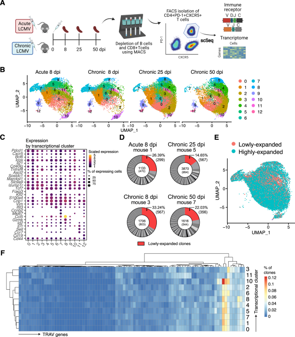


![Recombinant Anti-p53 antibody [E26] KO Tested (ab32389) | Abcam](https://www.abcam.com/ps/products/32/ab32389/Images/ab32389-389819-antip53antibodyimmunocytochemistryimmunofluorescencehap1human.jpg)


![Physiologically Based Pharmacokinetic Modeling of FTY720 (2-Amino-2[2-(-4-octylphenyl)ethyl]propane-1,3-diol hydrochloride) in...](https://dmd.aspetjournals.org/sites/default/files/styles/medium/public/highwire/dmd/34/9.cover.gif?itok=HklfPAL9)