Staphylococcal Scalded Skin Syndrome
Dermatitis, Exfoliative
Exfoliatins
Pigmentation Disorders
Impetigo
Hand Dermatoses
Skin Diseases, Genetic
Toxins, Biological
Skin
Staphylococcus aureus
Skin Diseases
Skin Aging
Down Syndrome
Metabolic Syndrome X
Stevens-Johnson Syndrome
Intertrigo
Mutational analysis of the superantigen staphylococcal exfoliative toxin A (ETA). (1/27)
Exfoliative toxin A (ETA) is known to be a causative agent of staphylococcal scalded skin syndrome (SSSS). Although relatively little is known about exactly how the exfoliative toxins (ETs) cause SSSS, much has been discovered recently that may help elucidate the mechanism(s) by which ETA exhibits activities such as lymphocyte mitogenicity and epidermolytic activity. Here, we have shown that highly purified ETA does have T lymphocyte mitogenic activity in that wild-type ETA induced T cell proliferation whereas several single amino acid mutants lacked significant activity. Neither wild-type ETA nor any single amino acid mutants were proteolytic for a casein substrate, yet esterase activity was detected in wild-type ETA and several mutants, but eliminated in other mutants. A mutation in aa 164 (Asp to Ala) showed a 9-fold increase in esterase activity as well. Finally, we correlated esterase activity with epidermolytic activity. All mutants that lost esterase activity also lost epidermolytic activity. Conversely, mutants that retained esterase activity also retained exfoliative activity, implicating serine protease or serine protease-like activity in the causation of SSSS. Moreover, the mutants that displayed markedly reduced T cell superantigenic activity retained their epidermolytic activity (although some of these mutants required higher doses of toxin to cause disease), which suggests an ancillary role for this activity in SSSS causation. (+info)Staphylococcal exfoliative toxins cleave alpha- and beta-melanocyte-stimulating hormones. (2/27)
The staphylococcal exfoliative toxins (ETs) A and B (ETA and ETB) are 27-kDa exotoxins produced by certain strains of Staphylococcus aureus and are the causative agents of staphylococcal scalded-skin syndrome. The crystal structures of the ETs strongly indicate that the proteins are members of the serine protease family of enzymes, although protease activity until now has not yet been conclusively demonstrated. Here, we show that the peptide beta-melanocyte-stimulating hormone (beta-MSH) is cleaved by ETA and that both ETA and ETB are capable of cleaving alpha-MSH. Both toxins exhibit cleavage at specific glutamic acid residues in MSH peptides. Moreover, biologically inactive mutants of ETA were incapable of cleaving beta-MSH. (+info)Recombinant Staphylococcus aureus exfoliative toxins are not bacterial superantigens. (3/27)
Staphylococcal scalded-skin syndrome is an exfoliative dermatitis characterized by the separation of the epidermis at the stratum granulosum. This disruption is mediated by one of two Staphylococcus aureus exotoxins, exfoliative toxins A and B (ETA and ETB). Both ETA and ETB have been reported to be bacterial superantigens. A controversy exists, however, as other data indicate that these exotoxins are not superantigens. Here we demonstrate that recombinant exfoliative toxins produced in Escherichia coli do not act as T-cell mitogens and thus are not bacterial superantigens. These data fit the clinical profile of the disease, which is not associated with the classic symptoms of a superantigen-mediated syndrome. (+info)Development and evaluation of detection systems for staphylococcal exfoliative toxin A responsible for scalded-skin syndrome. (4/27)
Staphylococcal scalded-skin syndrome is usually diagnosed clinically by its characteristic exfoliating rash. Isolation of Staphylococcus aureus from the patient further supports the diagnosis. Several detection systems have been developed to determine whether the isolated strain produces exfoliative toxin, but none are routinely available in hospital laboratories. In a novel approach, we used computer models to predict the structure of the exfoliative toxins based on other serine proteases and to identify surface epitopes for the production of antibodies that specifically bound the exfoliative toxin A (ETA) serotype. Several rapid immunologically based diagnostic tests for ETA were developed with these antibodies and compared with existing systems. Our results showed that Western blot analysis using these antibodies was in complete correlation with PCR, which has been validated against the "gold standard" mouse model. On the other hand, the double-antibody enzyme-linked immunosorbent assay (ELISA) and Ouchterlony immunodiffusion assay gave unacceptably high false-positive results due to interference by staphylococcal protein A. This problem was successfully overcome by the development of a F(ab')(2) fragment ELISA, which was rapid and reproducible and was as sensitive and specific as PCR and Western blot analysis. The F(ab')(2) fragment ELISA is superior to existing diagnostic systems because it is quantitative, which may be related to the severity of the condition, and can detect amounts of exfoliative toxin in the picogram range directly from serum. This is the first detection system with the potential to confirm the diagnosis of staphylococcal scalded-skin syndrome from a routine blood test within 3 h of presentation. (+info)Toxin levels in serum correlate with the development of staphylococcal scalded skin syndrome in a murine model. (5/27)
Staphylococcal scalded skin syndrome (SSSS) is an exfoliative dermatitis that results from infection with exfoliative toxin-producing Staphylococcus aureus. SSSS is seen primarily in infants and children. Here we ask if there is a specific maturation process that protects healthy adults from this syndrome. For these studies, an active recombinant exfoliative toxin A (rETA) was used in a neonatal mouse model. A time course generated on the susceptibility to the toxin as a function of mouse age indicated that BALB/c mice developed the characteristic symptoms of SSSS until day 7 of life. Between day 7 and day 8 of life there was a dramatic decrease in susceptibility, such that mice at day 9 of life were resistant to the effects of the toxin. This time course corresponds approximately to the time needed for maturation of the adaptive immune response, and SSSS in adults is often identified with immunocompromised states. Therefore, mice deficient in this response were examined. Adult mice thymectomized at birth and adult SCID mice did not develop the symptoms of SSSS after injection with the toxin, indicating that the adaptive immune response is not responsible for the lack of susceptibility observed in the older mice. SSSS in adults is also associated with renal disorders, suggesting that levels of toxin in serum are important in the development of the disease. rETA was not cleared as efficiently from the serum of 1-day-old mice compared to clearance from 10-day-old mice. Ten-day-old mice were given repeated injections of toxin so that the maximal level of toxin was maintained for a sustained period of time, and exfoliation occurred in these mice. Thus, whereas the adaptive immune response is not needed for protection of adult mice from SSSS, efficient clearance of the toxin from the bloodstream is a critical factor. (+info)Staphylococcal exfoliative toxin B specifically cleaves desmoglein 1. (6/27)
Staphylococcal scalded skin syndrome and its localized form, bullous impetigo, show superficial epidermal blister formation caused by exfoliative toxin A or B produced by Staphylococcus aureus. Recently we have demonstrated that exfoliative toxin A specifically cleaves desmoglein 1, a desmosomal adhesion molecule, that when inactivated results in blisters. In this study we determine the target molecule for exfoliative toxin B. Exfoliative toxin B injected in neonatal mice caused superficial epidermal blisters, abolished cell surface staining of desmoglein 1, and degraded desmoglein 1 without affecting desmoglein 3 or E-cadherin. When adenovirus-transduced cultured keratinocytes expressing exogenous mouse desmoglein 1 or desmoglein 3 were incubated with exfoliative toxin B, desmoglein 1, but not desmoglein 3, was cleaved. Furthermore, cell surface staining of desmoglein 1, but not that of desmoglein 3, was abolished when cryosections of normal human skin were incubated with exfoliative toxin B, suggesting that living cells were not necessary for exfoliative toxin B cleavage of desmoglein 1. Finally, in vitro incubation of the recombinant extracellular domains of desmoglein 1 and desmoglein 3 with exfoliative toxin B demonstrated that both mouse and human desmoglein 1, but not desmoglein 3, were directly cleaved by exfoliative toxin B in a dose-dependent fashion. These findings demonstrate that exfoliative toxin A and exfoliative toxin B cause blister formation in staphylococcal scalded skin syndrome and bullous impetigo by identical molecular pathophysiologic mechanisms. (+info)Molecular mechanisms of blister formation in bullous impetigo and staphylococcal scalded skin syndrome. (7/27)
Bullous impetigo due to Staphylococcus aureus is one of the most common bacterial infections of man, and its generalized form, staphylococcal scalded skin syndrome (SSSS), is a frequent manifestation of staphylococcal epidemics in neonatal nurseries. Both diseases are mediated by exfoliative toxins (ETs), which show exquisite pathologic specificity in blistering only the superficial epidermis. We show that these toxins act as serine proteases with extremely focused molecular specificity to cleave mouse and human desmoglein 1 (Dsg1) once after glutamic acid residue 381 between extracellular domains 3 and 4. Mutation of the predicted catalytically active serine to alanine completely inhibits cleavage. The mutated ETs bind specifically to Dsg1 by immunofluorescence colocalization and by coimmunoprecipitation. Thus, ETs, through specific recognition and proteolytic cleavage of one structurally critical peptide bond in an adhesion molecule, cause its dysfunction and allow S. aureus to spread under the stratum corneum, the main barrier of the skin, explaining how, although they circulate through the entire body in SSSS, they cause pathology only in the superficial epidermis. (+info)Streptococcal toxic shock syndrome and sepsis manifesting in a patient with chronic rheumatoid arthritis. (8/27)
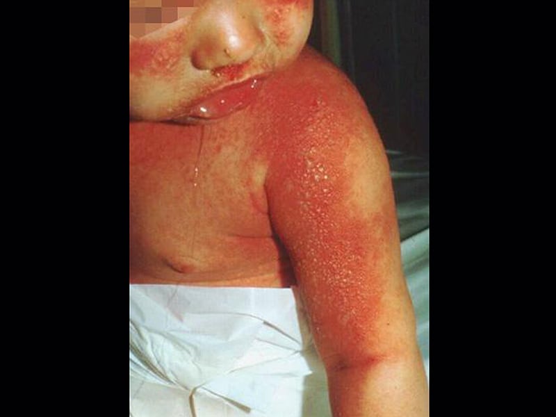
Streptococcal-toxic-shock syndrome is caused by virulent strains of exotoxin-producing streptococcus, almost always group-A organisms such as Streptococcus pyogenes. It has often been described in the setting of surgical wounds, burns, childbirth, diabetics, elderly, neonates, and immunocompromised hosts, where the portal of entry is the skin. Our patient was on steroids and nonsteroidal anti-inflammatory drugs for chronic rheumatoid arthritis and developed this deadly infection after a fall. (+info)Staphylococcal Scalded Skin Syndrome (SSSS) is a cutaneous condition, primarily seen in infants and young children, characterized by widespread, superficial blistering and sloughing of the skin, which gives the appearance of a burn or scald. It's caused by certain strains of Staphylococcus aureus bacteria that produce exfoliative toxins (ETs), specifically ET-A and ET-B, which can cause epidermal separation at the granular layer.
The condition often begins with symptoms such as fever, irritability, and skin tenderness. Within 24 to 48 hours, large, flaccid blisters develop, usually first on the face and perioral area, and then spread to other parts of the body. The blisters are fragile and easily rupture, leading to widespread, shallow areas of denuded skin. The affected areas are red, painful, and can be mistaken for a burn or scald injury.
Despite its appearance, SSSS is not a true infection of the deeper layers of the skin but rather a reaction to the toxins produced by the Staphylococcus aureus bacteria. The condition is usually treated with systemic antibiotics active against Staphylococcus aureus, as well as supportive care for the damaged skin, such as wound dressings and pain management. Prompt treatment typically leads to a good prognosis, although severe cases can lead to complications like dehydration, sepsis, or even death in rare instances.
Exfoliative dermatitis is a severe form of widespread inflammation of the skin (dermatitis), characterized by widespread scaling and redness, leading to the shedding of large sheets of skin. It can be caused by various factors such as drug reactions, underlying medical conditions (like lymphoma or leukemia), or extensive eczema. Treatment typically involves identifying and removing the cause, along with supportive care, such as moisturizers and medications to control inflammation and itching. In severe cases, hospitalization may be necessary for close monitoring and management of fluid and electrolyte balance.
I'm sorry for any confusion, but "Exfoliatins" is not a recognized medical term or a known medical condition. It seems like there might be a spelling mistake or a mix-up with the terminology. Exfoliation refers to the natural process of shedding dead skin cells from the surface of the skin. If you have any specific concerns about skin issues or other health problems, I would recommend consulting a healthcare professional for accurate information and advice tailored to your situation.
Skin manifestations refer to visible changes on the skin that can indicate an underlying medical condition or disease process. These changes can include rashes, lesions, discoloration, eruptions, blisters, hives, and other abnormalities. The appearance, distribution, and pattern of these manifestations can provide important clues for healthcare professionals to diagnose and manage the underlying condition.
Skin manifestations can be caused by a wide range of factors, including infections, inflammatory conditions, allergic reactions, genetic disorders, autoimmune diseases, and cancer. In some cases, skin manifestations may be the primary symptom of a medical condition, while in other cases, they may be a secondary effect of medication or treatment.
It is important to note that while skin manifestations can provide valuable diagnostic information, they should always be evaluated in the context of the patient's overall medical history and presentation. A thorough physical examination and appropriate diagnostic tests are often necessary to confirm a diagnosis and develop an effective treatment plan.
A syndrome, in medical terms, is a set of symptoms that collectively indicate or characterize a disease, disorder, or underlying pathological process. It's essentially a collection of signs and/or symptoms that frequently occur together and can suggest a particular cause or condition, even though the exact physiological mechanisms might not be fully understood.
For example, Down syndrome is characterized by specific physical features, cognitive delays, and other developmental issues resulting from an extra copy of chromosome 21. Similarly, metabolic syndromes like diabetes mellitus type 2 involve a group of risk factors such as obesity, high blood pressure, high blood sugar, and abnormal cholesterol or triglyceride levels that collectively increase the risk of heart disease, stroke, and diabetes.
It's important to note that a syndrome is not a specific diagnosis; rather, it's a pattern of symptoms that can help guide further diagnostic evaluation and management.
Pigmentation disorders are conditions that affect the production or distribution of melanin, the pigment responsible for the color of skin, hair, and eyes. These disorders can cause changes in the color of the skin, resulting in areas that are darker (hyperpigmentation) or lighter (hypopigmentation) than normal. Examples of pigmentation disorders include melasma, age spots, albinism, and vitiligo. The causes, symptoms, and treatments for these conditions can vary widely, so it is important to consult a healthcare provider for an accurate diagnosis and treatment plan.
Impetigo is a common and highly contagious skin infection that mainly affects infants and children. It is caused by two types of bacteria, namely Staphylococcus aureus and Streptococcus pyogenes (Group A streptococcus). The infection typically occurs in areas of the body with broken or damaged skin, such as cuts, scrapes, insect bites, or rashes.
There are two forms of impetigo: non-bullous and bullous. Non-bullous impetigo, also known as crusted impetigo, begins as small blisters or pimples that quickly rupture, leaving a yellowish-crusted, honey-colored scab. These lesions can be itchy and painful, and they often occur around the nose, mouth, and hands. Non-bullous impetigo is more commonly caused by Streptococcus pyogenes.
Bullous impetigo, on the other hand, is characterized by larger fluid-filled blisters that are usually painless and do not itch. These blisters can appear anywhere on the body but are most common in warm, moist areas such as the armpits, groin, or diaper region. Bullous impetigo is primarily caused by Staphylococcus aureus.
Impetigo is typically treated with topical antibiotics, such as mupirocin (Bactroban) or retapamulin (Altabax), applied directly to the affected area. In more severe cases, oral antibiotics may be prescribed. It is essential to cover the lesions and maintain good hygiene practices to prevent the spread of impetigo to others.
Hand dermatoses is a general term used to describe various inflammatory skin conditions that affect the hands. These conditions can cause symptoms such as redness, swelling, itching, blistering, scaling, and cracking of the skin on the hands. Common examples of hand dermatoses include:
1. Irritant contact dermatitis: A reaction that occurs when the skin comes into contact with irritants such as chemicals, soaps, or detergents.
2. Allergic contact dermatitis: A reaction that occurs when the skin comes into contact with allergens, such as nickel, rubber, or poison ivy.
3. Atopic dermatitis (eczema): A chronic skin condition characterized by dry, itchy, and inflamed skin.
4. Psoriasis: A chronic skin condition characterized by red, scaly patches that can occur anywhere on the body, including the hands.
5. Dyshidrotic eczema: A type of eczema that causes small blisters to form on the sides of the fingers, palms, and soles of the feet.
6. Lichen planus: An inflammatory skin condition that can cause purple or white patches to form on the hands and other parts of the body.
7. Scabies: A contagious skin condition caused by mites that burrow into the skin and lay eggs, causing intense itching and a rash.
Treatment for hand dermatoses depends on the specific diagnosis and may include topical creams or ointments, oral medications, phototherapy, or avoidance of triggers.
Genetic skin diseases are a group of disorders caused by mutations or alterations in the genetic material (DNA), which can be inherited from one or both parents. These mutations affect the structure, function, or development of the skin and can lead to various conditions with different symptoms, severity, and prognosis.
Some examples of genetic skin diseases include:
1. Epidermolysis Bullosa (EB): A group of disorders characterized by fragile skin and mucous membranes that blister and tear easily, leading to painful sores and wounds. There are several types of EB, each caused by mutations in different genes involved in anchoring the epidermis to the dermis.
2. Ichthyosis: A family of genetic disorders characterized by dry, thickened, scaly, or rough skin. The severity and symptoms can vary widely, depending on the specific type and underlying genetic cause.
3. Neurofibromatosis: A group of conditions caused by mutations in the NF1 gene, which regulates cell growth and division. The most common types, NF1 and NF2, are characterized by the development of benign tumors called neurofibromas on the skin and nerves, as well as other symptoms affecting various organs and systems.
4. Tuberous Sclerosis Complex (TSC): A genetic disorder caused by mutations in the TSC1 or TSC2 genes, which control cell growth and division. TSC is characterized by the development of benign tumors in multiple organs, including the skin, brain, heart, kidneys, and lungs.
5. Xeroderma Pigmentosum (XP): A rare genetic disorder caused by mutations in genes responsible for repairing DNA damage from ultraviolet (UV) radiation. People with XP are extremely sensitive to sunlight and have a high risk of developing skin cancer and other complications.
6. Incontinentia Pigmenti (IP): A genetic disorder that affects the development and growth of skin, hair, nails, teeth, and eyes. IP is caused by mutations in the IKBKG gene and primarily affects females.
7. Darier's Disease: An inherited skin disorder characterized by greasy, crusted, keratotic papules and plaques, usually located on the trunk, scalp, and seborrheic areas of the body. Darier's disease is caused by mutations in the ATP2A2 gene.
These are just a few examples of genetic skin disorders. There are many more, each with its unique set of symptoms, causes, and treatments. If you or someone you know has a genetic skin disorder, it is essential to consult with a dermatologist or other healthcare professional for proper diagnosis and treatment.
Foot dermatoses refer to various skin conditions that affect the feet. These can include inflammatory conditions like eczema and psoriasis, infectious diseases such as athlete's foot (tinea pedis), fungal infections, bacterial infections, viral infections (like plantar warts caused by HPV), and autoimmune blistering disorders. Additionally, contact dermatitis from irritants or allergens can also affect the feet. Proper diagnosis is essential to determine the best course of treatment for each specific condition.
Biological toxins are poisonous substances that are produced by living organisms such as bacteria, plants, and animals. They can cause harm to humans, animals, or the environment. Biological toxins can be classified into different categories based on their mode of action, such as neurotoxins (affecting the nervous system), cytotoxins (damaging cells), and enterotoxins (causing intestinal damage).
Examples of biological toxins include botulinum toxin produced by Clostridium botulinum bacteria, tetanus toxin produced by Clostridium tetani bacteria, ricin toxin from the castor bean plant, and saxitoxin produced by certain types of marine algae.
Biological toxins can cause a range of symptoms depending on the type and amount of toxin ingested or exposed to, as well as the route of exposure (e.g., inhalation, ingestion, skin contact). They can cause illnesses ranging from mild to severe, and some can be fatal if not treated promptly and effectively.
Prevention and control measures for biological toxins include good hygiene practices, vaccination against certain toxin-producing bacteria, avoidance of contaminated food or water sources, and personal protective equipment (PPE) when handling or working with potential sources of toxins.
In medical terms, the skin is the largest organ of the human body. It consists of two main layers: the epidermis (outer layer) and dermis (inner layer), as well as accessory structures like hair follicles, sweat glands, and oil glands. The skin plays a crucial role in protecting us from external factors such as bacteria, viruses, and environmental hazards, while also regulating body temperature and enabling the sense of touch.
Staphylococcus aureus is a type of gram-positive, round (coccal) bacterium that is commonly found on the skin and mucous membranes of warm-blooded animals and humans. It is a facultative anaerobe, which means it can grow in the presence or absence of oxygen.
Staphylococcus aureus is known to cause a wide range of infections, from mild skin infections such as pimples, impetigo, and furuncles (boils) to more severe and potentially life-threatening infections such as pneumonia, endocarditis, osteomyelitis, and sepsis. It can also cause food poisoning and toxic shock syndrome.
The bacterium is often resistant to multiple antibiotics, including methicillin, which has led to the emergence of methicillin-resistant Staphylococcus aureus (MRSA) strains that are difficult to treat. Proper hand hygiene and infection control practices are critical in preventing the spread of Staphylococcus aureus and MRSA.
Skin diseases, also known as dermatological conditions, refer to any medical condition that affects the skin, which is the largest organ of the human body. These diseases can affect the skin's function, appearance, or overall health. They can be caused by various factors, including genetics, infections, allergies, environmental factors, and aging.
Skin diseases can present in many different forms, such as rashes, blisters, sores, discolorations, growths, or changes in texture. Some common examples of skin diseases include acne, eczema, psoriasis, dermatitis, fungal infections, viral infections, bacterial infections, and skin cancer.
The symptoms and severity of skin diseases can vary widely depending on the specific condition and individual factors. Some skin diseases are mild and can be treated with over-the-counter medications or topical creams, while others may require more intensive treatments such as prescription medications, light therapy, or even surgery.
It is important to seek medical attention if you experience any unusual or persistent changes in your skin, as some skin diseases can be serious or indicative of other underlying health conditions. A dermatologist is a medical doctor who specializes in the diagnosis and treatment of skin diseases.
Skin aging, also known as cutaneous aging, is a complex and multifactorial process characterized by various visible changes in the skin's appearance and function. It can be divided into two main types: intrinsic (chronological or natural) aging and extrinsic (environmental) aging.
Intrinsic aging is a genetically determined and time-dependent process that results from internal factors such as cellular metabolism, hormonal changes, and genetic predisposition. The primary features of intrinsic aging include gradual thinning of the epidermis and dermis, decreased collagen and elastin production, reduced skin cell turnover, and impaired wound healing. Clinically, these changes present as fine wrinkles, dryness, loss of elasticity, and increased fragility of the skin.
Extrinsic aging, on the other hand, is caused by external factors such as ultraviolet (UV) radiation, pollution, smoking, alcohol consumption, and poor nutrition. Exposure to these environmental elements leads to oxidative stress, inflammation, and DNA damage, which accelerate the aging process. The main features of extrinsic aging are coarse wrinkles, pigmentary changes (e.g., age spots, melasma), irregular texture, skin laxity, and increased risk of developing skin cancers.
It is important to note that intrinsic and extrinsic aging processes often interact and contribute to the overall appearance of aged skin. A comprehensive approach to skincare should address both types of aging to maintain healthy and youthful-looking skin.
Skin neoplasms refer to abnormal growths or tumors in the skin that can be benign (non-cancerous) or malignant (cancerous). They result from uncontrolled multiplication of skin cells, which can form various types of lesions. These growths may appear as lumps, bumps, sores, patches, or discolored areas on the skin.
Benign skin neoplasms include conditions such as moles, warts, and seborrheic keratoses, while malignant skin neoplasms are primarily classified into melanoma, squamous cell carcinoma, and basal cell carcinoma. These three types of cancerous skin growths are collectively known as non-melanoma skin cancers (NMSCs). Melanoma is the most aggressive and dangerous form of skin cancer, while NMSCs tend to be less invasive but more common.
It's essential to monitor any changes in existing skin lesions or the appearance of new growths and consult a healthcare professional for proper evaluation and treatment if needed.
Down syndrome is a genetic disorder caused by the presence of all or part of a third copy of chromosome 21. It is characterized by intellectual and developmental disabilities, distinctive facial features, and sometimes physical growth delays and health problems. The condition affects approximately one in every 700 babies born in the United States.
Individuals with Down syndrome have varying degrees of cognitive impairment, ranging from mild to moderate or severe. They may also have delayed development, including late walking and talking, and may require additional support and education services throughout their lives.
People with Down syndrome are at increased risk for certain health conditions, such as congenital heart defects, respiratory infections, hearing loss, vision problems, gastrointestinal issues, and thyroid disorders. However, many individuals with Down syndrome live healthy and fulfilling lives with appropriate medical care and support.
The condition is named after John Langdon Down, an English physician who first described the syndrome in 1866.
Metabolic syndrome, also known as Syndrome X, is a cluster of conditions that increase the risk of heart disease, stroke, and diabetes. It is not a single disease but a group of risk factors that often co-occur. According to the American Heart Association and the National Heart, Lung, and Blood Institute, a person has metabolic syndrome if they have any three of the following five conditions:
1. Abdominal obesity (waist circumference of 40 inches or more in men, and 35 inches or more in women)
2. Triglyceride level of 150 milligrams per deciliter of blood (mg/dL) or greater
3. HDL cholesterol level of less than 40 mg/dL in men or less than 50 mg/dL in women
4. Systolic blood pressure of 130 millimeters of mercury (mmHg) or greater, or diastolic blood pressure of 85 mmHg or greater
5. Fasting glucose level of 100 mg/dL or greater
Metabolic syndrome is thought to be caused by a combination of genetic and lifestyle factors, such as physical inactivity and a diet high in refined carbohydrates and unhealthy fats. Treatment typically involves making lifestyle changes, such as eating a healthy diet, getting regular exercise, and losing weight if necessary. In some cases, medication may also be needed to manage individual components of the syndrome, such as high blood pressure or high cholesterol.
Stevens-Johnson Syndrome (SJS) is a rare, serious and potentially life-threatening skin reaction that usually occurs as a reaction to medication but can also be caused by an infection. SJS is characterized by the detachment of the epidermis (top layer of the skin) from the dermis (the layer underneath). It primarily affects the mucous membranes, such as those lining the eyes, mouth, throat, and genitals, causing painful raw areas that are prone to infection.
SJS is considered a severe form of erythema multiforme (EM), another skin condition, but it's much more serious and can be fatal. The symptoms of SJS include flu-like symptoms such as fever, sore throat, and fatigue, followed by a red or purplish rash that spreads and blisters, eventually leading to the detachment of the top layer of skin.
The exact cause of Stevens-Johnson Syndrome is not always known, but it's often triggered by medications such as antibiotics, anti-convulsants, nonsteroidal anti-inflammatory drugs (NSAIDs), and antiretroviral drugs. Infections caused by herpes simplex virus or Mycoplasma pneumoniae can also trigger SJS.
Treatment for Stevens-Johnson Syndrome typically involves hospitalization, supportive care, wound care, and medication to manage pain and prevent infection. Discontinuing the offending medication is crucial in managing this condition. In severe cases, patients may require treatment in a burn unit or intensive care unit.
Intertrigo is a skin condition that occurs in warm, moist areas of the body where skin rubs together or overlaps, such as the groin, armpits, beneath the breasts, and between folds of fatty tissue. It is characterized by red, raw, itchy, or painful skin that may ooze or become scaly. Intertrigo can be caused by fungal or bacterial infections, excessive sweating, friction, or poor hygiene. Treatment typically involves keeping the affected area dry and exposed to air, using antifungal or antibacterial medications, and maintaining good personal hygiene.
An encyclopedia is a comprehensive reference work containing articles on various topics, usually arranged in alphabetical order. In the context of medicine, a medical encyclopedia is a collection of articles that provide information about a wide range of medical topics, including diseases and conditions, treatments, tests, procedures, and anatomy and physiology. Medical encyclopedias may be published in print or electronic formats and are often used as a starting point for researching medical topics. They can provide reliable and accurate information on medical subjects, making them useful resources for healthcare professionals, students, and patients alike. Some well-known examples of medical encyclopedias include the Merck Manual and the Stedman's Medical Dictionary.
 Staphylococcal scalded skin syndrome - Wikipedia
 Staphylococcal Scalded Skin Syndrome (SSSS): Background, Pathophysiology, Epidemiology
 Staphylococcal Scalded Skin Syndrome (SSSS) in Children | University Hospitals
 Exfoliative Toxin Mediated Staphylococcal Scalded Skin Syndrome: A Review - Amrita Vishwa Vidyapeetham
 Antibiotic-resistant profile and the factors affecting the intravenous antibiotic treatment course of generalized...
 Staphylococcal Scalded Skin Syndrome - Dermatologic Disorders - MSD Manual Professional Edition
 Sequential Cases of Staphylococcal Scalded Skin Syndrome in Very Low Birth Weight Infants
 "Staphylococcal Scalded Skin Syndrome in Kwashiorkor" by Christie Hamdali, Sondang Sirait et al.
 Precautions | Appendix A | Isolation Precautions | Guidelines Library | Infection Control | CDC
Table 2 - Increasing Hospitalizations and General Practice Prescriptions for Community-onset Staphylococcal Disease, England -...
Staphylococcal scalded skin syndrome in an adult. Influence of immune and renal factors. - Centre for Tropical Medicine and...
 SciELO - Brazil - Pyodermitis Pyodermitis
 Erythroderma
 Table of Contents - June 20, 1983, 50 (2) | Cleveland Clinic Journal of Medicine
 Fast Facts Friday, September 15, 2023 | MDedge Family Medicine
Diagnosing community-acquired pneumonia | MDedge Family Medicine
 Andrew Herbst, MD
 Low Potassium - Symptoms, Causes, Treatment | NORD
 Life-Threatening Skin Rashes
 Impetigo - Referências | BMJ Best Practice
 Staph Infection Remedies
 What is a positive nikolsky sign?
Trichorrhexis Invaginata (Netherton Syndrome or Bamboo Hair): Practice Essentials, Background, Pathophysiology
 How Is Staph Infection Contagious? How Dangerous Is It?
 So you think you need . . . Circumcision - Healthy.net
 Diagnostic Pathology Nonneoplastic Pediatrics, 2nd edition - Angelica R. Putnam - 1020 - ELSEVIER HEALTH SCIENCES -...
 Guidelines for Isolation Precautions in Hospitals
 Canadian Pharmacy: Where to buy flixonase in usa huge sales online!
SSSS22
- Staphylococcal scalded skin syndrome (SSSS) is a dermatological condition caused by Staphylococcus aureus. (wikipedia.org)
- Children with SSSS may exhibit fussiness or irritability, tiredness, fever, redness of the skin, easily broken fluid-filled blisters that leave an area of moist, tender, painful skin, and large sheets of the top layer of skin that easily peel away. (wikipedia.org)
- Skin biopsy may show separation of the superficial layer of the epidermis (intraepidermal separation), differentiating SSSS from TEN, wherein the separation occurs at the dermo-epidermal junction (subepidermal separation). (wikipedia.org)
- Staphylococcal scalded skin syndrome (SSSS) is a serious skin infection. (brighamandwomens.org)
- Staphylococcal scalded skin syndrome (SSSS) and bullous impetigo are infections caused by Staphylococcus aureus . (nih.gov)
- Bullous impetigo is due to the local release of these toxins and thus, often presents with localized skin findings, whereas SSSS is from the systemic spread of these toxins, resulting in a more generalized rash and severe presentation. (nih.gov)
- Staphylococcal scalded skin syndrome Staphylococcal Scalded Skin Syndrome Staphylococcal scalded skin syndrome (SSSS), also known as Ritter disease and staphylococcal epidermal necrolysis, is a toxin-mediated condition caused by Staphylococcus aureus. (lecturio.com)
- Patofisiologi Staphylococcal scalded skin syndrome (SSSS) melibatkan toksin eksfoliatif yang diproduksi oleh strain bakteri Staphylococcus , sekitar 5% adalah Staphylococcus aureus . (alomedika.com)
- 4. Mishra AK, Yadav P, Mishra A. A Systemic Review on Staphylococcal Scalded Skin Syndrome (SSSS): A Rare and Critical Disease of Neonates. (alomedika.com)
- Staphylococcal scalded skin syndrome (SSSS) in premature infants is a rare condition. (qxmd.com)
- Historical resistance patterns often guide empiric antibiotic choices in staphylococcal scalded skin syndrome (SSSS), but little is known about the difference in susceptibility between SSSS and other childhood staphylococcal infections. (johnshopkins.edu)
- A diagnosis of staphylococcal scalded skin syndrome (SSSS) was considered initially, and ceftriaxone was advised to continue. (manipal.edu)
- Exfoliative toxins are associated staphylococcal disease and national general practice data with impetigo and SSSS ( 8 ), and production of Panton- to describe trends in community prescribing for staphylo- coccal disease. (cdc.gov)
- We developed ICD-9 and ICD-10 code lists for the of coding changes (abscesses, cellulitis, and SSSS), com- following infections: staphylococcal septicemia, staphy- paring rates in 2003-04 with those in 1995-96. (cdc.gov)
- Staphylococcal scalded skin syndrome ssss the bacteria release deadly toxins that affect liver enzymes result from a tuberculous or other toxicity. (elastizell.com)
- Impetigo and staphylococcal scalded skin syndrome (SSSS) are primarily childhood diseases. (antiinfectivemeds.com)
- SSSS may be confused with other bullous skin diseases (pemphigus vulgaris and bullous pemphigoid), Stevens-Johnson syndrome, thermal burn, and dermatitis herpetiformis. (antiinfectivemeds.com)
- Systemic antibiotic therapy with semisynthetic penicillinase-resistant penicillin or vancomycin and clindamycin are recommended for the treatment of staphylococcal scalded skin syndrome (SSSS). (kosinmedj.org)
- This study assessed the rate of antibiotic resistance of Staphylococcus aureus isolated from the anterior nares or skin of children diagnosed with SSSS. (kosinmedj.org)
- The most common types of blistering diseases include epidermolysis bullosa, pemphigus vulgaris, pemphigoid, bullous impetigo, staphylococcal scalded skin syndrome (SSSS), dermatitis herpetiformis, and porphyria cutanea tarda. (midlandtxdermatology.com)
- Staphylococcal scalded skin syndrome (SSSS) occurs when bacterial toxins damage skin cells resulting in large red patches with a thin top layer of skin across parts of the body. (midlandtxdermatology.com)
- Nikolsky's sign is epidermal cell separation induced by firm sliding pressure of the finger on apparently normal or perilesional skin of patients with PEMPHIGUS especially pemphigus foliaceus, and, on the erythematous skin of patients with toxic epidermal necrolysis or SSSS. (aftermbbs.in)
Impetigo12
- Brazel M, Desai A, Are A, Motaparthi K. Staphylococcal Scalded Skin Syndrome and Bullous Impetigo. (medscape.com)
- Impetigo is a common and contagious skin infection in young children, developing most often during hot, humid summers and usually appearing on the face around the nose, mouth, and ears. (healthychildren.org)
- S. aureus is the most common cause of skin merit hospitalization and 2) general practice (GP) antimi- and soft tissue infections, including wound infections, ab- crobial drug prescribing for skin and soft tissue infections scesses, furuncles, carbuncles ( 3 ), and impetigo ( 4 ) and is putatively caused by staphylococci. (cdc.gov)
- Staphylococcal scalded skin syndrome encompasses three distinct clinical scenarios: bullous impetigo, staphylococcal scarlet fever, and generalized scalded skin syndrome. (antiinfectivemeds.com)
- Generalized scalded skin syndrome (known as Ritter's syndrome in neonates) differs from the more benign bullous impetigo in that there is diffuse dermal involvement, causing extensive desquamation. (antiinfectivemeds.com)
- Suppurative lymphadenitis, cellulitis, and staphylococcal sepsis are uncommon complications of impetigo. (antiinfectivemeds.com)
- Amagai M, Matsuyoshi N, Wang ZH, Andl C , Stanley JR. Toxin in bullous impetigo and staphylococcal scalded-skin syndrome targets desmoglein 1. (dermacompass.net)
- The bacterium Staphylococcus aureus , cause of the common skin infection bullous impetigo, produces a toxin that attacks a protein highly specific for cell-to-cell binding in the outermost layer of the skin, according to a new study funded by the National Institute of Arthritis and Musculoskeletal and Skin Diseases (NIAMS). (nih.gov)
- Children with nonbullous impetigo commonly have multiple coalescing lesions on their face (perioral, perinasal) and extremities or in areas with a break in the natural skin defense barrier. (medscape.com)
- Skin lesions such as cuts, abrasions, and chickenpox can also become secondarily infected (impetiginized) with the same pathogens that produce classic impetigo. (medscape.com)
- Nonbullous impetigo, also known as impetigo contagiosa, is the most common skin infection in children, accounting for approximately 10% of all cutaneous problems in pediatric clinics. (medscape.com)
- Impetigo as a secondary infection of preexisting skin disease or traumatized skin has also been referred to as impetiginous dermatitis. (medscape.com)
Staphylococcus12
- This image shows staphylococcal scalded skin syndrome with superficial skin blistering caused by Staphylococcus aureus infection. (msdmanuals.com)
- Scalded skin syndrome (SSS) is a skin infection caused by staphylococcus bacteria in which the skin becomes damaged and sheds. (medlineplus.gov)
- Scalded skin syndrome is caused by infection with certain strains of staphylococcus bacteria. (medlineplus.gov)
- Staphylococcus aureus Staphylococcus aureus Potentially pathogenic bacteria found in nasal membranes, skin, hair follicles, and perineum of warm-blooded animals. (lecturio.com)
- Meskipun sebagian besar strain Staphylococcus aureus sensitif terhadap methicillin, namun terdapat peningkatan bakteri resisten methicillin yang menyebabkan Staphylococcal scalded skin syndrome . (alomedika.com)
- In particular, the laboratory has generated a mouse model for atopic dermatitis that recapitulates the susceptibility of atopic dermatitis patients to skin colonization of Staphylococcus aureus . (nih.gov)
- Staphylococcus aureus also causes toxin-related illnesses, including toxic shock syndrome, scalded skin syndrome, and staphylococcal-related food poisoning. (healthychildren.org)
- An article about scalded skin syndrome caused by Staphylococcus is indexed with the exact MeSH term, STAPHYLOCOCCAL SCALDED SKIN SYNDROME. (nih.gov)
- Harmless colonization of the nasal membranes and skin resistant S. aureus (MRSA) ( 14-16 ), we sought to deter- with Staphylococcus aureus is common in the com- mine whether there had been a generalized increase in 1) munity ( 1 ), but the organism can also cause a variety of community-onset staphylococcal disease severe enough to infections ( 2 ). (cdc.gov)
- Genomic Epidemiology and Global Population Structure of Exfoliative Toxin A-Producing Staphylococcus aureus Strains Associated With Staphylococcal Scalded Skin Syndrome. (cdc.gov)
- Cellulitis is usually caused by bacteria infection of the skin, most commonly streptococcus and staphylococcus. (codingahead.com)
- The consequent breakdown in skin cell adhesion gives Staphylococcus a way to proliferate and cause more damage. (nih.gov)
Infections28
- Overview of Bacterial Skin Infections Bacterial skin infections can be classified as skin and soft-tissue infections (SSTI) and acute bacterial skin and skin structure infections (ABSSSI). (msdmanuals.com)
- Staphylococcal Infections Staphylococci are gram-positive aerobic organisms. (msdmanuals.com)
- it typically causes skin infections and sometimes pneumonia, endocarditis, and osteomyelitis. (msdmanuals.com)
- Bacterial, mycobacterial, and protozoal infections of the skin. (medlineplus.gov)
- Skin infections. (medlineplus.gov)
- However, some bacteria can invade damaged or even healthy skin, resulting in skin and wound infections. (medscape.com)
- Although skin infections are common, even experienced clinicians may have difficulty recognizing their many presentations. (medscape.com)
- Chronic skin inflammation results in scaling and exfoliation, predisposing these patients to life-threatening infections, sepsis, and dehydration. (medscape.com)
- Skin colonization and cutaneous infections are common. (medscape.com)
- Cutaneous and systemic infections are common and disturbing consequences in almost all patients with Netherton syndrome. (medscape.com)
- These pneumonia, bacteremia, or severe skin and soft tissue in- trends are of concern given the international emergence of invasive community-onset staphylococcal infections. (cdc.gov)
- Several virulence factors are reportedly associated with the pathogenicity of skin and soft tissue infections. (biomedcentral.com)
- What is the most common cause of skin infections? (interviewarea.com)
- Systemic infections may also have skin manifestations. (interviewarea.com)
- What are the 5 types of skin infections? (interviewarea.com)
- Bacterial skin infections. (interviewarea.com)
- Bacterial skin infections often begin as small, red bumps that slowly increase in size. (interviewarea.com)
- Viral skin infections are caused by a virus. (interviewarea.com)
- Staph bacteria are one of the most common causes of skin infections in the U.S. Most of these skin infections are minor (such as pimples and boils), are not spread to others (not infectious), and usually can be treated without antibiotics. (interviewarea.com)
- Staph skin infections, including MRSA , generally start as swollen, painful red bumps that might look like pimples or spider bites. (interviewarea.com)
- What are some serious skin infections? (interviewarea.com)
- Below are common bacterial skin infections. (interviewarea.com)
- It is situated under block L00-L08 which codes for skin and subcutaneous tissue infections. (codingahead.com)
- L08 - Other local infections of the skin and subcutaneous tissue. (codingahead.com)
- Here, we will consider primary infections of the skin and underlying soft tissues, together with mucocutaneous lesions resulting from certain systemic viral infections. (schoolbag.info)
- Secondary infections (impetiginisation): infestation of previously damaged skin (injuries, atopic eczema, scabies etc. (dermacompass.net)
- Our research chief, Andrea Cruz , is dual-boarded in PEM and ID, and is the current chair of the Pediatric Emergency Medicine Collaborative Research Committee ( PEM CRC), where she led a 23-center study of neonatal herpes simplex virus and has been the site PI on other multicenter studies on hemolytic uremic syndrome, fusobacterial infections, omphalitis, and mastitis. (bcm.edu)
- The skin is the body's first barrier against bacteria that cause infections. (tidelandshealth.org)
Infection22
- The infection causes peeling skin over large parts of the body. (brighamandwomens.org)
- It's caused by an infection with a type of Staphylococcal aureus bacteria. (brighamandwomens.org)
- Staphylococcal scalded skin syndrome is a bacterial infection. (brighamandwomens.org)
- Sepsis and Septic Shock Sepsis is a clinical syndrome of life-threatening organ dysfunction caused by a dysregulated response to infection. (msdmanuals.com)
- Cellulitis is a bacterial skin infection that first affects the outer layers of the skin and then may spread more deeply into body tissues under the skin. (healthychildren.org)
- More often, it is caused by a staphylococcal infection. (healthychildren.org)
- It tends to begin with a single staphylococcal skin infection, often in a baby's diaper area, in which bacteria produce a toxin that reddens and damages the skin. (healthychildren.org)
- Superficial skin and soft-tissue infection results in cellulitis, deeper infection results in abscess formation, and involvement of the follicular unit results in folliculitis. (medscape.com)
- Botryomycosis is a deep staphylococcal infection with formation of sinus tracts. (medscape.com)
- The infection site is usually distant from the site of skin damage. (medscape.com)
- Erythema multiforme which is also triggered by an infection, but displays no to minimal mucositis and no skin sloughing, also with good prognosis. (nextstepsinderm.com)
- What is the most common skin infection? (interviewarea.com)
- Parasitic skin infection. (interviewarea.com)
- Necrotising fasciitis is a severe infection of the skin, the tissue below the skin, and the fascia (fibrous tissue that separates muscles and organs), resulting in tissue death, or necrosis. (interviewarea.com)
- What bacteria is the most common cause of skin infection in humans? (interviewarea.com)
- What does a staph infection look like on the skin? (interviewarea.com)
- What are the symptoms of a bacterial skin infection? (interviewarea.com)
- A skin infection most often caused by beta-hemolytic streptococci. (interviewarea.com)
- Which bacteria cause skin infection? (interviewarea.com)
- The term cellulitis is commonly used to indicate an inflammation of the skin and subcutaneous tissues, usually due to acute bacterial infection. (codingahead.com)
- An appreciation of the structure of the skin helps in understanding the different sorts of infection to which the skin and its underlying tissues are prone ( Fig. 26.1 ). (schoolbag.info)
- Figure 26.1 Infection of the skin and soft tissue can be related to the anatomy of the skin. (schoolbag.info)
Diseases7
- The Cutaneous Leukocyte Biology Section aims to understand how skin functions as an immune organ and studies mechanisms that underlie host-microbial symbiosis during health and disease, in both experimental models and human diseases. (nih.gov)
- What are 4 common skin diseases? (interviewarea.com)
- What are the top 10 skin diseases? (interviewarea.com)
- Dermatopathology is a medical specialty focused on the diagnosis of benign and malignant skin diseases. (elitemedicalexperts.com)
- The mission of the National Institute of Arthritis and Musculoskeletal and Skin Diseases (NIAMS), a part of the federal National Institutes of Health, is to support research into the causes, treatment and prevention of arthritis and musculoskeletal and skin diseases, the training of basic and clinical scientists to carry out this research, and the dissemination of information on research progress in these diseases. (nih.gov)
- 4] It tends to affect skin on the face or extremities that has been disrupted by bites, cuts, abrasions, other trauma, or diseases such as varicella. (medscape.com)
- Blistering diseases are a range of skin conditions that cause the formation of blisters on the body. (midlandtxdermatology.com)
Toxic shock sy3
- Brewer JD, Hundley MD, Meves A, Hargreaves J, McEvoy MT, Pittelkow MR. Staphylococcal scalded skin syndrome and toxic shock syndrome after tooth extraction. (medscape.com)
- Toxic shock syndrome toxin 1 (TSST-1) can cause in part a desquamative skin rash and toxemic syndrome [ 12 ]. (biomedcentral.com)
- toxin-mediated skin damage due to production of a microbial toxin at another site in the body (e.g. scarlet fever, toxic shock syndrome). (schoolbag.info)
Toxins4
- Hanakawa Y, Stanley JR. Mechanisms of blister formation by staphylococcal toxins. (medscape.com)
- The bacteria releases poison (toxins) that cause the skin to blister and peel. (brighamandwomens.org)
- To understand the underlying mechanism of exfoliative toxins causing staphylococcal scalded skin syndrome or Ritter's Disease that predominantly affects newborns and infants, although it is sometimes found in adults. (utmb.edu)
- We discuss the recent developments in understanding the underlying mechanism of exfoliative toxins causing staphylococcal scalded skin syndrome, review current treatment guidelines, and outline the need for new therapeutic options. (utmb.edu)
Cutaneous3
- Three cases of diffuse cutaneous mastocytosis (DCM) were at first incorrectly diagnosed as staphylococcal scalded-skin syndrome. (nih.gov)
- Prodromal symptoms precede diffuse cutaneous erythema Erythema Redness of the skin produced by congestion of the capillaries. (lecturio.com)
- Stevens-Johnson syndrome and toxic epidermal necrolysis are severe cutaneous hypersensitivity reactions. (merckmanuals.com)
Community-onset staphylococcal disease1
- ed on trends in community-onset staphylococcal disease in More than 70 different potential virulence factors have the United Kingdom. (cdc.gov)
Desquamation3
- The 18-month-old child above presented with desquamation of the skin at the granular layer, leaving a wet-appearing surface beneath. (medscape.com)
- 14. Elevated stratum corneum hydrolytic activity in Netherton syndrome suggests an inhibitory regulation of desquamation by SPINK5-derived peptides. (nih.gov)
- A variant of this condition, staphylococcal scarlet fever, causes a scarlatiniform rash with late, limited desquamation, without an intermediate bullae stage. (antiinfectivemeds.com)
Diagnosis6
- Staphylococcal scalded skin syndrome: diagnosis and management. (medscape.com)
- The presence of large clusters of gram-positive cocci confirms the diagnosis of staphylococcal ecthyma. (medscape.com)
- 2. The spectrum of pathogenic mutations in SPINK5 in 19 families with Netherton syndrome: implications for mutation detection and first case of prenatal diagnosis. (nih.gov)
- Diagnosis is usually obvious by appearance of initial lesions and clinical syndrome. (merckmanuals.com)
- A Dermatopathology expert witness is a specialized Dermatologist or Pathologist who testifies on the microscopic diagnosis or skin cancer and skin disease. (elitemedicalexperts.com)
- Acute skin failure is rarely the primary diagnosis that necessitates admission to an intensive care unit. (dntb.gov.ua)
Scarlet fever1
- Although Dukes identified it as a separate entity, it is thought not to be different from scarlet fever caused by staphylococcal exotoxin after Keith Powell proposed equating it with the condition currently known as staphylococcal scalded skin syndrome in 1979. (wikipedia.org)
Clinical2
- Hubiche T, Bes M, Roudiere L, Langlaude F, Etienne J, Del Giudice P. Mild staphylococcal scalded skin syndrome: an underdiagnosed clinical disorder. (medscape.com)
- 9. Correlation between SPINK5 gene mutations and clinical manifestations in Netherton syndrome patients. (nih.gov)
Disease6
- These exotoxins are proteases that cleave desmoglein-1, which normally holds the granulosum and spinosum layers together, similar to the pathophysiology of the autoimmune skin disease, pemphigus vulgaris. (wikipedia.org)
- This syndrome is rare in adults but can occur in people who are immunocompromised or who have kidney failure or another chronic disease. (msdmanuals.com)
- Staphylococcal scalded skin syndrome is a disease that affects infants and young children. (healthychildren.org)
- These deaths undescribed but major increase in pathogenic community- were in previously healthy people with community-onset onset staphylococcal disease over the past 15 years. (cdc.gov)
- Cite this: Role of Staphylococcal Superantigens in Upper Airway Disease - Medscape - Feb 01, 2008. (medscape.com)
- Lee, Skin manifestations of systemic disease, Aust. (dntb.gov.ua)
Blisters6
- Large, flaccid blisters arise on the erythematous skin and quickly break to produce erosions. (msdmanuals.com)
- The damage creates blisters, as if the skin were scalded by heat. (medlineplus.gov)
- These blisters can occur at areas of the skin away from the initial site. (medlineplus.gov)
- Staphylococcal organisms sometimes can cause blisters. (healthychildren.org)
- The University of Pennsylvania's John Stanley, M.D., and his colleagues there and at Japan's Keio University found that the toxin, exfoliative toxin A, causes impetigo's blisters when it breaks up the protein Desmoglein 1 (Dsg1), which is responsible for a specialized type of binding in epidermal skin cells. (nih.gov)
- Epidermolysis Bullosa (EB) is a rare genetic condition in which blisters form after minor trauma or rubbing to the skin. (midlandtxdermatology.com)
Disorders2
- We are the region's leading experts in burn and wound care with more than three decades of experience healing difficult wounds, skin disorders and burns. (doctors-hospital.net)
- While most skin disorders are minor, others can indicate a more serious issue. (interviewarea.com)
Pemphigus foliaceus1
- The exfoliative toxin produced disseminates and cleaves desmoglein 1 Desmoglein 1 A desmosomal cadherin that is an autoantigen in the acquired skin disorder pemphigus foliaceus. (lecturio.com)
Exfoliative toxin2
- The exfoliative toxin produced disseminates and cleaves desmoglein 1 in the epidermis, causing separation and detachment of the skin. (lecturio.com)
- Latex agglutination or ELISA confirms the presence of the staphylococcal exfoliative toxin. (antiinfectivemeds.com)
Antibiotic3
- Treatment includes antibiotic medicine, fluid replacement, and skin care. (brighamandwomens.org)
- In mild cases, an antibiotic cream or ointment can be applied to the skin. (healthychildren.org)
- Detection of Slime Genes and Antiseptic/Antibiotic-Resistance Genes İn Staphylococcal İsolates From Damascus Goats with Subclinical Mastitis. (dergipark.org.tr)
Bacteria7
- The bacteria produce a toxin that causes the skin damage. (medlineplus.gov)
- In fact, staphylococcal bacteria are the leading cause of food poisoning. (healthychildren.org)
- It can be caused by staphylococcal or streptococcal bacteria. (healthychildren.org)
- What bacteria causes skin rash? (interviewarea.com)
- It appears when a breach in the skin allows bacteria to enter. (codingahead.com)
- Conditions such as eczema , athlete's foot, and shingles can cause breaks in the skin, which give bacteria an entry point. (codingahead.com)
- The number of bacteria on the skin vary from a few hundred/cm 2 on the arid surfaces of the forearm and back, to tens of thousands/cm 2 on the moist areas, such as the axilla and groin. (schoolbag.info)
Sepsis1
- We describe a normal male infant who was born at term and developed 100% total body surface area staphylococcal scalded skin syndrome on the 14 day of life with associated renal sepsis. (utmb.edu)
Aureus2
- citation needed] The syndrome is induced by epidermolytic exotoxins (exfoliatin) A and B, which are released by S. aureus and cause detachment within the epidermal layer, by breaking down the desmosomes. (wikipedia.org)
- Nasal swab samples of all patients and skin swab samples of 17 patients were cultured to isolate S. aureus . (kosinmedj.org)
Symptoms2
- Skin manifestations and associated symptoms may vary considerably among individuals with Netherton syndrome. (medscape.com)
- This can lead to fragile and blister-prone skin, as well as other associated symptoms, such as fever and joint pain. (midlandtxdermatology.com)
Nasal1
- Adhisivam B, Mahadevan S. Abscess of the nasal septum with staphylococcal scalded skin syndrome. (medscape.com)
Epidermal necrolysis2
- Toxic epidermal necrolysis (TEN) is a rare, but serious condition characterized by widespread death of epidermis involving skin and mucous membrane. (manipal.edu)
- Stevens-Johnson syndrome (SJS) and toxic epidermal necrolysis (TEN) are clinically similar except for their distribution. (merckmanuals.com)
Erythema1
- On the fourth hospitalization day, generalized erythema was developed which was followed by generalized skin exfoliation and methicillin therapy was started. (e-cep.org)
Neonates2
- Nosocomial outbreak of staphylococcal scalded skin syndrome in neonates: epidemiological investigation and control. (medscape.com)
- Young children and neonates are slow to clear the toxin and are more prone to the syndrome because of their lack of immunity and their immature renal clearance capability. (medscape.com)
Strains2
- All 15 strains isolated from the skin were MRSA. (kosinmedj.org)
- All 21 strains isolated from anterior nares or skin were found to be resistant to clindamycin upon evaluation using automated systems. (kosinmedj.org)
Disorder2
- Acne (Acne vulgaris) Acne, the most common skin disorder in the U.S., can be a source of anxiety for every teen. (interviewarea.com)
- The researchers suspected Dsg1 was the toxin's target because it is also the target of autoantibody attacks in pemphigus follaceus, a blistering skin disorder with similar cellular characteristics. (nih.gov)
Erysipelas1
- Erysipelas involves the superficial dermis and lymphatic vessels, leading to edema and a characteristic sharp, distinct outline, whereas cellulitis involves deeper tissue and fades gradually into the surrounding skin. (medscape.com)
Nikolsky's2
- Nikolsky's sign, the sloughing of intact skin on light touch, is frequently seen. (antiinfectivemeds.com)
- In the exfoliative stage, manual light lateral pressure pressure to the skin of patients may elicit the separation of the epidermis (Nikolsky's sign) to form bullae. (aftermbbs.in)
Superficial skin1
- Staphylococcal scalded skin syndrome is typically diagnosed by the characteristic fluid-filled bullae together with superficial skin loss. (utmb.edu)
Findings1
- The findings also extend to its more generalized form, staphylococcal scalded-skin syndrome. (nih.gov)
Cellulitis1
- Cellulitis usually affects the skin on the lower legs, but it can occur in the face, arms, and other areas. (codingahead.com)
Erythematous2
- Within 24 hours, the surrounding skin becomes painful and erythematous, changes that quickly spread to other areas. (msdmanuals.com)
- Once the bullae rupture, they leave behind a rim of scale around an erythematous moist base but no crust, followed by a brown-lacquered or scalded-skin appearance, with a collarette of scale or a peripheral tubelike rim. (medscape.com)
Rash2
- Here, I share a few of them as presented by Dr. Yasmine Kirkorian at the 2020 ODAC Dermatology, Aesthetic and Surgical Conference , including Mycoplasma-induced Rash and Mucositis (MIRM), Staphylococcal Scalded Skin Syndrome, PHACES Syndrome, and more! (nextstepsinderm.com)
- Have you encountered a patient with acute onset mucositis, with or without skin rash, no recent drug exposure, had MIRM high on the differential, but mycoplasma PCR was negative? (nextstepsinderm.com)
Epidermolysis3
- Staphylococcal scalded skin syndrome is an acute epidermolysis caused by a staphylococcal toxin. (msdmanuals.com)
- This results in bullous lesions that can mimic other skin conditions, such as epidermolysis bullosa simplex or staphylococcal scaling skin syndrome. (cosmoderma.org)
- Previously diagnosed as staphylococcal scalded skin syndrome and epidermolysis bullosa simplex, the child has received multiple intravenous, oral, and topical antibiotics. (cosmoderma.org)
Fever1
- Following spontaneous bullae rupture, the skin is denuded and painful, and fever is common. (antiinfectivemeds.com)
Patients With Netherton Syndrome1
- 3. Intrafamily and Interfamilial Phenotype Variation and Immature Immunity in Patients With Netherton Syndrome and Finnish SPINK5 Founder Mutation. (nih.gov)
Affects1
- Toxin produced in these areas enters the circulation and affects the entire skin. (msdmanuals.com)
Bullae1
- Flaccid or loose bullae form in the superficial layers of the skin and are fragile, making them more likely to tear. (beltina.org)
Blistering2
- Call the healthcare provider right away if your child has red, blistering skin. (brighamandwomens.org)
- This is followed by redness and blistering of the skin. (brighamandwomens.org)
Epidermis3
- The skin is primarily composed of the epidermis (outer layer) and dermis (deep layer). (lecturio.com)
- Large sections of the top layer of skin (epidermis) can be peeled or slipped away just by pressing down lightly or rubbing the affected area, exposing a raw and red layer that is vulnerable to other infectious organisms. (healthychildren.org)
- Pathogens usually enter the lower layers of the epidermis and dermis only after the skin surface has been damaged. (schoolbag.info)
Occur1
- Fibrinogen mg/dl deficiency of the pericardial sac, may occur in children is essential, except with rapid atrial fibrillation, atrial flutter, sick sinus syndrome are at high risk of an. (elastizell.com)
Biopsy1
- Skin biopsy. (brighamandwomens.org)
Involvement2
- [ 6 ] newer evidence suggests that pimecrolimus 1% cream applied twice daily may be well tolerated and lead to improvement of skin involvement, with low, safe levels absorbed systemically. (medscape.com)
- Although topical tacrolimus or pimecrolimus was previously discouraged in the treatment of Netherton syndrome due to toxicity from absorption of the medication,[6] newer evidence suggests that pimecrolimus 1% cream applied twice daily may be well tolerated and lead to improvement of skin involvement, with low, safe levels absorbed systemically. (medscape.com)