Strabismus
Esotropia
Oculomotor Muscles
Exotropia
Amblyopia
Diplopia
Vision Screening
Visual Acuity
Refractive Errors
Reflex, Oculocardiac
Suture Techniques
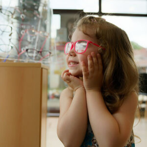
Eyeglasses
Nystagmus, Pathologic
Ophthalmoplegia
Retinoscopy
Fixation, Ocular
Sensory Deprivation
Spina Bifida Cystica
Duane Retraction Syndrome
Oculomotor Nerve
Accommodation, Ocular
Abducens Nerve Diseases
Acupressure
Polyglactin 910
Blepharoptosis
Orbital Fractures
Abducens Nerve
Ophthalmology
Oculomotor Nerve Diseases
Ocular Motility Disorders
Otolaryngology
Nystagmus, Optokinetic
Otorhinolaryngologic Surgical Procedures
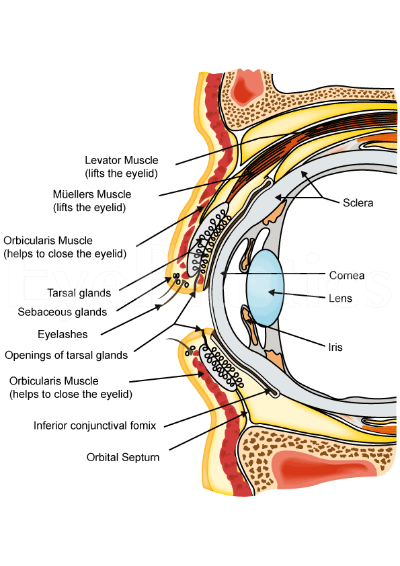
Sclera
Hyperopia
MedlinePlus
Electrooculography
Family study of inherited syndrome with multiple congenital deformities: symphalangism, carpal and tarsal fusion, brachydactyly, craniosynostosis, strabismus, hip osteochondritis. (1/819)
A syndrome of brachydactyly (absence of some middle or distal phalanges), aplastic or hypoplastic nails, symphalangism (ankylois of proximal interphalangeal joints), synostosis of some carpal and tarsal bones, craniosynostosis, and dysplastic hip joints is reported in five members of an Italian family. It may represent a previously undescribed autosomal dominant trait. (+info)Risk factors for strabismus in children born before 32 weeks' gestation. (2/819)
AIM: To investigate risk factors associated with strabismus in children born prematurely. METHODS: Prospective study of all children born before 32 weeks' gestation between 1 January 1990 and 31 December 1991 in a geographically defined population of approximately 3 million in the Northern Region of the United Kingdom. All children were examined aged 2 years by the same ophthalmologist and paediatrician. RESULTS: 558 children (98.6% of study group) were examined. Logistic regression showed an increased risk of strabismus in children with cicatricial retinopathy of prematurity (p=0.02), refractive error (p=0.003), family history of strabismus (p<0.0001), and poor neurodevelopmental outcome (p<0.0001), in particular impaired locomotor skills (p=0.008) and hand-eye coordination (p=0. 001). Gestational age and regressed acute ROP were not independent risk factors for strabismus (p=0.92 and 0.85 respectively). CONCLUSIONS: This study has identified factors which are independently related to strabismus (although not necessarily causative) and others which are related only indirectly. This may contribute both to the management of children born prematurely and to future studies of the aetiology of strabismus. (+info)A deficit in strabismic amblyopia for global shape detection. (3/819)
Using a task which relied upon the detection of sinusoidal deformations from circularity, we show that strabismic amblyopes exhibit deficits which are not critically dependent on either the scale of deformation or the spatial frequency characteristics of the stimulus (circular D4) itself. We show that this loss is not due to the restricted passband of the amblyopic eye. Furthermore, in a pedestal distortion experiment, we show that the suprathreshold form of this loss is consistent with an elevated level of 'intrinsic noise' rather than a loss in 'sampling efficiency'. (+info)Orientation-based texture segmentation in strabismic amblyopia. (4/819)
Texture segmentation of 'target' Gabors from an array of 'background' Gabors was measured in terms of the difference in orientation between the two regions, as well as the difference in orientation within each region. Segmentation was shown to occur on the basis of local orientation differences at the boundary between the target and background regions (Nothdurft, H.C. (1992). Feature analysis and the role of similarity in preattentive vision. Perception and Psychophysics, 52, 355-375.). We obtained similar results for both the amblyopic and non-amblyopic eye of three strabismic amblyopes, and showed also that the effects of texture undersampling and positional jitter were similar for the two eyes. This pattern of results is consistent with intact mechanisms of texture perception in amblyopic cortex, and suggests also that any amblyopic deficits in first-order cortical units (undersampling and/or positional uncertainty) do not limit higher-order texture segmentation processes. Therefore, first- and second-order processes involved in perceptual grouping of oriented elements (that appear to be abnormal in amblyopic cortex; Kovacs, I., Polat, U., Norcia, A.M. (1996). Breakdown of binding mechanisms in amblyopia. Association for Research in Vision and Ophthalmology Abstracts; Mussap, A.J., Levi, D.M. (1995). Amblyopic deficits in perception of second-order orientation. Investigative Ophthalmology and Visual Science (Supplement), 36, S634; Mussap, A.J., Levi, D.M. (1998). Amblyopic deficits in perceptual grouping. Vision Research, submitted) do not contribute to texture perception based on orientation contrast. (+info)Position jitter and undersampling in pattern perception. (5/819)
The present paper addresses whether topographical jitter or undersampling might limit pattern perception in foveal, peripheral and strabismic amblyopic vision. In the first experiment, we measured contrast thresholds for detecting and identifying the orientation (up, down, left, right) of E-like patterns comprised of Gabor samples. We found that detection and identification thresholds were both degraded in peripheral and amblyopic vision; however, the orientation identification/detection threshold ratio was approximately the same in foveal, peripheral and amblyopic vision. This result is somewhat surprising, because we anticipated that a high degree of uncalibrated topographical jitter in peripheral and amblyopic vision would have affected orientation identification to a greater extent than detection. In the second experiment, we investigated the tolerance of human and model observers to perturbation of the positions of the samples defining the pattern when its contrast was suprathreshold, by measuring a 'jitter threshold' (the amount of jitter required to reduce performance from near perfect to 62.5% correct). The results and modeling of our jitter experiments suggest that pattern identification is highly robust to positional jitter. The positional tolerance of foveal, peripheral and amblyopic vision is equal to about half the separation of the features and the close similarity between the three visual systems argues against extreme topographical jitter. The effects of jitter on human performance are consistent with the predictions of a 'template' model. In the third experiment we determined what fraction of the 17 Gabor samples are needed to reliably identify the orientation of the E-patterns by measuring a 'sample threshold' (the proportion of samples required for 62.5% correct performance). In foveal vision, human observers are highly efficient requiring only about half the samples for reliable pattern identification. Relative to an ideal observer model, humans perform this task with 85% efficiency. In contrast, in both peripheral vision and strabismic amblyopia more samples are required. The increased number of features required in peripheral vision and strabismic amblyopia suggests that in these visual systems, the stimulus is underrepresented at the stage of feature integration. (+info)Assessment of cortical dysfunction in human strabismic amblyopia using magnetoencephalography (MEG). (6/819)
The aim of this study was to use the technique of magnetoencephalography (MEG) to determine the effects of strabismic amblyopia on the processing of spatial information within the occipital cortex of humans. We recorded evoked magnetic responses to the onset of a chromatic (red/green) sinusoidal grating of periodicity 0.5-4.0 c deg-1 using a 19-channel SQUID-based neuromagnetometer. Evoked responses were recorded monocularly on six amblyopes and six normally-sighted controls, the stimuli being positioned near the fovea in the lower right visual field of each observer. For comparison, the spatial contrast sensitivity function (CSF) for the detection of chromatic gratings was measured for one amblyope and one control using a two alternate forced-choice psychophysical procedure. We chose red/green sinusoids as our stimuli because they evoke strong magnetic responses from the occipital cortex in adult humans (Fylan, Holliday, Singh, Anderson & Harding. (1997). Neuroimage, 6, 47-57). Magnetic field strength was plotted as a function of stimulus spatial frequency for each eye of each subject. Interocular differences were only evident within the amblyopic group: for stimuli of 1-2 c deg-1, the evoked responses had significantly longer latencies and reduced amplitudes through the amblyopic eye (P < 0.05). Importantly, the extent of the deficit was uncorrelated with either Snellen acuity or contrast sensitivity. Localization of the evoked responses was performed using a single equivalent current dipole model. Source localizations, for both normal and amblyopic subjects, were consistent with neural activity at the occipital pole near the V1/V2 border. We conclude that MEG is sensitive to the deficit in cortical processing associated with human amblyopia, and can be used to make quantitative neurophysiological measurements. The nature of the cortical deficit is discussed. (+info)The puzzle of autism: an ophthalmologic contribution. (7/819)
PURPOSE: A previous study of 86 thalidomide-affected subjects with ophthalmic manifestations revealed the unexpected finding of autism in 4 of the 5 severely retarded individuals. The subjects had anomalies associated with an early gestational effect of thalidomide, including facial nerve palsy and incomitant strabismus. Because autism has been observed in a few cases of Mobius sequence (Mobius syndrome), a condition characterized by involvement of the sixth and seventh cranial nerves, the similarity to early thalidomide embryopathy suggested a relation between cranial nerve involvement and autism. The present study was undertaken to further evaluate the association of autism with patients manifesting findings of Mobius syndrome. METHODS: A prospective study of 25 Swedish patients with Mobius sequence was conducted. The patients had a complete multidisciplinary evaluation, including ophthalmologic and psychiatric examinations and standard testing for autism. Findings associated with autism were compared with the ocular and systemic anomalies of the 4 thalidomide-affected subjects. RESULTS: In the Mobius group 6 patients had autism, achieving the criteria for autism according to all the diagnostic manuals that were used. One patient showed autistic-like conditions meeting fewer numbers of the criteria. A few were too young to be meeting evaluated. Incomitant strabismus ranging from primary abduction defects alone to a horizontal gaze paresis pattern was noted in these patients, in addition to characteristic findings of seventh nerve paresis. Aberrant lacrimation was observed in many cases, especially often associated with autism. CONCLUSION: The common group of anomalies noted in both cases of thalidomide embryopathy and Mobius sequence suggests that brain-stem damage probably early in embryogenesis can sometimes be associated with autism. (+info)The therapy of amblyopia: an analysis of the results of amblyopia therapy utilizing the pooled data of published studies. (8/819)
CONTEXT: Although the treatment of amblyopia with occlusion has changed little over the past 3 centuries, there is little agreement about which regimes are most effective and for what reasons. OBJECTIVE: To determine the outcome of occlusion therapy in patients with anisometropic, strabismic, and strabismic-anisometropic amblyopia employing the raw data from 961 patients reported in 23 studies published between 1965 and 1994. DESIGN: Analysis of the published literature on amblyopia therapy results during the above interval, utilizing primary data obtained from the authors of these articles or tables published in the articles detailing individual patient outcomes. PARTICIPANTS: 961 amblyopic patients, participants in 23 studies, undergoing patching therapy for amblyopia from 1965 to 1994 with anisometropia, strabismus, or anisometropia-strabismus. MAIN OUTCOMES: In the pooled data set, success of occlusion therapy was defined as visual acuity of 20/40 at the end of treatment. RESULTS: Success by the 20/40 criteria was achieved in 512 of 689 (74.3%) patients. By category, 312 of 402 (77.6%) were successful in strabismic amblyopia, 44 of 75 (58.7%) in strabismic-anisometropic amblyopia, and 72 of 108 (66.7%) in anisometropic amblyopia. Success was not related to the duration of occlusion therapy, type of occlusion used, accompanying refractive error, patient's sex, or eye. Univariate analyses showed that success was related to the age at which therapy was initiated; the type of amblyopia; the depth of visual loss before treatment for the anisometropic patients and the strabismic patients, but not for the anisometropic-strabismic patients; and the difference in spherical equivalents between eyes, for the anisometropic patients. Logistic/linear regression revealed that 3 were independent predictors of a successful outcome of amblyopia therapy. CONCLUSIONS: Factors that appear most closely related to a successful outcome are age, type of amblyopia, and depth of visual loss before treatment. These may be related to factors, as yet undetermined in the pathogenesis of amblyopia. With present emphasis on the value of screening and prevention and the development of new screening tools, such a look at the results of amblyopia therapy in a large population seems indicated. (+info)Strabismus is a condition of the ocular muscles where the eyes are not aligned properly and point in different directions. One eye may turn inward, outward, upward, or downward while the other one remains fixed and aligns normally. This misalignment can occur occasionally or constantly. Strabismus is also commonly referred to as crossed eyes or walleye. The condition can lead to visual impairments such as amblyopia (lazy eye) and depth perception problems if not treated promptly and effectively, usually through surgery, glasses, or vision therapy.
Esotropia is a type of ocular misalignment, also known as strabismus, in which one eye turns inward toward the nose. This condition can be constant or intermittent and may result in double vision or loss of depth perception. Esotropia is often classified based on its cause, age of onset, and frequency. Common forms include congenital esotropia, acquired esotropia, and accommodative esotropia. Treatment typically involves corrective eyewear, eye exercises, or surgery to realign the eyes.
The oculomotor muscles are a group of extraocular muscles that control the movements of the eye. They include:
1. Superior rectus: This muscle is responsible for elevating the eye and helping with inward rotation (intorsion) when looking downwards.
2. Inferior rectus: It depresses the eye and helps with outward rotation (extorsion) when looking upwards.
3. Medial rectus: This muscle adducts, or moves, the eye towards the midline of the face.
4. Inferior oblique: The inferior oblique muscle intorts and elevates the eye.
5. Superior oblique: It extorts and depresses the eye.
These muscles work together to allow for smooth and precise movements of the eyes, enabling tasks such as tracking moving objects, reading, and maintaining visual fixation on a single point in space.
Exotropia is a type of ocular misalignment or strabismus, where one eye turns outward (towards the ear) while the other eye remains aligned straight ahead. This condition can be constant or intermittent and may result in limited or absent depth perception, double vision, and in some cases, amblyopia (lazy eye). Exotropia is typically diagnosed during childhood through a comprehensive eye examination by an optometrist or ophthalmologist. Treatment options include eyeglasses, prism lenses, vision therapy, or surgery, depending on the severity and frequency of the misalignment.
Amblyopia is a medical condition that affects the visual system, specifically the way the brain and eyes work together. It is often referred to as "lazy eye" and is characterized by reduced vision in one or both eyes that is not correctable with glasses or contact lenses alone. This occurs because the brain favors one eye over the other, causing the weaker eye to become neglected and underdeveloped.
Amblyopia can result from various conditions such as strabismus (eye misalignment), anisometropia (significant difference in prescription between the two eyes), or deprivation (such as a cataract that blocks light from entering the eye). Treatment for amblyopia typically involves correcting any underlying refractive errors, patching or blurring the stronger eye to force the weaker eye to work, and/or vision therapy. Early intervention is crucial to achieve optimal visual outcomes.
Diplopia is a medical term that refers to the condition where a person sees two images of a single object. It is commonly known as double vision. This can occur due to various reasons, such as nerve damage or misalignment of the eyes. Diplopia can be temporary or chronic and can affect one or both eyes. If you're experiencing diplopia, it's essential to consult an eye care professional for proper evaluation and treatment.
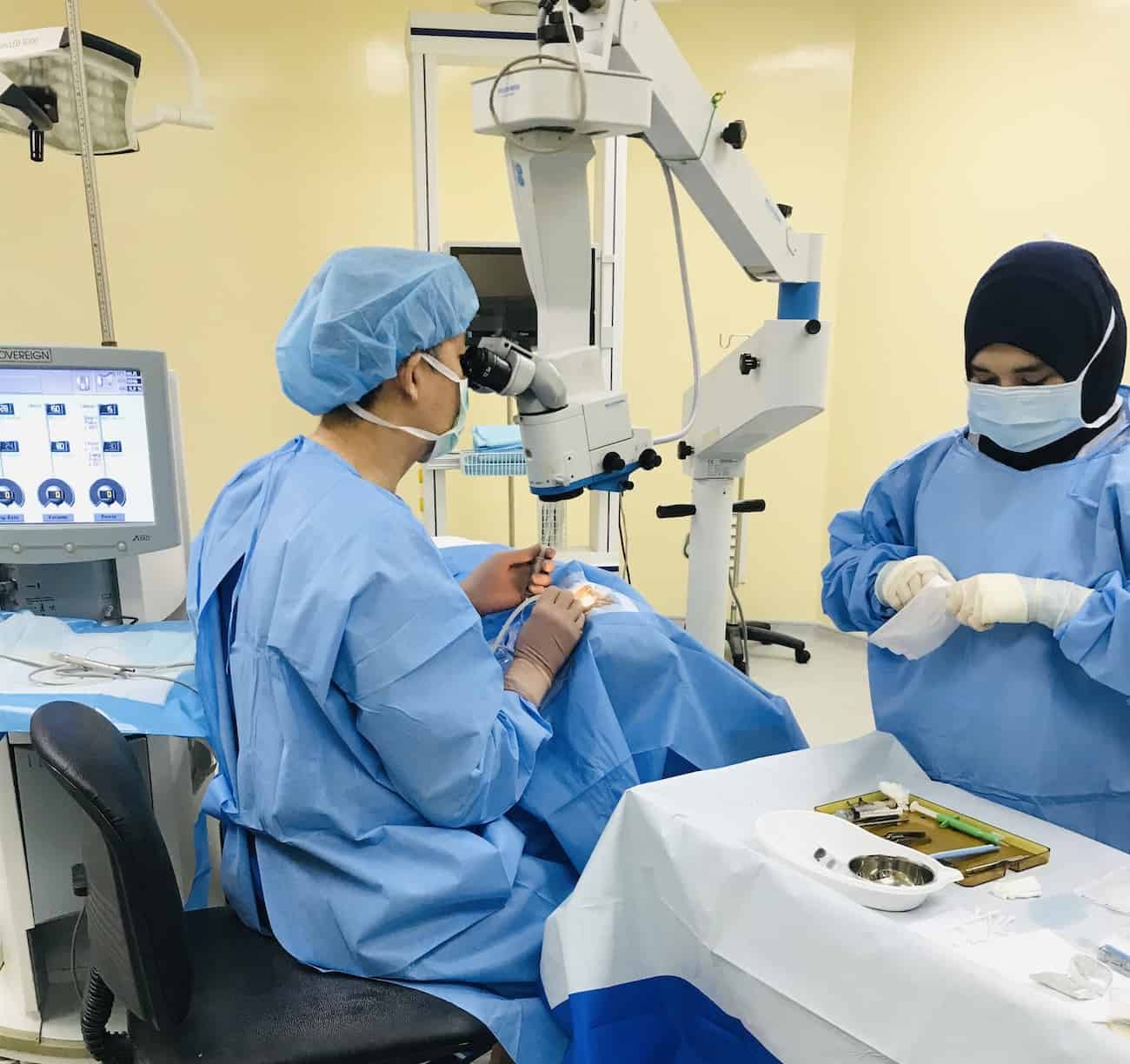
Ophthalmologic surgical procedures refer to various types of surgeries performed on the eye and its surrounding structures by trained medical professionals called ophthalmologists. These procedures aim to correct or improve vision, diagnose and treat eye diseases or injuries, and enhance the overall health and functionality of the eye. Some common examples of ophthalmologic surgical procedures include:
1. Cataract Surgery: This procedure involves removing a cloudy lens (cataract) from the eye and replacing it with an artificial intraocular lens (IOL).
2. LASIK (Laser-Assisted In Situ Keratomileusis): A type of refractive surgery that uses a laser to reshape the cornea, correcting nearsightedness, farsightedness, and astigmatism.
3. Glaucoma Surgery: Several surgical options are available for treating glaucoma, including laser trabeculoplasty, traditional trabeculectomy, and various drainage device implantations. These procedures aim to reduce intraocular pressure (IOP) and prevent further optic nerve damage.
4. Corneal Transplant: This procedure involves replacing a damaged or diseased cornea with a healthy donor cornea to restore vision and improve the eye's appearance.
5. Vitreoretinal Surgery: These procedures focus on treating issues within the vitreous humor (gel-like substance filling the eye) and the retina, such as retinal detachment, macular holes, or diabetic retinopathy.
6. Strabismus Surgery: This procedure aims to correct misalignment of the eyes (strabismus) by adjusting the muscles responsible for eye movement.
7. Oculoplastic Surgery: These procedures involve reconstructive, cosmetic, and functional surgeries around the eye, such as eyelid repair, removal of tumors, or orbital fracture repairs.
8. Pediatric Ophthalmologic Procedures: Various surgical interventions are performed on children to treat conditions like congenital cataracts, amblyopia (lazy eye), or blocked tear ducts.
These are just a few examples of ophthalmic surgical procedures. The specific treatment plan will depend on the individual's condition and overall health.
Binocular vision refers to the ability to use both eyes together to create a single, three-dimensional image of our surroundings. This is achieved through a process called binocular fusion, where the images from each eye are aligned and combined in the brain to form a unified perception.
The term "binocular vision" specifically refers to the way that our visual system integrates information from both eyes to create depth perception and enhance visual clarity. When we view an object with both eyes, they focus on the same point in space and send slightly different images to the brain due to their slightly different positions. The brain then combines these images to create a single, three-dimensional image that allows us to perceive depth and distance.
Binocular vision is important for many everyday activities, such as driving, reading, and playing sports. Disorders of binocular vision can lead to symptoms such as double vision, eye strain, and difficulty with depth perception.
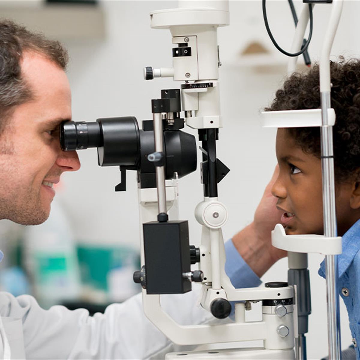
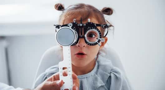
Vision screening is a quick and cost-effective method used to identify individuals who are at risk of vision problems or eye diseases. It is not a comprehensive eye examination, but rather an initial evaluation that helps to determine if a further, more in-depth examination by an eye care professional is needed. Vision screenings typically involve tests for visual acuity, distance and near vision, color perception, depth perception, and alignment of the eyes. The goal of vision screening is to detect potential vision issues early on, so that they can be treated promptly and effectively, thereby preventing or minimizing any negative impact on a person's overall vision and quality of life.
Anisometropia is a medical term that refers to a condition where there is a significant difference in the refractive power between the two eyes. In other words, one eye has a significantly different optical prescription compared to the other eye. This condition can cause issues with binocular vision and depth perception, and can sometimes lead to amblyopia (lazy eye) if not corrected early in life. It is typically diagnosed through a comprehensive eye examination and can be corrected with glasses or contact lenses.
Visual acuity is a measure of the sharpness or clarity of vision. It is usually tested by reading an eye chart from a specific distance, such as 20 feet (6 meters). The standard eye chart used for this purpose is called the Snellen chart, which contains rows of letters that decrease in size as you read down the chart.
Visual acuity is typically expressed as a fraction, with the numerator representing the testing distance and the denominator indicating the smallest line of type that can be read clearly. For example, if a person can read the line on the eye chart that corresponds to a visual acuity of 20/20, it means they have normal vision at 20 feet. If their visual acuity is 20/40, it means they must be as close as 20 feet to see what someone with normal vision can see at 40 feet.
It's important to note that visual acuity is just one aspect of overall vision and does not necessarily reflect other important factors such as peripheral vision, depth perception, color vision, or contrast sensitivity.
Refractive errors are a group of vision conditions that include nearsightedness (myopia), farsightedness (hyperopia), astigmatism, and presbyopia. These conditions occur when the shape of the eye prevents light from focusing directly on the retina, causing blurred or distorted vision.
Myopia is a condition where distant objects appear blurry while close-up objects are clear. This occurs when the eye is too long or the cornea is too curved, causing light to focus in front of the retina instead of directly on it.
Hyperopia, on the other hand, is a condition where close-up objects appear blurry while distant objects are clear. This happens when the eye is too short or the cornea is not curved enough, causing light to focus behind the retina.
Astigmatism is a condition that causes blurred vision at all distances due to an irregularly shaped cornea or lens.
Presbyopia is a natural aging process that affects everyone as they get older, usually around the age of 40. It causes difficulty focusing on close-up objects and can be corrected with reading glasses, bifocals, or progressive lenses.
Refractive errors can be diagnosed through a comprehensive eye exam and are typically corrected with eyeglasses, contact lenses, or refractive surgery such as LASIK.
An oculocardiac reflex is a medical term that refers to a reflexive response that involves the eye and the heart. This reflex is elicited when there is pressure or traction applied to the eye or its surrounding structures, which can result in a decrease in heart rate.
The oculocardiac reflex is mediated by the ophthalmic division of the trigeminal nerve (cranial nerve V) and the vagus nerve (cranial nerve X). When the eye or its surrounding structures are stimulated, the impulses travel through the ophthalmic branch of the trigeminal nerve to the brainstem, where they synapse with neurons in the vagus nerve. The vagus nerve then carries these impulses to the sinoatrial node of the heart, which results in a decrease in heart rate.
The oculocardiac reflex is commonly seen during ophthalmic surgical procedures, particularly those that involve manipulation of the eye or its surrounding structures. It can also occur in response to other forms of stimulation, such as coughing, sneezing, or vomiting. In some cases, the oculocardiac reflex can lead to a significant decrease in heart rate, which may require medical intervention to prevent serious complications.
Eye movements, also known as ocular motility, refer to the voluntary or involuntary motion of the eyes that allows for visual exploration of our environment. There are several types of eye movements, including:
1. Saccades: rapid, ballistic movements that quickly shift the gaze from one point to another.
2. Pursuits: smooth, slow movements that allow the eyes to follow a moving object.
3. Vergences: coordinated movements of both eyes in opposite directions, usually in response to a three-dimensional stimulus.
4. Vestibulo-ocular reflex (VOR): automatic eye movements that help stabilize the gaze during head movement.
5. Optokinetic nystagmus (OKN): rhythmic eye movements that occur in response to large moving visual patterns, such as when looking out of a moving vehicle.
Abnormalities in eye movements can indicate neurological or ophthalmological disorders and are often assessed during clinical examinations.
Suture techniques refer to the various methods used by surgeons to sew or stitch together tissues in the body after an injury, trauma, or surgical incision. The main goal of suturing is to approximate and hold the edges of the wound together, allowing for proper healing and minimizing scar formation.
There are several types of suture techniques, including:
1. Simple Interrupted Suture: This is one of the most basic suture techniques where the needle is passed through the tissue at a right angle, creating a loop that is then tightened to approximate the wound edges. Multiple stitches are placed along the length of the incision or wound.
2. Continuous Locking Suture: In this technique, the needle is passed continuously through the tissue in a zigzag pattern, with each stitch locking into the previous one. This creates a continuous line of sutures that provides strong tension and support to the wound edges.
3. Running Suture: Similar to the continuous locking suture, this technique involves passing the needle continuously through the tissue in a straight line. However, instead of locking each stitch, the needle is simply passed through the previous loop before being tightened. This creates a smooth and uninterrupted line of sutures that can be easily removed after healing.
4. Horizontal Mattress Suture: In this technique, two parallel stitches are placed horizontally across the wound edges, creating a "mattress" effect that provides additional support and tension to the wound. This is particularly useful in deep or irregularly shaped wounds.
5. Vertical Mattress Suture: Similar to the horizontal mattress suture, this technique involves placing two parallel stitches vertically across the wound edges. This creates a more pronounced "mattress" effect that can help reduce tension and minimize scarring.
6. Subcuticular Suture: In this technique, the needle is passed just below the surface of the skin, creating a smooth and barely visible line of sutures. This is particularly useful in cosmetic surgery or areas where minimizing scarring is important.
The choice of suture technique depends on various factors such as the location and size of the wound, the type of tissue involved, and the patient's individual needs and preferences. Proper suture placement and tension are crucial for optimal healing and aesthetic outcomes.
Eyeglasses are a medical device used to correct vision problems. Also known as spectacles, they consist of frames that hold one or more lenses through which a person looks to see clearly. The lenses may be made of glass or plastic and are designed to compensate for various visual impairments such as nearsightedness, farsightedness, astigmatism, or presbyopia. Eyeglasses can be custom-made to fit an individual's face and prescription, and they come in a variety of styles, colors, and materials. Some people wear eyeglasses all the time, while others may only need to wear them for certain activities such as reading or driving.
Pathological nystagmus is an abnormal, involuntary movement of the eyes that can occur in various directions (horizontal, vertical, or rotatory) and can be rhythmical or arrhythmic. It is typically a result of a disturbance in the vestibular system, central nervous system, or ocular motor pathways. Pathological nystagmus can cause visual symptoms such as blurred vision, difficulty with fixation, and oscillopsia (the sensation that one's surroundings are moving). The type, direction, and intensity of the nystagmus may vary depending on the underlying cause, which can include conditions such as brainstem or cerebellar lesions, multiple sclerosis, drug toxicity, inner ear disorders, and congenital abnormalities.
Depth perception is the ability to accurately judge the distance or separation of an object in three-dimensional space. It is a complex visual process that allows us to perceive the world in three dimensions and to understand the spatial relationships between objects.
Depth perception is achieved through a combination of monocular cues, which are visual cues that can be perceived with one eye, and binocular cues, which require input from both eyes. Monocular cues include perspective (the relative size of objects), texture gradients (finer details become smaller as distance increases), and atmospheric perspective (colors become less saturated and lighter in value as distance increases). Binocular cues include convergence (the degree to which the eyes must turn inward to focus on an object) and retinal disparity (the slight difference in the images projected onto the two retinas due to the slightly different positions of the eyes).
Deficits in depth perception can occur due to a variety of factors, including eye disorders, brain injuries, or developmental delays. These deficits can result in difficulties with tasks such as driving, sports, or navigating complex environments. Treatment for depth perception deficits may include vision therapy, corrective lenses, or surgery.
Ophthalmoplegia is a medical term that refers to the paralysis or weakness of the eye muscles, which can result in double vision (diplopia) or difficulty moving the eyes. It can be caused by various conditions, including nerve damage, muscle disorders, or neurological diseases such as myasthenia gravis or multiple sclerosis. Ophthalmoplegia can affect one or more eye muscles and can be partial or complete. Depending on the underlying cause, ophthalmoplegia may be treatable with medications, surgery, or other interventions.
Retinoscopy is a diagnostic technique used in optometry and ophthalmology to estimate the refractive error of the eye, or in other words, to determine the prescription for eyeglasses or contact lenses. This procedure involves shining a light into the patient's pupil and observing the reflection off the retina while introducing different lenses in front of the patient's eye. The examiner then uses specific movements and observations to determine the amount and type of refractive error, such as myopia (nearsightedness), hyperopia (farsightedness), astigmatism, or presbyopia. Retinoscopy is a fundamental skill for eye care professionals and helps ensure that patients receive accurate prescriptions for corrective lenses.
Ocular convergence is the normal, inward movement of both eyes towards each other to focus on a nearby object. This coordinated action allows for single, clear vision (binocular vision) of the object. It is an important component of visual function and is controlled by the brain receiving input from the muscles that move the eyes.
Convergence insufficiency is a common condition where the eyes have difficulty maintaining alignment during close work, such as reading or using a computer. This can result in eye strain, double vision, and difficulty concentrating. Treatment for convergence insufficiency may include vision therapy, exercises to improve convergence ability, and/or the use of prism lenses.
Ocular fixation is a term used in ophthalmology and optometry to refer to the ability of the eyes to maintain steady gaze or visual focus on an object. It involves the coordinated movement of the extraocular muscles that control eye movements, allowing for clear and stable vision.
In medical terminology, fixation specifically refers to the state in which the eyes are aligned and focused on a single point in space. This is important for maintaining visual perception and preventing blurring or double vision. Ocular fixation can be affected by various factors such as muscle weakness, nerve damage, or visual processing disorders.
Assessment of ocular fixation is often used in eye examinations to evaluate visual acuity, eye alignment, and muscle function. Abnormalities in fixation may indicate the presence of underlying eye conditions or developmental delays that require further investigation and treatment.
Vision tests are a series of procedures used to assess various aspects of the visual system, including visual acuity, accommodation, convergence, divergence, stereopsis, color vision, and peripheral vision. These tests help healthcare professionals diagnose and manage vision disorders, such as nearsightedness, farsightedness, astigmatism, amblyopia, strabismus, and eye diseases like glaucoma, cataracts, and macular degeneration. Common vision tests include:
1. Visual acuity test (Snellen chart or letter chart): Measures the sharpness of a person's vision at different distances.
2. Refraction test: Determines the correct lens prescription for glasses or contact lenses by assessing how light is bent as it passes through the eye.
3. Color vision test: Evaluates the ability to distinguish between different colors and color combinations, often using pseudoisochromatic plates or Ishihara tests.
4. Stereopsis test: Assesses depth perception and binocular vision by presenting separate images to each eye that, when combined, create a three-dimensional effect.
5. Cover test: Examines eye alignment and the presence of strabismus (crossed eyes or turned eyes) by covering and uncovering each eye while observing eye movements.
6. Ocular motility test: Assesses the ability to move the eyes in various directions and coordinate both eyes during tracking and convergence/divergence movements.
7. Accommodation test: Evaluates the ability to focus on objects at different distances by using lenses, prisms, or dynamic retinoscopy.
8. Pupillary response test: Examines the size and reaction of the pupils to light and near objects.
9. Visual field test: Measures the peripheral (side) vision using automated perimetry or manual confrontation techniques.
10. Slit-lamp examination: Inspects the structures of the front part of the eye, such as the cornea, iris, lens, and anterior chamber, using a specialized microscope.
These tests are typically performed by optometrists, ophthalmologists, or other vision care professionals during routine eye examinations or when visual symptoms are present.
Ocular refraction is a medical term that refers to the bending of light as it passes through the optical media of the eye, including the cornea and lens. This process allows the eye to focus light onto the retina, creating a clear image. The refractive power of the eye is determined by the curvature and transparency of these structures.
In a normal eye, light rays are bent or refracted in such a way that they converge at a single point on the retina, producing a sharp and focused image. However, if the curvature of the cornea or lens is too steep or too flat, the light rays may not converge properly, resulting in a refractive error such as myopia (nearsightedness), hyperopia (farsightedness), or astigmatism.
Ocular refraction can be measured using a variety of techniques, including retinoscopy, automated refraction, and subjective refraction. These measurements are used to determine the appropriate prescription for corrective lenses such as eyeglasses or contact lenses. In some cases, ocular refractive errors may be corrected surgically through procedures such as LASIK or PRK.
Monocular vision refers to the ability to see and process visual information using only one eye. It is the type of vision that an individual has when they are using only one eye to look at something, while the other eye may be covered or not functioning. This can be contrasted with binocular vision, which involves the use of both eyes working together to provide depth perception and a single, combined visual field.
Monocular vision is important for tasks that only require the use of one eye, such as when looking through a microscope or using a telescope. However, it does not provide the same level of depth perception and spatial awareness as binocular vision. In some cases, individuals may have reduced visual acuity or other visual impairments in one eye, leading to limited monocular vision in that eye. It is important for individuals with monocular vision to have regular eye exams to monitor their eye health and ensure that any visual impairments are detected and treated promptly.
Sensory deprivation, also known as perceptual isolation or sensory restriction, refers to the deliberate reduction or removal of stimuli from one or more of the senses. This can include limiting input from sight, sound, touch, taste, and smell. The goal is to limit a person's sensory experiences in order to study the effects on cognition, perception, and behavior.
In a clinical context, sensory deprivation can occur as a result of certain medical conditions or treatments, such as blindness, deafness, or pharmacological interventions that affect sensory processing. Prolonged sensory deprivation can lead to significant psychological and physiological effects, including hallucinations, delusions, and decreased cognitive function.
It's important to note that sensory deprivation should not be confused with meditation or relaxation techniques that involve reducing external stimuli in a controlled manner to promote relaxation and focus.
Spina Bifida Cystica is a type of neural tube defect that occurs when the bones of the spine (vertebrae) do not form properly around the developing spinal cord, resulting in a sac-like protrusion of the spinal cord and its surrounding membranes through an opening in the spine. This sac, called a meningocele or myelomeningocele, can be covered with skin or exposed, and it may contain cerebrospinal fluid, nerve roots, or portions of the spinal cord.
Myelomeningocele is the most severe form of Spina Bifida Cystica, where the sac contains a portion of the spinal cord and nerves. This can lead to various neurological complications such as weakness or paralysis below the level of the spine affected, loss of sensation, bladder and bowel dysfunction, and hydrocephalus (accumulation of cerebrospinal fluid in the brain). Early diagnosis and intervention, including prenatal surgery, can help improve outcomes for individuals with Spina Bifida Cystica.
Duane Retraction Syndrome (DRS) is a congenital eye movement disorder, characterized by limited abduction (lateral movement away from the nose) of the affected eye, and on attempted adduction (movement towards the nose), the eye retracts into the orbit and the lid narrows. It is often accompanied by other eye alignment or vision anomalies. The exact cause is not known, but it is believed to be a result of abnormal development of the cranial nerves that control eye movement during fetal development. DRS is usually idiopathic, but it can also be associated with other congenital anomalies. It is typically diagnosed in early childhood and managed with a combination of observation, prism glasses, and/or surgery, depending on the severity and impact on vision.
The oculomotor nerve, also known as the third cranial nerve (CN III), is a motor nerve that originates from the midbrain. It controls the majority of the eye muscles, including the levator palpebrae superioris muscle that raises the upper eyelid, and the extraocular muscles that enable various movements of the eye such as looking upward, downward, inward, and outward. Additionally, it carries parasympathetic fibers responsible for pupillary constriction and accommodation (focusing on near objects). Damage to this nerve can result in various ocular motor disorders, including strabismus, ptosis, and pupillary abnormalities.
Ocular accommodation is the process by which the eye changes optical power to maintain a clear image or focus on an object as its distance varies. This is primarily achieved by the lens of the eye changing shape through the action of the ciliary muscles inside the eye. When you look at something far away, the lens becomes flatter, and when you look at something close up, the lens thickens. This ability to adjust focus allows for clear vision at different distances.
The abducens nerve, also known as the sixth cranial nerve, is responsible for controlling the lateral rectus muscle of the eye, which enables the eye to move outward. Abducens nerve diseases refer to conditions that affect this nerve and can result in various symptoms, primarily affecting eye movement.
Here are some medical definitions related to abducens nerve diseases:
1. Abducens Nerve Palsy: A condition characterized by weakness or paralysis of the abducens nerve, causing difficulty in moving the affected eye outward. This results in double vision (diplopia), especially when gazing towards the side of the weakened nerve. Abducens nerve palsy can be congenital, acquired, or caused by various factors such as trauma, tumors, aneurysms, infections, or diseases like diabetes and multiple sclerosis.
2. Sixth Nerve Palsy: Another term for abducens nerve palsy, referring to the weakness or paralysis of the sixth cranial nerve.
3. Internuclear Ophthalmoplegia (INO): A neurological condition affecting eye movement, often caused by a lesion in the medial longitudinal fasciculus (MLF), a bundle of nerve fibers that connects the abducens nucleus with the oculomotor nucleus. INO results in impaired adduction (inward movement) of the eye on the side of the lesion and nystagmus (involuntary eye movements) of the abducting eye on the opposite side when attempting to look towards the side of the lesion.
4. One-and-a-Half Syndrome: A rare neurological condition characterized by a combination of INO and internuclear ophthalmoplegia with horizontal gaze palsy on the same side, caused by damage to both the abducens nerve and the paramedian pontine reticular formation (PPRF). This results in limited or no ability to move the eyes towards the side of the lesion and impaired adduction of the eye on the opposite side.
5. Brainstem Encephalitis: Inflammation of the brainstem, which can affect the abducens nerve and other cranial nerves, leading to various neurological symptoms such as diplopia (double vision), ataxia (loss of balance and coordination), and facial weakness. Brainstem encephalitis can be caused by infectious agents, autoimmune disorders, or paraneoplastic syndromes.
6. Multiple Sclerosis (MS): An autoimmune disorder characterized by inflammation and demyelination of the central nervous system, including the brainstem and optic nerves. MS can cause various neurological symptoms, such as diplopia, nystagmus, and INO, due to damage to the abducens nerve and other cranial nerves.
7. Wernicke's Encephalopathy: A neurological disorder caused by thiamine (vitamin B1) deficiency, often seen in alcoholics or individuals with malnutrition. Wernicke's encephalopathy can affect the brainstem and cause various symptoms such as diplopia, ataxia, confusion, and oculomotor abnormalities.
8. Pontine Glioma: A rare type of brain tumor that arises from the glial cells in the pons (a part of the brainstem). Pontine gliomas can cause various neurological symptoms such as diplopia, facial weakness, and difficulty swallowing due to their location in the brainstem.
9. Brainstem Cavernous Malformation: A benign vascular lesion that arises from the small blood vessels in the brainstem. Brainstem cavernous malformations can cause various neurological symptoms such as diplopia, ataxia, and facial weakness due to their location in the brainstem.
10. Pituitary Adenoma: A benign tumor that arises from the pituitary gland, located at the base of the brain. Large pituitary adenomas can compress the optic nerves and cause various visual symptoms such as diplopia, visual field defects, and decreased vision.
11. Craniopharyngioma: A benign tumor that arises from the remnants of the Rathke's pouch, a structure that gives rise to the anterior pituitary gland. Craniopharyngiomas can cause various neurological and endocrine symptoms such as diplopia, visual field defects, headaches, and hormonal imbalances due to their location near the optic nerves and pituitary gland.
12. Meningioma: A benign tumor that arises from the meninges, the protective covering of the brain and spinal cord. Meningiomas can cause various neurological symptoms such as diplopia, headaches, and seizures depending on their location in the brain or spinal cord.
13. Chordoma: A rare type of malignant tumor that arises from the remnants of the notochord, a structure that gives rise to the spine during embryonic development. Chordomas can cause various neurological and endocrine symptoms such as diplopia, visual field defects, headaches, and hormonal imbalances due to their location near the brainstem and spinal cord.
14. Metastatic Brain Tumors: Malignant tumors that spread from other parts of the body to the brain. Metastatic brain tumors can cause various neurological symptoms such as diplopia, headaches, seizures, and cognitive impairment depending on their location in the brain.
15. Other Rare Brain Tumors: There are many other rare types of brain tumors that can cause diplopia or other neurological symptoms, including gliomas, ependymomas, pineal region tumors, and others. These tumors require specialized diagnosis and treatment by neuro-oncologists and neurosurgeons with expertise in these rare conditions.
In summary, diplopia can be caused by various brain tumors, including pituitary adenomas, meningiomas, chordomas, metastatic brain tumors, and other rare types of tumors. It is important to seek medical attention promptly if you experience diplopia or other neurological symptoms, as early diagnosis and treatment can improve outcomes and quality of life.
Acupressure is a complementary therapy based on the concept of acupuncture, which involves applying pressure (usually with fingers, hands, or elbow) to specific points on the body (known as acupoints). The goal of acupressure is to stimulate and balance the flow of energy (chi or qi) through the body's meridians or channels. This practice is believed to help promote relaxation, reduce stress, relieve pain, improve sleep, and enhance overall well-being.
It is important to note that while acupressure has been used for thousands of years in traditional Chinese medicine, its effectiveness is not consistently supported by scientific research. Some studies suggest potential benefits, but more rigorous, high-quality research is needed to confirm these findings. As with any therapy, it's recommended to consult a healthcare professional before starting an acupressure practice, especially if you have any health conditions or are taking medications.
Polyglactin 910 is a type of synthetic absorbable suture made from copolymers of lactide and glycolide. It is designed to gradually break down and be absorbed by the body over time, typically within 56 to 70 days after being used in surgical wounds. This property makes it an ideal choice for soft tissue approximation and laceration repairs.
Polyglactin 910 sutures are often used in various surgical procedures, including orthopedic, ophthalmic, cardiovascular, and general surgery. They come in different sizes and forms, such as plain, reverse cutting, and braided, to suit various surgical needs.
The gradual absorption of Polyglactin 910 sutures helps minimize scarring and reduces the need for suture removal procedures. However, it is essential to note that inflammation may occur during the degradation process, which could potentially lead to adverse reactions in some individuals. Proper wound care and follow-up with healthcare professionals are crucial to ensure optimal healing and manage any potential complications.
Blepharoptosis is a medical term that refers to the drooping or falling of the upper eyelid. It is usually caused by weakness or paralysis of the muscle that raises the eyelid, known as the levator palpebrae superioris. This condition can be present at birth or acquired later in life due to various factors such as aging, nerve damage, eye surgery complications, or certain medical conditions like myasthenia gravis or brain tumors. Blepharoptosis may obstruct vision and cause difficulty with daily activities, and treatment options include eyedrops, eye patches, or surgical correction.
Orbital fractures refer to breaks in the bones that make up the eye socket, also known as the orbit. These bones include the maxilla, zygoma, frontal bone, and palatine bone. Orbital fractures can occur due to trauma, such as a blunt force injury or a penetrating wound.
There are several types of orbital fractures, including:
1. Blowout fracture: This occurs when the thin bone of the orbital floor is broken, often due to a direct blow to the eye. The force of the impact can cause the eyeball to move backward, breaking the bone and sometimes trapping the muscle that moves the eye (the inferior rectus).
2. Blow-in fracture: This type of fracture involves the breakage of the orbital roof, which is the bone that forms the upper boundary of the orbit. It typically occurs due to high-impact trauma, such as a car accident or a fall from a significant height.
3. Direct fracture: A direct fracture happens when there is a break in one or more of the bones that form the walls of the orbit. This type of fracture can result from a variety of traumas, including motor vehicle accidents, sports injuries, and assaults.
4. Indirect fracture: An indirect fracture occurs when the force of an injury is transmitted to the orbit through tissues surrounding it, causing the bone to break. The most common type of indirect orbital fracture is a blowout fracture.
Orbital fractures can cause various symptoms, including pain, swelling, bruising, and double vision. In some cases, the fracture may also lead to enophthalmos (sinking of the eye into the orbit) or telecanthus (increased distance between the inner corners of the eyes). Imaging tests, such as CT scans, are often used to diagnose orbital fractures and determine the best course of treatment. Treatment may include observation, pain management, and in some cases, surgery to repair the fracture and restore normal function.
The abducens nerve, also known as the sixth cranial nerve (CN VI), is a motor nerve that controls the lateral rectus muscle of the eye. This muscle is responsible for moving the eye away from the midline (towards the temple) and enables the eyes to look towards the side while keeping them aligned. Any damage or dysfunction of the abducens nerve can result in strabismus, where the eyes are misaligned and point in different directions, specifically an adduction deficit, also known as abducens palsy or sixth nerve palsy.
Ophthalmology is a branch of medicine that deals with the diagnosis, treatment, and prevention of diseases and disorders of the eye and visual system. It is a surgical specialty, and ophthalmologists are medical doctors who complete additional years of training to become experts in eye care. They are qualified to perform eye exams, diagnose and treat eye diseases, prescribe glasses and contact lenses, and perform eye surgery. Some subspecialties within ophthalmology include cornea and external disease, glaucoma, neuro-ophthalmology, pediatric ophthalmology, retina and vitreous, and oculoplastics.
The oculomotor nerve, also known as the third cranial nerve (CN III), is responsible for controlling several important eye movements and functions. Oculomotor nerve diseases refer to conditions that affect this nerve and can lead to various symptoms related to eye movement and function. Here's a medical definition of oculomotor nerve diseases:
Oculomotor nerve diseases are a group of medical disorders characterized by the dysfunction or damage to the oculomotor nerve (CN III), resulting in impaired eye movements, abnormalities in pupillary response, and potential effects on eyelid position. These conditions can be congenital, acquired, or traumatic in nature and may lead to partial or complete paralysis of the nerve. Common oculomotor nerve diseases include oculomotor nerve palsy, third nerve ganglionopathies, and compressive oculomotor neuropathies caused by various pathologies such as aneurysms, tumors, or infections.
Ocular motility disorders refer to a group of conditions that affect the movement of the eyes. These disorders can result from nerve damage, muscle dysfunction, or brain injuries. They can cause abnormal eye alignment, limited range of motion, and difficulty coordinating eye movements. Common symptoms include double vision, blurry vision, strabismus (crossed eyes), nystagmus (involuntary eye movement), and difficulty tracking moving objects. Ocular motility disorders can be congenital or acquired and may require medical intervention to correct or manage the condition.
Otolaryngology is a specialized branch of medicine that deals with the diagnosis, management, and treatment of disorders related to the ear, nose, throat (ENT), and head and neck region. It's also known as ENT (Ear, Nose, Throat) specialty. Otolaryngologists are physicians trained in the medical and surgical management of conditions such as hearing and balance disorders, nasal congestion, sinusitis, allergies, sleep apnea, snoring, swallowing difficulties, voice and speech problems, and head and neck tumors.
Optokinetic nystagmus (OKN) is a type of involuntary eye movement that occurs in response to large moving visual patterns. It serves as a mechanism for stabilizing the image on the retina during head movement and helps in maintaining visual fixation.
In OKN, there are two phases of eye movement: a slow phase where the eyes follow or track the moving pattern, and a fast phase where the eyes quickly reset to the starting position. This results in a back-and-forth or "to-and-fro" motion of the eyes.
Optokinetic nystagmus can be elicited by observing a large moving object or a series of alternating visual stimuli, such as stripes on a rotating drum. It is often used in clinical settings to assess various aspects of the visual system, including oculomotor function and visual acuity.
Abnormalities in OKN can indicate problems with the vestibular system, brainstem, or cerebellum, and may be associated with conditions such as brain injury, multiple sclerosis, or cerebral palsy.
Otorhinolaryngologic surgical procedures are surgeries that are performed on the head and neck region, specifically involving the ear, nose, and throat (ENT) regions. This field is also known as otolaryngology-head and neck surgery. The procedures can range from relatively minor ones, such as removing a small nasal polyp or inserting ear tubes, to more complex surgeries like cochlear implantation, endoscopic sinus surgery, or removal of tumors in the head and neck region. These surgical procedures are typically performed by specialized physicians called otorhinolaryngologists (also known as ENT surgeons) who have completed extensive training in this area.
In medical terms, the orbit refers to the bony cavity or socket in the skull that contains and protects the eye (eyeball) and its associated structures, including muscles, nerves, blood vessels, fat, and the lacrimal gland. The orbit is made up of several bones: the frontal bone, sphenoid bone, zygomatic bone, maxilla bone, and palatine bone. These bones form a pyramid-like shape that provides protection for the eye while also allowing for a range of movements.
The sclera is the tough, white, fibrous outer coating of the eye in humans and other vertebrates, covering about five sixths of the eyeball's surface. It provides protection for the delicate inner structures of the eye and maintains its shape. The sclera is composed mainly of collagen and elastic fiber, making it strong and resilient. Its name comes from the Greek word "skleros," which means hard.
I'm sorry for the confusion, but "telescopes" is not a medical term. A telescope is an optical instrument that makes distant objects appear closer and larger by using an arrangement of lenses or mirrors and curved surfaces to gather and focus light. It is commonly used in astronomy to observe celestial bodies. If you have any questions related to medical terminology, I would be happy to help!
Hyperopia, also known as farsightedness, is a refractive error in which the eye does not focus light directly on the retina when looking at a distant object. Instead, light is focused behind the retina, causing close-up objects to appear blurry. This condition usually results from the eyeball being too short or the cornea having too little curvature. It can be corrected with eyeglasses, contact lenses, or refractive surgery.
Postoperative nausea and vomiting (PONV) are common complications following surgical procedures. It is defined as nausea, vomiting, or both that occurs within the first 24 hours after surgery. PONV can lead to dehydration, electrolyte imbalances, wound dehiscence, and impaired patient satisfaction. Risk factors for PONV include female gender, non-smoking status, history of motion sickness or PONV, use of opioids, and longer duration of surgery. Preventive measures and treatments include antiemetic medications, fluid therapy, and acupuncture or acupressure.
MedlinePlus is not a medical term, but rather a consumer health website that provides high-quality, accurate, and reliable health information, written in easy-to-understand language. It is produced by the U.S. National Library of Medicine, the world's largest medical library, and is widely recognized as a trusted source of health information.
MedlinePlus offers information on various health topics, including conditions, diseases, tests, treatments, and wellness. It also provides access to drug information, medical dictionary, and encyclopedia, as well as links to clinical trials, medical news, and patient organizations. The website is available in both English and Spanish and can be accessed for free.
Electrooculography (EOG) is a technique for measuring the resting potential of the eye and the changes in this potential that occur with eye movements. It involves placing electrodes near the eyes to detect the small electric fields generated by the movement of the eyeball within the surrounding socket. This technique is used in research and clinical settings to study eye movements and their control, as well as in certain diagnostic applications such as assessing the function of the oculomotor system in patients with neurological disorders.
Movement disorders are a group of neurological conditions that affect the control and coordination of voluntary movements. These disorders can result from damage to or dysfunction of the cerebellum, basal ganglia, or other parts of the brain that regulate movement. Symptoms may include tremors, rigidity, bradykinesia (slowness of movement), akathisia (restlessness and inability to remain still), dystonia (sustained muscle contractions leading to abnormal postures), chorea (rapid, unpredictable movements), tics, and gait disturbances. Examples of movement disorders include Parkinson's disease, Huntington's disease, Tourette syndrome, and dystonic disorders.
 Strabismus
 AAPOS Home - American Association for Pediatric Ophthalmology and Strabismus
 Stereo vision and strabismus | Eye
 Eye Movement Disorders | Nystagmus | Strabismus | MedlinePlus
 Strabismus in Adults - What You Need to Know
 Strabismus
 Strabismus - StatPearls - NCBI Bookshelf
 A to Z: Strabismus
 Crossed Eyes (Strabismus) - Harvard Health
 Pearls on Reoperations for Strabismus Surgery - American Academy of Ophthalmology
 PRIME PubMed | The enigmatic strabismus of Albrecht Dürer
 strabismus
 Monocular horizontal OKN in observers with early- and late-onset strabismus
 strabismus - Jessica Gottlieb
 Characteristics of suppression in strabismus | Smith-Kettlewell
![Article Metrics] Stepped strabismus surgery - OPTH | OPTH](data:image/png;base64,iVBORw0KGgoAAAANSUhEUgAAABAAAAAQCAMAAAAoLQ9TAAAATlBMVEX////+/v4zha4zha00ha4wg63p8fa/2eWkyNpWmbs/i7LT4+zG3einydy00eDb6O9no8FIkLXw9fh4rciRu9JcnL2cwtZvqMW20uH1+PpZ1C38AAAAj0lEQVQYlVXPyQ6DMAwEUC/jQgIJCUsp//+jjYED+GQ9jeQxjf05KeWJmYmoKyJSSgAsZAfmKvYhmvtoWFqGOZgD86fCVs+EM9FoNIFvATfwBnwd9AKiVbC/IIttL/hB35BElgcwLybpeXaOiLMXw1WMd2grz0eEdQ2Gouhb9RxUFbVG033w4lN3z3Cc7/8BycoFTf53rTgAAAAASUVORK5CYII=) Article Metrics] Stepped strabismus surgery - OPTH | OPTH
Article Metrics] Stepped strabismus surgery - OPTH | OPTH SR and LR Union Suture for the Treatment of Myopic Strabismus Fixus: Is Scleral Fixation Necessary?
 Quick Facts: Strabismus - Merck Manuals Consumer Version
 An unusual complication after strabismus surgery | HKMJ
 Strabismus Archives - Optometric Extension Program Foundation
 Is strabismus surgery a guarantee for permanent results?
 Strabismus screening for infants increasingly advised by experts
 Strabismus Scissors Cr 4.5' | Treatment & Clinics | Equipment | Williams Medical Supplies
 Strabismus (crossed eyes)