Brain Stem Infarctions
Differential Threshold
Down Syndrome
Metabolic Syndrome X
Plant Nectar
A case of glaucoma associated with Sturge-Weber syndrome and Nevus of Ota. (1/61)
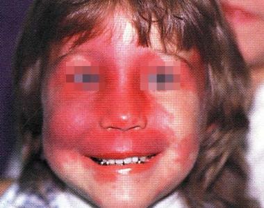
The Sturge-Weber syndrome consists of a unilateral port-wine hemangioma of the skin along the trigeminal distribution and is accompanied by an ipsilateral leptomeningeal angioma. Glaucoma is present in approximately half of the cases. The Nevus of Ota is a melanocytic pigmentary disorder, most commonly involving the area innervated by the trigeminal nerve. Elevated intraocular pressure, with or without glaucomatous damage, is observed in 10% of the cases. We report the first case of glaucoma associated with Sturge-Weber syndrome and Nevus of Ota in Korea. (+info)Sturge-Weber disease with repercussion on the prenatal development of the cerebral hemisphere. (2/61)
Sturge-Weber syndrome was diagnosed in a neonate on basis of a characteristic port-wine stain. In the absence of any acute neurologic episode, MR images obtained when the infant was aged 3 months showed a typical pial vascular dysplasia, as well as prominent hypotrophy of the homolateral hemisphere. Areas suggesting the presence of developmental dysplasia of the cerebral mantel were found in association with the typical pial vascular anomaly. The prenatal effect of Sturge-Weber disease on normal brain development may best be explored by using a better evaluation with cerebral imaging shortly after birth. (+info)Prophylactic antiepileptic treatment in Sturge-Weber disease. (3/61)
PURPOSE: In Sturge-Weber disease, motor and cognitive defects are supposed to result mostly from severe epilepsy. They might, therefore be partly prevented by prophylactic antiepileptic drug treatment. This condition constitutes a possible model for the study of prophylactic drug treatment in severe epilepsy. In the present study, we compared the outcome of patients treated prospectively with phenobarbitone before the first seizure, with those referred following the first seizure, in order to identify the issues related to the evaluation of prophylactic treatment of severe epilepsy. METHODS: Motor and cognitive outcome were compared in patients treated prophylactically with phenobarbitone (16 cases) and in those treated following the first seizures (21 cases). RESULTS: Whereas the incidence of motor deficit was similar in both groups (44 vs. 52%), that of mental retardation was lower in the group treated prophylactically (76.2 vs. 43.7%, P< 0.05). The major methodological issues encountered included the small number of patients identified at birth that could be included in the study, the need for randomization taking into account the size of the angioma, and the choice of the prophylactic medication, including the occurrence of epilepsy together with the course of motor and cognitive functions among the endpoints. CONCLUSION: Prophylactic anti-epileptic drug treatment is worth considering for Sturge-Weber disease, but a randomized prospective study is necessary to determine this. It should be multicentric, take in account the size of the angioma, and decide what the most appropriate medication should be. (+info)Pulsed dye laser for Sturge-Weber syndrome. (4/61)
Port wine stain of Sturge-Weber syndrome represents a cosmetic prejudice with social consequences. We have treated eight patients with a 585 nm pulsed dye laser. According to our experience, the treatment is not risky provided that adequate care is taken; the cosmetic result on the V1 port wine stain component is satisfactory. (+info)Contrast-enhanced fluid-attenuated inversion recovery imaging for leptomeningeal disease in children. (5/61)
BACKGROUND AND PURPOSE: To develop an MR imaging method that improves detection of leptomeningeal disease when compared with the current reference standard, contrast-enhanced T1-weighted imaging. METHODS: We investigated the cases of 10 children who were at high risk of intracranial leptomeningeal disease (Sturge-Weber syndrome and medulloblastoma). The cases of Sturge-Weber syndrome were investigated by using one MR imaging examination, and the cases of medulloblastoma were investigated by using four MR imaging examinations performed over 18 months. In all cases, contrast-enhanced fluid-attenuated inversion recovery (FLAIR) images were acquired in addition to the routine sequences. The parameters of the FLAIR sequence were chosen to maximize the T1 component of the signal intensity, to maximize detection of leptomeningeal enhancement. We made subjective and objective assessments of the presence and extent of leptomeningeal disease as shown on contrast-enhanced T1-weighted images and contrast-enhanced FLAIR images. RESULTS: In three of the four cases of Sturge-Weber syndrome, the T1 and FLAIR sequences showed comparable extent of leptomeningeal enhancement. For one child, FLAIR images showed unexpected bilateral disease and more extensive leptomeningeal enhancement on the clinically suspected side. In four of six cases of medulloblastoma, no leptomeningeal enhancement was shown on any examinations during the 18-month period. In two cases, FLAIR images showed more extensive leptomeningeal enhancement when compared with T1-weighted images. CONCLUSION: Contrast-enhanced FLAIR imaging seems to improve detection of leptomeningeal disease when compared with routine contrast-enhanced T1-weighted imaging. This seems to be partly because of suppression of signal intensity from normal vascular structures on the surface of the brain by FLAIR, which allows easier visualization of abnormal leptomeninges. We think that these findings can be extrapolated to the investigation of leptomeningeal disease of all causes and at all ages. (+info)Discrepancy between [18F]fluorodeoxyglucose and 11C-methionine positron emission tomography findings in Sturge-Weber syndrome--case report. (6/61)
Cerebral blood flow and metabolism were evaluated in an adult with symptomatic intractable epilepsy and Sturge-Weber syndrome (SWS) manifesting as angiomas in the left cerebral hemisphere. 99mTc-ethylcysteinate dimer single photon emission computed tomography detected reduced blood flow in the entire left cerebral hemisphere, and [18F]fluorodeoxyglucose positron emission tomography (PET) showed decreased glucose metabolism in the left cerebral hemisphere. These findings indicated hypofunction of the left cerebral hemisphere, which caused the right hemiparesis. 11C-methionine (11C-Met) PET revealed high 11C-Met accumulation in the angiomas in the left cerebral hemisphere. Immunostaining for glial fibrillary acidic protein showed positive reaction in the lesions. Gliosis is a likely mechanism for the 11C-Met accumulation, which is possibly associated with progressive calcification in the angiomas and retarded growth of patients with SWS occurring over many years. (+info)Early characteristics of Sturge-Weber syndrome shown by perfusion MR imaging and proton MR spectroscopic imaging. (7/61)
We report the case of a 9-month-old boy with Sturge-Weber syndrome and new onset of seizure. Perfusion MR imaging showed early changes compatible with impaired venous drainage in the affected hemisphere, whereas proton MR spectroscopic imaging revealed a focal parietal area of elevated choline without significant alteration of N-acetylaspartate levels. The perfusion and subtle metabolic abnormalities are comparable with the extent of the overlying leptomeningeal enhancement, illustrating the early pathophysiological manifestation of this disease. (+info)Total oral rehabilitation in a patient with portwine stains. (8/61)
Sturge-Weber syndrome is an uncommon condition characterized by presence of Portwine stains on the face along with ocular disorders, mental retardation, oral involvement and leptomeningeal angiomas. A report of a case with atypical manifestations of this syndrome along with a step-by-step protocol oral rehabilitation of such patients is described. (+info)Brainstem infarctions refer to the damage or death of brain tissue in the brainstem due to lack of blood supply, resulting in a localized injury known as an infarction. The brainstem is a critical region that controls essential functions such as breathing, heart rate, and consciousness. Infarctions in this area can result in various symptoms depending on the location and extent of damage, which may include:
1. Hemiparesis or paralysis on one side of the body
2. Cranial nerve dysfunction, leading to double vision, slurred speech, or facial weakness
3. Difficulty swallowing or speaking
4. Unstable blood pressure and heart rate
5. Altered level of consciousness, ranging from confusion to coma
6. Abnormal muscle tone and reflexes
7. Respiratory disturbances, such as irregular breathing patterns or apnea (cessation of breathing)
Brainstem infarctions can be caused by various conditions, including atherosclerosis, embolism, vasospasm, or small vessel disease. Prompt diagnosis and treatment are crucial to minimize the risk of long-term disability or death.
A syndrome, in medical terms, is a set of symptoms that collectively indicate or characterize a disease, disorder, or underlying pathological process. It's essentially a collection of signs and/or symptoms that frequently occur together and can suggest a particular cause or condition, even though the exact physiological mechanisms might not be fully understood.
For example, Down syndrome is characterized by specific physical features, cognitive delays, and other developmental issues resulting from an extra copy of chromosome 21. Similarly, metabolic syndromes like diabetes mellitus type 2 involve a group of risk factors such as obesity, high blood pressure, high blood sugar, and abnormal cholesterol or triglyceride levels that collectively increase the risk of heart disease, stroke, and diabetes.
It's important to note that a syndrome is not a specific diagnosis; rather, it's a pattern of symptoms that can help guide further diagnostic evaluation and management.
The Differential Threshold, also known as the Just Noticeable Difference (JND), is the minimum change in a stimulus that can be detected or perceived as different from another stimulus by an average human observer. It is a fundamental concept in psychophysics, which deals with the relationship between physical stimuli and the sensations and perceptions they produce.
The differential threshold is typically measured using methods such as the method of limits or the method of constant stimuli, in which the intensity of a stimulus is gradually increased or decreased until the observer can reliably detect a difference. The difference between the original stimulus and the barely detectable difference is then taken as the differential threshold.
The differential threshold can vary depending on a number of factors, including the type of stimulus (e.g., visual, auditory, tactile), the intensity of the original stimulus, the observer's attention and expectations, and individual differences in sensory sensitivity. Understanding the differential threshold is important for many applications, such as designing sensory aids for people with hearing or vision impairments, optimizing the design of multimedia systems, and developing more effective methods for detecting subtle changes in physiological signals.
"Bone retroversion" is not a widely recognized medical term or concept with a specific definition. The term "retroversion" is used in anatomy to describe the position of an organ or structure when it is turned backward or inward. In relation to bones, retroversion typically describes the orientation of a bone or joint when it is angled or positioned in such a way that its posterior (back) aspect faces more anteriorly (toward the front).
However, I was unable to find a widely accepted medical definition for "bone retroversion" as a specific pathological or anatomical condition. It's possible that the term may be used in a more specialized context within certain medical subspecialties. If you have more context or information about where this term is being used, I may be able to provide a more precise answer.
Down syndrome is a genetic disorder caused by the presence of all or part of a third copy of chromosome 21. It is characterized by intellectual and developmental disabilities, distinctive facial features, and sometimes physical growth delays and health problems. The condition affects approximately one in every 700 babies born in the United States.
Individuals with Down syndrome have varying degrees of cognitive impairment, ranging from mild to moderate or severe. They may also have delayed development, including late walking and talking, and may require additional support and education services throughout their lives.
People with Down syndrome are at increased risk for certain health conditions, such as congenital heart defects, respiratory infections, hearing loss, vision problems, gastrointestinal issues, and thyroid disorders. However, many individuals with Down syndrome live healthy and fulfilling lives with appropriate medical care and support.
The condition is named after John Langdon Down, an English physician who first described the syndrome in 1866.
Size perception in a medical context typically refers to the way an individual's brain interprets and perceives the size or volume of various stimuli. This can include visual stimuli, such as objects or distances, as well as tactile stimuli, like the size of an object being held or touched.
Disorders in size perception can occur due to neurological conditions, brain injuries, or certain developmental disorders. For example, individuals with visual agnosia may have difficulty recognizing or perceiving the size of objects they see, even though their eyes are functioning normally. Similarly, those with somatoparaphrenia may not recognize the size of their own limbs due to damage in specific areas of the brain.
It's important to note that while 'size perception' is not a medical term per se, it can still be used in a medical or clinical context to describe these types of symptoms and conditions.
Metabolic syndrome, also known as Syndrome X, is a cluster of conditions that increase the risk of heart disease, stroke, and diabetes. It is not a single disease but a group of risk factors that often co-occur. According to the American Heart Association and the National Heart, Lung, and Blood Institute, a person has metabolic syndrome if they have any three of the following five conditions:
1. Abdominal obesity (waist circumference of 40 inches or more in men, and 35 inches or more in women)
2. Triglyceride level of 150 milligrams per deciliter of blood (mg/dL) or greater
3. HDL cholesterol level of less than 40 mg/dL in men or less than 50 mg/dL in women
4. Systolic blood pressure of 130 millimeters of mercury (mmHg) or greater, or diastolic blood pressure of 85 mmHg or greater
5. Fasting glucose level of 100 mg/dL or greater
Metabolic syndrome is thought to be caused by a combination of genetic and lifestyle factors, such as physical inactivity and a diet high in refined carbohydrates and unhealthy fats. Treatment typically involves making lifestyle changes, such as eating a healthy diet, getting regular exercise, and losing weight if necessary. In some cases, medication may also be needed to manage individual components of the syndrome, such as high blood pressure or high cholesterol.
I apologize for any confusion, but "Plant Nectar" is not a term used in medical definitions. Nectar is a sweet liquid produced by plants in flowers to attract pollinators such as insects and birds. It's a sugar-rich substance that serves as a source of energy for these animals. While it may have some nutritional value, it's not something that would be relevant to medical definitions or human health in a direct sense.
 Sturge-Weber syndrome
 Sturge-Weber syndrome: MedlinePlus Genetics
 Sturge-Weber Syndrome (for Parents) - Primary Children's Hospital
 Sturge-Weber Syndrome: Practice Essentials, Background, Pathophysiology
 Neurocutaneous Disorders: Tuberous Sclerosis Complex and Sturge-Weber Syndrome
 Sturge-Weber Syndrome | FACES
 Sturge-Weber Syndrome - American Association for Pediatric Ophthalmology and Strabismus
 Accelerated myelination in early Sturge-Weber syndrome: MRI-SPECT correlations
 Sturge-Weber syndrome
 Ophthalmic Alterations in the Sturge-Weber Syndrome, Klippel-Trenaunay Syndrome, and the Phakomatosis Pigmentovascularis: An...
 Sturge-Weber Syndrome Types : New to SWF : The Sturge-Weber Foundation
 Treatment Services at the Hunter Nelson Sturge-Weber Syndrome Center | Kennedy Krieger Institute
 Port-wine Birthmarks and Sturge-Weber Syndrome: The Challenge of Diagnosis and Management | AAP Journal Blogs | American...
 Sturge-Weber Syndrome | Encyclopedia MDPI
 Sturge-Weber syndrome: a case report
 Sturge-Weber syndrome | Taber's Medical Dictionary
 Sturge-Weber Syndrome - Pediatrics - MSD Manual Professional Edition
 Non-penetrating deep sclerectomy for glaucoma associated with Sturge-Weber syndrome. | Read by QxMD
 Course 10: Diagnosis and Management of Sturge-Weber Syndrome - The VBF Educate E-Learning Center
 Sturge-Weber and Klippel-Trenaunay Syndrome With Nevus of Ota and Ito | JAMA Dermatology | JAMA Network
 1st case of Sturge Weber Syndrome in India to be treated using the cell based therapy - Mon Voyage
 Find a Doctor
 Molecules | Free Full-Text | Use of Cannabidiol in the Treatment of Epilepsy: Efficacy and Security in Clinical Trials