Superior Vena Cava Syndrome
Vena Cava, Superior
Brachiocephalic Veins
Phlebography
Vena Cava, Inferior
Tomography, X-Ray Computed
Stents
Azygos Vein
Vena Cava Filters
Carcinoma, Bronchogenic
The management of non-small-cell lung cancer: a case history. (1/180)
Accurate assessment and treatment of the patient with lung cancer requires a team approach involving respiratory physicians, cardiothoracic surgeons, oncologists and the palliative care team. Adequate staging and assessment of prognostic factors are essential before deciding what treatment is appropriate for an individual patient. Surgery is the mainstay of treatment for early disease. Patients with medically inoperable stage 1 (T1, T2, N0) tumours should be considered for radical radiotherapy; additional chemotherapy in early stage disease may offer an additional survival advantage, but its overall role can only be assessed by further clinical trials. In more locally advanced tumours radical radiotherapy has never been formally tested. It is however, often used in patients where the tumour can be encompassed safely within a radiation field. This will depend on total dose and fractionation schedule as well as the volume of tissue irradiated. Neo-adjuvant chemotherapy prolongs survival in these patients. As only a few patients are cured, symptom control and quality of life are usually the most important goals of management and can be achieved by a variety of interventions. It is disappointing that in such a common disease less than 5% of patients are entered into clinical trials. Without such evidence the therapeutic outcomes in NSCLC cannot be improved. (+info)Mediastinal fibromatosis presenting with superior vena cava syndrome. (2/180)
We encountered a fatal case of mediastinal fibromatosis in a 67-year-old female in whom there was aggressive infiltration into the large vessels, nerves and pericardium. She presented with the superior vena cava syndrome, Horner's syndrome, paralysis of bilateral vocal cords and diaphragm and heart failure. Mediastinoscopical examination revealed an extremely firm tumor adhering to the sternum, trachea and brachiocephalic artery. She died of severe heart failure due to the disturbed dilatation of the heart and ventilatory insufficiency. Although mediastinal fibromatosis is very uncommon and sometimes difficult to diagnose at an early stage, physicians should be aware of this disease for the differential diagnosis of mediastinal tumors. (+info)Superior vena cava syndrome as a complication of transvenous permanent pacemaker implantation. (3/180)
Venous thrombosis induced by a transvenous permanent pacemaker is a common complication. However, superior vena cava (SVC) syndrome caused by pacemaker leads is only occasionally seen and its prevalence has been estimated to be less than 1 in 1000 pacemaker patients. Herein, we report a Taiwanese patient of high grade AV block, who presented with SVC syndrome 2 years after transvenous permanent pacemaker implantation. This case features fibrotic stenosis of the junction of right brachiocephalic trunk and SVC, and an extensive thrombus formation resulting in complete obliteration of the left brachiocephalic vein. The collateral circulation was so delicate that he still could lead a rather normal life, even if anticoagulant therapy proved to be ineffective from an angiographic point of view. (+info)Coronary artery bypass and superior vena cava syndrome. (4/180)
Superior vena cava syndrome is the obstruction of the superior vena cava or its main tributaries by benign or malignant lesions. The syndrome causes edema and engorgement of the vessels on the face, neck, and arms, nonproductive cough, and dyspnea. We discuss the case of a 48-year-old obese diabetic woman who was admitted with unstable angina. She had previously been diagnosed with superior vena cava syndrome. Urgent coronary artery bypass grafting was necessary Although thousands of coronary artery bypasses are performed every year, there are not many reports on patients with superior vena cava syndrome who successfully undergo cardiopulmonary bypass and coronary artery grafting with an internal mammary artery as the conduit. The results of the case and alternative recommended methods are discussed. (+info)Superior vena cava obstruction caused by radiation induced venous fibrosis. (5/180)
Superior vena cava syndrome is most often caused by lung carcinoma. Two cases are described in whom venous obstruction in the superior mediastinum was caused by local vascular fibrosis due to radiotherapy five and seven years earlier. The development of radiation injury to greater vessels is discussed, together with the possibilities for treatment of superior vena cava syndrome. (+info)Successful resection of intracardiac invasive thymoma with right ventricular inflow tract occlusion. (6/180)
A 72-year-old man presenting with the superior vena cava syndrome and intracardiac mass was admitted to our hospital. The mass was resected and confirmed to be invasive thymoma. Three years later, he was re-admitted with recurrence into the intracardiac space without any changes in the anterior mediastinum mass. The mass occupied the right atrial cavity and protruded into the right ventricle, causing right ventricular inflow tract obstruction. He underwent re-operation and irradiation. His postoperative course was uneventful, and he has remained alive. Invasive thymoma with intracardiac extension is extremely rare. (+info)Reconstruction of the superior vena cava with the aid of an extraluminal venovenous jugulo-atrial shunt. (7/180)
A 57-year-old woman had chronic benign superior vena cava syndrome related to the long-term use of multiple central venous catheters for chemotherapy. Treatment included resection of the obstructed segment and repair of the superior vena cava with an autologous pericardial patch. Intraoperatively, return venous flow was maintained with an extraluminal venovenous jugulo-atrial shunt. The shunt relieved upper-body hypertension and congestion, resulting in early extubation and a short, smooth postoperative course. (+info)Lymphocutaneous fistula as a long-term complication of multiple central venous catheter placement. (8/180)
We report a case of a lymphocutaneous fistula in a 19-month-old boy who had been a premature neonate, born in the 23rd week of gestation. The fistula, an apparent complication of central venous line placement during the patient's first 5 months of life, was composed of a distinct lymphatic vessel bundle in the right supraclavicular region, with its exit point at the posterior aspect of the right shoulder. The drainage ceased immediately after resection and repair of a 1-cm obstruction in the superior vena cava. (+info)Superior Vena Cava Syndrome (SVCS) is a medical condition characterized by the obstruction of the superior vena cava (SVC), which is the large vein that carries blood from the upper body to the heart. This obstruction can be caused by cancerous tumors, thrombosis (blood clots), or other compressive factors.
The obstruction results in the impaired flow of blood from the head, neck, arms, and upper chest, leading to a variety of symptoms such as swelling of the face, neck, and upper extremities; shortness of breath; cough; chest pain; and distended veins visible on the skin surface. In severe cases, SVCS can cause life-threatening complications like cerebral edema (swelling of the brain) or pulmonary edema (fluid accumulation in the lungs).
Immediate medical attention is required for individuals with suspected SVCS to prevent further complications and to manage the underlying cause. Treatment options may include chemotherapy, radiation therapy, anticoagulation therapy, or surgery, depending on the etiology of the obstruction.
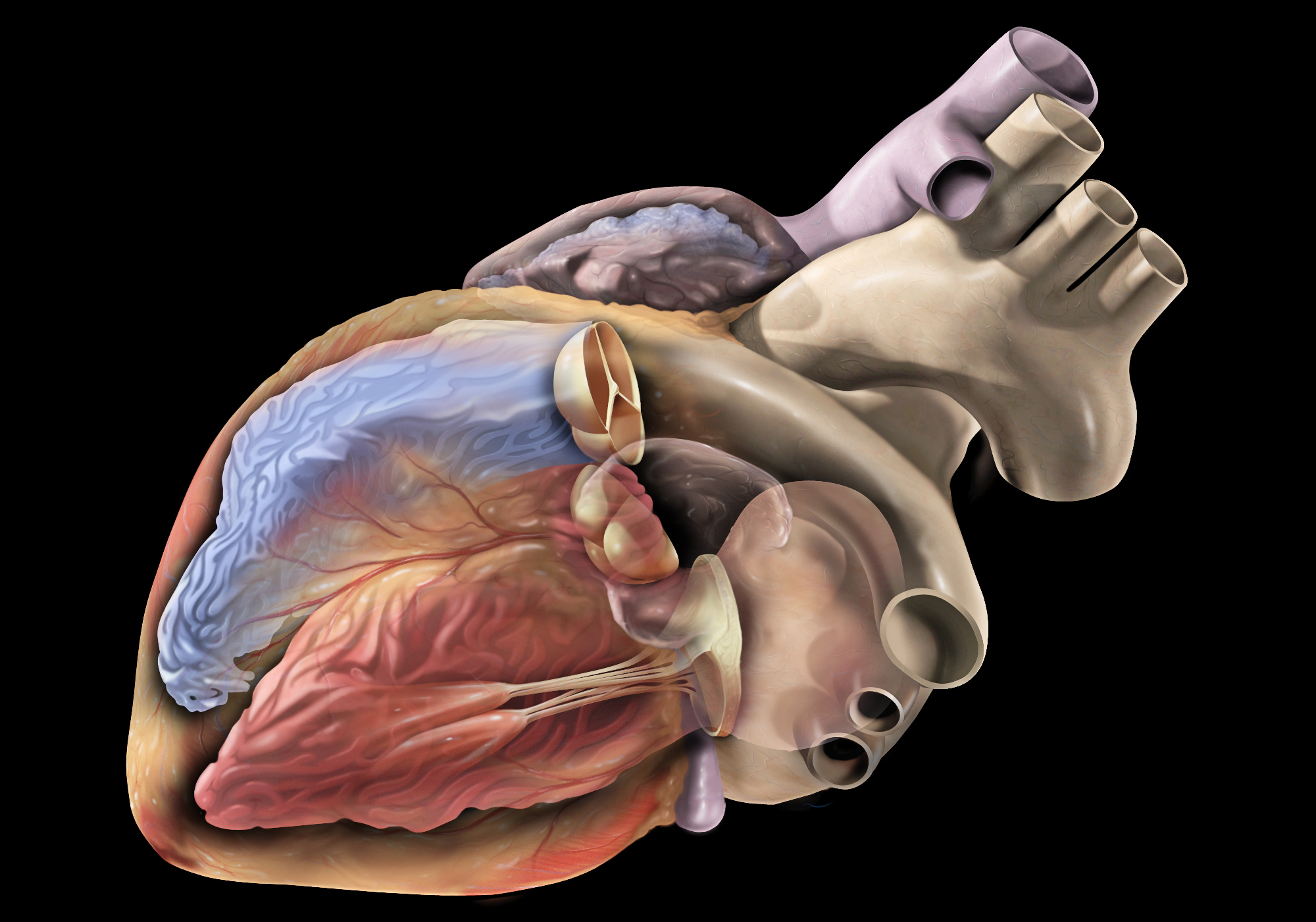
The superior vena cava is a large vein that carries deoxygenated blood from the upper half of the body to the right atrium of the heart. It is formed by the union of the left and right brachiocephalic veins (also known as the internal jugular and subclavian veins) near the base of the neck. The superior vena cava runs posteriorly to the sternum and enters the upper right portion of the right atrium, just posterior to the opening of the inferior vena cava. It plays a crucial role in the circulatory system by allowing blood returning from the head, neck, upper limbs, and thorax to bypass the liver before entering the heart.
The brachiocephalic veins, also known as the innominate veins, are large veins in the human body. They are formed by the union of the subclavian vein and the internal jugular vein on each side of the body. The resulting vein then carries blood from the upper limbs, head, and neck to the superior vena cava, which is the large vein that returns blood to the heart.
Here's a more detailed medical definition:
The brachiocephalic veins are paired venous structures that result from the union of the subclavian vein and the internal jugular vein on each side of the body. These veins are located in the superior mediastinum, near the base of the neck, and are typically about 2 to 3 centimeters in length. The brachiocephalic veins receive blood from several sources, including the upper extremities, head, neck, and thoracic wall. They then transport this blood to the superior vena cava, which is a large vein that returns blood to the right atrium of the heart.
It's worth noting that the brachiocephalic veins are subject to various pathological conditions, including thrombosis (blood clots), stenosis (narrowing), and compression by nearby structures such as the first rib or the scalene muscles. These conditions can lead to a variety of symptoms, including swelling, pain, and difficulty breathing.
Mediastinal neoplasms refer to abnormal growths or tumors located in the mediastinum, which is the central compartment of the thoracic cavity that lies between the lungs and contains various vital structures such as the heart, esophagus, trachea, blood vessels, lymph nodes, and nerves. Mediastinal neoplasms can be benign (non-cancerous) or malignant (cancerous), and they can arise from any of the tissues or organs within the mediastinum.
Benign mediastinal neoplasms may include thymomas, lipomas, neurofibromas, or teratomas, among others. These tumors are typically slow-growing and rarely spread to other parts of the body. However, they can still cause symptoms or complications by compressing adjacent structures within the mediastinum, such as the airways, blood vessels, or nerves.
Malignant mediastinal neoplasms are cancerous tumors that can invade and destroy surrounding tissues and may spread (metastasize) to other parts of the body. Common types of malignant mediastinal neoplasms include thymic carcinomas, lymphomas, germ cell tumors, and neuroendocrine tumors. These tumors often require aggressive treatment, such as surgery, radiation therapy, and chemotherapy, to control their growth and spread.
It is important to note that mediastinal neoplasms can present with various symptoms depending on their location, size, and type. Some patients may be asymptomatic, while others may experience cough, chest pain, difficulty breathing, hoarseness, or swallowing difficulties. A thorough diagnostic workup, including imaging studies and biopsies, is necessary to confirm the diagnosis and determine the best course of treatment for mediastinal neoplasms.
Phlebography is a medical imaging technique used to visualize and assess the veins, particularly in the legs. It involves the injection of a contrast agent into the veins, followed by X-ray imaging to capture the flow of the contrast material through the veins. This allows doctors to identify any abnormalities such as blood clots, blockages, or malformations in the venous system.
There are different types of phlebography, including ascending phlebography (where the contrast agent is injected into a foot vein and travels up the leg) and descending phlebography (where the contrast agent is injected into a vein in the groin or neck and travels down the leg).
Phlebography is an invasive procedure that requires careful preparation and monitoring, and it is typically performed by radiologists or vascular specialists. It has largely been replaced by non-invasive imaging techniques such as ultrasound and CT angiography in many clinical settings.
Mediastinal diseases refer to a group of conditions that affect the mediastinum, which is the area in the chest separating the lungs and containing various vital structures such as the heart, esophagus, trachea, thymus gland, lymph nodes, blood vessels, and nerves. These diseases can be benign or malignant (cancerous) and may cause symptoms due to compression or invasion of surrounding tissues. Examples of mediastinal diseases include:
1. Mediastinal tumors: Abnormal growths in the mediastinum, which can be benign or malignant. Common types include thymomas, germ cell tumors, lymphomas, and neurogenic tumors.
2. Mediastinitis: Inflammation of the mediastinal tissues, often caused by infections, trauma, or complications from medical procedures.
3. Enlarged lymph nodes: Abnormal swelling of the lymph nodes in the mediastinum can be a sign of various conditions, including infections, cancer, and autoimmune disorders.
4. Mediastinal cysts: Fluid-filled sacs that develop in the mediastinum, which are usually benign but may cause symptoms due to compression or infection.
5. Aneurysms or dissections of the aorta: Abnormal weakening or tearing of the aortic wall within the mediastinum, which can lead to life-threatening complications if not treated promptly.
6. Esophageal diseases: Conditions affecting the esophagus, such as tumors, strictures, or motility disorders, may present with symptoms related to the mediastinum.
7. Thyroid disorders: Enlargement of the thyroid gland (goiter) can extend into the mediastinum and cause compression symptoms.
8. Hematomas or effusions: Accumulation of blood (hematoma) or fluid (effusion) in the mediastinal space due to trauma, surgery, or other underlying conditions.
Early diagnosis and appropriate treatment are crucial for managing mediastinal diseases and improving patient outcomes.
Tuberculous empyema is a specific type of empyema, which is a collection of pus in the pleural space (the space between the lungs and the chest wall). It is caused by the Mycobacterium tuberculosis bacterium, which is the same bacterium that causes tuberculosis (TB) of the lungs.
In tuberculous empyema, the bacteria spread from the lungs to the pleural space, where they cause an infection and inflammation. This can lead to the accumulation of pus and the development of a chronic empyema. Symptoms may include chest pain, cough, fever, and difficulty breathing. Treatment typically involves a prolonged course of multiple antibiotics to kill the bacteria, as well as drainage of the pus from the pleural space. In some cases, surgery may be necessary to remove the infected tissue and prevent recurrence.
The inferior vena cava (IVC) is the largest vein in the human body that carries deoxygenated blood from the lower extremities, pelvis, and abdomen to the right atrium of the heart. It is formed by the union of the left and right common iliac veins at the level of the fifth lumbar vertebra. The inferior vena cava is a retroperitoneal structure, meaning it lies behind the peritoneum, the lining that covers the abdominal cavity. It ascends through the posterior abdominal wall and passes through the central tendon of the diaphragm to enter the thoracic cavity.
The inferior vena cava is composed of three parts:
1. The infrarenal portion, which lies below the renal veins
2. The renal portion, which receives blood from the renal veins
3. The suprahepatic portion, which lies above the liver and receives blood from the hepatic veins before draining into the right atrium of the heart.
The inferior vena cava plays a crucial role in maintaining venous return to the heart and contributing to cardiovascular function.
Mediastinitis is a medical condition that refers to the inflammation of the mediastinum, which is the area in the chest that separates the lungs and contains various vital structures such as the heart, esophagus, trachea, thymus gland, and major blood vessels. Mediastinitis can be caused by bacterial or fungal infections, trauma, or complications from medical procedures such as esophageal surgery or heart catheterization.
The symptoms of mediastinitis may include chest pain, fever, difficulty swallowing, shortness of breath, cough, and neck stiffness. The diagnosis is typically made through imaging tests such as X-rays, CT scans, or MRI scans, and confirmed with laboratory tests that identify the causative organism. Treatment usually involves antibiotics or antifungal medications to eliminate the infection, along with supportive care such as pain management, fluids, and nutrition. In severe cases, surgery may be necessary to drain infected fluid or remove damaged tissue.
X-ray computed tomography (CT or CAT scan) is a medical imaging method that uses computer-processed combinations of many X-ray images taken from different angles to produce cross-sectional (tomographic) images (virtual "slices") of the body. These cross-sectional images can then be used to display detailed internal views of organs, bones, and soft tissues in the body.
The term "computed tomography" is used instead of "CT scan" or "CAT scan" because the machines take a series of X-ray measurements from different angles around the body and then use a computer to process these data to create detailed images of internal structures within the body.
CT scanning is a noninvasive, painless medical test that helps physicians diagnose and treat medical conditions. CT imaging provides detailed information about many types of tissue including lung, bone, soft tissue and blood vessels. CT examinations can be performed on every part of the body for a variety of reasons including diagnosis, surgical planning, and monitoring of therapeutic responses.
In computed tomography (CT), an X-ray source and detector rotate around the patient, measuring the X-ray attenuation at many different angles. A computer uses this data to construct a cross-sectional image by the process of reconstruction. This technique is called "tomography". The term "computed" refers to the use of a computer to reconstruct the images.
CT has become an important tool in medical imaging and diagnosis, allowing radiologists and other physicians to view detailed internal images of the body. It can help identify many different medical conditions including cancer, heart disease, lung nodules, liver tumors, and internal injuries from trauma. CT is also commonly used for guiding biopsies and other minimally invasive procedures.
In summary, X-ray computed tomography (CT or CAT scan) is a medical imaging technique that uses computer-processed combinations of many X-ray images taken from different angles to produce cross-sectional images of the body. It provides detailed internal views of organs, bones, and soft tissues in the body, allowing physicians to diagnose and treat medical conditions.
An aortic aneurysm is a medical condition characterized by the abnormal widening or bulging of the wall of the aorta, which is the largest artery in the body. The aorta carries oxygenated blood from the heart to the rest of the body. When the aortic wall weakens, it can stretch and balloon out, forming an aneurysm.
Aortic aneurysms can occur anywhere along the aorta but are most commonly found in the abdominal section (abdominal aortic aneurysm) or the chest area (thoracic aortic aneurysm). The size and location of the aneurysm, as well as the patient's overall health, determine the risk of rupture and associated complications.
Aneurysms often do not cause symptoms until they become large or rupture. Symptoms may include:
* Pain in the chest, back, or abdomen
* Pulsating sensation in the abdomen
* Difficulty breathing
* Hoarseness
* Coughing or vomiting
Risk factors for aortic aneurysms include age, smoking, high blood pressure, family history, and certain genetic conditions. Treatment options depend on the size and location of the aneurysm and may include monitoring, medication, or surgical repair.
A stent is a small mesh tube that's used to treat narrow or weak arteries. Arteries are blood vessels that carry blood away from your heart to other parts of your body. A stent is placed in an artery as part of a procedure called angioplasty. Angioplasty restores blood flow through narrowed or blocked arteries by inflating a tiny balloon inside the blocked artery to widen it.
The stent is then inserted into the widened artery to keep it open. The stent is usually made of metal, but some are coated with medication that is slowly and continuously released to help prevent the formation of scar tissue in the artery. This can reduce the chance of the artery narrowing again.
Stents are also used in other parts of the body, such as the neck (carotid artery) and kidneys (renal artery), to help maintain blood flow and prevent blockages. They can also be used in the urinary system to treat conditions like ureteropelvic junction obstruction or narrowing of the urethra.
The azygos vein is a large, unpaired venous structure in the thoracic cavity of the human body. It begins as the ascending lumbar vein, which receives blood from the lower extremities and abdominal organs. As it enters the thorax through the diaphragm, it becomes the azygos vein and continues to ascend along the vertebral column.
The azygos vein receives blood from various tributaries, including the intercostal veins, esophageal veins, mediastinal veins, and bronchial veins. It then arches over the right mainstem bronchus and empties into the superior vena cava, which returns blood to the right atrium of the heart.
The azygos vein provides an important collateral pathway for venous return in cases where the inferior vena cava is obstructed or occluded. It also plays a role in the spread of certain thoracic diseases, such as tuberculosis and cancer.
Vena cava filters are medical devices that are implanted into the inferior vena cava, which is the largest vein in the body that returns blood from the lower half of the body to the heart. These filters are designed to trap blood clots that form in the deep veins of the legs (deep vein thrombosis or DVT) and prevent them from traveling to the lungs (pulmonary embolism or PE), which can be a life-threatening condition.
The filter is typically implanted using a catheter-based procedure, and it has legs or arms that extend out to trap the blood clots as they flow through the vein. Over time, the trapped clots may dissolve on their own or become organized and incorporated into the wall of the vein.
Vena cava filters are typically used in patients who are at high risk for PE but cannot take anticoagulation medication or have failed anticoagulation therapy. However, there is some controversy surrounding the use of these devices due to concerns about their long-term safety and effectiveness.
Carcinoma, bronchogenic is a medical term that refers to a type of lung cancer that originates in the bronchi, which are the branching tubes that carry air into the lungs. It is the most common form of lung cancer and can be further classified into different types based on the specific cell type involved, such as squamous cell carcinoma, adenocarcinoma, or large cell carcinoma.
Bronchogenic carcinomas are often associated with smoking and exposure to environmental pollutants, although they can also occur in non-smokers. Symptoms may include coughing, chest pain, shortness of breath, wheezing, hoarseness, or unexplained weight loss. Treatment options depend on the stage and location of the cancer, as well as the patient's overall health and may include surgery, radiation therapy, chemotherapy, targeted therapy, or a combination of these approaches.
An encyclopedia is a comprehensive reference work containing articles on various topics, usually arranged in alphabetical order. In the context of medicine, a medical encyclopedia is a collection of articles that provide information about a wide range of medical topics, including diseases and conditions, treatments, tests, procedures, and anatomy and physiology. Medical encyclopedias may be published in print or electronic formats and are often used as a starting point for researching medical topics. They can provide reliable and accurate information on medical subjects, making them useful resources for healthcare professionals, students, and patients alike. Some well-known examples of medical encyclopedias include the Merck Manual and the Stedman's Medical Dictionary.
 Superior vena cava syndrome
Vein
Reinforcement
Superior vena cava
Pemberton's sign
Thymic carcinoma
Gardner fibroma
Mediastinal tumors
Raghib syndrome
List of films released posthumously
Thymoma
Deaths in July 2012
Plethora (medicine)
Primary mediastinal (thymic) large B cell lymphoma
Esophageal cancer
Histoplasmosis
Vein of Galen aneurysmal malformations
Deep vein thrombosis
Sherman Hemsley
Non-small-cell lung cancer
List of medical mnemonics
Causes of cancer pain
Lung cancer
Pancoast tumor
Dexamethasone
List of causes of shortness of breath
Index of oncology articles
Cancer pain
Childhood cancer
Oncological emergencies
Superior vena cava syndrome - Wikipedia
 Catheter-associated superior vena cava syndrome | CMAJ
 Superior Vena Cava Syndrome Imaging: Practice Essentials, Imaging Modalities
 Frontiers | Case report: Primary cardiac lymphoma manifesting as superior vena cava syndrome
The difficult approach to neoplastic superior vena cava syndrome: surgical option - The Journal of Cardiovascular Surgery 2003...
 Cutaneous findings leading to a diagnosis of superior vena cava syndrome: A case report and review of the literature
Superior Vena Cava Syndrome: Background, Pathophysiology, Etiology
 Superior Vena Cava (SVC) Syndrome | RK.MD
 Superior Vena Cava Syndrome- Causes, Symptoms, Diagnosis, treatment
 Guide To Treating Superior Vena Cava Syndrome - HealthPrep.com
Review of evolving etiologies, implications and treatment strategies for the superior vena cava syndrome | SpringerPlus | Full...
Palliative Cancer Care Guidelines: Palliative Care Standards, Cancer Pain, Dyspnea
 Catheter-related thrombosis of Superior Vena Cava in a patient with Superior Vena Cava Syndrome | Asploro Open Access...
Reinforcement - Wikipedia
 Superior vena cava syndrome due to intravascular thrombosis in a patient with rheumatoid arthritis without antiphospholipid...
![Fuchs H[au] - Search Results - PubMed](data:image/png;base64,iVBORw0KGgoAAAANSUhEUgAAABAAAAAQCAMAAAAoLQ9TAAAARVBMVEVHcEwoU45gYmYAUpQAUpRPYGVgYmZLXnJgYmYAUZUAUpRJXnIAUpQAUpRgYmYAUpRgYmZgYmZhYmYAUpQAUpQAUpRgYmaDiPJuAAAAFXRSTlMADOJ+6QewGO8/uTRqtH7GdFJ11p1bCL3TAAAAZUlEQVQYlV2PVw7AIAxDTeney7n/UcsoldX3E+VJOAboEi7MBpHWMs1ADlG8u7UYWauwyZFeRQVPOhG2o+aiwhByJxUx91Jxhje3iJSqGfHuLKI0+0TpXvY1twCOPlFh5pa/++MB0vIOBm+1zaoAAAAASUVORK5CYII=) Fuchs H[au] - Search Results - PubMed
Fuchs H[au] - Search Results - PubMed
 Superior vena cava syndrome and pacemaker leads. Explant by mechanical dissection system of extraction and percutaneous...
 Rigid Bronchoscopic Intervention for Central Airway Obstruction and Concurrent Superior Vena Cava Syndrome Caused by Small Cell...
Palliative Cancer Care Guidelines: Palliative Care Standards, Cancer Pain, Dyspnea
November 2014 Clinical Trends
 Small Cell Lung Cancer Treatment (PDQ®) - NCI
Pediatric Holt-Oram Syndrome Treatment & Management: Medical Care, Surgical Care, Long-Term Monitoring
Antiretroviral Therapy-associated Coccidioidal Meningitis - Volume 19, Number 1-January 2013 - Emerging Infectious Diseases...
 Pathology Outlines - Anatomy
 urofacial syndrome - Ontology Browser - Rat Genome Database
 How To Do Infraclavicular Subclavian Vein Cannulation, Ultrasound-Guided - Critical Care Medicine - Merck Manuals Professional...
 Thieme E-Journals - Indian Journal of Radiology and Imaging / Abstract
Clinical Trials Register
 Recognition of Common Childhood Malignancies | AAFP
 9.4 Akutte onkologiske tilstander - Helsebiblioteket
Obstruction9
- Superior vena cava syndrome (SVCS), is a group of symptoms caused by obstruction of the superior vena cava ("SVC"), a short, wide vessel carrying circulating blood into the heart. (wikipedia.org)
- Superior vena cava (SVC) syndrome (SVCS) is a constellation of symptoms that result from obstruction of the SVC (see the images from a single case, below). (medscape.com)
- Superior vena cava syndrome, which occurs in approximately 15,000 persons in the United States annually, consists of a collection of symptoms and signs resulting from the obstruction of the superior vena cava (SVC). (escholarship.org)
- Superior vena cava syndrome (SVCS) is obstruction of blood flow through the superior vena cava (SVC). (medscape.com)
- Anything that impairs SVC drainage by intrinsic obstruction (thrombosis, anatomic narrowing) or extrinsic compression (mediastinal masses, non-small cell lung cancer, non-Hodgkin's lymphoma, etc.) can result in this syndrome. (rk.md)
- The superior vena cava syndrome (SVCS) is defined as the set of signs and symptoms derived from superior vena cava obstruction, both intrinsic obstruction and extrinsic compression, which causes an increase in venous pressure in the upper body region. (asploro.com)
- B, Contrast-enhanced computed tomography shows near complete obstruction of the superior vena cava and lower endotracheal tumor completely closing the right main bronchus. (clinicalgate.com)
- Superior vena cava syndrome is characterized by superior vena cava obstruction with severe reduction in venous return. (romj.org)
- These images are a random sampling from a Bing search on the term "Superior Vena Cava Obstruction. (fpnotebook.com)
Thrombosis13
- Catheter-associated thrombosis is the most common noninfectious complication of implantable venous access devices and can cause superior vena cava syndrome. (cmaj.ca)
- Prophylactic approaches to catheter-associated thrombosis are not recommended, and the use of superior vena cava filters in deep vein thrombosis of the upper extremities should be avoided. (cmaj.ca)
- SVCS is caused by compression, invasion, and/or thrombosis of the superior vena cava and/or the brachiocephalic veins. (medscape.com)
- Iatrogenic thrombosis associated with central venous catheters is the most frequent cause of intraluminal occlusion of the superior vena cava. (escholarship.org)
- Torres-Pérez ME, Huerta-Torres KG, Vargas-Ledo JF, "Catheter-related thrombosis of Superior Vena Cava in a patient with Superior Vena Cava Syndrome", Asp Biomed Clin Case Rep, vol.1, no.1: 33-39, 2018. (asploro.com)
- The tomographic contrasted study demonstrated superior vena cava thrombosis. (asploro.com)
- Although thrombosis is a frequent manifestation in patients with blood coagulation alterations and patients with end stage chronic kidney disease, catheter-related thrombosis is a rare cause of thrombosis and superior vena cava syndrome whose must common cause is neoplastic. (asploro.com)
- Superior venous system stenosis (superior vena cava (SVC) - right subclavian vein - innominate vein - left subclavian vein) is a clinical situation that frequently appears in patients with long-term implanted cardiac stimulation devices, due to venous system thrombosis and in those with congenital heart disease who need corrective surgery, due to chronic complications inherent to surgical techniques. (hvt-journal.com)
- We present a retrospective series of six consecutive patients with SVC syndrome studied in a single center from 2012 to 2021.Three of them presented with thrombosis related to pacing or defibrillation electrodes and the other three presented with complications derived from Mustard or Senning techniques in patients with pacemakers and D-transposition of the great arteries. (hvt-journal.com)
- Anti cardiolipin antibody was done to rule out antiphospholipid antibody (APLA) syndrome which has a known association with rheumatoid arthritis to cause intravascular thrombosis but was negative. (romj.org)
- There are case reports where RA is associated with SVC syndrome but only when it is associated mediastinal lymphadenopathy or SVC thrombosis due to APLA Syndrome. (romj.org)
- We could not found any other previous case report in the literature where rheumatoid arthritis was found to be the cause for SVC thrombosis without any associated antiphospholipid antibody (APLA) syndrome. (romj.org)
- Further contrast enhanced computerized tomography (CECT) with angiography of chest was done which revealed a non opacification of right sided superior vena cava with hypodense filling defect noted within suggestive of chronic thrombosis and multiple dilated collateral venous channels ( Figure 2 and 3). (romj.org)
Occlusion3
- Venogram shows almost complete occlusion of the superior vena cava with dramatic collateral drainage through the left superior intercostal vein. (medscape.com)
- Originally described in 1757 by William Hunter in a patient afflicted with a saccular aneurysm of the ascending aorta secondary to syphilis [ 2 ], this condition is characterized by compromised blood flow in the vena cava because of extrinsic compression or intraluminal occlusion. (escholarship.org)
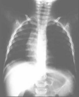
- D. R. Northrip, B. K. Bohman and K. Tsueda, "Total Airway Occlusion and Superior Vena Cava Syndrome in a Child with Anterior Mediastinal Tumour," Anesthesia & Analgesia, Vol. 65, No. 10, 1986, pp. 1079-1082. (scirp.org)
Venous9
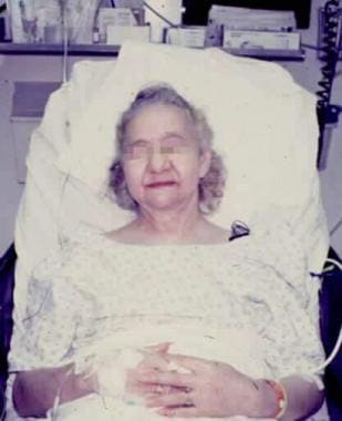
- Photographs of the head and upper chest of a 53-year-old man with catheter-associated superior vena cava syndrome, showing (A) facial and neck plethora, and (B) a prominent superficial venous pattern on the chest. (cmaj.ca)
- The superior vena cava (SVC) appeared almost completely occluded immediately above the right atrium distal to the catheter tip, with extensive venous collaterals in the mediastinum, suggestive of SVC syndrome ( Figure 2 ). (cmaj.ca)
- Timely diagnosis of superior vena cava syndrome and treatment of the underlying disease are critical, for increased cervical venous pressure can compromise the larynx and pharynx. (escholarship.org)
- The second pathway is the internal mammary venous system plus tributaries and secondary communications to the superior and inferior epigastric veins. (medscape.com)
- Superior vena cava (SVC) syndrome is characterized by facial plethora, upper body venous engorgement, chest pain, soft tissue swelling, dyspnea, and even neurologic changes. (rk.md)
- The superior vena cava (SVC) in the main conduit for venous drainage of the head, neck, upper extremities, and upper thorax, its main auxiliary vessel, the azygos vein, enters in the SVC just above the pericardial reflection, other collateral systems are the internal mammary veins and the esophageal vascular plexus. (asploro.com)
- In clinical practice, venous system stenosis may manifest as a SVC syndrome. (hvt-journal.com)
- Ultrasound-guided cannulation of the subclavian vein uses real-time (dynamic) ultrasound to guide venipuncture and a guidewire (Seldinger technique) to thread a central venous catheter through the subclavian vein and into the superior vena cava. (merckmanuals.com)
- The subclavian vein may be less preferred for stiff catheters (because of difficulty achieving the sharp turn into the superior van cava) or large-bore hemodialysis catheters (which can cause venous stenosis that renders the ipsilateral arm unsuitable for arteriovenous shunt placement). (merckmanuals.com)
SVCS2
- If a person's superior vena cava is partially blocked or compressed, superior vena cava syndrome (SVCS) occurs. (drsuvadipchakrabarti.com)
- Superior vena cava syndrome (SVCS) is only observed in 0.6%-3.5% of the patient population (2, 4, 5). (hvt-journal.com)
Symptoms6
- A 64-year-old man presented with symptoms indicative of superior vena cava syndrome. (frontiersin.org)
- Given the patient's symptoms suggestive of superior vena cava syndrome (SVC), a broad differential diagnosis were considered. (frontiersin.org)
- Symptoms may result from local invasion or compression of adjacent thoracic structures, such as compression involving the esophagus causing dysphagia, compression involving the laryngeal nerves causing hoarseness, or compression involving the superior vena cava causing facial edema and distension of the superficial veins of the head and neck. (cancer.gov)
- Primary care physicians should be alert for possible presenting signs and symptoms of childhood malignancy, particularly in patients with Down syndrome or other congenital and familial conditions associated with an increased risk of cancer. (aafp.org)
- We report on the case of a 55-year-old man diagnosed with advanced thymoma, who, during the progression of his disease, developed signs and symptoms suggesting Good's syndrome and pure red cell aplasia. (ecancer.org)
- Patients usually present with symptoms secondary to local compression, such as respiratory symptoms or superior vena cava syndrome. (ecancer.org)
Implantable2
- Keywords: transvenous lead extraction, balloon angioplasty, cardiac implantable electronic devices, superior vena cava stent. (hvt-journal.com)
- One study explored the syndrome's relationship as a complication of an implantable cardioverter defibrillator, while another study examined superior vena cava syndrome and skin color change with circulatory assist devices. (medscape.com)
Clinical6
- Clinical superior vena cava syndrome manifestations remitted. (asploro.com)
- A clinical diagnosis of superior vena cava (SVC) syndrome was made. (romj.org)
- For more in-depth clinical information, see Superior Vena Cava Syndrome . (medscape.com)
- Al-Qattan MM, Abou Al-Shaar H. Molecular basis of the clinical features of Holt-Oram syndrome resulting from missense and extended protein mutations of the TBX5 gene as well as TBX5 intragenic duplications. (medscape.com)
- Ogur G, Gul D, Lenk MK, Imirzalioglu N, Alpay F, Ogur E. Variable clinical expression of Holt-Oram syndrome in three generations. (medscape.com)
- TBX5 genotyping has high sensitivity and specificity for Holt-Oram syndrome (HOS) if stringent diagnostic criteria are used in assigning the clinical diagnosis. (medscape.com)
Lymph4
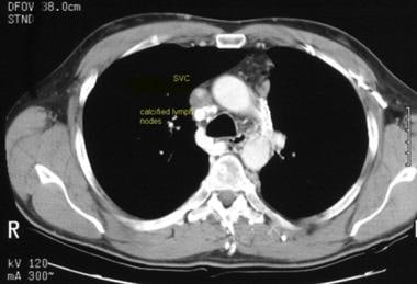
- CT scan shows a narrowed superior vena cava with adjacent calcified lymph nodes and posterior soft tissue thickening. (medscape.com)
- Additional known masses of the middle or right anterior mediastinum causing superior vena cava syndrome include enlarged paratracheal lymph nodes, lymphoma, leiomyosarcomas, carcinoids, germ cell tumors, fibrosing mediastinitis, intrathoracic goiter, thymoma, and aortic aneurysm. (escholarship.org)
- There may be the spreading of cancer to the lymph nodes surrounding the superior vena cava. (drsuvadipchakrabarti.com)
- Compression of the vena cava may result from extra luminal compression by mass present in middle or anterior mediastinum including right paratracheal lymph nodes, lymphoma, thymoma, aortic aneurysm or any inflammatory process leading to fibrosing mediastinitis [1-5]. (romj.org)
Cardiac4
- Malignant causes included primary or secondary cardiac tumors, lung cancer, mediastinal tumors, or lymphoma, which may obstruct or compress the superior vena cava. (frontiersin.org)
- Patients with Holt-Oram syndrome may require dietary modification because of their specific cardiac abnormality. (medscape.com)
- Malignant hyperthermia-like manifestations in a two-month-old child with Holt-Oram syndrome undergoing cardiac surgery. (medscape.com)
- A computed tomography (CT) scan showed a 2-cm right middle lobe nodule adjacent to the cardiac border and mediastinal lymphadenopathy abutting the superior vena cava and anterior pericardium ( Figure 1 ). (cdc.gov)
Etiology1
- In general, treating SVC syndrome depends on the etiology. (rk.md)
Compression2
- Resolution of superior vena cava syndrome is directly related to the treatment of the underlying compression. (wikipedia.org)
- Describe the following oncologic emergencies: superior vena cava syndrome, spinal cord compression, hypercalcemia, tumor lysis syndrome, syndrome of inappropriate antidiuretic hormone, and neutropenic fever. (nursingworld.org)
Occurs2
- Chylous effusion also occurs with the superior vena cava syndrome. (msdmanuals.com)
- Hypoplastic left heart syndrome occurs when parts of the left side of the heart (mitral valve, left ventricle, aortic valve, and aorta) do not develop completely. (medlineplus.gov)
Collateral1
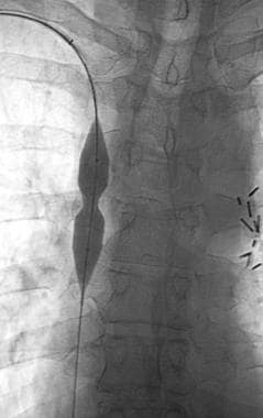
- Venogram obtained after stenting shows a widely patent superior vena cava with no collateral drainage. (medscape.com)
Babies with hypoplastic left hea5
- About 10% of babies with hypoplastic left heart syndrome also have other birth defects. (medlineplus.gov)
- In babies with hypoplastic left heart syndrome, blood leaving the right side of the heart through the pulmonary artery travels through the ductus arteriosus to the aorta. (medlineplus.gov)
- Often, babies with hypoplastic left heart syndrome also have an atrial septal defect , which is a hole between the left and right upper chambers (atria) of the heart. (cdc.gov)
- In babies with hypoplastic left heart syndrome, the left side of the heart cannot pump oxygen-rich blood to the body properly. (cdc.gov)
- However, among babies with hypoplastic left heart syndrome, when these openings close, it becomes hard for oxygen-rich blood to get to the rest of the body. (cdc.gov)
Malignant1
- Over 80% of cases are caused by malignant tumors compressing the superior vena cava. (wikipedia.org)
Patient's1
- The examination of a patient suspected of having superior vena cava syndrome depends on the patient's prior medical history. (medscape.com)
Respiratory1
- Respiratory insufficiency and hypoventilation syndromes 32. (muni.cz)
20191
- La información más reciente sobre el nuevo Coronavirus de 2019, incluidas las clínicas de vacunación para niños de 6 meses en adelante. (stanfordchildrens.org)
Posterior2
- The animals were anesthetized and held up the incision and visualization of the posterior vena cava for blood puncture vacuum in tubes containing EDTA. (bvsalud.org)
- Os animais foram anestesiados e procedeu-se a incisão e visualização da veia cava posterior. (bvsalud.org)
Stent3
- A Palmaz P308 stent mounted on a 12-mm balloon was deployed in the superior vena cava after it was predilated to 8 mm. (medscape.com)
- Our objective was to perform an extraction of the pacemaker and defibrillation electrodes, to allow the passage of a support wire to achieve the implantation of the endovascular stent(s) to correct the SVC syndrome. (hvt-journal.com)
- Combined treatment of lead extraction and endovascular stent implantation corrected the syndrome in all cases. (hvt-journal.com)
Neck1
- Superior vena cava (SVC) carries blood from the head, neck and upper limb to the heart. (romj.org)
Inferior2
- The patient underwent a chest CT-scan, followed by a confirmatory transesophageal echocardiogram (TEE) which revealed the presence of a prominent, heterogeneous, partially non-enhancing, right atrial mass, measuring 66 × 41 × 37 mm, partially disrupting inferior vena cava flow and obstructing the superior vena cava ( Figures 1 , 2 , Supplementary Video S1 ). (frontiersin.org)
- Inferior vena cava-syndrome. (nih.gov)
Aortic aneurysm1
- William Hunter first described the syndrome in 1757 in a patient with syphilitic aortic aneurysm. (medscape.com)
Thoracic1
- Cases of other less common etiologies: pulmonary amoebiasis, antiphospholipid syndrome, chronic lymphocytic leukemia, thoracic actinomycosis, invasive epidermoid carcinoma, cutaneous polyarteritis, and merkel cell carcinoma [4-10] have also been reported. (escholarship.org)
Tumour2
- The superior vena cava may get pressure from a tumour in the chest. (drsuvadipchakrabarti.com)
- There may be a blockage in the superior vena cava due to the growth of a tumour in the vein. (drsuvadipchakrabarti.com)
Pleural1
- Yellow nail syndrome is a rare disorder causing chronic exudative pleural effusions, lymphedema, and dystrophic yellow nails-all thought to be the result of impaired lymphatic drainage. (msdmanuals.com)
Blood4
- If the ductus arteriosus is allowed to close in a baby with hypoplastic left heart syndrome, the baby may quickly die because no blood will be pumped to the body. (medlineplus.gov)
- Hypoplastic (pronounced hi-puh-PLAS-tik) left heart syndrome or HLHS is a birth defect that affects normal blood flow through the heart. (cdc.gov)
- Hypoplastic left heart syndrome (HLHS) is a birth defect that affects normal blood flow through the heart. (cdc.gov)
- During the first few days of life for a baby with hypoplastic left heart syndrome, the oxygen-rich blood bypasses the poorly functioning left side of the heart through the patent ductus arteriosus and the patent foramen ovale. (cdc.gov)
Patient1
- Patient 2 was an 8-year-old boy with hyperimmunoglobulin-E (Job) syndrome due to mutation in signal transducer and activator of transcription-3 in whom a right-sided pulmonary abscess developed and failed to improve after 1 month of antibacterial therapy. (cdc.gov)
Physical1
- Physical exam usually establishes the presence of superior vena cava syndrome. (escholarship.org)
Atrial3
- TBX5 loss-of-function mutation contributes to atrial fibrillation and atypical Holt-Oram syndrome. (medscape.com)
- A gain-of-function TBX5 mutation is associated with atypical Holt-Oram syndrome and paroxysmal atrial fibrillation. (medscape.com)
- A 2-dimensional echocardiographic picture taken from subxiphoid window showing a large secundum atrial septal defect (arrow) in a 7-year-old boy with Holt-Oram syndrome. (medscape.com)
Myasthenia1
- On other occasions, they may appear with a paraneoplastic syndrome [ 2 ], including myasthenia gravis, pure red cell aplasia, connective tissue disorders, and hypogammaglobulinaemia/Good's syndrome [ 1 , 2 ]. (ecancer.org)
Presentation2
- In the outpatient setting, recognizing the early cutaneous presentation of superior vena cava syndrome requires a high index of suspicion. (escholarship.org)
- We report a case of postmortem examination-proven antiretroviral therapy (ART)-associated coccidioidomycosis manifesting as atypical lymphocytic meningitis, which we believe represents a rare presentation of immune reconstitution inflammatory syndrome (IRIS). (cdc.gov)
Defect1
- Hypoplastic left heart syndrome is one type of congenital heart defect. (cdc.gov)
Genetic5
- KLF13 is a genetic modifier of the Holt-Oram syndrome gene TBX5. (medscape.com)
- Li B, Chen S, Sun K, Xu R, Wu Y. Genetic analyses identified a SALL4 gene mutation associated with Holt-Oram syndrome. (medscape.com)
- Some genetic syndromes requiring increased vigilance for cancer are also discussed. (aafp.org)
- A cardiomelic developmental field has also been postulated to relate the genetic heterogeneity of HOS (and other similar syndromes) to a cascade of molecules, including the brachyury, sonic hedgehog, bone morphogenetic protein, retinoic acid receptor, and transforming growth factor beta families. (medscape.com)
- It is also associated with some genetic diseases such as Turner syndrome, Jacobsen syndrome, trisomy 13 and 18. (medlineplus.gov)
Hypoplastic7
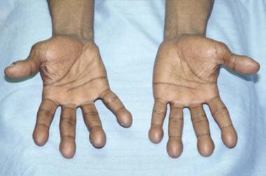
- Photograph showing hypoplastic right thumb of the right hand of a 6-month-old infant with Holt-Oram syndrome. (medscape.com)
- Babies with known hypoplastic left heart syndrome are usually started on a medicine to keep the ductus arteriosus open. (medlineplus.gov)
- The Centers for Disease Control and Prevention (CDC) estimates that each year about 1,025 babies in the United States are born with hypoplastic left heart syndrome. (cdc.gov)
- 1 In other words, about 1 out of every 3,841 babies born in the United States each year is born with hypoplastic left heart syndrome. (cdc.gov)
- Hypoplastic left heart syndrome may be diagnosed during pregnancy or soon after the baby is born. (cdc.gov)
- Hypoplastic left heart syndrome may be diagnosed during pregnancy with an ultrasound, (which creates pictures of the body). (cdc.gov)
- Some findings from the ultrasound may make the health care provider suspect a baby may have hypoplastic left heart syndrome. (cdc.gov)
Vein1
- A major vein in a person's body is the superior vena cava. (drsuvadipchakrabarti.com)
Mediastinum1
- The superior vena cava (SVC) is formed in the upper middle part of the mediastinum by the junction of the brachiocephalic veins. (medscape.com)
Patients6
- Between 1989 and 1995 6 -patients -with -superior -vena -cava syn-drome under-went sur-gical treat-ment for tho-racic -tumors. (minervamedica.it)
- A report identified this syndrome in 4% of patients with radial longitudinal deficiency. (medscape.com)
- In this retrospective study we performed a chart review of patients with a confirmed diagnosis of BS who were followed at the Behçet's syndrome outpatient clinic, Rheumatology Research Center, Shariati Hospital, Tehran, Iran, between 2015 and 2017. (researchsquare.com)
- The presence of inter-current infections, especially diarrhoea and pneumonia, in the presence of lymphocyte B depletion and hypogammaglobulinaemia is known as Good's syndrome and may affect up to 5% of patients with thymoma. (ecancer.org)
- While anaemia is present in 50%-86% of patients with Good's syndrome, only 41.9% of cases present pure red cell aplasia. (ecancer.org)
- Good's syndrome affects at least 5% of patients with thymomas [ 3 ]. (ecancer.org)
Chest1
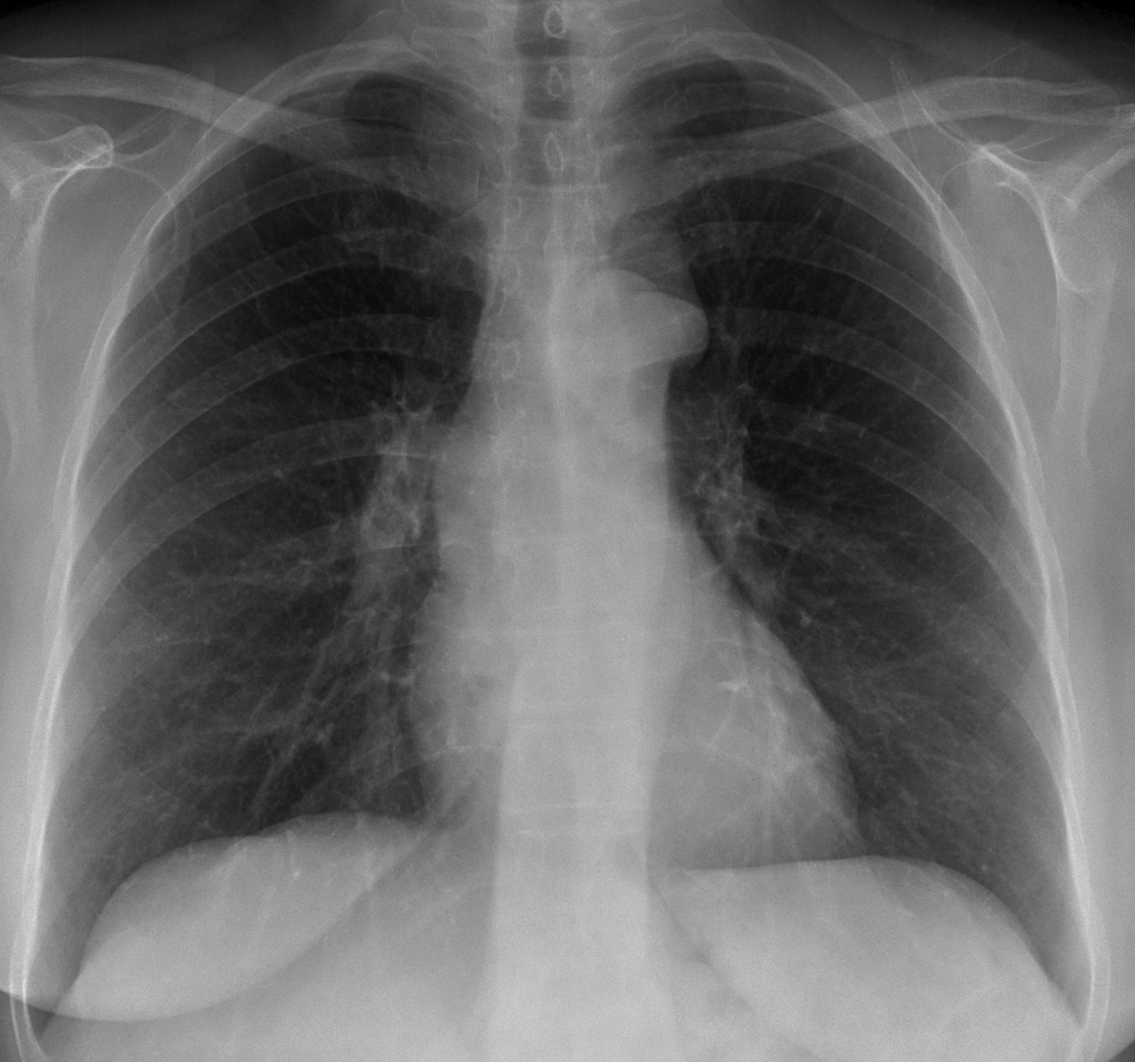
- However, 16% of people with SVC syndrome have a normal chest X-ray. (wikipedia.org)