Teratoma
Testicular Neoplasms
Dermoid Cyst
Neoplasms, Germ Cell and Embryonal
Dysgerminoma
Ovarian Neoplasms
Retroperitoneal Neoplasms
Embryonic Stem Cells
Germinoma
Isochromosomes
Mesonephroma
Struma Ovarii
Coccyx
Induced Pluripotent Stem Cells
Retroperitoneal Space
Cell Differentiation
alpha-Fetoproteins
Endodermal Sinus Tumor
Neoplasms, Multiple Primary
Octamer Transcription Factor-3
SOXB1 Transcription Factors
Choriocarcinoma
Paraneoplastic Syndromes, Nervous System
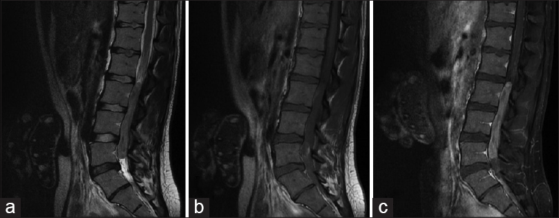
Spinal Neoplasms
Tomography, X-Ray Computed
Twins, Conjoined
Seminoma
Carcinoma, Embryonal
Carcinoid heart disease from ovarian primary presenting with acute pericarditis and biventricular failure. (1/1513)
A case is described of a 54 year old woman who had acute pericarditis with large exudative effusion accompanied by severe right and left ventricular failure. The patient was finally diagnosed with carcinoid heart disease from an ovarian carcinoid teratoma. She was treated with octreotide--a somatostatin analogue--followed by radical surgical resection of the neoplasm. At one year follow up only mild carcinoid tricuspid regurgitation remained. Only 16 cases of carcinoid heart disease from an ovarian primary have been described in literature. Moreover clinically manifest acute, nonmetastatic pericarditis and left heart failure are not considered as possible presentations of carcinoid heart disease, whatever the origin. In a recent series a small pericardial effusion was considered an infrequent and unexpected echocardiographic finding in carcinoid heart patients. One case of "carcinoid pericarditis" has previously been described as a consequence of pericardial metastasis. Left sided heart involvement is usually caused by bronchial carcinoids or patency of foramen ovale; both were excluded in the case presented. (+info)Regulation of I-branched poly-N-acetyllactosamine synthesis. Concerted actions by I-extension enzyme, I-branching enzyme, and beta1,4-galactosyltransferase I. (2/1513)
I-branched poly-N-acetyllactosamine is a unique carbohydrate composed of N-acetyllactosamine branches attached to linear poly-N-acetyllactosamine, which is synthesized by I-branching beta1, 6-N-acetylglucosaminyltransferase. I-branched poly-N-acetyllactosamine can carry bivalent functional oligosaccharides such as sialyl Lewisx, which provide much better carbohydrate ligands than monovalent functional oligosaccharides. In the present study, we first demonstrate that I-branching beta1, 6-N-acetylglucosaminyltransferase cloned from human PA-1 embryonic carcinoma cells transfers beta1,6-linked GlcNAc preferentially to galactosyl residues of N-acetyllactosamine close to nonreducing terminals. We then demonstrate that among various beta1, 4-galactosyltransferases (beta4Gal-Ts), beta4Gal-TI is most efficient in adding a galactose to linear and branched poly-N-acetyllactosamines. When a beta1,6-GlcNAc branched poly-N-acetyllactosamine was incubated with a mixture of beta4Gal-TI and i-extension beta1,3-N-acetylglucosaminyltransferase, the major product was the oligosaccharide with one N-acetyllactosamine extension on the linear Galbeta1-->4GlcNAcbeta1-->3 side chain. Only a minor product contained galactosylated I-branch without N-acetyllactosamine extension. This finding was explained by the fact that beta4Gal-TI adds a galactose poorly to beta1,6-GlcNAc attached to linear poly-N-acetyllactosamines, while beta1, 3-N-acetylglucosaminyltransferase and beta4Gal-TI efficiently add N-acetyllactosamine to linear poly-N-acetyllactosamines. Together, these results strongly suggest that galactosylation of I-branch is a rate-limiting step in I-branched poly-N-acetyllactosamine synthesis, allowing poly-N-acetyllactosamine extension mostly along the linear poly-N-acetyllactosamine side chain. These findings are entirely consistent with previous findings that poly-N-acetyllactosamines in human erythrocytes, PA-1 embryonic carcinoma cells, and rabbit erythrocytes contain multiple, short I-branches. (+info)Ovarian teratomas appearing as solid masses on ultrasonography. (3/1513)
The purposes of this study were to evaluate the prevalence and imaging characteristics of ovarian teratomas that appear as solid masses on ultrasonography and to compare the ultrasonographic imaging features of the tumors with their pathologic findings. The ultrasonographic images of 202 ovarian teratomas were reviewed retrospectively. Solid-appearing masses were selected from among them and were evaluated in terms of internal echotexture, the presence or lack of peripheral hypoechogenicity, posterior sonic attenuation, and tumoral calcification. Seventy-six (37.6%) masses of 202 belonged to the atypical solid-appearing masses on ultrasonography. Of 76 masses, 57 (75.0%) had peripheral hypoechogenicity; 38 masses had well-defined thin hypoechoic rims, whereas 19 had poorly demarcated peripheral hypoechogenicity. Posterior sonic attenuation was evident in 18 (23.7%) masses. The presence of peripheral hypoechogenicity, which is suggestive of the fluid portion of the tumor, might be one of the characteristic findings of solid-appearing ovarian teratomas on ultrasonography. (+info)Microdissection-based analysis of mature ovarian teratoma. (4/1513)
The genotypic features of mature ovarian teratomas (MOTs) are controversial. Early studies detected a homozygous genotype in MOTs suggesting that these tumors are composed of germ cells that have undergone meiosis I. Other studies, however, revealed a heterozygous genotype in a substantial proportion of MOTs suggesting an origin either from premeiotic germ cells or from a somatic cell line. In view of the complex morphology of MOTs and to increase the sensitivity of teratoma genotyping, we applied tissue microdissection before genetic analysis of teratomatous tissue. This approach allowed selective analysis of different heterotopic tissue elements as well as the lymphoid tissues within MOTs the origin of which is unknown. After DNA extraction, the tissue samples were polymerase chain reaction amplified using a random panel of highly informative genetic markers for different chromosomes to evaluate heterozygosity versus homozygosity. In all seven cases that were analyzed, heterotopic tissues consistently revealed a homozygous genotype with several markers; in two cases, heterozygosity was detected with a single marker, indicating a meiotic recombination event. Lymphoid aggregates within MOTs were heterozygous and derived from host tissue rather than from teratomatous growth. However, well differentiated thymic tissue was consistently homozygous, suggesting lymphoid differentiation capability of MOTs. We conclude that potential pitfalls in genotyping of teratomas including meiotic recombination and host cell participation can be avoided by a microdissection-based approach in combination with a panel of genetic markers. (+info)Antenatal sonographic diagnosis of epignathus at 15 weeks of pregnancy. (5/1513)
Epignathus is a rare, benign, congenital teratoma of the hard palate. Most of these teratomas are unidirectional and protrude through the mouth. Hence, the prognosis depends on the size of the tumor and the degree of face distortion and airway obstruction that it causes. However, some epignathi protrude bidirectionally, involving and destroying the brain tissue, resulting in a poor prognosis. This report presents a case of ultrasonographic detection of a bidirectional epignathus at 15 weeks of pregnancy. (+info)Pathogenesis of testicular germ cell tumours. (6/1513)
Human germ cell tumours comprise a heterogeneous group of neoplasms. In the testis, three entities are distinguished, the teratomas-yolk sac tumours of the infantile testis, the seminomas and nonseminomas of adolescents and adults, and the spermatocytic seminomas. Studies on epidemiology, histology, clinical behaviour, and chromosomal constitution of these tumours support the concept of distinct entities derived from germ cells but each with a different pathogenesis. Either the teratomas of the infantile testis show no chromosomal aberrations, or display a pattern of over- and under-representation of (parts of) chromosomes as detected in the yolk sac tumours of the infantile testis. In contrast, the seminomas and nonseminomas reveal a consistent pattern of losses and gains, that is, chromosomes 11, 13 and 18, and 7, 8 and X, respectively, that is different from that found in the infantile testis teratomas and yolk sac tumours. The most consistent structural chromosomal abnormality is an isochromosome 12p. Tumours lacking i(12p) have other structural abnormalities of 12p, among them amplification of 12p11.2-p12.1. The pathogenetically relevant genes on 12p11.2-p12.1 are probably on a fragment of about 1.7 mb. Gain of 12p sequences may be related to invasive growth. Gain of chromosome 9 is the only consistent chromosomal anomaly of spermatocytic seminomas. Infantile teratomas and spermatocytic seminomas are benign tumours. Infantile yolk sac tumour is a malignant germ cell tumour. Seminomas and nonseminomas are malignant, and the most common cancer in young Caucasian males. The cure rate of seminomas and non-seminomas with radio- and chemotherapy is over 90%, which is higher than that of any other solid cancer in adults. In addition, the precursor lesions of these tumours can be treated readily, justifying efforts to develop means for early diagnosis. Finally, the pathogenetic relationship between seminomas and nonseminomas, and the available animal models for the three groups of testicular germ cell tumours are discussed. (+info)Forced expression of the homeobox-containing gene Pem blocks differentiation of embryonic stem cells. (7/1513)
Similarities in the differentiation of mouse embryos and ES cell embryoid bodies suggest that aspects of early mammalian embryogenesis can be studied in ES cell embryoid bodies. In an effort to understand the regulation of cellular differentiation during early mouse embryogenesis, we altered the expression of the Pem homeobox-containing gene in ES cells. Pem is normally expressed in the preimplantation embryo and expressed in a lineage-restricted fashion following implantation, suggesting a role for Pem in regulating cellular differentiation in the early embryo. Here, we show that the forced expression of Pem from the mouse Pgk-1 promoter in ES cells blocks the in vitro and in vivo differentiation of the cells. In particular, embryoid bodies produced from these Pgk-Pem ES cells do not differentiate into primitive endoderm or embryonic ectoderm, which are prominent features of early embryoid bodies from normal ES cells. This Pgk-Pem phenotype is also different from the null phenotype, as embryoid bodies derived from ES cells in which endogenous Pem gene expression has been blocked show a pattern of differentiation similar to that of normal ES cells. When the Pgk-Pem ES cells were introduced into subcutaneous sites of nude mice, only undifferentiated EC-like cells were found in the teratomas derived from the injected cells. The Pem-dependent block of ES cell differentiation appears to be cell autonomous; Pgk-Pem ES cells did not differentiate when mixed with normal, differentiating ES cells. A block to ES cell differentiation, resulting from the forced expression of Pem, can also be produced by the forced expression of the nonhomeodomain region of Pem. These studies are consistent with a role for Pem in regulating the transition between undifferentiated and differentiated cells of the early mouse embryo. (+info)Characterization of the model for experimental testicular teratoma in 129/SvJ-mice. (8/1513)
An animal model of experimental testicular teratoma has been established to study how a teratoma affects the host testis and how the host testis reacts against the teratoma. 129/SvJ-mice were used as experimental animals. To induce the experimental testicular teratoma, male gonadal ridges from 12-day-old 129/SvJ-mouse fetuses were grafted into the testes of adult mice for 1-12 weeks. The developing tumour was analysed by light and electron microscopy and by immunocytochemical localization of transcription factors SOX9 and c-kit, glial fibrillary acidic protein (GFAP) and type IV collagen. Testicular teratoma was observed in 36 out of 124 testes with implanted fetal gonadal ridges (frequency 29%). One spontaneous testicular teratoma was observed in this material from 70 male mice (1.5%). One week after implantation intracordal clusters of cells were seen in embryonic testicular cords of the graft as the first sign of testicular teratomas. Four weeks after implantation the embryonic testicular cords had totally disappeared from grafts with teratomas, and the tumour tissue had enlarged the testis and invaded the interstitium of the host testis. It consisted of solitary pieces of immature cartilage as well as of glial cells and of primitive neuroepithelium. Six to eight weeks after implantation the tumour tissue had expanded so that the enlarged testis could be detected by macroscopic enlargement of the scrotum. The testicular tissue of the host had practically disappeared, and only solitary disrupted seminiferous tubules of the host were seen surrounding the teratoma. Neuroepithelial structures of some teratomas cultured for 8 weeks had cells with a granular nucleus as a sign of obvious apoptosis. Eleven to 12 weeks after implantation the growth of the teratoma had stopped, and the histology corresponded to that of a mature cystic teratoma. GFAP, SOX9 and type IV collagen were strongly positive in some parts of the tumours cultured for 4 and 8 weeks, while only occasional c-kit-positive areas were observed in tumours cultured for 8 weeks. As conclusions: (1) the metastasizing capacity of the experimental testicular teratoma is very low during 12 weeks, but the behaviour of the tumour in the testicular tissue of the graft is invasive; (2) the growth of experimental testicular teratomas cease 6-8 weeks after implantation of the fetal gonadal ridges with the obvious apoptosis of the immature tissue components; (3) the model of experimental testicular teratoma in the mouse is suitable for studying how the teratoma affects the host testis and how the host testis reacts to teratoma. (+info)A teratoma is a type of germ cell tumor, which is a broad category of tumors that originate from the reproductive cells. A teratoma contains developed tissues from all three embryonic germ layers: ectoderm, mesoderm, and endoderm. This means that a teratoma can contain various types of tissue such as hair, teeth, bone, and even more complex organs like eyes, thyroid, or neural tissue.
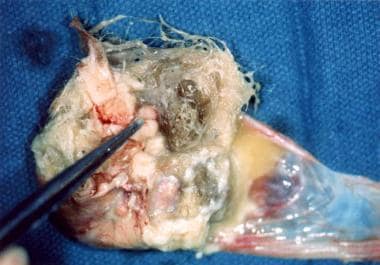
Teratomas are usually benign (non-cancerous), but they can sometimes be malignant (cancerous) and can spread to other parts of the body. They can occur anywhere in the body, but they're most commonly found in the ovaries and testicles. When found in these areas, they are typically removed surgically.
Teratomas can also occur in other locations such as the sacrum, coccyx (tailbone), mediastinum (the area between the lungs), and pineal gland (a small gland in the brain). These types of teratomas can be more complex to treat due to their location and potential to cause damage to nearby structures.
Testicular neoplasms are abnormal growths or tumors in the testicle that can be benign (non-cancerous) or malignant (cancerous). They are a type of genitourinary cancer, which affects the reproductive and urinary systems. Testicular neoplasms can occur in men of any age but are most commonly found in young adults between the ages of 15 and 40.
Testicular neoplasms can be classified into two main categories: germ cell tumors and non-germ cell tumors. Germ cell tumors, which arise from the cells that give rise to sperm, are further divided into seminomas and non-seminomas. Seminomas are typically slow-growing and have a good prognosis, while non-seminomas tend to grow more quickly and can spread to other parts of the body.
Non-germ cell tumors are less common than germ cell tumors and include Leydig cell tumors, Sertoli cell tumors, and lymphomas. These tumors can have a variety of clinical behaviors, ranging from benign to malignant.
Testicular neoplasms often present as a painless mass or swelling in the testicle. Other symptoms may include a feeling of heaviness or discomfort in the scrotum, a dull ache in the lower abdomen or groin, and breast enlargement (gynecomastia).
Diagnosis typically involves a physical examination, imaging studies such as ultrasound or CT scan, and blood tests to detect tumor markers. Treatment options depend on the type and stage of the neoplasm but may include surgery, radiation therapy, chemotherapy, or a combination of these modalities. Regular self-examinations of the testicles are recommended for early detection and improved outcomes.
The sacrococcygeal region is the lower part of the back where the spine ends, specifically referring to the area where the sacrum (a triangular bone at the base of the spine formed by the fusion of several vertebrae) meets the coccyx (also known as the tailbone). This region is located at the very bottom of the spine and is susceptible to injury or trauma due to its position and role in supporting the body's weight. It is also a common site for birth defects, particularly in newborns.
A dermoid cyst is a type of benign (non-cancerous) growth that typically develops during embryonic development. It is a congenital condition, which means it is present at birth, although it may not become apparent until later in life. Dermoid cysts are most commonly found in the skin or the ovaries of women, but they can also occur in other areas of the body, such as the spine or the brain.
Dermoid cysts form when cells that are destined to develop into skin and its associated structures, such as hair follicles and sweat glands, become trapped during fetal development. These cells continue to grow and multiply, forming a sac-like structure that contains various types of tissue, including skin, fat, hair, and sometimes even teeth or bone.
Dermoid cysts are usually slow-growing and may not cause any symptoms unless they become infected or rupture. In some cases, they may cause pain or discomfort if they press on nearby structures. Treatment typically involves surgical removal of the cyst to prevent complications and alleviate symptoms.
Mediastinal neoplasms refer to abnormal growths or tumors located in the mediastinum, which is the central compartment of the thoracic cavity that lies between the lungs and contains various vital structures such as the heart, esophagus, trachea, blood vessels, lymph nodes, and nerves. Mediastinal neoplasms can be benign (non-cancerous) or malignant (cancerous), and they can arise from any of the tissues or organs within the mediastinum.
Benign mediastinal neoplasms may include thymomas, lipomas, neurofibromas, or teratomas, among others. These tumors are typically slow-growing and rarely spread to other parts of the body. However, they can still cause symptoms or complications by compressing adjacent structures within the mediastinum, such as the airways, blood vessels, or nerves.
Malignant mediastinal neoplasms are cancerous tumors that can invade and destroy surrounding tissues and may spread (metastasize) to other parts of the body. Common types of malignant mediastinal neoplasms include thymic carcinomas, lymphomas, germ cell tumors, and neuroendocrine tumors. These tumors often require aggressive treatment, such as surgery, radiation therapy, and chemotherapy, to control their growth and spread.
It is important to note that mediastinal neoplasms can present with various symptoms depending on their location, size, and type. Some patients may be asymptomatic, while others may experience cough, chest pain, difficulty breathing, hoarseness, or swallowing difficulties. A thorough diagnostic workup, including imaging studies and biopsies, is necessary to confirm the diagnosis and determine the best course of treatment for mediastinal neoplasms.
Neoplasms, germ cell and embryonal are types of tumors that originate from the abnormal growth of cells. Here's a brief medical definition for each:
1. Neoplasms: Neoplasms refer to abnormal tissue growths or masses, which can be benign (non-cancerous) or malignant (cancerous). They result from uncontrolled cell division and may invade surrounding tissues or spread to other parts of the body through a process called metastasis.
2. Germ Cell Tumors: These are rare tumors that develop from the germ cells, which give rise to sperm and eggs in the reproductive organs (ovaries and testes). They can be benign or malignant and may occur in both children and adults. Germ cell tumors can also arise outside of the reproductive organs, a condition known as extragonadal germ cell tumors.
3. Embryonal Tumors: These are a type of malignant neoplasm that primarily affects infants and young children. They develop from embryonic cells, which are immature cells present during fetal development. Embryonal tumors can occur in various organs, including the brain (medulloblastomas), nervous system (primitive neuroectodermal tumors or PNETs), and other areas like the kidneys and liver.
It is essential to note that these conditions require professional medical evaluation and treatment by healthcare professionals with expertise in oncology and related fields.
Dysgerminoma is a type of germ cell tumor that develops in the ovaries. It is a malignant (cancerous) tumor that primarily affects girls and women of reproductive age, although it can occur at any age. Dysgerminomas are composed of large, round, or polygonal cells with clear cytoplasm and distinct cell borders, arranged in nests or sheets. They may also contain lymphoid aggregates and may produce hormones such as estrogen or testosterone.
Dysgerminomas are usually unilateral (affecting one ovary), but they can be bilateral (affecting both ovaries) in about 10-15% of cases. They tend to grow and spread rapidly, so early detection and treatment are crucial for a favorable prognosis.
The standard treatment for dysgerminoma is surgical removal of the affected ovary or ovaries, followed by chemotherapy with agents such as bleomycin, etoposide, and cisplatin (BEP). With appropriate treatment, the five-year survival rate for patients with dysgerminoma is high, ranging from 80% to 95%.
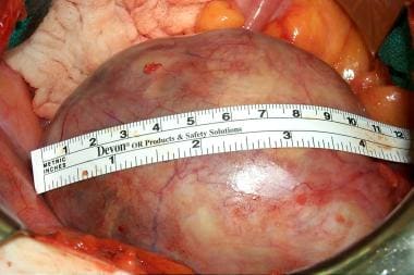
Ovarian neoplasms refer to abnormal growths or tumors in the ovary, which can be benign (non-cancerous) or malignant (cancerous). These growths can originate from various cell types within the ovary, including epithelial cells, germ cells, and stromal cells. Ovarian neoplasms are often classified based on their cell type of origin, histological features, and potential for invasive or metastatic behavior.
Epithelial ovarian neoplasms are the most common type and can be further categorized into several subtypes, such as serous, mucinous, endometrioid, clear cell, and Brenner tumors. Some of these epithelial tumors have a higher risk of becoming malignant and spreading to other parts of the body.
Germ cell ovarian neoplasms arise from the cells that give rise to eggs (oocytes) and can include teratomas, dysgerminomas, yolk sac tumors, and embryonal carcinomas. Stromal ovarian neoplasms develop from the connective tissue cells supporting the ovary and can include granulosa cell tumors, thecomas, and fibromas.
It is essential to diagnose and treat ovarian neoplasms promptly, as some malignant forms can be aggressive and potentially life-threatening if not managed appropriately. Regular gynecological exams, imaging studies, and tumor marker tests are often used for early detection and monitoring of ovarian neoplasms. Treatment options may include surgery, chemotherapy, or radiation therapy, depending on the type, stage, and patient's overall health condition.
Retroperitoneal neoplasms refer to abnormal growths or tumors that develop in the retroperitoneal space. This is the area located behind the peritoneum, which is the membrane that lines the abdominal cavity and covers the abdominal organs. The retroperitoneal space contains several vital structures such as the kidneys, adrenal glands, pancreas, aorta, and lymphatic vessels.
Retroperitoneal neoplasms can be benign or malignant (cancerous). Malignant retroperitoneal neoplasms are often aggressive and can invade surrounding tissues and organs, leading to various complications. Common types of retroperitoneal neoplasms include lymphomas, sarcomas, and metastatic tumors from other primary sites. Symptoms may vary depending on the size and location of the tumor but can include abdominal or back pain, weight loss, and swelling in the legs. Diagnosis typically involves imaging studies such as CT scans or MRI, followed by a biopsy to determine the type and grade of the tumor. Treatment options may include surgery, radiation therapy, chemotherapy, or a combination of these approaches.
Embryonic stem cells are a type of pluripotent stem cell that are derived from the inner cell mass of a blastocyst, which is a very early-stage embryo. These cells have the ability to differentiate into any cell type in the body, making them a promising area of research for regenerative medicine and the study of human development and disease. Embryonic stem cells are typically obtained from surplus embryos created during in vitro fertilization (IVF) procedures, with the consent of the donors. The use of embryonic stem cells is a controversial issue due to ethical concerns surrounding the destruction of human embryos.
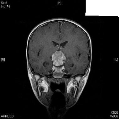
A germinoma is a type of tumor that develops in the brain or the spine, primarily in the pituitary gland or pineal gland. It is a rare form of primary central nervous system (CNS) cancer and is classified as a type of germ cell tumor. These tumors arise from cells that normally develop into sperm or eggs, which can migrate to unusual locations during embryonic development.
Germinomas are highly sensitive to radiation therapy and chemotherapy, making them generally treatable and curable with appropriate medical intervention. Symptoms of a germinoma may include headaches, nausea, vomiting, visual disturbances, hormonal imbalances, and neurological deficits, depending on the location and size of the tumor. Diagnosis typically involves imaging studies like MRI or CT scans, followed by a biopsy to confirm the presence of malignant cells.
I could not find a medical definition for "Severe Teratoid Abnormalities" as it is not a widely recognized or established medical term. However, the term "teratoid" is used in medical contexts to describe abnormal growths or tumors that contain a mixture of tissue types, such as skin, muscle, bone, and nerve cells. A severe teratoid abnormality would likely refer to a particularly serious or extreme case of such a condition.
If you are looking for information on a specific medical condition or issue, I would be happy to help you try to find more accurate and relevant information. Can you please provide me with more context or details?
Isochromosomes are abnormal chromosomes that contain identical arms on both sides, instead of having one arm longer than the other. This occurs due to an error in cell division where the centromere, the region where the chromatids (the two copies of chromosome) are attached, is duplicated and then separated improperly. As a result, each new chromosome has two identical arms.
Isochromosomes can lead to genetic disorders because they can disrupt the balance of genes on the chromosome. For example, if an isochromosome forms for chromosome 18 (i(18)), there will be three copies of the genes on one arm and only one copy on the other arm, leading to an overexpression of some genes and a loss of expression of others. This can cause developmental abnormalities and intellectual disabilities.
Isochromosomes are often associated with certain types of cancer, as well as genetic disorders such as Turner syndrome and Klinefelter syndrome.
Mesonephroma is a very rare type of kidney tumor that originates from the mesonephric duct remnants, which are the embryonic precursors of the male reproductive system. This tumor typically affects older adults and is more common in men than women.
Mesonephromas are usually slow-growing and asymptomatic, making them difficult to detect at an early stage. When symptoms do occur, they may include flank pain, hematuria (blood in the urine), a palpable abdominal mass, and weight loss.
On imaging studies such as CT or MRI scans, mesonephromas typically appear as well-circumscribed masses within the kidney. The diagnosis is usually confirmed through a biopsy or surgical excision of the tumor.
Mesonephromas are composed of tubular structures lined with cuboidal to low columnar epithelial cells, often with clear cytoplasm. They may also contain areas of necrosis and hemorrhage. The treatment of mesonephroma typically involves surgical excision, and the prognosis is generally favorable, with a low risk of recurrence or metastasis. However, long-term follow-up is recommended due to the rarity and limited data on this type of tumor.
Struma ovarii is a rare type of ovarian tumor, which is composed predominantly of thyroid tissue and accounts for less than 1% of all ovarian neoplasms. It is classified as a specialized form of monodermal teratoma (a type of germ cell tumor). Despite being composed mainly of thyroid tissue, struma ovarii may produce and release thyroid hormones, leading to symptoms associated with hyperthyroidism in some cases.
Struma ovarii can be asymptomatic or present with various symptoms such as abdominal pain, distension, or menstrual irregularities. In rare instances, it might undergo malignant transformation into a thyroid-like carcinoma known as strumal carcinoid or thyroid carcinoma of the ovary. The definitive diagnosis is usually established through histopathological examination following surgical resection.
The coccyx, also known as the tailbone, is the small triangular bone at the bottom of the spine in humans and other primates. It is formed by the fusion of several small vertebrae and serves to attach muscles and ligaments in the pelvic region. The coccyx can be a source of pain and discomfort if it is injured or becomes inflamed.
Pluripotent stem cells are a type of undifferentiated stem cell that have the ability to differentiate into any cell type of the three germ layers (endoderm, mesoderm, and ectoderm) of a developing embryo. These cells can give rise to all the cell types that make up the human body, with the exception of those that form the extra-embryonic tissues such as the placenta.
Pluripotent stem cells are characterized by their ability to self-renew, which means they can divide and produce more pluripotent stem cells, and differentiate, which means they can give rise to specialized cell types with specific functions. Pluripotent stem cells can be derived from embryos at the blastocyst stage of development or generated in the lab through a process called induced pluripotency, where adult cells are reprogrammed to have the properties of embryonic stem cells.
Pluripotent stem cells hold great promise for regenerative medicine and tissue engineering because they can be used to generate large numbers of specific cell types that can potentially replace or repair damaged or diseased tissues in the body. However, their use is still a subject of ethical debate due to concerns about the source of embryonic stem cells and the potential risks associated with their use in clinical applications.
An ovarian cyst is a sac or pouch filled with fluid that forms on the ovary. Ovarian cysts are quite common in women during their childbearing years, and they often cause no symptoms. In most cases, ovarian cysts disappear without treatment over a few months. However, larger or persistent cysts may require medical intervention, including surgical removal.
There are various types of ovarian cysts, such as functional cysts (follicular and corpus luteum cysts), which develop during the menstrual cycle due to hormonal changes, and non-functional cysts (dermoid cysts, endometriomas, and cystadenomas), which can form due to different causes.
While many ovarian cysts are benign, some may have malignant potential or indicate an underlying medical condition like polycystic ovary syndrome (PCOS). Regular gynecological check-ups, including pelvic examinations and ultrasounds, can help detect and monitor ovarian cysts.
Induced Pluripotent Stem Cells (iPSCs) are a type of pluripotent stem cells that are generated from somatic cells, such as skin or blood cells, through the introduction of specific genes encoding transcription factors. These reprogrammed cells exhibit similar characteristics to embryonic stem cells, including the ability to differentiate into any cell type of the three germ layers (endoderm, mesoderm, and ectoderm). The discovery and development of iPSCs have opened up new possibilities in regenerative medicine, drug testing and development, and disease modeling, while avoiding ethical concerns associated with embryonic stem cells.
The retroperitoneal space refers to the area within the abdominal cavity that is located behind (retro) the peritoneum, which is the smooth serous membrane that lines the inner wall of the abdomen and covers the abdominal organs. This space is divided into several compartments and contains vital structures such as the kidneys, adrenal glands, pancreas, duodenum, aorta, and vena cava.
The retroperitoneal space can be further categorized into two regions:
1. The posterior pararenal space, which is lateral to the psoas muscle and contains fat tissue.
2. The perirenal space, which surrounds the kidneys and adrenal glands and is filled with fatty connective tissue.
Disorders or conditions affecting the retroperitoneal space may include infections, tumors, hematomas, or inflammation, which can lead to various symptoms depending on the specific structures involved. Imaging techniques such as CT scans or MRI are commonly used to diagnose and assess retroperitoneal pathologies.
Cell differentiation is the process by which a less specialized cell, or stem cell, becomes a more specialized cell type with specific functions and structures. This process involves changes in gene expression, which are regulated by various intracellular signaling pathways and transcription factors. Differentiation results in the development of distinct cell types that make up tissues and organs in multicellular organisms. It is a crucial aspect of embryonic development, tissue repair, and maintenance of homeostasis in the body.
Alpha-fetoprotein (AFP) is a protein produced by the yolk sac and the liver during fetal development. In adults, AFP is normally present in very low levels in the blood. However, abnormal production of AFP can occur in certain medical conditions, such as:
* Liver cancer or hepatocellular carcinoma (HCC)
* Germ cell tumors, including non-seminomatous testicular cancer and ovarian cancer
* Hepatitis or liver inflammation
* Certain types of benign liver disease, such as cirrhosis or hepatic adenomas
Elevated levels of AFP in the blood can be detected through a simple blood test. This test is often used as a tumor marker to help diagnose and monitor certain types of cancer, particularly HCC. However, it's important to note that an elevated AFP level alone is not enough to diagnose cancer, and further testing is usually needed to confirm the diagnosis. Additionally, some non-cancerous conditions can also cause elevated AFP levels, so it's important to interpret the test results in the context of the individual's medical history and other diagnostic tests.
An Endodermal Sinus Tumor (EST) is a type of germ cell tumor, which is a rare cancer that occurs most frequently in the ovaries or testicles but can also occur in other parts of the body. EST is also known as a yolk sac tumor because it resembles the yolk sac of an embryo.
ESTs are highly aggressive and fast-growing tumors that typically affect children and young adults, with a peak incidence in the first decade of life. These tumors can produce various proteins and substances, such as alpha-fetoprotein (AFP), which can be used as markers for diagnosis and monitoring treatment response.
The symptoms of EST depend on the location of the tumor but may include abdominal pain or swelling, constipation, nausea, vomiting, and irregular menstrual periods in females. Treatment typically involves surgical removal of the tumor, followed by chemotherapy to kill any remaining cancer cells. The prognosis for EST depends on several factors, including the stage of the disease at diagnosis, the patient's age, and the response to treatment.
Multiple primary neoplasms refer to the occurrence of more than one primary malignant tumor in an individual, where each tumor is unrelated to the other and originates from separate cells or organs. This differs from metastatic cancer, where a single malignancy spreads to multiple sites in the body. Multiple primary neoplasms can be synchronous (occurring at the same time) or metachronous (occurring at different times). The risk of developing multiple primary neoplasms increases with age and is associated with certain genetic predispositions, environmental factors, and lifestyle choices such as smoking and alcohol consumption.
Octamer Transcription Factor-3 (OTF-3 or Oct3) is a specific protein that belongs to the class of POU domain transcription factors. These proteins play crucial roles in the regulation of gene expression during cell growth, development, and differentiation. The "POU" name refers to the presence of two conserved domains - a POU-specific domain and a POU homeodomain - that recognize and bind to specific DNA sequences called octamer motifs, which are involved in controlling the transcription of target genes.
Oct3, encoded by the Pou2f1 gene, is widely expressed in various tissues, including lymphoid cells, neurons, and embryonic stem cells. It has been shown to regulate the expression of several genes that are essential for cell survival, proliferation, and differentiation. Dysregulation of Oct3 has been implicated in several diseases, such as cancers and neurological disorders.
In summary, Octamer Transcription Factor-3 (Oct3) is a POU domain transcription factor that binds to octamer motifs in DNA and regulates the expression of target genes involved in cell growth, development, and differentiation.
SOXB1 transcription factors are a subgroup of the SOX (SRY-related HMG box) family of transcription factors, which are characterized by a conserved high mobility group (HMG) box DNA-binding domain. The SOXB1 subfamily includes SOX1, SOX2, and SOX3, which play crucial roles during embryonic development and in the maintenance of stem cells. They regulate gene expression by binding to specific DNA sequences and interacting with other transcription factors and cofactors. SOXB1 proteins have been implicated in various biological processes, such as neurogenesis, eye development, and sex determination. Dysregulation of SOXB1 transcription factors has been associated with several human diseases, including cancer.
Choriocarcinoma is a rapidly growing and invasive type of gestational trophoblastic disease (GTD), which are abnormal growths that develop in the tissues that are supposed to become the placenta during pregnancy. It occurs when a malignant tumor develops from trophoblast cells, which are normally found in the developing embryo and help to form the placenta.
Choriocarcinoma can occur after any type of pregnancy, including normal pregnancies, molar pregnancies (a rare mass that forms inside the uterus after conception), or ectopic pregnancies (when a fertilized egg implants outside the uterus). It is characterized by the presence of both trophoblastic and cancerous cells, which can produce human chorionic gonadotropin (hCG) hormone.
Choriocarcinoma can spread quickly to other parts of the body, such as the lungs, liver, brain, or vagina, through the bloodstream. It is important to diagnose and treat choriocarcinoma early to prevent serious complications and improve the chances of a successful treatment outcome. Treatment typically involves surgery, chemotherapy, or radiation therapy.
Paraneoplastic syndromes of the nervous system are a group of rare disorders that occur in some individuals with cancer. These syndromes are caused by an immune system response to the cancer tumor, which can lead to the damage or destruction of nerve cells. The immune system produces antibodies and/or activated immune cells that attack the neural tissue, leading to neurological symptoms.
Paraneoplastic syndromes can affect any part of the nervous system, including the brain, spinal cord, peripheral nerves, and muscles. Symptoms vary depending on the specific syndrome and the location of the affected nerve tissue. Some common neurological symptoms include muscle weakness, numbness or tingling, seizures, memory loss, confusion, difficulty speaking or swallowing, visual disturbances, and coordination problems.
Paraneoplastic syndromes are often associated with specific types of cancer, such as small cell lung cancer, breast cancer, ovarian cancer, and lymphoma. Diagnosis can be challenging because the symptoms may precede the discovery of the underlying cancer. A combination of clinical evaluation, imaging studies, laboratory tests, and sometimes a brain biopsy may be necessary to confirm the diagnosis.
Treatment typically involves addressing the underlying cancer with surgery, chemotherapy, or radiation therapy. Immunosuppressive therapies may also be used to manage the immune response that is causing the neurological symptoms. While treatment can help alleviate symptoms and improve quality of life, paraneoplastic syndromes are often difficult to cure completely.
Spinal neoplasms refer to abnormal growths or tumors found within the spinal column, which can be benign (non-cancerous) or malignant (cancerous). These tumors can originate in the spine itself, called primary spinal neoplasms, or they can spread to the spine from other parts of the body, known as secondary or metastatic spinal neoplasms. Spinal neoplasms can cause various symptoms, such as back pain, neurological deficits, and even paralysis, depending on their location and size. Early diagnosis and treatment are crucial to prevent or minimize long-term complications and improve the patient's prognosis.
X-ray computed tomography (CT or CAT scan) is a medical imaging method that uses computer-processed combinations of many X-ray images taken from different angles to produce cross-sectional (tomographic) images (virtual "slices") of the body. These cross-sectional images can then be used to display detailed internal views of organs, bones, and soft tissues in the body.
The term "computed tomography" is used instead of "CT scan" or "CAT scan" because the machines take a series of X-ray measurements from different angles around the body and then use a computer to process these data to create detailed images of internal structures within the body.
CT scanning is a noninvasive, painless medical test that helps physicians diagnose and treat medical conditions. CT imaging provides detailed information about many types of tissue including lung, bone, soft tissue and blood vessels. CT examinations can be performed on every part of the body for a variety of reasons including diagnosis, surgical planning, and monitoring of therapeutic responses.
In computed tomography (CT), an X-ray source and detector rotate around the patient, measuring the X-ray attenuation at many different angles. A computer uses this data to construct a cross-sectional image by the process of reconstruction. This technique is called "tomography". The term "computed" refers to the use of a computer to reconstruct the images.
CT has become an important tool in medical imaging and diagnosis, allowing radiologists and other physicians to view detailed internal images of the body. It can help identify many different medical conditions including cancer, heart disease, lung nodules, liver tumors, and internal injuries from trauma. CT is also commonly used for guiding biopsies and other minimally invasive procedures.
In summary, X-ray computed tomography (CT or CAT scan) is a medical imaging technique that uses computer-processed combinations of many X-ray images taken from different angles to produce cross-sectional images of the body. It provides detailed internal views of organs, bones, and soft tissues in the body, allowing physicians to diagnose and treat medical conditions.
Conjoined twins, also known as Siamese twins, are a rare type of monozygotic (identical) twins who are born physically connected to each other. They develop from a single fertilized egg that fails to fully separate, resulting in various degrees of fusion between their bodies. The point of connection and the extent of sharing body parts can vary greatly between sets of conjoined twins. Some may be connected at the chest, abdomen, or hips, while others may share vital organs such as the heart or brain. Treatment options depend on the type of conjunction and whether separation is possible without causing harm to either twin. Conjoined twins occur in about 1 in every 200,000 live births.
Seminoma is a type of germ cell tumor that develops in the testicle. It is a malignant tumor, meaning it can spread to other parts of the body if left untreated. Seminomas are typically slow-growing and tend to remain localized to the testicle for a longer period compared to other types of testicular cancer. They usually occur in men between the ages of 25 and 45 but can develop at any age.
Seminomas can be classified into two main subtypes: classical seminoma and spermatocytic seminoma. Classical seminoma is more common and typically responds well to treatment, while spermatocytic seminoma is rarer and tends to have a better prognosis with a lower risk of spreading.
Seminomas are usually treated with surgery to remove the affected testicle (orchiectomy), followed by radiation therapy or chemotherapy to kill any remaining cancer cells. The prognosis for seminoma is generally good, especially when caught and treated early. Regular self-examinations of the testicles can help detect any lumps or abnormalities that may indicate the presence of a seminoma or other type of testicular cancer.
Embryonal carcinoma is a rare and aggressive type of cancer that arises from primitive germ cells. It typically occurs in the gonads (ovaries or testicles), but can also occur in other areas of the body such as the mediastinum, retroperitoneum, or sacrococcygeal region.
Embryonal carcinoma is called "embryonal" because the cancerous cells resemble those found in an embryo during early stages of development. These cells are capable of differentiating into various cell types, which can lead to a mix of cell types within the tumor.
Embryonal carcinoma is a highly malignant tumor that tends to grow and spread quickly. It can metastasize to other parts of the body, including the lungs, liver, brain, and bones. Treatment typically involves surgical removal of the tumor, followed by chemotherapy and/or radiation therapy to kill any remaining cancer cells.
Prognosis for embryonal carcinoma depends on several factors, including the stage of the disease at diagnosis, the location of the tumor, and the patient's overall health. In general, this type of cancer has a poor prognosis, with a high risk of recurrence even after treatment.
"Plesiomonas" is a genus of gram-negative, facultatively anaerobic, rod-shaped bacteria that are commonly found in aquatic environments. The most well-known species is Plesiomonas shigelloides, which is a potential human pathogen. It can cause gastroenteritis, with symptoms such as diarrhea, abdominal cramps, nausea, and vomiting. The bacteria are often transmitted through the consumption of contaminated food or water. However, it's worth noting that Plesiomonas infections are relatively rare and are more commonly seen in tropical and subtropical regions.
 Teratoma
Immature teratoma
Sacrococcygeal teratoma
Growing teratoma syndrome
Epignathus
Currarino syndrome
Homunculus
Lucy, the Daughter of the Devil
Choristoma
Uterus-like mass
Mediastinal tumors
Hamartoma
Imperforate anus
Fabian Udekwu
Herb Green
Ritam Chowdhury
Metanephric dysplastic hematoma of the sacral region
List of vaginal tumors
Induced stem cells
Pulmonary hypoplasia
Presacral space
Monstrous birth
Induced pluripotent stem cell
Santosh G. Honavar
Coccyx
Ovarian germ cell tumors
Bladder augmentation
Blair Lewis
Germ cell tumor
Postpartum psychosis
Tumors14
- Teratomas occur in the coccyx in about one in 30,000 newborns, making them one of the most common tumors in this age group. (wikipedia.org)
- Ovarian teratomas represent about a quarter of ovarian tumors and are typically noticed during middle age. (wikipedia.org)
- Teratomas are a germ cell tumors composed of two or more tissues which originate from ectoderm, endoderm or mesoderm. (medscape.com)
- Women with benign (non-cancerous) germ cell tumors such as mature teratomas (dermoid cysts) are cured by removing the part of the ovary that has the tumor (ovarian cystectomy) or by removing the entire ovary. (cancer.org)
- Teratomas, for those of you not as repulsed/fascinated by them as I am, are benign tumors in which germ cells gone awry grow random body parts like teeth, hair, bone and soft tissue like muscle, thyroid and skin. (thehistoryblog.com)
- Sacrococcygeal teratomas are the most common type of germ cell tumors (both benign and malignant ) diagnosed in neonates , infants , and children younger than 4 years. (iiab.me)
- Neonatal tumors present at birth protruding from the sacral site and are usually mature or immature teratomas. (iiab.me)
- Objective - Teratomas with sacrococcygeal, mediastinal and gonadal locations are the most frequently occurring pediatric germ cell tumors. (cejpaediatrics.com)
- Anatomically, teratomas are divided into gonadal or extragonadal lesions and histologically they are classified as mature or immature tumors. (ox.ac.uk)
- Sacrococcygeal teratoma (SCT) is one of the most common fetal tumors, with a birth prevalence varying from 1 in 22000 to 1 in 40000 live births. (sogr.ro)
- For example, teratomas and germ cell tumors mainly occur in the tailbone. (medicalnewstoday.com)
- Purpose: To examine associations between parental occupation and childhood germ cell tumors (GCTs) in offspring while distinguishing by common histologic subtype (i.e., yolk sac tumor and teratoma). (cdc.gov)
- Teratomas are embryonal tumors that normally arise from germ cells and are typically benign. (asu.edu)
- 8. Mature teratoma arising in intraabdominal undescended testis in an infant with previous inguinal exploration: case report and review of intraabdominal testicular tumors in children. (nih.gov)
Ovary8
- Teratomata typically form in the tailbone (where it is known as a sacrococcygeal teratoma), ovary, or testicle. (wikipedia.org)
- A struma ovarii (also known as goitre of the ovary or ovarian goiter) is a rare form of mature teratoma that contains mostly thyroid tissue. (wikipedia.org)
- Mature cystic teratoma of the ovary: a clinicopathologic study of 283 cases. (medscape.com)
- Ayhan A, Bukulmez O, Genc C, Karamursel BS, Ayhan A. Mature cystic teratomas of the ovary: case series from one institution over 34 years. (medscape.com)
- I am a 25 year old women and was recently diagnosed with Stage 1A immature teratoma grade 3 (high grade) germ cell cancer after having a 22cm tumor removed, along with my right ovary and Fallopian tube removed. (cancer.org)
- Mature teratomas are usually detected in the ovary, while they are very rarely found in the vagina. (imrpress.com)
- Elevated serum SCC antigen levels were also found in four out of 19 patients with mature cystic teratoma of the ovary. (bmj.com)
- Teratomas are the most common type of germ cell tumor to develop in extragonadal (not in the ovary or testes) areas. (akronchildrens.org)
Tumor17
- A teratoma is a tumor made up of several different types of tissue, such as hair, muscle, teeth, or bone. (wikipedia.org)
- Immature teratoma has one of the lowest rates of somatic mutation of any tumor type and results from one of five mechanisms of meiotic failure. (wikipedia.org)
- Through genetic studies of exome sequence, it was found that gliomatosis is genetically identical to the parent ovarian tumor and developed from cells that disseminate from the ovarian teratoma. (wikipedia.org)
- The prognosis for people with malignant teratomas is based on the size of the tumor, its location and the age of the patient. (medlineplus.gov)
- A carcinoid tumor arising from a mature cystic teratoma in a 25-year-old patient: a case study. (medscape.com)
- A Sacrococcygeal Teratoma is a tumor that grows at the base of the spine. (nih.gov)
- She may not have died from the teratoma directly, but the large tumor probably made her appear pregnant and may have factored into her early death. (thehistoryblog.com)
- Existing tumor markers for testicular germ cell tumor (TGCT) cannot detect the presence of pure teratoma. (nih.gov)
- A teratoma of the pineal region in a 20-year-old Australian Aborigine is presented in which an unusual location of the straight sinus and tentorium cerebelli suggests that the tumor arose before 4 months of gestation. (edu.au)
- Sacrococcygeal teratoma (SCT) is a type of tumor known as a teratoma that develops at the base of the coccyx (tailbone) and is thought to be derived from the primitive streak . (iiab.me)
- Historically, sacrococcygeal teratomas present in 2 clinical patterns related to the child's age, tumor location, and likelihood of tumor malignancy. (iiab.me)
- The tumor was resected en-block with the uterus and rectum. (fujita-hu.ac.jp)
- The histopathological diagnosis of the ovarian tumor was an immature teratoma ( Fig. 1C , 1D ). (e-jyms.org)
- Teratoma has little to absolutely no response to chemo since it is a malignant and/or benign tumor. (testicularcancersociety.org)
- Mixed embryonal carcinoma - teratoma is defined a rare kind of germ cell tumor, especially in children. (biomedcentral.com)
- Doctors usually treat teratomas with surgery because chemotherapy doesn't work in a benign tumor. (akronchildrens.org)
- 6. Testicular germ cell tumor composed of placental site trophoblastic tumor and teratoma. (nih.gov)
Cysts4
- Mature teratomas include dermoid cysts and are generally benign. (wikipedia.org)
- Cysts within mature teratomas may have partially-developed organ systems: reports include cases of partial cranial bones, long bones and a rudimentary, beating heart. (wikipedia.org)
- A malignant teratoma is a type of cancer consisting of cysts that contain one or more of the three primary embryonic germ layers: ectoderm, mesoderm, and endoderm. (medlineplus.gov)
- The MRI findings in vaginal teratoma are easily confused with other vaginal cysts. (imrpress.com)
Embryonal7
- Immature teratoma is the malignant counterpart of the mature teratoma and contains immature tissues which typically show primitive or embryonal neuroectodermal histopathology. (wikipedia.org)
- Teratomas of embryonal origin are most often found in babies at birth, in young children, and, since the advent of ultrasound imaging, in fetuses. (wikipedia.org)
- SCTs can be non-harmful tumours, immature teratomas (tumour), containing different proportion of embryonal tissues, or malignant (very infectious) teratomas. (isuog.org)
- Objective: a rare case report with early puberty due to mixed embryonal carcinoma - teratoma. (biomedcentral.com)
- Histologica showed mixed embryonal carcinoma - teratoma. (biomedcentral.com)
- Mixed embryonal carcinoma - teratoma can be caused testosterone excretion and early puberty. (biomedcentral.com)
- 11. [On the combination of Kartagener syndrome and malignant embryonal testis teratoma]. (nih.gov)
Associated with ovarian teratoma1
- Background: Anti-NMDA-receptor (anti-NMDAR) encephalitis is often associated with ovarian teratoma (OT). (uni-luebeck.de)
Cystic teratomas1
- Mature teratomas generally are benign, with 0.17-2% of mature cystic teratomas becoming malignant. (wikipedia.org)
Cervical7
- The most diagnosed fetal teratomas are sacrococcygeal teratoma (Altman types I, II, and III) and cervical (neck) teratoma. (wikipedia.org)
- To better understand their generation, we aimed to characterize and identify human germinal centres actively participating in NMDAR-specific autoimmunization by sampling patient blood, CSF, ovarian teratoma tissue and, directly from the putative site of human CNS lymphatic drainage, cervical lymph nodes. (unifi.it)
- By contrast, NR1-IgG secretion was observed neither from cervical lymph nodes in disease controls nor in patients with adequately resected ovarian teratomas. (unifi.it)
- provide anatomical and functional evidence of NMDAR-autoantibody production from active germinal centres within human cervical lymph nodes and ovarian teratomas, using multimodal analyses of tissues obtained directly from patients with NMDAR-antibody encephalitis. (unifi.it)
- Approximately 20% of giant cervical teratoma causes airway compression. (cejpaediatrics.com)
- We report a congenital cervical teratoma with partial airway compression diagnosed post-natally in a preterm infant. (cejpaediatrics.com)
- Conclusion - Teratoma in infancy may present in an unusual cervical location. (cejpaediatrics.com)
Fetal3
- Because these teratomas project from the fetal body into the surrounding amniotic fluid, they can be seen during routine prenatal ultrasound exams. (wikipedia.org)
- The prognosis is related to three factors: the development of fetal hydrops, whether the teratoma is benign or malignant, and the size of the tumour. (isuog.org)
- Fetal teratoma--diagnosis and management]. (nih.gov)
Encephalitis5
- The Immunopathogenesis of Ovarian Teratoma-associated NMDAR-Antibody Encephalitis: Mechanisms of B Cell Immunoreactions Inform a Rational Approach to Gynaecologic Operative Management. (ox.ac.uk)
- Anti- N -methyl-D-aspartate receptor (NMDAR) encephalitis is a severe autoimmune paraneoplastic syndrome associated with ovarian teratomas. (e-jyms.org)
- We present a case report of three patients diagnosed with anti-NMDAR encephalitis accompanied by ovarian teratomas at Ajou University Hospital in Korea. (e-jyms.org)
- This is a case report of a rare autoimmune anti-NMDAR encephalitis associated with ovarian neoplasms, including immature teratoma. (e-jyms.org)
- Under the suspicion of anti-NMDAR encephalitis, removal of the ovarian teratoma was performed ( Fig. 1B ). (e-jyms.org)
Bilateral3
- Fetus in fetu differs from fetiform teratoma in having an apparent spine and bilateral symmetry. (wikipedia.org)
- For the convenience of detecting contralateral ovaries (more than 50% of teratomas are bilateral successive diseases), it has been replaced and surpassed by four-dimensional vaginal color Doppler ultrasound. (xjat.com)
- 19. Bilateral testicular teratoma in Klinefelter's syndrome. (nih.gov)
Malignant teratoma1
- 13. A histochemical and immunohistological study of a testicular malignant teratoma containing embryonic and extraembryonic elements in various stages of development. (nih.gov)
Immature teratomas3
- Immature teratomas may be cancerous. (wikipedia.org)
- Mature and immature teratomas are frequently cystic, while malignant forms are predominantly solid with extensive blood vessel formation. (isuog.org)
- Sacrococcygeal teratomas are benign 75% of the time, malignant 12% of the time, and the remainder are considered "immature teratomas" that share benign and malignant features. (iiab.me)
Testicular teratoma1
- A testicular teratoma may present as a painless lump. (wikipedia.org)
Sacrococcygeal teratomas3
- Sacrococcygeal teratomas should be differentiated from neural tube defects, specifically meningoceles or meningomyeloceles. (medscape.com)
- Makin EC, Hyett J, Ade-Ajayi N, Patel S, Nicolaides K, Davenport M. Outcome of antenatally diagnosed sacrococcygeal teratomas: single-center experience (1993-2004). (medscape.com)
- Benign sacrococcygeal teratomas are more likely to develop in younger children who are less than 5 months old, and older children are more likely to develop malignant sacrococcygeal teratomas. (iiab.me)
Fetus in f4
- citation needed] Fetus in fetu and fetiform teratoma are rare forms of mature teratomas that include one or more components resembling a malformed fetus. (wikipedia.org)
- It has been noted that fetiform teratoma is reported more often (by gynecologists) in ovarian teratomas, and fetus in fetu is reported more often (by general surgeons) in retroperitoneal teratomas. (wikipedia.org)
- In many cases, the fetus in fetu is reported to occupy a fluid-filled cyst within a mature teratoma. (wikipedia.org)
- Regardless of whether fetus in fetu and fetiform teratoma are one entity or two, they are distinct from and not to be confused with ectopic pregnancy. (wikipedia.org)
Testis3
Gonadal1
- Gonadal teratomas: a review and speculation. (medscape.com)
Benign ovarian2
- Histological examination confirmed a benign ovarian teratoma. (medscape.com)
- Linder D, McCaw BK, Hecht F. Parthenogenic origin of benign ovarian teratomas. (medscape.com)
Congenital1
- The MRI in pre- and postnatal diagnosis of congenital sacrococcygeal teratoma]. (nih.gov)
Ovarian neoplasms3
- Introduction Cystic ovarian teratomas comprise 20% of all ovarian neoplasms, and are commonly encountered in patients between 20 and 40 years of age. (medscape.com)
- Mature cystic ovarian teratomas comprise 20% of all ovarian neoplasms and are encountered in the second or third decade of life. (medscape.com)
- It is a paraneoplastic syndrome accompanied by ovarian neoplasms, mostly mature teratoma. (e-jyms.org)
Tissues2
- A dermoid cyst is a mature cystic teratoma containing hair (sometimes very abundant) and other structures characteristic of normal skin and other tissues derived from the ectoderm. (wikipedia.org)
- Teratomas are composed of multiple tissues foreign to the organ or site in which they arise. (ox.ac.uk)
Chemotherapy2
- Testicular and immature ovarian teratomas are also frequently treated with chemotherapy. (wikipedia.org)
- Because malignant teratomas have usually spread by the time of diagnosis, systemic chemotherapy is needed. (medlineplus.gov)
Metastatic1
- Complications of not removing the coccyx may include both recurrence of the teratoma [6] and metastatic cancer . (iiab.me)
Diagnosis4
- Swamy R, Embleton N, Hale J. Sacrococcygeal teratoma over two decades: birth prevalence, prenatal diagnosis and clinical outcomes. (medscape.com)
- Pelvic ultrasound suggested pelvic abscess but after laparotomy and histological analysis of a bulky uterus removed a diagnosis of mature uterine teratoma was confirmed. (medscape.com)
- Although it is rare, uterine teratoma should be considered in differential diagnosis to any patient with uterine mass even without typical radiological findings. (medscape.com)
- However, upon diagnosis, all patients underwent surgical removal of the ovarian teratoma followed by intensive immunotherapy. (e-jyms.org)
Cancerous2
- In adults, testicular teratomas are generally cancerous. (wikipedia.org)
- Immature teratoma is malignant, or cancerous. (webmd.com)
Lymphoid1
- Multiplex histology suggested tertiary lymphoid architectures in ovarian teratomas with dense B cell foci expressing the germinal centre marker BCL6, CD21(+) follicular dendritic cells, and the NR1 subunit, alongside lymphatic vessels and high endothelial vasculature. (unifi.it)
Case6
- Conclusions An unusual case of torsion of an ovarian teratoma presenting with abdominal pain and generalized pruritus, believed to be due to an antibody-mediated response, was resolved after surgical removal of the cyst. (medscape.com)
- We present an unusual case of a twisted ovarian teratoma in a woman who presented as an emergency with an acute abdomen and generalized pruritus. (medscape.com)
- Sacrococcygeal teratoma with intraspinal extension: A case series and review of literature. (nih.gov)
- In the case of CNS U2-60, Klaus and Ericksen note that "it is not possible to determine if the lesion was benign or malignant, but a teratoma of this complexity and size likely impacted morbidity via impeded circulation. (thehistoryblog.com)
- We report the rare case of a 34-year-old woman with primary cystic teratoma of the rectum, pinpointed in a colonoscopy. (fujita-hu.ac.jp)
- 4. [Testicular mature teratoma with onset during childhood and removed 40 years later: a case report]. (nih.gov)
Uncommon3
- Ovarian teratomas that metastasize to the liver are uncommon in mice. (nih.gov)
- Alas, teratoma peppers are not all that uncommon . (artifacting.com)
- While not uncommon, teratomas make the news because of how creepy they are - they're not absorbed "evil" twins, no matter what the headlines might say - and how infrequently they're found. (thehistoryblog.com)
Infancy1
- Teratomas in infancy and childhood. (medscape.com)
Symptoms1
- When Do Symptoms of Sacrococcygeal teratoma Begin? (nih.gov)
Immunoglobulin1
- Consistent with this immunoglobulin class bias, ovarian teratoma samples showed intratumoral production of both NR1-IgG and NR1-IgA and, by single cell RNA sequencing, contained expanded highly-mutated IgA clones with an ovarian teratoma-restricted B cell population. (unifi.it)
Abdominal pain1
- An 18-year-old woman presented with abdominal pain associated with a 6 cm left ovarian teratoma observed by an abdominal computed tomography (CT) scan ( Fig. 1A ). (e-jyms.org)
Serum4
- Serum Small RNA Sequencing and miR-375 Assay Do Not Identify the Presence of Pure Teratoma at Postchemotherapy Retroperitoneal Lymph Node Dissection. (nih.gov)
- The purpose of this study was to explore the role of serum miRNA, including miR-375, in detecting the presence of teratoma at postchemotherapy retroperitoneal lymph node dissection (PC-RPLND). (nih.gov)
- We prospectively collected presurgical serum from 40 TGCT patients undergoing PC-RPLND (21 with teratoma at RPLND and 19 with no evidence of disease). (nih.gov)
- Our results confirm prior reports that serum miR-375 cannot predict teratoma, and suggest that there may not exist a predictive serum miRNA for teratoma. (nih.gov)
Embryo1
- Teratoma, when viewed under a microscope, looks like the three layers of a developing embryo. (webmd.com)
Ovaries2
- The term is most often applied to teratoma on the skull sutures and in the ovaries of females. (wikipedia.org)
- Placing the ovaries in plastic bags can completely remove the teratoma tissue without contaminating the abdominal cavity. (xjat.com)
Vaginal1
- Only several cases of vaginal mature teratomas have been reported. (imrpress.com)
Excision1
- Complete surgical excision is the treatment of choice for neonatal teratomas. (ox.ac.uk)
Presacral2
- A sacrococcygeal teratoma (SCT) is a tumour arising from the presacral area (located in the lower part of the back). (isuog.org)
- The Currarino triad (OMIM 176450), due to an autosomal dominant mutation in the MNX1 gene, consists of a presacral mass (usually a mature teratoma or anterior meningocele), anorectal malformation and sacral dysgenesis. (iiab.me)
Histopathological1
- Histopathological findings showed cystic teratoma ruptured into the rectum from the rectal wall. (fujita-hu.ac.jp)
Lesions1
- Teratomas are mainy isolated lesions and may occur anywhere in the body. (ox.ac.uk)
Surgical1
- Professor Chen Fenglin pointed out that the traditional surgical method for teratoma is to remove the affected side appendix by open surgery. (xjat.com)
Childhood1
- 14. [Genesis and malignancy of teratomas in childhood. (nih.gov)


![10 Best Clinics for Esophageal Cancer Treatment in Thailand [2023 Prices]](https://www.mymeditravel.com/cdn-cgi/image/f=auto,fit=contain,quality=75/uploads/property/gallery/5af2758efa6b7e04401f8c27/5af51dadfa6b7e4212052361/preview.jpg)