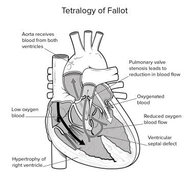
Tetralogy of Fallot
Pulmonary Valve Insufficiency
Pulmonary Valve Stenosis
Pulmonary Atresia
Heart Defects, Congenital
Ventricular Function, Right
Heart Septal Defects, Ventricular
Cyanosis
Trilogy of Fallot
Ventricular Dysfunction, Right
Ventricular Outflow Obstruction
Pulmonary Artery
Postoperative Complications
Pulmonary Subvalvular Stenosis
Abnormalities, Multiple
Echocardiography
Heart Ventricles
Tricuspid Valve
Heart Septal Defects
Hypertrophy, Right Ventricular
Electrocardiography
Tachycardia, Ectopic Junctional
Chromosomes, Human, Pair 22
Dilatation, Pathologic
Truncus Arteriosus, Persistent
Tachycardia, Ventricular
Reoperation
Heart Valve Prosthesis Implantation
Bethanidine
Arrhythmias, Cardiac
Coronary Vessel Anomalies
Echocardiography, Doppler
Cardiac Catheterization
Follow-Up Studies
Treatment Outcome
Malta
Blalock-Taussig Procedure
Magnetic Resonance Imaging, Cine
Double Outlet Right Ventricle
Stroke Volume
Cardiopulmonary Bypass
Transposition of Great Vessels
Heart Septal Defects, Atrial
Balloon Valvuloplasty
Retrospective Studies
Outcome of pregnancy in women with congenital shunt lesions. (1/500)
OBJECTIVE: To evaluate the outcome of pregnancy in women with congenital shunt lesions. SETTING: Retrospective study in a tertiary care centre. METHODS: Pregnancy history was obtained by a standardised questionnaire and medical records were reviewed. PATIENTS: 175 women were identified, at a mean (SD) age of 42 (14) years. Pregnancies occurred in 126 women: 50 with an atrial septal defect, 22 with a ventricular septal defect, 22 with an atrioventricular septal defect, 19 with tetralogy of Fallot, and 13 with other complex shunt lesions. RESULTS: 309 pregnancies were reported by 126 woman (2.5 (1.6) pregnancies per woman). The shortening fraction of the systemic ventricle was 40 (8)%, and 98% were in New York Heart Association class I-II at last follow up. Spontaneous abortions occurred in 17% of pregnancies (abortion rate, 0.4 (0.9) per woman). Gestational age of the 241 newborn infants was 8.8 (0.8) months. There were no maternal deaths related to pregnancy. Pre-eclampsia and embolic events were observed in 1.3% and 0.6%, respectively of all pregnancies. Women with complex shunt lesions more often underwent caesarean section (70% v 15-30%, p = 0.005) and gave birth to smaller babies for equivalent gestation (2577 (671) g v 3016 (572) to 3207 (610) g, p < 0.05). The recurrence risk of congenital heart disease was 2.5%. CONCLUSIONS: The outcome of pregnancy is favourable in women with congenital shunt lesions if their functional class and their systolic ventricular function are good. Such patients can be reassured. (+info)Echocardiographic and morphological correlations in tetralogy of Fallot. (2/500)
AIMS: Our aim was to clarify the location and structure of the outlet septum relative to the free-standing subpulmonary infundibulum in the setting of tetralogy of Fallot and to examine its relationship to the other components of the subpulmonary outflow tract, determining their potential influence on clinical outcome. METHODS AND RESULTS: We studied prospectively 41 patients with tetralogy of Fallot (mean age 14 +/- 10.9 months) prior to surgical repair, and compared them with 15 patients undergoing closure of a ventricular septal defect associated with malalignment of the outlet septum but no subpulmonary infundibular stenosis (Eisenmenger ventricular septal defect), and 20 healthy controls. We also examined available autopsied hearts from cases with uncorrected tetralogy of Fallot (8) and Eisenmenger ventricular septal defect (13). Data were indexed for body surface area, and diameter of the tricuspid valve, respectively. The overall length of the subpulmonary infundibulum, including the extent of the muscular outlet septum, was significantly greater for patients with tetralogy of Fallot compared to normals (2.34 +/- 0.6 vs 1.46 +/- 0.34 cm/BSA0.5, P<0.001), whereas the difference between those with tetralogy of Fallot and an Eisenmenger ventricular septal defect was confined to the degree of narrowing of the subpulmonary outlet (0.43 +/- 0.22 vs 2.17 +/- 0.64 cm/BSA0.5, P<0.001). Within the tetralogy of Fallot group, there were linear relationships between deviation of the outlet septum (r= -0.61, P<0.005) and the diameter of the pulmonary valvar orifice (r=0.75, P<0.001), suggesting that growth of the pulmonary arteries may be related to this feature. When patients requiring a transannular patch as part of their surgical repair were compared with those not needing this procedure, differences were found in the diameter of the pulmonary valvar orifice and the pulmonary trunk, but not in the dimensions of the outlet septum. CONCLUSION: The position of the outlet septum in relationship to the remainder of the muscular subpulmonary infundibulum represents a hallmark of tetralogy of Fallot, permitting its differentiation from Eisenmenger ventricular septal defects and normal hearts. (+info)Decreased left ventricular filling pressure 8 months after corrective surgery in a 55-year-old man with tetralogy of Fallot: adaptation for increased preload. (3/500)
A 55-year-old man with tetralogy of Fallot underwent corrective surgery. Left ventricular filling pressure increased markedly with increased left ventricular volume one month after surgery, then decreased over the next 7 months, presumably due to increased left ventricular compliance. (+info)Disopyramide improves hypoxia in patients with tetralogy of Fallot through a negative inotropic action. (4/500)
The hemodynamic and right ventricular volumetric effects of disopyramide were investigated in patients with tetralogy of Fallot (TF). Intracardiac pressure and oxygen saturation were measured, before and after intravenous administration of disopyramide (2 mg/kg) in 7 patients who had not had previous surgery. Right ventricular volume and the diameter of its outflow tract were analyzed in these 7 and in a further 4 patients with a previous shunt. Aortic oxygen saturation increased from 90.4+/-7.5 (mean+/-SD) to 94.1+/-5.5% (p<0.05) with an increase in pulmonary blood flow and pressure. The systolic pressure gradient between the main pulmonary artery and the right ventricle decreased from 59+/-8 to 42+/-9 mmHg (p<0.01). Aortic pressure fell from 77+/-5 to 67+/-4 mmHg (p<0.05). Systemic vascular resistance increased from 15.3+/-2.2 to 19.4+/-3.3 u x m2 (p<0.05). Pulmonary vascular resistance remained unchanged. The diastolic and systolic diameter indices of the right ventricular outflow tract increased from 17.8+/-3.8 to 20.5+/-3.4 and from 6.5+/-3.0 to 10.4+/-2.2 mm/m2, respectively (p<0.01), whereas the right ventricular ejection fraction decreased. Disopyramide improves systemic oxygen saturation in patients with TF through its negative inotropic action on the right ventricle. (+info)The myocardial profile of the cytosolic isozymes of creatine kinase is apparently not related to cyanosis in congenital heart disease. (5/500)
BACKGROUND: CKMB, the cardiac-specific heterodimer of cytosolic creatine-kinase (CK), is developmentally and physiologically regulated, tissue hypoxia being a proposed regulator. In patients with cyanotic heart disease the myocardium is perfused with partially saturated blood. We questioned whether the myocardium of cyanotic subjects contains higher proportions of CKMB. MATERIALS AND METHODS: CK activity, the distribution of cytosolic CK isozymes, activity of lactic dehydrogenase (LDH), and tissue protein content were determined in obstructive tissues removed at corrective surgery of patients with congenital heart defects. Cyanotic (n = 13) and acyanotic (n = 12) subjects were compared. RESULTS: In cyanotic and acyanotic patients, CK activity was 8.4 +/- 0.6 and 7.6 +/- 0.6 IU/mg protein and the proportion of CKMB was 21 +/- 1.4 and 22 +/- 2. 0% (mean +/- S.E.M), respectively. In the two groups of patients, the activity related to the B subunit corresponded to the steady-state level of the CKBmRNA. The tissue content of protein and the activities of CK and LDH were similar in cyanotic and acyanotic subjects and increased with the age. CONCLUSIONS: The lack of difference in CKMB distribution between the cyanotic and acyanotic patients may either indicate that hypooxygenation is not a regulator of CK isozyme expression, or may be attributed to the already high proportion of this isozyme in hypertrophied, obstructive tissues. Recruitment of additional CKMB, in the cyanotic hearts, may thus not be required. (+info)Accuracy of electrocardiographic and echocardiographic indices in predicting life threatening ventricular arrhythmias in patients operated for tetralogy of Fallot. (6/500)
OBJECTIVE: To validate the accuracy of the prognostic significance of non-invasive clinical diagnostic indices as predictors of sustained ventricular tachycardia (sVT) or fibrillation (VF) in patients undergoing repair for tetralogy of Fallot. METHODS: One way analysis of variance and pairwise comparison of the values with the Bonferroni correction, logistic multivariate analysis, and ordinal logistic analysis were used to study quantitative electrocardiographic and echocardiographic variables in 66 patients who had undergone surgery for tetralogy of Fallot by ventriculotomy at a mean (SD) age of 11.8 (9.5) years. The mean (SD) period of follow up was 16.1 (5.7) years after surgery. RESULTS: Four groups of patients were identified by ECG and 24 hour Holter monitoring: 19 (28.7%) without ventricular arrhythmias, 34 (51.5%) with minor ventricular arrhythmias, seven (10.6%) with non-sustained ventricular tachycardia (nsVT), and six (9.0%) with sVT or VF. One way analysis indicated significant differences in QT dispersion (QTd) and end diastolic volume of the right ventricle (EDVRV) among the groups. Univariate logistic analysis showed EDVRV, QTd, and QRS duration to be significantly associated with sVT or VF. Stepwise multivariate analysis and ordinal logistic analysis showed QTd to be preferable to QRS duration as an indicator, because it was unrelated to EDVRV, and was capable of separating different probability curves for nsVT as opposed to sVT or VF. CONCLUSIONS: Stratification of patients undergoing corrective surgery for tetralogy of Fallot and at risk of life threatening arrhythmias is possible by simple and inexpensive means, which provide sensitive and specific indices. (+info)Reduced heart rate variability following repair of tetralogy of Fallot. (7/500)
OBJECTIVE: To examine autonomic function as assessed by heart rate variability in patients 10 or more years after repair of tetralogy of Fallot, and to relate this to cardiac structure, function, and electrocardiographic indices. METHODS: Heart rate variability was measured by standard time domain techniques on a 24 hour Holter ECG in 28 patients, aged 12 to 34 years (mean 19.5), who had undergone repair of tetralogy of Fallot at least 10 years previously. Echocardiography was performed to assess left ventricular size and function, right ventricular size and pressure, and any proximal pulmonary arterial stenosis. Right ventricular function was evaluated by radionuclide scan. QRS duration, QT interval, and QT dispersion were measured on a standard 12 lead ECG. Measurements of heart rate variability were compared with values from 28 age matched healthy controls (mean age 19.9 years). Interrelations between variables were assessed using Pearson correlation coefficients and stepwise regression analysis. RESULTS: Heart rate variability was reduced, compared with values for age matched normal controls, in 12 of the 28 patients. Reduced heart rate variability was associated with increased age, increased right ventricular size and pressure, and widening of the QRS complex. CONCLUSIONS: Reduced heart rate variability is a feature following repair of tetralogy of Fallot. It is associated with increasing age, impaired right ventricular haemodynamics, and widening of the QRS complex. Under these circumstances, reduced heart rate variability may be a marker for deteriorating right ventricular function. Increased QRS duration has been identified as a risk factor for sudden death following repair of tetralogy of Fallot, and impaired cardiac autonomic control may be one of the mechanisms involved. (+info)Tetralogy of Fallot in the fetus: findings at targeted sonography. (8/500)
OBJECTIVES: To evaluate the findings of tetralogy of Fallot in various fetal sonographic views. METHODS: We reviewed the fetal sonograms and medical records of 20 fetuses with prenatal diagnosis of tetralogy of Fallot. We analyzed the indications for targeted sonography, the abnormalities seen in various sonographic views, the postnatal echocardiographic and angiographic findings and autopsy findings. RESULTS: The most common indication for targeted sonography was an abnormal (n = 12) or inadequate (n = 3) finding on sonographic screening in which the abnormality was most frequently found on the three-vessel view (n = 9). The key pathological features of tetralogy of Fallot were uniformly demonstrated in the ventricular outflow tract, three-vessel and short-axis views. The ductus arteriosus was small in 70% of cases and not identifiable in the remaining fetuses. In three of six fetuses with no identifiable ductus, the ductus was shown to be absent at autopsy. The direction of ductal flow was variable. CONCLUSION: The key features of tetralogy of Fallot were always demonstrable in the ventricular outflow tract, three-vessel and short-axis views. The most common reason for referral was the abnormal three-vessel view. (+info)Tetralogy of Fallot is a congenital heart defect that consists of four components: ventricular septal defect (a hole between the lower chambers of the heart), pulmonary stenosis (narrowing of the pulmonary valve and outflow tract), overriding aorta (the aorta lies directly over the ventricular septal defect), and right ventricular hypertrophy (thickening of the right ventricular muscle). This condition results in insufficient oxygenation of the blood, leading to cyanosis (bluish discoloration of the skin and mucous membranes) and other symptoms such as shortness of breath, fatigue, and poor growth. Treatment typically involves surgical repair, which is usually performed during infancy or early childhood.
Pulmonary Valve Insufficiency, also known as Pulmonary Regurgitation, is a cardiac condition in which the pulmonary valve located between the right ventricle and the pulmonary artery does not close properly. This leads to the backward leakage or regurgitation of blood from the pulmonary artery into the right ventricle during diastole, causing an increased volume load on the right ventricle.
The severity of Pulmonary Valve Insufficiency can vary from mild to severe and may be caused by congenital heart defects, infective endocarditis, Marfan syndrome, rheumatic heart disease, or as a result of aging, or following certain cardiac procedures such as pulmonary valvotomy or ventriculostomy.
Mild Pulmonary Valve Insufficiency may not cause any symptoms and may only require periodic monitoring. However, severe Pulmonary Valve Insufficiency can lead to right-sided heart failure, arrhythmias, and other complications if left untreated. Treatment options for Pulmonary Valve Insufficiency include medication, surgical repair or replacement of the pulmonary valve, or a combination of these approaches.
The pulmonary valve, also known as the pulmonic valve, is a semilunar valve located at the exit of the right ventricle of the heart and the beginning of the pulmonary artery. It has three cusps or leaflets that prevent the backflow of blood from the pulmonary artery into the right ventricle during ventricular diastole, ensuring unidirectional flow of blood towards the lungs for oxygenation.
Pulmonary Valve Stenosis is a cardiac condition where the pulmonary valve, located between the right ventricle and the pulmonary artery, has a narrowed opening. This stenosis (narrowing) can cause obstruction of blood flow from the right ventricle to the lungs. The narrowing can be caused by a fusion of the valve leaflets, thickened or calcified valve leaflets, or rarely, a dysplastic valve.
The severity of Pulmonary Valve Stenosis is classified based on the gradient pressure across the valve, which is measured during an echocardiogram. A mild stenosis has a gradient of less than 30 mmHg, moderate stenosis has a gradient between 30-59 mmHg, and severe stenosis has a gradient of 60 mmHg or higher.
Mild Pulmonary Valve Stenosis may not require treatment, while more severe cases may need to be treated with balloon valvuloplasty or surgical valve replacement. If left untreated, Pulmonary Valve Stenosis can lead to right ventricular hypertrophy, heart failure, and other complications.
Pulmonary atresia is a congenital heart defect where the pulmonary valve, which controls blood flow from the right ventricle to the lungs, doesn't form properly and instead of being open, there is a membranous obstruction or atresia. This results in an absence of communication between the right ventricle and the pulmonary artery.
The right ventricle is often small and underdeveloped due to this condition, and blood flow to the lungs can be severely limited. In some cases, there may be additional heart defects present, such as a ventricular septal defect (a hole between the two lower chambers of the heart) or patent ductus arteriosus (an abnormal connection between the pulmonary artery and the aorta).
Pulmonary atresia can range from mild to severe, and treatment options depend on the specific anatomy and physiology of each individual case. Treatment may include medications, catheter-based procedures, or open-heart surgery, and in some cases, a heart transplant may be necessary.
Congenital heart defects (CHDs) are structural abnormalities in the heart that are present at birth. They can affect any part of the heart's structure, including the walls of the heart, the valves inside the heart, and the major blood vessels that lead to and from the heart.
Congenital heart defects can range from mild to severe and can cause various symptoms depending on the type and severity of the defect. Some common symptoms of CHDs include cyanosis (a bluish tint to the skin, lips, and fingernails), shortness of breath, fatigue, poor feeding, and slow growth in infants and children.
There are many different types of congenital heart defects, including:
1. Septal defects: These are holes in the walls that separate the four chambers of the heart. The two most common septal defects are atrial septal defect (ASD) and ventricular septal defect (VSD).
2. Valve abnormalities: These include narrowed or leaky valves, which can affect blood flow through the heart.
3. Obstruction defects: These occur when blood flow is blocked or restricted due to narrowing or absence of a part of the heart's structure. Examples include pulmonary stenosis and coarctation of the aorta.
4. Cyanotic heart defects: These cause a lack of oxygen in the blood, leading to cyanosis. Examples include tetralogy of Fallot and transposition of the great arteries.
The causes of congenital heart defects are not fully understood, but genetic factors and environmental influences during pregnancy may play a role. Some CHDs can be detected before birth through prenatal testing, while others may not be diagnosed until after birth or later in childhood. Treatment for CHDs may include medication, surgery, or other interventions to improve blood flow and oxygenation of the body's tissues.
Right Ventricular Function refers to the ability of the right ventricle (RV) of the heart to receive and eject blood during the cardiac cycle. The right ventricle is one of the four chambers of the heart and is responsible for pumping deoxygenated blood from the body to the lungs for re-oxygenation.
Right ventricular function can be assessed by measuring various parameters such as:
1. Right Ventricular Ejection Fraction (RVEF): It is the percentage of blood that is ejected from the right ventricle during each heartbeat. A normal RVEF ranges from 45-75%.
2. Right Ventricular Systolic Function: It refers to the ability of the right ventricle to contract and eject blood during systole (contraction phase). This can be assessed by measuring the tricuspid annular plane systolic excursion (TAPSE) or tissue Doppler imaging.
3. Right Ventricular Diastolic Function: It refers to the ability of the right ventricle to relax and fill with blood during diastole (relaxation phase). This can be assessed by measuring the right ventricular inflow pattern, tricuspid valve E/A ratio, or deceleration time.
4. Right Ventricular Afterload: It refers to the pressure that the right ventricle must overcome to eject blood into the pulmonary artery. Increased afterload can impair right ventricular function.
Abnormalities in right ventricular function can lead to various cardiovascular conditions such as pulmonary hypertension, heart failure, and arrhythmias.
A ventricular septal defect (VSD) is a type of congenital heart defect that involves a hole in the wall separating the two lower chambers of the heart, the ventricles. This defect allows oxygenated blood from the left ventricle to mix with deoxygenated blood in the right ventricle, leading to inefficient oxygenation of the body's tissues. The size and location of the hole can vary, and symptoms may range from none to severe, depending on the size of the defect and the amount of blood that is able to shunt between the ventricles. Small VSDs may close on their own over time, while larger defects usually require medical intervention, such as medication or surgery, to prevent complications like pulmonary hypertension and heart failure.
Cyanosis is a medical term that refers to the bluish discoloration of the skin and mucous membranes due to an insufficient amount of oxygen in the blood. This occurs when the level of deoxygenated hemoglobin (the form of hemoglobin that has released its oxygen) in the blood is increased, causing a blue or purple tint to appear, especially in the lips, fingertips, and nail beds.
Cyanosis can be central or peripheral. Central cyanosis affects the entire body and results from low levels of oxygen in the arterial blood, often due to heart or lung conditions that impair oxygen exchange. Peripheral cyanosis is localized to the extremities, usually caused by poor circulation or cold exposure, which can lead to sluggish blood flow and slow oxygen uptake in the tissues.
It's important to note that cyanosis may not always be visually apparent, particularly in individuals with darker skin tones. In these cases, other signs of hypoxia (low oxygen levels) should be considered for proper diagnosis and treatment.
Cardiac surgical procedures are operations that are performed on the heart or great vessels (the aorta and vena cava) by cardiothoracic surgeons. These surgeries are often complex and require a high level of skill and expertise. Some common reasons for cardiac surgical procedures include:
1. Coronary artery bypass grafting (CABG): This is a surgery to improve blood flow to the heart in patients with coronary artery disease. During the procedure, a healthy blood vessel from another part of the body is used to create a detour around the blocked or narrowed portion of the coronary artery.
2. Valve repair or replacement: The heart has four valves that control blood flow through and out of the heart. If one or more of these valves become damaged or diseased, they may need to be repaired or replaced. This can be done using artificial valves or valves from animal or human donors.
3. Aneurysm repair: An aneurysm is a weakened area in the wall of an artery that can bulge out and potentially rupture. If an aneurysm occurs in the aorta, it may require surgical repair to prevent rupture.
4. Heart transplantation: In some cases, heart failure may be so severe that a heart transplant is necessary. This involves removing the diseased heart and replacing it with a healthy donor heart.
5. Arrhythmia surgery: Certain types of abnormal heart rhythms (arrhythmias) may require surgical treatment. One such procedure is called the Maze procedure, which involves creating a pattern of scar tissue in the heart to disrupt the abnormal electrical signals that cause the arrhythmia.
6. Congenital heart defect repair: Some people are born with structural problems in their hearts that require surgical correction. These may include holes between the chambers of the heart or abnormal blood vessels.
Cardiac surgical procedures carry risks, including bleeding, infection, stroke, and death. However, for many patients, these surgeries can significantly improve their quality of life and longevity.
Tetralogy of Fallot is a congenital heart defect that consists of four cardiac abnormalities: ventricular septal defect (a hole between the right and left ventricles), pulmonary stenosis (narrowing of the pulmonary valve and outflow tract), overriding aorta (the aorta is positioned over both ventricles instead of just the left one), and right ventricular hypertrophy (thickening of the right ventricular muscle). This condition results in insufficient oxygenation of the blood, causing cyanosis (bluish discoloration of the skin and mucous membranes) and other symptoms such as shortness of breath, fatigue, and poor growth. Treatment typically involves surgical repair, usually done during infancy or early childhood.
Right ventricular dysfunction is a condition characterized by the impaired ability of the right ventricle (one of the two pumping chambers in the heart) to fill with blood during the diastolic phase or eject blood during the systolic phase. This results in reduced cardiac output from the right ventricle, which can lead to various complications such as fluid accumulation in the body, particularly in the abdomen and lower extremities, and ultimately congestive heart failure if left untreated.
Right ventricular dysfunction can be caused by various factors, including damage to the heart muscle due to a heart attack, high blood pressure in the lungs (pulmonary hypertension), chronic lung diseases, congenital heart defects, viral infections, and certain medications. Symptoms of right ventricular dysfunction may include shortness of breath, fatigue, swelling in the legs, ankles, or abdomen, and a decreased tolerance for physical activity.
Diagnosis of right ventricular dysfunction typically involves a combination of medical history, physical examination, imaging tests such as echocardiography, cardiac MRI, or CT scan, and other diagnostic procedures such as electrocardiogram (ECG) or cardiac catheterization. Treatment options depend on the underlying cause but may include medications to reduce fluid buildup, improve heart function, and manage symptoms, as well as lifestyle modifications such as reducing salt intake and increasing physical activity levels. In severe cases, more invasive treatments such as surgery or implantable devices like pacemakers or ventricular assist devices may be necessary.
Ventricular outflow obstruction is a term used in cardiology to describe a condition where there is an obstruction or narrowing in the flow of blood as it exits the heart's ventricles (the lower chambers of the heart). This obstruction can occur due to various reasons such as congenital heart defects, hypertrophic cardiomyopathy, or calcification of the aortic valve.
In a normal heart, the left ventricle pumps oxygenated blood into the aorta through the aortic valve, and the right ventricle pumps deoxygenated blood into the pulmonary artery through the pulmonic valve. Any obstruction in these outflow tracts can lead to increased pressure within the ventricles, which can result in various symptoms such as shortness of breath, chest pain, dizziness, or fatigue.
The severity of the obstruction and the resulting symptoms can vary depending on the location and extent of the narrowing. Treatment options may include medications, surgical procedures, or catheter-based interventions to alleviate the obstruction and improve blood flow.
The pulmonary artery is a large blood vessel that carries deoxygenated blood from the right ventricle of the heart to the lungs for oxygenation. It divides into two main branches, the right and left pulmonary arteries, which further divide into smaller vessels called arterioles, and then into a vast network of capillaries in the lungs where gas exchange occurs. The thin walls of these capillaries allow oxygen to diffuse into the blood and carbon dioxide to diffuse out, making the blood oxygen-rich before it is pumped back to the left side of the heart through the pulmonary veins. This process is crucial for maintaining proper oxygenation of the body's tissues and organs.
Angiocardiography is a medical procedure used to examine the heart and blood vessels, particularly the chambers of the heart and the valves between them. It involves injecting a contrast agent into the bloodstream and taking X-ray images as the agent flows through the heart. This allows doctors to visualize any abnormalities such as blockages, narrowing, or leakage in the heart valves or blood vessels.
There are different types of angiocardiography, including:
* Left heart catheterization (LHC): A thin tube called a catheter is inserted into a vein in the arm or groin and threaded through to the left side of the heart to measure pressure and oxygen levels.
* Right heart catheterization (RHC): Similar to LHC, but the catheter is threaded through to the right side of the heart to measure pressure and oxygen levels there.
* Selective angiocardiography: A catheter is used to inject the contrast agent into specific blood vessels or chambers of the heart to get a more detailed view.
Angiocardiography can help diagnose and evaluate various heart conditions, including congenital heart defects, coronary artery disease, cardiomyopathy, and valvular heart disease. It is an invasive procedure that carries some risks, such as bleeding, infection, and damage to blood vessels or heart tissue. However, it can provide valuable information for diagnosing and treating heart conditions.
Postoperative complications refer to any unfavorable condition or event that occurs during the recovery period after a surgical procedure. These complications can vary in severity and may include, but are not limited to:
1. Infection: This can occur at the site of the incision or inside the body, such as pneumonia or urinary tract infection.
2. Bleeding: Excessive bleeding (hemorrhage) can lead to a drop in blood pressure and may require further surgical intervention.
3. Blood clots: These can form in the deep veins of the legs (deep vein thrombosis) and can potentially travel to the lungs (pulmonary embolism).
4. Wound dehiscence: This is when the surgical wound opens up, which can lead to infection and further complications.
5. Pulmonary issues: These include atelectasis (collapsed lung), pneumonia, or respiratory failure.
6. Cardiovascular problems: These include abnormal heart rhythms (arrhythmias), heart attack, or stroke.
7. Renal failure: This can occur due to various reasons such as dehydration, blood loss, or the use of certain medications.
8. Pain management issues: Inadequate pain control can lead to increased stress, anxiety, and decreased mobility.
9. Nausea and vomiting: These can be caused by anesthesia, opioid pain medication, or other factors.
10. Delirium: This is a state of confusion and disorientation that can occur in the elderly or those with certain medical conditions.
Prompt identification and management of these complications are crucial to ensure the best possible outcome for the patient.
Pulmonary subvalvular stenosis is a rare cardiac condition that refers to the narrowing or obstruction of the pulmonary valve or the outflow tract below it, within the right ventricle of the heart. This results in restricted blood flow from the right ventricle to the pulmonary artery and subsequently to the lungs.
The narrowing can be caused by various factors such as a membranous shelf-like structure (dysplasia), a fibrous ring, or a tunnel-like narrowing of the outflow tract (tunneling). The severity of the stenosis may vary from mild to severe, and symptoms can range from shortness of breath, fatigue, and chest pain to more serious complications like heart failure or arrhythmias.
Diagnosis typically involves imaging tests such as echocardiography, cardiac MRI, or cardiac catheterization. Treatment options depend on the severity of the stenosis and may include monitoring, medications, or invasive procedures such as balloon dilation or surgical repair.
Cineangiography is a medical imaging technique used to visualize the blood flow in the heart and cardiovascular system. It involves the injection of a contrast agent into the bloodstream while X-ray images are taken in quick succession, creating a movie-like sequence that shows the movement of the contrast through the blood vessels and chambers of the heart. This technique is often used to diagnose and evaluate various heart conditions, such as coronary artery disease, valvular heart disease, and congenital heart defects.
The procedure typically involves threading a catheter through a blood vessel in the arm or leg and guiding it to the heart. Once in place, the contrast agent is injected, and X-ray images are taken using a specialized X-ray machine called a fluoroscope. The images captured during cineangiography can help doctors identify areas of narrowing or blockage in the coronary arteries, abnormalities in heart valves, and other cardiovascular problems.
Cineangiography is an invasive procedure that carries some risks, such as bleeding, infection, and reactions to the contrast agent. However, it can provide valuable information for diagnosing and treating heart conditions, and may be recommended when other diagnostic tests have been inconclusive.
'Abnormalities, Multiple' is a broad term that refers to the presence of two or more structural or functional anomalies in an individual. These abnormalities can be present at birth (congenital) or can develop later in life (acquired). They can affect various organs and systems of the body and can vary greatly in severity and impact on a person's health and well-being.
Multiple abnormalities can occur due to genetic factors, environmental influences, or a combination of both. Chromosomal abnormalities, gene mutations, exposure to teratogens (substances that cause birth defects), and maternal infections during pregnancy are some of the common causes of multiple congenital abnormalities.
Examples of multiple congenital abnormalities include Down syndrome, Turner syndrome, and VATER/VACTERL association. Acquired multiple abnormalities can result from conditions such as trauma, infection, degenerative diseases, or cancer.
The medical evaluation and management of individuals with multiple abnormalities depend on the specific abnormalities present and their impact on the individual's health and functioning. A multidisciplinary team of healthcare professionals is often involved in the care of these individuals to address their complex needs.
Echocardiography is a medical procedure that uses sound waves to produce detailed images of the heart's structure, function, and motion. It is a non-invasive test that can help diagnose various heart conditions, such as valve problems, heart muscle damage, blood clots, and congenital heart defects.
During an echocardiogram, a transducer (a device that sends and receives sound waves) is placed on the chest or passed through the esophagus to obtain images of the heart. The sound waves produced by the transducer bounce off the heart structures and return to the transducer, which then converts them into electrical signals that are processed to create images of the heart.
There are several types of echocardiograms, including:
* Transthoracic echocardiography (TTE): This is the most common type of echocardiogram and involves placing the transducer on the chest.
* Transesophageal echocardiography (TEE): This type of echocardiogram involves passing a specialized transducer through the esophagus to obtain images of the heart from a closer proximity.
* Stress echocardiography: This type of echocardiogram is performed during exercise or medication-induced stress to assess how the heart functions under stress.
* Doppler echocardiography: This type of echocardiogram uses sound waves to measure blood flow and velocity in the heart and blood vessels.
Echocardiography is a valuable tool for diagnosing and managing various heart conditions, as it provides detailed information about the structure and function of the heart. It is generally safe, non-invasive, and painless, making it a popular choice for doctors and patients alike.
The heart ventricles are the two lower chambers of the heart that receive blood from the atria and pump it to the lungs or the rest of the body. The right ventricle pumps deoxygenated blood to the lungs, while the left ventricle pumps oxygenated blood to the rest of the body. Both ventricles have thick, muscular walls to generate the pressure necessary to pump blood through the circulatory system.
The tricuspid valve is the heart valve that separates the right atrium and the right ventricle in the human heart. It is called "tricuspid" because it has three leaflets or cusps, which are also referred to as flaps or segments. These cusps are named anterior, posterior, and septal. The tricuspid valve's function is to prevent the backflow of blood from the ventricle into the atrium during systole, ensuring unidirectional flow of blood through the heart.
A heart septal defect is a type of congenital heart defect, which means it is present at birth. It involves an abnormal opening in the septum, the wall that separates the two sides of the heart. This opening allows oxygen-rich blood to leak into the oxygen-poor blood chambers in the heart.
There are several types of heart septal defects, including:
1. Atrial Septal Defect (ASD): A hole in the atrial septum, the wall between the two upper chambers of the heart (the right and left atria).
2. Ventricular Septal Defect (VSD): A hole in the ventricular septum, the wall between the two lower chambers of the heart (the right and left ventricles).
3. Atrioventricular Septal Defect (AVSD): A combination of an ASD and a VSD, often accompanied by malformation of the mitral and/or tricuspid valves.
The severity of a heart septal defect depends on the size of the opening and its location in the septum. Small defects may cause no symptoms and may close on their own over time. Larger defects can lead to complications, such as heart failure, pulmonary hypertension, or infective endocarditis, and may require medical or surgical intervention.
Right ventricular hypertrophy (RVH) is a medical condition characterized by an enlargement and thickening (hypertrophy) of the right ventricle of the heart. The right ventricle is one of the four chambers of the heart that is responsible for pumping deoxygenated blood to the lungs through the pulmonary artery.
In response to increased workload or pressure overload, such as in chronic lung diseases, pulmonary hypertension, or congenital heart defects, the right ventricle may undergo hypertrophy. This results in an increase in the size and thickness of the right ventricular muscle, which can impair its ability to fill with blood and pump it efficiently to the lungs.
RVH can be diagnosed through various tests, including electrocardiogram (ECG), echocardiography, cardiac magnetic resonance imaging (MRI), or cardiac catheterization. Treatment of RVH depends on the underlying cause and may include medications, oxygen therapy, surgery, or other interventions to reduce the workload on the right ventricle and improve its function.
Electrocardiography (ECG or EKG) is a medical procedure that records the electrical activity of the heart. It provides a graphic representation of the electrical changes that occur during each heartbeat. The resulting tracing, called an electrocardiogram, can reveal information about the heart's rate and rhythm, as well as any damage to its cells or abnormalities in its conduction system.
During an ECG, small electrodes are placed on the skin of the chest, arms, and legs. These electrodes detect the electrical signals produced by the heart and transmit them to a machine that amplifies and records them. The procedure is non-invasive, painless, and quick, usually taking only a few minutes.
ECGs are commonly used to diagnose and monitor various heart conditions, including arrhythmias, coronary artery disease, heart attacks, and electrolyte imbalances. They can also be used to evaluate the effectiveness of certain medications or treatments.
Tachycardia refers to a rapid heart rate, typically defined as over 100 beats per minute in adults. Ectopic junctional tachycardia (EJT) is a specific type of abnormal heart rhythm that originates from the junction between the atria (the upper chambers of the heart) and ventricles (the lower chambers).
In EJT, the electrical impulse arises from an ectopic focus (an area outside of the normal conduction system) located in or near the atrioventricular (AV) node. This results in a rapid heart rate that can range from 100 to 250 beats per minute.
EJT is often seen in patients after cardiac surgery, and it can also occur in other conditions such as myocarditis, digoxin toxicity, or following congenital heart disease repair. It may cause symptoms such as palpitations, shortness of breath, chest discomfort, or dizziness. Treatment options for EJT include medications, cardioversion, or ablation therapy, depending on the underlying cause and severity of symptoms.
A newborn infant is a baby who is within the first 28 days of life. This period is also referred to as the neonatal period. Newborns require specialized care and attention due to their immature bodily systems and increased vulnerability to various health issues. They are closely monitored for signs of well-being, growth, and development during this critical time.
Human chromosome pair 22 consists of two rod-shaped structures present in the nucleus of each cell in the human body. Each chromosome is made up of DNA tightly coiled around histone proteins, forming a complex structure called a chromatin.
Chromosome pair 22 is one of the 22 autosomal pairs of human chromosomes, meaning they are not sex chromosomes (X or Y). Chromosome 22 is the second smallest human chromosome, with each arm of the chromosome designated as p and q. The short arm is labeled "p," and the long arm is labeled "q."
Chromosome 22 contains several genes that are associated with various genetic disorders, including DiGeorge syndrome, velocardiofacial syndrome, and cat-eye syndrome, which result from deletions or duplications of specific regions on the chromosome. Additionally, chromosome 22 is the location of the NRXN1 gene, which has been associated with an increased risk for autism spectrum disorder (ASD) and schizophrenia when deleted or disrupted.
Understanding the genetic makeup of human chromosome pair 22 can provide valuable insights into human genetics, evolution, and disease susceptibility, as well as inform medical diagnoses, treatments, and research.
Pathologic dilatation refers to an abnormal and excessive widening or enlargement of a body cavity or organ, which can result from various medical conditions. This abnormal dilation can occur in different parts of the body, including the blood vessels, digestive tract, airways, or heart chambers.
In the context of the cardiovascular system, pathologic dilatation may indicate a weakening or thinning of the heart muscle, leading to an enlarged chamber that can no longer pump blood efficiently. This condition is often associated with various heart diseases, such as cardiomyopathy, valvular heart disease, or long-standing high blood pressure.
In the gastrointestinal tract, pathologic dilatation may occur due to mechanical obstruction, neuromuscular disorders, or inflammatory conditions that affect the normal motility of the intestines. Examples include megacolon in Hirschsprung's disease, toxic megacolon in ulcerative colitis, or volvulus (twisting) of the bowel.
Pathologic dilatation can lead to various complications, such as reduced organ function, impaired circulation, and increased risk of infection or perforation. Treatment depends on the underlying cause and may involve medications, surgery, or other interventions to address the root problem and prevent further enlargement.
Persistent Truncus Arteriosus is a rare congenital heart defect that is characterized by the failure of the truncus arteriosus to divide into the separate pulmonary artery and aorta during fetal development. This results in a single large vessel, the truncus arteriosus, which gives rise to both the systemic and pulmonary circulations.
The truncus arteriosus contains a single semilunar valve, instead of the two separate semilunar valves (pulmonary and aortic) found in a normal heart. Additionally, there is often a ventricular septal defect (VSD), a hole in the wall between the two lower chambers of the heart, present.
This condition leads to mixing of oxygenated and deoxygenated blood within the truncus arteriosus, resulting in cyanosis (bluish discoloration of the skin and mucous membranes) and decreased oxygen delivery to the body. Symptoms typically appear soon after birth and may include difficulty breathing, poor feeding, rapid heart rate, and failure to thrive.
Persistent truncus arteriosus is usually treated with surgical repair in infancy or early childhood to separate the pulmonary and systemic circulations, close the VSD, and reconstruct the great vessels as needed.
Cardiovascular surgical procedures refer to a range of surgeries performed on the heart and blood vessels to treat or manage various cardiovascular conditions. These surgeries can be open or minimally invasive, and they aim to correct structural abnormalities, improve blood flow, or replace damaged or diseased parts of the cardiovascular system.
Some common types of cardiovascular surgical procedures include:
1. Coronary artery bypass grafting (CABG): This surgery involves taking a healthy blood vessel from another part of the body and using it to create a detour around a blocked or narrowed coronary artery, improving blood flow to the heart muscle.
2. Heart valve repair or replacement: When one or more heart valves become damaged or diseased, they may not open or close properly, leading to reduced blood flow or leakage of blood backward through the valve. In these cases, surgeons may repair or replace the affected valve with a mechanical or biological prosthetic valve.
3. Aneurysm repair: An aneurysm is a weakened area in the wall of an artery that can bulge and potentially rupture, causing severe bleeding. Surgeons can repair an aneurysm by reinforcing the weakened area with a graft or by replacing the affected section of the blood vessel.
4. Heart transplant: In cases where heart failure is irreversible and all other treatment options have been exhausted, a heart transplant may be necessary. This procedure involves removing the damaged heart and replacing it with a healthy donor heart.
5. Ventricular assist devices (VADs): These are mechanical pumps that can be implanted to help support heart function in patients with advanced heart failure who are not candidates for heart transplants. VADs can help improve blood flow, reduce symptoms, and increase the patient's quality of life.
6. Minimally invasive procedures: Advances in technology have led to the development of several minimally invasive cardiovascular surgical procedures, such as robotic-assisted heart surgery, video-assisted thoracoscopic surgery (VATS), and transcatheter aortic valve replacement (TAVR). These techniques typically involve smaller incisions, reduced blood loss, shorter hospital stays, and faster recovery times compared to traditional open-heart surgeries.
Ventricular Tachycardia (VT) is a rapid heart rhythm that originates from the ventricles, the lower chambers of the heart. It is defined as three or more consecutive ventricular beats at a rate of 120 beats per minute or greater in a resting adult. This abnormal heart rhythm can cause the heart to pump less effectively, leading to inadequate blood flow to the body and potentially life-threatening conditions such as hypotension, shock, or cardiac arrest.
VT can be classified into three types based on its duration, hemodynamic stability, and response to treatment:
1. Non-sustained VT (NSVT): It lasts for less than 30 seconds and is usually well tolerated without causing significant symptoms or hemodynamic instability.
2. Sustained VT (SVT): It lasts for more than 30 seconds, causes symptoms such as palpitations, dizziness, shortness of breath, or chest pain, and may lead to hemodynamic instability.
3. Pulseless VT: It is a type of sustained VT that does not produce a pulse, blood pressure, or adequate cardiac output, requiring immediate electrical cardioversion or defibrillation to restore a normal heart rhythm.
VT can occur in people with various underlying heart conditions such as coronary artery disease, cardiomyopathy, valvular heart disease, congenital heart defects, and electrolyte imbalances. It can also be triggered by certain medications, substance abuse, or electrical abnormalities in the heart. Prompt diagnosis and treatment of VT are crucial to prevent complications and improve outcomes.
The postoperative period is the time following a surgical procedure during which the patient's response to the surgery and anesthesia is monitored, and any complications or adverse effects are managed. This period can vary in length depending on the type of surgery and the individual patient's needs, but it typically includes the immediate recovery phase in the post-anesthesia care unit (PACU) or recovery room, as well as any additional time spent in the hospital for monitoring and management of pain, wound healing, and other aspects of postoperative care.
The goals of postoperative care are to ensure the patient's safety and comfort, promote optimal healing and rehabilitation, and minimize the risk of complications such as infection, bleeding, or other postoperative issues. The specific interventions and treatments provided during this period will depend on a variety of factors, including the type and extent of surgery performed, the patient's overall health and medical history, and any individualized care plans developed in consultation with the patient and their healthcare team.
A reoperation is a surgical procedure that is performed again on a patient who has already undergone a previous operation for the same or related condition. Reoperations may be required due to various reasons, such as inadequate initial treatment, disease recurrence, infection, or complications from the first surgery. The nature and complexity of a reoperation can vary widely depending on the specific circumstances, but it often carries higher risks and potential complications compared to the original operation.
Heart valve prosthesis implantation is a surgical procedure where an artificial heart valve is inserted to replace a damaged or malfunctioning native heart valve. This can be necessary for patients with valvular heart disease, including stenosis (narrowing) or regurgitation (leaking), who do not respond to medical management and are at risk of heart failure or other complications.
There are two main types of artificial heart valves used in prosthesis implantation: mechanical valves and biological valves. Mechanical valves are made of synthetic materials, such as carbon and metal, and can last a long time but require lifelong anticoagulation therapy to prevent blood clots from forming. Biological valves, on the other hand, are made from animal or human tissue and typically do not require anticoagulation therapy but may have a limited lifespan and may need to be replaced in the future.
The decision to undergo heart valve prosthesis implantation is based on several factors, including the patient's age, overall health, type and severity of valvular disease, and personal preferences. The procedure can be performed through traditional open-heart surgery or minimally invasive techniques, such as robotic-assisted surgery or transcatheter aortic valve replacement (TAVR). Recovery time varies depending on the approach used and individual patient factors.
Bethanidine is a non-cardioselective, moderately potent, short-acting antihypertensive drug. It belongs to the class of medications known as ganglionic blockers, which work by blocking the action of certain nerves in the body, leading to a decrease in blood pressure.
Bethanidine is used to treat high blood pressure and has been used in the management of symptoms associated with congestive heart failure. However, its use has declined over the years due to the availability of safer and more effective antihypertensive medications.
Like other ganglionic blockers, bethanidine can cause side effects such as dry mouth, blurred vision, constipation, difficulty urinating, dizziness, and weakness. It should be used with caution in patients with certain medical conditions, including kidney or liver disease, narrow-angle glaucoma, and bladder neck obstruction.
It is important to note that bethanidine is not commonly used in clinical practice today due to its potential for serious side effects and the availability of safer alternatives.
Cardiac arrhythmias are abnormal heart rhythms that result from disturbances in the electrical conduction system of the heart. The heart's normal rhythm is controlled by an electrical signal that originates in the sinoatrial (SA) node, located in the right atrium. This signal travels through the atrioventricular (AV) node and into the ventricles, causing them to contract and pump blood throughout the body.
An arrhythmia occurs when there is a disruption in this electrical pathway or when the heart's natural pacemaker produces an abnormal rhythm. This can cause the heart to beat too fast (tachycardia), too slow (bradycardia), or irregularly.
There are several types of cardiac arrhythmias, including:
1. Atrial fibrillation: A rapid and irregular heartbeat that starts in the atria (the upper chambers of the heart).
2. Atrial flutter: A rapid but regular heartbeat that starts in the atria.
3. Supraventricular tachycardia (SVT): A rapid heartbeat that starts above the ventricles, usually in the atria or AV node.
4. Ventricular tachycardia: A rapid and potentially life-threatening heart rhythm that originates in the ventricles.
5. Ventricular fibrillation: A chaotic and disorganized electrical activity in the ventricles, which can be fatal if not treated immediately.
6. Heart block: A delay or interruption in the conduction of electrical signals from the atria to the ventricles.
Cardiac arrhythmias can cause various symptoms, such as palpitations, dizziness, shortness of breath, chest pain, and fatigue. In some cases, they may not cause any symptoms and go unnoticed. However, if left untreated, certain types of arrhythmias can lead to serious complications, including stroke, heart failure, or even sudden cardiac death.
Treatment for cardiac arrhythmias depends on the type, severity, and underlying causes. Options may include lifestyle changes, medications, cardioversion (electrical shock therapy), catheter ablation, implantable devices such as pacemakers or defibrillators, and surgery. It is essential to consult a healthcare professional for proper evaluation and management of cardiac arrhythmias.
Coronary vessel anomalies refer to abnormalities in the structure, origin, or course of the coronary arteries or veins. These vessels are responsible for delivering oxygenated blood to the heart muscle. Some common types of coronary vessel anomalies include:
1. Anomalous Origin of the Coronary Artery (AOCA): This occurs when one or both of the coronary arteries originate from an abnormal location in the aorta. The left coronary artery may arise from the right sinus of Valsalva, while the right coronary artery may arise from the left sinus of Valsalva. This can lead to ischemia (reduced blood flow) and potentially life-threatening complications such as sudden cardiac death.
2. Coronary Artery Fistula: A fistula is an abnormal connection between a coronary artery and another chamber or vessel in the heart. Blood flows directly from the high-pressure coronary artery into a low-pressure chamber, bypassing the capillaries and leading to a steal phenomenon where oxygenated blood is diverted away from the heart muscle.
3. Coronary Artery Aneurysm: An aneurysm is a localized dilation or bulging of the coronary artery wall. This can lead to complications such as thrombosis (blood clot formation), embolism (blockage caused by a clot that travels to another location), or rupture, which can be life-threatening.
4. Myocardial Bridge: In this condition, a segment of the coronary artery passes between the muscle fibers of the heart, instead of running along its surface. This can cause compression of the artery during systole (contraction) and lead to ischemia.
5. Kawasaki Disease: Although not strictly an anomaly, Kawasaki disease is a pediatric illness that can result in coronary artery aneurysms and other complications if left untreated.
Coronary vessel anomalies may be asymptomatic or present with symptoms such as chest pain, shortness of breath, palpitations, or syncope (fainting). Diagnosis typically involves imaging techniques such as coronary angiography, computed tomography (CT) angiography, or magnetic resonance angiography. Treatment depends on the specific anomaly and may involve medications, percutaneous interventions, or surgical correction.
Doppler echocardiography is a type of ultrasound test that uses high-frequency sound waves to produce detailed images of the heart and its blood vessels. It measures the direction and speed of blood flow in the heart and major blood vessels leading to and from the heart. This helps to evaluate various conditions such as valve problems, congenital heart defects, and heart muscle diseases.
In Doppler echocardiography, a small handheld device called a transducer is placed on the chest, which emits sound waves that bounce off the heart and blood vessels. The transducer then picks up the returning echoes, which are processed by a computer to create moving images of the heart.
The Doppler effect is used to measure the speed and direction of blood flow. This occurs when the frequency of the sound waves changes as they bounce off moving objects, such as red blood cells. By analyzing these changes, the ultrasound machine can calculate the velocity and direction of blood flow in different parts of the heart.
Doppler echocardiography is a non-invasive test that does not require any needles or dyes. It is generally safe and painless, although patients may experience some discomfort from the pressure applied by the transducer on the chest. The test usually takes about 30 to 60 minutes to complete.
Cardiac catheterization is a medical procedure used to diagnose and treat cardiovascular conditions. In this procedure, a thin, flexible tube called a catheter is inserted into a blood vessel in the arm or leg and threaded up to the heart. The catheter can be used to perform various diagnostic tests, such as measuring the pressure inside the heart chambers and assessing the function of the heart valves.
Cardiac catheterization can also be used to treat certain cardiovascular conditions, such as narrowed or blocked arteries. In these cases, a balloon or stent may be inserted through the catheter to open up the blood vessel and improve blood flow. This procedure is known as angioplasty or percutaneous coronary intervention (PCI).
Cardiac catheterization is typically performed in a hospital cardiac catheterization laboratory by a team of healthcare professionals, including cardiologists, radiologists, and nurses. The procedure may be done under local anesthesia with sedation or general anesthesia, depending on the individual patient's needs and preferences.
Overall, cardiac catheterization is a valuable tool in the diagnosis and treatment of various heart conditions, and it can help improve symptoms, reduce complications, and prolong life for many patients.
Follow-up studies are a type of longitudinal research that involve repeated observations or measurements of the same variables over a period of time, in order to understand their long-term effects or outcomes. In medical context, follow-up studies are often used to evaluate the safety and efficacy of medical treatments, interventions, or procedures.
In a typical follow-up study, a group of individuals (called a cohort) who have received a particular treatment or intervention are identified and then followed over time through periodic assessments or data collection. The data collected may include information on clinical outcomes, adverse events, changes in symptoms or functional status, and other relevant measures.
The results of follow-up studies can provide important insights into the long-term benefits and risks of medical interventions, as well as help to identify factors that may influence treatment effectiveness or patient outcomes. However, it is important to note that follow-up studies can be subject to various biases and limitations, such as loss to follow-up, recall bias, and changes in clinical practice over time, which must be carefully considered when interpreting the results.
Treatment outcome is a term used to describe the result or effect of medical treatment on a patient's health status. It can be measured in various ways, such as through symptoms improvement, disease remission, reduced disability, improved quality of life, or survival rates. The treatment outcome helps healthcare providers evaluate the effectiveness of a particular treatment plan and make informed decisions about future care. It is also used in clinical research to compare the efficacy of different treatments and improve patient care.
I'm sorry for any confusion, but "Malta" is not a medical term. It is a country located in Southern Europe, consisting of an archipelago in the Mediterranean Sea. If you have any questions about medical terminology or concepts, I would be happy to help with those!
The Blalock-Taussig (BT) procedure is a type of open-heart surgery used to treat cyanotic heart diseases in infants and children. It is primarily used as a palliative treatment for Tetralogy of Fallot, Truncus Arteriosus, and other complex congenital heart defects.
In this procedure, a connection is created between the subclavian artery (a major artery that branches off from the aorta) and the pulmonary artery (the blood vessel that carries oxygen-depleted blood from the heart to the lungs). This connection helps increase the amount of oxygen-rich blood flowing to the lungs, which in turn improves the oxygen saturation levels in the body.
The Blalock-Taussig procedure is typically performed as a temporary measure until a more definitive surgical repair can be carried out, usually when the child is older and has grown larger. The connection created during the BT procedure may be maintained using a synthetic tube (shunt) or by directly sewing the subclavian artery to the pulmonary artery.
The Blalock-Taussig procedure was first performed in 1945 by Drs. Alfred Blalock and Helen Taussig at Johns Hopkins Hospital, and it has since become a standard surgical technique for treating cyanotic heart diseases in infants and children.
Magnetic Resonance Imaging (MRI) is a non-invasive diagnostic technique that uses a strong magnetic field and radio waves to create detailed cross-sectional images of the body's internal structures. In MRI, Cine is a specific mode of imaging that allows for the evaluation of moving structures, such as the heart, by acquiring and displaying a series of images in rapid succession. This technique is particularly useful in cardiac imaging, where it can help assess heart function, valve function, and blood flow. The term "Cine" refers to the continuous playback of these images, similar to watching a movie, allowing doctors to evaluate motion and timing within the heart.
Double outlet right ventricle (DORV) is a congenital heart defect in which both great vessels (the aorta and the pulmonary artery) arise from the right ventricle. In a normal heart, the aorta arises from the left ventricle and the pulmonary artery arises from the right ventricle.
In DORV, there is a communication between the two ventricles (a ventricular septal defect), which allows oxygen-rich blood to mix with oxygen-poor blood. The location of this ventricular septal defect and the relationship of the great vessels to each other determine the physiology and the clinical manifestations of DORV.
DORV is a complex congenital heart defect that can range from mild to severe, and it often requires surgical intervention to improve blood flow and oxygenation. The prognosis for individuals with DORV depends on various factors, including the specific type of DORV, associated cardiac anomalies, and the timing and success of treatment.
Stroke volume is a term used in cardiovascular physiology and medicine. It refers to the amount of blood that is pumped out of the left ventricle of the heart during each contraction (systole). Specifically, it is the difference between the volume of blood in the left ventricle at the end of diastole (when the ventricle is filled with blood) and the volume at the end of systole (when the ventricle has contracted and ejected its contents into the aorta).
Stroke volume is an important measure of heart function, as it reflects the ability of the heart to pump blood effectively to the rest of the body. A low stroke volume may indicate that the heart is not pumping efficiently, while a high stroke volume may suggest that the heart is working too hard. Stroke volume can be affected by various factors, including heart disease, high blood pressure, and physical fitness level.
The formula for calculating stroke volume is:
Stroke Volume = End-Diastolic Volume - End-Systolic Volume
Where end-diastolic volume (EDV) is the volume of blood in the left ventricle at the end of diastole, and end-systolic volume (ESV) is the volume of blood in the left ventricle at the end of systole.
Cardiopulmonary bypass (CPB) is a medical procedure that temporarily takes over the functions of the heart and lungs during major heart surgery. It allows the surgeon to operate on a still, bloodless heart.
During CPB, the patient's blood is circulated outside the body with the help of a heart-lung machine. The machine pumps the blood through a oxygenator, where it is oxygenated and then returned to the body. This bypasses the heart and lungs, hence the name "cardiopulmonary bypass."
CPB involves several components, including a pump, oxygenator, heat exchanger, and tubing. The patient's blood is drained from the heart through cannulas (tubes) and passed through the oxygenator, where it is oxygenated and carbon dioxide is removed. The oxygenated blood is then warmed to body temperature in a heat exchanger before being pumped back into the body.
While on CPB, the patient's heart is stopped with the help of cardioplegia solution, which is infused directly into the coronary arteries. This helps to protect the heart muscle during surgery. The surgeon can then operate on a still and bloodless heart, allowing for more precise surgical repair.
After the surgery is complete, the patient is gradually weaned off CPB, and the heart is restarted with the help of electrical stimulation or medication. The patient's condition is closely monitored during this time to ensure that their heart and lungs are functioning properly.
While CPB has revolutionized heart surgery and allowed for more complex procedures to be performed, it is not without risks. These include bleeding, infection, stroke, kidney damage, and inflammation. However, with advances in technology and technique, the risks associated with CPB have been significantly reduced over time.
Transposition of the Great Vessels is a congenital heart defect in which the two main vessels that carry blood from the heart to the rest of the body are switched in position. Normally, the aorta arises from the left ventricle and carries oxygenated blood to the body, while the pulmonary artery arises from the right ventricle and carries deoxygenated blood to the lungs. In transposition of the great vessels, the aorta arises from the right ventricle and the pulmonary artery arises from the left ventricle. This results in oxygen-poor blood being pumped to the body and oxygen-rich blood being recirculated back to the lungs, which can lead to serious health problems and is often fatal if not corrected through surgery soon after birth.
Atrial septal defect (ASD) is a type of congenital heart defect that involves the septum, which is the wall that separates the two upper chambers of the heart (atria). An ASD is a hole or abnormal opening in the atrial septum, allowing oxygen-rich blood to leak into the oxygen-poor blood chambers in the heart. This leads to an overload of blood in the right side of the heart, which can cause enlargement of the heart and increased work for the right ventricle.
ASDs can vary in size, and small defects may not cause any symptoms or require treatment. Larger defects, however, can result in symptoms such as shortness of breath, fatigue, and heart rhythm abnormalities. Over time, if left untreated, ASDs can lead to complications like pulmonary hypertension, atrial fibrillation, and stroke.
Treatment for ASD typically involves surgical closure of the defect or catheter-based procedures using devices to close the hole. The choice of treatment depends on factors such as the size and location of the defect, the patient's age and overall health, and the presence of any coexisting conditions.
Palliative care is a type of medical care that focuses on relieving the pain, symptoms, and stress of serious illnesses. The goal is to improve quality of life for both the patient and their family. It is provided by a team of doctors, nurses, and other specialists who work together to address the physical, emotional, social, and spiritual needs of the patient. Palliative care can be provided at any stage of an illness, alongside curative treatments, and is not dependent on prognosis.
The World Health Organization (WHO) defines palliative care as: "an approach that improves the quality of life of patients and their families facing the problems associated with life-threatening illness, through the prevention and relief of suffering by means of early identification and impeccable assessment and treatment of pain and other problems, physical, psychological and spiritual."
Balloon valvuloplasty is a medical procedure used to treat heart valve stenosis or narrowing. It involves the use of a thin, flexible tube (catheter) with a balloon at its tip, which is guided through a blood vessel to the narrowed heart valve. Once in position, the balloon is inflated to stretch and widen the valve opening, improving blood flow. After the valve is widened, the balloon is deflated and the catheter is removed. This procedure can be performed on various heart valves, including the aortic, mitral, and pulmonary valves.
Retrospective studies, also known as retrospective research or looking back studies, are a type of observational study that examines data from the past to draw conclusions about possible causal relationships between risk factors and outcomes. In these studies, researchers analyze existing records, medical charts, or previously collected data to test a hypothesis or answer a specific research question.
Retrospective studies can be useful for generating hypotheses and identifying trends, but they have limitations compared to prospective studies, which follow participants forward in time from exposure to outcome. Retrospective studies are subject to biases such as recall bias, selection bias, and information bias, which can affect the validity of the results. Therefore, retrospective studies should be interpreted with caution and used primarily to generate hypotheses for further testing in prospective studies.
 Tetralogy of Fallot
Helen B. Taussig
Heart
Multan Institute of Cardiology
List of OMIM disorder codes
Blue baby syndrome
Sussex Spaniel
Squatting position
Trilogy of Fallot
Homeobox protein Nkx-2.5
Subclavian steal syndrome
Phonocardiogram
Riaz Haider
PITX2
Absent pulmonary valve syndrome
Right atrial enlargement
Tinman (gene)
Dennis McEldowney
NEPSY
MYL4
Pulmonary atresia with ventricular septal defect
SNX8
Panangipalli Venugopal
Townes-Brocks syndrome
HAND2
John W. Kirklin
JAG1
Down syndrome
1q21.1 duplication syndrome
1q21.1 deletion syndrome
Tetralogy of Fallot - Wikipedia
 Tetralogy of Fallot: MedlinePlus Medical Encyclopedia
 Tetralogy of Fallot
 Risk stratification in surgically repaired tetralogy of Fallot
 Tetralogy of Fallot With Pulmonary Stenosis Differential Diagnoses
TOF (Tetralogy of Fallot) | Rady Children's Hospital
 tetralogy of Fallot
Prevalence of significant congenital heart defects in children of parents with Fallot's tetralogy
Pregnancy, fertility, and recurrence risk in corrected tetralogy of Fallot | Heart
 Lexi M. - Tetralogy of Fallot | Cardiology Story
 Tetralogy of Fallot: Ethan - Tiny Tickers
Considerations in pursuing the optimal timing for pulmonary valve replacement in repaired tetralogy of Fallot | Heart
The Nager acrofacial dysostosis syndrome with the tetralogy of Fallot. | Journal of Medical Genetics
 Risk stratification of ventricular arrhythmias in repaired tetralogy of Fallot | Revista Española de Cardiología
An excess of tetralogy of Fallot in Malta. | Journal of Epidemiology & Community Health
Tetralogy of Fallot - Erin Ocampo, aged 1½ - Surgery Diary, July 2002
 Information for Fallot's tetralogy
 Table 22. Adults With Complex Congenital Heart Disease [Ebstein's Anomaly, Tetralogy of Fallot, Complex Cyanotic Congenital...
 Noninvasive Predictors of Ventricular Arrhythmias in Patients With Tetralogy of Fallot Undergoing Pulmonary Valve Replacement |...
 CHD | Late gadolinium enhancement and adverse outcomes in a contemporary cohort of adult survivors of tetralogy of Fallot
 Tetralogy of Fallot - Pediatrics - MSD Manual Professional Edition
 An Integrated Platform for Dynamic Cardiac Simulation and Image Processing: Application to Personalised Tetralogy of Fallot...
Using contracting band to improve right ventricle ejection fraction for patients with repaired tetralogy of Fallot: a modeling...
 Tetralogy of Fallot (TOF) - Willis-Knighton Health System - Shreveport - Bossier City - Louisiana
 Tetralogy of Fallot | Riley Children's Health
Tetralogy of Fallot in Dogs & Cats
 7 Questions Answered About Tetralogy of Fallot Surgery | Sri Ramakrishna Hospital
 Utility of Cardiac Magnetic Resonance Imaging in Predicting Atrial Arrhythmias in Repaired Tetralogy of Fallot.
 Unifocalization Heart Animation for tetralogy of Fallot with pulmonary atresia and MAPCAs - Stanford Children's Health -...
 Tetralogy of Fallot - WikEM
Child with tetralogy2
- If a child with tetralogy of Fallot becomes blue, immediately place the child on their side or back and put the knees up to the chest. (medlineplus.gov)
- To the first year of life the child with tetralogy of Fallot is fully formed specific appearance: a bluish tinge is clearly visible not only on the skin and the sclera of the eyes, ears. (vsebolezni.com)
Features of tetralogy2
- The clinical features of tetralogy of Fallot are directly related to the severity of the anatomic defects. (medscape.com)
- The four features of tetralogy of Fallot are: 1. (3danatomyseries.com)
Aorta9
- In tetralogy of Fallot, the aorta is between the left and right ventricles, directly over the VSD. (smartdraw.com)
- Tetralogy of Fallot is characterised by a ventricular septal defect with an overriding aorta and anterior deviation of the outlet septum, creating pulmonary stenosis and resulting in right ventricular hypertrophy. (bmj.com)
- Tetralogy of Fallot consists of 4 features: a large ventricular septal defect, right ventricular outflow tract obstruction and pulmonic valve stenosis, right ventricular hypertrophy, and over-riding of the aorta. (msdmanuals.com)
- In some cases of Tetralogy of Fallot, the pulmonic stenosis is so great, that the easiest path for the blood to take is from the right ventricle, through the hole in the septum to the left ventricle and out the aorta. (ethosvet.com)
- An abnormal arrangement of the aorta characterizes tetralogy of Fallot. (sriramakrishnahospital.com)
- Tetralogy of Fallot, which is one of the most common congenital heart disorders, comprises right ventricular (RV) outflow tract obstruction (RVOTO) (infundibular stenosis), ventricular septal defect (VSD), aorta dextroposition, and RV hypertrophy (see the image below). (medscape.com)
- Patients with tetralogy of Fallot have a VSD, RV infundibular stenosis, RVH, and an enlarged aorta (in about half of patients, it overrides the septum). (medical-definitions.net)
- Tetralogy of Fallot is a congenital heart condition characterized by four specific defects: pulmonary stenosis, hypertrophy of the right ventricule, a ventricular septal defect, and an overriding aorta. (arfaalawgroup.com)
- They determined the relative risk (RR) of having offspring with any CHD, or one of 17 types of severe CHD, or one of the five most common types of severe CHD (univentricular heart, transposition of the great arteries , atrioventricular septum defect, coarctation of the aorta , and Tetralogy of Fallot). (medscape.com)
Symptoms7
- Wide variation in the basic anatomic morphology, pathophysiology, clinical signs and symptoms, and surgical methods of therapy is noted for tetralogy of Fallot (TOF). (medscape.com)
- Sometimes tetralogy of Fallot is not diagnosed until maturity because the abnormalities and symptoms are so mild. (sriramakrishnahospital.com)
- The severity of the blockage in blood flow causes a wide range of symptoms in patients with tetralogy of Fallot. (sriramakrishnahospital.com)
- What are the signs and symptoms of tetralogy of fallot? (medical-definitions.net)
- Signs and symptoms of tetralogy of fallot - Most adults with tetralogy of Fallot who have had surgery to fix it don't have many symptoms unless they develop right heart failure or arrhythmias. (medical-definitions.net)
- The reason why tetralogy of Fallot should be operated on as soon as possible is because its symptoms are severe. (steptohealth.com)
- Symptoms of Tetralogy of Fallot vary at birth, ranging from nonexistent to severe. (arfaalawgroup.com)
Cardiovascular2
- Babies born with tetralogy of fallot must need heart (cardiovascular) surgery to repair the condition. (sriramakrishnahospital.com)
- Report from the Japanese Society of Pediatric Cardiology and Cardiac Surgery Research Committee on Cardiovascular Function in Adult Patients with Congenital Heart Disease: Mechanism of aortic root dilation and cardiovascular function in patients with tetralogy of Fallot. (cvs.com)
Pathophysiology2
Infants4
- Right ventricular outflow tract stenting in tetralogy of Fallot infants with risk factors for early primary repair. (medscape.com)
- Cardiac surgeons at Riley at IU Health employ the newest technologies and innovations in treating tetralogy of Fallot, offering better outcomes for more infants. (rileychildrens.org)
- Available at: https://www.dynamed.com/condition/tetralogy-of-fallot-in-infants-and-children. (cvs.com)
- Most infants with tetralogy of Fallot require some type of surgical procedure. (medscape.com)
Fallot's1
- Cardiac catheterisation led to a diagnosis of Fallot's tetralogy and severe pulmonary oedema. (3danatomyseries.com)
Arthur Fallot2
- A further description was published in 1888 by the French physician Étienne-Louis Arthur Fallot, after whom it is named. (wikipedia.org)
- It is named after Etienne-Louis Arthur Fallot, who first described this condition in 1888. (rileychildrens.org)
Cardiac10
- Tetralogy of Fallot (TOF), formerly known as Steno-Fallot tetralogy, is a congenital heart defect characterized by four specific cardiac defects. (wikipedia.org)
- Thankfully, most forms of TOF and related heart problems can now be successfully treated in patients of all ages and sizes, and most patients can expect to live healthy and full lives.However, when it comes to Tetralogy of Fallot treatment, surgery remains a major cardiac operation that is best treated by surgeons with experience and specific training in newborn and infant heart surgery. (rchsd.org)
- Residual cardiac defects in combination with the circulatory changes observed during pregnancy (increased cardiac output and reduced systemic vascular resistance and blood pressure) may affect pregnancy in women with corrected tetralogy of Fallot. (bmj.com)
- Preventing ventricular arrhythmias and sudden cardiac death in adults with repaired tetralogy of Fallot: a never-ending story? (revespcardiol.org)
- Occasionally, patients with tetralogy of Fallot require more invasive techniques such as cardiac catheterization to help diagnose and plan for surgical repair. (rileychildrens.org)
- Tetralogy of Fallot is an uncommon congenital cardiac abnormality that can affect both cats and dogs. (ethosvet.com)
- Scholars@Duke publication: Utility of Cardiac Magnetic Resonance Imaging in Predicting Atrial Arrhythmias in Repaired Tetralogy of Fallot. (duke.edu)
- The mortality rate in untreated patients reaches 50% by age 6 years, but in the present era of cardiac surgery, children with simple forms of tetralogy of Fallot enjoy good long-term survival with an excellent quality of life. (medscape.com)
- This paper presents an image-based classification method, and applies it to classification of cardiac MRI scans of individuals with Tetralogy of Fallot (TOF). (johnshopkins.edu)
- Extra-cardiac diagnoses and postnatal outcomes of fetal tetralogy of fallot. (bvsalud.org)
Cases of tetralogy2
- Before the 1960s surgical procedures were performed in older children, while nowadays almost all cases of tetralogy of Fallot are corrected during infancy. (bmj.com)
- In most cases of tetralogy of Fallot, the cause is not known although in some patients, genetic factors play a role. (3danatomyseries.com)
Repair of tetralogy4
- Effect of transannular patching on outcome after repair of tetralogy of Fallot. (medscape.com)
- Nollert G, Fischlein T, Bouterwek S, Bohmer C, Klinner W, Reichart B. Long-term survival in patients with repair of tetralogy of Fallot: 36-year follow-up of 490 survivors of the first year after surgical repair. (medscape.com)
- Gellis L, Banka P, Marshall A, Emani S, Porras D. Transcatheter balloon dilation for recurrent right ventricular outflow tract obstruction following valve-sparing repair of tetralogy of Fallot. (medscape.com)
- Total repair of tetralogy of Fallot usually includes a VSD patch and an enlarging RV outflow tract patch. (medical-definitions.net)
Characteristic of tetralogy1
- Additional methods of examination - ultrasound, Doppler ultrasound, heart catheterization - allows you to specify the diagnosis, identifying the main changes in the heart and direction of blood flow, which is characteristic of tetralogy of Fallot. (vsebolezni.com)
Diagnosis5
- A diagnosis can be made by an echocardiogram, which is an ultrasound that shows the structural abnormalities associated with tetralogy of Fallot. (rileychildrens.org)
- Diagnosis of tetralogy of Fallot typically occurs within the first few months of life. (sriramakrishnahospital.com)
- Despite their "awareness" of, tetralogy of Fallot needs to clarify the diagnosis. (vsebolezni.com)
- Tetralogy of Fallot can cause fatal complications if it is left untreated, and a prompt diagnosis is essential to a good prognosis. (arfaalawgroup.com)
- After trying to get pregnant for years, this family had to deal with a difficult diagnosis of tetralogy of Fallot at their scheduled pre-natal visit. (relx.com)
Patients16
- Indeed, sudden death of presumed arrhythmic etiology is the most frequent mode of demise in patients with surgically repaired tetralogy of Fallot, often in early-to-mid adulthood. (nih.gov)
- This review highlights some of the complexities involved in risk stratifying patients with tetralogy of Fallot, underscoring the importance of avoiding oversimplification by means of a rigid algorithm that dictates therapy. (nih.gov)
- Shah RR, Poommipanit P, Law MA, Amin Z. Anchor balloon, buddy wire, and wire and sheath techniques to deploy percutaneous pulmonary valves in tetralogy of fallot patients. (medscape.com)
- Contemporary predictors of death and sustained ventricular tachycardia in patients with repaired tetralogy of Fallot enrolled in the INDICATOR cohort. (medscape.com)
- Although complications did occur in five of 26 (19%) of the patients with a corrected tetralogy of Fallot, pregnancy was generally well tolerated in this largest report so far. (bmj.com)
- In developed countries tetralogy of Fallot has been corrected surgically in almost all patients. (bmj.com)
- 1- 3 Although patients with corrected tetralogy of Fallot achieve a good quality of life, late complications do occur in 10-15% at 20 years after the initial repair, of which the most important are right sided heart failure caused by pulmonary regurgitation and supraventricular or ventricular arrhythmia. (bmj.com)
- We have read the interesting article from Bokma and colleagues 1 documenting the outcomes of pulmonary valve replacement (PVR) in patients with repaired tetralogy of Fallot (rTOF). (bmj.com)
- Risk stratification of ventricular arrhythmias in patients with repaired tetralogy of Fallot (rTOF) remains unresolved. (revespcardiol.org)
- Objectives: This study sought to test the hypothesis that a vectorcardiographic parameter, the QRS vector magnitude (QRSVm), can risk stratify those patients at risk for sustained spontaneous ventricular arrhythmias (VAs) or ventricular arrhythmia inducibility (VAI) in a large cohort of patients with tetralogy of Fallot (TOF). (lu.se)
- Using contracting band to improve right ventricle ejection fraction for patients with repaired tetralogy of Fallot: a modeling study using patient-specific CMR-based 2-layer anisotropic models of human right and left ventricles. (umassmed.edu)
- Arrhythmias are the leading cause of morbidity and mortality in repaired tetralogy of Fallot (TOF), and over 20% of these patients will develop a sustained atrial arrhythmia during their lifetimes. (duke.edu)
- 90% of untreated tetralogy patients succumb by the age of 10 years 6. (impedia.net)
- Tetralogy of Fallot or TOF is a congenital heart condition where patients have four heart abnormalities. (osmosis.org)
- Tetralogy of Fallot in adult patients: a case study) points out. (steptohealth.com)
- Influence of staged repair and primary repair on outcomes in patients with complete atrioventricular septal defect and tetralogy of Fallot: a systematic review and meta-analysis. (nih.gov)
Cyanosis4
- In some children with unrepaired tetralogy of Fallot, most often those several months up to 2 years of age, sudden episodes of profound cyanosis and hypoxia (hypercyanotic or "tet" spells) may occur, which may be lethal. (msdmanuals.com)
- In cases where outflow obstruction is minimal, cyanosis may be inapparent (pink tetralogy) resulting in delayed presentation, even into adulthood, although this is rare. (impedia.net)
- Specialist, just glancing briefly at such a child, I would say that the reason "cinemacast" is most likely tetralogy of Fallot - a congenital anomaly of the heart, which refer to "blue" vices due to pronounced cyanosis. (vsebolezni.com)
- Tetralogy of Fallot is the single most common form of congenital heart disease that causes cyanosis (bluish discoloration from lack of oxygen). (relx.com)
Surgical4
- The structural defects associated with tetralogy of Fallot require surgical repair. (rileychildrens.org)
- MEDICAL ANIMATION TRANSCRIPT: The treatments for Tetralogy of Fallot are surgical procedures to help fix the following four heart problems. (nucleusmedicalmedia.com)
- Contemporary surgical management of complete atrioventricular septal defect with tetralogy of Fallot in Japan. (nih.gov)
- Complete atrioventricular septal defect associated with tetralogy of fallot: surgical indications and results. (nih.gov)
Arrhythmias2
- Arya S, Kovach J, Singh H, Karpawich PP. Arrhythmias and sudden death among older children and young adults following tetralogy of Fallot repair in the current era: are previously reported risk factors still applicable? (medscape.com)
- Low incidence of arrhythmias in the right ventricular infundibulum sparing approach to tetralogy of Fallot repair. (medscape.com)
Defects8
- People with tetralogy of Fallot are more likely to also have other congenital defects. (medlineplus.gov)
- Children with tetralogy of Fallot are more likely to have chromosome disorders, such as Down syndrome, Alagille syndrome, and DiGeorge syndrome (a condition that causes heart defects, low calcium levels, and poor immune function). (medlineplus.gov)
- The condition is known as a tetralogy for the perfect storm of four heart defects that commonly occur together. (chp.edu)
- Tetralogy of Fallot is characterized by four heart defects that cause insufficient blood to reach the lungs and oxygen-depleted blood to be sent to the body. (rileychildrens.org)
- Tetralogy of Fallot is a combination of four heart defects that alter the flow of blood through the heart. (rileychildrens.org)
- Available at: https://www.heart.org/en/health-topics/congenital-heart-defects/about-congenital-heart-defects/tetralogy-of-fallot. (cvs.com)
- Tetralogy of Fallot affects the blood flow in the heart and is made up of four different heart defects: VSD Pulmonary stenosis: A narrowing of the pulmonary valve and main pulmonary artery. (rainbowkids.com)
- Tetralogy of Fallot (TOF) is a set of 4 congenital heart defects that happen together. (chkd.org)
20192
- 2019. https://evidence.unboundmedicine.com/evidence/view/EBMG/453753/all/Tetralogy_of_Fallot___Image. (unboundmedicine.com)
- Recuperado en 31 de marzo de 2019, de http://www.scielo.org.mx/scielo.php?script=sci_arttext &pid=S0188-21982013000200004&lng=es&tlng=es. (steptohealth.com)
Typically3
- The cause of tetralogy of Fallot is typically not known. (wikipedia.org)
- Surgery to repair tetralogy of Fallot is done when the infant is very young, typically before 6 months of age. (medlineplus.gov)
- Tetralogy of Fallot is typically diagnosed via imaging studies. (arfaalawgroup.com)
Pulmonary stenosis3
- Other conditions to consider when evaluating a patient with suspected Tetralogy of Fallot with pulmonary stenosis include acute anemia, asthma and reactive airway disease, bacteremia and sepsis, cardiogenic shock, Ebstein malformation of the tricuspid valve, pseudotruncus arteriosus, pulmonary atresia, septic shock, and ventricular septal defect (VSD). (medscape.com)
- Hill G. Repair and follow-up of Tetralogy of Fallot with pulmonary stenosis. (medscape.com)
- This condition causes infundibular pulmonary stenosis (IPS), as the article Tetralogy of Fallot: Case report and literature review points out. (steptohealth.com)
Pentalogy2
- Sometimes the condition is called pentalogy of Fallot," says Dr. Drickman. (chp.edu)
- If there is an ASD, the whole thing is called the "pentalogy of Fallot. (medical-definitions.net)
Cyanotic5
- Tetralogy of Fallot is rare, but it is the most common form of cyanotic congenital heart disease. (medlineplus.gov)
- Stenting of the right ventricular outflow tract in the high-risk infant with cyanotic teratology of Fallot. (medscape.com)
- Tetralogy of Fallot is a common cause of cyanotic episodes, making a child appear blue when oxygen-poor blood is pumped to the body instead of the lungs. (chp.edu)
- Tetralogy of Fallot (TOF) is one of the most common cyanotic congenital heart conditions 1,5 and continues to be a major source of morbidity. (impedia.net)
- For example, if an infant's skin appears blue, they may have a cyanotic condition known as Tetralogy of Fallot. (arfaalawgroup.com)
Atrioventricular3
- A rare congenital heart anomaly in which there is coexistence of tetralogy of Fallot and complete atrioventricular septal defect. (nih.gov)
- Repair of complete atrioventricular septal defect with tetralogy of fallot: our experience and literature review. (nih.gov)
- 8 years) mortality risk was found among children born to Asian/Pacific Islander mothers for encephalocele (HR 2.6), tetralogy of Fallot, and atrioventricular septal defect (HRs 1.6-1.8) and among children born to American Indian/Alaska Native mothers for encephalocele (HR 2.8), whereas a significantly decreased childhood mortality risk was found among children born to Asian/Pacific Islander mothers for cleft lip with or without cleft palate (HR 0.6). (cdc.gov)
Abnormalities1
- Tetralogy of Fallot , or TOF, is a congenital disorder characterized by four heart abnormalities. (osmosis.org)
Surgery9
- There are two major forms of Tetralogy of Fallot surgery: one is known as a shunt operation and one is known as a complete repair . (rchsd.org)
- Tetralogy of Fallot is treated with surgery. (wkhs.com)
- Even after a successful open-heart surgery, all children born with tetralogy of Fallot require lifelong medical management. (rileychildrens.org)
- Babies born with tetralogy of Fallot require emergency surgery. (sriramakrishnahospital.com)
- Adults who were diagnosed with tetralogy of Fallot as children but did not have surgery to correct the condition as children may be candidates for this procedure. (sriramakrishnahospital.com)
- If your baby was born prematurely or had underdeveloped pulmonary arteries, this tetralogy of fallot surgery may be recommended. (sriramakrishnahospital.com)
- Most adults with tetralogy of Fallot have had surgery, usually to fix the RV outflow and close the VSD. (medical-definitions.net)
- Tetralogy of Fallot is repaired with open-heart surgery, either soon after birth or in infancy. (nucleusmedicalmedia.com)
- A person who undergoes surgery to treat tetralogy of Fallot will soon be able to lead a normal life. (steptohealth.com)
Surgically1
- To determine in women with surgically corrected tetralogy of Fallot the risk of pregnancy for mother and fetus, whether fertility was compromised, and the recurrence risk of congenital heart disease. (bmj.com)
Risk Factors1
- To complement sound clinical judgment, a quantitative probabilistic approach is presented, which considers the body of literature from primary prevention ICD trials, risk factors identified by observational studies, the value of programmed ventricular stimulation and insights gleaned from studying ICD recipients with tetralogy of Fallot. (nih.gov)
Lifelong medical1
- Tetralogy of Fallot is a congenital disability that requires lifelong medical monitoring and may result in activity limitations. (sriramakrishnahospital.com)
Children3
- Only operative treatment - that is the verdict of experts for children diagnosed with tetralogy of Fallot. (vsebolezni.com)
- In later infancy, however, children with Tetralogy of Fallot often present with bluish skin due to insufficient oxygenation and may have instances where they turn blue, become limp, have difficulty breathing, and lose consciousness. (arfaalawgroup.com)
- With proper treatment, most children with Tetralogy of Fallot go on to live relatively normal lives. (arfaalawgroup.com)
Common congenital1
- Tetralogy of Fallot is a fairly common congenital heart disease. (steptohealth.com)
Babies3
- Tetralogy of Fallot is a rare heart defect which occurs in about 5 out every 10,000 babies. (smartdraw.com)
- Tetralogy of Fallot appears in an estimated 1,660 babies born in the United States each year, according to the Centers for Disease Control and Prevention. (chp.edu)
- One example is tetralogy of Fallot, a rare defect that occurs in only four out of every 10,000 babies born. (rileychildrens.org)
Treatment3
- Our team has been evaluating and providing Tetralogy of Fallot treatment for more than 30 years. (rchsd.org)
- What are the treatment options recommended for tetralogy of fallot? (sriramakrishnahospital.com)
- To find out more about treatment for Tetralogy of Fallot, talk to your healthcare provider. (nucleusmedicalmedia.com)