Thalamus
Thalamic Nuclei
Ventral Thalamic Nuclei
Mediodorsal Thalamic Nucleus
Thalamic Diseases
Brain
Intralaminar Thalamic Nuclei
Anterior Thalamic Nuclei
Posterior Thalamic Nuclei
Magnetic Resonance Imaging
Midline Thalamic Nuclei
Cerebral Cortex
Geniculate Bodies
Brain Mapping
Pulvinar
Neurons
Basal Ganglia
Lateral Thalamic Nuclei
Vibrissae
Spinothalamic Tracts
Diencephalon
Afferent Pathways
Somatosensory Cortex
Epilepsy, Absence
Putamen
Auditory Pathways
Caudate Nucleus
Mamillary Bodies
Subthalamus
Image Processing, Computer-Assisted
Globus Pallidus
Brain Stem
Nerve Net
Rats, Sprague-Dawley
Stereotaxic Techniques
Rats, Long-Evans
Action Potentials
Limbic System
Deep Brain Stimulation
Electroencephalography
Neural Inhibition
Cerebellum
Functional Laterality
Visual Pathways
Cats
Tomography, Emission-Computed
Prosencephalon
Reticular Formation
Tremor
Efferent Pathways
Autoradiography
Auditory Cortex
Electrodes, Implanted
Trigeminal Nuclei
Atrophy
Corpus Striatum
Evoked Potentials
Amygdala
Synaptic Transmission
Myoclonic Epilepsy, Juvenile
Basal Ganglia Diseases
Evoked Potentials, Somatosensory
Mesencephalon
Positron-Emission Tomography
Wakefulness
Diffusion Magnetic Resonance Imaging
Pons
Brain Chemistry
Essential Tremor
Muscimol
Diffusion Tensor Imaging
Pain
Models, Neurological
Hippocampus
Internal Capsule
Receptors, GABA-A
Macaca fascicularis
Neocortex
Electrophysiology
Prefrontal Cortex
Cerebral Infarction
Photic Stimulation
Frontal Lobe
Thiamine Deficiency
Synapses
Source of inappropriate receptive fields in cortical somatotopic maps from rats that sustained neonatal forelimb removal. (1/2839)
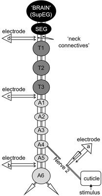
Previously this laboratory demonstrated that forelimb removal at birth in rats results in the invasion of the cuneate nucleus by sciatic nerve axons and the development of cuneothalamic cells with receptive fields that include both the forelimb-stump and the hindlimb. However, unit-cluster recordings from primary somatosensory cortex (SI) of these animals revealed few sites in the forelimb-stump representation where responses to hindlimb stimulation also could be recorded. Recently we reported that hindlimb inputs to the SI forelimb-stump representation are suppressed functionally in neonatally amputated rats and that GABAergic inhibition is involved in this process. The present study was undertaken to assess the role that intracortical projections from the SI hindlimb representation may play in the functional reorganization of the SI forelimb-stump field in these animals. The SI forelimb-stump representation was mapped during gamma-aminobutyric acid (GABA)-receptor blockade, both before and after electrolytic destruction of the SI hindlimb representation. Analysis of eight amputated rats showed that 75.8% of 264 stump recording sites possessed hindlimb receptive fields before destruction of the SI hindlimb. After the lesions, significantly fewer sites (13.2% of 197) were responsive to hindlimb stimulation (P < 0.0001). Electrolytic destruction of the SI lower-jaw representation in four additional control rats with neonatal forelimb amputation did not significantly reduce the percentage of hindlimb-responsive sites in the SI stump field during GABA-receptor blockade (P = 0.98). Similar results were obtained from three manipulated rats in which the SI hindlimb representation was silenced temporarily with a local cobalt chloride injection. Analysis of response latencies to sciatic nerve stimulation in the hindlimb and forelimb-stump representations suggested that the intracortical pathway(s) mediating the hindlimb responses in the forelimb-stump field may be polysynaptic. The mean latency to sciatic nerve stimulation at responsive sites in the GABA-receptor blocked SI stump representation of neonatally amputated rats was significantly longer than that for recording sites in the hindlimb representation [26.3 +/- 8.1 (SD) ms vs. 10.8 +/- 2.4 ms, respectively, P < 0.0001]. These results suggest that hindlimb input to the SI forelimb-stump representation detected in GABA-blocked cortices of neonatally forelimb amputated rats originates primarily from the SI hindlimb representation. (+info)Corticofugal amplification of facilitative auditory responses of subcortical combination-sensitive neurons in the mustached bat. (2/2839)
Recent studies on the bat's auditory system indicate that the corticofugal system mediates a highly focused positive feedback to physiologically "matched" subcortical neurons, and widespread lateral inhibition to physiologically "unmatched" subcortical neurons, to adjust and improve information processing. These findings have solved the controversy in physiological data, accumulated since 1962, of corticofugal effects on subcortical auditory neurons: inhibitory, excitatory, or both (an inhibitory effect is much more frequent than an excitatory effect). In the mustached bat, Pteronotus parnellii parnellii, the inferior colliculus, medial geniculate body, and auditory cortex each have "FM-FM" neurons, which are "combination-sensitive" and are tuned to specific time delays (echo delays) of echo FM components from the FM components of an emitted biosonar pulse. FM-FM neurons are more complex in response properties than cortical neurons which primarily respond to single tones. In the present study, we found that inactivation of the entire FM-FM area in the cortex, including neurons both physiologically matched and unmatched with subcortical FM-FM neurons, on the average reduced the facilitative responses to paired FM sounds by 82% for thalamic FM-FM neurons and by 66% for collicular FM-FM neurons. The corticofugal influence on the facilitative responses of subcortical combination-sensitive neurons is much larger than that on the excitatory responses of subcortical neurons primarily responding to single tones. Therefore we propose the hypothesis that, in general, the processing of complex sounds by combination-sensitive neurons more heavily depends on the corticofugal system than that by single-tone sensitive neurons. (+info)Distinct populations of NMDA receptors at subcortical and cortical inputs to principal cells of the lateral amygdala. (3/2839)
Fear conditioning involves the transmission of sensory stimuli to the amygdala from the thalamus and cortex. These input synapses are prime candidates for sites of plasticity critical to the learning in fear conditioning. Because N-methyl-D-aspartate (NMDA)-dependent mechanisms have been implicated in fear learning, we investigated the contribution of NMDA receptors to synaptic transmission at putative cortical and thalamic inputs using visualized whole cell recording in amygdala brain slices. Whereas NMDA receptors are present at both of these pathways, differences were observed. First, the alpha-amino-3-hydroxy-5-methyl-4-isoxazolepropionic acid-receptor-mediated component of the synaptic response, relative to the NMDA component, is smaller at thalamic than cortical input synapses. Second, thalamic NMDA responses are more sensitive to Mg2+. These findings suggest that there are distinct populations of NMDA receptors at cortical and thalamic inputs to the lateral amygdala. Differences such as these might underlie unique contributions of the two pathways to fear conditioning. (+info)N-Methyl-D-aspartate antagonists and apoptotic cell death triggered by head trauma in developing rat brain. (4/2839)
Morbidity and mortality from head trauma is highest among children. No animal model mimicking traumatic brain injury in children has yet been established, and the mechanisms of neuronal degeneration after traumatic injury to the developing brain are not understood. In infant rats subjected to percussion head trauma, two types of brain damage could be characterized. The first type or primary damage evolved within 4 hr and occurred by an excitotoxic mechanism. The second type or secondary damage evolved within 6-24 hr and occurred by an apoptotic mechanism. Primary damage remained localized to the parietal cortex at the site of impact. Secondary damage affected distant sites such as the cingulate/retrosplenial cortex, subiculum, frontal cortex, thalamus and striatum. Secondary apoptotic damage was more severe than primary excitotoxic damage. Morphometric analysis demonstrated that the N-methyl-D-aspartate receptor antagonists 3-(2-carboxypiperazin-4-yl)-propyl-1-phosphonate and dizocilpine protected against primary excitotoxic damage but increased severity of secondary apoptotic damage. 2-Sulfo-alpha-phenyl-N-tert-butyl-nitrone, a free radical scavenger, did not affect primary excitotoxic damage but mitigated apoptotic damage. These observations demonstrate that apoptosis and not excitotoxicity determine neuropathologic outcome after traumatic injury to the developing brain. Whereas free radical scavengers may prove useful in therapy of head trauma in children, N-methyl-D-aspartate antagonists should be avoided because of their propensity to increase severity of apoptotic damage. (+info)Secondary glioblastoma remarkably reduced by steroid administration after anaplastic transformation from gliomatosis cerebri--case report. (5/2839)
A 45-year-old female presented with gliomatosis cerebri manifesting as hemiballismus-like involuntary movement in the arm, motor weakness in the leg, and hypesthesia in her left side. Computed tomography showed only diffuse swelling of the right cerebral hemisphere, but T2-weighted magnetic resonance imaging revealed a diffuse lesion spreading from the right thalamus to the temporal, parietal, and occipital lobes on the same side. No abnormal enhancement was recognized. Cerebral angiography showed no specific finding. A right occipital lobectomy was performed to confirm the diagnosis of gliomatosis cerebri. Anaplastic transformation was recognized 5 months later. The disease did not resolve with radiation or interferon administration, but steroid therapy achieved remarkably effective tumor regression. The patient died due to pneumonia. Autopsy showed the features of diffuse glioblastoma. Steroid therapy may be an effective treatment for gliomatosis cerebri before the terminal stage. (+info)Blind smell: brain activation induced by an undetected air-borne chemical. (6/2839)
EEG and behavioural evidence suggests that air-borne chemicals can affect the nervous system without being consciously detected. EEG and behaviour, however, do not specify which brain structures are involved in chemical sensing that occurs below a threshold of conscious detection. Here we used functional MRI to localize brain activation induced by high and low concentrations of the air-borne compound oestra-1,3,5(10),16-tetraen-3yl acetate. Following presentations of both concentrations, eight of eight subjects reported verbally that they could not detect any odour (P = 0.004). Forced choice detection performed during the presentations revealed above-chance detection of the high concentration, but no better than chance detection of the low concentration compound. Both concentrations induced significant brain activation, primarily in the anterior medial thalamus and inferior frontal gyrus. Activation in the inferior frontal gyrus during the high concentration condition was significantly greater in the right than in the left hemisphere (P = 0.03). A trend towards greater thalamic activation was observed for the high concentration than the low concentration compound (P = 0.08). These findings localize human brain activation that was induced by an undetectable air-borne chemical (the low concentration compound). (+info)Functional neuropsychophysiological asymmetry in schizophrenia: a review and reorientation. (7/2839)
In reviewing the neuropsychophysiological evidence of functional asymmetry it is proposed that schizophrenia is characterized by a greater dispersion of leftward and rightward asymmetries. The two extremes are represented by active (left greater than right) and withdrawn (right greater than left) syndromes, as is the case with psychometric schizotypy. Syndrome-asymmetry relations extended beyond fronto-temporal systems to include posterior activity, infracortical motoneuron excitability, and individual differences in interhemispheric connectivity and directional biases. Central to these are lateral imbalances in thalamo-cortical and callosal arousal systems, while centrality to schizophrenia follows evidence of reversals in asymmetry with changes in symptom profile, clinical recovery, and neuroleptic treatment. Affinities are found in intact animals from challenge-induced turning tendencies representing coordinated activity of attentional, motor, and reinforcement systems. In both patients and animals, neuroleptics have reciprocal interhemispheric effects, with a bidirectionality that depends on syndrome or endogenous turning preference. Bidirectionality implicates nonspecific thalamic system (NSTS) and not limbic projections. It is proposed that the asymmetries arise from endogenous influences of genes, hormones, and early experience including stressors on NSTS asymmetry, and these underpin approach/withdrawal behavior that is manifested in temperament, personality, and clinical syndrome, and which precedes language development. (+info)Defects in thalamocortical axon pathfinding correlate with altered cell domains in Mash-1-deficient mice. (8/2839)
We have analyzed the pathfinding of thalamocortical axons (TCAs) from dorsal thalamus to neocortex in relation to specific cell domains in the forebrain of wild-type and Mash-1-deficient mice. In wild-type mice, we identified four cell domains that constitute the proximal part of the TCA pathway. These domains are distinguished by patterns of gene expression and by the presence of neurons retrogradely labeled from dorsal thalamus. Since the cells that form these domains are generated in forebrain proliferative zones that express high levels of Mash-1, we studied Mash-1 mutant mice to assess the potential roles of these domains in TCA pathfinding. In null mutants, each of the domains is altered: the two Pax-6 domains, one in ventral thalamus and one in hypothalamus, are expanded in size; a complementary RPTP(delta) domain in ventral thalamus is correspondingly reduced and the normally graded expression of RPTP(delta) in that domain is no longer apparent. In ventral telencephalon, a domain characterized in the wild type by Netrin-1 and Nkx-2.1 expression and by retrogradely labeled neurons is absent in the mutant. Defects in TCA pathfinding are localized to the borders of each of these altered domains. Many TCAs fail to enter the expanded, ventral thalamic Pax-6 domain that constitutes the most proximal part of the TCA pathway, and form a dense whorl at the border between dorsal and ventral thalamus. A proportion of TCAs do extend further distally into ventral thalamus, but many of these stall at an aberrant, abrupt border of high RPTP(delta) expression. A small proportion of TCAs extend around the RPTP(delta) domain and reach the ventral thalamic-hypothalamic border, but few of these axons turn at that border to extend into the ventral telencephalon. These findings demonstrate that Mash-1 is required for the normal development of cell domains that in turn are required for normal TCA pathfinding. In addition, these findings support the hypothesis that ventral telencephalic neurons and their axons guide TCAs through ventral thalamus and into ventral telencephalon. (+info)The thalamus is a large, paired structure in the brain that serves as a relay station for sensory and motor signals to the cerebral cortex. It is located in the dorsal part of the diencephalon and is made up of two symmetrical halves, each connected to the corresponding cerebral hemisphere.
The thalamus receives inputs from almost all senses, except for the olfactory system, and processes them before sending them to specific areas in the cortex. It also plays a role in regulating consciousness, sleep, and alertness. Additionally, the thalamus is involved in motor control by relaying information between the cerebellum and the motor cortex.
The thalamus is divided into several nuclei, each with distinct connections and functions. Some of these nuclei are involved in sensory processing, while others are involved in motor function or regulation of emotions and cognition. Overall, the thalamus plays a critical role in integrating information from various brain regions and modulating cognitive and emotional processes.
Thalamic nuclei refer to specific groupings of neurons within the thalamus, a key relay station in the brain that receives sensory information from various parts of the body and transmits it to the cerebral cortex for processing. The thalamus is divided into several distinct nuclei, each with its own unique functions and connections. These nuclei can be broadly categorized into three groups:
1. Sensory relay nuclei: These nuclei receive sensory information from different modalities such as vision, audition, touch, and taste, and project this information to specific areas of the cerebral cortex for further processing. Examples include the lateral geniculate nucleus (vision), medial geniculate nucleus (audition), and ventral posterior nucleus (touch and taste).
2. Association nuclei: These nuclei are involved in higher-order cognitive functions, such as attention, memory, and executive control. They receive inputs from various cortical areas and project back to those same areas, forming closed loops that facilitate information processing and integration. Examples include the mediodorsal nucleus and pulvinar.
3. Motor relay nuclei: These nuclei are involved in motor control and coordination. They receive inputs from the cerebral cortex and basal ganglia and project to the brainstem and spinal cord, helping to regulate movement and posture. Examples include the ventral anterior and ventral lateral nuclei.
Overall, thalamic nuclei play a crucial role in integrating sensory, motor, and cognitive information, allowing for adaptive behavior and conscious experience.
The ventral thalamic nuclei are a group of nuclei located in the ventral part of the thalamus, a region of the diencephalon in the brain. These nuclei play a crucial role in sensory and motor functions, as well as cognitive processes such as attention and memory. They include several subnuclei, such as the ventral anterior (VA), ventral lateral (VL), ventral medial (VM), and ventral posterior (VP) nuclei.
The ventral anterior and ventral lateral nuclei are involved in motor control and receive inputs from the basal ganglia, cerebellum, and cortex. They project to the premotor and motor areas of the cortex, contributing to the planning, initiation, and execution of movements.
The ventral medial nucleus is associated with emotional processing and receives inputs from the limbic system, including the amygdala and hippocampus. It projects to the prefrontal cortex and cingulate gyrus, contributing to the regulation of emotions and motivation.
The ventral posterior nuclei are involved in sensory processing, particularly for tactile and proprioceptive information. They receive inputs from the spinal cord and brainstem and project to the primary somatosensory cortex, where they contribute to the perception of touch, pressure, temperature, and body position.
Overall, the ventral thalamic nuclei are an essential component of the neural circuits involved in sensory, motor, and cognitive functions, and their dysfunction has been implicated in various neurological and psychiatric disorders.
The mediodorsal thalamic nucleus (MDTN) is a collection of neurons located in the dorsal part of the thalamus, a region of the brain that serves as a relay station for sensory and motor signals to the cerebral cortex. The MDTN is primarily involved in cognitive functions such as memory, attention, and emotion regulation.
The MDTN receives inputs from various regions of the brain, including the prefrontal cortex, amygdala, and hippocampus, and projects to the same areas of the cerebral cortex. It has been implicated in several neurological and psychiatric conditions, such as Alzheimer's disease, Parkinson's disease, schizophrenia, and depression.
Anatomically, the MDTN is divided into several subnuclei, including the parvocellular, magnocellular, and intermediate parts, each with distinct connectivity patterns and functions. Overall, the MDTN plays a crucial role in integrating information from different brain regions to facilitate higher-order cognitive processes.
Thalamic diseases refer to conditions that affect the thalamus, which is a part of the brain that acts as a relay station for sensory and motor signals to the cerebral cortex. The thalamus plays a crucial role in regulating consciousness, sleep, and alertness. Thalamic diseases can cause a variety of symptoms depending on the specific area of the thalamus that is affected. These symptoms may include sensory disturbances, motor impairment, cognitive changes, and altered levels of consciousness. Examples of thalamic diseases include stroke, tumors, multiple sclerosis, infections, and degenerative disorders such as dementia and Parkinson's disease. Treatment for thalamic diseases depends on the underlying cause and may include medications, surgery, or rehabilitation therapy.
The brain is the central organ of the nervous system, responsible for receiving and processing sensory information, regulating vital functions, and controlling behavior, movement, and cognition. It is divided into several distinct regions, each with specific functions:
1. Cerebrum: The largest part of the brain, responsible for higher cognitive functions such as thinking, learning, memory, language, and perception. It is divided into two hemispheres, each controlling the opposite side of the body.
2. Cerebellum: Located at the back of the brain, it is responsible for coordinating muscle movements, maintaining balance, and fine-tuning motor skills.
3. Brainstem: Connects the cerebrum and cerebellum to the spinal cord, controlling vital functions such as breathing, heart rate, and blood pressure. It also serves as a relay center for sensory information and motor commands between the brain and the rest of the body.
4. Diencephalon: A region that includes the thalamus (a major sensory relay station) and hypothalamus (regulates hormones, temperature, hunger, thirst, and sleep).
5. Limbic system: A group of structures involved in emotional processing, memory formation, and motivation, including the hippocampus, amygdala, and cingulate gyrus.
The brain is composed of billions of interconnected neurons that communicate through electrical and chemical signals. It is protected by the skull and surrounded by three layers of membranes called meninges, as well as cerebrospinal fluid that provides cushioning and nutrients.
The Intralaminar Thalamic Nuclei are a group of nuclei located within the thalamus, a part of the brain that serves as a relay station for sensory and motor signals. These nuclei are situated between the laminae (layers) of the thalamus and are characterized by their intricate internal organization. They play a crucial role in various functions, including attention, consciousness, and sleep-wake regulation. The Intralaminar Thalamic Nuclei have extensive connections with the cerebral cortex and other subcortical structures, making them an essential component of the brain's neural circuitry.
Neural pathways, also known as nerve tracts or fasciculi, refer to the highly organized and specialized routes through which nerve impulses travel within the nervous system. These pathways are formed by groups of neurons (nerve cells) that are connected in a series, creating a continuous communication network for electrical signals to transmit information between different regions of the brain, spinal cord, and peripheral nerves.
Neural pathways can be classified into two main types: sensory (afferent) and motor (efferent). Sensory neural pathways carry sensory information from various receptors in the body (such as those for touch, temperature, pain, and vision) to the brain for processing. Motor neural pathways, on the other hand, transmit signals from the brain to the muscles and glands, controlling movements and other effector functions.
The formation of these neural pathways is crucial for normal nervous system function, as it enables efficient communication between different parts of the body and allows for complex behaviors, cognitive processes, and adaptive responses to internal and external stimuli.
The anterior thalamic nuclei are a group of nuclei in the thalamus, which is a part of the brain. The thalamus serves as a relay station for sensory and motor signals to the cerebral cortex. The anterior thalamic nuclei, specifically, are involved in various functions such as memory, navigation, and arousal. They receive inputs from the hippocampus and other limbic structures and project to the cingulate gyrus and other areas of the cerebral cortex. The anterior thalamic nuclei have been implicated in several neurological and psychiatric conditions, including epilepsy, Alzheimer's disease, and schizophrenia.
The posterior thalamic nuclei are a group of nuclei located in the dorsal part of the thalamus, a major relay center in the brain. These nuclei include the lateroposterior nucleus (LP), pulvinar, and the medial and lateral geniculate bodies (MGN, LGN). They play crucial roles in processing and integrating sensory information, particularly from visual and auditory pathways, as well as motor and cognitive functions.
1. Lateroposterior nucleus (LP): This nucleus is involved in the processing of somatosensory information, which includes touch, pain, temperature, and proprioception (body position sense). It receives input from the cerebellum and sends outputs to the parietal cortex, contributing to the perception of body movement and position.
2. Pulvinar: The pulvinar is the largest nucleus in the thalamus and is primarily involved in visual processing. It receives inputs from multiple sources, including the retina, superior colliculus, and visual cortex, and sends outputs to various areas of the visual cortex. The pulvinar plays a critical role in attentional selection, object recognition, and scene perception.
3. Medial geniculate body (MGN): This nucleus is a part of the auditory pathway and receives input from the inferior colliculus in the midbrain. The MGN sends outputs to the primary auditory cortex, where sound processing and interpretation occur.
4. Lateral geniculate body (LGN): The LGN is a critical component of the visual pathway, receiving direct input from the retina and sending outputs to the primary visual cortex. It contains six layers, with alternating ON and OFF layers that process information from corresponding regions of the visual field.
In summary, the posterior thalamic nuclei are essential for sensory processing, attention, and perception in various modalities, including vision, audition, and somatosensation.
Medical Definition:
Magnetic Resonance Imaging (MRI) is a non-invasive diagnostic imaging technique that uses a strong magnetic field and radio waves to create detailed cross-sectional or three-dimensional images of the internal structures of the body. The patient lies within a large, cylindrical magnet, and the scanner detects changes in the direction of the magnetic field caused by protons in the body. These changes are then converted into detailed images that help medical professionals to diagnose and monitor various medical conditions, such as tumors, injuries, or diseases affecting the brain, spinal cord, heart, blood vessels, joints, and other internal organs. MRI does not use radiation like computed tomography (CT) scans.
The midline thalamic nuclei are a group of nuclei located in the thalamus, which is a part of the diencephalon in the brain. The thalamus serves as a relay station for sensory and motor signals to the cerebral cortex. The midline thalamic nuclei are situated in the most medial portion of the thalamus, along the midline. They include several distinct nuclei, such as the paraventricular nucleus, the reuniens nucleus, the rhomboid nucleus, and the central medial nucleus. These nuclei have complex connections with various brain regions, including the hypothalamus, the hippocampus, and the prefrontal cortex. They are involved in a variety of functions, such as memory, emotion, and sleep regulation.
The cerebral cortex is the outermost layer of the brain, characterized by its intricate folded structure and wrinkled appearance. It is a region of great importance as it plays a key role in higher cognitive functions such as perception, consciousness, thought, memory, language, and attention. The cerebral cortex is divided into two hemispheres, each containing four lobes: the frontal, parietal, temporal, and occipital lobes. These areas are responsible for different functions, with some regions specializing in sensory processing while others are involved in motor control or associative functions. The cerebral cortex is composed of gray matter, which contains neuronal cell bodies, and is covered by a layer of white matter that consists mainly of myelinated nerve fibers.
The geniculate bodies are part of the auditory pathway in the brainstem. They are two small, rounded eminences located on the lateral side of the upper pons, near the junction with the midbrain. The geniculate bodies are divided into an anterior and a posterior portion, known as the anterior and posterior geniculate bodies, respectively.
The anterior geniculate body receives inputs from the contralateral cochlear nucleus via the trapezoid body, and it is involved in the processing of sound localization. The posterior geniculate body receives inputs from the inferior colliculus via the lateral lemniscus and is involved in the processing of auditory information for conscious perception.
Overall, the geniculate bodies play a critical role in the processing and transmission of auditory information to higher brain areas for further analysis and interpretation.
Brain mapping is a broad term that refers to the techniques used to understand the structure and function of the brain. It involves creating maps of the various cognitive, emotional, and behavioral processes in the brain by correlating these processes with physical locations or activities within the nervous system. Brain mapping can be accomplished through a variety of methods, including functional magnetic resonance imaging (fMRI), positron emission tomography (PET) scans, electroencephalography (EEG), and others. These techniques allow researchers to observe which areas of the brain are active during different tasks or thoughts, helping to shed light on how the brain processes information and contributes to our experiences and behaviors. Brain mapping is an important area of research in neuroscience, with potential applications in the diagnosis and treatment of neurological and psychiatric disorders.
The pulvinar is a part of the brain that is located in the thalamus, which is a structure situated deep within the brain. The pulvinar plays a crucial role in visual processing and attention. It is the largest nucleus in the thalamus and is composed of several subdivisions, each with distinct connections to different areas of the cerebral cortex.
The pulvinar receives inputs from various sources, including the retina, superior colliculus, and visual cortex. It then sends outputs to multiple regions of the visual cortex, as well as other parts of the brain involved in attention and awareness. The pulvinar has been shown to modulate the flow of information between different areas of the visual system, allowing for the integration of visual information with other sensory inputs and attentional processes.
Damage to the pulvinar can result in a variety of visual deficits, including impairments in visual attention, object recognition, and motion perception. Additionally, some studies have suggested that the pulvinar may be involved in the regulation of consciousness and arousal.
Neurons, also known as nerve cells or neurocytes, are specialized cells that constitute the basic unit of the nervous system. They are responsible for receiving, processing, and transmitting information and signals within the body. Neurons have three main parts: the dendrites, the cell body (soma), and the axon. The dendrites receive signals from other neurons or sensory receptors, while the axon transmits these signals to other neurons, muscles, or glands. The junction between two neurons is called a synapse, where neurotransmitters are released to transmit the signal across the gap (synaptic cleft) to the next neuron. Neurons vary in size, shape, and structure depending on their function and location within the nervous system.
The basal ganglia are a group of interconnected nuclei, or clusters of neurons, located in the base of the brain. They play a crucial role in regulating motor function, cognition, and emotion. The main components of the basal ganglia include the striatum (made up of the caudate nucleus, putamen, and ventral striatum), globus pallidus (divided into external and internal segments), subthalamic nucleus, and substantia nigra (with its pars compacta and pars reticulata).
The basal ganglia receive input from various regions of the cerebral cortex and other brain areas. They process this information and send output back to the thalamus and cortex, helping to modulate and coordinate movement. The basal ganglia also contribute to higher cognitive functions such as learning, decision-making, and habit formation. Dysfunction in the basal ganglia can lead to neurological disorders like Parkinson's disease, Huntington's disease, and dystonia.
The lateral thalamic nuclei are a group of nuclei located in the dorsolateral part of the thalamus, a major relay station for sensory and motor signals in the brain. These nuclei include the lateral dorsal nucleus, lateral posterior nucleus, and pulvinar. They play a role in various functions such as attention, awareness, and visuospatial processing. Damage to these nuclei can result in neurological disorders like neglect syndrome, where patients have difficulty attending to stimuli on one side of their body or environment.
Vibrissae are stiff, tactile hairs that are highly sensitive to touch and movement. They are primarily found in various mammals, including humans (in the form of eyelashes and eyebrows), but they are especially prominent in certain animals such as cats, rats, and seals. These hairs are deeply embedded in skin and have a rich supply of nerve endings that provide the animal with detailed information about its environment. They are often used for detecting nearby objects, navigating in the dark, and maintaining balance.
The spinothalamic tracts are a pair of white matter tracts in the spinal cord that carry sensory information from the body to the brain. They are responsible for transmitting pain, temperature, and crude touch sensation. The tracts consist of two components: the lateral spinothalamic tract, which carries information about pain and temperature, and the anterior spinothalamic tract, which carries information about touch and pressure. These tracts decussate (cross to the opposite side) at the level of the spinal cord where they enter, and then ascend to the thalamus, where the information is relayed to the sensory cortex for processing.
The diencephalon is a term used in anatomy to refer to the part of the brain that lies between the cerebrum and the midbrain. It includes several important structures, such as the thalamus, hypothalamus, epithalamus, and subthalamus.
The thalamus is a major relay station for sensory information, receiving input from all senses except smell and sending it to the appropriate areas of the cerebral cortex. The hypothalamus plays a crucial role in regulating various bodily functions, including hunger, thirst, body temperature, and sleep-wake cycles. It also produces hormones that regulate mood, growth, and development.
The epithalamus contains the pineal gland, which produces melatonin, a hormone that helps regulate sleep-wake cycles. The subthalamus is involved in motor control and coordination.
Overall, the diencephalon plays a critical role in integrating sensory information, regulating autonomic functions, and modulating behavior and emotion.
Afferent pathways, also known as sensory pathways, refer to the neural connections that transmit sensory information from the peripheral nervous system to the central nervous system (CNS), specifically to the brain and spinal cord. These pathways are responsible for carrying various types of sensory information, such as touch, temperature, pain, pressure, vibration, hearing, vision, and taste, to the CNS for processing and interpretation.
The afferent pathways begin with sensory receptors located throughout the body, which detect changes in the environment and convert them into electrical signals. These signals are then transmitted via afferent neurons, also known as sensory neurons, to the spinal cord or brainstem. Within the CNS, the information is further processed and integrated with other neural inputs before being relayed to higher cognitive centers for conscious awareness and response.
Understanding the anatomy and physiology of afferent pathways is essential for diagnosing and treating various neurological conditions that affect sensory function, such as neuropathies, spinal cord injuries, and brain disorders.
The somatosensory cortex is a part of the brain located in the postcentral gyrus of the parietal lobe, which is responsible for processing sensory information from the body. It receives and integrates tactile, proprioceptive, and thermoception inputs from the skin, muscles, joints, and internal organs, allowing us to perceive and interpret touch, pressure, pain, temperature, vibration, position, and movement of our body parts. The somatosensory cortex is organized in a map-like manner, known as the sensory homunculus, where each body part is represented according to its relative sensitivity and density of innervation. This organization allows for precise localization and discrimination of tactile stimuli across the body surface.
Absence epilepsy is a type of epilepsy characterized by recurrent brief episodes of "absences," or staring spells, that can last from a few seconds to several minutes. These episodes are often accompanied by subtle body movements such as lip smacking or eyelid flutters. Absence epilepsy is most commonly diagnosed in children and adolescents, and it is more common in girls than boys.
The seizures in absence epilepsy are caused by abnormal electrical activity in the brain, specifically in a part of the brain called the cortex. These abnormal electrical discharges occur in a pattern that involves both sides of the brain simultaneously. This differs from other types of epilepsy, which may involve only one side of the brain or specific areas within a single hemisphere.
Absence seizures are typically brief and do not cause confusion or disorientation after they end. However, if they occur frequently, they can interfere with learning and social development. In some cases, absence epilepsy may be associated with other types of seizures, such as generalized tonic-clonic (grand mal) seizures or myoclonic jerks.
The diagnosis of absence epilepsy is usually made based on the characteristic symptoms and the results of an electroencephalogram (EEG), which can detect the abnormal electrical activity in the brain during a seizure. Treatment typically involves medication to control the seizures, such as ethosuximide or valproic acid. In some cases, a ketogenic diet may also be recommended as an alternative treatment option.
The putamen is a round, egg-shaped structure that is a part of the basal ganglia, located in the forebrain. It is situated laterally to the globus pallidus and medially to the internal capsule. The putamen plays a crucial role in regulating movement and is involved in various functions such as learning, motivation, and habit formation.
It receives input from the cerebral cortex via the corticostriatal pathway and sends output to the globus pallidus and substantia nigra pars reticulata, which are also part of the basal ganglia circuitry. The putamen is heavily innervated by dopaminergic neurons from the substantia nigra pars compacta, and degeneration of these neurons in Parkinson's disease leads to a significant reduction in dopamine levels in the putamen, resulting in motor dysfunction.
Auditory pathways refer to the series of structures and nerves in the body that are involved in processing sound and transmitting it to the brain for interpretation. The process begins when sound waves enter the ear and cause vibrations in the eardrum, which then move the bones in the middle ear. These movements stimulate hair cells in the cochlea, a spiral-shaped structure in the inner ear, causing them to release neurotransmitters that activate auditory nerve fibers.
The auditory nerve carries these signals to the brainstem, where they are relayed through several additional structures before reaching the auditory cortex in the temporal lobe of the brain. Here, the signals are processed and interpreted as sounds, allowing us to hear and understand speech, music, and other environmental noises.
Damage or dysfunction at any point along the auditory pathway can lead to hearing loss or impairment.
The caudate nucleus is a part of the brain located within the basal ganglia, a group of structures that are important for movement control and cognition. It has a distinctive C-shaped appearance and plays a role in various functions such as learning, memory, emotion, and motivation. The caudate nucleus receives inputs from several areas of the cerebral cortex and sends outputs to other basal ganglia structures, contributing to the regulation of motor behavior and higher cognitive processes.
The mamillary bodies are a pair of small, round structures located in the hypothalamus region of the brain. They play a crucial role in the limbic system, which is involved in emotions, memory, and learning. Specifically, the mamillary bodies are part of the circuit that forms the Papez circuit, a neural network responsible for memory and cognitive functions.
The mamillary bodies receive inputs from several brain regions, including the hippocampus, anterior thalamic nuclei, and cingulate gyrus. They then project this information to the thalamus, which in turn sends it to the cerebral cortex for further processing.
Damage to the mamillary bodies can result in memory impairment, as seen in patients with Korsakoff's syndrome, a condition often associated with chronic alcohol abuse.
The subthalamus is a region in the brain that is located deep beneath the thalamus and above the midbrain. It is a part of the basal ganglia, which are a group of structures involved in the control of movement. The subthalamus contains several different types of neurons, including glutamatergic and GABAergic neurons, and plays a role in regulating movement, reward, and motivation. It is also thought to be involved in the pathophysiology of certain neurological disorders such as Parkinson's disease.
The subthalamic nucleus (STN) is a specific structure within the subthalamus that has been the target of deep brain stimulation surgery for the treatment of movement disorders like Parkinson's disease and dystonia. The STN is responsible for regulating the activity of other structures in the basal ganglia, and its overactivity can lead to symptoms such as tremors, rigidity, and difficulty initiating movements. By implanting electrodes in the STN and delivering electrical impulses, deep brain stimulation can help to regulate the activity of the STN and alleviate some of these symptoms.
Computer-assisted image processing is a medical term that refers to the use of computer systems and specialized software to improve, analyze, and interpret medical images obtained through various imaging techniques such as X-ray, CT (computed tomography), MRI (magnetic resonance imaging), ultrasound, and others.
The process typically involves several steps, including image acquisition, enhancement, segmentation, restoration, and analysis. Image processing algorithms can be used to enhance the quality of medical images by adjusting contrast, brightness, and sharpness, as well as removing noise and artifacts that may interfere with accurate diagnosis. Segmentation techniques can be used to isolate specific regions or structures of interest within an image, allowing for more detailed analysis.
Computer-assisted image processing has numerous applications in medical imaging, including detection and characterization of lesions, tumors, and other abnormalities; assessment of organ function and morphology; and guidance of interventional procedures such as biopsies and surgeries. By automating and standardizing image analysis tasks, computer-assisted image processing can help to improve diagnostic accuracy, efficiency, and consistency, while reducing the potential for human error.
Physical stimulation, in a medical context, refers to the application of external forces or agents to the body or its tissues to elicit a response. This can include various forms of touch, pressure, temperature, vibration, or electrical currents. The purpose of physical stimulation may be therapeutic, as in the case of massage or physical therapy, or diagnostic, as in the use of reflex tests. It is also used in research settings to study physiological responses and mechanisms.
In a broader sense, physical stimulation can also refer to the body's exposure to physical activity or exercise, which can have numerous health benefits, including improving cardiovascular function, increasing muscle strength and flexibility, and reducing the risk of chronic diseases.
The Globus Pallidus is a structure in the brain that is part of the basal ganglia, a group of nuclei associated with movement control and other functions. It has two main subdivisions: the external (GPe) and internal (GPi) segments. The GPe receives input from the striatum and sends inhibitory projections to the subthalamic nucleus, while the GPi sends inhibitory projections to the thalamus, which in turn projects to the cerebral cortex. These connections allow for the regulation of motor activity, with abnormal functioning of the Globus Pallidus being implicated in various movement disorders such as Parkinson's disease and Huntington's disease.
The brainstem is the lower part of the brain that connects to the spinal cord. It consists of the midbrain, pons, and medulla oblongata. The brainstem controls many vital functions such as heart rate, breathing, and blood pressure. It also serves as a relay center for sensory and motor information between the cerebral cortex and the rest of the body. Additionally, several cranial nerves originate from the brainstem, including those that control eye movements, facial movements, and hearing.
A nerve net, also known as a neural net or neuronal network, is not a medical term per se, but rather a concept in neuroscience and artificial intelligence (AI). It refers to a complex network of interconnected neurons that process and transmit information. In the context of the human body, the nervous system can be thought of as a type of nerve net, with the brain and spinal cord serving as the central processing unit and peripheral nerves carrying signals to and from various parts of the body.
In the field of AI, artificial neural networks are computational models inspired by the structure and function of biological nerve nets. These models consist of interconnected nodes or "neurons" that process information and learn patterns through a process of training and adaptation. They have been used in a variety of applications, including image recognition, natural language processing, and machine learning.
Sprague-Dawley rats are a strain of albino laboratory rats that are widely used in scientific research. They were first developed by researchers H.H. Sprague and R.C. Dawley in the early 20th century, and have since become one of the most commonly used rat strains in biomedical research due to their relatively large size, ease of handling, and consistent genetic background.
Sprague-Dawley rats are outbred, which means that they are genetically diverse and do not suffer from the same limitations as inbred strains, which can have reduced fertility and increased susceptibility to certain diseases. They are also characterized by their docile nature and low levels of aggression, making them easier to handle and study than some other rat strains.
These rats are used in a wide variety of research areas, including toxicology, pharmacology, nutrition, cancer, and behavioral studies. Because they are genetically diverse, Sprague-Dawley rats can be used to model a range of human diseases and conditions, making them an important tool in the development of new drugs and therapies.
Stereotaxic techniques are minimally invasive surgical procedures used in neuroscience and neurology that allow for precise targeting and manipulation of structures within the brain. These methods use a stereotactic frame, which is attached to the skull and provides a three-dimensional coordinate system to guide the placement of instruments such as electrodes, cannulas, or radiation sources. The main goal is to reach specific brain areas with high precision and accuracy, minimizing damage to surrounding tissues. Stereotaxic techniques are widely used in research, diagnosis, and treatment of various neurological disorders, including movement disorders, pain management, epilepsy, and psychiatric conditions.
"Long-Evans" is a strain of laboratory rats commonly used in scientific research. They are named after their developers, the scientists Long and Evans. This strain is albino, with a brownish-black hood over their eyes and ears, and they have an agouti (salt-and-pepper) color on their backs. They are often used as a model organism due to their size, ease of handling, and genetic similarity to humans. However, I couldn't find any specific medical definition related to "Long-Evans rats" as they are not a medical condition or disease.
An action potential is a brief electrical signal that travels along the membrane of a nerve cell (neuron) or muscle cell. It is initiated by a rapid, localized change in the permeability of the cell membrane to specific ions, such as sodium and potassium, resulting in a rapid influx of sodium ions and a subsequent efflux of potassium ions. This ion movement causes a brief reversal of the electrical potential across the membrane, which is known as depolarization. The action potential then propagates along the cell membrane as a wave, allowing the electrical signal to be transmitted over long distances within the body. Action potentials play a crucial role in the communication and functioning of the nervous system and muscle tissue.
The limbic system is a complex set of structures in the brain that includes the hippocampus, amygdala, fornix, cingulate gyrus, and other nearby areas. It's associated with emotional responses, instinctual behaviors, motivation, long-term memory formation, and olfaction (smell). The limbic system is also involved in the modulation of visceral functions and drives, such as hunger, thirst, and sexual drive.
The structures within the limbic system communicate with each other and with other parts of the brain, particularly the hypothalamus and the cortex, to regulate various physiological and psychological processes. Dysfunctions in the limbic system can lead to a range of neurological and psychiatric conditions, including depression, anxiety disorders, post-traumatic stress disorder (PTSD), and certain types of memory impairment.
Deep brain stimulation (DBS) is a surgical procedure that involves the implantation of a medical device called a neurostimulator, which sends electrical impulses to specific targets in the brain. The impulses help to regulate abnormal brain activity, and can be used to treat a variety of neurological conditions, including Parkinson's disease, essential tremor, dystonia, and obsessive-compulsive disorder.
During the procedure, electrodes are implanted into the brain and connected to the neurostimulator, which is typically implanted in the chest. The neurostimulator can be programmed to deliver electrical impulses at varying frequencies, amplitudes, and pulse widths, depending on the specific needs of the patient.
DBS is generally considered a safe and effective treatment option for many patients with neurological conditions, although it does carry some risks, such as infection, bleeding, and hardware complications. It is typically reserved for patients who have not responded well to other forms of treatment, or who experience significant side effects from medication.
Electroencephalography (EEG) is a medical procedure that records electrical activity in the brain. It uses small, metal discs called electrodes, which are attached to the scalp with paste or a specialized cap. These electrodes detect tiny electrical charges that result from the activity of brain cells, and the EEG machine then amplifies and records these signals.
EEG is used to diagnose various conditions related to the brain, such as seizures, sleep disorders, head injuries, infections, and degenerative diseases like Alzheimer's or Parkinson's. It can also be used during surgery to monitor brain activity and ensure that surgical procedures do not interfere with vital functions.
EEG is a safe and non-invasive procedure that typically takes about 30 minutes to an hour to complete, although longer recordings may be necessary in some cases. Patients are usually asked to relax and remain still during the test, as movement can affect the quality of the recording.
Electric stimulation, also known as electrical nerve stimulation or neuromuscular electrical stimulation, is a therapeutic treatment that uses low-voltage electrical currents to stimulate nerves and muscles. It is often used to help manage pain, promote healing, and improve muscle strength and mobility. The electrical impulses can be delivered through electrodes placed on the skin or directly implanted into the body.
In a medical context, electric stimulation may be used for various purposes such as:
1. Pain management: Electric stimulation can help to block pain signals from reaching the brain and promote the release of endorphins, which are natural painkillers produced by the body.
2. Muscle rehabilitation: Electric stimulation can help to strengthen muscles that have become weak due to injury, illness, or surgery. It can also help to prevent muscle atrophy and improve range of motion.
3. Wound healing: Electric stimulation can promote tissue growth and help to speed up the healing process in wounds, ulcers, and other types of injuries.
4. Urinary incontinence: Electric stimulation can be used to strengthen the muscles that control urination and reduce symptoms of urinary incontinence.
5. Migraine prevention: Electric stimulation can be used as a preventive treatment for migraines by applying electrical impulses to specific nerves in the head and neck.
It is important to note that electric stimulation should only be administered under the guidance of a qualified healthcare professional, as improper use can cause harm or discomfort.
Neural inhibition is a process in the nervous system that decreases or prevents the activity of neurons (nerve cells) in order to regulate and control communication within the nervous system. It is a fundamental mechanism that allows for the balance of excitation and inhibition necessary for normal neural function. Inhibitory neurotransmitters, such as GABA (gamma-aminobutyric acid) and glycine, are released from the presynaptic neuron and bind to receptors on the postsynaptic neuron, reducing its likelihood of firing an action potential. This results in a decrease in neural activity and can have various effects depending on the specific neurons and brain regions involved. Neural inhibition is crucial for many functions including motor control, sensory processing, attention, memory, and emotional regulation.
The cerebellum is a part of the brain that lies behind the brainstem and is involved in the regulation of motor movements, balance, and coordination. It contains two hemispheres and a central portion called the vermis. The cerebellum receives input from sensory systems and other areas of the brain and spinal cord and sends output to motor areas of the brain. Damage to the cerebellum can result in problems with movement, balance, and coordination.
Functional laterality, in a medical context, refers to the preferential use or performance of one side of the body over the other for specific functions. This is often demonstrated in hand dominance, where an individual may be right-handed or left-handed, meaning they primarily use their right or left hand for tasks such as writing, eating, or throwing.
However, functional laterality can also apply to other bodily functions and structures, including the eyes (ocular dominance), ears (auditory dominance), or legs. It's important to note that functional laterality is not a strict binary concept; some individuals may exhibit mixed dominance or no strong preference for one side over the other.
In clinical settings, assessing functional laterality can be useful in diagnosing and treating various neurological conditions, such as stroke or traumatic brain injury, where understanding any resulting lateralized impairments can inform rehabilitation strategies.
Visual pathways, also known as the visual system or the optic pathway, refer to the series of specialized neurons in the nervous system that transmit visual information from the eyes to the brain. This complex network includes the retina, optic nerve, optic chiasma, optic tract, lateral geniculate nucleus, pulvinar, and the primary and secondary visual cortices located in the occipital lobe of the brain.
The process begins when light enters the eye and strikes the photoreceptor cells (rods and cones) in the retina, converting the light energy into electrical signals. These signals are then transmitted to bipolar cells and subsequently to ganglion cells, whose axons form the optic nerve. The fibers from each eye's nasal hemiretina cross at the optic chiasma, while those from the temporal hemiretina continue without crossing. This results in the formation of the optic tract, which carries visual information from both eyes to the opposite side of the brain.
The majority of fibers in the optic tract synapse with neurons in the lateral geniculate nucleus (LGN), a part of the thalamus. The LGN sends this information to the primary visual cortex, also known as V1 or Brodmann area 17, located in the occipital lobe. Here, simple features like lines and edges are initially processed. Further processing occurs in secondary (V2) and tertiary (V3-V5) visual cortices, where more complex features such as shape, motion, and depth are analyzed. Ultimately, this information is integrated to form our perception of the visual world.
"Cat" is a common name that refers to various species of small carnivorous mammals that belong to the family Felidae. The domestic cat, also known as Felis catus or Felis silvestris catus, is a popular pet and companion animal. It is a subspecies of the wildcat, which is found in Europe, Africa, and Asia.
Domestic cats are often kept as pets because of their companionship, playful behavior, and ability to hunt vermin. They are also valued for their ability to provide emotional support and therapy to people. Cats are obligate carnivores, which means that they require a diet that consists mainly of meat to meet their nutritional needs.
Cats are known for their agility, sharp senses, and predatory instincts. They have retractable claws, which they use for hunting and self-defense. Cats also have a keen sense of smell, hearing, and vision, which allow them to detect prey and navigate their environment.
In medical terms, cats can be hosts to various parasites and diseases that can affect humans and other animals. Some common feline diseases include rabies, feline leukemia virus (FeLV), feline immunodeficiency virus (FIV), and toxoplasmosis. It is important for cat owners to keep their pets healthy and up-to-date on vaccinations and preventative treatments to protect both the cats and their human companions.
Cerebrovascular circulation refers to the network of blood vessels that supply oxygenated blood and nutrients to the brain tissue, and remove waste products. It includes the internal carotid arteries, vertebral arteries, circle of Willis, and the intracranial arteries that branch off from them.
The internal carotid arteries and vertebral arteries merge to form the circle of Willis, a polygonal network of vessels located at the base of the brain. The anterior cerebral artery, middle cerebral artery, posterior cerebral artery, and communicating arteries are the major vessels that branch off from the circle of Willis and supply blood to different regions of the brain.
Interruptions or abnormalities in the cerebrovascular circulation can lead to various neurological conditions such as stroke, transient ischemic attack (TIA), and vascular dementia.
Emission computed tomography (ECT) is a type of tomographic imaging technique in which an emission signal from within the body is detected to create cross-sectional images of that signal's distribution. In Emission-Computed Tomography (ECT), a radionuclide is introduced into the body, usually through injection, inhalation or ingestion. The radionuclide emits gamma rays that are then detected by external gamma cameras.
The data collected from these cameras is then used to create cross-sectional images of the distribution of the radiopharmaceutical within the body. This allows for the identification and quantification of functional information about specific organs or systems within the body, such as blood flow, metabolic activity, or receptor density.
One common type of Emission-Computed Tomography is Single Photon Emission Computed Tomography (SPECT), which uses a single gamma camera that rotates around the patient to collect data from multiple angles. Another type is Positron Emission Tomography (PET), which uses positron-emitting radionuclides and detects the coincident gamma rays emitted by the annihilation of positrons and electrons.
Overall, ECT is a valuable tool in medical imaging for diagnosing and monitoring various diseases, including cancer, heart disease, and neurological disorders.
The prosencephalon is a term used in the field of neuroembryology, which refers to the developmental stage of the forebrain in the embryonic nervous system. It is one of the three primary vesicles that form during the initial stages of neurulation, along with the mesencephalon (midbrain) and rhombencephalon (hindbrain).
The prosencephalon further differentiates into two secondary vesicles: the telencephalon and diencephalon. The telencephalon gives rise to structures such as the cerebral cortex, basal ganglia, and olfactory bulbs, while the diencephalon develops into structures like the thalamus, hypothalamus, and epithalamus.
It is important to note that 'prosencephalon' itself is not used as a medical term in adult neuroanatomy, but it is crucial for understanding the development of the human brain during embryogenesis.
Cerebral dominance is a concept in neuropsychology that refers to the specialization of one hemisphere of the brain over the other for certain cognitive functions. In most people, the left hemisphere is dominant for language functions such as speaking and understanding spoken or written language, while the right hemisphere is dominant for non-verbal functions such as spatial ability, face recognition, and artistic ability.
Cerebral dominance does not mean that the non-dominant hemisphere is incapable of performing the functions of the dominant hemisphere, but rather that it is less efficient or specialized in those areas. The concept of cerebral dominance has been used to explain individual differences in cognitive abilities and learning styles, as well as the laterality of brain damage and its effects on cognition and behavior.
It's important to note that cerebral dominance is a complex phenomenon that can vary between individuals and can be influenced by various factors such as genetics, environment, and experience. Additionally, recent research has challenged the strict lateralization of functions and suggested that there is more functional overlap and interaction between the two hemispheres than previously thought.
Acoustic stimulation refers to the use of sound waves or vibrations to elicit a response in an individual, typically for the purpose of assessing or treating hearing, balance, or neurological disorders. In a medical context, acoustic stimulation may involve presenting pure tones, speech sounds, or other types of auditory signals through headphones, speakers, or specialized devices such as bone conduction transducers.
The response to acoustic stimulation can be measured using various techniques, including electrophysiological tests like auditory brainstem responses (ABRs) or otoacoustic emissions (OAEs), behavioral observations, or functional imaging methods like fMRI. Acoustic stimulation is also used in therapeutic settings, such as auditory training programs for hearing impairment or vestibular rehabilitation for balance disorders.
It's important to note that acoustic stimulation should be administered under the guidance of a qualified healthcare professional to ensure safety and effectiveness.
The reticular formation is not a single structure but rather a complex network of interconnected neurons located in the brainstem, extending from the medulla oblongata through the pons and mesencephalon (midbrain) up to the diencephalon (thalamus and hypothalamus). It forms part of the reticular activating system, which is involved in regulating arousal, awareness, and sleep-wake cycles.
The reticular formation plays a crucial role in various functions such as:
1. Modulation of sensory input: The neurons in the reticular formation receive inputs from all senses (visual, auditory, tactile, etc.) and help filter and prioritize this information before it reaches higher cognitive areas.
2. Control of motor function: The reticular formation contributes to the regulation of muscle tone, posture, and locomotion by modulating the activity of motor neurons in the spinal cord.
3. Regulation of autonomic functions: The reticular formation is involved in controlling heart rate, blood pressure, respiration, and other visceral functions through its connections with the autonomic nervous system.
4. Consciousness and arousal: The ascending reticular activating system (ARAS) originates from the reticular formation and projects to the thalamus and cerebral cortex, where it helps maintain wakefulness and arousal. Damage to the ARAS can lead to coma or other states of altered consciousness.
5. Sleep-wake cycle regulation: The reticular formation contains cells that release neurotransmitters like histamine, serotonin, and orexin/hypocretin, which are essential for sleep-wake regulation. Dysfunction in these circuits has been implicated in various sleep disorders, such as narcolepsy and insomnia.
A tremor is an involuntary, rhythmic muscle contraction and relaxation that causes a shaking movement. It's a type of motion disorder that can affect any part of your body, but it most often occurs in your hands. Tremors can be harmless, but they can also be a symptom of a more serious neurological disorder. The cause of tremors isn't always known, but they can be the result of damage to the brain from a stroke, multiple sclerosis, or trauma. Certain medications, alcohol abuse, and drug withdrawal can also cause tremors. In some cases, tremors may be inherited and run in families.
Tremors can be classified based on their cause, appearance, and the situation in which they occur. The two most common types of tremors are:
* Resting tremors, which occur when your muscles are relaxed, such as when your hands are resting on your lap. Parkinson's disease is a common cause of this type of tremor.
* Action tremors, which occur with purposeful movement, such as when you're trying to hold something or when you're using a utensil. Essential tremor, the most common type of tremor, is an action tremor.
Tremors can also be classified based on their frequency (how often they occur) and amplitude (the size of the movement). High-frequency tremors are faster and smaller in amplitude, while low-frequency tremors are slower and larger in amplitude.
In general, tremors are not a life-threatening condition, but they can be embarrassing or make it difficult to perform daily activities. In some cases, tremors may indicate a more serious underlying condition that requires treatment. If you're concerned about tremors or have any questions about your symptoms, it's important to speak with a healthcare provider for an accurate diagnosis and appropriate treatment.
Efferent pathways refer to the neural connections that carry signals from the central nervous system (CNS), which includes the brain and spinal cord, to the peripheral effectors such as muscles and glands. These pathways are responsible for the initiation and control of motor responses, as well as regulating various autonomic functions.
Efferent pathways can be divided into two main types:
1. Somatic efferent pathways: These pathways carry signals from the CNS to the skeletal muscles, enabling voluntary movements and postural control. The final common pathway for somatic motor innervation is the alpha-motor neuron, which synapses directly onto skeletal muscle fibers.
2. Autonomic efferent pathways: These pathways regulate the function of internal organs, smooth muscles, and glands. They are further divided into two subtypes: sympathetic and parasympathetic. The sympathetic system is responsible for the 'fight or flight' response, while the parasympathetic system promotes rest and digestion. Both systems use a two-neuron chain to transmit signals from the CNS to the effector organs. The preganglionic neuron has its cell body in the CNS and synapses with the postganglionic neuron in an autonomic ganglion located near the effector organ. The postganglionic neuron then innervates the target organ or tissue.
In summary, efferent pathways are the neural connections that carry signals from the CNS to peripheral effectors, enabling motor responses and regulating various autonomic functions. They can be divided into somatic and autonomic efferent pathways, with further subdivisions within the autonomic system.
Autoradiography is a medical imaging technique used to visualize and localize the distribution of radioactively labeled compounds within tissues or organisms. In this process, the subject is first exposed to a radioactive tracer that binds to specific molecules or structures of interest. The tissue is then placed in close contact with a radiation-sensitive film or detector, such as X-ray film or an imaging plate.
As the radioactive atoms decay, they emit particles (such as beta particles) that interact with the film or detector, causing chemical changes and leaving behind a visible image of the distribution of the labeled compound. The resulting autoradiogram provides information about the location, quantity, and sometimes even the identity of the molecules or structures that have taken up the radioactive tracer.
Autoradiography has been widely used in various fields of biology and medical research, including pharmacology, neuroscience, genetics, and cell biology, to study processes such as protein-DNA interactions, gene expression, drug metabolism, and neuronal connectivity. However, due to the use of radioactive materials and potential hazards associated with them, this technique has been gradually replaced by non-radioactive alternatives like fluorescence in situ hybridization (FISH) or immunofluorescence techniques.
The auditory cortex is the region of the brain that is responsible for processing and analyzing sounds, including speech. It is located in the temporal lobe of the cerebral cortex, specifically within the Heschl's gyrus and the surrounding areas. The auditory cortex receives input from the auditory nerve, which carries sound information from the inner ear to the brain.
The auditory cortex is divided into several subregions that are responsible for different aspects of sound processing, such as pitch, volume, and location. These regions work together to help us recognize and interpret sounds in our environment, allowing us to communicate with others and respond appropriately to our surroundings. Damage to the auditory cortex can result in hearing loss or difficulty understanding speech.
Implanted electrodes are medical devices that are surgically placed inside the body to interface directly with nerves, neurons, or other electrically excitable tissue for various therapeutic purposes. These electrodes can be used to stimulate or record electrical activity from specific areas of the body, depending on their design and application.
There are several types of implanted electrodes, including:
1. Deep Brain Stimulation (DBS) electrodes: These are placed deep within the brain to treat movement disorders such as Parkinson's disease, essential tremor, and dystonia. DBS electrodes deliver electrical impulses that modulate abnormal neural activity in targeted brain regions.
2. Spinal Cord Stimulation (SCS) electrodes: These are implanted along the spinal cord to treat chronic pain syndromes. SCS electrodes emit low-level electrical pulses that interfere with pain signals traveling to the brain, providing relief for patients.
3. Cochlear Implant electrodes: These are surgically inserted into the cochlea of the inner ear to restore hearing in individuals with severe to profound hearing loss. The electrodes stimulate the auditory nerve directly, bypassing damaged hair cells within the cochlea.
4. Retinal Implant electrodes: These are implanted in the retina to treat certain forms of blindness caused by degenerative eye diseases like retinitis pigmentosa. The electrodes convert visual information from a camera into electrical signals, which stimulate remaining retinal cells and transmit the information to the brain via the optic nerve.
5. Sacral Nerve Stimulation (SNS) electrodes: These are placed near the sacral nerves in the lower back to treat urinary or fecal incontinence and overactive bladder syndrome. SNS electrodes deliver electrical impulses that regulate the function of the affected muscles and nerves.
6. Vagus Nerve Stimulation (VNS) electrodes: These are wrapped around the vagus nerve in the neck to treat epilepsy and depression. VNS electrodes provide intermittent electrical stimulation to the vagus nerve, which has connections to various regions of the brain involved in these conditions.
Overall, implanted electrodes serve as a crucial component in many neuromodulation therapies, offering an effective treatment option for numerous neurological and sensory disorders.
The trigeminal nuclei are a collection of sensory nerve cell bodies (nuclei) located in the brainstem that receive and process sensory information from the face and head, including pain, temperature, touch, and proprioception. There are four main trigeminal nuclei: the ophthalmic, maxillary, mandibular, and mesencephalic nuclei. Each nucleus is responsible for processing sensory information from specific areas of the face and head. The trigeminal nerve (cranial nerve V) carries these sensory signals to the brainstem, where they synapse with neurons in the trigeminal nuclei before being relayed to higher brain centers for further processing.
Reaction time, in the context of medicine and physiology, refers to the time period between the presentation of a stimulus and the subsequent initiation of a response. This complex process involves the central nervous system, particularly the brain, which perceives the stimulus, processes it, and then sends signals to the appropriate muscles or glands to react.
There are different types of reaction times, including simple reaction time (responding to a single, expected stimulus) and choice reaction time (choosing an appropriate response from multiple possibilities). These measures can be used in clinical settings to assess various aspects of neurological function, such as cognitive processing speed, motor control, and alertness.
However, it is important to note that reaction times can be influenced by several factors, including age, fatigue, attention, and the use of certain medications or substances.
Atrophy is a medical term that refers to the decrease in size and wasting of an organ or tissue due to the disappearance of cells, shrinkage of cells, or decreased number of cells. This process can be caused by various factors such as disuse, aging, degeneration, injury, or disease.
For example, if a muscle is immobilized for an extended period, it may undergo atrophy due to lack of use. Similarly, certain medical conditions like diabetes, cancer, and heart failure can lead to the wasting away of various tissues and organs in the body.
Atrophy can also occur as a result of natural aging processes, leading to decreased muscle mass and strength in older adults. In general, atrophy is characterized by a decrease in the volume or weight of an organ or tissue, which can have significant impacts on its function and overall health.
The corpus striatum is a part of the brain that plays a crucial role in movement, learning, and cognition. It consists of two structures called the caudate nucleus and the putamen, which are surrounded by the external and internal segments of the globus pallidus. Together, these structures form the basal ganglia, a group of interconnected neurons that help regulate voluntary movement.
The corpus striatum receives input from various parts of the brain, including the cerebral cortex, thalamus, and other brainstem nuclei. It processes this information and sends output to the globus pallidus and substantia nigra, which then project to the thalamus and back to the cerebral cortex. This feedback loop helps coordinate and fine-tune movements, allowing for smooth and coordinated actions.
Damage to the corpus striatum can result in movement disorders such as Parkinson's disease, Huntington's disease, and dystonia. These conditions are characterized by abnormal involuntary movements, muscle stiffness, and difficulty initiating or controlling voluntary movements.
Evoked potentials (EPs) are medical tests that measure the electrical activity in the brain or spinal cord in response to specific sensory stimuli, such as sight, sound, or touch. These tests are often used to help diagnose and monitor conditions that affect the nervous system, such as multiple sclerosis, brainstem tumors, and spinal cord injuries.
There are several types of EPs, including:
1. Visual Evoked Potentials (VEPs): These are used to assess the function of the visual pathway from the eyes to the back of the brain. A patient is typically asked to look at a patterned image or flashing light while electrodes placed on the scalp record the electrical responses.
2. Brainstem Auditory Evoked Potentials (BAEPs): These are used to evaluate the function of the auditory nerve and brainstem. Clicking sounds are presented to one or both ears, and electrodes placed on the scalp measure the response.
3. Somatosensory Evoked Potentials (SSEPs): These are used to assess the function of the peripheral nerves and spinal cord. Small electrical shocks are applied to a nerve at the wrist or ankle, and electrodes placed on the scalp record the response as it travels up the spinal cord to the brain.
4. Motor Evoked Potentials (MEPs): These are used to assess the function of the motor pathways in the brain and spinal cord. A magnetic or electrical stimulus is applied to the brain or spinal cord, and electrodes placed on a muscle measure the response as it travels down the motor pathway.
EPs can help identify abnormalities in the nervous system that may not be apparent through other diagnostic tests, such as imaging studies or clinical examinations. They are generally safe, non-invasive procedures with few risks or side effects.
The amygdala is an almond-shaped group of nuclei located deep within the temporal lobe of the brain, specifically in the anterior portion of the temporal lobes and near the hippocampus. It forms a key component of the limbic system and plays a crucial role in processing emotions, particularly fear and anxiety. The amygdala is involved in the integration of sensory information with emotional responses, memory formation, and decision-making processes.
In response to emotionally charged stimuli, the amygdala can modulate various physiological functions, such as heart rate, blood pressure, and stress hormone release, via its connections to the hypothalamus and brainstem. Additionally, it contributes to social behaviors, including recognizing emotional facial expressions and responding appropriately to social cues. Dysfunctions in amygdala function have been implicated in several psychiatric and neurological conditions, such as anxiety disorders, depression, post-traumatic stress disorder (PTSD), and autism spectrum disorder (ASD).
Synaptic transmission is the process by which a neuron communicates with another cell, such as another neuron or a muscle cell, across a junction called a synapse. It involves the release of neurotransmitters from the presynaptic terminal of the neuron, which then cross the synaptic cleft and bind to receptors on the postsynaptic cell, leading to changes in the electrical or chemical properties of the target cell. This process is critical for the transmission of signals within the nervous system and for controlling various physiological functions in the body.
Gamma-Aminobutyric Acid (GABA) is a major inhibitory neurotransmitter in the mammalian central nervous system. It plays a crucial role in regulating neuronal excitability and preventing excessive neuronal firing, which helps to maintain neural homeostasis and reduce the risk of seizures. GABA functions by binding to specific receptors (GABA-A, GABA-B, and GABA-C) on the postsynaptic membrane, leading to hyperpolarization of the neuronal membrane and reduced neurotransmitter release from presynaptic terminals.
In addition to its role in the central nervous system, GABA has also been identified as a neurotransmitter in the peripheral nervous system, where it is involved in regulating various physiological processes such as muscle relaxation, hormone secretion, and immune function.
GABA can be synthesized in neurons from glutamate, an excitatory neurotransmitter, through the action of the enzyme glutamic acid decarboxylase (GAD). Once synthesized, GABA is stored in synaptic vesicles and released into the synapse upon neuronal activation. After release, GABA can be taken up by surrounding glial cells or degraded by the enzyme GABA transaminase (GABA-T) into succinic semialdehyde, which is further metabolized to form succinate and enter the Krebs cycle for energy production.
Dysregulation of GABAergic neurotransmission has been implicated in various neurological and psychiatric disorders, including epilepsy, anxiety, depression, and sleep disturbances. Therefore, modulating GABAergic signaling through pharmacological interventions or other therapeutic approaches may offer potential benefits for the treatment of these conditions.
Juvenile Myoclonic Epilepsy (JME) is a genetic condition that is characterized by the occurrence of myoclonic seizures, which are sudden, brief, shock-like jerks of muscles typically occurring in the arms and legs. These seizures usually begin in adolescence or early adulthood, between 12 to 18 years of age.
JME is a type of generalized epilepsy, meaning that it involves abnormal electrical activity throughout the brain rather than just one area. In addition to myoclonic seizures, individuals with JME may also experience absence seizures (brief periods of staring and unresponsiveness) and/or tonic-clonic seizures (generalized convulsions).
The condition is often inherited in an autosomal dominant manner, meaning that a child has a 50% chance of inheriting the gene mutation from a parent with JME. However, not all cases are familial, and some may result from new genetic changes (mutations) that occur spontaneously.
JME is typically treated with anticonvulsant medications such as valproate or lamotrigine to control seizures. Lifestyle modifications, including avoiding sleep deprivation, stress, and excessive alcohol consumption, may also help reduce the frequency of seizures. With appropriate treatment, most individuals with JME can lead normal or near-normal lives.
Basal ganglia diseases are a group of neurological disorders that affect the function of the basal ganglia, which are clusters of nerve cells located deep within the brain. The basal ganglia play a crucial role in controlling movement and coordination. When they are damaged or degenerate, it can result in various motor symptoms such as tremors, rigidity, bradykinesia (slowness of movement), and difficulty with balance and walking.
Some examples of basal ganglia diseases include:
1. Parkinson's disease - a progressive disorder that affects movement due to the death of dopamine-producing cells in the basal ganglia.
2. Huntington's disease - an inherited neurodegenerative disorder that causes uncontrolled movements, emotional problems, and cognitive decline.
3. Dystonia - a movement disorder characterized by sustained or intermittent muscle contractions that cause twisting and repetitive movements or abnormal postures.
4. Wilson's disease - a rare genetic disorder that causes excessive copper accumulation in the liver and brain, leading to neurological and psychiatric symptoms.
5. Progressive supranuclear palsy (PSP) - a rare brain disorder that affects movement, gait, and balance, as well as speech and swallowing.
6. Corticobasal degeneration (CBD) - a rare neurological disorder characterized by progressive loss of nerve cells in the cerebral cortex and basal ganglia, leading to stiffness, rigidity, and difficulty with movement and coordination.
Treatment for basal ganglia diseases varies depending on the specific diagnosis and symptoms but may include medication, surgery, physical therapy, or a combination of these approaches.
Somatosensory evoked potentials (SEPs) are electrical signals generated in the brain and spinal cord in response to the stimulation of peripheral nerves. These responses are recorded and measured to assess the functioning of the somatosensory system, which is responsible for processing sensations such as touch, temperature, vibration, and proprioception (the sense of the position and movement of body parts).
SEPs are typically elicited by applying electrical stimuli to peripheral nerves in the arms or legs. The resulting neural responses are then recorded using electrodes placed on the scalp or other locations on the body. These recordings can provide valuable information about the integrity and function of the nervous system, and are often used in clinical settings to diagnose and monitor conditions such as nerve damage, spinal cord injury, multiple sclerosis, and other neurological disorders.
SEPs can be further categorized based on the specific type of stimulus used and the location of the recording electrodes. For example, short-latency SEPs (SLSEPs) are those that occur within the first 50 milliseconds after stimulation, and are typically recorded from the scalp over the primary sensory cortex. These responses reflect the earliest stages of sensory processing and can be used to assess the integrity of the peripheral nerves and the ascending sensory pathways in the spinal cord.
In contrast, long-latency SEPs (LLSEPs) occur after 50 milliseconds and are typically recorded from more posterior regions of the scalp over the parietal cortex. These responses reflect later stages of sensory processing and can be used to assess higher-level cognitive functions such as attention, memory, and perception.
Overall, SEPs provide a valuable tool for clinicians and researchers seeking to understand the functioning of the somatosensory system and diagnose or monitor neurological disorders.
The mesencephalon, also known as the midbrain, is the middle portion of the brainstem that connects the hindbrain (rhombencephalon) and the forebrain (prosencephalon). It plays a crucial role in several important functions including motor control, vision, hearing, and the regulation of consciousness and sleep-wake cycles. The mesencephalon contains several important structures such as the cerebral aqueduct, tectum, tegmentum, cerebral peduncles, and several cranial nerve nuclei (III and IV).
Positron-Emission Tomography (PET) is a type of nuclear medicine imaging that uses small amounts of radioactive material, called a radiotracer, to produce detailed, three-dimensional images. This technique measures metabolic activity within the body, such as sugar metabolism, to help distinguish between healthy and diseased tissue, identify cancerous cells, or examine the function of organs.
During a PET scan, the patient is injected with a radiotracer, typically a sugar-based compound labeled with a positron-emitting radioisotope, such as fluorine-18 (^18^F). The radiotracer accumulates in cells that are metabolically active, like cancer cells. As the radiotracer decays, it emits positrons, which then collide with electrons in nearby tissue, producing gamma rays. A special camera, called a PET scanner, detects these gamma rays and uses this information to create detailed images of the body's internal structures and processes.
PET is often used in conjunction with computed tomography (CT) or magnetic resonance imaging (MRI) to provide both functional and anatomical information, allowing for more accurate diagnosis and treatment planning. Common applications include detecting cancer recurrence, staging and monitoring cancer, evaluating heart function, and assessing brain function in conditions like dementia and epilepsy.
Wakefulness is a state of consciousness in which an individual is alert and aware of their surroundings. It is characterized by the ability to perceive, process, and respond to stimuli in a purposeful manner. In a medical context, wakefulness is often assessed using measures such as the electroencephalogram (EEG) to evaluate brain activity patterns associated with consciousness.
Wakefulness is regulated by several interconnected neural networks that promote arousal and attention. These networks include the ascending reticular activating system (ARAS), which consists of a group of neurons located in the brainstem that project to the thalamus and cerebral cortex, as well as other regions involved in regulating arousal and attention, such as the basal forebrain and hypothalamus.
Disorders of wakefulness can result from various underlying conditions, including neurological disorders, sleep disorders, medication side effects, or other medical conditions that affect brain function. Examples of such disorders include narcolepsy, insomnia, hypersomnia, and various forms of encephalopathy or brain injury.
Diffusion Magnetic Resonance Imaging (MRI) is a non-invasive medical imaging technique that uses magnetic fields and radio waves to produce detailed images of the body's internal structures, particularly the brain and nervous system. In diffusion MRI, the movement of water molecules in biological tissues is measured and analyzed to generate contrast in the images based on the microstructural properties of the tissue.
Diffusion MRI is unique because it allows for the measurement of water diffusion in various directions, which can reveal important information about the organization and integrity of nerve fibers in the brain. This technique has been widely used in research and clinical settings to study a variety of neurological conditions, including stroke, traumatic brain injury, multiple sclerosis, and neurodegenerative diseases such as Alzheimer's disease.
In summary, diffusion MRI is a specialized type of MRI that measures the movement of water molecules in biological tissues to generate detailed images of the body's internal structures, particularly the brain and nervous system. It provides valuable information about the microstructural properties of tissues and has important applications in both research and clinical settings.
The pons is a part of the brainstem that lies between the medulla oblongata and the midbrain. Its name comes from the Latin word "ponte" which means "bridge," as it serves to connect these two regions of the brainstem. The pons contains several important structures, including nerve fibers that carry signals between the cerebellum (the part of the brain responsible for coordinating muscle movements) and the rest of the nervous system. It also contains nuclei (clusters of neurons) that help regulate various functions such as respiration, sleep, and facial movements.
Brain chemistry refers to the chemical processes that occur within the brain, particularly those involving neurotransmitters, neuromodulators, and neuropeptides. These chemicals are responsible for transmitting signals between neurons (nerve cells) in the brain, allowing for various cognitive, emotional, and physical functions.
Neurotransmitters are chemical messengers that transmit signals across the synapse (the tiny gap between two neurons). Examples of neurotransmitters include dopamine, serotonin, norepinephrine, GABA (gamma-aminobutyric acid), and glutamate. Each neurotransmitter has a specific role in brain function, such as regulating mood, motivation, attention, memory, and movement.
Neuromodulators are chemicals that modify the effects of neurotransmitters on neurons. They can enhance or inhibit the transmission of signals between neurons, thereby modulating brain activity. Examples of neuromodulators include acetylcholine, histamine, and substance P.
Neuropeptides are small protein-like molecules that act as neurotransmitters or neuromodulators. They play a role in various physiological functions, such as pain perception, stress response, and reward processing. Examples of neuropeptides include endorphins, enkephalins, and oxytocin.
Abnormalities in brain chemistry can lead to various neurological and psychiatric conditions, such as depression, anxiety disorders, schizophrenia, Parkinson's disease, and Alzheimer's disease. Understanding brain chemistry is crucial for developing effective treatments for these conditions.
Afferent neurons, also known as sensory neurons, are a type of nerve cell that conducts impulses or signals from peripheral receptors towards the central nervous system (CNS), which includes the brain and spinal cord. These neurons are responsible for transmitting sensory information such as touch, temperature, pain, sound, and light to the CNS for processing and interpretation. Afferent neurons have specialized receptor endings that detect changes in the environment and convert them into electrical signals, which are then transmitted to the CNS via synapses with other neurons. Once the signals reach the CNS, they are processed and integrated with other information to produce a response or reaction to the stimulus.
Essential tremor is a type of involuntary tremor, or shaking, that primarily affects the hands and arms. It can also affect the head, vocal cords, and other parts of the body. Essential tremor is often confused with Parkinson's disease, as they share some similar symptoms, but essential tremor is generally not associated with other neurological conditions.
The tremors associated with essential tremor typically occur when a person is performing voluntary movements, such as writing, eating, or using tools. The shaking may also occur at rest, but this is less common. Essential tremor usually worsens with stress, fatigue, and age.
While the exact cause of essential tremor is not known, it appears to have a genetic component, as it tends to run in families. In some cases, essential tremor may be related to alcohol use or other factors. There is no cure for essential tremor, but medications and lifestyle changes can help manage the symptoms and improve quality of life.
Muscimol is defined as a cyclic psychoactive ingredient found in certain mushrooms, including Amanita muscaria and Amanita pantherina. It acts as a potent agonist at GABA-A receptors, which are involved in inhibitory neurotransmission in the central nervous system. Muscimol can cause symptoms such as altered consciousness, delirium, hallucinations, and seizures. It is used in research but has no medical applications.
Diffusion Tensor Imaging (DTI) is a type of magnetic resonance imaging (MRI) technique that allows for the measurement and visualization of water diffusion in biological tissues, particularly in the brain. DTI provides information about the microstructural organization and integrity of nerve fibers within the brain by measuring the directionality of water diffusion in the brain's white matter tracts.
In DTI, a tensor is used to describe the three-dimensional diffusion properties of water molecules in each voxel (three-dimensional pixel) of an MRI image. The tensor provides information about the magnitude and direction of water diffusion, which can be used to calculate various diffusion metrics such as fractional anisotropy (FA), mean diffusivity (MD), axial diffusivity (AD), and radial diffusivity (RD). These metrics provide insights into the structural properties of nerve fibers, including their orientation, density, and integrity.
DTI has numerous clinical applications, such as in the diagnosis and monitoring of neurological disorders like multiple sclerosis, traumatic brain injury, and neurodegenerative diseases. It can also be used for presurgical planning to identify critical white matter tracts that need to be preserved during surgery.
Pain is an unpleasant sensory and emotional experience associated with actual or potential tissue damage, or described in terms of such damage. It is a complex phenomenon that can result from various stimuli, such as thermal, mechanical, or chemical irritation, and it can be acute or chronic. The perception of pain involves the activation of specialized nerve cells called nociceptors, which transmit signals to the brain via the spinal cord. These signals are then processed in different regions of the brain, leading to the conscious experience of pain. It's important to note that pain is a highly individual and subjective experience, and its perception can vary widely among individuals.
Neurological models are simplified representations or simulations of various aspects of the nervous system, including its structure, function, and processes. These models can be theoretical, computational, or physical and are used to understand, explain, and predict neurological phenomena. They may focus on specific neurological diseases, disorders, or functions, such as memory, learning, or movement. The goal of these models is to provide insights into the complex workings of the nervous system that cannot be easily observed or understood through direct examination alone.
The hippocampus is a complex, curved formation in the brain that resembles a seahorse (hence its name, from the Greek word "hippos" meaning horse and "kampos" meaning sea monster). It's part of the limbic system and plays crucial roles in the formation of memories, particularly long-term ones.
This region is involved in spatial navigation and cognitive maps, allowing us to recognize locations and remember how to get to them. Additionally, it's one of the first areas affected by Alzheimer's disease, which often results in memory loss as an early symptom.
Anatomically, it consists of two main parts: the Ammon's horn (or cornu ammonis) and the dentate gyrus. These structures are made up of distinct types of neurons that contribute to different aspects of learning and memory.
The internal capsule is a critical structure in the brain that consists of a bundle of white matter fibers (nerve tracts) located deep within the cerebral hemispheres. It serves as a major pathway for the transmission of motor, sensory, and cognitive information between different regions of the brain. The internal capsule is divided into several segments, including the anterior limb, genu, posterior limb, and retrolentiform and sublentiform parts.
The fibers within the internal capsule can be categorized into three groups: corticopontine fibers, corticospinal and corticobulbar fibers, and thalamocortical fibers. Corticopontine fibers originate from the cerebral cortex and terminate in the pons. Corticospinal and corticobulbar fibers are responsible for motor functions, with corticospinal fibers controlling movements of the trunk and limbs, while corticobulbar fibers control movements of the face and head. Thalamocortical fibers carry sensory information from the thalamus to the cerebral cortex.
Damage to the internal capsule can result in various neurological deficits, depending on the specific location and extent of the injury. These may include motor impairments, sensory loss, cognitive dysfunction, or a combination of these symptoms.
GABA-A receptors are ligand-gated ion channels in the membrane of neuronal cells. They are the primary mediators of fast inhibitory synaptic transmission in the central nervous system. When the neurotransmitter gamma-aminobutyric acid (GABA) binds to these receptors, it opens an ion channel that allows chloride ions to flow into the neuron, resulting in hyperpolarization of the membrane and decreased excitability of the neuron. This inhibitory effect helps to regulate neural activity and maintain a balance between excitation and inhibition in the nervous system. GABA-A receptors are composed of multiple subunits, and the specific combination of subunits can determine the receptor's properties, such as its sensitivity to different drugs or neurotransmitters.
"Macaca fascicularis" is the scientific name for the crab-eating macaque, also known as the long-tailed macaque. It's a species of monkey that is native to Southeast Asia. They are called "crab-eating" macaques because they are known to eat crabs and other crustaceans. These monkeys are omnivorous and their diet also includes fruits, seeds, insects, and occasionally smaller vertebrates.
Crab-eating macaques are highly adaptable and can be found in a wide range of habitats, including forests, grasslands, and wetlands. They are also known to live in close proximity to human settlements and are often considered pests due to their tendency to raid crops and steal food from humans.
These monkeys are social animals and live in large groups called troops. They have a complex social structure with a clear hierarchy and dominant males. Crab-eating macaques are also known for their intelligence and problem-solving abilities.
In medical research, crab-eating macaques are often used as animal models due to their close genetic relationship to humans. They are used in studies related to infectious diseases, neuroscience, and reproductive biology, among others.
An axon is a long, slender extension of a neuron (a type of nerve cell) that conducts electrical impulses (nerve impulses) away from the cell body to target cells, such as other neurons or muscle cells. Axons can vary in length from a few micrometers to over a meter long and are typically surrounded by a myelin sheath, which helps to insulate and protect the axon and allows for faster transmission of nerve impulses.
Axons play a critical role in the functioning of the nervous system, as they provide the means by which neurons communicate with one another and with other cells in the body. Damage to axons can result in serious neurological problems, such as those seen in spinal cord injuries or neurodegenerative diseases like multiple sclerosis.
'Animal behavior' refers to the actions or responses of animals to various stimuli, including their interactions with the environment and other individuals. It is the study of the actions of animals, whether they are instinctual, learned, or a combination of both. Animal behavior includes communication, mating, foraging, predator avoidance, and social organization, among other things. The scientific study of animal behavior is called ethology. This field seeks to understand the evolutionary basis for behaviors as well as their physiological and psychological mechanisms.
The neocortex, also known as the isocortex, is the most recently evolved and outermost layer of the cerebral cortex in mammalian brains. It plays a crucial role in higher cognitive functions such as sensory perception, spatial reasoning, conscious thought, language, and memory. The neocortex is characterized by its six-layered structure and is divided into several functional regions, including the primary motor, somatosensory, and visual cortices. It is highly expanded in humans and other primates, reflecting our advanced cognitive abilities compared to other animals.
Electrophysiology is a branch of medicine that deals with the electrical activities of the body, particularly the heart. In a medical context, electrophysiology studies (EPS) are performed to assess abnormal heart rhythms (arrhythmias) and to evaluate the effectiveness of certain treatments, such as medication or pacemakers.
During an EPS, electrode catheters are inserted into the heart through blood vessels in the groin or neck. These catheters can record the electrical activity of the heart and stimulate it to help identify the source of the arrhythmia. The information gathered during the study can help doctors determine the best course of treatment for each patient.
In addition to cardiac electrophysiology, there are also other subspecialties within electrophysiology, such as neuromuscular electrophysiology, which deals with the electrical activity of the nervous system and muscles.
The prefrontal cortex is the anterior (frontal) part of the frontal lobe in the brain, involved in higher-order cognitive processes such as planning complex cognitive behavior, personality expression, decision making, and moderating social behavior. It also plays a significant role in working memory and executive functions. The prefrontal cortex is divided into several subregions, each associated with specific cognitive and emotional functions. Damage to the prefrontal cortex can result in various impairments, including difficulties with planning, decision making, and social behavior regulation.
Cerebral infarction, also known as a "stroke" or "brain attack," is the sudden death of brain cells caused by the interruption of their blood supply. It is most commonly caused by a blockage in one of the blood vessels supplying the brain (an ischemic stroke), but can also result from a hemorrhage in or around the brain (a hemorrhagic stroke).
Ischemic strokes occur when a blood clot or other particle blocks a cerebral artery, cutting off blood flow to a part of the brain. The lack of oxygen and nutrients causes nearby brain cells to die. Hemorrhagic strokes occur when a weakened blood vessel ruptures, causing bleeding within or around the brain. This bleeding can put pressure on surrounding brain tissues, leading to cell death.
Symptoms of cerebral infarction depend on the location and extent of the affected brain tissue but may include sudden weakness or numbness in the face, arm, or leg; difficulty speaking or understanding speech; vision problems; loss of balance or coordination; and severe headache with no known cause. Immediate medical attention is crucial for proper diagnosis and treatment to minimize potential long-term damage or disability.
Photic stimulation is a medical term that refers to the exposure of the eyes to light, specifically repetitive pulses of light, which is used as a method in various research and clinical settings. In neuroscience, it's often used in studies related to vision, circadian rhythms, and brain function.
In a clinical context, photic stimulation is sometimes used in the diagnosis of certain medical conditions such as seizure disorders (like epilepsy). By observing the response of the brain to this light stimulus, doctors can gain valuable insights into the functioning of the brain and the presence of any neurological disorders.
However, it's important to note that photic stimulation should be conducted under the supervision of a trained healthcare professional, as improper use can potentially trigger seizures in individuals who are susceptible to them.
The frontal lobe is the largest lobes of the human brain, located at the front part of each cerebral hemisphere and situated in front of the parietal and temporal lobes. It plays a crucial role in higher cognitive functions such as decision making, problem solving, planning, parts of social behavior, emotional expressions, physical reactions, and motor function. The frontal lobe is also responsible for what's known as "executive functions," which include the ability to focus attention, understand rules, switch focus, plan actions, and inhibit inappropriate behaviors. It is divided into five areas, each with its own specific functions: the primary motor cortex, premotor cortex, Broca's area, prefrontal cortex, and orbitofrontal cortex. Damage to the frontal lobe can result in a wide range of impairments, depending on the location and extent of the injury.
Thiamine deficiency, also known as beriberi, is a condition that results from inadequate intake or impaired absorption of thiamine (vitamin B1), which is essential for energy metabolism and nerve function. This deficiency can lead to various symptoms such as peripheral neuropathy, muscle weakness, heart failure, and in severe cases, Wernicke-Korsakoff syndrome, a neurological disorder associated with alcoholism. Thiamine deficiency is commonly found in populations with poor nutrition, alcohol dependence, and gastrointestinal disorders affecting nutrient absorption.
A synapse is a structure in the nervous system that allows for the transmission of signals from one neuron (nerve cell) to another. It is the point where the axon terminal of one neuron meets the dendrite or cell body of another, and it is here that neurotransmitters are released and received. The synapse includes both the presynaptic and postsynaptic elements, as well as the cleft between them.
At the presynaptic side, an action potential travels down the axon and triggers the release of neurotransmitters into the synaptic cleft through exocytosis. These neurotransmitters then bind to receptors on the postsynaptic side, which can either excite or inhibit the receiving neuron. The strength of the signal between two neurons is determined by the number and efficiency of these synapses.
Synapses play a crucial role in the functioning of the nervous system, allowing for the integration and processing of information from various sources. They are also dynamic structures that can undergo changes in response to experience or injury, which has important implications for learning, memory, and recovery from neurological disorders.
 Thalamus
Thalamus Ltd
Dorsal nucleus of thalamus
Stria medullaris of thalamus
Mind: Path to Thalamus
Anterior nuclei of thalamus
Medullary laminae of thalamus
Lateral posterior nucleus of thalamus
Lateral dorsal nucleus of thalamus
Taenia thalami
Anterolateral region of the motor thalamus
Bruttia Crispina
Eretna
Flurry (company)
Nonverbal learning disorder
Somaliland campaign
Julia Maesa
Affective neuroscience
Iazyges
January 20
Avidius Cassius
Crash (magazine)
Jacob Collier
Brain
Ottoman Army (1861-1922)
Battle of Fornovo
Ottoman Empire in World War I
Ottoman Caliphate
Committee of Union and Progress
North Macedonia
Thalamus - Wikipedia
 Thalamus on Pandora | Radio, Songs & Lyrics
 Leukoencephalopathy with thalamus and brainstem involvement and high lactate: MedlinePlus Genetics
Thalamus - ExoticA
Paraventricular Thalamus Controls Behavior during Motivational Conflict | Journal of Neuroscience
THALAMUS Subterfuge = Digipak =
 Entwickler: Thalamus
 MIND: Path to Thalamus review | Adventure Gamers
 A Path To Thalamus EP by SECOND SIDE on MP3, WAV, FLAC, AIFF & ALAC at Juno Download
CAG ChR2 Venus inj thalamus RichardHakim - Berkeley Neuroscience
 GameStone - Zzap! World 64 - Thalamus
 Short-term plasticity in the human visual thalamus | eLife
 Ictal recruitment of anterior nucleus of thalamus in human focal epilepsy | bioRxiv
 PT - PARATAENIAL NUCLEUS OF THALAMUS - BrainMaps.org - Interactive Brain Atlas, Neuroanatomy, Brain Mapping
Diffusion tensor imaging of thalamus correlates with cognition in CADASIL without dementia | Neurology
PT - PARATAENIAL NUCLEUS OF THALAMUS - BrainMaps.org - Interactive Brain Atlas, Neuroanatomy, Brain Mapping
 Frontiers | Meditation reduces pain-related neural activity in the anterior cingulate cortex, insula, secondary somatosensory...
Gamma rhythms and visual information in mouse V1 specifically modulated by somatostatin+ neurons in reticular thalamus | eLife
 Mind: Path to Thalamus - Ross's Game List - Accursed Farms
Study of projections from the entopeduncular nucleus to the thalamus of the rat<...
 The sensory thalamus and visual midbrain in mouse lemurs. - SBRI
Critical role for the mediodorsal thalamus in permitting rapid reward-guided updating in stochastic reward environments. -...
 Two Distinct Circuits Drive Inhibition in the Sensory Thalamus of the Brain, Study Finds
 Protein kinase C regulates tonic GABAA receptor-mediated inhibition in the hippocampus and thalamus.
-
...
Protein kinase C regulates tonic GABAA receptor-mediated inhibition in the hippocampus and thalamus.
-
...
 A brain circuit in the thalamus helps us hold information in mind - MIT McGovern Institute
Dual projections of single cholinergic and aminergic brainstem neurons to the thalamus and basal forebrain in the rat -...
 THALAMUS - Revyuh
Thalamus - TheGIConnection
 Brain and Nervous System (for Teens) - Norton Children's
Nuclei11
- The lateral part of the thalamus is the phylogenetically newest part of the thalamus (neothalamus), and includes the lateral nuclei, the pulvinar and the medial and lateral geniculate nuclei. (wikipedia.org)
- Multiple nuclei in the thalamus are involved in processing visual signals and relaying them to the cortex. (elifesciences.org)
- There are also two somatosensory thalamocortical nuclei - that is, nuclei that transmit sensory information from the thalamus to the neocortex. (technologynetworks.com)
- Cholinergic neurons projecting to both the basal forebrain and thalamus were found in the pedunculopontine and laterodorsal tegmental nuclei, representing an average of 8.0% of the total cholinergic cell population in these nuclei. (mcmaster.ca)
- In addition to the above the thalamus contains the following nuclei. (brainkart.com)
- 2. The midline nuclei consist of scattered cells that lie between the medial part of the thalamus and the ependyma of the third ventricle. (brainkart.com)
- The thalamus is made up of various structures called nuclei, which each contain projections into different parts of the brain. (biologydictionary.net)
- There are up to 60 different nuclei that have been identified in the thalamus. (biologydictionary.net)
- Different nuclei within the thalamus are important for passing on different types of information to the appropriate targets in the brain. (biologydictionary.net)
- The anterior nuclei are a collection of nuclei found at the very back of the thalamus. (biologydictionary.net)
- These nuclei associate with the hippocampus and represent one of the regions of the thalamus that plays a role in regulating emotions. (biologydictionary.net)
Path to Thalamus2
- Somewhere along the development path, COMA: A Mind Adventure was renamed MIND: Path to Thalamus , and I suspect this is probably symbolic of the game's main weakness-a bit of identity crisis tossed in with some misplaced ambition. (adventuregamers.com)
- My latest dive into the genre was Mind: Path to Thalamus from a team of three, Carlos Coronado, Dani Navarro, and Luka Nieto. (indiegamebundles.com)
Basal ganglia2
- Studies show the importance of the basal ganglia and thalamus in memory, attention, selection and monitoring of information. (bvsalud.org)
- 2002). Some studies have documented brain dysfunction with corresponding cranial computed tomography (CT) findings after ethylene glycol ingestion, such as low-density areas in the basal ganglia, thalami, midbrain, and upper pons. (cdc.gov)
Neurons in the thalamus2
- Recordings from dentate gyrus granule cells in the hippocampus and dorsal lateral geniculate relay neurons in the thalamus demonstrated that PKC activation caused downregulation of tonic GABAA R-mediated inhibition. (ucl.ac.uk)
- These data suggest that through dual projections, cholinergic and aminergic brainstem neurons can concurrently modulate the activity of neurons in the thalamus and basal forebrain during cortical arousal. (mcmaster.ca)
Dorsal5
- There are areas of white matter in the thalamus including the stratum zonale that covers the dorsal surface, and the external and internal medullary laminae. (wikipedia.org)
- Here we show a role for the paraventricular thalamus, a nucleus of the dorsal midline thalamus, in the arbitration of appetitive and aversive behavior during motivational conflict. (jneurosci.org)
- The anterior thalamus is divided into three sections: ventral, dorsal, and medial. (mit.edu)
- The reticular nucleus, earlier described as part of the dorsal thalamus, is now regarded as part of the ventral thalamus. (brainkart.com)
- The dorsomedial nucleus (or the medial dorsal nucleus) is a large nucleus of the thalamus. (biologydictionary.net)
Brainstem2
- Both thalamus and basal forebrain are known to receive projections from brainstem cholinergic and aminergic neuronal pools that are involved in wake/sleep regulation. (mcmaster.ca)
- The limbic system allows for complex interactions between the cortex, the thalamus, the hypothalamus, and the brainstem. (medscape.com)
Hypothalamus4
- The thalamus is separated from the hypothalamus below by a narrow depression in the lateral wall of the third ventricle, the hypothalamic sulcus. (blogspot.com)
- Due to the similarity between their names, the thalamus and the hypothalamus are sometimes confused. (biologydictionary.net)
- The hypothalamus is found immediately below the thalamus (hypo- means 'under' in Ancient Greek). (biologydictionary.net)
- The diencephalon mainly consists of the thalamus and hypothalamus. (medscape.com)
Hippocampus and thalamus2
- Protein kinase C regulates tonic GABAA receptor-mediated inhibition in the hippocampus and thalamus. (ucl.ac.uk)
- These findings were corroborated by experiments on human embryonic kidney 293 cells expressing recombinant α4β2δ GABAA Rs, which represent a key extrasynaptic GABAA R isoform in the hippocampus and thalamus. (ucl.ac.uk)
Diencephalon1
- The thalamus represents the most rostral part of the diencephalon. (blogspot.com)
Sensory thalamus1
- The sensory thalamus and visual midbrain in mouse lemurs. (sbri.fr)
Nucleus reticularis thalami2
- While the nucleus reticularis thalami (nRT) has been implicated in selective attention, its role in modulating gamma activity in the visual cortex remains unknown. (elifesciences.org)
- In addition, ETX (up to 1 mM) had no effect on the low-threshold Ca 2+ current, I T , of TC neurons of the rat ventrobasal (VB) thalamus and LGN and in neurons of the rat nucleus reticularis thalami nor on the high-threshold Ca 2+ current in TC neurons of the rat LGN. (aston.ac.uk)
Cerebral10
- Nerve fibers project out of the thalamus to the cerebral cortex in all directions, allowing hub-like exchanges of information. (wikipedia.org)
- The thalamus is a paired structure of gray matter located in the forebrain which is superior to the midbrain, near the center of the brain, with nerve fibers projecting out to the cerebral cortex in all directions. (wikipedia.org)
- Some people have the artery of Percheron, which is a rare anatomic variation in which a single arterial trunk arises from the posterior cerebral artery to supply both parts of the thalamus. (wikipedia.org)
- The thalamus is connected to the cerebral cortex via the thalamocortical radiations. (wikipedia.org)
- The thalamus has multiple functions, generally believed to act as a relay station, or hub, relaying information between different subcortical areas and the cerebral cortex. (wikipedia.org)
- The thalamus is believed to both process sensory information as well as relay it-each of the primary sensory relay areas receives strong feedback connections from the cerebral cortex. (wikipedia.org)
- The thalamus is the main conductor of information that passes between the cerebral cortex Cerebral cortex The cerebral cortex is the largest and most developed part of the human brain and CNS. (lecturio.com)
- The paired structure of the thalamus is located just above the brain stem in the cerebral hemisphere. (biologydictionary.net)
- The thalamus sits centrally under the cerebral cortex . (wikipedia.org)
- Similar to the motor examination, the sensory examination is designed to localize dysfunction and help determine whether the problem is in the cerebral cortex, thalamus, sensory pathways in the brain or spinal cord, or peripheral nerves. (msdmanuals.com)
Cortex5
- citation needed] For the visual system, for example, inputs from the retina are sent to the lateral geniculate nucleus of the thalamus, which in turn projects to the visual cortex in the occipital lobe. (wikipedia.org)
- By thoroughly overviewing previous findings, we hypothesized that meditation reduces pain-related neural activity in the ACC, insula, secondary somatosensory cortex, and thalamus. (frontiersin.org)
- Almost every sight, sound, taste and touch we perceive travels to our brain's cortex via the thalamus. (technologynetworks.com)
- Not only does sensory information pass through the thalamus, it is also processed and transformed by the thalamus so our cortex can better understand and interpret these signals from the world around us. (technologynetworks.com)
- Proton MRS was performed in the thalamus and rostral anterior cingulate cortex (rACC) of FMS and concentrations of a wide spectrum of metabolites were assessed. (bvsalud.org)
Medial3
- The medial surface of the thalamus constitutes the upper part of the lateral wall of the third ventricle, and is connected to the corresponding surface of the opposite thalamus by a flattened gray band, the interthalamic adhesion. (wikipedia.org)
- The thalamus derives its blood supply from a number of arteries: the polar artery (posterior communicating artery), paramedian thalamic-subthalamic arteries, inferolateral (thalamogeniculate) arteries, and posterior (medial and lateral) choroidal arteries. (wikipedia.org)
- 3. The medial and lateral geniculate bodies (traditionally described under metathalamus) are now included as part of the thalamus. (brainkart.com)
Subcortical structures1
- Nevertheless, other subcortical structures, such as the thalamus, amygdala and pallidum can differentiate schizophrenia patients from controls. (unibas.ch)
Brain9
- One key brain region linked to this type of memory is the anterior thalamus, which is primarily involved in spatial memory - memory of our surroundings and how to navigate them. (mit.edu)
- The thalamus, a small structure located near the center of the brain, contributes to working memory and many other executive functions, such as planning and attention. (mit.edu)
- They found that the AD thalamus is involved in creating mental maps of physical spaces, while the AV thalamus helps the brain to distinguish these memories from other memories of similar spaces. (mit.edu)
- Located in the central part of the brain, the thalamus receives sensory messages, such as touch, from the body, and sends the messages to the appropriate part of the brain to be interpreted. (kidshealth.org)
- The thalamus is certainly a very important relay station in the brain and undoubtedly an important subcortical integrator as well. (blogspot.com)
- The thalamus is a paired, symmetrical structure that is found at the base of the cerebrum, just above the brain stem. (biologydictionary.net)
- The thalamus acts in concert with many regions of the brain. (biologydictionary.net)
- This is distinct from the role of the thalamus, which relays sensory information to the appropriate part of the brain. (biologydictionary.net)
- Further, anti-SGC IgG levels negatively correlated with metabolites such as scyllo-inositol in thalamus and rACC as well as with total choline and macromolecule 12 in thalamus, thus linking anti-SGC IgG levels to the concentration of metabolites in the brain of FMS. (bvsalud.org)
Putamen2
- Previous DTI studies in CADASIL demonstrated abnormalities in normal-appearing white matter, thalamus, and putamen and correlations with the Mini-Mental State Examination (MMSE). (neurology.org)
- Mean diffusivity was increased in white matter lesions, normal-appearing white matter, and normal-appearing gray matter (thalamus, putamen, and globus pallidus). (neurology.org)
Anterior nucleus3
- The role of the anterior nucleus of the thalamus (ANT) - a node that is integral to the limbic network is unclear. (biorxiv.org)
- The gray matter of the thalamus is divided internally by a somewhat myelinated band, the internal medullary lamina, which opens into a Y at the anterior pole of the thalamus to effectively demarcate the anterior nucleus (AN) (Fig-3). (blogspot.com)
- From the hippocampus, signals are relayed via the fornix to the mammillary bodies and via the mammillothalamic tract to the anterior nucleus of the thalamus. (medscape.com)
Bilateral1
- The thalamus is a bilateral, symmetrical structure. (biologydictionary.net)
Epithalamus1
- Due to their different ontogenetic origins, the epithalamus and the perithalamus are formally distinguished from the thalamus proper. (wikipedia.org)
Mammillothalamic tract1
- The thalamus has many connections to the hippocampus via the mammillothalamic tract, this tract comprises the mammillary bodies and fornix. (wikipedia.org)
Structures1
- These latter structures, different in structure from the major part of the thalamus, have been grouped together into the allothalamus as opposed to the isothalamus. (wikipedia.org)
Consists1
- The thalamus consists mainly of grey matter. (brainkart.com)
Role6
- citation needed] The thalamus also plays an important role in regulating states of sleep and wakefulness. (wikipedia.org)
- Here we show a role for the paraventricular thalamus (PVT) in behavioral control during motivational conflict. (jneurosci.org)
- Critical role for the mediodorsal thalamus in permitting rapid reward-guided updating in stochastic reward environments. (ox.ac.uk)
- It is theorized that the thalamus plays a major role in consciousness itself. (technologynetworks.com)
- In a study published last year, Feng, Roy and Zhang studied the role of the anterodorsal (AD) thalamus and anteroventral (AV) thalamus in memory formation. (mit.edu)
- In their new study, the researchers wanted to look more deeply at the AV thalamus, exploring its role in a spatial working memory task. (mit.edu)
Main1
- The grey matter of the thalamus is subdivided into three main parts by a Y - shaped sheet of white matter which is called the internal medullary lamina (Fig. 13.3). (brainkart.com)
Important3
- The paraventricular thalamus (PVT) may be especially important for behavioral control during conflict. (jneurosci.org)
- Feng's lab has recently been investigating a region of the thalamus known as the anterior thalamus, which has important roles in memory and spatial navigation. (mit.edu)
- The thalamus is often described as a relay station because it is an important transfer hub of information. (biologydictionary.net)
Implement2
- We also implement our best practice of holding weekly meetings with all our clients, to work on the evolution of their marketing strategy and how to best execute it with Thalamus. (thalamus.global)
- Thalamus is very quick to implement. (thalamus.global)
Recruitment1
- How Thalamus' Technology innovates and optimizes Diversity, Equity, and Inclusion in GME Recruitment. (thalamusgme.com)
Page1
- Here's my little page about the software publishing company Thalamus (now known as Thalamus Digital). (gamestone.co.uk)
Activity1
- In humans, studies have revealed age-related decline in anterior thalamus activity, which is correlated with lower performance on spatial memory tasks. (mit.edu)
Working memory1
- Previous studies in mice have shown that damage to the anterior thalamus leads to impairments in spatial working memory. (mit.edu)
Navigate1
- In a study of mice, MIT researchers have identified a circuit in the anterior thalamus that is necessary for remembering how to navigate a maze. (mit.edu)
Consciousness1
- The thalamus is also thought to contribute to our consciousness and alertness, as well as controlling the sleep-wake cycle. (biologydictionary.net)
Human1
- Here, we asked whether short-term ocular dominance plasticity affects the human visual thalamus. (elifesciences.org)
Application1
- Thalamus is the premier cloud-based interview management platform designed specifically for application to Graduate Medical Education training programs. (thalamusgme.com)
Band1
- Subterfuge is the second full-length album from the Swedish Hard Rock/Classic Rock band Thalamus. (recordheaven.net)
Found1
- Moreover, we found comparable medium effect sizes for group-related comparison of the thalamus in the two analytical methods. (unibas.ch)
Process1
- Finally, at Thalamus, we conduct an adequacy process to comply with the requirements of the GDPR and each of the DP regulations of the markets in which we operate. (thalamus.global)