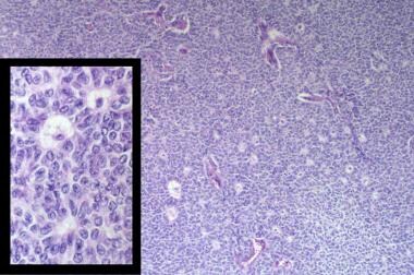
Thecoma
Sex Cord-Gonadal Stromal Tumors
Encyclopedias as Topic
Granulosa Cell Tumor
Sertoli Cell Tumor
Luteoma
Malignant fibrothecomatous tumour of the ovary: diagnostic value of anti-inhibin immunostaining. (1/26)
Malignant ovarian tumours of the fibrothecoma group are rare. The clinicopathological features of a case of ovarian malignant fibrothecoma in which there was metastatic disease in the small intestine and peritoneum at presentation are described. A number of differential diagnoses were considered but positive immunohistochemical staining of the resected ovarian and small intestinal neoplasms with anti-inhibin was of value in confirming a sex cord-stromal tumour and in excluding other lesions. The two tumours were also ultrastructurally identical. Classical malignant fibrothecomas are said to show four or more mitotic figures per 10 high power fields (HPF). Although the intestinal secondary was mitotically active, the primary ovarian tumour contained only one to two mitoses per 10 HPF, showing that formal mitotic counts are not an absolute indicator of malignant behaviour in this group of tumours. (+info)Frequent activation of AKT2 and induction of apoptosis by inhibition of phosphoinositide-3-OH kinase/Akt pathway in human ovarian cancer. (2/26)
We previously demonstrated that AKT2, a member of protein kinase B family, is activated by a number of growth factors via Ras and PI 3-kinase signaling pathways. Here, we report the frequent activation of AKT2 in human primary ovarian cancer and induction of apoptosis by inhibition of phosphoinositide-3-OH kinase (PI 3-kinase)/Akt pathway. In vitro AKT2 kinase assay analyses in 91 ovarian cancer specimens revealed elevated levels of AKT2 activity (>3-fold) in 33 cases (36.3%). The majority of tumors displaying activated AKT2 were high grade and stages III and IV. Immunostaining and Western blot analyses using a phospho-ser-473 Akt antibody that detects the activated form of AKT2 (AKT2 phosphorylated at serine-474) confirmed the frequent activation of AKT2 in ovarian cancer specimens. Phosphorylated AKT2 in tumor specimens localized to the cell membrane and cytoplasm but not the nucleus. To address the mechanism of AKT2 activation, we measured in vitro PI 3-kinase activity in 43 ovarian cancer specimens, including the 33 cases displaying elevated AKT2 activation. High levels of PI 3-kinase activity were observed in 20 cases, 15 of which also exhibited AKT2 activation. The remaining five cases displayed elevated AKT1 activation. Among the cases with elevated AKT2, but not PI 3-kinase activity (18 cases), three showed down-regulation of PTEN protein expression. Inhibition of PI 3-kinase/AKT2 by wortmannin or LY294002 induces apoptosis in ovarian cancer cells exhibiting activation of the PI 3-kinase/AKT2 pathway. These findings demonstrate for the first time that activation of AKT2 is a common occurrence in human ovarian cancer and that PI 3-kinase/Akt pathway may be an important target for ovarian cancer intervention. (+info)Malignant theca cell tumor of rat. (3/26)
A large-sized ovarian tumor of theca cell origin was found in a female rat. The mass was located in the right ovary position. Histologically, the tumor was covered by thin fibrous capsule and consisted of a solid area and an abundant necrotic area. Tumor cells were arranged in a storiform or whorled pattern. Connective tissue elements occasionally presented as bundles of dense collagen fibers. Fusiform to elongated cells had oval- to spindle-shaped nuclei with indistinct nucleoli. Large round nuclei and mitotic figures were scattered throughout the tumor cells. These cells were stained positively with S-100 but negatively with vimentin and a-smooth muscle actin. Tumor cells with abundant cytoplasm sometimes contained multiple small-sized lipid vacuoles. (+info)Malignant granulosa-theca cell tumor in a two-year-old Miniature Horse. (4/26)
A 2-year-old female Miniature Horse that presented with a history of progressive weight loss, depression, and diarrhea was diagnosed at necropsy with a highly malignant abdominal neoplasm involving the left ovary, kidneys, adrenal glands, intestines, and various abdominal and thoracic lymph nodes. Microscopic examination of these masses revealed large pleomorphic cells that stained positive for vimentin and inhibin and negative for epithelial membrane antigen and placental alkaline phosphatase. Ultrastructural examination of the cells revealed a high nucleocytoplasmic ratio and indented euchromatic nuclei with large nucleoli. Based on the gross, microscopic, immunohistochemical, and ultrastructural features, the neoplasm was identified as a malignant granulosa-theca cell tumor, a rare neoplasm in young horses. (+info)Overexpression of human chorionic gonadotropin causes multiple reproductive defects in transgenic mice. (5/26)
Human CG is a pregnancy marker secreted by the placenta, and it utilizes the same receptors as does LH. Human CG is a heterodimer, and its subunits are expressed in tissues other than placenta. Similarly, LH/hCG receptors are also expressed in multiple tissues; however, the physiological significance of this expression is unknown. Free hCGbeta is efficiently secreted in vitro in transfected cells and is highly expressed in many human cancers; however, the biological effects of free hCGbeta in vivo are unknown. To study in vivo consequences of elevated levels of free hCGbeta and hCG dimer in both male and female reproductive physiology, we used mouse metallothionein 1 promoter to generate multiple lines of transgenic mice that overexpressed either one or both subunits of hCG. Although mice expressing the glycoprotein hormone alpha subunit are normal and fertile, both male and female transgenic mice overexpressing only the hormone-specific hCGbeta subunit are infertile. The hCGbeta subunit-expressing transgenic female mice progressively develop cystic ovaries, whereas the male transgenic mice are infertile but otherwise are not phenotypically discernible. In contrast, both the male and female transgenic mice coexpressing high levels of the hCG subunits (i.e., the hCG dimer) demonstrate multiple reproductive defects. The male transgenic mice have Leydig cell hyperplasia, very high levels of serum testosterone, reduced testis size, and dramatically enlarged seminal vesicles and are infertile and display overly aggressive behavior when caged with females. The female transgenic mice are also infertile, have elevated levels of serum estradiol, and progressively develop hemorrhagic and cystic ovaries with thecal layer enlargement and stromal cell proliferation and degenerating kidneys. These results suggest that the in vivo biological effects of ectopically expressed free hCGbeta subunit are distinct from those of the hCG dimer and are gender specific. These transgenic mice are useful models for studying the biology of free hCGbeta subunit, for further analyzing the gain of function effects of hCG during early Leydig cell development, and for studying the roles of hCG in ovarian and kidney pathophysiology and function. (+info)Ovarian hematoma in an 11-year-old Thoroughbred-Hanovarian mare. (6/26)
An aggressive mare, presented for prebreeding examination, was found to have a significantly enlarged ovary (soccer ball size). A granulosa thecal cell tumor was initially suspected. Following laboratory and repeated clinical examination, the mare was diagnosed with an ovarian hematoma, which regressed with treatment. (+info)Meigs' syndrome with elevated CA 125: case report. (7/26)
CONTEXT: Meigs' syndrome consists of a benign ovarian tumor accompanied by ascites and hydrothorax. Elevated serum CA 125 levels in postmenopausal women with solid adnexal masses, ascites and pleural effusion are highly suggestive for malignant ovarian tumor. However, patients with Meigs' syndrome can also have elevated serum CA 125 levels. The authors report a case of Meigs' syndrome with elevated CA 125 level. OBJECTIVE: This is a case report of Meigs' syndrome with elevated CA 125 level. CASE REPORT: A 65-year-old Brazilian woman had presented progressive dyspnea, weight loss and decline in general condition over the 7 months preceding admission to our service. In another hospital, the patient had been submitted to thoracic drainage due to pleural effusion. With recurrence of the pleural effusion and increase in abdominal volume due to ascites and a pelvic mass, the patient sought our service. Transvaginal ultrasound showed an extensive adnexal solid mass of 16.4 x 10.8 cm located in the pelvis without exact limits, and the serum CA 125 level was elevated. With a preoperative diagnosis of ovarian carcinoma, the patient was submitted to exploratory laparotomy, which revealed a left ovarian tumor. The frozen section diagnosis was thecoma. Total abdominal hysterectomy with bilateral salpingo-oophorectomy was performed. The histology of the specimen confirmed the diagnosis of thecoma. The patient was asymptomatic with a normal serum CA 125 level 20 months after the operation. (+info)Bilateral occurrence of granulosa-theca cell tumors in an Arabian mare. (8/26)
An Arabian mare was referred for right granulosa-theca cell tumor (GTCT) evaluation. The mare was presented 4.5 years later for a left GTCT, after successfully conceiving and delivering a normal foal in the interim. The concurrent or nonconcurrent occurrence of bilateral GTCT in mares appears to be rare. (+info)A thecoma is a type of ovarian sex cord-stromal tumor, which are rare tumors that develop from the supporting cells (stromal cells) or the cells that produce hormones (sex cord cells) in the ovary. These tumors account for about 2% of all ovarian tumors.
Thecomas specifically arise from stromal cells that produce estrogen and other sex hormones. They are typically slow-growing and may not cause any symptoms, or they may cause symptoms related to hormonal imbalances such as irregular menstrual periods, vaginal bleeding, or postmenopausal bleeding. In some cases, thecomas can also grow large enough to cause abdominal discomfort or bloating.
Most thecomas are benign (non-cancerous), but a small percentage of them can be malignant (cancerous) and may spread to other parts of the body. Treatment for thecomas typically involves surgical removal of the tumor, and in some cases, hormonal therapy or chemotherapy may also be recommended.
Sex cord-gonadal stromal tumors are a type of rare cancer that develops in the cells of the ovaries or testicles that produce hormones and help to form ova or sperm. These tumors can be benign (noncancerous) or malignant (cancerous), and they can occur in both males and females, although they are more common in females.
There are several subtypes of sex cord-gonadal stromal tumors, including granulosa cell tumors, thecomas, fibromas, Sertoli cell tumors, Leydig cell tumors, and gonadoblastomas. The symptoms and treatment options for these tumors depend on several factors, including the type and stage of the tumor, the patient's age and overall health, and whether the tumor is producing hormones.
Common symptoms of sex cord-gonadal stromal tumors may include abdominal pain or swelling, bloating, irregular menstrual periods, vaginal bleeding, or a feeling of fullness in the abdomen. In some cases, these tumors may produce hormones that can cause additional symptoms, such as breast tenderness, acne, or voice deepening.
Treatment for sex cord-gonadal stromal tumors typically involves surgery to remove the tumor and any affected tissue. Depending on the stage and type of the tumor, additional treatments such as chemotherapy or radiation therapy may also be recommended. Regular follow-up care is important to monitor for recurrence and manage any long-term effects of treatment.
An encyclopedia is a comprehensive reference work containing articles on various topics, usually arranged in alphabetical order. In the context of medicine, a medical encyclopedia is a collection of articles that provide information about a wide range of medical topics, including diseases and conditions, treatments, tests, procedures, and anatomy and physiology. Medical encyclopedias may be published in print or electronic formats and are often used as a starting point for researching medical topics. They can provide reliable and accurate information on medical subjects, making them useful resources for healthcare professionals, students, and patients alike. Some well-known examples of medical encyclopedias include the Merck Manual and the Stedman's Medical Dictionary.
A Granulosa Cell Tumor is a type of sex cord-stromal tumor, which are uncommon neoplasms that arise from the supporting cells of the ovary or testis. These tumors account for approximately 5% of all ovarian tumors and can occur at any age, but they are most commonly found in perimenopausal and postmenopausal women.
Granulosa cell tumors originate from the granulosa cells, which are normally responsible for producing estrogen and supporting the development of the egg within the ovarian follicle. These tumors can be functional, meaning they produce hormones, or nonfunctional. Functional granulosa cell tumors often secrete estrogen, leading to symptoms such as irregular menstrual periods, postmenopausal bleeding, and, in rare cases, the development of male characteristics (virilization) due to androgen production.
Granulosa cell tumors are typically slow-growing and can vary in size. They are often diagnosed at an early stage because they cause symptoms related to hormonal imbalances or, less commonly, due to abdominal pain or distention caused by the growing mass. The diagnosis is usually confirmed through imaging studies (such as ultrasound, CT, or MRI) and a biopsy or surgical removal of the tumor, followed by histopathological examination.
Treatment for granulosa cell tumors typically involves surgery to remove the tumor and, in some cases, adjacent organs if there is evidence of spread. The role of chemotherapy and radiation therapy is less clear, but they may be used in certain situations, such as advanced-stage disease or high-risk features. Regular follow-up with imaging studies and tumor marker measurements (such as inhibin) is essential due to the risk of recurrence, even many years after initial treatment.
A Sertoli cell tumor is a rare type of sex-cord stromal tumor that develops in the testicles or, more rarely, in the ovaries. These tumors arise from the Sertoli cells, which are specialized cells within the testicle that help to nurture and protect the developing sperm cells. In the ovary, Sertoli cell tumors are thought to arise from similar cells that are part of the supporting tissue in the ovary.
Sertoli cell tumors can occur in people of any age but are most commonly found in middle-aged adults. They are usually slow-growing and may not cause any symptoms, especially if they are small. However, larger tumors or those that have spread (metastasized) may cause various symptoms depending on their location and size.
Symptoms of a Sertoli cell tumor can include:
* A painless lump or swelling in the testicle or ovary
* Abdominal pain or discomfort
* Bloating or a feeling of fullness in the abdomen
* Changes in bowel habits or urinary frequency
* Pain during sexual intercourse (in women)
* Hormonal imbalances, such as gynecomastia (breast development) in men or menstrual irregularities in women.
Diagnosis of a Sertoli cell tumor typically involves a combination of imaging tests, such as ultrasound, CT scan, or MRI, and blood tests to check for elevated levels of certain hormones that may be produced by the tumor. A biopsy may also be performed to confirm the diagnosis and determine the tumor's grade and stage.
Treatment for Sertoli cell tumors typically involves surgical removal of the tumor, along with any affected lymph nodes or other tissues. Additional treatments, such as radiation therapy or chemotherapy, may be recommended in cases where the tumor has spread or is at a higher risk of recurrence. Regular follow-up care is also important to monitor for any signs of recurrence or new tumors.
A luteoma is a benign ovarian tumor that is composed of luteinized cells, which are typically found in the corpus luteum of the ovary. The corpus luteum is a temporary endocrine structure that forms during the menstrual cycle and produces progesterone to support pregnancy.
Luteomas are rare tumors that usually occur in women of reproductive age, particularly those who have used fertility drugs or who have had prolonged exposure to high levels of estrogen. They can be asymptomatic or may cause symptoms such as abdominal pain, bloating, and menstrual irregularities.
Luteomas are typically diagnosed through imaging studies such as ultrasound or CT scan, and the diagnosis is confirmed through biopsy or surgical removal of the tumor. Treatment usually involves surgical removal of the tumor, and the prognosis is generally good, with a low risk of recurrence. However, luteomas can produce high levels of hormones that may cause virilization or other endocrine abnormalities, so follow-up care is important to monitor for any potential complications.
Theca cells are specialized cells that are part of the follicle where the egg matures in the ovary. They are located in the outer layer of the follicle and play an important role in producing hormones necessary for the growth and development of the follicle and the egg within it. Specifically, they produce androgens, such as testosterone, which are then converted into estrogens by another type of cells in the follicle called granulosa cells. These hormones help to thicken the lining of the uterus in preparation for a possible pregnancy. In some cases, theca cells can become overactive and produce too much testosterone, leading to conditions such as polycystic ovary syndrome (PCOS).
 Thecoma
Sex cord-gonadal stromal tumour
Leydig cell tumour
Theca of follicle
Reinke crystals
Ovarian cancer
List of MeSH codes (C04)
List of MeSH codes (C13)
List of MeSH codes (C19)
International Classification of Diseases for Oncology
Testicular cancer
Thecoma - Wikipedia
 Sex Cord Stromal Ovary Tumor Pathology: Sex Cord-Stromal Tumors, Granulosa Cell Tumor Group, Thecoma-Fibroma Group
 Ovarian Thecoma - Symptoms, Treatment & Support - Without a Ribbon
 Rare Gynecological Cancers: Granulosa Cell Tumors and Thecoma
 Buy The Coma 2: Vicious Sisters | Xbox
 The Coma: Cutting Class (Game) - Giant Bomb
 NGC 4921: Outskirts of the Coma Cluster | The Planetary Society
 Block 7 Reproduction Creighton Univ W Xpl Pt 4 - ProProfs Quiz
 101 Must-See Cosmic Objects: The Coma galaxy cluster
 Testicular Cancer - EPIDEMIOLOGY AETIOLOGY PATHOLOGY - Uroweb
 The Humble Store: Great games. Fantastic prices. Support charity.
 ESA Science & Technology - Ground-based image of the Coma Galaxy Cluster (Abell 1656)
 The Coma 2
 The Coma: Recut (PlayStation 4) Review - Page 1 - Cubed3
 The Coma 2: Vicious Sisters: A Traumatic Take on Eldritch Horror - Console Monster
 XIX - Female Genital System and the Breast Flashcards by Paula Victoria Catherine Cheng | Brainscape
 Journal of Astrophysics and Astronomy | Indian Academy of Sciences
Indian Journal of Pathology and Microbiology (IJPM): Table of Contents
 Thallium - Therapeutic Relationship | Taber's® Cyclopedic Medical Dictionary, 24e | F.A. Davis PT Collection | McGraw Hill...
Chen, P. H.<...
 Namespace
Plus it
 Escape A Psychotic Killer In The Coma II | Nintendo Insider
 Pagina 8 - Endometriosi.it
Code System Concept
 tumor
tumor
Bio2Vec
 Sclerosing stromal tumour of ovary - A clinicopathological and immunohistochemical study of five cases - Fingerprint
-...
Sclerosing stromal tumour of ovary - A clinicopathological and immunohistochemical study of five cases - Fingerprint
-...
 Overview of ovarian tumors
Fibroma3
- Ovarian thecoma-fibroma groups are uncommon sex cord-stromal neoplasms, constituting 1.0%-4.0% of all ovarian tumors. (bvsalud.org)
- A combination of surface epithelial and thecoma-fibroma group is very rarely encountered. (bvsalud.org)
- Etiology Thecoma-fibroma tumors are a closely related group of benign tumors that arise from ovarian stroma and are often difficult for the imager and even pathologist to distinguish. (symptoma.com)
Ovarian9
- What is Ovarian Thecoma or Thecoma of the Ovary? (withoutaribbon.org)
- Ovarian thecoma is a rare type of benign (non-cancerous) tumour that accounts for approximately 1% of benign ovarian tumours. (withoutaribbon.org)
- However, cases of ovarian thecoma in children and adolescents are extremely rare. (withoutaribbon.org)
- And most common cases of ovarian thecoma are found in post-menopausal women. (withoutaribbon.org)
- However, Certain hereditary and genetic mutations (faulty alterations in the genes) are believed to be responsible for the development of ovarian Thecoma. (withoutaribbon.org)
- Surgery is the most common and effective treatment for ovarian thecoma. (withoutaribbon.org)
- What Support can we Give for Ovarian Thecoma? (withoutaribbon.org)
- Ovarian Thecoma is rare cancer, meaning it is not as well known as other forms of cancer. (withoutaribbon.org)
- If you suffer from rare cancer such as Ovarian Thecoma, we can help and support you through your journey thanks to the generous donations we receive. (withoutaribbon.org)
Ovary1
- Incidental FOXL2 mutated adult granulosa cell tumour of the ovary with thecoma-like foci. (nih.gov)
Tumors1
- In honor of Gynecological Cancer Awareness Month this September, we're spreading the word about two lesser-known gynecological cancers: granulosa cell tumors and thecoma. (avantgynecology.com)
Develop1
- A fifth of patients diagnosed with a thecoma may also develop endometrial carcinoma, a common form of uterine cancer. (avantgynecology.com)
Form1
- During the recent apparition of Comet Halley in 1985-86 a transient ionic event in the form of a blob of H 2 O + emission was recorded in thecoma at ∼ 0 h UT on 1986 March 13. (ias.ac.in)