Cavernous Sinus Thrombosis
Lemierre Syndrome
Fusobacterium necrophorum
Phlebitis
Jugular Veins
Cephapirin
Postphlebitic Syndrome
Orbital Cellulitis
Catheterization, Peripheral
Behcet Syndrome
Phlebography
Stanozolol
Femoral Vein
Heparinoids
Mesenteric Veins
Extravasation of Diagnostic and Therapeutic Materials
Catheterization, Central Venous
Ultrasonography, Doppler, Duplex
Venous Insufficiency
Radionuclide Imaging
Catheter-Related Infections
Ultrasonography
Heparin
Sepsis
Tomography, X-Ray Computed
Treatment Outcome
Postoperative Complications
Pain
Effect of intravenous dextran 70 and pneumatic leg compression on incidence of postoperative pulmonary embolism. (1/774)
The incidence of pulmonary embolism and deep vein thrombosis was measured in 50 matched pairs of patients undergoing common surgical procedures with preoperative and postoperative ventilation-perfusion lung scans and the fibrinogen uptake test. One patient in each pair was treated with intravenous dextran 70 and pneumatic leggings. The incidence of pulmonary embolism among the treated patients was significantly reduced from 24% to 8%, but the incidence of deep vein thrombosis was not significantly reduced (34% to 24%). (+info)The intrarenal vascular lesions associated with primary antiphospholipid syndrome. (2/774)
Even 10 yr after the identification of the antiphospholipid syndrome (APS), renal involvement in the course of APS is still relatively unrecognized, and is probably underestimated. The association of anticardiolipin antibodies and/or lupus anticoagulant with the development of a vaso-occlusive process involving numerous organs is now confirmed. In a multicenter study, 16 cases of "primary" APS (PAPS) were found and followed for 5 yr or more, all with renal biopsy. In all 16 cases of PAPS, there was a vascular nephropathy characterized by small vessel vaso-occlusive lesions associated with fibrous intimal hyperplasia of interlobular arteries (12 patients), recanalizing thrombi in arteries and arterioles (six patients), and focal cortical atrophy (10 patients). In combination, these led to progressive destruction of the kidney, accelerated by acute glomerular and arteriolar microangiopathy in five patients. Focal cortical atrophy is a distinctive lesion, present in 10 biopsies, and likely represents the histologic and functional renal analogue to the multiple cerebral infarcts detected on imaging studies. The clinical hallmark of this vascular nephropathy in PAPS is systemic hypertension, only variably associated with renal insufficiency, proteinuria, or hematuria. The ensemble of histologic renal lesions defined in this study should aid in the separation of the lesions found in cases of secondary APS, especially systemic lupus erythematosus, into those lesions related to APS and those related to the underlying disease. (+info)Thrombophilia as a multigenic disease. (3/774)
BACKGROUND AND OBJECTIVE: Venous thrombosis is a common disease annually affecting 1 in 1000 individuals. The multifactorial nature of the disease is illustrated by the frequent identification of one or more predisposing genetic and/or environmental risk factors in thrombosis patients. Most of the genetic defects known today affect the function of the natural anticoagulant pathways and in particular the protein C system. This presentation focuses on the importance of the genetic factors in the pathogenesis of inherited thrombophilia with particular emphasis on those defects which affect the protein C system. INFORMATION SOURCES: Published results in articles covered by the Medline database have been integrated with our original studies in the field of thrombophilia. STATE OF THE ART AND PERSPECTIVES: The risk of venous thrombosis is increased when the hemostatic balance between pro- and anti-coagulant forces is shifted in favor of coagulation. When this is caused by an inherited defect, the resulting hypercoagulable state is a lifelong risk factor for thrombosis. Resistance to activated protein C (APC resistance) is the most common inherited hypercoagulable state found to be associated with venous thrombosis. It is caused by a single point mutation in the factor V (FV) gene, which predicts the substitution of Arg506 with a Gln. Arg506 is one of three APC-cleavage sites and the mutation results in the loss of this APC-cleavage site. The mutation is only found in Caucasians but the prevalence of the mutant FV allele (FV:Q506) varies between countries. It is found to be highly prevalent (up to 15%) in Scandinavian populations, in areas with high incidence of thrombosis. FV:Q506 is associated with a 5-10-fold increased risk of thrombosis and is found in 20-60% of Caucasian patients with thrombosis. The second most common inherited risk factor for thrombosis is a point mutation (G20210A) in the 3' untranslated region of the prothrombin gene. This mutation is present in approximately 2% of healthy individuals and in 6-7% of thrombosis patients, suggesting it to be a mild risk factor of thrombosis. Other less common genetic risk factors for thrombosis are the deficiencies of natural anticoagulant proteins such as antithrombin, protein C or protein S. Such defects are present in less than 1% of healthy individuals and together they account for 5-10% of genetic defects found in patients with venous thrombosis. Owing to the high prevalence of inherited APC resistance (FV:Q506) and of the G20210A mutation in the prothrombin gene, combinations of genetic defects are relatively common in the general population. As each genetic defect is an independent risk factor for thrombosis, individuals with multiple defects have a highly increased risk of thrombosis. As a consequence, multiple defects are often found in patients with thrombosis. (+info)Factor V Leiden and antibodies against phospholipids and protein S in a young woman with recurrent thromboses and abortion. (4/774)
We describe the case of a 39-year-old woman who suffered two iliofemoral venous thromboses, a cerebral ischemic infarct and recurrent fetal loss. Initial studies showed high levels of antiphospholipid antibodies (APAs) and a moderate thrombocytopenia. After her second miscarriage, laboratory diagnosis revealed that the woman was heterozygous for the factor V Leiden mutation and had a functional protein S deficiency as well as anti-protein S and anti-beta 2-glycoprotein I antibodies. The impairment of the protein C pathway at various points could well explain the recurrent thromboses in the patient and supports the role of a disturbed protein C system in the pathophysiology of thrombosis in patients with APAs. (+info)Incidence of deep vein thrombosis and leg oedema in patients with strokes. (5/774)
In a series of 26 patients with strokes 13 had deep vein thrombosis (DVT) in the leg, demonstrated by fibrinogen scanning. In 10 patients the thrombosis was in the paralysed leg but the degree of paralysis was unrelated to the tendency to develop DVT, which usually occurred about the third day. Leg oedema in 10 patients was unrelated to the DVT. (+info)Usefulness of D-dimer, blood gas, and respiratory rate measurements for excluding pulmonary embolism. (6/774)
BACKGROUND: A study was undertaken to assess the usefulness of the SimpliRED D-dimer test, arterial oxygen tension, and respiratory rate measurement for excluding pulmonary embolism (PE) and venous thromboembolism (VTE). METHODS: Lung scans were performed in 517 consecutive medical inpatients with suspected acute PE over a one year period. Predetermined end points for objectively diagnosed PE in order of precedence were (1) a post mortem diagnosis, (2) a positive pulmonary angiogram, (3) a high probability ventilation perfusion lung scan when the pretest probability was also high, and (4) the unanimous opinion of an adjudication committee. Deep vein thrombosis (DVT) was diagnosed by standard ultrasound and venography. RESULTS: A total of 40 cases of PE and 37 cases of DVT were objectively diagnosed. The predictive value of a negative SimpliRED test for excluding objectively diagnosed PE was 0.99 (error rate 2/249), that of PaO2 of > or = 80 mm Hg (10.7 kPa) was 0.97 (error rate 5/160), and that of a respiratory rate of < or = 20/min was 0.95 (error rate 14/308). The best combination of findings for excluding PE was a negative SimpliRED test and PaO2 > or = 80 mm Hg, which gave a predictive value of 1.0 (error rate 0/93). The predictive value of a negative SimpliRED test for excluding VTE was 0.98 (error rate 5/249). CONCLUSIONS: All three of these observations are helpful in excluding PE. When any two parameters were normal, PE was very unlikely. In patients with a negative SimpliRED test and PaO2 of > or = 80 mm Hg a lung scan is usually unnecessary. Application of this approach for triage in the preliminary assessment of suspected PE could lead to a reduced rate of false positive diagnoses and considerable resource savings. (+info)Effect of prolonged infusion on vein calibre: a prospective study. (7/774)
Infusion thrombophlebitis is common and is the principal limitation to intravenous nutrition (IVN) via a peripheral vein, yet its precise pathogenesis is unclear. Prospective observations were performed on patients in whom a hypertonic, acidic, nutritional emulsion was infused via fine-bore polyurethane catheters placed in peripheral veins. B mode ultrasound was used to determine vein calibre and proved to be a useful means for serial examination during intravenous infusion. Contrary to previous reports, no evidence of venospasm was observed. It is suggested that previous evidence of venoconstriction is erroneous and that other mechanisms are responsible for thrombophlebitis. (+info)Combined oral contraceptives, smoking, and cardiovascular risk. (8/774)
STUDY OBJECTIVE: To assess age specific incidence and mortality of stroke, acute myocardial infarction (AMI), and idiopathic venous thromboembolism (VTE) associated with use of modern low dose combined oral contraceptives (OCs) and the interaction with smoking. DESIGN: Hospital-based case-control study. SETTING: Hospitals in Oxford region in the United Kingdom, which covered a defined population, during the period 1989-1993. METHODS: Relative risk estimates from the WHO Collaborative Study and observed incidence rates from the Oxford region were used to estimate age specific incidence of each disease among women without cardiovascular risk factors and model total cardiovascular incidence and mortality. RESULTS: Among women who did not use OCs, smoke nor had any other cardiovascular risk factors, total incidence of stroke and AMI were less than 2 events per 100,000 woman years in those aged 20-24 years and rose exponentially with age to 8 events per 100,000 among women aged 40-44 years. Incidence of idiopathic VTE among women who did not use OCs rose linearly with age (from 3.3 per 100,000 at ages 20-24 years to 5.8 per 100,000 at ages 40-44 years). The increased risk of idiopathic VTE associated with OC use among non-smokers constituted over 90% of all cardiovascular events for women aged 20-24 years and more than 60% in those aged 40-44 years. Fatal cardiovascular events were dominated by haemorrhagic stroke and AMI, and among OC users who smoked these two diseases accounted for 80% of cardiovascular deaths among women aged 20-24 years, rising to 97% among those aged 40-44 years. Cardiovascular mortality associated with smoking was greater than that associated with OC use at all ages. Attributable risk associated with OC use was 1 death per 370,000 users annually among women aged 20-24 years, 1 per 170,000 at ages 30-34 years, and 1 per 37,000 at ages 40-44 years. Among smokers, the cardiovascular mortality attributable to OC use was estimated to be about 1 per 100,000 users annually among women aged less than 35 years, and about 1 per 10,000 users annually among those above the age of 35 years. CONCLUSION: The incidence of fatal cardiovascular events among women aged less than 35 years is low. The VTE risk associated with OC use is the largest contributor to OC induced adverse effects. The potentially avoidable excess VTE risk associated with the newer progestogens desogestrel and gestodene would account for a substantial proportion of total cardiovascular morbidity in this age group. For women over age 35 years the absolute risks associated with OC use and smoking are greater because of the steeply rising incidence of arterial diseases. The combination of smoking and OC use among such women is associated with particularly increased risks. Any potential reduction in AMI or stroke risk with use of third generation OCs would be a more important consideration among older compared with younger women, particularly if they smoke. However, the mortality associated with smoking is far greater than that associated with OC use (of any type) at all ages. (+info)Thrombophlebitis is a medical condition characterized by the inflammation and clotting of blood in a vein, usually in the legs. The term thrombophlebitis comes from two words: "thrombo" which means blood clot, and "phlebitis" which refers to inflammation of the vein.
The condition can occur in superficial or deep veins. Superficial thrombophlebitis affects the veins just below the skin's surface, while deep vein thrombophlebitis (DVT) occurs in the deeper veins. DVT is a more serious condition as it can lead to complications such as pulmonary embolism if the blood clot breaks off and travels to the lungs.
Symptoms of thrombophlebitis may include redness, warmth, pain, swelling, or discomfort in the affected area. In some cases, there may be visible surface veins that are hard, tender, or ropy to touch. If left untreated, thrombophlebitis can lead to chronic venous insufficiency and other long-term complications. Treatment typically involves medications such as anticoagulants, antiplatelet agents, or thrombolytics, along with compression stockings and other supportive measures.
Cavernous sinus thrombosis is a medical condition that refers to the formation of a blood clot (thrombus) in the cavernous sinuses, which are located near the base of the brain and are important for draining blood from the face and brain. This condition can occur as a complication of an infection in the facial area or sinuses, or it can be associated with other medical conditions such as cancer or trauma.
Symptoms of cavernous sinus thrombosis may include headache, fever, eye pain, swelling or bulging of the eyes, double vision, and decreased vision. If left untreated, this condition can lead to serious complications such as meningitis, brain abscess, or even death. Treatment typically involves administering antibiotics to treat any underlying infection and anticoagulants to prevent further clot formation. In some cases, surgery may be necessary to remove the clot.
Fusobacterium infections are diseases or conditions caused by the bacterial genus Fusobacterium, which are gram-negative, anaerobic bacilli. These bacteria are commonly found as normal flora in the oral cavity, gastrointestinal tract, and female genital tract. However, under certain circumstances, they can cause infections, particularly in individuals with weakened immune systems or underlying medical conditions.
Fusobacterium infections can manifest in various forms, including:
1. Oral infections: Fusobacterium nucleatum is the most common species associated with oral infections, such as periodontitis, abscesses, and Ludwig's angina.
2. Respiratory tract infections: Fusobacterium necrophorum can cause lung abscesses, empyema, and bronchitis.
3. Bloodstream infections (bacteremia): Fusobacterium species can enter the bloodstream through various routes, such as dental procedures or invasive medical procedures, leading to bacteremia. This condition can be particularly dangerous for individuals with compromised immune systems or underlying medical conditions.
4. Intra-abdominal infections: Fusobacterium species can cause intra-abdominal abscesses, peritonitis, and appendicitis.
5. Skin and soft tissue infections: Fusobacterium species can cause cellulitis, myositis, and necrotizing fasciitis.
6. Bone and joint infections: Fusobacterium species can cause osteomyelitis and septic arthritis.
7. Central nervous system infections: Fusobacterium species can cause meningitis and brain abscesses, although these are rare.
Fusobacterium infections can be challenging to treat due to their anaerobic nature and resistance to certain antibiotics. Therefore, it is essential to seek medical attention if you suspect a Fusobacterium infection. Treatment typically involves the use of appropriate antibiotics, such as metronidazole or clindamycin, and sometimes surgical intervention may be necessary.
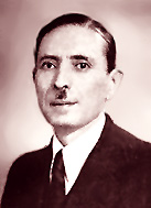
Lemierre Syndrome, also known as post-anginal septicemia or necrobacillosis, is a rare but serious medical condition that typically follows a recent pharyngitis (throat infection) or upper respiratory tract infection. It is characterized by the spread of infection from the oropharynx to the internal jugular vein and subsequent septicemia (bloodstream infection), leading to metastatic infectious complications, most commonly affecting the lungs. The causative organism is usually a bacterium called Fusobacterium necrophorum.
The syndrome was first described by French physician André Lemierre in 1936. Symptoms may include fever, chills, severe neck pain and stiffness, difficulty swallowing, swelling of the jaw or neck, shortness of breath, and the formation of abscesses in various parts of the body. Rapid diagnosis and appropriate antibiotic treatment are crucial to prevent potentially life-threatening complications.
Fusobacterium necrophorum is a gram-negative, anaerobic, non-spore forming rod-shaped bacterium. It is a normal inhabitant of the oral cavity, gastrointestinal tract and urogenital tract of humans and animals. However, it can cause various infections in humans, particularly in individuals with compromised immune systems.
Fusobacterium necrophorum is well known for its association with severe clinical conditions such as Lemierre's syndrome, which is a rare but life-threatening condition characterized by septic thrombophlebitis of the internal jugular vein and metastatic infections. It can also cause other suppurative infections including bronchitis, pneumonia, meningitis, brain abscesses, and septicemia. In addition, Fusobacterium necrophorum has been implicated in the pathogenesis of certain types of periodontal disease and is a significant cause of bacterial peritonitis in cirrhotic patients.
Phlebitis is a medical term that refers to the inflammation of a vein, usually occurring in the legs. The inflammation can be caused by blood clots (thrombophlebitis) or other conditions that cause irritation and swelling in the vein's lining. Symptoms may include redness, warmth, pain, and swelling in the affected area. In some cases, phlebitis may lead to serious complications such as deep vein thrombosis (DVT) or pulmonary embolism (PE), so it is essential to seek medical attention if you suspect you have this condition.
The jugular veins are a pair of large, superficial veins that carry blood from the head and neck to the heart. They are located in the neck and are easily visible when looking at the side of a person's neck. The external jugular vein runs along the surface of the muscles in the neck, while the internal jugular vein runs within the carotid sheath along with the carotid artery and the vagus nerve.
The jugular veins are important in clinical examinations because they can provide information about a person's cardiovascular function and intracranial pressure. For example, distention of the jugular veins may indicate heart failure or increased intracranial pressure, while decreased venous pulsations may suggest a low blood pressure or shock.
It is important to note that medical conditions such as deep vein thrombosis (DVT) can also affect the jugular veins and can lead to serious complications if not treated promptly.
Varicose veins are defined as enlarged, swollen, and twisting veins often appearing blue or dark purple, which usually occur in the legs. They are caused by weakened valves and vein walls that can't effectively push blood back toward the heart. This results in a buildup of blood, causing the veins to bulge and become varicose.
The condition is generally harmless but may cause symptoms like aching, burning, muscle cramp, or a feeling of heaviness in the legs. In some cases, varicose veins can lead to more serious problems, such as skin ulcers, blood clots, or chronic venous insufficiency. Treatment options include lifestyle changes, compression stockings, and medical procedures like sclerotherapy, laser surgery, or endovenous ablation.
Cephapirin is a type of antibiotic that belongs to the class of cephalosporins. It is used to treat various bacterial infections, including respiratory tract infections, skin and soft tissue infections, bone and joint infections, and genitourinary tract infections. Cephapirin works by interfering with the bacteria's ability to form a cell wall, which results in bacterial death.
Like other cephalosporins, cephapirin is generally well-tolerated and has a broad spectrum of activity against many different types of bacteria. However, it may cause side effects such as nausea, diarrhea, vomiting, and allergic reactions in some people. It is important to take cephapirin exactly as directed by a healthcare provider, and to complete the full course of treatment even if symptoms improve before all of the medication has been taken.
It's worth noting that Cephapirin is not a commonly used antibiotic now a days, due to the availability of other cephalosporins which are more effective and have less side effects.
Postphlebitic syndrome, also known as postthrombotic syndrome or post-thrombotic limb, is a long-term complication that can occur after deep vein thrombosis (DVT). It's characterized by chronic venous insufficiency due to damage in the valves and walls of the affected veins. This results in impaired return of blood from the extremities back to the heart, leading to symptoms such as:
1. Swelling (edema) in the affected limb, usually the lower leg or calf.
2. Pain, aching, or cramping in the legs.
3. Heaviness or fatigue in the legs.
4. Skin changes like redness, warmth, or itchiness.
5. Development of venous ulcers or sores, particularly around the ankles.
The severity of postphlebitic syndrome can vary from mild to severe and may significantly impact a person's quality of life. Risk factors for developing this condition include having had a previous DVT, obesity, older age, lack of physical activity, and a family history of blood clotting disorders. Early diagnosis and appropriate management of deep vein thrombosis can help reduce the risk of developing postphlebitic syndrome.
A pulmonary embolism (PE) is a medical condition that occurs when a blood clot, often formed in the deep veins of the legs (deep vein thrombosis), breaks off and travels to the lungs, blocking one or more pulmonary arteries. This blockage can lead to various symptoms such as shortness of breath, chest pain, rapid heart rate, and coughing up blood. In severe cases, it can cause life-threatening complications like low oxygen levels, hypotension, and even death if not promptly diagnosed and treated with anticoagulant medications or thrombolytic therapy to dissolve the clot.
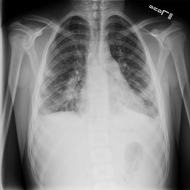
Orbital cellulitis is a serious infection involving the soft tissues within the orbit (the bony cavity containing the eye). This condition can cause symptoms such as eyelid swelling, redness, warmth, pain, and impaired eye movement. It may also be accompanied by fever, decreased vision, or altered mental status in severe cases. Orbital cellulitis often results from the spread of infection from nearby structures, such as the sinuses. Immediate medical attention is required to prevent potential complications like vision loss or intracranial infections. Treatment typically involves antibiotics and, in some cases, surgical intervention.
Peripheral catheterization is a medical procedure that involves the insertion of a thin, flexible tube (catheter) into a peripheral vein, which is a blood vessel located outside of the chest and abdomen. This type of catheterization is typically performed to administer medications, fluids, or nutritional support, or to monitor various physiological parameters such as central venous pressure.
Peripheral catheters are usually inserted into veins in the hands or arms, although they can also be placed in other peripheral veins. The procedure is typically performed using aseptic technique to minimize the risk of infection. Once the catheter is in place, it may be secured with a dressing or suture to prevent movement and dislodgement.
Peripheral catheterization is a relatively safe and common procedure that is routinely performed in hospitals, clinics, and other healthcare settings. However, like any medical procedure, it carries a small risk of complications such as infection, bleeding, or damage to the vein or surrounding tissues.
The saphenous vein is a term used in anatomical description to refer to the great or small saphenous veins, which are superficial veins located in the lower extremities of the human body.
The great saphenous vein (GSV) is the longest vein in the body and originates from the medial aspect of the foot, ascending along the medial side of the leg and thigh, and drains into the femoral vein at the saphenofemoral junction, located in the upper third of the thigh.
The small saphenous vein (SSV) is a shorter vein that originates from the lateral aspect of the foot, ascends along the posterior calf, and drains into the popliteal vein at the saphenopopliteal junction, located in the popliteal fossa.
These veins are often used as conduits for coronary artery bypass grafting (CABG) surgery due to their consistent anatomy and length.
Behçet syndrome is a rare inflammatory disease that can cause symptoms in various parts of the body. It's characterized by recurrent mouth sores (aphthous ulcers), genital sores, and inflammation of the eyes (uveitis). The condition may also cause skin lesions, joint pain and swelling, and inflammation of the digestive tract, brain, or spinal cord.
The exact cause of Behçet syndrome is not known, but it's thought to be an autoimmune disorder, in which the body's immune system mistakenly attacks its own healthy cells and tissues. The condition tends to affect men more often than women and typically develops during a person's 20s or 30s.
There is no cure for Behçet syndrome, but treatments can help manage symptoms and prevent complications. Treatment options may include medications such as corticosteroids, immunosuppressants, and biologics to reduce inflammation, as well as pain relievers and other supportive therapies.
Phlebography is a medical imaging technique used to visualize and assess the veins, particularly in the legs. It involves the injection of a contrast agent into the veins, followed by X-ray imaging to capture the flow of the contrast material through the veins. This allows doctors to identify any abnormalities such as blood clots, blockages, or malformations in the venous system.
There are different types of phlebography, including ascending phlebography (where the contrast agent is injected into a foot vein and travels up the leg) and descending phlebography (where the contrast agent is injected into a vein in the groin or neck and travels down the leg).
Phlebography is an invasive procedure that requires careful preparation and monitoring, and it is typically performed by radiologists or vascular specialists. It has largely been replaced by non-invasive imaging techniques such as ultrasound and CT angiography in many clinical settings.
Stanozolol is a synthetic anabolic-androgenic steroid (AAS) derivative of dihydrotestosterone (DHT). It is commonly used in medicine for the treatment of hereditary angioedema and was formerly used to promote muscle growth in weakened or catabolic patients. Stanozolol has a high anabolic and moderate androgenic activity, with reduced estrogenic properties compared to testosterone. Its chemical formula is (17α-methyl-5α-androstano[2,3-c]pyrazol-17β-ol). It is important to note that the use of Stanozolol for performance enhancement is considered illegal and subject to severe penalties in many countries, including disqualification from sports events and criminal charges.
The femoral vein is the large vein that runs through the thigh and carries oxygen-depleted blood from the lower limbs back to the heart. It is located in the femoral triangle, along with the femoral artery and nerve. The femoral vein begins at the knee as the popliteal vein, which then joins with the deep vein of the thigh to form the femoral vein. As it moves up the leg, it is joined by several other veins, including the great saphenous vein, before it becomes the external iliac vein at the inguinal ligament in the groin.
Heparinoids are a group of substances that have similar properties to heparin, a highly sulfated glycosaminoglycan found in mast cells and basophils. Heparin is a powerful anticoagulant that works by accelerating the action of an enzyme called antithrombin III, which inhibits the formation of blood clots.
Heparinoids are often used as alternative anticoagulants to heparin in clinical settings. They have similar mechanisms of action and can also inhibit the coagulation cascade, preventing the formation of blood clots. However, heparinoids have a lower anticoagulant activity than heparin and may have different side effect profiles.
Examples of heparinoids include low molecular weight heparins (LMWHs), fondaparinux, and danaparoid. LMWHs are derived from standard heparin by chemical or enzymatic depolymerization and have a lower molecular weight than heparin. They have a more predictable anticoagulant response and longer half-life than standard heparin, making them useful for outpatient treatment of deep vein thrombosis and pulmonary embolism.
Fondaparinux is a synthetic pentasaccharide that selectively binds to antithrombin III and enhances its inhibitory activity against factor Xa, a key enzyme in the coagulation cascade. It has a long half-life and predictable pharmacokinetics, making it useful for the prevention and treatment of venous thromboembolism.
Danaparoid is a mixture of heparan sulfate, dermatan sulfate, and chondroitin sulfate derived from pig intestinal mucosa. It has a lower anticoagulant activity than heparin but a longer half-life and less frequent dosing requirements. Danaparoid is used for the prevention and treatment of venous thromboembolism, as well as for the management of heparin-induced thrombocytopenia (HIT), a rare but serious complication of heparin therapy.
The mesenteric veins are a set of blood vessels that are responsible for draining deoxygenated blood from the small and large intestines. There are two main mesenteric veins: the superior mesenteric vein and the inferior mesenteric vein. The superior mesenteric vein drains blood from the majority of the small intestine, as well as the ascending colon and proximal two-thirds of the transverse colon. The inferior mesenteric vein drains blood from the distal third of the transverse colon, descending colon, sigmoid colon, and rectum. These veins ultimately drain into the portal vein, which carries the blood to the liver for further processing.
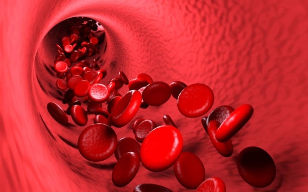
Veins are blood vessels that carry deoxygenated blood from the tissues back to the heart. They have a lower pressure than arteries and contain valves to prevent the backflow of blood. Veins have a thin, flexible wall with a larger lumen compared to arteries, allowing them to accommodate more blood volume. The color of veins is often blue or green due to the absorption characteristics of light and the reduced oxygen content in the blood they carry.
Thrombosis is the formation of a blood clot (thrombus) inside a blood vessel, obstructing the flow of blood through the circulatory system. When a clot forms in an artery, it can cut off the supply of oxygen and nutrients to the tissues served by that artery, leading to damage or tissue death. If a thrombus forms in the heart, it can cause a heart attack. If a thrombus breaks off and travels through the bloodstream, it can lodge in a smaller vessel, causing blockage and potentially leading to damage in the organ that the vessel supplies. This is known as an embolism.
Thrombosis can occur due to various factors such as injury to the blood vessel wall, abnormalities in blood flow, or changes in the composition of the blood. Certain medical conditions, medications, and lifestyle factors can increase the risk of thrombosis. Treatment typically involves anticoagulant or thrombolytic therapy to dissolve or prevent further growth of the clot, as well as addressing any underlying causes.
Extravasation of diagnostic and therapeutic materials refers to the unintended leakage or escape of these substances from the intended vasculature into the surrounding tissues. This can occur during the administration of various medical treatments, such as chemotherapy, contrast agents for imaging studies, or other injectable medications.
The extravasation can result in a range of complications, depending on the type and volume of the material that has leaked, as well as the location and sensitivity of the surrounding tissues. Possible consequences include local tissue damage, inflammation, pain, and potential long-term effects such as fibrosis or necrosis.
Prompt recognition and management of extravasation are essential to minimize these complications. Treatment may involve local cooling or heating, the use of hyaluronidase or other agents to facilitate dispersion of the extravasated material, or surgical intervention in severe cases.
Central venous catheterization is a medical procedure in which a flexible tube called a catheter is inserted into a large vein in the body, usually in the neck (internal jugular vein), chest (subclavian vein), or groin (femoral vein). The catheter is threaded through the vein until it reaches a central location, such as the superior vena cava or the right atrium of the heart.
Central venous catheterization may be performed for several reasons, including:
1. To administer medications, fluids, or nutritional support directly into the bloodstream.
2. To monitor central venous pressure (CVP), which can help assess a patient's volume status and cardiac function.
3. To draw blood samples for laboratory tests.
4. To deliver chemotherapy drugs or other medications that may be harmful to peripheral veins.
5. To provide access for hemodialysis or other long-term therapies.
The procedure requires careful attention to sterile technique to minimize the risk of infection, and it is usually performed under local anesthesia with sedation or general anesthesia. Complications of central venous catheterization may include bleeding, infection, pneumothorax (collapsed lung), arterial puncture, and catheter-related bloodstream infections (CRBSI).
Ultrasonography, Doppler, and Duplex are diagnostic medical techniques that use sound waves to create images of internal body structures and assess their function. Here are the definitions for each:
1. Ultrasonography: Also known as ultrasound, this is a non-invasive imaging technique that uses high-frequency sound waves to produce images of internal organs and tissues. A small handheld device called a transducer is placed on the skin surface, which emits and receives sound waves. The returning echoes are then processed to create real-time visual images of the internal structures.
2. Doppler: This is a type of ultrasound that measures the velocity and direction of blood flow in the body by analyzing the frequency shift of the reflected sound waves. It can be used to assess blood flow in various parts of the body, such as the heart, arteries, and veins.
3. Duplex: Duplex ultrasonography is a combination of both gray-scale ultrasound and Doppler ultrasound. It provides detailed images of internal structures, as well as information about blood flow velocity and direction. This technique is often used to evaluate conditions such as deep vein thrombosis, carotid artery stenosis, and peripheral arterial disease.
In summary, ultrasonography is a diagnostic imaging technique that uses sound waves to create images of internal structures, Doppler is a type of ultrasound that measures blood flow velocity and direction, and duplex is a combination of both techniques that provides detailed images and information about blood flow.
Venous insufficiency is a medical condition that occurs when the veins, particularly in the legs, have difficulty returning blood back to the heart due to impaired valve function or obstruction in the vein. This results in blood pooling in the veins, leading to symptoms such as varicose veins, swelling, skin changes, and ulcers. Prolonged venous insufficiency can cause chronic pain and affect the quality of life if left untreated.
Radionuclide imaging, also known as nuclear medicine, is a medical imaging technique that uses small amounts of radioactive material, called radionuclides or radiopharmaceuticals, to diagnose and treat various diseases and conditions. The radionuclides are introduced into the body through injection, inhalation, or ingestion and accumulate in specific organs or tissues. A special camera then detects the gamma rays emitted by these radionuclides and converts them into images that provide information about the structure and function of the organ or tissue being studied.
Radionuclide imaging can be used to evaluate a wide range of medical conditions, including heart disease, cancer, neurological disorders, gastrointestinal disorders, and bone diseases. The technique is non-invasive and generally safe, with minimal exposure to radiation. However, it should only be performed by qualified healthcare professionals in accordance with established guidelines and regulations.
Venous thrombosis is a medical condition characterized by the formation of a blood clot (thrombus) in the deep veins, often in the legs (deep vein thrombosis or DVT), but it can also occur in other parts of the body such as the arms, pelvis, or lungs (pulmonary embolism).
The formation of a venous thrombus can be caused by various factors, including injury to the blood vessel wall, changes in blood flow, and alterations in the composition of the blood. These factors can lead to the activation of clotting factors and platelets, which can result in the formation of a clot that blocks the vein.
Symptoms of venous thrombosis may include swelling, pain, warmth, and redness in the affected area. In some cases, the clot can dislodge and travel to other parts of the body, causing potentially life-threatening complications such as pulmonary embolism.
Risk factors for venous thrombosis include advanced age, obesity, smoking, pregnancy, use of hormonal contraceptives or hormone replacement therapy, cancer, recent surgery or trauma, prolonged immobility, and a history of previous venous thromboembolism. Treatment typically involves the use of anticoagulant medications to prevent further clotting and dissolve existing clots.
Catheter-related infections are infections that occur due to the presence of a catheter, a flexible tube that is inserted into the body to perform various medical functions such as draining urine or administering medication. These infections can affect any part of the body where a catheter is inserted, including the bladder, bloodstream, heart, and lungs.
The most common type of catheter-related infection is a catheter-associated urinary tract infection (CAUTI), which occurs when bacteria enter the urinary tract through the catheter and cause an infection. Symptoms of CAUTI may include fever, chills, pain or burning during urination, and cloudy or foul-smelling urine.
Other types of catheter-related infections include catheter-associated bloodstream infections (CLABSI), which can occur when bacteria enter the bloodstream through the catheter, and catheter-related pulmonary infections, which can occur when secretions from the respiratory tract enter the lungs through a catheter.
Catheter-related infections are a significant concern in healthcare settings, as they can lead to serious complications such as sepsis, organ failure, and even death. Proper catheter insertion and maintenance techniques, as well as regular monitoring for signs of infection, can help prevent these types of infections.
Anticoagulants are a class of medications that work to prevent the formation of blood clots in the body. They do this by inhibiting the coagulation cascade, which is a series of chemical reactions that lead to the formation of a clot. Anticoagulants can be given orally, intravenously, or subcutaneously, depending on the specific drug and the individual patient's needs.
There are several different types of anticoagulants, including:
1. Heparin: This is a naturally occurring anticoagulant that is often used in hospitalized patients who require immediate anticoagulation. It works by activating an enzyme called antithrombin III, which inhibits the formation of clots.
2. Low molecular weight heparin (LMWH): LMWH is a form of heparin that has been broken down into smaller molecules. It has a longer half-life than standard heparin and can be given once or twice daily by subcutaneous injection.
3. Direct oral anticoagulants (DOACs): These are newer oral anticoagulants that work by directly inhibiting specific clotting factors in the coagulation cascade. Examples include apixaban, rivaroxaban, and dabigatran.
4. Vitamin K antagonists: These are older oral anticoagulants that work by inhibiting the action of vitamin K, which is necessary for the formation of clotting factors. Warfarin is an example of a vitamin K antagonist.
Anticoagulants are used to prevent and treat a variety of conditions, including deep vein thrombosis (DVT), pulmonary embolism (PE), atrial fibrillation, and prosthetic heart valve thrombosis. It is important to note that anticoagulants can increase the risk of bleeding, so they must be used with caution and regular monitoring of blood clotting times may be required.
A syndrome, in medical terms, is a set of symptoms that collectively indicate or characterize a disease, disorder, or underlying pathological process. It's essentially a collection of signs and/or symptoms that frequently occur together and can suggest a particular cause or condition, even though the exact physiological mechanisms might not be fully understood.
For example, Down syndrome is characterized by specific physical features, cognitive delays, and other developmental issues resulting from an extra copy of chromosome 21. Similarly, metabolic syndromes like diabetes mellitus type 2 involve a group of risk factors such as obesity, high blood pressure, high blood sugar, and abnormal cholesterol or triglyceride levels that collectively increase the risk of heart disease, stroke, and diabetes.
It's important to note that a syndrome is not a specific diagnosis; rather, it's a pattern of symptoms that can help guide further diagnostic evaluation and management.
In medical terms, the leg refers to the lower portion of the human body that extends from the knee down to the foot. It includes the thigh (femur), lower leg (tibia and fibula), foot, and ankle. The leg is primarily responsible for supporting the body's weight and enabling movements such as standing, walking, running, and jumping.
The leg contains several important structures, including bones, muscles, tendons, ligaments, blood vessels, nerves, and joints. These structures work together to provide stability, support, and mobility to the lower extremity. Common medical conditions that can affect the leg include fractures, sprains, strains, infections, peripheral artery disease, and neurological disorders.
Ultrasonography, also known as sonography, is a diagnostic medical procedure that uses high-frequency sound waves (ultrasound) to produce dynamic images of organs, tissues, or blood flow inside the body. These images are captured in real-time and can be used to assess the size, shape, and structure of various internal structures, as well as detect any abnormalities such as tumors, cysts, or inflammation.
During an ultrasonography procedure, a small handheld device called a transducer is placed on the patient's skin, which emits and receives sound waves. The transducer sends high-frequency sound waves into the body, and these waves bounce back off internal structures and are recorded by the transducer. The recorded data is then processed and transformed into visual images that can be interpreted by a medical professional.
Ultrasonography is a non-invasive, painless, and safe procedure that does not use radiation like other imaging techniques such as CT scans or X-rays. It is commonly used to diagnose and monitor conditions in various parts of the body, including the abdomen, pelvis, heart, blood vessels, and musculoskeletal system.
Heparin is defined as a highly sulfated glycosaminoglycan (a type of polysaccharide) that is widely present in many tissues, but is most commonly derived from the mucosal tissues of mammalian lungs or intestinal mucosa. It is an anticoagulant that acts as an inhibitor of several enzymes involved in the blood coagulation cascade, primarily by activating antithrombin III which then neutralizes thrombin and other clotting factors.
Heparin is used medically to prevent and treat thromboembolic disorders such as deep vein thrombosis, pulmonary embolism, and certain types of heart attacks. It can also be used during hemodialysis, cardiac bypass surgery, and other medical procedures to prevent the formation of blood clots.
It's important to note that while heparin is a powerful anticoagulant, it does not have any fibrinolytic activity, meaning it cannot dissolve existing blood clots. Instead, it prevents new clots from forming and stops existing clots from growing larger.
Angiography is a medical procedure in which an x-ray image is taken to visualize the internal structure of blood vessels, arteries, or veins. This is done by injecting a radiopaque contrast agent (dye) into the blood vessel using a thin, flexible catheter. The dye makes the blood vessels visible on an x-ray image, allowing doctors to diagnose and treat various medical conditions such as blockages, narrowing, or malformations of the blood vessels.
There are several types of angiography, including:
* Cardiac angiography (also called coronary angiography) - used to examine the blood vessels of the heart
* Cerebral angiography - used to examine the blood vessels of the brain
* Peripheral angiography - used to examine the blood vessels in the limbs or other parts of the body.
Angiography is typically performed by a radiologist, cardiologist, or vascular surgeon in a hospital setting. It can help diagnose conditions such as coronary artery disease, aneurysms, and peripheral arterial disease, among others.
Sepsis is a life-threatening condition that arises when the body's response to an infection injures its own tissues and organs. It is characterized by a whole-body inflammatory state (systemic inflammation) that can lead to blood clotting issues, tissue damage, and multiple organ failure.
Sepsis happens when an infection you already have triggers a chain reaction throughout your body. Infections that lead to sepsis most often start in the lungs, urinary tract, skin, or gastrointestinal tract.
Sepsis is a medical emergency. If you suspect sepsis, seek immediate medical attention. Early recognition and treatment of sepsis are crucial to improve outcomes. Treatment usually involves antibiotics, intravenous fluids, and may require oxygen, medication to raise blood pressure, and corticosteroids. In severe cases, surgery may be required to clear the infection.
Diagnostic errors refer to inaccurate or delayed diagnoses of a patient's medical condition, which can lead to improper or unnecessary treatment and potentially serious harm to the patient. These errors can occur due to various factors such as lack of clinical knowledge, failure to consider all possible diagnoses, inadequate communication between healthcare providers and patients, and problems with testing or interpretation of test results. Diagnostic errors are a significant cause of preventable harm in medical care and have been identified as a priority area for quality improvement efforts.
Anti-bacterial agents, also known as antibiotics, are a type of medication used to treat infections caused by bacteria. These agents work by either killing the bacteria or inhibiting their growth and reproduction. There are several different classes of anti-bacterial agents, including penicillins, cephalosporins, fluoroquinolones, macrolides, and tetracyclines, among others. Each class of antibiotic has a specific mechanism of action and is used to treat certain types of bacterial infections. It's important to note that anti-bacterial agents are not effective against viral infections, such as the common cold or flu. Misuse and overuse of antibiotics can lead to antibiotic resistance, which is a significant global health concern.
X-ray computed tomography (CT or CAT scan) is a medical imaging method that uses computer-processed combinations of many X-ray images taken from different angles to produce cross-sectional (tomographic) images (virtual "slices") of the body. These cross-sectional images can then be used to display detailed internal views of organs, bones, and soft tissues in the body.
The term "computed tomography" is used instead of "CT scan" or "CAT scan" because the machines take a series of X-ray measurements from different angles around the body and then use a computer to process these data to create detailed images of internal structures within the body.
CT scanning is a noninvasive, painless medical test that helps physicians diagnose and treat medical conditions. CT imaging provides detailed information about many types of tissue including lung, bone, soft tissue and blood vessels. CT examinations can be performed on every part of the body for a variety of reasons including diagnosis, surgical planning, and monitoring of therapeutic responses.
In computed tomography (CT), an X-ray source and detector rotate around the patient, measuring the X-ray attenuation at many different angles. A computer uses this data to construct a cross-sectional image by the process of reconstruction. This technique is called "tomography". The term "computed" refers to the use of a computer to reconstruct the images.
CT has become an important tool in medical imaging and diagnosis, allowing radiologists and other physicians to view detailed internal images of the body. It can help identify many different medical conditions including cancer, heart disease, lung nodules, liver tumors, and internal injuries from trauma. CT is also commonly used for guiding biopsies and other minimally invasive procedures.
In summary, X-ray computed tomography (CT or CAT scan) is a medical imaging technique that uses computer-processed combinations of many X-ray images taken from different angles to produce cross-sectional images of the body. It provides detailed internal views of organs, bones, and soft tissues in the body, allowing physicians to diagnose and treat medical conditions.
Treatment outcome is a term used to describe the result or effect of medical treatment on a patient's health status. It can be measured in various ways, such as through symptoms improvement, disease remission, reduced disability, improved quality of life, or survival rates. The treatment outcome helps healthcare providers evaluate the effectiveness of a particular treatment plan and make informed decisions about future care. It is also used in clinical research to compare the efficacy of different treatments and improve patient care.
Postoperative complications refer to any unfavorable condition or event that occurs during the recovery period after a surgical procedure. These complications can vary in severity and may include, but are not limited to:
1. Infection: This can occur at the site of the incision or inside the body, such as pneumonia or urinary tract infection.
2. Bleeding: Excessive bleeding (hemorrhage) can lead to a drop in blood pressure and may require further surgical intervention.
3. Blood clots: These can form in the deep veins of the legs (deep vein thrombosis) and can potentially travel to the lungs (pulmonary embolism).
4. Wound dehiscence: This is when the surgical wound opens up, which can lead to infection and further complications.
5. Pulmonary issues: These include atelectasis (collapsed lung), pneumonia, or respiratory failure.
6. Cardiovascular problems: These include abnormal heart rhythms (arrhythmias), heart attack, or stroke.
7. Renal failure: This can occur due to various reasons such as dehydration, blood loss, or the use of certain medications.
8. Pain management issues: Inadequate pain control can lead to increased stress, anxiety, and decreased mobility.
9. Nausea and vomiting: These can be caused by anesthesia, opioid pain medication, or other factors.
10. Delirium: This is a state of confusion and disorientation that can occur in the elderly or those with certain medical conditions.
Prompt identification and management of these complications are crucial to ensure the best possible outcome for the patient.
A contusion is a medical term for a bruise. It's a type of injury that occurs when blood vessels become damaged or broken as a result of trauma to the body. This trauma can be caused by a variety of things, such as a fall, a blow, or a hit. When the blood vessels are damaged, blood leaks into the surrounding tissues, causing the area to become discolored and swollen.
Contusions can occur anywhere on the body, but they are most common in areas that are more likely to be injured, such as the knees, elbows, and hands. In some cases, a contusion may be accompanied by other injuries, such as fractures or sprains.
Most contusions will heal on their own within a few days or weeks, depending on the severity of the injury. Treatment typically involves rest, ice, compression, and elevation (RICE) to help reduce swelling and pain. In some cases, over-the-counter pain medications may also be recommended to help manage discomfort.
If you suspect that you have a contusion, it's important to seek medical attention if the injury is severe or if you experience symptoms such as difficulty breathing, chest pain, or loss of consciousness. These could be signs of a more serious injury and require immediate medical attention.
Pain is an unpleasant sensory and emotional experience associated with actual or potential tissue damage, or described in terms of such damage. It is a complex phenomenon that can result from various stimuli, such as thermal, mechanical, or chemical irritation, and it can be acute or chronic. The perception of pain involves the activation of specialized nerve cells called nociceptors, which transmit signals to the brain via the spinal cord. These signals are then processed in different regions of the brain, leading to the conscious experience of pain. It's important to note that pain is a highly individual and subjective experience, and its perception can vary widely among individuals.
In medical terms, the foot is the part of the lower limb that is distal to the leg and below the ankle, extending from the tarsus to the toes. It is primarily responsible for supporting body weight and facilitating movement through push-off during walking or running. The foot is a complex structure made up of 26 bones, 33 joints, and numerous muscles, tendons, ligaments, and nerves that work together to provide stability, balance, and flexibility. It can be divided into three main parts: the hindfoot, which contains the talus and calcaneus (heel) bones; the midfoot, which includes the navicular, cuboid, and cuneiform bones; and the forefoot, which consists of the metatarsals and phalanges that form the toes.
 Thrombophlebitis
Septic thrombophlebitis
Superficial thrombophlebitis
Septic pelvic thrombophlebitis
Polyestriol phosphate
Cerebral venous sinus thrombosis
List of ICD-9 codes 390-459: diseases of the circulatory system
Anaerobic infection
Complication (medicine)
Great saphenous vein
Venous access
Peabody's sign
Perianal hematoma
Varicose veins
Venous thrombosis
Vein
Metronidazole
Blood irradiation therapy
Vascular disease
David Weiss (novelist)
Deaths in April 2001
Hughes-Stovin syndrome
Ireneusz Roszkowski
Lemierre's syndrome
Danger triangle of the face
Phlebothrombosis
Pseudothrombophlebitis
Phlebitis
Ice axe
Tom Fletcher (baseball)
Thrombophlebitis - Wikipedia
 Thrombophlebitis: Background, Pathophysiology, Etiology
 milk leg
thrombophlebitis - Symptoms, Treatments and Resources for thrombophlebitis
 Thrombophlebitis | Sparrow
 Thrombophlebitis
 What Are Thrombophlebitis Symptoms? - Uniprix
 thrombophlebitides - definition and meaning
Migratory Thrombophlebitis and Acute Q Fever - Volume 10, Number 3-March 2004 - Emerging Infectious Diseases journal - CDC
 Sinus thrombophlebitis; infammatory diseases of the venous sinuses of the dura mater., by Alfred Braun | The Online Books Page
HIE Multimedia - Thrombophlebitis
 Pancreatic cancer presenting with paraneoplastic thrombophlebitis-case report • JML Journal of Medicine and Life
ICD-9-CM Diagnosis Code 670.32 : Puerperal septic thrombophlebitis, delivered, with mention of postpartum complication
 Incidence of deep vein thrombosis in patients diagnosed with superficial thrombophlebitis after presenting to an emergency...
Thrombophlebitis and Thrombosis in Postpartum - RNpedia
 A Visual Guide to Vein and Artery Problems
 Visual Guide to Vein and Artery Problems
 Thrombophlebitis Definition - BirthForMen
 Treatment for thrombophlebitis
Thrombophlebitis: Background, Pathophysiology, Epidemiology
Thrombophlebitis - Atlas of Ultrasound
 Living With Thrombophlebitis | ImWithoutStress
 veins - Healthy.net
 Thrombophlebitis Migrans With Serious Implications
 Dr Johan Blignaut | Superficial thrombophlebitis
 Green Veins: Why They're That Color, Plus Conditions to Be Aware Of
 Superficial Thrombophlebitis medications & coupons | Optum Perks
Lateral Sinus Thrombophlebitis: Review of Literature
 Thrombophlebitis or Venous Thrombosis - Aurses Healthcare
 Superficial venous thrombophlebitis | Evidence-Based Medicine Guidelines
Septic thrombophlebitis4
- Conversion into a infected form of phlebitis known as septic thrombophlebitis. (veinsurgery.co.za)
- Clinical and microbiological course of GNB septic thrombophlebitis in critically ill patients. (biomedcentral.com)
- Ceccarelli G, Giuliano S, Falcone M, Venditti M. Follow-up blood cultures: a 2.0 diagnostic tool in patients with Gram-negative bacteremia and septic thrombophlebitis. (biomedcentral.com)
- Since Lemierre's syndrome occurs after an oropharyngeal infection with the development and subsequent embolization of septic thrombophlebitis of the internal jugular vein, it could also be considered an endovascular infection, similar to infective endocarditis. (lu.se)
Pulmonary6
- Specific disorders associated with thrombophlebitis include superficial thrombophlebitis which affects veins near the skin surface, deep vein thrombosis which affects deeper veins, and pulmonary embolism. (wikipedia.org)
- Superficial thrombophlebitis is usually self-limiting and rarely leads to pulmonary embolism. (health-care-clinic.org)
- After some types of surgery, especially major abdominal or pelvic operations, prophylactic doses of anticoagulants may reduce the risk of deep-vein thrombophlebitis and pulmonary embolism. (health-care-clinic.org)
- Deep thrombophlebitis, or deep vein thrombosis, in particular must be treated quickly, as it can cause a pulmonary embolism. (uniprix.com)
- However, thrombophlebitis and pulmonary embolisms have been occasionally reported ( 6 - 8 ). (cdc.gov)
- The most common complication of thrombophlebitis is pulmonary embolism. (birthformen.com)
Phlebitis4
- Thrombophlebitis is a phlebitis (inflammation of a vein) related to a thrombus (blood clot). (wikipedia.org)
- Thrombophlebitis often begins with localized inflammation alone (phlebitis), but such inflammation rapidly provokes thrombus formation. (health-care-clinic.org)
- The word "thrombophlebitis" comes from two roots: thrombus, which means clot, and phlebitis, which means vein inflammation. (uniprix.com)
- I80.03 is a billable ICD code used to specify a diagnosis of phlebitis and thrombophlebitis of superficial vessels of lower extremities, bilateral. (icd.codes)
Condition is called thrombophlebitis2
- If the inflammation is associated with the formation of a thrombus (a blood clot) in your vein, the condition is called thrombophlebitis. (taylortransformation.com)
- If the vein swells, the condition is called thrombophlebitis. (medlineplus.gov)
Diagnosis4
- The diagnosis for thrombophlebitis is primarily based on the appearance of the affected area. (wikipedia.org)
- Diagnosis of superficial thrombophlebitis is based on physical examination (redness and warmth over affected area, palpable vein, and pain during palpation or compression). (health-care-clinic.org)
- Before you start the treatment of thrombophlebitis of lower extremities, it is necessary to conduct a competent diagnosis. (vsebolezni.com)
- Diagnosis by ultrasound scanning is essential to exclude deep venous thrombosis and confirm the extent of the superficial thrombophlebitis. (phlebolymphology.org)
Often associated with thrombophlebitis1
- The following symptoms or signs are often associated with thrombophlebitis, although thrombophlebitis is not restricted to the veins of the legs. (wikipedia.org)
Symptoms5
- What Are Thrombophlebitis Symptoms? (uniprix.com)
- You need to know the symptoms and risk factors of thrombophlebitis so that you can act as quickly as possible. (uniprix.com)
- Deep thrombophlebitis may not cause any symptoms, which happens in about half of patients. (uniprix.com)
- Contact your provider if you have symptoms of thrombophlebitis. (adam.com)
- The treatment of superficial thrombophlebitis depends on the cause, extent, and symptoms. (veinsurgery.co.za)
Formation of a blood clot3
- Thrombophlebitis involves the formation of a blood clot in the presence of venous inflammation or injury. (medscape.com)
- Thrombophlebitis is a condition that involves the formation of a blood clot in a vein. (birthformen.com)
- Thrombophlebitis can affect several deep veins of the lower extremities, since there is the factor that triggers the formation of a blood clot. (vsebolezni.com)
Complication2
- If required, you may also be treated for chronic venous insufficiency, as this condition can sometimes lead to a clot and become a longer term complication of deep thrombophlebitis. (uniprix.com)
- Thrombophlebitis is a known complication of varicose veins. (veinsurgery.co.za)
Thrombosis7
- The affected vein might be near the surface of the skin (superficial thrombophlebitis) or deep within a muscle (deep vein thrombosis, or DVT). (sparrow.org)
- Objective To determine the incidence of deep vein thrombosis (DVT) in patients diagnosed with superficial thrombophlebitis (STP) after presenting to an outpatient DVT service. (bmj.com)
- These include venous thromboembolism (VTE), deep vein thrombosis (DVT), and superficial thrombophlebitis. (birthformen.com)
- The affected veins can be found close to the surface of the skin and cause a superficial thrombophlebitis or deep into muscles, it will cause deep vein thrombosis. (standardfirstaidcourses.ca)
- Deep thrombophlebitis (known as deep venous thrombosis or DVT) affects your veins farther below your skin's surface. (taylortransformation.com)
- Superficial thrombophlebitis is a manifestation of thrombosis that involves the superficial venous system of the lower limb. (phlebolymphology.org)
- When superficial thrombophlebitis coexists with deep vein thrombosis, or when the main trunk of the saphenous veins in the vicinity of the junctions is affected, treatment with low molecular weight heparins should be initiated. (phlebolymphology.org)
Cases of thrombophlebitis2
- Some cases of thrombophlebitis are a result of injury to the wall of the vein from needles or IV fluids or the spread of blood cancer. (health-care-clinic.org)
- Medical evaluation showed no basis for relating the cases of thrombophlebitis and vocal cord nodules to chemical exposures. (cdc.gov)
Prevent thrombophlebitis2
- Routine changing of intravenous (IV) lines helps to prevent thrombophlebitis related to IVs. (adam.com)
- The best way to prevent thrombophlebitis is to keep your legs elevated when you are pregnant. (birthformen.com)
Migratory Thrombophlebitis3
- When it occurs repeatedly in different locations, it is known as thrombophlebitis migrans (migratory thrombophlebitis). (wikipedia.org)
- The patient was treated with subcutaneous heparin and diclophenac, and fever and migratory thrombophlebitis subsided. (cdc.gov)
- This patient is unique in that he had acute Q fever with migratory thrombophlebitis. (cdc.gov)
Phlebothrombosis1
- When phlebothrombosis thrombus is fixed unsteadily, but the second stage of the disease and is called thrombophlebitis. (vsebolezni.com)
Inflammation of a vein1
- Thrombophlebitis is swelling (inflammation) of a vein. (adam.com)
Migrans5
- Thrombophlebitis migrans can be a sign of malignancy - Trousseau sign of malignancy. (wikipedia.org)
- When thrombophlebitis migrans was first described with its serious associations by French doctor Armand Trousseau around 1860s clotting cascades were not understood as they are today. (veinremedy.com)
- But true thrombophlebitis migrans occurs without such provoking factors. (veinremedy.com)
- If you have a history of repeated inflamed surface clots in varying places it is important to exclude thrombophlebitis migrans. (veinremedy.com)
- With its serious implications thrombophlebitis migrans should be thoroughly investigated. (veinremedy.com)
Skin's surface1
- To diagnose thrombophlebitis, your doctor will ask you about your discomfort and look for affected veins near your skin's surface. (sparrow.org)
Varicose vein1
- For the superficial, localized, mildly tender area of thrombophlebitis that occurs in a varicose vein, treatment with mild analgesics, such as aspirin or ibuprofen, and the use of some type of support bandage or stocking is usually sufficient. (veinsurgery.co.za)
Affects3
- Deep-vein thrombophlebitis affects small veins, such as the soleal venous sinuses, or large veins, such as the vena cava, and the femoral, iliac, and subclavian veins. (health-care-clinic.org)
- Superficial thrombophlebitis is a less serious form of thrombophlebitis that affects only the surface veins. (birthformen.com)
- Superficial thrombophlebitis (ST) is a relatively common inflammatory process associated with a blood clot (thrombus) that affects the superficial veins (veins that are close to the surface of the body). (sigvaris.com)
Veins near1
- Thrombophlebitis may affect deeper, larger veins or veins near the skin surface . (adam.com)
Iliofemoral1
- Doppler ultrasonography is used to identify reduced blood flow to a specific area and any obstruction to venous flow, particularly in iliofemoral deep-vein thrombophlebitis. (health-care-clinic.org)
Tenderness2
- Superficial thrombophlebitis within the saphenous vein system manifests as midcalf pain, tenderness, redness, and warmth along the vein. (rnpedia.com)
- In superficial thrombophlebitis, the affected vein can be felt (it feels harder than normal), and may be seen as a reddish line under your skin, with localized swelling, pain, and tenderness to the touch. (taylortransformation.com)
Occurs2
- prolonged traveling (sitting) may promote a blood clot leading to thrombophlebitis but this occurs relatively less. (wikipedia.org)
- In general, treatment may include the following: Low molecular weight heparin Warfarin Surgery Nonsteroidal anti-inflammatory medications (NSAIDS) (Ibuprofen) Support stockings Thrombophlebitis occurs almost equally between women and men, though males do have a slightly higher possibility. (wikipedia.org)
Complications3
- Complications from superficial thrombophlebitis are rare. (sparrow.org)
- This condition has few risks of complications but shouldn't be ignored, as it could lead to deep thrombophlebitis, which is a more serious problem that results in blood clots in the deep veins. (uniprix.com)
- Most women who experience thrombophlebitis during pregnancy will have no long-term complications. (birthformen.com)
Lower extremities2
- When venous blood vascular become inflamed, and can form a clot - so there is a deep vein thrombophlebitis of the lower extremities. (vsebolezni.com)
- After diagnosing the disease the doctor should immediately begin treatment of thrombophlebitis, particularly in lesions of the lower extremities. (vsebolezni.com)
Thrombus1
- An acute condition characterized by inflammation and thrombus formation, thrombophlebitis may occur in deep (intermuscular or intramuscular) or superficial (subcutaneous [S.C.]) veins. (health-care-clinic.org)
Medications1
- Superficial thrombophlebitis is sometimes treated with blood-thinning medications, too. (sparrow.org)
Acute1
- After the acute episode of deep-vein thrombophlebitis subsides, the patient may resume activity while wearing antiembolism stockings that were applied before he got out of bed. (health-care-clinic.org)
Deep6
- Although deep-vein thrombophlebitis may occur asymptomatically, it may also produce severe pain, fever, chills, malaise and, possibly, swelling and cyanosis of the affected arm or leg. (health-care-clinic.org)
- it's more sensitive than ultrasound in detecting deep-vein thrombophlebitis. (health-care-clinic.org)
- Rarely, deep-vein thrombophlebitis may cause complete venous occlusion, which necessitates venous interruption through simple ligation to vein plication, or clipping. (health-care-clinic.org)
- In the majority of cases, deep thrombophlebitis appears in a vein of the legs and particularly in the calf. (uniprix.com)
- Thrombophlebitis can be either superficial or deep. (taylortransformation.com)
- DVT is a much more serious condition than superficial thrombophlebitis because the veins affected are larger and located deep within the muscles of your leg. (taylortransformation.com)
Diagnose2
- To diagnose thrombophlebitis, your doctor will start by checking your medical history and examining you. (uniprix.com)
- The best method to diagnose Venous Thrombophlebitis is to examine the veins with sonography combined with doppler, also called duplex ultrasound of the veins. (aurseshealthcare.com)
Clot formation1
- Thrombophlebitis is an inflammation of the vascular endothelium with clot formation on the vessel wall. (rnpedia.com)
Ultrasound1
- Some allow thrombophlebitis be treated as an outpatient with the proviso that the constant passage of a patient an ultrasound. (vsebolezni.com)
Clinical2
- Superficial thrombophlebitis is diagnosed by a doctor on clinical grounds. (veinsurgery.co.za)
- The clinical manifestations of suppurative intracranial thrombophlebitis depend on the sinus involved, the involvement of anatomical structures within the sinus, and coexisting central nervous system infection. (hku.hk)
Risk10
- High estrogen states such as pregnancy, estrogen replacement therapy, or oral contraceptives are associated with an increased risk of thrombophlebitis. (wikipedia.org)
- Those with familial clotting disorders such as protein S deficiency, protein C deficiency, or factor V Leiden are also at increased risk of thrombophlebitis. (wikipedia.org)
- See your doctor right away if you have a red, swollen or tender vein - especially if you have one or more risk factors for thrombophlebitis. (sparrow.org)
- Sitting during a long flight or car ride can cause your ankles and calves to swell and increases your risk of thrombophlebitis. (sparrow.org)
- What are the main risk factors of thrombophlebitis? (uniprix.com)
- Women over 35 who take oral contraceptives , especially women with other risk factors, have a greater risk of developing thrombophlebitis. (uniprix.com)
- There are several risk factors for thrombophlebitis during pregnancy. (birthformen.com)
- If you do develop thrombophlebitis, it is important to seek treatment early to reduce your risk of developing PE. (birthformen.com)
- The inactivity of a person like sitting for hours in a flight or riding in a car can cause swelling of the ankles and calves and it increases the risk of thrombophlebitis in the veins found in the legs. (standardfirstaidcourses.ca)
- Treatment of varicose veins would minimize your risk of suffering thrombophlebitis. (veinsurgery.co.za)
Vasculitis2
- Thrombophlebitis can be found in people with vasculitis including Behçet's disease. (wikipedia.org)
- What are the differential diagnoses for Vasculitis and Thrombophlebitis? (medscape.com)
Incidence1
- The incidence of superficial thrombophlebitis (STP) in the general population ranges from 3% to 11%, 1-5 although this is considered to be an underestimate as only the more symptomatic cases seek medical attention. (phlebolymphology.org)
Blood clots1
- In veins, blood clots are commonly referred to as thrombophlebitis since it is associated with local inflammation. (aurseshealthcare.com)
Inactivity1
- Thrombophlebitis can be caused by surgery, trauma, long periods of inactivity and people with varicose veins are susceptible to this condition. (standardfirstaidcourses.ca)
Redness1
- More severe thrombophlebitis, usually indicated by more severe pain, redness, and more extensive vein area involved , should be treated with elevation of the leg and application of large, hot, wet compresses. (veinsurgery.co.za)
Disorders1
- See the specific disorders associated with thrombophlebitis for other preventive measures. (health-care-clinic.org)
Painful1
- Although thrombophlebitis is not usually serious, it can be painful and uncomfortable. (birthformen.com)
Superficial veins1
- Superficial thrombophlebitis simply means blood clotting and inflammation in the superficial veins. (veinsurgery.co.za)
Swelling and irritation1
- Superficial thrombophlebitis (ST or SVT) is swelling and irritation of a vein near the surface of your body caused by a blood clot. (hhma.org)
Infection1
- Thrombophlebitis may result from infection or injury that prompts excessive clotting in the veins. (health-care-clinic.org)
Treatment1
- Prompt treatment can treat thrombophlebitis and its other forms. (adam.com)