Coronary Thrombosis
Sinus Thrombosis, Intracranial
Intracranial Thrombosis
Carotid Artery Thrombosis
Sagittal Sinus Thrombosis
Phlebography
Intracranial Embolism and Thrombosis
Iliac Vein
Thrombophilia
Femoral Vein
Upper Extremity Deep Vein Thrombosis
Heparin
Factor V
Hemostasis
Heparin, Low-Molecular-Weight
Cavernous Sinus Thrombosis
Blood Coagulation
Antiphospholipid Syndrome
Vena Cava, Inferior
Thromboembolism
Platelet Aggregation Inhibitors
Lateral Sinus Thrombosis
Bleeding Time
Thrombectomy
Fibrin Fibrinogen Degradation Products
Stents
Protein S Deficiency
Treatment Outcome
Platelet Aggregation
Mesenteric Veins
Subclavian Vein
Drug-Eluting Stents
Prothrombin
Risk Factors
Popliteal Vein
Jugular Veins
Blood Platelets
Venous Thromboembolism
Antibodies, Antiphospholipid
Protein C Deficiency
Cranial Sinuses
Activated Protein C Resistance
Lupus Coagulation Inhibitor
Platelet Activation
Thromboplastin
Thrombolytic Therapy
Warfarin
Follow-Up Studies
Splenic Vein
Ultrasonography, Doppler, Duplex
Budd-Chiari Syndrome
Antithrombin III Deficiency
Retrospective Studies
Axillary Vein
Mesenteric Vascular Occlusion
Protein C
Postthrombotic Syndrome
Aspirin
Catheterization, Central Venous
Fibrin
Vena Cava Filters
Fibrinogen
Postphlebitic Syndrome
Antithrombins
Arteriovenous Shunt, Surgical
Prospective Studies
Ticlopidine
Partial Thromboplastin Time
Tomography, X-Ray Computed
Angioplasty, Balloon, Coronary
Sirolimus
Catheters, Indwelling
Coronary Restenosis
Antibodies, Anticardiolipin
Stockings, Compression
Thrombocythemia, Essential
Catheterization, Peripheral
Blood Coagulation Factors
Enoxaparin
Blood Coagulation Disorders
Superior Sagittal Sinus
Protein S
Incidence
beta 2-Glycoprotein I
Tissue Plasminogen Activator
Arterial Occlusive Diseases
Plethysmography, Impedance
Intermittent Pneumatic Compression Devices
Blood Vessel Prosthesis
Disseminated Intravascular Coagulation
Myocardial Infarction
Disease Models, Animal
Platelet Adhesiveness
Risk Assessment
Nadroparin
Pregnancy Complications, Cardiovascular
Behcet Syndrome
von Willebrand Factor
Antithrombin III
Coronary Angiography
Ultrasonography, Doppler, Color
Polycythemia Vera
Constriction, Pathologic
Hepatic Artery
Prothrombin Time
Ultrasonography, Doppler
Puerperal Disorders
Pregnancy Complications, Hematologic
Pipecolic Acids
Lower Extremity
Thrombomodulin
Catheterization
Ultrasonography, Interventional
Cerebral Angiography
Platelet Glycoprotein GPIIb-IIIa Complex
Fatal Outcome
P-Selectin
Contraceptives, Oral
Aneurysm
Blood Coagulation Disorders, Inherited
Case-Control Studies
Acenocoumarol
Factor V Deficiency
Polytetrafluoroethylene
Coronary Artery Disease
Coated Materials, Biocompatible
Plasminogen Activator Inhibitor 1
Thrombin Time
Blood Vessel Prosthesis Implantation
Reoperation
Infarction
Urokinase-Type Plasminogen Activator
Endothelium, Vascular
Liver Transplantation
Platelet Factor 4
Factor Xa
Brachiocephalic Veins
Receptors, Purinergic P2Y12
Embolism and Thrombosis
Hirudins
Magnetic Resonance Angiography
Carotid Arteries
Ultrasonography
Portasystemic Shunt, Surgical
Heart Valve Prosthesis
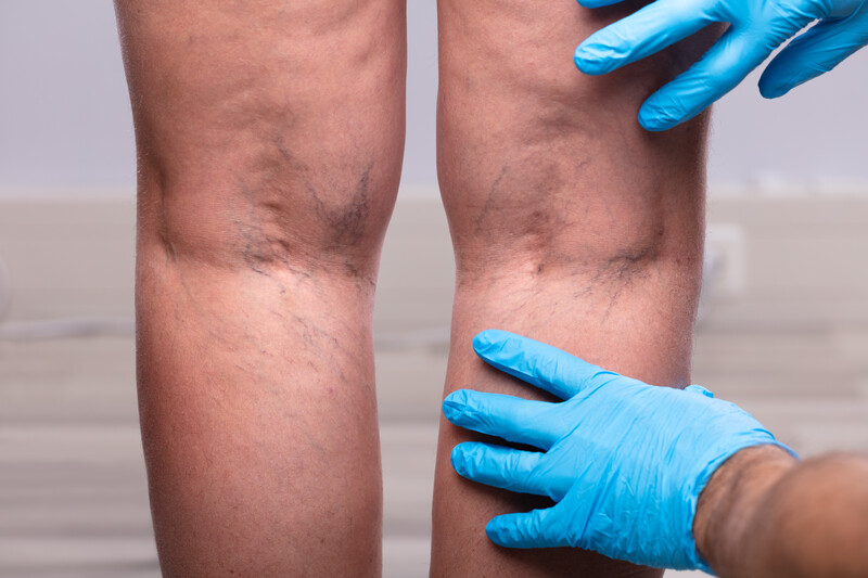
Venous Insufficiency
Cardiovascular Agents
Percutaneous Coronary Intervention
Blood Coagulation Factor Inhibitors
Platelet Function Tests
Plasminogen Activators
Metals
Magnetic Resonance Imaging
International Normalized Ratio
Renal Dialysis
Angioplasty, Balloon
Streptokinase
Hypertension, Portal
Dogs
Prosthesis Failure
Predictive Value of Tests
Factor XII Deficiency
Paclitaxel
Rupture, Spontaneous
Kaplan-Meier Estimate
Hirudin Therapy
Cohort Studies
Systemic infection with Alaria americana (Trematoda). (1/5349)
Alaria americana is a trematode, the adult of which is found in mammalian carnivores. The first case of disseminated human infection by the mesocercarial stage of this worm occurred in a 24-year-old man. The infection possibly was acquired by the eating of inadequately cooked frogs, which are intermediate hosts of the worm. The diagnosis was made during life by lung biopsy and confirmed at autopsy. The mesocercariae were present in the stomach wall, lymph nodes, liver, myocardium, pancreas and surrounding adipose tissue, spleen, kidney, lungs, brain and spinal cord. There was no host reaction to the parasites. Granulomas were present in the stomach wall, lymph nodes and liver, but the worms were not identified in them. Hypersensitivity vasculitis and a bleeding diathesis due to disseminated intravascular coagulation and a circulating anticoagulant caused his death 8 days after the onset of his illness. (+info)Antiphospholipid, anti-beta 2-glycoprotein-I and anti-oxidized-low-density-lipoprotein antibodies in antiphospholipid syndrome. (2/5349)
Antiphospholipid antibodies (aPL), anti-beta 2-glycoprotein I (anti-beta 2-GPI) and anti-oxidized-low-density lipoprotein (LDL) antibodies are all implicated in the pathogenesis of antiphospholipid syndrome. To investigate whether different autoantibodies or combinations thereof produced distinct effects related to their antigenic specificities, we examined the frequencies of antiphospholipid syndrome (APS)-related features in the presence of different antibodies [aPL, beta 2-GPI, anti-oxidized low density lipoprotein (LDL)] in 125 patients with APS. Median follow-up was 72 months: 58 patients were diagnosed as primary APS and 67 as APS plus systemic lupus erythematosus (SLE). Anticardiolipin antibodies (aCL), anti-beta 2-GPI and anti-oxidized LDL antibodies were determined by ELISA; lupus anticoagulant (LA) by standard coagulometric methods. Univariate analysis showed that patients positive for anti-beta 2-GPI had a higher risk of recurrent thrombotic events (OR = 3.64, 95% CI, p = 0.01) and pregnancy loss (OR = 2.99, 95% CI, p = 0.004). Patients positive for anti-oxidized LDL antibodies had a 2.24-fold increase in the risk of arterial thrombosis (2.24, 95% CI, p = 0.03) and lower risk of thrombocytopenia (OR = 0.41 95% CI, p = 0.04). Patients positive for aCL antibodies had a higher risk of pregnancy loss (OR = 4.62 95% CI, p = 0.001). When these data were tested by multivariate logistic regression, the association between anti-beta 2-GPI and pregnancy loss and the negative association between anti-oxidized LDL antibodies and thrombocytopenia disappeared. (+info)Blood-borne tissue factor: another view of thrombosis. (3/5349)
Arterial thrombosis is considered to arise from the interaction of tissue factor (TF) in the vascular wall with platelets and coagulation factors in circulating blood. According to this paradigm, coagulation is initiated after a vessel is damaged and blood is exposed to vessel-wall TF. We have examined thrombus formation on pig arterial media (which contains no stainable TF) and on collagen-coated glass slides (which are devoid of TF) exposed to flowing native human blood. In both systems the thrombi that formed during a 5-min perfusion stained intensely for TF, much of which was not associated with cells. Antibodies against TF caused approximately 70% reduction in the amount of thrombus formed on the pig arterial media and also reduced thrombi on the collagen-coated glass slides. TF deposited on the slides was active, as there was abundant fibrin in the thrombi. Factor VIIai, a potent inhibitor of TF, essentially abolished fibrin production and markedly reduced the mass of the thrombi. Immunoelectron microscopy revealed TF-positive membrane vesicles that we frequently observed in large clusters near the surface of platelets. TF, measured by factor Xa formation, was extracted from whole blood and plasma of healthy subjects. By using immunostaining, TF-containing neutrophils and monocytes were identified in peripheral blood; our data raise the possibility that leukocytes are the main source of blood TF. We suggest that blood-borne TF is inherently thrombogenic and may be involved in thrombus propagation at the site of vascular injury. (+info)Endothelial implants inhibit intimal hyperplasia after porcine angioplasty. (4/5349)
The perivascular implantation of tissue-engineered endothelial cells around injured arteries offers an opportunity to study fundamental vascular physiology as well as restore and improve tissue function. Cell source is an important issue because the ability to implant either xenogeneic or allogeneic cells would greatly enhance the clinical applications of tissue-engineered grafts. We investigated the biological and immunological responses to endothelial cell xenografts and allografts in pigs 4 weeks after angioplasty of the carotid arteries. Porcine or bovine aortic endothelial cells were cultured within Gelfoam matrices and implanted in the perivascular space of 42 injured arteries. Both porcine and bovine endothelial cell grafts reduced the restenosis index compared with control by 54% and 46%, respectively. Perivascular heparin release devices, formulated to release heparin at twice the rate of release of heparan sulfate proteoglycan from endothelial cell implants, produced no significant reduction in the restenosis index. Endothelial cell implants also reduced occlusive thrombosis compared with control and heparin release devices. Host immune responses to endothelial implants were investigated by immunohistochemical examination of explanted devices and by immunocytochemistry of serum samples. The bovine cell grafts displayed infiltration of leukocytes, consisting primarily of lymphocytes, and caused an increase in antibodies detected in serum samples. Reduced cellular infiltration and no humoral response were detected in animals that received allografts. Despite the difference in immune response, the biological effects of xenografts or allografts did not differ significantly. (+info)The tourniquet in total knee arthroplasty. A prospective, randomised study. (5/5349)
We assessed the influence of the use of a tourniquet in total knee arthroplasty in a prospective, randomised study. After satisfying exclusion criteria, we divided 77 patients into two groups, one to undergo surgery with a tourniquet and one without. Both groups were well matched. The mean change in knee flexion in the group that had surgery without a tourniquet was significantly better at one week (p = 0.03) than in the other group, but movement was similar at six weeks and at four months. There was no significant difference in the surgical time, postoperative pain, need for analgesia, the volume collected in the drains, postoperative swelling, and the incidence of wound complications or of deep-venous thrombosis. We conclude that the use of a tourniquet is safe and that current practice can be continued. (+info)The value of late computed tomographic scanning in identification of vascular abnormalities after abdominal aortic aneurysm repair. (6/5349)
PURPOSE: The purpose of this study was to determine the prevalence of late arterial abnormalities after aortic aneurysm repair and thus to suggest a routine for postoperative radiologic follow-up examination and to establish reference criteria for endovascular repair. METHODS: Computed tomographic (CT) scan follow-up examination was obtained at 8 to 9 years after abdominal aortic aneurysm (AAA) repair on a cohort of patients enrolled in the Canadian Aneurysm Study. The original registry consisted of 680 patients who underwent repair of nonruptured AAA. When the request for CT scan follow-up examination was sent in 1994, 251 patients were alive and potentially available for CT scan follow-up examination and 94 patients agreed to undergo abdominal and thoracic CT scanning procedures. Each scan was interpreted independently by two vascular radiologists. RESULTS: For analysis, the aorta was divided into five defined segments and an aneurysm was defined as a more than 50% enlargement from the expected normal value as defined in the reporting standards for aneurysms. With this strict definition, 64.9% of patients had aneurysmal dilatation and the abnormality was considered as a possible indication for surgical repair in 13.8%. Of the 39 patients who underwent initial repair with a tube graft, 12 (30.8%) were found to have an iliac aneurysm and six of these aneurysms (15.4%) were of possible surgical significance. Graft dilatation was observed from the time of operation (median graft size of 18 mm) to a median size of 22 mm as measured by means of CT scanning at follow-up examination. Fluid or thrombus was seen around the graft in 28% of the cases, and bowel was believed to be intimately associated with the graft in 7%. CONCLUSION: Late follow-up CT scans after AAA repair often show vascular abnormalities. Most of these abnormalities are not clinically significant, but, in 13.8% of patients, the thoracic or abdominal aortic segment was aneurysmal and, in 15.4% of patients who underwent tube graft placement, one of the iliac arteries was significantly abnormal to warrant consideration for surgical repair. On the basis of these findings, a routine CT follow-up examination after 5 years is recommended. This study provides a population-based study for comparison with the results of endovascular repair. (+info)Immunohistochemical analysis of arterial wall cellular infiltration in Buerger's disease (endarteritis obliterans). (7/5349)
PURPOSE: The diagnosis of Buerger's disease has depended on clinical symptoms and angiographic findings, whereas pathologic findings are considered to be of secondary importance. Arteries from patients with Buerger's tissue were analyzed histologically, including immunophenotyping of the infiltrating cells, to elucidate the nature of Buerger's disease as a vasculitis. METHODS: Thirty-three specimens from nine patients, in whom Buerger's disease was diagnosed on the basis of our clinical and angiographic criteria between 1980 and 1995 at Nagoya University Hospital, were studied. Immunohistochemical studies were performed on paraffin-embedded tissue with a labeled streptoavidin-biotin method. RESULTS: The general architecture of vessel walls was well preserved regardless of the stage of disease, and cell infiltration was observed mainly in the thrombus and the intima. Among infiltrating cells, CD3(+) T cells greatly outnumbered CD20(+) B cells. CD68(+) macrophages or S-100(+) dendritic cells were detected, especially in the intima during acute and subacute stages. All cases except one showed infiltration by the human leukocyte antigen-D region (HLA-DR) antigen-bearing macrophages and dendritic cells in the intima. Immunoglobulins G, A, and M (IgG, IgA, IgM) and complement factors 3d and 4c (C3d, C4c) were deposited along the internal elastic lamina. CONCLUSION: Buerger's disease is strictly an endarteritis that is introduced by T-cell mediated cellular immunity and by B-cell mediated humoral immunity associated with activation of macrophages or dendritic cells in the intima. (+info)The intrarenal vascular lesions associated with primary antiphospholipid syndrome. (8/5349)
Even 10 yr after the identification of the antiphospholipid syndrome (APS), renal involvement in the course of APS is still relatively unrecognized, and is probably underestimated. The association of anticardiolipin antibodies and/or lupus anticoagulant with the development of a vaso-occlusive process involving numerous organs is now confirmed. In a multicenter study, 16 cases of "primary" APS (PAPS) were found and followed for 5 yr or more, all with renal biopsy. In all 16 cases of PAPS, there was a vascular nephropathy characterized by small vessel vaso-occlusive lesions associated with fibrous intimal hyperplasia of interlobular arteries (12 patients), recanalizing thrombi in arteries and arterioles (six patients), and focal cortical atrophy (10 patients). In combination, these led to progressive destruction of the kidney, accelerated by acute glomerular and arteriolar microangiopathy in five patients. Focal cortical atrophy is a distinctive lesion, present in 10 biopsies, and likely represents the histologic and functional renal analogue to the multiple cerebral infarcts detected on imaging studies. The clinical hallmark of this vascular nephropathy in PAPS is systemic hypertension, only variably associated with renal insufficiency, proteinuria, or hematuria. The ensemble of histologic renal lesions defined in this study should aid in the separation of the lesions found in cases of secondary APS, especially systemic lupus erythematosus, into those lesions related to APS and those related to the underlying disease. (+info)Thrombosis is the formation of a blood clot (thrombus) inside a blood vessel, obstructing the flow of blood through the circulatory system. When a clot forms in an artery, it can cut off the supply of oxygen and nutrients to the tissues served by that artery, leading to damage or tissue death. If a thrombus forms in the heart, it can cause a heart attack. If a thrombus breaks off and travels through the bloodstream, it can lodge in a smaller vessel, causing blockage and potentially leading to damage in the organ that the vessel supplies. This is known as an embolism.
Thrombosis can occur due to various factors such as injury to the blood vessel wall, abnormalities in blood flow, or changes in the composition of the blood. Certain medical conditions, medications, and lifestyle factors can increase the risk of thrombosis. Treatment typically involves anticoagulant or thrombolytic therapy to dissolve or prevent further growth of the clot, as well as addressing any underlying causes.
Venous thrombosis is a medical condition characterized by the formation of a blood clot (thrombus) in the deep veins, often in the legs (deep vein thrombosis or DVT), but it can also occur in other parts of the body such as the arms, pelvis, or lungs (pulmonary embolism).
The formation of a venous thrombus can be caused by various factors, including injury to the blood vessel wall, changes in blood flow, and alterations in the composition of the blood. These factors can lead to the activation of clotting factors and platelets, which can result in the formation of a clot that blocks the vein.
Symptoms of venous thrombosis may include swelling, pain, warmth, and redness in the affected area. In some cases, the clot can dislodge and travel to other parts of the body, causing potentially life-threatening complications such as pulmonary embolism.
Risk factors for venous thrombosis include advanced age, obesity, smoking, pregnancy, use of hormonal contraceptives or hormone replacement therapy, cancer, recent surgery or trauma, prolonged immobility, and a history of previous venous thromboembolism. Treatment typically involves the use of anticoagulant medications to prevent further clotting and dissolve existing clots.
Coronary thrombosis is a medical condition that refers to the formation of a blood clot (thrombus) inside a coronary artery, which supplies oxygenated blood to the heart muscle. The development of a thrombus can partially or completely obstruct blood flow, leading to insufficient oxygen supply to the heart muscle. This can cause chest pain (angina) or a heart attack (myocardial infarction), depending on the severity and duration of the blockage.
Coronary thrombosis often results from the rupture of an atherosclerotic plaque, a buildup of cholesterol, fat, calcium, and other substances in the inner lining (endothelium) of the coronary artery. The ruptured plaque exposes the underlying tissue to the bloodstream, triggering the coagulation cascade and resulting in the formation of a thrombus.
Immediate medical attention is crucial for managing coronary thrombosis, as timely treatment can help restore blood flow, prevent further damage to the heart muscle, and reduce the risk of complications such as heart failure or life-threatening arrhythmias. Treatment options may include medications, such as antiplatelet agents, anticoagulants, and thrombolytic drugs, or interventional procedures like angioplasty and stenting to open the blocked artery. In some cases, surgical intervention, such as coronary artery bypass grafting (CABG), may be necessary.
Intracranial sinus thrombosis is a medical condition characterized by the formation of a blood clot (thrombus) within the intracranial venous sinuses, which are responsible for draining blood from the brain. The condition can lead to various neurological symptoms and complications, such as increased intracranial pressure, headaches, seizures, visual disturbances, and altered consciousness. Intracranial sinus thrombosis may result from various factors, including hypercoagulable states, infections, trauma, and malignancies. Immediate medical attention is necessary for proper diagnosis and treatment to prevent potential long-term neurological damage or even death.
Intracranial thrombosis refers to the formation of a blood clot (thrombus) within the intracranial vessels, which supply blood to the brain. This condition can occur in any of the cerebral arteries or veins and can lead to serious complications such as ischemic stroke, transient ischemic attack (TIA), or venous sinus thrombosis.
The formation of an intracranial thrombus can be caused by various factors, including atherosclerosis, cardiac embolism, vasculitis, sickle cell disease, hypercoagulable states, and head trauma. Symptoms may vary depending on the location and extent of the thrombosis but often include sudden onset of headache, weakness or numbness in the face or limbs, difficulty speaking or understanding speech, vision changes, and loss of balance or coordination.
Diagnosis of intracranial thrombosis typically involves imaging studies such as computed tomography (CT) angiography, magnetic resonance angiography (MRA), or digital subtraction angiography (DSA). Treatment options may include anticoagulation therapy, thrombolysis, endovascular intervention, or surgical intervention, depending on the underlying cause and severity of the condition.
Carotid artery thrombosis is a medical condition characterized by the formation of a blood clot (thrombus) inside the carotid artery, which is one of the major blood vessels that supplies oxygenated blood to the head and neck. This condition can lead to serious complications such as a stroke or transient ischemic attack (TIA), also known as a "mini-stroke," if the clot dislodges and travels to the brain, blocking the flow of blood and oxygen.
Carotid artery thrombosis can result from various factors, including atherosclerosis (the buildup of fats, cholesterol, and other substances in the artery walls), hypertension (high blood pressure), diabetes, smoking, and genetic predisposition. Symptoms may include neck pain or stiffness, weakness or numbness in the face or limbs, difficulty speaking or understanding speech, vision problems, and sudden severe headaches. Diagnosis typically involves imaging tests such as ultrasound, CT angiography, or MRI angiography. Treatment options may include anticoagulant or antiplatelet medications, endovascular procedures to remove the clot, or surgery to clean out the artery (carotid endarterectomy).
Anticoagulants are a class of medications that work to prevent the formation of blood clots in the body. They do this by inhibiting the coagulation cascade, which is a series of chemical reactions that lead to the formation of a clot. Anticoagulants can be given orally, intravenously, or subcutaneously, depending on the specific drug and the individual patient's needs.
There are several different types of anticoagulants, including:
1. Heparin: This is a naturally occurring anticoagulant that is often used in hospitalized patients who require immediate anticoagulation. It works by activating an enzyme called antithrombin III, which inhibits the formation of clots.
2. Low molecular weight heparin (LMWH): LMWH is a form of heparin that has been broken down into smaller molecules. It has a longer half-life than standard heparin and can be given once or twice daily by subcutaneous injection.
3. Direct oral anticoagulants (DOACs): These are newer oral anticoagulants that work by directly inhibiting specific clotting factors in the coagulation cascade. Examples include apixaban, rivaroxaban, and dabigatran.
4. Vitamin K antagonists: These are older oral anticoagulants that work by inhibiting the action of vitamin K, which is necessary for the formation of clotting factors. Warfarin is an example of a vitamin K antagonist.
Anticoagulants are used to prevent and treat a variety of conditions, including deep vein thrombosis (DVT), pulmonary embolism (PE), atrial fibrillation, and prosthetic heart valve thrombosis. It is important to note that anticoagulants can increase the risk of bleeding, so they must be used with caution and regular monitoring of blood clotting times may be required.
Sagittal sinus thrombosis is a medical condition that refers to the formation of a blood clot (thrombus) in the sagittal sinus, which is a venous structure located in the brain. The sagittal sinus runs along the midline of the brain and receives blood from the superficial veins of the brain.
Sagittal sinus thrombosis can occur as a result of various conditions, such as head trauma, infection, cancer, or certain medical disorders that cause hypercoagulability (an increased tendency to form blood clots). The formation of a blood clot in the sagittal sinus can obstruct the flow of blood from the brain, leading to symptoms such as headache, seizures, altered consciousness, and focal neurological deficits.
Diagnosis of sagittal sinus thrombosis typically involves imaging studies such as computed tomography (CT) or magnetic resonance imaging (MRI) scans, which can show the presence of a blood clot in the sagittal sinus. Treatment may involve administering anticoagulant medications to prevent further growth of the blood clot and reduce the risk of complications such as pulmonary embolism or cerebral infarction. In some cases, surgical intervention may be necessary to remove the blood clot or alleviate pressure on the brain.
Phlebography is a medical imaging technique used to visualize and assess the veins, particularly in the legs. It involves the injection of a contrast agent into the veins, followed by X-ray imaging to capture the flow of the contrast material through the veins. This allows doctors to identify any abnormalities such as blood clots, blockages, or malformations in the venous system.
There are different types of phlebography, including ascending phlebography (where the contrast agent is injected into a foot vein and travels up the leg) and descending phlebography (where the contrast agent is injected into a vein in the groin or neck and travels down the leg).
Phlebography is an invasive procedure that requires careful preparation and monitoring, and it is typically performed by radiologists or vascular specialists. It has largely been replaced by non-invasive imaging techniques such as ultrasound and CT angiography in many clinical settings.
1. Intracranial Embolism: This is a medical condition that occurs when a blood clot or other particle (embolus) formed elsewhere in the body, travels through the bloodstream and lodges itself in the intracranial blood vessels, blocking the flow of blood to a part of the brain. This can lead to various neurological symptoms such as weakness, numbness, speech difficulties, or even loss of consciousness, depending on the severity and location of the blockage.
2. Intracranial Thrombosis: This is a medical condition that occurs when a blood clot (thrombus) forms within the intracranial blood vessels. The clot can partially or completely obstruct the flow of blood, leading to various symptoms such as headache, confusion, seizures, or neurological deficits, depending on the severity and location of the thrombosis. Intracranial thrombosis can occur due to various factors including atherosclerosis, hypertension, diabetes, and other medical conditions that increase the risk of blood clot formation.
The iliac veins are a pair of large veins in the human body that carry deoxygenated blood from the lower extremities and the pelvic area back to the heart. They are formed by the union of the common iliac veins, which receive blood from the lower abdomen and legs, at the level of the fifth lumbar vertebra.
The combined iliac vein is called the inferior vena cava, which continues upward to the right atrium of the heart. The iliac veins are located deep within the pelvis, lateral to the corresponding iliac arteries, and are accompanied by the iliac lymphatic vessels.
The left common iliac vein is longer than the right because it must cross the left common iliac artery to join the right common iliac vein. The external and internal iliac veins are the two branches of the common iliac vein, with the external iliac vein carrying blood from the lower limbs and the internal iliac vein carrying blood from the pelvic organs.
It is essential to maintain proper blood flow in the iliac veins to prevent deep vein thrombosis (DVT), a condition that can lead to serious complications such as pulmonary embolism.
Thrombophilia is a medical condition characterized by an increased tendency to form blood clots (thrombi) due to various genetic or acquired abnormalities in the coagulation system. These abnormalities can lead to a hypercoagulable state, which can cause thrombosis in both veins and arteries. Commonly identified thrombophilias include factor V Leiden mutation, prothrombin G20210A mutation, antithrombin deficiency, protein C deficiency, and protein S deficiency.
Acquired thrombophilias can be caused by various factors such as antiphospholipid antibody syndrome (APS), malignancies, pregnancy, oral contraceptive use, hormone replacement therapy, and certain medical conditions like inflammatory bowel disease or nephrotic syndrome.
It is essential to diagnose thrombophilia accurately, as it may influence the management of venous thromboembolism (VTE) events and guide decisions regarding prophylactic anticoagulation in high-risk situations.
The femoral vein is the large vein that runs through the thigh and carries oxygen-depleted blood from the lower limbs back to the heart. It is located in the femoral triangle, along with the femoral artery and nerve. The femoral vein begins at the knee as the popliteal vein, which then joins with the deep vein of the thigh to form the femoral vein. As it moves up the leg, it is joined by several other veins, including the great saphenous vein, before it becomes the external iliac vein at the inguinal ligament in the groin.
Upper extremity deep vein thrombosis (UEDVT) is a medical condition that refers to the formation of a blood clot (thrombus) in the deep veins located in the arm or shoulder. This condition can occur due to various reasons, including trauma, surgery, cancer, certain medications, and underlying medical conditions that increase the risk of blood clotting.
The deep veins are larger vessels that run through the body's muscles and are surrounded by fascia, a connective tissue. UEDVT can cause partial or complete blockage of blood flow in the affected vein, leading to swelling, pain, redness, warmth, and decreased function in the arm or hand. In some cases, the clot can break off and travel to the lungs, causing a potentially life-threatening condition called pulmonary embolism (PE).
Diagnosis of UEDVT typically involves a physical exam, medical history, and imaging tests such as ultrasound, CT scan, or MRI. Treatment may include anticoagulant medications to prevent the clot from growing or breaking off, thrombolytic therapy to dissolve the clot, or surgical intervention in severe cases. Compression stockings or other devices may also be used to help improve blood flow and reduce swelling.
The portal vein is the large venous trunk that carries blood from the gastrointestinal tract, spleen, pancreas, and gallbladder to the liver. It is formed by the union of the superior mesenteric vein (draining the small intestine and a portion of the large intestine) and the splenic vein (draining the spleen and pancreas). The portal vein then divides into right and left branches within the liver, where the blood flows through the sinusoids and gets enriched with oxygen and nutrients before being drained by the hepatic veins into the inferior vena cava. This unique arrangement allows the liver to process and detoxify the absorbed nutrients, remove waste products, and regulate metabolic homeostasis.
Cerebral veins are the blood vessels that carry deoxygenated blood from the brain to the dural venous sinuses, which are located between the layers of tissue covering the brain. The largest cerebral vein is the superior sagittal sinus, which runs along the top of the brain. Other major cerebral veins include the straight sinus, transverse sinus, sigmoid sinus, and cavernous sinus. These veins receive blood from smaller veins called venules that drain the surface and deep structures of the brain. The cerebral veins play an important role in maintaining normal circulation and pressure within the brain.
Heparin is defined as a highly sulfated glycosaminoglycan (a type of polysaccharide) that is widely present in many tissues, but is most commonly derived from the mucosal tissues of mammalian lungs or intestinal mucosa. It is an anticoagulant that acts as an inhibitor of several enzymes involved in the blood coagulation cascade, primarily by activating antithrombin III which then neutralizes thrombin and other clotting factors.
Heparin is used medically to prevent and treat thromboembolic disorders such as deep vein thrombosis, pulmonary embolism, and certain types of heart attacks. It can also be used during hemodialysis, cardiac bypass surgery, and other medical procedures to prevent the formation of blood clots.
It's important to note that while heparin is a powerful anticoagulant, it does not have any fibrinolytic activity, meaning it cannot dissolve existing blood clots. Instead, it prevents new clots from forming and stops existing clots from growing larger.
Factor V, also known as proaccelerin or labile factor, is a protein involved in the coagulation cascade, which is a series of chemical reactions that leads to the formation of a blood clot. Factor V acts as a cofactor for the activation of Factor X to Factor Xa, which is a critical step in the coagulation cascade.
When blood vessels are damaged, the coagulation cascade is initiated to prevent excessive bleeding. During this process, Factor V is activated by thrombin, another protein involved in coagulation, and then forms a complex with activated Factor X and calcium ions on the surface of platelets or other cells. This complex converts prothrombin to thrombin, which then converts fibrinogen to fibrin to form a stable clot.
Deficiency or dysfunction of Factor V can lead to bleeding disorders such as hemophilia B or factor V deficiency, while mutations in the gene encoding Factor V can increase the risk of thrombosis, as seen in the Factor V Leiden mutation.
Fibrinolytic agents are medications that dissolve or break down blood clots by activating plasminogen, which is converted into plasmin. Plasmin is a proteolytic enzyme that degrades fibrin, the structural protein in blood clots. Fibrinolytic agents are used medically to treat conditions such as acute ischemic stroke, deep vein thrombosis, pulmonary embolism, and myocardial infarction (heart attack) by restoring blood flow in occluded vessels. Examples of fibrinolytic agents include alteplase, reteplase, and tenecteplase. It is important to note that these medications carry a risk of bleeding complications and should be administered with caution.
Hemostasis is the physiological process that occurs to stop bleeding (bleeding control) when a blood vessel is damaged. This involves the interaction of platelets, vasoconstriction, and blood clotting factors leading to the formation of a clot. The ultimate goal of hemostasis is to maintain the integrity of the vascular system while preventing excessive blood loss.
Low-molecular-weight heparin (LMWH) is a type of heparin used as an anticoagulant, which refers to a group of medications that prevent the formation of blood clots. Heparin is a naturally occurring substance in the body, and low-molecular-weight heparins are obtained through the depolymerization of standard heparin.
LMWH has a lower molecular weight than standard heparin, which results in several pharmacological differences. LMWHs have a more predictable dose response, longer half-life, and higher bioavailability when administered subcutaneously compared to standard heparin. They also exhibit greater anti-factor Xa activity relative to their antithrombin (anti-IIa) activity, which contributes to their anticoagulant effects.
LMWHs are used for the prevention and treatment of deep vein thrombosis (DVT), pulmonary embolism (PE), and other thromboembolic disorders. Common LMWHs include enoxaparin, dalteparin, tinzaparin, and nadroparin.
It is essential to monitor the patient's kidney function when using LMWH since they are primarily cleared by the kidneys. In patients with renal impairment, dose adjustments or alternative anticoagulants may be necessary to reduce the risk of bleeding complications.
Cavernous sinus thrombosis is a medical condition that refers to the formation of a blood clot (thrombus) in the cavernous sinuses, which are located near the base of the brain and are important for draining blood from the face and brain. This condition can occur as a complication of an infection in the facial area or sinuses, or it can be associated with other medical conditions such as cancer or trauma.
Symptoms of cavernous sinus thrombosis may include headache, fever, eye pain, swelling or bulging of the eyes, double vision, and decreased vision. If left untreated, this condition can lead to serious complications such as meningitis, brain abscess, or even death. Treatment typically involves administering antibiotics to treat any underlying infection and anticoagulants to prevent further clot formation. In some cases, surgery may be necessary to remove the clot.
Blood coagulation, also known as blood clotting, is a complex process that occurs in the body to prevent excessive bleeding when a blood vessel is damaged. This process involves several different proteins and chemical reactions that ultimately lead to the formation of a clot.
The coagulation cascade is initiated when blood comes into contact with tissue factor, which is exposed after damage to the blood vessel wall. This triggers a series of enzymatic reactions that activate clotting factors, leading to the formation of a fibrin clot. Fibrin is a protein that forms a mesh-like structure that traps platelets and red blood cells to form a stable clot.
Once the bleeding has stopped, the coagulation process is regulated and inhibited to prevent excessive clotting. The fibrinolytic system degrades the clot over time, allowing for the restoration of normal blood flow.
Abnormalities in the blood coagulation process can lead to bleeding disorders or thrombotic disorders such as deep vein thrombosis and pulmonary embolism.
Antiphospholipid syndrome (APS) is an autoimmune disorder characterized by the presence of antiphospholipid antibodies in the blood. These antibodies are directed against phospholipids, a type of fat molecule found in cell membranes and plasma lipoproteins. The presence of these antibodies can lead to abnormal blood clotting, which can cause serious complications such as stroke, heart attack, deep vein thrombosis, and pulmonary embolism.
APS can occur either on its own (primary APS) or in conjunction with other autoimmune disorders, such as systemic lupus erythematosus (secondary APS). The exact cause of APS is not fully understood, but it is believed to involve a combination of genetic and environmental factors.
Symptoms of APS can vary widely depending on the location and severity of the blood clots. They may include:
* Recurrent miscarriages or stillbirths
* Blood clots in the legs, lungs, or other parts of the body
* Skin ulcers or lesions
* Headaches, seizures, or stroke-like symptoms
* Kidney problems
* Heart valve abnormalities
Diagnosis of APS typically involves blood tests to detect the presence of antiphospholipid antibodies. Treatment may include medications to prevent blood clots, such as anticoagulants and antiplatelet agents, as well as management of any underlying autoimmune disorders.
The inferior vena cava (IVC) is the largest vein in the human body that carries deoxygenated blood from the lower extremities, pelvis, and abdomen to the right atrium of the heart. It is formed by the union of the left and right common iliac veins at the level of the fifth lumbar vertebra. The inferior vena cava is a retroperitoneal structure, meaning it lies behind the peritoneum, the lining that covers the abdominal cavity. It ascends through the posterior abdominal wall and passes through the central tendon of the diaphragm to enter the thoracic cavity.
The inferior vena cava is composed of three parts:
1. The infrarenal portion, which lies below the renal veins
2. The renal portion, which receives blood from the renal veins
3. The suprahepatic portion, which lies above the liver and receives blood from the hepatic veins before draining into the right atrium of the heart.
The inferior vena cava plays a crucial role in maintaining venous return to the heart and contributing to cardiovascular function.
Thromboembolism is a medical condition that refers to the obstruction of a blood vessel by a thrombus (blood clot) that has formed elsewhere in the body and then been transported by the bloodstream to a narrower vessel, where it becomes lodged. This process can occur in various parts of the body, leading to different types of thromboembolisms:
1. Deep Vein Thrombosis (DVT): A thrombus forms in the deep veins, usually in the legs or pelvis, and then breaks off and travels to the lungs, causing a pulmonary embolism.
2. Pulmonary Embolism (PE): A thrombus formed elsewhere, often in the deep veins of the legs, dislodges and travels to the lungs, blocking one or more pulmonary arteries. This can lead to shortness of breath, chest pain, and potentially life-threatening complications if not treated promptly.
3. Cerebral Embolism: A thrombus formed in another part of the body, such as the heart or carotid artery, dislodges and travels to the brain, causing a stroke or transient ischemic attack (TIA).
4. Arterial Thromboembolism: A thrombus forms in an artery and breaks off, traveling to another part of the body and blocking blood flow to an organ or tissue, leading to potential damage or loss of function. Examples include mesenteric ischemia (intestinal damage due to blocked blood flow) and retinal artery occlusion (vision loss due to blocked blood flow in the eye).
Prevention, early detection, and appropriate treatment are crucial for managing thromboembolism and reducing the risk of severe complications.
Platelet aggregation inhibitors are a class of medications that prevent platelets (small blood cells involved in clotting) from sticking together and forming a clot. These drugs work by interfering with the ability of platelets to adhere to each other and to the damaged vessel wall, thereby reducing the risk of thrombosis (blood clot formation).
Platelet aggregation inhibitors are often prescribed for people who have an increased risk of developing blood clots due to various medical conditions such as atrial fibrillation, coronary artery disease, peripheral artery disease, stroke, or a history of heart attack. They may also be used in patients undergoing certain medical procedures, such as angioplasty and stenting, to prevent blood clot formation in the stents.
Examples of platelet aggregation inhibitors include:
1. Aspirin: A nonsteroidal anti-inflammatory drug (NSAID) that irreversibly inhibits the enzyme cyclooxygenase, which is involved in platelet activation and aggregation.
2. Clopidogrel (Plavix): A P2Y12 receptor antagonist that selectively blocks ADP-induced platelet activation and aggregation.
3. Prasugrel (Effient): A third-generation thienopyridine P2Y12 receptor antagonist, similar to clopidogrel but with faster onset and greater potency.
4. Ticagrelor (Brilinta): A direct-acting P2Y12 receptor antagonist that does not require metabolic activation and has a reversible binding profile.
5. Dipyridamole (Persantine): An antiplatelet agent that inhibits platelet aggregation by increasing cyclic adenosine monophosphate (cAMP) levels in platelets, which leads to decreased platelet reactivity.
6. Iloprost (Ventavis): A prostacyclin analogue that inhibits platelet aggregation and causes vasodilation, often used in the treatment of pulmonary arterial hypertension.
7. Cilostazol (Pletal): A phosphodiesterase III inhibitor that increases cAMP levels in platelets, leading to decreased platelet activation and aggregation, as well as vasodilation.
8. Ticlopidine (Ticlid): An older P2Y12 receptor antagonist with a slower onset of action and more frequent side effects compared to clopidogrel or prasugrel.
Lateral sinus thrombosis, also known as sigmoid sinus thrombosis, is a medical condition characterized by the formation of a blood clot (thrombus) in the lateral or sigmoid sinus, which are venous structures located in the skull that help drain blood from the brain.
The lateral sinuses are situated near the mastoid process of the temporal bone and can become thrombosed due to various reasons such as infection (often associated with ear or mastoid infections), trauma, tumors, or other underlying medical conditions that increase the risk of blood clot formation.
Symptoms of lateral sinus thrombosis may include headache, fever, neck stiffness, altered mental status, and signs of increased intracranial pressure such as papilledema (swelling of the optic nerve disc). Diagnosis is typically made with the help of imaging studies like CT or MRI scans, and treatment often involves anticoagulation therapy to prevent clot expansion and potential complications. In some cases, surgical intervention may be necessary to remove the clot or manage any underlying conditions.
Bleeding time is a medical test that measures the time it takes for a small blood vessel to stop bleeding after being cut. It's used to evaluate platelet function and the effectiveness of blood clotting. The most common method used to measure bleeding time is the Ivy method, which involves making a standardized incision on the forearm and measuring the time it takes for the bleeding to stop. A normal bleeding time ranges from 2 to 9 minutes, but this can vary depending on the specific method used. Prolonged bleeding time may indicate an impairment in platelet function or clotting factor deficiency.
A thrombectomy is a medical procedure that involves the removal of a blood clot (thrombus) from a blood vessel. This is typically performed to restore blood flow in cases where the clot is causing significant blockage, which can lead to serious complications such as tissue damage or organ dysfunction.
During a thrombectomy, a surgeon makes an incision and accesses the affected blood vessel, often with the help of imaging guidance. Specialized tools are then used to extract the clot, after which the blood vessel is usually repaired. Thrombectomies can be performed on various blood vessels throughout the body, including those in the brain, heart, lungs, and limbs.
This procedure may be recommended for patients with deep vein thrombosis (DVT), pulmonary embolism (PE), or certain types of stroke, depending on the specific circumstances and the patient's overall health. It is generally considered when anticoagulation therapy or clot-dissolving medications are not sufficient or appropriate to treat the blood clot.
Fibrin(ogen) degradation products (FDPs) are a group of proteins that result from the breakdown of fibrinogen and fibrin, which are key components of blood clots. This process occurs during the normal physiological process of fibrinolysis, where clots are dissolved to maintain blood flow.
FDPs can be measured in the blood as a marker for the activation of the coagulation and fibrinolytic systems. Elevated levels of FDPs may indicate the presence of a disorder that causes abnormal clotting or bleeding, such as disseminated intravascular coagulation (DIC), deep vein thrombosis (DVT), pulmonary embolism (PE), or certain types of cancer.
It is important to note that FDPs are not specific to any particular disorder and their measurement should be interpreted in conjunction with other clinical and laboratory findings.
A stent is a small mesh tube that's used to treat narrow or weak arteries. Arteries are blood vessels that carry blood away from your heart to other parts of your body. A stent is placed in an artery as part of a procedure called angioplasty. Angioplasty restores blood flow through narrowed or blocked arteries by inflating a tiny balloon inside the blocked artery to widen it.
The stent is then inserted into the widened artery to keep it open. The stent is usually made of metal, but some are coated with medication that is slowly and continuously released to help prevent the formation of scar tissue in the artery. This can reduce the chance of the artery narrowing again.
Stents are also used in other parts of the body, such as the neck (carotid artery) and kidneys (renal artery), to help maintain blood flow and prevent blockages. They can also be used in the urinary system to treat conditions like ureteropelvic junction obstruction or narrowing of the urethra.
Protein S deficiency is a genetic disorder that affects the body's ability to coagulate blood properly. Protein S is a naturally occurring protein in the blood that helps regulate the clotting process by deactivating clotting factors when they are no longer needed. When Protein S levels are too low, it can lead to an increased risk of abnormal blood clots forming within blood vessels, a condition known as thrombophilia.
There are three types of Protein S deficiency: Type I (quantitative deficiency), Type II (qualitative deficiency), and Type III (dysfunctional protein). These types refer to the amount or function of Protein S in the blood. In Type I, there is a decrease in both free and total Protein S levels. In Type II, there is a decrease in functional Protein S despite normal total Protein S levels. In Type III, there is a decrease in free Protein S with normal total Protein S levels.
Protein S deficiency can be inherited or acquired. Inherited forms of the disorder are caused by genetic mutations and are usually present from birth. Acquired forms of Protein S deficiency can develop later in life due to certain medical conditions, such as liver disease, vitamin K deficiency, or the use of certain medications that affect blood clotting.
Symptoms of Protein S deficiency may include recurrent blood clots, usually in the legs (deep vein thrombosis) or lungs (pulmonary embolism), skin discoloration, pain, and swelling in the affected area. In severe cases, it can lead to complications such as chronic leg ulcers, pulmonary hypertension, or damage to the heart or lungs.
Diagnosis of Protein S deficiency typically involves blood tests to measure Protein S levels and function. Treatment may include anticoagulant medications to prevent blood clots from forming or growing larger. Lifestyle modifications such as regular exercise, maintaining a healthy weight, and avoiding smoking can also help reduce the risk of blood clots in people with Protein S deficiency.
Treatment outcome is a term used to describe the result or effect of medical treatment on a patient's health status. It can be measured in various ways, such as through symptoms improvement, disease remission, reduced disability, improved quality of life, or survival rates. The treatment outcome helps healthcare providers evaluate the effectiveness of a particular treatment plan and make informed decisions about future care. It is also used in clinical research to compare the efficacy of different treatments and improve patient care.
Platelet aggregation is the clumping together of platelets (thrombocytes) in the blood, which is an essential step in the process of hemostasis (the stopping of bleeding) after injury to a blood vessel. When the inner lining of a blood vessel is damaged, exposure of subendothelial collagen and tissue factor triggers platelet activation. Activated platelets change shape, become sticky, and release the contents of their granules, which include ADP (adenosine diphosphate).
ADP then acts as a chemical mediator to attract and bind additional platelets to the site of injury, leading to platelet aggregation. This forms a plug that seals the damaged vessel and prevents further blood loss. Platelet aggregation is also a crucial component in the formation of blood clots (thrombosis) within blood vessels, which can have pathological consequences such as heart attacks and strokes if they obstruct blood flow to vital organs.
The mesenteric veins are a set of blood vessels that are responsible for draining deoxygenated blood from the small and large intestines. There are two main mesenteric veins: the superior mesenteric vein and the inferior mesenteric vein. The superior mesenteric vein drains blood from the majority of the small intestine, as well as the ascending colon and proximal two-thirds of the transverse colon. The inferior mesenteric vein drains blood from the distal third of the transverse colon, descending colon, sigmoid colon, and rectum. These veins ultimately drain into the portal vein, which carries the blood to the liver for further processing.
The subclavian vein is a large venous structure that carries deoxygenated blood from the upper limb and part of the thorax back to the heart. It forms when the axillary vein passes through the narrow space between the first rib and the clavicle (collarbone), becoming the subclavian vein.
On the left side, the subclavian vein joins with the internal jugular vein to form the brachiocephalic vein, while on the right side, the subclavian vein directly merges with the internal jugular vein to create the brachiocephalic vein. These brachiocephalic veins then unite to form the superior vena cava, which drains blood into the right atrium of the heart.
The subclavian vein is an essential structure for venous access in various medical procedures and interventions, such as placing central venous catheters or performing blood tests.
Drug-eluting stents (DES) are medical devices used in the treatment of coronary artery disease. They are small, flexible tubes that are coated with a medication that is slowly released (eluted) over time to prevent the formation of scar tissue and reduce the risk of renarrowing (restenosis) of the artery after it has been treated with angioplasty and stenting.
The stent is typically placed in a narrowed or blocked coronary artery during a percutaneous coronary intervention (PCI) procedure, such as angioplasty, to open up the blood vessel and improve blood flow to the heart muscle. The medication on the DES helps to prevent the growth of smooth muscle cells and the formation of scar tissue in the artery, which can cause restenosis and require additional treatments.
The most commonly used medications on DES are sirolimus, paclitaxel, zotarolimus, and everolimus. These drugs work by inhibiting the growth of smooth muscle cells and reducing inflammation in the artery. While DES have been shown to reduce the risk of restenosis compared to bare-metal stents, they also carry a small increased risk of late stent thrombosis (blood clots forming in the stent), which can lead to serious complications such as heart attack or stroke. Therefore, patients who receive DES are typically prescribed long-term antiplatelet therapy to reduce this risk.
Prothrombin is a protein present in blood plasma, and it's also known as coagulation factor II. It plays a crucial role in the coagulation cascade, which is a complex series of reactions that leads to the formation of a blood clot.
When an injury occurs, the coagulation cascade is initiated to prevent excessive blood loss. Prothrombin is converted into its active form, thrombin, by another factor called factor Xa in the presence of calcium ions, phospholipids, and factor Va. Thrombin then catalyzes the conversion of fibrinogen into fibrin, forming a stable clot.
Prothrombin levels can be measured through a blood test, which is often used to diagnose or monitor conditions related to bleeding or coagulation disorders, such as liver disease or vitamin K deficiency.
Medical Definition:
"Risk factors" are any attribute, characteristic or exposure of an individual that increases the likelihood of developing a disease or injury. They can be divided into modifiable and non-modifiable risk factors. Modifiable risk factors are those that can be changed through lifestyle choices or medical treatment, while non-modifiable risk factors are inherent traits such as age, gender, or genetic predisposition. Examples of modifiable risk factors include smoking, alcohol consumption, physical inactivity, and unhealthy diet, while non-modifiable risk factors include age, sex, and family history. It is important to note that having a risk factor does not guarantee that a person will develop the disease, but rather indicates an increased susceptibility.
The popliteal vein is the continuation of the tibial and fibular (or anterior and posterior tibial) veins, forming in the lower leg's back portion or popliteal fossa. It carries blood from the leg towards the heart. The popliteal vein is located deep within the body and is accompanied by the popliteal artery, which supplies oxygenated blood to the lower leg. This venous structure is a crucial part of the venous system in the lower extremities and is often assessed during physical examinations for signs of venous insufficiency or deep vein thrombosis (DVT).
The jugular veins are a pair of large, superficial veins that carry blood from the head and neck to the heart. They are located in the neck and are easily visible when looking at the side of a person's neck. The external jugular vein runs along the surface of the muscles in the neck, while the internal jugular vein runs within the carotid sheath along with the carotid artery and the vagus nerve.
The jugular veins are important in clinical examinations because they can provide information about a person's cardiovascular function and intracranial pressure. For example, distention of the jugular veins may indicate heart failure or increased intracranial pressure, while decreased venous pulsations may suggest a low blood pressure or shock.
It is important to note that medical conditions such as deep vein thrombosis (DVT) can also affect the jugular veins and can lead to serious complications if not treated promptly.
Blood platelets, also known as thrombocytes, are small, colorless cell fragments in our blood that play an essential role in normal blood clotting. They are formed in the bone marrow from large cells called megakaryocytes and circulate in the blood in an inactive state until they are needed to help stop bleeding. When a blood vessel is damaged, platelets become activated and change shape, releasing chemicals that attract more platelets to the site of injury. These activated platelets then stick together to form a plug, or clot, that seals the wound and prevents further blood loss. In addition to their role in clotting, platelets also help to promote healing by releasing growth factors that stimulate the growth of new tissue.
Venous Thromboembolism (VTE) is a medical condition that includes both deep vein thrombosis (DVT) and pulmonary embolism (PE). DVT is a blood clot that forms in the deep veins, usually in the legs, while PE occurs when a clot breaks off and travels to the lungs, blocking a pulmonary artery or one of its branches. This condition can be life-threatening if not diagnosed and treated promptly.
The medical definition of Venous Thromboembolism is:
"The formation of a blood clot (thrombus) in a deep vein, most commonly in the legs, which can then dislodge and travel to the lungs, causing a potentially life-threatening blockage of the pulmonary artery or one of its branches (pulmonary embolism). VTE is a complex disorder resulting from an interplay of genetic and environmental factors that affect the balance between thrombosis and fibrinolysis."
Some common risk factors for VTE include immobility, surgery, trauma, cancer, hormonal therapy, pregnancy, advanced age, and inherited or acquired thrombophilia. Symptoms of DVT may include swelling, pain, warmth, and redness in the affected limb, while symptoms of PE can range from shortness of breath and chest pain to coughing up blood or even sudden death. Diagnosis typically involves a combination of clinical assessment, imaging studies (such as ultrasound, CT scan, or MRI), and laboratory tests (such as D-dimer). Treatment usually includes anticoagulation therapy to prevent further clot formation and reduce the risk of recurrence.
Hemorrhage is defined in the medical context as an excessive loss of blood from the circulatory system, which can occur due to various reasons such as injury, surgery, or underlying health conditions that affect blood clotting or the integrity of blood vessels. The bleeding may be internal, external, visible, or concealed, and it can vary in severity from minor to life-threatening, depending on the location and extent of the bleeding. Hemorrhage is a serious medical emergency that requires immediate attention and treatment to prevent further blood loss, organ damage, and potential death.
Antiphospholipid antibodies are a type of autoantibody that targets and binds to certain proteins found in the blood that attach to phospholipids (a type of fat molecule). These antibodies are associated with an increased risk of developing antiphospholipid syndrome, a disorder characterized by abnormal blood clotting.
There are several types of antiphospholipid antibodies, including:
1. Lupus anticoagulant: This type of antiphospholipid antibody can interfere with blood clotting tests and may increase the risk of thrombosis (blood clots) in both arteries and veins.
2. Anticardiolipin antibodies: These antibodies target a specific phospholipid called cardiolipin, which is found in the inner membrane of mitochondria. High levels of anticardiolipin antibodies are associated with an increased risk of thrombosis and pregnancy complications such as recurrent miscarriage.
3. Anti-β2 glycoprotein I antibodies: These antibodies target a protein called β2 glycoprotein I, which binds to negatively charged phospholipids on the surface of cells. High levels of anti-β2 glycoprotein I antibodies are associated with an increased risk of thrombosis and pregnancy complications.
The exact mechanism by which antiphospholipid antibodies cause blood clotting is not fully understood, but it is thought to involve the activation of platelets, the inhibition of natural anticoagulants, and the promotion of inflammation. Antiphospholipid syndrome can be treated with medications that thin the blood or prevent clots from forming, such as aspirin, warfarin, or heparin.
Protein C deficiency is a genetic disorder that affects the body's ability to control blood clotting. Protein C is a protein in the blood that helps regulate the formation of blood clots. When blood clots form too easily or do not dissolve properly, they can block blood vessels and lead to serious medical conditions such as deep vein thrombosis (DVT) or pulmonary embolism (PE).
People with protein C deficiency have lower than normal levels of this protein in their blood, which can increase their risk of developing abnormal blood clots. The condition is usually inherited and present from birth, but it may not cause any symptoms until later in life, such as during pregnancy, after surgery, or due to other factors that increase the risk of blood clots.
Protein C deficiency can be classified into two types: type I and type II. Type I deficiency is characterized by lower than normal levels of both functional and immunoreactive protein C in the blood. Type II deficiency is characterized by normal or near-normal levels of immunoreactive protein C, but reduced functional activity.
Protein C deficiency can be diagnosed through blood tests that measure the level and function of protein C in the blood. Treatment may include anticoagulant medications to prevent blood clots from forming or dissolve existing ones. Regular monitoring of protein C levels and careful management of risk factors for blood clots are also important parts of managing this condition.
Cranial sinuses are a part of the venous system in the human head. They are air-filled spaces located within the skull and are named according to their location. The cranial sinuses include:
1. Superior sagittal sinus: It runs along the top of the brain, inside the skull, and drains blood from the scalp and the veins of the brain.
2. Inferior sagittal sinus: It runs along the bottom of the brain and drains into the straight sinus.
3. Straight sinus: It is located at the back of the brain and receives blood from the inferior sagittal sinus and great cerebral vein.
4. Occipital sinuses: They are located at the back of the head and drain blood from the scalp and skull.
5. Cavernous sinuses: They are located on each side of the brain, near the temple, and receive blood from the eye and surrounding areas.
6. Sphenoparietal sinus: It is a small sinus that drains blood from the front part of the brain into the cavernous sinus.
7. Petrosquamosal sinuses: They are located near the ear and drain blood from the scalp and skull.
The cranial sinuses play an essential role in draining blood from the brain and protecting it from injury.
Activated Protein C (APC) resistance is a condition in which the body's natural anticoagulant system is impaired, leading to an increased risk of thrombosis or blood clot formation. APC is an enzyme that plays a crucial role in regulating blood coagulation by inactivating clotting factors Va and VIIIa.
APC resistance is most commonly caused by a genetic mutation in the Factor V gene, known as Factor V Leiden. This mutation results in the production of a variant form of Factor V called Factor V Leiden, which is resistant to APC-mediated inactivation. As a result, the body's ability to regulate blood clotting is impaired, leading to an increased risk of thrombosis.
APC resistance can be measured by performing a functional assay that compares the activity of APC in normal plasma versus plasma from a patient with suspected APC resistance. The assay measures the rate of inactivation of Factor Va by APC, and a reduced rate of inactivation indicates APC resistance.
It is important to note that not all individuals with APC resistance will develop thrombosis, and other factors such as age, obesity, pregnancy, oral contraceptive use, and smoking can increase the risk of thrombosis in individuals with APC resistance.
I'm sorry for any confusion, but "Lupus Coagulation Inhibitor" is not a recognized medical term or concept in hematology or immunology.
Systemic Lupus Erythematosus (SLE), often referred to simply as lupus, is an autoimmune disease where the body's immune system mistakenly attacks healthy tissue. However, this does not result in a specific coagulation inhibitor.
If you're asking about lupus anticoagulants, these are antibodies that can interfere with clotting tests but paradoxically increase the risk of blood clots in vivo. They are sometimes seen in patients with SLE and other autoimmune diseases.
Please provide more context if you meant something else, so I can give a more accurate response.
Platelet activation is the process by which platelets (also known as thrombocytes) become biologically active and change from their inactive discoid shape to a spherical shape with pseudopodia, resulting in the release of chemical mediators that are involved in hemostasis and thrombosis. This process is initiated by various stimuli such as exposure to subendothelial collagen, von Willebrand factor, or thrombin during vascular injury, leading to platelet aggregation and the formation of a platelet plug to stop bleeding. Platelet activation also plays a role in inflammation, immune response, and wound healing.
Thromboplastin is a substance that activates the coagulation cascade, leading to the formation of a clot (thrombus). It's primarily found in damaged or injured tissues and blood vessels, as well as in platelets (thrombocytes). There are two types of thromboplastin:
1. Extrinsic thromboplastin (also known as tissue factor): This is a transmembrane glycoprotein that is primarily found in subendothelial cells and released upon injury to the blood vessels. It initiates the extrinsic pathway of coagulation by binding to and activating Factor VII, ultimately leading to the formation of thrombin and fibrin clots.
2. Intrinsic thromboplastin (also known as plasma thromboplastin or factor III): This term is used less frequently and refers to a labile phospholipid component present in platelet membranes, which plays a role in the intrinsic pathway of coagulation.
In clinical settings, the term "thromboplastin" often refers to reagents used in laboratory tests like the prothrombin time (PT) and activated partial thromboplastin time (aPTT). These reagents contain a source of tissue factor and calcium ions to initiate and monitor the coagulation process.
In the field of medicine, "time factors" refer to the duration of symptoms or time elapsed since the onset of a medical condition, which can have significant implications for diagnosis and treatment. Understanding time factors is crucial in determining the progression of a disease, evaluating the effectiveness of treatments, and making critical decisions regarding patient care.
For example, in stroke management, "time is brain," meaning that rapid intervention within a specific time frame (usually within 4.5 hours) is essential to administering tissue plasminogen activator (tPA), a clot-busting drug that can minimize brain damage and improve patient outcomes. Similarly, in trauma care, the "golden hour" concept emphasizes the importance of providing definitive care within the first 60 minutes after injury to increase survival rates and reduce morbidity.
Time factors also play a role in monitoring the progression of chronic conditions like diabetes or heart disease, where regular follow-ups and assessments help determine appropriate treatment adjustments and prevent complications. In infectious diseases, time factors are crucial for initiating antibiotic therapy and identifying potential outbreaks to control their spread.
Overall, "time factors" encompass the significance of recognizing and acting promptly in various medical scenarios to optimize patient outcomes and provide effective care.
Thrombolytic therapy, also known as thrombolysis, is a medical treatment that uses medications called thrombolytics or fibrinolytics to dissolve or break down blood clots (thrombi) in blood vessels. These clots can obstruct the flow of blood to vital organs such as the heart, lungs, or brain, leading to serious conditions like myocardial infarction (heart attack), pulmonary embolism, or ischemic stroke.
The goal of thrombolytic therapy is to restore blood flow as quickly and efficiently as possible to prevent further damage to the affected organ and potentially save lives. Commonly used thrombolytic drugs include alteplase (tPA), reteplase, and tenecteplase. It's essential to administer these medications as soon as possible after the onset of symptoms for optimal treatment outcomes. However, there are risks associated with thrombolytic therapy, such as an increased chance of bleeding complications, which must be carefully weighed against its benefits in each individual case.
Warfarin is a anticoagulant medication that works by inhibiting the vitamin K-dependent activation of several coagulation factors (factors II, VII, IX, and X). This results in prolonged clotting times and reduced thrombus formation. It is commonly used to prevent and treat blood clots in conditions such as atrial fibrillation, deep vein thrombosis, and pulmonary embolism. Warfarin is also known by its brand names Coumadin and Jantoven.
It's important to note that warfarin has a narrow therapeutic index, meaning that the difference between an effective dose and a toxic one is small. Therefore, it requires careful monitoring of the patient's coagulation status through regular blood tests (INR) to ensure that the dosage is appropriate and to minimize the risk of bleeding complications.
Follow-up studies are a type of longitudinal research that involve repeated observations or measurements of the same variables over a period of time, in order to understand their long-term effects or outcomes. In medical context, follow-up studies are often used to evaluate the safety and efficacy of medical treatments, interventions, or procedures.
In a typical follow-up study, a group of individuals (called a cohort) who have received a particular treatment or intervention are identified and then followed over time through periodic assessments or data collection. The data collected may include information on clinical outcomes, adverse events, changes in symptoms or functional status, and other relevant measures.
The results of follow-up studies can provide important insights into the long-term benefits and risks of medical interventions, as well as help to identify factors that may influence treatment effectiveness or patient outcomes. However, it is important to note that follow-up studies can be subject to various biases and limitations, such as loss to follow-up, recall bias, and changes in clinical practice over time, which must be carefully considered when interpreting the results.
The renal veins are a pair of large veins that carry oxygen-depleted blood and waste products from the kidneys to the inferior vena cava, which is the largest vein in the body that returns blood to the heart. The renal veins are formed by the union of several smaller veins that drain blood from different parts of the kidney.
In humans, the right renal vein is shorter and passes directly into the inferior vena cava, while the left renal vein is longer and passes in front of the aorta before entering the inferior vena cava. The left renal vein also receives blood from the gonadal (testicular or ovarian) veins, suprarenal (adrenal) veins, and the lumbar veins.
It is important to note that the renal veins are vulnerable to compression by surrounding structures, such as the overlying artery or a tumor, which can lead to renal vein thrombosis, a serious condition that requires prompt medical attention.
The splenic vein is a large, thin-walled vein that carries oxygenated blood from the spleen and pancreas to the liver. It is formed by the union of several smaller veins that drain the upper part of the stomach, the pancreas, and the left side of the colon (splenic flexure). The splenic vein runs along the top border of the pancreas and merges with the superior mesenteric vein to form the portal vein. This venous system allows for the filtration and detoxification of blood by the liver before it is distributed to the rest of the body.
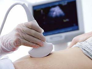
Ultrasonography, Doppler, and Duplex are diagnostic medical techniques that use sound waves to create images of internal body structures and assess their function. Here are the definitions for each:
1. Ultrasonography: Also known as ultrasound, this is a non-invasive imaging technique that uses high-frequency sound waves to produce images of internal organs and tissues. A small handheld device called a transducer is placed on the skin surface, which emits and receives sound waves. The returning echoes are then processed to create real-time visual images of the internal structures.
2. Doppler: This is a type of ultrasound that measures the velocity and direction of blood flow in the body by analyzing the frequency shift of the reflected sound waves. It can be used to assess blood flow in various parts of the body, such as the heart, arteries, and veins.
3. Duplex: Duplex ultrasonography is a combination of both gray-scale ultrasound and Doppler ultrasound. It provides detailed images of internal structures, as well as information about blood flow velocity and direction. This technique is often used to evaluate conditions such as deep vein thrombosis, carotid artery stenosis, and peripheral arterial disease.
In summary, ultrasonography is a diagnostic imaging technique that uses sound waves to create images of internal structures, Doppler is a type of ultrasound that measures blood flow velocity and direction, and duplex is a combination of both techniques that provides detailed images and information about blood flow.
Budd-Chiari syndrome is a rare condition characterized by the obstruction of the hepatic veins, which are the blood vessels that carry blood from the liver to the heart. This obstruction can be caused by blood clots, tumors, or other abnormalities, and it can lead to a backflow of blood in the liver, resulting in various symptoms such as abdominal pain, swelling, and liver enlargement. In severe cases, Budd-Chiari syndrome can cause liver failure and other complications if left untreated. The diagnosis of this condition typically involves imaging tests such as ultrasound, CT scan, or MRI, and treatment may include anticoagulation therapy, thrombolytic therapy, or surgical intervention to remove the obstruction.
Antithrombin III (ATIII) deficiency is a genetic disorder that affects the body's ability to regulate blood clotting. ATIII is a protein produced in the liver that inhibits the activity of thrombin and other coagulation factors, preventing excessive clot formation.
People with ATIII deficiency have lower than normal levels of this protein, which can lead to an increased risk of developing abnormal blood clots (thrombosis) in veins, particularly deep vein thrombosis (DVT) and pulmonary embolism (PE). These clots can cause serious complications, including damage to the affected veins, organ damage, and even death.
ATIII deficiency can be classified into two types: type I and type II. Type I is characterized by a quantitative decrease in ATIII levels, while type II is characterized by a qualitative defect that results in reduced functional activity of the protein.
The condition is usually inherited in an autosomal dominant manner, meaning that a person has a 50% chance of inheriting the gene mutation from an affected parent. However, some cases may occur spontaneously due to new mutations in the ATIII gene. Treatment for ATIII deficiency typically involves anticoagulation therapy with medications such as heparin or warfarin to prevent blood clots from forming.
Retrospective studies, also known as retrospective research or looking back studies, are a type of observational study that examines data from the past to draw conclusions about possible causal relationships between risk factors and outcomes. In these studies, researchers analyze existing records, medical charts, or previously collected data to test a hypothesis or answer a specific research question.
Retrospective studies can be useful for generating hypotheses and identifying trends, but they have limitations compared to prospective studies, which follow participants forward in time from exposure to outcome. Retrospective studies are subject to biases such as recall bias, selection bias, and information bias, which can affect the validity of the results. Therefore, retrospective studies should be interpreted with caution and used primarily to generate hypotheses for further testing in prospective studies.
Fibrinolysis is the natural process in the body that leads to the dissolution of blood clots. It is a vital part of hemostasis, the process that regulates bleeding and wound healing. Fibrinolysis occurs when plasminogen activators convert plasminogen to plasmin, an enzyme that breaks down fibrin, the insoluble protein mesh that forms the structure of a blood clot. This process helps to prevent excessive clotting and maintains the fluidity of the blood. In medical settings, fibrinolysis can also refer to the therapeutic use of drugs that stimulate this process to dissolve unwanted or harmful blood clots, such as those that cause deep vein thrombosis or pulmonary embolism.
The axillary vein is a large vein that runs through the axilla or armpit region. It is formed by the union of the brachial vein and the basilic vein at the lower border of the teres major muscle. The axillary vein carries deoxygenated blood from the upper limb, chest wall, and breast towards the heart. As it moves proximally, it becomes continuous with the subclavian vein to form the brachiocephalic vein. It is accompanied by the axillary artery and forms part of the important neurovascular bundle in the axilla.
Mesenteric vascular occlusion refers to the blockage or obstruction of the blood vessels that supply the intestines, specifically the mesenteric arteries and veins. This condition can result in insufficient blood flow to the intestines, leading to ischemia (inadequate oxygen supply) and potential necrosis (tissue death).
There are two primary types of mesenteric vascular occlusion:
1. Mesenteric arterial occlusion: This occurs when the mesenteric artery, which carries oxygenated blood from the heart to the intestines, becomes blocked. The most common causes include atherosclerosis (plaque buildup in the arteries), embolism (a clot or particle that travels from another part of the body and lodges in the artery), and thrombosis (a blood clot forming directly in the artery).
2. Mesenteric venous occlusion: This happens when the mesenteric vein, which returns deoxygenated blood from the intestines to the heart, becomes obstructed. The most common causes include thrombophlebitis (inflammation and clot formation in the vein), tumors, or abdominal trauma.
Symptoms of mesenteric vascular occlusion may include severe abdominal pain, nausea, vomiting, diarrhea, and bloody stools. Rapid diagnosis and treatment are crucial to prevent intestinal tissue damage and potential life-threatening complications such as sepsis or shock. Treatment options typically involve surgical intervention, anticoagulation therapy, or endovascular procedures to restore blood flow.
Protein C is a vitamin K-dependent protease that functions as an important regulator of coagulation and inflammation. It is a plasma protein produced in the liver that, when activated, degrades clotting factors Va and VIIIa to limit thrombus formation and prevent excessive blood clotting. Protein C also has anti-inflammatory properties by inhibiting the release of pro-inflammatory cytokines and reducing endothelial cell activation. Inherited or acquired deficiencies in Protein C can lead to an increased risk of thrombosis, a condition characterized by abnormal blood clot formation within blood vessels.
Postthrombotic syndrome (PTS), also known as postphlebitic syndrome, is a chronic complication that can occur after deep vein thrombosis (DVT). It's characterized by a combination of symptoms including pain, swelling, cramping, itching, and skin changes in the affected limb. PTS happens when the damaged valves in the veins are unable to properly move blood back to the heart, leading to venous hypertension and fluid accumulation in the lower extremities.
The symptoms of PTS can vary in severity, but they often worsen with prolonged standing or sitting. In some cases, patients may develop open sores (ulcers) on the skin, particularly around the ankles. The risk of developing PTS is higher in individuals who have experienced a recurrent DVT, those with more extensive clotting, and those who do not receive appropriate anticoagulation therapy after their initial DVT diagnosis.
Preventive measures such as early mobilization, use of compression stockings, and maintaining adequate anticoagulation can help reduce the risk of developing PTS following a DVT.
Aspirin is the common name for acetylsalicylic acid, which is a medication used to relieve pain, reduce inflammation, and lower fever. It works by inhibiting the activity of an enzyme called cyclooxygenase (COX), which is involved in the production of prostaglandins, hormone-like substances that cause inflammation and pain. Aspirin also has an antiplatelet effect, which means it can help prevent blood clots from forming. This makes it useful for preventing heart attacks and strokes.
Aspirin is available over-the-counter in various forms, including tablets, capsules, and chewable tablets. It is also available in prescription strengths for certain medical conditions. As with any medication, aspirin should be taken as directed by a healthcare provider, and its use should be avoided in children and teenagers with viral infections due to the risk of Reye's syndrome, a rare but serious condition that can affect the liver and brain.
Central venous catheterization is a medical procedure in which a flexible tube called a catheter is inserted into a large vein in the body, usually in the neck (internal jugular vein), chest (subclavian vein), or groin (femoral vein). The catheter is threaded through the vein until it reaches a central location, such as the superior vena cava or the right atrium of the heart.
Central venous catheterization may be performed for several reasons, including:
1. To administer medications, fluids, or nutritional support directly into the bloodstream.
2. To monitor central venous pressure (CVP), which can help assess a patient's volume status and cardiac function.
3. To draw blood samples for laboratory tests.
4. To deliver chemotherapy drugs or other medications that may be harmful to peripheral veins.
5. To provide access for hemodialysis or other long-term therapies.
The procedure requires careful attention to sterile technique to minimize the risk of infection, and it is usually performed under local anesthesia with sedation or general anesthesia. Complications of central venous catheterization may include bleeding, infection, pneumothorax (collapsed lung), arterial puncture, and catheter-related bloodstream infections (CRBSI).
Graft occlusion in the context of vascular surgery refers to the complete or partial blockage of a blood vessel that has been surgically replaced or repaired with a graft. The graft can be made from either synthetic materials or autologous tissue (taken from another part of the patient's body).
Graft occlusion can occur due to various reasons, including:
1. Thrombosis: Formation of a blood clot within the graft, which can obstruct blood flow.
2. Intimal hyperplasia: Overgrowth of the inner lining (intima) of the graft or the adjacent native vessel, causing narrowing of the lumen and reducing blood flow.
3. Atherosclerosis: Deposition of cholesterol and other substances in the walls of the graft, leading to hardening and narrowing of the vessel.
4. Infection: Bacterial or fungal infection of the graft can cause inflammation, weakening, and ultimately occlusion of the graft.
5. Mechanical factors: Kinking, twisting, or compression of the graft can lead to obstruction of blood flow.
Graft occlusion is a significant complication following vascular surgery, as it can result in reduced perfusion to downstream tissues and organs, leading to ischemia (lack of oxygen supply) and potential tissue damage or loss.
Fibrin is defined as a protein that is formed from fibrinogen during the clotting of blood. It plays an essential role in the formation of blood clots, also known as a clotting or coagulation cascade. When an injury occurs and bleeding starts, fibrin threads form a net-like structure that entraps platelets and red blood cells to create a stable clot, preventing further loss of blood.
The process of forming fibrin from fibrinogen is initiated by thrombin, another protein involved in the coagulation cascade. Thrombin cleaves fibrinogen into fibrin monomers, which then polymerize to form long strands of fibrin. These strands cross-link with each other through a process catalyzed by factor XIIIa, forming a stable clot that protects the wound and promotes healing.
It is important to note that abnormalities in fibrin formation or breakdown can lead to bleeding disorders or thrombotic conditions, respectively. Proper regulation of fibrin production and degradation is crucial for maintaining healthy hemostasis and preventing excessive clotting or bleeding.
Vena cava filters are medical devices that are implanted into the inferior vena cava, which is the largest vein in the body that returns blood from the lower half of the body to the heart. These filters are designed to trap blood clots that form in the deep veins of the legs (deep vein thrombosis or DVT) and prevent them from traveling to the lungs (pulmonary embolism or PE), which can be a life-threatening condition.
The filter is typically implanted using a catheter-based procedure, and it has legs or arms that extend out to trap the blood clots as they flow through the vein. Over time, the trapped clots may dissolve on their own or become organized and incorporated into the wall of the vein.
Vena cava filters are typically used in patients who are at high risk for PE but cannot take anticoagulation medication or have failed anticoagulation therapy. However, there is some controversy surrounding the use of these devices due to concerns about their long-term safety and effectiveness.
Fibrinogen is a soluble protein present in plasma, synthesized by the liver. It plays an essential role in blood coagulation. When an injury occurs, fibrinogen gets converted into insoluble fibrin by the action of thrombin, forming a fibrin clot that helps to stop bleeding from the injured site. Therefore, fibrinogen is crucial for hemostasis, which is the process of stopping bleeding and starting the healing process after an injury.
Postphlebitic syndrome, also known as postthrombotic syndrome or post-thrombotic limb, is a long-term complication that can occur after deep vein thrombosis (DVT). It's characterized by chronic venous insufficiency due to damage in the valves and walls of the affected veins. This results in impaired return of blood from the extremities back to the heart, leading to symptoms such as:
1. Swelling (edema) in the affected limb, usually the lower leg or calf.
2. Pain, aching, or cramping in the legs.
3. Heaviness or fatigue in the legs.
4. Skin changes like redness, warmth, or itchiness.
5. Development of venous ulcers or sores, particularly around the ankles.
The severity of postphlebitic syndrome can vary from mild to severe and may significantly impact a person's quality of life. Risk factors for developing this condition include having had a previous DVT, obesity, older age, lack of physical activity, and a family history of blood clotting disorders. Early diagnosis and appropriate management of deep vein thrombosis can help reduce the risk of developing postphlebitic syndrome.
Antithrombins are substances that prevent the formation or promote the dissolution of blood clots (thrombi). They include:
1. Anticoagulants: These are medications that reduce the ability of the blood to clot. Examples include heparin, warfarin, and direct oral anticoagulants (DOACs) such as apixaban, rivaroxaban, and dabigatran.
2. Thrombolytic agents: These are medications that break down existing blood clots. Examples include alteplase, reteplase, and tenecteplase.
3. Fibrinolytics: These are a type of thrombolytic agent that specifically target fibrin, a protein involved in the formation of blood clots.
4. Natural anticoagulants: These are substances produced by the body to regulate blood clotting. Examples include antithrombin III, protein C, and protein S.
Antithrombins are used in the prevention and treatment of various thromboembolic disorders, such as deep vein thrombosis (DVT), pulmonary embolism (PE), stroke, and myocardial infarction (heart attack). It is important to note that while antithrombins can help prevent or dissolve blood clots, they also increase the risk of bleeding, so their use must be carefully monitored.
An arteriovenous shunt is a surgically created connection between an artery and a vein. This procedure is typically performed to reroute blood flow or to provide vascular access for various medical treatments. In a surgical setting, the creation of an arteriovenous shunt involves connecting an artery directly to a vein, bypassing the capillary network in between.
There are different types of arteriovenous shunts used for specific medical purposes:
1. Arteriovenous Fistula (AVF): This is a surgical connection created between an artery and a vein, usually in the arm or leg. The procedure involves dissecting both the artery and vein, then suturing them directly together. Over time, the increased blood flow to the vein causes it to dilate and thicken, making it suitable for repeated needle punctures during hemodialysis treatments for patients with kidney failure.
2. Arteriovenous Graft (AVG): An arteriovenous graft is a synthetic tube used to connect an artery and a vein when a direct AVF cannot be created due to insufficient vessel size or poor quality. The graft can be made of various materials, such as polytetrafluoroethylene (PTFE) or Dacron. Grafts are more prone to infection and clotting compared to native AVFs but remain an essential option for patients requiring hemodialysis access.
3. Central Venous Catheter (CVC): A central venous catheter is a flexible tube inserted into a large vein, often in the neck or groin, and advanced towards the heart. CVCs can be used as temporary arteriovenous shunts for patients who require immediate hemodialysis access but do not have time to wait for an AVF or AVG to mature. However, they are associated with higher risks of infection and thrombosis compared to native AVFs and AVGs.
In summary, a surgical arteriovenous shunt is a connection between an artery and a vein established through a medical procedure. The primary purpose of these shunts is to provide vascular access for hemodialysis in patients with end-stage renal disease or to serve as temporary access when native AVFs or AVGs are not feasible.
Prospective studies, also known as longitudinal studies, are a type of cohort study in which data is collected forward in time, following a group of individuals who share a common characteristic or exposure over a period of time. The researchers clearly define the study population and exposure of interest at the beginning of the study and follow up with the participants to determine the outcomes that develop over time. This type of study design allows for the investigation of causal relationships between exposures and outcomes, as well as the identification of risk factors and the estimation of disease incidence rates. Prospective studies are particularly useful in epidemiology and medical research when studying diseases with long latency periods or rare outcomes.
Ticlopidine is defined as a platelet aggregation inhibitor drug, which works by preventing certain types of blood cells (platelets) from sticking together to form clots. It is used to reduce the risk of stroke and heart attack in patients who have already had a stroke or have peripheral arterial disease.
Ticlopidine is a thienopyridine derivative that selectively inhibits platelet activation and aggregation by blocking the ADP (adenosine diphosphate) receptor on the platelet surface. This action prevents the formation of platelet plugs, which can lead to the development of blood clots in the arteries.
Ticlopidine is available in oral form as tablets and is typically taken twice daily. Common side effects include diarrhea, skin rash, and itching. More serious side effects, such as neutropenia (low white blood cell count), thrombotic thrombocytopenic purpura (TTP), and aplastic anemia, are rare but can be life-threatening.
Due to the risk of serious side effects, ticlopidine is usually reserved for use in patients who cannot tolerate or have failed other antiplatelet therapies, such as aspirin or clopidogrel. It is important to monitor patients taking ticlopidine closely for signs of adverse reactions and to follow the prescribing instructions carefully.
Partial Thromboplastin Time (PTT) is a medical laboratory test that measures the time it takes for blood to clot. It's more specifically a measure of the intrinsic and common pathways of the coagulation cascade, which are the series of chemical reactions that lead to the formation of a clot.
The test involves adding a partial thromboplastin reagent (an activator of the intrinsic pathway) and calcium to plasma, and then measuring the time it takes for a fibrin clot to form. This is compared to a control sample, and the ratio of the two times is calculated.
The PTT test is often used to help diagnose bleeding disorders or abnormal blood clotting, such as hemophilia or disseminated intravascular coagulation (DIC). It can also be used to monitor the effectiveness of anticoagulant therapy, such as heparin. Prolonged PTT results may indicate a bleeding disorder or an increased risk of bleeding, while shortened PTT results may indicate a hypercoagulable state and an increased risk of thrombosis.
Recurrence, in a medical context, refers to the return of symptoms or signs of a disease after a period of improvement or remission. It indicates that the condition has not been fully eradicated and may require further treatment. Recurrence is often used to describe situations where a disease such as cancer comes back after initial treatment, but it can also apply to other medical conditions. The likelihood of recurrence varies depending on the type of disease and individual patient factors.
X-ray computed tomography (CT or CAT scan) is a medical imaging method that uses computer-processed combinations of many X-ray images taken from different angles to produce cross-sectional (tomographic) images (virtual "slices") of the body. These cross-sectional images can then be used to display detailed internal views of organs, bones, and soft tissues in the body.
The term "computed tomography" is used instead of "CT scan" or "CAT scan" because the machines take a series of X-ray measurements from different angles around the body and then use a computer to process these data to create detailed images of internal structures within the body.
CT scanning is a noninvasive, painless medical test that helps physicians diagnose and treat medical conditions. CT imaging provides detailed information about many types of tissue including lung, bone, soft tissue and blood vessels. CT examinations can be performed on every part of the body for a variety of reasons including diagnosis, surgical planning, and monitoring of therapeutic responses.
In computed tomography (CT), an X-ray source and detector rotate around the patient, measuring the X-ray attenuation at many different angles. A computer uses this data to construct a cross-sectional image by the process of reconstruction. This technique is called "tomography". The term "computed" refers to the use of a computer to reconstruct the images.
CT has become an important tool in medical imaging and diagnosis, allowing radiologists and other physicians to view detailed internal images of the body. It can help identify many different medical conditions including cancer, heart disease, lung nodules, liver tumors, and internal injuries from trauma. CT is also commonly used for guiding biopsies and other minimally invasive procedures.
In summary, X-ray computed tomography (CT or CAT scan) is a medical imaging technique that uses computer-processed combinations of many X-ray images taken from different angles to produce cross-sectional images of the body. It provides detailed internal views of organs, bones, and soft tissues in the body, allowing physicians to diagnose and treat medical conditions.
Blood coagulation tests, also known as coagulation studies or clotting tests, are a series of medical tests used to evaluate the blood's ability to clot. These tests measure the functioning of various clotting factors and regulatory proteins involved in the coagulation cascade, which is a complex process that leads to the formation of a blood clot to prevent excessive bleeding.
The most commonly performed coagulation tests include:
1. Prothrombin Time (PT): Measures the time it takes for a sample of plasma to clot after the addition of calcium and tissue factor, which activates the extrinsic pathway of coagulation. The PT is reported in seconds and can be converted to an International Normalized Ratio (INR) to monitor anticoagulant therapy.
2. Activated Partial Thromboplastin Time (aPTT): Measures the time it takes for a sample of plasma to clot after the addition of calcium, phospholipid, and a contact activator, which activates the intrinsic pathway of coagulation. The aPTT is reported in seconds and is used to monitor heparin therapy.
3. Thrombin Time (TT): Measures the time it takes for a sample of plasma to clot after the addition of thrombin, which directly converts fibrinogen to fibrin. The TT is reported in seconds and can be used to detect the presence of fibrin degradation products or abnormalities in fibrinogen function.
4. Fibrinogen Level: Measures the amount of fibrinogen, a protein involved in clot formation, present in the blood. The level is reported in grams per liter (g/L) and can be used to assess bleeding risk or the effectiveness of fibrinogen replacement therapy.
5. D-dimer Level: Measures the amount of D-dimer, a protein fragment produced during the breakdown of a blood clot, present in the blood. The level is reported in micrograms per milliliter (µg/mL) and can be used to diagnose or exclude venous thromboembolism (VTE), such as deep vein thrombosis (DVT) or pulmonary embolism (PE).
These tests are important for the diagnosis, management, and monitoring of various bleeding and clotting disorders. They can help identify the underlying cause of abnormal bleeding or clotting, guide appropriate treatment decisions, and monitor the effectiveness of therapy. It is essential to interpret these test results in conjunction with a patient's clinical presentation and medical history.
Coronary balloon angioplasty is a minimally invasive medical procedure used to widen narrowed or obstructed coronary arteries (the blood vessels that supply oxygen-rich blood to the heart muscle) and improve blood flow to the heart. This procedure is typically performed in conjunction with the insertion of a stent, a small mesh tube that helps keep the artery open.
During coronary balloon angioplasty, a thin, flexible catheter with a deflated balloon at its tip is inserted into a blood vessel, usually through a small incision in the groin or arm. The catheter is then guided to the narrowed or obstructed section of the coronary artery. Once in position, the balloon is inflated to compress the plaque against the artery wall and widen the lumen (the inner space) of the artery. This helps restore blood flow to the heart muscle.
The procedure is typically performed under local anesthesia and conscious sedation to minimize discomfort. Coronary balloon angioplasty is a relatively safe and effective treatment for many people with coronary artery disease, although complications such as bleeding, infection, or re-narrowing of the artery (restenosis) can occur in some cases.
Sirolimus is a medication that belongs to a class of drugs called immunosuppressants. It is also known as rapamycin. Sirolimus works by inhibiting the mammalian target of rapamycin (mTOR), which is a protein that plays a key role in cell growth and division.
Sirolimus is primarily used to prevent rejection of transplanted organs, such as kidneys, livers, and hearts. It works by suppressing the activity of the immune system, which can help to reduce the risk of the body rejecting the transplanted organ. Sirolimus is often used in combination with other immunosuppressive drugs, such as corticosteroids and calcineurin inhibitors.
Sirolimus is also being studied for its potential therapeutic benefits in a variety of other conditions, including cancer, tuberous sclerosis complex, and lymphangioleiomyomatosis. However, more research is needed to fully understand the safety and efficacy of sirolimus in these contexts.
It's important to note that sirolimus can have significant side effects, including increased risk of infections, mouth sores, high blood pressure, and kidney damage. Therefore, it should only be used under the close supervision of a healthcare provider.
Indwelling catheters, also known as Foley catheters, are medical devices that are inserted into the bladder to drain urine. They have a small balloon at the tip that is inflated with water once the catheter is in the correct position in the bladder, allowing it to remain in place and continuously drain urine. Indwelling catheters are typically used for patients who are unable to empty their bladders on their own, such as those who are bedridden or have nerve damage that affects bladder function. They are also used during and after certain surgical procedures. Prolonged use of indwelling catheters can increase the risk of urinary tract infections and other complications.
Coronary restenosis is the re-narrowing or re-occlusion of a coronary artery after a previous successful procedure to open or widen the artery, such as angioplasty or stenting. This narrowing is usually caused by the excessive growth of scar tissue or smooth muscle cells in the artery lining, which can occur spontaneously or as a response to the initial procedure. Restenosis can lead to recurrent symptoms of coronary artery disease, such as chest pain or shortness of breath, and may require additional medical intervention.
Anticardiolipin antibodies are a type of autoantibody that targets and binds to cardiolipin, a phospholipid component found in the inner mitochondrial membrane of cells. These antibodies are clinically significant because they have been associated with a variety of autoimmune disorders, including antiphospholipid syndrome (APS).
APS is a condition characterized by recurrent blood clots, pregnancy losses, and thrombocytopenia (low platelet count). Anticardiolipin antibodies are one of the three main types of autoantibodies found in APS, along with lupus anticoagulant and anti-β2 glycoprotein I antibodies.
The presence of high levels of anticardiolipin antibodies in the blood can lead to abnormal blood clotting, which can cause serious complications such as deep vein thrombosis, pulmonary embolism, and stroke. Anticardiolipin antibodies can also contribute to pregnancy losses by causing placental insufficiency or abnormal blood clotting in the placenta.
Anticardiolipin antibodies are typically detected through a blood test that measures their levels in the serum. A positive result is usually confirmed with a second test performed at least 12 weeks later to establish persistence. Treatment for anticardiolipin antibody-related disorders typically involves anticoagulation therapy to prevent blood clots and other complications.
Compression stockings are a specialized type of hosiery that applies pressure to your legs, promoting better blood flow. They are tightest at the ankle and gradually become less constrictive up the leg. This gradient compression helps to counteract the force of gravity and promote venous return, reducing the pooling of blood in the lower extremities.
Compression stockings are often used to help prevent or treat various conditions related to poor circulation, including:
1. Varicose veins: Enlarged, swollen, and twisting veins that are easily visible just under the surface of the skin.
2. Deep vein thrombosis (DVT): A blood clot that forms in the deep veins, usually in the legs. Compression stockings can help reduce the risk of DVT after certain surgeries or during long periods of immobilization.
3. Edema: Swelling in the legs and ankles due to fluid buildup.
4. Chronic venous insufficiency: A condition where the veins have difficulty returning blood from the legs back to the heart, leading to symptoms like leg pain, swelling, and skin changes.
5. Post-thrombotic syndrome (PTS): A long-term complication of DVT characterized by chronic leg pain, swelling, and skin ulcers. Compression stockings can help manage symptoms and prevent further complications.
There are different levels of compression available, ranging from mild (15-20 mmHg) to extra firm (50-60 mmHg). Your healthcare provider will recommend the appropriate level based on your specific condition and needs. It is essential to wear compression stockings correctly for them to be effective and avoid skin irritation or other complications.
Essential thrombocythemia (ET) is a myeloproliferative neoplasm (MPN), a type of blood cancer characterized by the overproduction of platelets (thrombocytosis) in the bone marrow. In ET, there is an excessive proliferation of megakaryocytes, the precursor cells that produce platelets. This leads to increased platelet counts in the peripheral blood, which can increase the risk of blood clots (thrombosis) and bleeding episodes (hemorrhage).
The term "essential" is used to indicate that the cause of this condition is not known or idiopathic. ET is primarily a disease of older adults, but it can also occur in younger individuals. The diagnosis of essential thrombocythemia requires careful evaluation and exclusion of secondary causes of thrombocytosis, such as reactive conditions, inflammation, or other myeloproliferative neoplasms.
The clinical presentation of ET can vary widely among patients. Some individuals may be asymptomatic and discovered only during routine blood tests, while others may experience symptoms related to thrombosis or bleeding. Common symptoms include headaches, visual disturbances, dizziness, weakness, numbness, or tingling in the extremities, if there are complications due to blood clots in the brain or other parts of the body. Excessive bruising, nosebleeds, or blood in the stool can indicate bleeding complications.
Treatment for essential thrombocythemia is aimed at reducing the risk of thrombosis and managing symptoms. Hydroxyurea is a commonly used medication to lower platelet counts, while aspirin may be prescribed to decrease the risk of blood clots. In some cases, interferon-alpha or ruxolitinib might be considered as treatment options. Regular follow-up with a hematologist and monitoring of blood counts are essential for managing this condition and detecting potential complications early.
Peripheral catheterization is a medical procedure that involves the insertion of a thin, flexible tube (catheter) into a peripheral vein, which is a blood vessel located outside of the chest and abdomen. This type of catheterization is typically performed to administer medications, fluids, or nutritional support, or to monitor various physiological parameters such as central venous pressure.
Peripheral catheters are usually inserted into veins in the hands or arms, although they can also be placed in other peripheral veins. The procedure is typically performed using aseptic technique to minimize the risk of infection. Once the catheter is in place, it may be secured with a dressing or suture to prevent movement and dislodgement.
Peripheral catheterization is a relatively safe and common procedure that is routinely performed in hospitals, clinics, and other healthcare settings. However, like any medical procedure, it carries a small risk of complications such as infection, bleeding, or damage to the vein or surrounding tissues.
Blood coagulation factors, also known as clotting factors, are a group of proteins that play a crucial role in the blood coagulation process. They are essential for maintaining hemostasis, which is the body's ability to stop bleeding after injury.
There are 13 known blood coagulation factors, and they are designated by Roman numerals I through XIII. These factors are produced in the liver and are normally present in an inactive form in the blood. When there is an injury to a blood vessel, the coagulation process is initiated, leading to the activation of these factors in a specific order.
The coagulation cascade involves two pathways: the intrinsic and extrinsic pathways. The intrinsic pathway is activated when there is damage to the blood vessel itself, while the extrinsic pathway is activated by tissue factor released from damaged tissues. Both pathways converge at the common pathway, leading to the formation of a fibrin clot.
Blood coagulation factors work together in a complex series of reactions that involve activation, binding, and proteolysis. When one factor is activated, it activates the next factor in the cascade, and so on. This process continues until a stable fibrin clot is formed.
Deficiencies or abnormalities in blood coagulation factors can lead to bleeding disorders such as hemophilia or thrombosis. Hemophilia is a genetic disorder that affects one or more of the coagulation factors, leading to excessive bleeding and difficulty forming clots. Thrombosis, on the other hand, occurs when there is an abnormal formation of blood clots in the blood vessels, which can lead to serious complications such as stroke or pulmonary embolism.
Enoxaparin is a low molecular weight heparin (LMWH) medication that is used as an anticoagulant to prevent and treat blood clots. It works by binding to and inhibiting the activity of factor Xa, a clotting factor in the blood. This helps to reduce the risk of clot formation and can help to prevent conditions such as deep vein thrombosis (DVT) and pulmonary embolism (PE). Enoxaparin is typically given by injection under the skin (subcutaneously) and is available under the brand names Lovenox and Clexane, among others. It is important to follow the instructions of a healthcare professional when using enoxaparin, as it can increase the risk of bleeding.
Vascular patency is a term used in medicine to describe the state of a blood vessel (such as an artery or vein) being open, unobstructed, and allowing for the normal flow of blood. It is an important concept in the treatment and management of various cardiovascular conditions, such as peripheral artery disease, coronary artery disease, and deep vein thrombosis.
Maintaining vascular patency can help prevent serious complications like tissue damage, organ dysfunction, or even death. This may involve medical interventions such as administering blood-thinning medications to prevent clots, performing procedures to remove blockages, or using devices like stents to keep vessels open. Regular monitoring of vascular patency is also crucial for evaluating the effectiveness of treatments and adjusting care plans accordingly.
Veins are blood vessels that carry deoxygenated blood from the tissues back to the heart. They have a lower pressure than arteries and contain valves to prevent the backflow of blood. Veins have a thin, flexible wall with a larger lumen compared to arteries, allowing them to accommodate more blood volume. The color of veins is often blue or green due to the absorption characteristics of light and the reduced oxygen content in the blood they carry.
Blood coagulation disorders, also known as bleeding disorders or clotting disorders, refer to a group of medical conditions that affect the body's ability to form blood clots properly. Normally, when a blood vessel is injured, the body's coagulation system works to form a clot to stop the bleeding and promote healing.
In blood coagulation disorders, there can be either an increased tendency to bleed due to problems with the formation of clots (hemorrhagic disorder), or an increased tendency for clots to form inappropriately even without injury, leading to blockages in the blood vessels (thrombotic disorder).
Examples of hemorrhagic disorders include:
1. Hemophilia - a genetic disorder that affects the ability to form clots due to deficiencies in clotting factors VIII or IX.
2. Von Willebrand disease - another genetic disorder caused by a deficiency or abnormality of the von Willebrand factor, which helps platelets stick together to form a clot.
3. Liver diseases - can lead to decreased production of coagulation factors, increasing the risk of bleeding.
4. Disseminated intravascular coagulation (DIC) - a serious condition where clotting and bleeding occur simultaneously due to widespread activation of the coagulation system.
Examples of thrombotic disorders include:
1. Factor V Leiden mutation - a genetic disorder that increases the risk of inappropriate blood clot formation.
2. Antithrombin III deficiency - a genetic disorder that impairs the body's ability to break down clots, increasing the risk of thrombosis.
3. Protein C or S deficiencies - genetic disorders that lead to an increased risk of thrombosis due to impaired regulation of the coagulation system.
4. Antiphospholipid syndrome (APS) - an autoimmune disorder where the body produces antibodies against its own clotting factors, increasing the risk of thrombosis.
Treatment for blood coagulation disorders depends on the specific diagnosis and may include medications to manage bleeding or prevent clots, as well as lifestyle changes and monitoring to reduce the risk of complications.
An acute disease is a medical condition that has a rapid onset, develops quickly, and tends to be short in duration. Acute diseases can range from minor illnesses such as a common cold or flu, to more severe conditions such as pneumonia, meningitis, or a heart attack. These types of diseases often have clear symptoms that are easy to identify, and they may require immediate medical attention or treatment.
Acute diseases are typically caused by an external agent or factor, such as a bacterial or viral infection, a toxin, or an injury. They can also be the result of a sudden worsening of an existing chronic condition. In general, acute diseases are distinct from chronic diseases, which are long-term medical conditions that develop slowly over time and may require ongoing management and treatment.
Examples of acute diseases include:
* Acute bronchitis: a sudden inflammation of the airways in the lungs, often caused by a viral infection.
* Appendicitis: an inflammation of the appendix that can cause severe pain and requires surgical removal.
* Gastroenteritis: an inflammation of the stomach and intestines, often caused by a viral or bacterial infection.
* Migraine headaches: intense headaches that can last for hours or days, and are often accompanied by nausea, vomiting, and sensitivity to light and sound.
* Myocardial infarction (heart attack): a sudden blockage of blood flow to the heart muscle, often caused by a buildup of plaque in the coronary arteries.
* Pneumonia: an infection of the lungs that can cause coughing, chest pain, and difficulty breathing.
* Sinusitis: an inflammation of the sinuses, often caused by a viral or bacterial infection.
It's important to note that while some acute diseases may resolve on their own with rest and supportive care, others may require medical intervention or treatment to prevent complications and promote recovery. If you are experiencing symptoms of an acute disease, it is always best to seek medical attention to ensure proper diagnosis and treatment.
Thrombocytopenia is a medical condition characterized by an abnormally low platelet count (thrombocytes) in the blood. Platelets are small cell fragments that play a crucial role in blood clotting, helping to stop bleeding when a blood vessel is damaged. A healthy adult typically has a platelet count between 150,000 and 450,000 platelets per microliter of blood. Thrombocytopenia is usually diagnosed when the platelet count falls below 150,000 platelets/µL.
Thrombocytopenia can be classified into three main categories based on its underlying cause:
1. Immune thrombocytopenia (ITP): An autoimmune disorder where the immune system mistakenly attacks and destroys its own platelets, leading to a decreased platelet count. ITP can be further divided into primary or secondary forms, depending on whether it occurs alone or as a result of another medical condition or medication.
2. Decreased production: Thrombocytopenia can occur when there is insufficient production of platelets in the bone marrow due to various causes, such as viral infections, chemotherapy, radiation therapy, leukemia, aplastic anemia, or vitamin B12 or folate deficiency.
3. Increased destruction or consumption: Thrombocytopenia can also result from increased platelet destruction or consumption due to conditions like disseminated intravascular coagulation (DIC), thrombotic thrombocytopenic purpura (TTP), hemolytic uremic syndrome (HUS), or severe bacterial infections.
Symptoms of thrombocytopenia may include easy bruising, prolonged bleeding from cuts, spontaneous nosebleeds, bleeding gums, blood in urine or stools, and skin rashes like petechiae (small red or purple spots) or purpura (larger patches). The severity of symptoms can vary depending on the degree of thrombocytopenia and the presence of any underlying conditions. Treatment for thrombocytopenia depends on the cause and may include medications, transfusions, or addressing the underlying condition.
The Superior Sagittal Sinus is a medical term that refers to a venous sinus (a channel for blood flow) located in the superior part (highest portion) of the sagittal suture, which is the line along the top of the skull where the two parietal bones join in the middle. It runs from front to back, starting at the frontal bone and ending at the occipital bone, and it receives blood from veins that drain the cerebral hemispheres (the right and left halves of the brain).
The Superior Sagittal Sinus is an important structure in the circulatory system of the brain as it plays a critical role in draining venous blood from the cranial cavity. It also contains valveless venous channels that allow for the flow of cerebrospinal fluid (CSF) between the intracranial and extracranial compartments.
It is worth noting that any damage to this structure, such as through trauma or infection, can lead to serious neurological complications, including increased intracranial pressure, seizures, and even death.
Prosthesis design is a specialized field in medical device technology that involves creating and developing artificial substitutes to replace a missing body part, such as a limb, tooth, eye, or internal organ. The design process typically includes several stages: assessment of the patient's needs, selection of appropriate materials, creation of a prototype, testing and refinement, and final fabrication and fitting of the prosthesis.
The goal of prosthesis design is to create a device that functions as closely as possible to the natural body part it replaces, while also being comfortable, durable, and aesthetically pleasing for the patient. The design process may involve collaboration between medical professionals, engineers, and designers, and may take into account factors such as the patient's age, lifestyle, occupation, and overall health.
Prosthesis design can be highly complex, particularly for advanced devices such as robotic limbs or implantable organs. These devices often require sophisticated sensors, actuators, and control systems to mimic the natural functions of the body part they replace. As a result, prosthesis design is an active area of research and development in the medical field, with ongoing efforts to improve the functionality, comfort, and affordability of these devices for patients.
Protein S is a vitamin K-dependent protein found in the blood that functions as a natural anticoagulant. It plays a crucial role in regulating the body's clotting system by inhibiting the activation of coagulation factors, thereby preventing excessive blood clotting. Protein S also acts as a cofactor for activated protein C, which is another important anticoagulant protein.
Protein S exists in two forms: free and bound to a protein called C4b-binding protein (C4BP). Only the free form of Protein S has biological activity in inhibiting coagulation. Inherited or acquired deficiencies in Protein S can lead to an increased risk of thrombosis, or abnormal blood clot formation, which can cause various medical conditions such as deep vein thrombosis (DVT) and pulmonary embolism (PE). Regular monitoring of Protein S levels is essential for patients with a history of thrombotic events or those who have a family history of thrombophilia.
In epidemiology, the incidence of a disease is defined as the number of new cases of that disease within a specific population over a certain period of time. It is typically expressed as a rate, with the number of new cases in the numerator and the size of the population at risk in the denominator. Incidence provides information about the risk of developing a disease during a given time period and can be used to compare disease rates between different populations or to monitor trends in disease occurrence over time.
Beta 2-glycoprotein I, also known as apolipoprotein H, is a plasma protein that belongs to the family of proteins called immunoglobulin-binding proteins. It has a molecular weight of approximately 44 kDa and is composed of five domains with similar structures.
Beta 2-glycoprotein I is primarily produced in the liver and circulates in the bloodstream, where it plays a role in several physiological processes, including coagulation, complement activation, and lipid metabolism. It has been identified as an autoantigen in certain autoimmune disorders, such as antiphospholipid syndrome (APS), where autoantibodies against beta 2-glycoprotein I can cause blood clots, miscarriages, and other complications.
In medical terminology, the definition of "beta 2-glycoprotein I" is as follows:
A plasma protein that belongs to the family of immunoglobulin-binding proteins and has a molecular weight of approximately 44 kDa. It is primarily produced in the liver and circulates in the bloodstream, where it plays a role in several physiological processes, including coagulation, complement activation, and lipid metabolism. Autoantibodies against beta 2-glycoprotein I are associated with certain autoimmune disorders, such as antiphospholipid syndrome (APS), where they can cause blood clots, miscarriages, and other complications.
Tissue Plasminogen Activator (tPA) is a thrombolytic enzyme, which means it dissolves blood clots. It is naturally produced by the endothelial cells that line the interior surface of blood vessels. tPA activates plasminogen, a zymogen, to convert it into plasmin, a protease that breaks down fibrin, the structural protein in blood clots. This enzyme is used medically as a thrombolytic drug under various brand names, such as Activase and Alteplase, to treat conditions like acute ischemic stroke, pulmonary embolism, and deep vein thrombosis by dissolving the clots and restoring blood flow.
Arterial occlusive diseases are medical conditions characterized by the blockage or narrowing of the arteries, which can lead to a reduction in blood flow to various parts of the body. This reduction in blood flow can cause tissue damage and may result in serious complications such as tissue death (gangrene), organ dysfunction, or even death.
The most common cause of arterial occlusive diseases is atherosclerosis, which is the buildup of plaque made up of fat, cholesterol, calcium, and other substances in the inner lining of the artery walls. Over time, this plaque can harden and narrow the arteries, restricting blood flow. Other causes of arterial occlusive diseases include blood clots, emboli (tiny particles that travel through the bloodstream and lodge in smaller vessels), inflammation, trauma, and certain inherited conditions.
Symptoms of arterial occlusive diseases depend on the location and severity of the blockage. Common symptoms include:
* Pain, cramping, or fatigue in the affected limb, often triggered by exercise and relieved by rest (claudication)
* Numbness, tingling, or weakness in the affected limb
* Coldness or discoloration of the skin in the affected area
* Slow-healing sores or wounds on the toes, feet, or legs
* Erectile dysfunction in men
Treatment for arterial occlusive diseases may include lifestyle changes such as quitting smoking, exercising regularly, and eating a healthy diet. Medications to lower cholesterol, control blood pressure, prevent blood clots, or manage pain may also be prescribed. In severe cases, surgical procedures such as angioplasty, stenting, or bypass surgery may be necessary to restore blood flow.
Impedance plethysmography is a non-invasive method used to measure changes in blood volume or flow in a particular area of the body. It works by passing a small electrical current through the tissue and measuring the opposition (impedance) to that current, which varies with the amount of blood present in the area.
In impedance cardiography, this technique is used to estimate cardiac output, stroke volume, and other hemodynamic parameters. The changes in impedance are measured across the chest wall, which correlate with the ventricular ejection of blood during each heartbeat. This allows for the calculation of various cardiovascular variables, such as the amount of blood pumped by the heart per minute (cardiac output) and the resistance to blood flow in the systemic circulation (systemic vascular resistance).
Impedance plethysmography is a safe and reliable method for assessing cardiovascular function, and it has been widely used in clinical settings to evaluate patients with various cardiovascular disorders, including heart failure, hypertension, and peripheral arterial disease.
A bandage is a medical dressing or covering applied to a wound, injury, or sore with the intention of promoting healing or preventing infection. Bandages can be made of a variety of materials such as gauze, cotton, elastic, or adhesive tape and come in different sizes and shapes to accommodate various body parts. They can also have additional features like fasteners, non-slip surfaces, or transparent windows for monitoring the condition of the wound.
Bandages serve several purposes, including:
1. Absorbing drainage or exudate from the wound
2. Protecting the wound from external contaminants and bacteria
3. Securing other medical devices such as catheters or splints in place
4. Reducing swelling or promoting immobilization of the affected area
5. Providing compression to control bleeding or prevent fluid accumulation
6. Relieving pain by reducing pressure on sensitive nerves or structures.
Proper application and care of bandages are essential for effective wound healing and prevention of complications such as infection or delayed recovery.
Intermittent Pneumatic Compression (IPC) devices are medical devices that use inflatable garments to apply controlled pressure in a rhythmic, intermittent manner to the extremities of the body, most commonly used on the legs. These devices are designed to help improve venous and lymphatic flow, reduce edema (swelling), and prevent the formation of blood clots (deep vein thrombosis) in patients who are at risk.
The IPC device typically consists of a pump, hoses, and an air-filled garment that covers the affected limb. The pump regulates the pressure and inflation pattern, while the garment applies pressure to the limb. The compression cycle usually starts with low pressure and gradually increases to a peak pressure before decreasing again. This process is repeated at regular intervals, providing intermittent compression that mimics natural muscle contractions and helps promote blood flow.
IPC devices are often used in clinical settings such as hospitals and rehabilitation centers, but they can also be prescribed for home use. They are commonly recommended for patients who have undergone surgery, experienced trauma, or have conditions that increase their risk of developing blood clots, such as prolonged immobilization, varicose veins, or certain medical disorders.
It is essential to follow the healthcare provider's instructions when using IPC devices and report any discomfort, pain, or unusual symptoms during treatment.
A blood vessel prosthesis is a medical device that is used as a substitute for a damaged or diseased natural blood vessel. It is typically made of synthetic materials such as polyester, Dacron, or ePTFE (expanded polytetrafluoroethylene) and is designed to mimic the function of a native blood vessel by allowing the flow of blood through it.
Blood vessel prostheses are used in various surgical procedures, including coronary artery bypass grafting, peripheral arterial reconstruction, and the creation of arteriovenous fistulas for dialysis access. The choice of material and size of the prosthesis depends on several factors, such as the location and diameter of the vessel being replaced, the patient's age and overall health status, and the surgeon's preference.
It is important to note that while blood vessel prostheses can be effective in restoring blood flow, they may also carry risks such as infection, thrombosis (blood clot formation), and graft failure over time. Therefore, careful patient selection, surgical technique, and postoperative management are crucial for the success of these procedures.
Disseminated Intravascular Coagulation (DIC) is a complex medical condition characterized by the abnormal activation of the coagulation cascade, leading to the formation of blood clots in small blood vessels throughout the body. This process can result in the consumption of clotting factors and platelets, which can then lead to bleeding complications. DIC can be caused by a variety of underlying conditions, including sepsis, trauma, cancer, and obstetric emergencies.
The term "disseminated" refers to the widespread nature of the clotting activation, while "intravascular" indicates that the clotting is occurring within the blood vessels. The condition can manifest as both bleeding and clotting complications, which can make it challenging to diagnose and manage.
The diagnosis of DIC typically involves laboratory tests that evaluate coagulation factors, platelet count, fibrin degradation products, and other markers of coagulation activation. Treatment is focused on addressing the underlying cause of the condition while also managing any bleeding or clotting complications that may arise.
Myocardial infarction (MI), also known as a heart attack, is a medical condition characterized by the death of a segment of heart muscle (myocardium) due to the interruption of its blood supply. This interruption is most commonly caused by the blockage of a coronary artery by a blood clot formed on the top of an atherosclerotic plaque, which is a buildup of cholesterol and other substances in the inner lining of the artery.
The lack of oxygen and nutrients supply to the heart muscle tissue results in damage or death of the cardiac cells, causing the affected area to become necrotic. The extent and severity of the MI depend on the size of the affected area, the duration of the occlusion, and the presence of collateral circulation.
Symptoms of a myocardial infarction may include chest pain or discomfort, shortness of breath, nausea, lightheadedness, and sweating. Immediate medical attention is necessary to restore blood flow to the affected area and prevent further damage to the heart muscle. Treatment options for MI include medications, such as thrombolytics, antiplatelet agents, and pain relievers, as well as procedures such as percutaneous coronary intervention (PCI) or coronary artery bypass grafting (CABG).
Thrombin is a serine protease enzyme that plays a crucial role in the coagulation cascade, which is a complex series of biochemical reactions that leads to the formation of a blood clot (thrombus) to prevent excessive bleeding during an injury. Thrombin is formed from its precursor protein, prothrombin, through a process called activation, which involves cleavage by another enzyme called factor Xa.
Once activated, thrombin converts fibrinogen, a soluble plasma protein, into fibrin, an insoluble protein that forms the structural framework of a blood clot. Thrombin also activates other components of the coagulation cascade, such as factor XIII, which crosslinks and stabilizes the fibrin network, and platelets, which contribute to the formation and growth of the clot.
Thrombin has several regulatory mechanisms that control its activity, including feedback inhibition by antithrombin III, a plasma protein that inactivates thrombin and other serine proteases, and tissue factor pathway inhibitor (TFPI), which inhibits the activation of factor Xa, thereby preventing further thrombin formation.
Overall, thrombin is an essential enzyme in hemostasis, the process that maintains the balance between bleeding and clotting in the body. However, excessive or uncontrolled thrombin activity can lead to pathological conditions such as thrombosis, atherosclerosis, and disseminated intravascular coagulation (DIC).
Animal disease models are specialized animals, typically rodents such as mice or rats, that have been genetically engineered or exposed to certain conditions to develop symptoms and physiological changes similar to those seen in human diseases. These models are used in medical research to study the pathophysiology of diseases, identify potential therapeutic targets, test drug efficacy and safety, and understand disease mechanisms.
The genetic modifications can include knockout or knock-in mutations, transgenic expression of specific genes, or RNA interference techniques. The animals may also be exposed to environmental factors such as chemicals, radiation, or infectious agents to induce the disease state.
Examples of animal disease models include:
1. Mouse models of cancer: Genetically engineered mice that develop various types of tumors, allowing researchers to study cancer initiation, progression, and metastasis.
2. Alzheimer's disease models: Transgenic mice expressing mutant human genes associated with Alzheimer's disease, which exhibit amyloid plaque formation and cognitive decline.
3. Diabetes models: Obese and diabetic mouse strains like the NOD (non-obese diabetic) or db/db mice, used to study the development of type 1 and type 2 diabetes, respectively.
4. Cardiovascular disease models: Atherosclerosis-prone mice, such as ApoE-deficient or LDLR-deficient mice, that develop plaque buildup in their arteries when fed a high-fat diet.
5. Inflammatory bowel disease models: Mice with genetic mutations affecting intestinal barrier function and immune response, such as IL-10 knockout or SAMP1/YitFc mice, which develop colitis.
Animal disease models are essential tools in preclinical research, but it is important to recognize their limitations. Differences between species can affect the translatability of results from animal studies to human patients. Therefore, researchers must carefully consider the choice of model and interpret findings cautiously when applying them to human diseases.
Platelet adhesiveness refers to the ability of platelets, which are small blood cells that help your body form clots to prevent excessive bleeding, to stick to other cells or surfaces. This process is crucial in hemostasis, the process of stopping bleeding after injury to a blood vessel.
When the endothelium (the lining of blood vessels) is damaged, subendothelial structures are exposed, which can trigger platelet adhesion. Platelets then change shape and release chemical signals that cause other platelets to clump together, forming a platelet plug. This plug helps to seal the damaged vessel and prevent further bleeding.
Platelet adhesiveness is influenced by several factors, including the presence of von Willebrand factor (vWF), a protein in the blood that helps platelets bind to damaged vessels, and the expression of glycoprotein receptors on the surface of platelets. Abnormalities in platelet adhesiveness can lead to bleeding disorders or thrombotic conditions.
Angiography is a medical procedure in which an x-ray image is taken to visualize the internal structure of blood vessels, arteries, or veins. This is done by injecting a radiopaque contrast agent (dye) into the blood vessel using a thin, flexible catheter. The dye makes the blood vessels visible on an x-ray image, allowing doctors to diagnose and treat various medical conditions such as blockages, narrowing, or malformations of the blood vessels.
There are several types of angiography, including:
* Cardiac angiography (also called coronary angiography) - used to examine the blood vessels of the heart
* Cerebral angiography - used to examine the blood vessels of the brain
* Peripheral angiography - used to examine the blood vessels in the limbs or other parts of the body.
Angiography is typically performed by a radiologist, cardiologist, or vascular surgeon in a hospital setting. It can help diagnose conditions such as coronary artery disease, aneurysms, and peripheral arterial disease, among others.
Risk assessment in the medical context refers to the process of identifying, evaluating, and prioritizing risks to patients, healthcare workers, or the community related to healthcare delivery. It involves determining the likelihood and potential impact of adverse events or hazards, such as infectious diseases, medication errors, or medical devices failures, and implementing measures to mitigate or manage those risks. The goal of risk assessment is to promote safe and high-quality care by identifying areas for improvement and taking action to minimize harm.
A platelet count is a laboratory test that measures the number of platelets, also known as thrombocytes, in a sample of blood. Platelets are small, colorless cell fragments that circulate in the blood and play a crucial role in blood clotting. They help to stop bleeding by sticking together to form a plug at the site of an injured blood vessel.
A normal platelet count ranges from 150,000 to 450,000 platelets per microliter (µL) of blood. A lower than normal platelet count is called thrombocytopenia, while a higher than normal platelet count is known as thrombocytosis.
Abnormal platelet counts can be a sign of various medical conditions, including bleeding disorders, infections, certain medications, and some types of cancer. It is important to consult with a healthcare provider if you have any concerns about your platelet count or if you experience symptoms such as easy bruising, prolonged bleeding, or excessive menstrual flow.
Nadroparin is defined as a low molecular weight heparin (LMWH) drug, which is used as an anticoagulant. It is derived from unfractionated heparin and works by inhibiting the activity of coagulation factor Xa and to a lesser extent, thrombin. Nadroparin is commonly used for the prevention and treatment of deep vein thrombosis (DVT) and pulmonary embolism (PE), as well as for the management of unstable angina and non-Q wave myocardial infarction.
The drug is administered subcutaneously, and its anticoagulant effect is monitored by measuring the activated partial thromboplastin time (APTT) or anti-Xa activity. The half-life of nadroparin is approximately 4 hours, and it has a lower risk of heparin-induced thrombocytopenia (HIT) compared to unfractionated heparin.
It's important to note that the use of nadroparin or any other anticoagulant medication should be under the supervision of a healthcare professional, and patients should be closely monitored for bleeding risks and other potential adverse effects.
Cardiovascular complications in pregnancy refer to conditions that affect the heart and blood vessels, which can arise during pregnancy, childbirth, or after delivery. These complications can be pre-existing or new-onset and can range from mild to severe, potentially threatening the life of both the mother and the fetus. Some examples of cardiovascular complications in pregnancy include:
1. Hypertension disorders: This includes chronic hypertension (high blood pressure before pregnancy), gestational hypertension (high blood pressure that develops after 20 weeks of pregnancy), and preeclampsia/eclampsia (a pregnancy-specific disorder characterized by high blood pressure, proteinuria, and potential organ damage).
2. Cardiomyopathy: A condition in which the heart muscle becomes weakened, leading to an enlarged heart and reduced pumping efficiency. Peripartum cardiomyopathy is a specific type that occurs during pregnancy or in the months following delivery.
3. Arrhythmias: Irregularities in the heart's rhythm, such as tachycardia (rapid heartbeat) or bradycardia (slow heartbeat), can occur during pregnancy and may require medical intervention.
4. Valvular heart disease: Pre-existing valve disorders, like mitral stenosis or aortic insufficiency, can worsen during pregnancy due to increased blood volume and cardiac output. Additionally, new valve issues might develop during pregnancy.
5. Venous thromboembolism (VTE): Pregnancy increases the risk of developing blood clots in the veins, particularly deep vein thrombosis (DVT) or pulmonary embolism (PE).
6. Ischemic heart disease: Although rare, coronary artery disease and acute coronary syndrome can occur during pregnancy, especially in women with risk factors such as obesity, diabetes, or smoking history.
7. Heart failure: Severe cardiac dysfunction leading to fluid accumulation, shortness of breath, and reduced exercise tolerance may develop due to any of the above conditions or other underlying heart diseases.
Early recognition, monitoring, and appropriate management of these cardiovascular complications in pregnancy are crucial for maternal and fetal well-being.
Behçet syndrome is a rare inflammatory disease that can cause symptoms in various parts of the body. It's characterized by recurrent mouth sores (aphthous ulcers), genital sores, and inflammation of the eyes (uveitis). The condition may also cause skin lesions, joint pain and swelling, and inflammation of the digestive tract, brain, or spinal cord.
The exact cause of Behçet syndrome is not known, but it's thought to be an autoimmune disorder, in which the body's immune system mistakenly attacks its own healthy cells and tissues. The condition tends to affect men more often than women and typically develops during a person's 20s or 30s.
There is no cure for Behçet syndrome, but treatments can help manage symptoms and prevent complications. Treatment options may include medications such as corticosteroids, immunosuppressants, and biologics to reduce inflammation, as well as pain relievers and other supportive therapies.
Von Willebrand factor (vWF) is a large multimeric glycoprotein that plays a crucial role in hemostasis, the process which leads to the cessation of bleeding and the formation of a blood clot. It was named after Erik Adolf von Willebrand, a Finnish physician who first described the disorder associated with its deficiency, known as von Willebrand disease (vWD).
The primary functions of vWF include:
1. Platelet adhesion and aggregation: vWF mediates the initial attachment of platelets to damaged blood vessel walls by binding to exposed collagen fibers and then interacting with glycoprotein Ib (GPIb) receptors on the surface of platelets, facilitating platelet adhesion. Subsequently, vWF also promotes platelet-platelet interactions (aggregation) through its interaction with platelet glycoprotein IIb/IIIa (GPIIb/IIIa) receptors under high shear stress conditions found in areas of turbulent blood flow, such as arterioles and the capillary bed.
2. Transport and stabilization of coagulation factor VIII: vWF serves as a carrier protein for coagulation factor VIII (FVIII), protecting it from proteolytic degradation and maintaining its stability in circulation. This interaction between vWF and FVIII is essential for the proper functioning of the coagulation cascade, particularly in the context of vWD, where impaired FVIII function can lead to bleeding disorders.
3. Wound healing: vWF contributes to wound healing by promoting platelet adhesion and aggregation at the site of injury, which facilitates the formation of a provisional fibrin-based clot that serves as a scaffold for tissue repair and regeneration.
In summary, von Willebrand factor is a vital hemostatic protein involved in platelet adhesion, aggregation, coagulation factor VIII stabilization, and wound healing. Deficiencies or dysfunctions in vWF can lead to bleeding disorders such as von Willebrand disease.
Antithrombin III is a protein that inhibits the formation of blood clots (thrombi) in the body. It does this by inactivating several enzymes involved in coagulation, including thrombin and factor Xa. Antithrombin III is produced naturally by the liver and is also available as a medication for the prevention and treatment of thromboembolic disorders, such as deep vein thrombosis and pulmonary embolism. It works by binding to and neutralizing excess clotting factors in the bloodstream, thereby reducing the risk of clot formation.
Coronary angiography is a medical procedure that uses X-ray imaging to visualize the coronary arteries, which supply blood to the heart muscle. During the procedure, a thin, flexible catheter is inserted into an artery in the arm or groin and threaded through the blood vessels to the heart. A contrast dye is then injected through the catheter, and X-ray images are taken as the dye flows through the coronary arteries. These images can help doctors diagnose and treat various heart conditions, such as blockages or narrowing of the arteries, that can lead to chest pain or heart attacks. It is also known as coronary arteriography or cardiac catheterization.
Ultrasonography, Doppler, color is a type of diagnostic ultrasound technique that uses the Doppler effect to produce visual images of blood flow in vessels and the heart. The Doppler effect is the change in frequency or wavelength of a wave in relation to an observer who is moving relative to the source of the wave. In this context, it refers to the change in frequency of the ultrasound waves as they reflect off moving red blood cells.
In color Doppler ultrasonography, different colors are used to represent the direction and speed of blood flow. Red typically represents blood flowing toward the transducer (the device that sends and receives sound waves), while blue represents blood flowing away from the transducer. The intensity or brightness of the color is proportional to the velocity of blood flow.
Color Doppler ultrasonography is often used in conjunction with grayscale ultrasound imaging, which provides information about the structure and composition of tissues. Together, these techniques can help diagnose a wide range of conditions, including heart disease, blood clots, and abnormalities in blood flow.
Polycythemia Vera is a type of myeloproliferative neoplasm, a group of rare blood cancers. In Polycythemia Vera, the body produces too many red blood cells, leading to an increased risk of blood clots and thickening of the blood, which can cause various symptoms such as fatigue, headache, dizziness, and itching. It can also lead to enlargement of the spleen. The exact cause of Polycythemia Vera is not known, but it is associated with genetic mutations in the JAK2 gene in most cases. It is a progressive disease that can lead to complications such as bleeding, thrombosis, and transformation into acute leukemia if left untreated.
Pathological constriction refers to an abnormal narrowing or tightening of a body passage or organ, which can interfere with the normal flow of blood, air, or other substances through the area. This constriction can occur due to various reasons such as inflammation, scarring, or abnormal growths, and can affect different parts of the body, including blood vessels, airways, intestines, and ureters. Pathological constriction can lead to a range of symptoms and complications depending on its location and severity, and may require medical intervention to correct.
The hepatic artery is a branch of the celiac trunk or abdominal aorta that supplies oxygenated blood to the liver. It typically divides into two main branches, the right and left hepatic arteries, which further divide into smaller vessels to supply different regions of the liver. The hepatic artery also gives off branches to supply other organs such as the gallbladder, pancreas, and duodenum.
It's worth noting that there is significant variability in the anatomy of the hepatic artery, with some individuals having additional branches or variations in the origin of the vessel. This variability can have implications for surgical procedures involving the liver and surrounding organs.
Prothrombin time (PT) is a medical laboratory test that measures the time it takes for blood to clot. It's often used to evaluate the functioning of the extrinsic and common pathways of the coagulation system, which is responsible for blood clotting. Specifically, PT measures how long it takes for prothrombin (a protein produced by the liver) to be converted into thrombin, an enzyme that converts fibrinogen into fibrin and helps form a clot.
Prolonged PT may indicate a bleeding disorder or a deficiency in coagulation factors, such as vitamin K deficiency or the use of anticoagulant medications like warfarin. It's important to note that PT is often reported with an international normalized ratio (INR), which allows for standardization and comparison of results across different laboratories and reagent types.
Ultrasonography, Doppler refers to a non-invasive diagnostic medical procedure that uses high-frequency sound waves to create real-time images of the movement of blood flow through vessels, tissues, or heart valves. The Doppler effect is used to measure the frequency shift of the ultrasound waves as they bounce off moving red blood cells, which allows for the calculation of the speed and direction of blood flow. This technique is commonly used to diagnose and monitor various conditions such as deep vein thrombosis, carotid artery stenosis, heart valve abnormalities, and fetal heart development during pregnancy. It does not use radiation or contrast agents and is considered safe with minimal risks.
Puerperal disorders are a group of medical conditions that can affect women during the period following childbirth, also known as the puerperium. The puerperium typically lasts for six to eight weeks after delivery. These disorders can be complications of childbirth or postpartum infections and include:
1. Puerperal fever: This is a febrile illness that occurs during the puerperium, usually caused by a bacterial infection. The most common causative organisms are group A streptococcus, Staphylococcus aureus, and Escherichia coli.
2. Puerperal sepsis: This is a severe form of puerperal fever characterized by the presence of bacteria in the blood (bacteremia) and widespread inflammation throughout the body. It can lead to organ failure and even death if not treated promptly with antibiotics.
3. Puerperal endometritis: This is an infection of the lining of the uterus (endometrium) that occurs during the puerperium. Symptoms may include fever, abdominal pain, and foul-smelling vaginal discharge.
4. Puerperal mastitis: This is an inflammation of the breast tissue that can occur during lactation, often caused by a bacterial infection. It is more common in women who are breastfeeding but can also occur in non-lactating women.
5. Puerperal psychosis: This is a rare but serious mental health disorder that can occur after childbirth. It is characterized by symptoms such as delusions, hallucinations, and disorganized thinking.
6. Puerperal thromboembolism: This is a blood clot that forms during the puerperium, usually in the deep veins of the legs (deep vein thrombosis) or in the lungs (pulmonary embolism). It can be a serious complication of childbirth and requires prompt medical attention.
Overall, puerperal disorders are a significant cause of maternal morbidity and mortality worldwide, particularly in low-income countries where access to healthcare is limited. Prompt diagnosis and treatment are essential for improving outcomes and reducing the risk of long-term complications.
Hematologic pregnancy complications refer to disorders related to the blood and blood-forming tissues that occur during pregnancy. These complications can have serious consequences for both the mother and the fetus if not properly managed. Some common hematologic pregnancy complications include:
1. Anemia: A condition characterized by a decrease in the number of red blood cells or hemoglobin in the blood, which can lead to fatigue, weakness, and shortness of breath. Iron-deficiency anemia is the most common type of anemia during pregnancy.
2. Thrombocytopenia: A condition characterized by a decrease in the number of platelets (cells that help blood clot) in the blood. Mild thrombocytopenia is relatively common during pregnancy, but severe thrombocytopenia can increase the risk of bleeding during delivery.
3. Gestational thrombotic thrombocytopenic purpura (GTTP): A rare but serious disorder that can cause blood clots to form in small blood vessels throughout the body, leading to a decrease in the number of platelets and red blood cells. GTTP can cause serious complications such as stroke, kidney failure, and even death if not promptly diagnosed and treated.
4. Disseminated intravascular coagulation (DIC): A condition characterized by abnormal clotting and bleeding throughout the body. DIC can be triggered by various conditions such as severe infections, pregnancy complications, or cancer.
5. Hemolysis, elevated liver enzymes, and low platelets (HELLP) syndrome: A serious complication of pregnancy that can cause damage to the liver and lead to bleeding. HELLP syndrome is often associated with preeclampsia, a condition characterized by high blood pressure and damage to organs such as the liver and kidneys.
It's important for pregnant women to receive regular prenatal care to monitor for these and other potential complications, and to seek prompt medical attention if any concerning symptoms arise.
Pipicolic acid is not a term that refers to a specific medical condition or disease. Instead, it is a metabolite that is involved in the body's metabolic processes.
Pipicolic acid is a type of organic compound called a cyclic amino acid, which is derived from the amino acid lysine. It is produced in the liver and is excreted in urine. Pipicolic acid has been found to have various functions in the body, including regulating the metabolism of lipids and bile acids.
Abnormal levels of pipicolic acid in the body may be associated with certain medical conditions, such as liver disease or genetic disorders that affect amino acid metabolism. However, pipicolic acid is not typically used as a diagnostic marker for these conditions.
In summary, pipicolic acid is a cyclic amino acid produced in the liver and involved in various metabolic processes in the body. Abnormal levels of pipicolic acid may be associated with certain medical conditions but are not typically used as diagnostic markers.
The term "lower extremity" is used in the medical field to refer to the portion of the human body that includes the structures below the hip joint. This includes the thigh, lower leg, ankle, and foot. The lower extremities are responsible for weight-bearing and locomotion, allowing individuals to stand, walk, run, and jump. They contain many important structures such as bones, muscles, tendons, ligaments, nerves, and blood vessels.
Ferric compounds are inorganic compounds that contain the iron(III) cation, Fe3+. Iron(III) is a transition metal and can form stable compounds with various anions. Ferric compounds are often colored due to the d-d transitions of the iron ion. Examples of ferric compounds include ferric chloride (FeCl3), ferric sulfate (Fe2(SO4)3), and ferric oxide (Fe2O3). Ferric compounds have a variety of uses, including as catalysts, in dye production, and in medical applications.
Thrombomodulin is a protein that is found on the surface of endothelial cells, which line the interior surface of blood vessels. It plays an important role in the regulation of blood coagulation (clotting) and the activation of natural anticoagulant pathways. Thrombomodulin binds to thrombin, a protein involved in blood clotting, and changes its function from promoting coagulation to inhibiting it. This interaction also activates protein C, an important anticoagulant protein, which helps to prevent the excessive formation of blood clots. Thrombomodulin also has anti-inflammatory properties and is involved in the maintenance of the integrity of the endothelial cell lining.
"Noxae" is a term derived from Latin, which means "causes of damage or injury." In medical contexts, it is used to refer to harmful agents or factors that can cause harm, damage, or disease in an organism or a biological system. These harmful agents can include physical, chemical, or biological factors such as radiation, toxins, infectious microorganisms, and mechanical injuries.
Catheterization is a medical procedure in which a catheter (a flexible tube) is inserted into the body to treat various medical conditions or for diagnostic purposes. The specific definition can vary depending on the area of medicine and the particular procedure being discussed. Here are some common types of catheterization:
1. Urinary catheterization: This involves inserting a catheter through the urethra into the bladder to drain urine. It is often performed to manage urinary retention, monitor urine output in critically ill patients, or assist with surgical procedures.
2. Cardiac catheterization: A procedure where a catheter is inserted into a blood vessel, usually in the groin or arm, and guided to the heart. This allows for various diagnostic tests and treatments, such as measuring pressures within the heart chambers, assessing blood flow, or performing angioplasty and stenting of narrowed coronary arteries.
3. Central venous catheterization: A catheter is inserted into a large vein, typically in the neck, chest, or groin, to administer medications, fluids, or nutrition, or to monitor central venous pressure.
4. Peritoneal dialysis catheterization: A catheter is placed into the abdominal cavity for individuals undergoing peritoneal dialysis, a type of kidney replacement therapy.
5. Neurological catheterization: In some cases, a catheter may be inserted into the cerebrospinal fluid space (lumbar puncture) or the brain's ventricular system (ventriculostomy) to diagnose or treat various neurological conditions.
These are just a few examples of catheterization procedures in medicine. The specific definition and purpose will depend on the medical context and the particular organ or body system involved.
Interventional ultrasonography is a medical procedure that involves the use of real-time ultrasound imaging to guide minimally invasive diagnostic and therapeutic interventions. This technique combines the advantages of ultrasound, such as its non-ionizing nature (no radiation exposure), relatively low cost, and portability, with the ability to perform precise and targeted procedures.
In interventional ultrasonography, a specialized physician called an interventional radiologist or an interventional sonographer uses high-frequency sound waves to create detailed images of internal organs and tissues. These images help guide the placement of needles, catheters, or other instruments used during the procedure. Common interventions include biopsies (tissue sampling), fluid drainage, tumor ablation, and targeted drug delivery.
The real-time visualization provided by ultrasonography allows for increased accuracy and safety during these procedures, minimizing complications and reducing recovery time compared to traditional surgical approaches. Additionally, interventional ultrasonography can be performed on an outpatient basis, further contributing to its appeal as a less invasive alternative in many clinical scenarios.
Cerebral angiography is a medical procedure that involves taking X-ray images of the blood vessels in the brain after injecting a contrast dye into them. This procedure helps doctors to diagnose and treat various conditions affecting the blood vessels in the brain, such as aneurysms, arteriovenous malformations, and stenosis (narrowing of the blood vessels).
During the procedure, a catheter is inserted into an artery in the leg and threaded through the body to the blood vessels in the neck or brain. The contrast dye is then injected through the catheter, and X-ray images are taken to visualize the blood flow through the brain's blood vessels.
Cerebral angiography provides detailed images of the blood vessels in the brain, allowing doctors to identify any abnormalities or blockages that may be causing symptoms or increasing the risk of stroke. Based on the results of the cerebral angiography, doctors can develop a treatment plan to address these issues and prevent further complications.
The platelet glycoprotein GPIIb-IIIa complex, also known as integrin αIIbβ3 or CD41/CD61, is a heterodimeric transmembrane receptor found on the surface of platelets and megakaryocytes. It plays a crucial role in platelet aggregation and thrombus formation during hemostasis and pathological conditions such as arterial thrombosis.
The GPIIb-IIIa complex is composed of two non-covalently associated subunits, GPIIb (αIIb or CD41) and IIIa (β3 or CD61). Upon platelet activation by various agonists like ADP, thrombin, or collagen, the GPIIb-IIIa complex undergoes a conformational change that allows it to bind fibrinogen, von Willebrand factor, and other adhesive proteins. This binding event leads to platelet aggregation and the formation of a hemostatic plug or pathological thrombus.
Inhibition of the GPIIb-IIIa complex has been a target for antiplatelet therapy in the prevention and treatment of arterial thrombosis, such as myocardial infarction and stroke. Several pharmacological agents, including monoclonal antibodies and small molecule antagonists, have been developed to block this complex and reduce platelet aggregation.
A fatal outcome is a term used in medical context to describe a situation where a disease, injury, or illness results in the death of an individual. It is the most severe and unfortunate possible outcome of any medical condition, and is often used as a measure of the severity and prognosis of various diseases and injuries. In clinical trials and research, fatal outcome may be used as an endpoint to evaluate the effectiveness and safety of different treatments or interventions.
P-Selectin is a type of cell adhesion molecule, specifically a member of the selectin family, that is involved in the inflammatory response. It is primarily expressed on the surface of activated platelets and endothelial cells. P-Selectin plays a crucial role in the initial interaction between leukocytes (white blood cells) and the vascular endothelium, which is an essential step in the recruitment of leukocytes to sites of inflammation or injury. This process helps to mediate the rolling and adhesion of leukocytes to the endothelial surface, facilitating their extravasation into the surrounding tissue. P-Selectin's function is regulated by its interaction with specific ligands on the surface of leukocytes, such as PSGL-1 (P-Selectin Glycoprotein Ligand-1).
Oral contraceptives, also known as "birth control pills," are medications taken by mouth to prevent pregnancy. They contain synthetic hormones that mimic the effects of natural hormones estrogen and progesterone in a woman's body, thereby preventing ovulation, fertilization, or implantation of a fertilized egg in the uterus.
There are two main types of oral contraceptives: combined pills, which contain both estrogen and progestin, and mini-pills, which contain only progestin. Combined pills work by preventing ovulation, thickening cervical mucus to make it harder for sperm to reach the egg, and thinning the lining of the uterus to make it less likely for a fertilized egg to implant. Mini-pills work mainly by thickening cervical mucus and changing the lining of the uterus.
Oral contraceptives are highly effective when used correctly, but they do not protect against sexually transmitted infections (STIs). It is important to use them consistently and as directed by a healthcare provider. Side effects may include nausea, breast tenderness, headaches, mood changes, and irregular menstrual bleeding. In rare cases, oral contraceptives may increase the risk of serious health problems such as blood clots, stroke, or liver tumors. However, for most women, the benefits of using oral contraceptives outweigh the risks.
An aneurysm is a localized, balloon-like bulge in the wall of a blood vessel. It occurs when the pressure inside the vessel causes a weakened area to swell and become enlarged. Aneurysms can develop in any blood vessel, but they are most common in arteries at the base of the brain (cerebral aneurysm) and the main artery carrying blood from the heart to the rest of the body (aortic aneurysm).
Aneurysms can be classified as saccular or fusiform, depending on their shape. A saccular aneurysm is a round or oval bulge that projects from the side of a blood vessel, while a fusiform aneurysm is a dilated segment of a blood vessel that is uniform in width and involves all three layers of the arterial wall.
The size and location of an aneurysm can affect its risk of rupture. Generally, larger aneurysms are more likely to rupture than smaller ones. Aneurysms located in areas with high blood pressure or where the vessel branches are also at higher risk of rupture.
Ruptured aneurysms can cause life-threatening bleeding and require immediate medical attention. Symptoms of a ruptured aneurysm may include sudden severe headache, neck stiffness, nausea, vomiting, blurred vision, or loss of consciousness. Unruptured aneurysms may not cause any symptoms and are often discovered during routine imaging tests for other conditions.
Treatment options for aneurysms depend on their size, location, and risk of rupture. Small, unruptured aneurysms may be monitored with regular imaging tests to check for growth or changes. Larger or symptomatic aneurysms may require surgical intervention, such as clipping or coiling, to prevent rupture and reduce the risk of complications.
Thrombocytosis is a medical condition characterized by an abnormally high platelet count (also known as thrombocytes) in the blood. Platelets are small cell fragments that play a crucial role in blood clotting. A normal platelet count ranges from 150,000 to 450,000 platelets per microliter of blood. Thrombocytosis is typically defined as a platelet count exceeding 450,000-500,000 platelets/µL.
Thrombocytosis can be classified into two types: reactive (or secondary) thrombocytosis and primary (or essential) thrombocytosis. Reactive thrombocytosis is more common and occurs as a response to an underlying condition, such as infection, inflammation, surgery, or certain types of cancer. Primary thrombocytosis, on the other hand, is caused by intrinsic abnormalities in the bone marrow cells responsible for platelet production (megakaryocytes), and it is often associated with myeloproliferative neoplasms like essential thrombocythemia.
While mild thrombocytosis may not cause any symptoms, higher platelet counts can increase the risk of blood clots (thrombosis) and bleeding disorders due to excessive platelet aggregation. Symptoms of thrombocytosis may include headaches, dizziness, visual disturbances, or chest pain if a blood clot forms in the brain or heart. Bleeding symptoms can manifest as easy bruising, nosebleeds, or gastrointestinal bleeding.
Treatment for thrombocytosis depends on the underlying cause and the severity of the condition. In cases of reactive thrombocytosis, treating the underlying disorder often resolves the high platelet count. For primary thrombocytosis, medications like aspirin or cytoreductive therapy (such as hydroxyurea) may be used to reduce the risk of blood clots and control platelet production. Regular monitoring of platelet counts is essential for managing this condition and preventing potential complications.
The femoral artery is the major blood vessel that supplies oxygenated blood to the lower extremity of the human body. It is a continuation of the external iliac artery and becomes the popliteal artery as it passes through the adductor hiatus in the adductor magnus muscle of the thigh.
The femoral artery is located in the femoral triangle, which is bound by the sartorius muscle anteriorly, the adductor longus muscle medially, and the biceps femoris muscle posteriorly. It can be easily palpated in the groin region, making it a common site for taking blood samples, measuring blood pressure, and performing surgical procedures such as femoral artery catheterization and bypass grafting.
The femoral artery gives off several branches that supply blood to the lower limb, including the deep femoral artery, the superficial femoral artery, and the profunda femoris artery. These branches provide blood to the muscles, bones, skin, and other tissues of the leg, ankle, and foot.
Vascular surgical procedures are operations that are performed to treat conditions and diseases related to the vascular system, which includes the arteries, veins, and capillaries. These procedures can be invasive or minimally invasive and are often used to treat conditions such as peripheral artery disease, carotid artery stenosis, aortic aneurysms, and venous insufficiency.
Some examples of vascular surgical procedures include:
* Endarterectomy: a procedure to remove plaque buildup from the inside of an artery
* Bypass surgery: creating a new path for blood to flow around a blocked or narrowed artery
* Angioplasty and stenting: using a balloon to open a narrowed artery and placing a stent to keep it open
* Aneurysm repair: surgically repairing an aneurysm, a weakened area in the wall of an artery that has bulged out and filled with blood
* Embolectomy: removing a blood clot from a blood vessel
* Thrombectomy: removing a blood clot from a vein
These procedures are typically performed by vascular surgeons, who are trained in the diagnosis and treatment of vascular diseases.
Blood coagulation disorders, inherited, also known as coagulopathies, are genetic conditions that affect the body's ability to form blood clots in response to injury or damage to blood vessels. These disorders can lead to excessive bleeding or hemorrhage, and in some cases, abnormal clotting.
There are several types of inherited blood coagulation disorders, including:
1. Hemophilia A and B: These are X-linked recessive disorders that affect the production of factors VIII and IX, respectively, which are essential for normal blood clotting. People with hemophilia may experience prolonged bleeding after injury or surgery, and spontaneous bleeding into joints and muscles.
2. Von Willebrand disease: This is the most common inherited coagulation disorder, affecting both men and women. It results from a deficiency or abnormality of von Willebrand factor, a protein that helps platelets stick to damaged blood vessels and assists in the activation of factor VIII. People with von Willebrand disease may experience excessive bleeding after injury, surgery, or dental work.
3. Factor XI deficiency: This is an autosomal recessive disorder that affects the production of factor XI, a protein involved in the intrinsic pathway of blood coagulation. People with factor XI deficiency may have a mild to moderate bleeding tendency, particularly after surgery or trauma.
4. Rare coagulation factor deficiencies: There are several other rare inherited coagulation disorders that affect the production of other clotting factors, such as factors II, V, VII, X, and XIII. These conditions can lead to a range of bleeding symptoms, from mild to severe.
Inherited blood coagulation disorders are usually diagnosed through a combination of medical history, physical examination, and laboratory tests that measure the levels and function of clotting factors in the blood. Treatment may include replacement therapy with purified clotting factor concentrates, medications to control bleeding, and management of bleeding symptoms as they arise.
A case-control study is an observational research design used to identify risk factors or causes of a disease or health outcome. In this type of study, individuals with the disease or condition (cases) are compared with similar individuals who do not have the disease or condition (controls). The exposure history or other characteristics of interest are then compared between the two groups to determine if there is an association between the exposure and the disease.
Case-control studies are often used when it is not feasible or ethical to conduct a randomized controlled trial, as they can provide valuable insights into potential causes of diseases or health outcomes in a relatively short period of time and at a lower cost than other study designs. However, because case-control studies rely on retrospective data collection, they are subject to biases such as recall bias and selection bias, which can affect the validity of the results. Therefore, it is important to carefully design and conduct case-control studies to minimize these potential sources of bias.
Acenocoumarol is an anticoagulant medication that is used to prevent and treat blood clots. It works by inhibiting the formation of vitamin K-dependent clotting factors, which are necessary for normal blood coagulation. This results in a prolonged bleeding time and reduced risk of blood clots.
Acenocoumarol is a coumarin derivative and is available under various brand names, including Sintrom and Nicoumalone. It is typically administered orally in the form of tablets and its effects are monitored through regular blood tests to ensure that the dosage is appropriate and that the risk of bleeding complications is minimized.
Common side effects of acenocoumarol include easy bruising, nosebleeds, and skin rashes. It may also interact with a variety of other medications, including antibiotics, antifungals, and certain herbal supplements, so it is important to inform your healthcare provider of all medications and supplements you are taking before starting acenocoumarol therapy.
It is important to note that acenocoumarol has a narrow therapeutic index, meaning that the difference between an effective dose and a toxic dose is relatively small. Therefore, it is essential to follow your healthcare provider's instructions carefully when taking this medication and to have regular blood tests to monitor its effects on your coagulation status.
Factor V deficiency is a rare bleeding disorder that is caused by a mutation in the gene that produces coagulation factor V, a protein involved in the clotting process. This condition can lead to excessive bleeding following injury or surgery, and may also cause menorrhagia (heavy menstrual periods) in women.
Factor V deficiency is inherited in an autosomal recessive manner, meaning that an individual must inherit two copies of the mutated gene (one from each parent) in order to develop the condition. People who inherit only one copy of the mutated gene are carriers and may have a milder form of the disorder or no symptoms at all.
Treatment for factor V deficiency typically involves replacement therapy with fresh frozen plasma or clotting factor concentrates, which can help to reduce bleeding episodes and prevent complications. In some cases, medications such as desmopressin or antifibrinolytics may also be used to manage the condition.
Polytetrafluoroethylene (PTFE) is not inherently a medical term, but it is a chemical compound with significant uses in the medical field. Medically, PTFE is often referred to by its brand name, Teflon. It is a synthetic fluoropolymer used in various medical applications due to its unique properties such as high resistance to heat, electrical and chemical interaction, and exceptional non-reactivity with body tissues.
PTFE can be found in medical devices like catheters, where it reduces friction, making insertion easier and minimizing trauma. It is also used in orthopedic and dental implants, drug delivery systems, and sutures due to its biocompatibility and non-adhesive nature.
Coronary artery disease (CAD) is a medical condition in which the coronary arteries, which supply oxygen-rich blood to the heart muscle, become narrowed or blocked due to the buildup of cholesterol, fatty deposits, and other substances, known as plaque. Over time, this buildup can cause the arteries to harden and narrow (a process called atherosclerosis), reducing blood flow to the heart muscle.
The reduction in blood flow can lead to various symptoms and complications, including:
1. Angina (chest pain or discomfort) - This occurs when the heart muscle doesn't receive enough oxygen-rich blood, causing pain, pressure, or discomfort in the chest, arms, neck, jaw, or back.
2. Shortness of breath - When the heart isn't receiving adequate blood flow, it can't pump blood efficiently to meet the body's demands, leading to shortness of breath during physical activities or at rest.
3. Heart attack - If a piece of plaque ruptures or breaks off in a coronary artery, a blood clot can form and block the artery, causing a heart attack (myocardial infarction). This can damage or destroy part of the heart muscle.
4. Heart failure - Chronic reduced blood flow to the heart muscle can weaken it over time, leading to heart failure, a condition in which the heart can't pump blood efficiently to meet the body's needs.
5. Arrhythmias - Reduced blood flow and damage to the heart muscle can lead to abnormal heart rhythms (arrhythmias), which can be life-threatening if not treated promptly.
Coronary artery disease is typically diagnosed through a combination of medical history, physical examination, and diagnostic tests such as electrocardiograms (ECGs), stress testing, cardiac catheterization, and imaging studies like coronary computed tomography angiography (CCTA). Treatment options for CAD include lifestyle modifications, medications, medical procedures, and surgery.
Cell-derived microparticles (CDMs), also known as microvesicles or microparticles, are small membrane-bound particles that are released from the cell surface upon activation or apoptosis of various cell types, including platelets, leukocytes, endothelial cells, and red blood cells. CDMs range in size from 0.1 to 1.0 micrometers in diameter and contain a variety of bioactive molecules, such as lipids, proteins, and nucleic acids, which can be transferred to neighboring or distant cells, thereby modulating their function.
CDMs have been implicated in various physiological and pathological processes, including coagulation, inflammation, immune response, angiogenesis, and cancer progression. They have also emerged as potential biomarkers for various diseases, such as cardiovascular disease, sepsis, and cancer, due to their distinct molecular signature and abundance in body fluids, such as blood, urine, and cerebrospinal fluid.
The mechanisms of CDM formation and release are complex and involve several cellular processes, including cytoskeletal rearrangement, membrane budding, and vesicle shedding. The molecular composition of CDMs reflects their cellular origin and activation state, and can be analyzed by various techniques, such as flow cytometry, proteomics, and transcriptomics, to gain insights into their biological functions and clinical relevance.
Biocompatible coated materials refer to surfaces or substances that are treated or engineered with a layer or film designed to interact safely and effectively with living tissues or biological systems, without causing harm or adverse reactions. The coating material is typically composed of biomaterials that can withstand the conditions of the specific application while promoting a positive response from the body.
The purpose of these coatings may vary depending on the medical device or application. For example, they might be used to enhance the lubricity and wear resistance of implantable devices, reduce the risk of infection, promote integration with surrounding tissues, control drug release, or prevent the formation of biofilms.
Biocompatible coated materials must undergo rigorous testing and evaluation to ensure their safety and efficacy in various clinical settings. This includes assessing potential cytotoxicity, genotoxicity, sensitization, hemocompatibility, carcinogenicity, and other factors that could impact the body's response to the material.
Examples of biocompatible coating materials include:
1. Hydrogels: Cross-linked networks of hydrophilic polymers that can be used for drug delivery, tissue engineering, or as lubricious coatings on medical devices.
2. Self-assembling monolayers (SAMs): Organosilane or thiol-based molecules that form a stable, well-ordered film on surfaces, which can be further functionalized to promote specific biological interactions.
3. Poly(ethylene glycol) (PEG): A biocompatible polymer often used as a coating material due to its ability to reduce protein adsorption and cell attachment, making it useful for preventing biofouling or thrombosis on medical devices.
4. Bioactive glass: A type of biomaterial composed of silica-based glasses that can stimulate bone growth and healing when used as a coating material in orthopedic or dental applications.
5. Drug-eluting coatings: Biocompatible polymers impregnated with therapeutic agents, designed to release the drug over time to promote healing, prevent infection, or inhibit restenosis in various medical devices.
Plasminogen Activator Inhibitor 1 (PAI-1) is a protein involved in the regulation of fibrinolysis, which is the body's natural process of breaking down blood clots. PAI-1 inhibits tissue plasminogen activator (tPA) and urokinase-type plasminogen activator (uPA), two enzymes that convert plasminogen to plasmin, which degrades fibrin clots. Therefore, PAI-1 acts as a natural antagonist of the fibrinolytic system, promoting clot formation and stability. Increased levels of PAI-1 have been associated with thrombotic disorders, such as deep vein thrombosis and pulmonary embolism.
Thrombin time (TT) is a medical laboratory test that measures the time it takes for a clot to form after thrombin, an enzyme that converts fibrinogen to fibrin in the final step of the coagulation cascade, is added to a plasma sample. This test is used to evaluate the efficiency of the conversion of fibrinogen to fibrin and can be used to detect the presence of abnormalities in the coagulation system, such as the presence of heparin or dysfibrinogenemia. Increased thrombin time may indicate the presence of a systemic anticoagulant or a deficiency in fibrinogen.
Blood vessel prosthesis implantation is a surgical procedure in which an artificial blood vessel, also known as a vascular graft or prosthetic graft, is inserted into the body to replace a damaged or diseased native blood vessel. The prosthetic graft can be made from various materials such as Dacron (polyester), PTFE (polytetrafluoroethylene), or bovine/human tissue.
The implantation of a blood vessel prosthesis is typically performed to treat conditions that cause narrowing or blockage of the blood vessels, such as atherosclerosis, aneurysms, or traumatic injuries. The procedure may be used to bypass blocked arteries in the legs (peripheral artery disease), heart (coronary artery bypass surgery), or neck (carotid endarterectomy). It can also be used to replace damaged veins for hemodialysis access in patients with kidney failure.
The success of blood vessel prosthesis implantation depends on various factors, including the patient's overall health, the location and extent of the vascular disease, and the type of graft material used. Possible complications include infection, bleeding, graft thrombosis (clotting), and graft failure, which may require further surgical intervention or endovascular treatments.
A reoperation is a surgical procedure that is performed again on a patient who has already undergone a previous operation for the same or related condition. Reoperations may be required due to various reasons, such as inadequate initial treatment, disease recurrence, infection, or complications from the first surgery. The nature and complexity of a reoperation can vary widely depending on the specific circumstances, but it often carries higher risks and potential complications compared to the original operation.
Infarction is the term used in medicine to describe the death of tissue (also known as an "area of necrosis") due to the lack of blood supply. This can occur when a blood vessel that supplies oxygen and nutrients to a particular area of the body becomes blocked or obstructed, leading to the deprivation of oxygen and nutrients necessary for the survival of cells in that region.
The blockage in the blood vessel is usually caused by a clot (thrombus) or an embolus, which is a small particle that travels through the bloodstream and lodges in a smaller vessel. The severity and extent of infarction depend on several factors, including the size and location of the affected blood vessel, the duration of the obstruction, and the presence of collateral circulation (alternative blood vessels that can compensate for the blocked one).
Common examples of infarctions include myocardial infarction (heart attack), cerebral infarction (stroke), and pulmonary infarction (lung tissue death due to obstruction in the lung's blood vessels). Infarctions can lead to various symptoms, depending on the affected organ or tissue, and may require medical intervention to manage complications and prevent further damage.
Urokinase-type plasminogen activator (uPA) is a serine protease enzyme that plays a crucial role in the degradation of the extracellular matrix and cell migration. It catalyzes the conversion of plasminogen to plasmin, which then breaks down various proteins in the extracellular matrix, leading to tissue remodeling and repair.
uPA is synthesized as a single-chain molecule, pro-uPA, which is activated by cleavage into two chains, forming the mature and active enzyme. uPA binds to its specific receptor, uPAR, on the cell surface, where it exerts its proteolytic activity.
Abnormal regulation of uPA and uPAR has been implicated in various pathological conditions, including cancer, where they contribute to tumor invasion and metastasis. Therefore, uPA is a potential target for therapeutic intervention in cancer and other diseases associated with excessive extracellular matrix degradation.
Varicose veins are defined as enlarged, swollen, and twisting veins often appearing blue or dark purple, which usually occur in the legs. They are caused by weakened valves and vein walls that can't effectively push blood back toward the heart. This results in a buildup of blood, causing the veins to bulge and become varicose.
The condition is generally harmless but may cause symptoms like aching, burning, muscle cramp, or a feeling of heaviness in the legs. In some cases, varicose veins can lead to more serious problems, such as skin ulcers, blood clots, or chronic venous insufficiency. Treatment options include lifestyle changes, compression stockings, and medical procedures like sclerotherapy, laser surgery, or endovenous ablation.
The endothelium is a thin layer of simple squamous epithelial cells that lines the interior surface of blood vessels, lymphatic vessels, and heart chambers. The vascular endothelium, specifically, refers to the endothelial cells that line the blood vessels. These cells play a crucial role in maintaining vascular homeostasis by regulating vasomotor tone, coagulation, platelet activation, inflammation, and permeability of the vessel wall. They also contribute to the growth and repair of the vascular system and are involved in various pathological processes such as atherosclerosis, hypertension, and diabetes.
Liver transplantation is a surgical procedure in which a diseased or failing liver is replaced with a healthy one from a deceased donor or, less commonly, a portion of a liver from a living donor. The goal of the procedure is to restore normal liver function and improve the patient's overall health and quality of life.
Liver transplantation may be recommended for individuals with end-stage liver disease, acute liver failure, certain genetic liver disorders, or liver cancers that cannot be treated effectively with other therapies. The procedure involves complex surgery to remove the diseased liver and implant the new one, followed by a period of recovery and close medical monitoring to ensure proper function and minimize the risk of complications.
The success of liver transplantation has improved significantly in recent years due to advances in surgical techniques, immunosuppressive medications, and post-transplant care. However, it remains a major operation with significant risks and challenges, including the need for lifelong immunosuppression to prevent rejection of the new liver, as well as potential complications such as infection, bleeding, and organ failure.
Platelet Factor 4 (PF4), also known as CXCL4, is a chemokine that is primarily secreted by activated platelets and involved in hemostasis and inflammation. It is a small protein with a molecular weight of approximately 8 kDa and is stored in the alpha granules of resting platelets. Upon activation, platelets release PF4 into the bloodstream, where it plays a role in attracting immune cells to sites of injury or infection.
PF4 can bind to various negatively charged molecules, including heparin, DNA, and RNA, which can lead to the formation of immune complexes. In some cases, these immune complexes can trigger an abnormal immune response, resulting in conditions such as heparin-induced thrombocytopenia (HIT) or vaccine-induced immune thrombotic thrombocytopenia (VITT).
In summary, Platelet Factor 4 is a chemokine released by activated platelets that plays a role in hemostasis and inflammation but can also contribute to the development of certain immune-related disorders.
Factor Xa is a serine protease that plays a crucial role in the coagulation cascade, which is a series of reactions that lead to the formation of a blood clot. It is one of the activated forms of Factor X, a pro-protein that is converted to Factor Xa through the action of other enzymes in the coagulation cascade.
Factor Xa functions as a key component of the prothrombinase complex, which also includes calcium ions, phospholipids, and activated Factor V (also known as Activated Protein C or APC). This complex is responsible for converting prothrombin to thrombin, which then converts fibrinogen to fibrin, forming a stable clot.
Inhibitors of Factor Xa are used as anticoagulants in the prevention and treatment of thromboembolic disorders such as deep vein thrombosis and pulmonary embolism. These drugs work by selectively inhibiting Factor Xa, thereby preventing the formation of the prothrombinase complex and reducing the risk of clot formation.
The brachiocephalic veins, also known as the innominate veins, are large veins in the human body. They are formed by the union of the subclavian vein and the internal jugular vein on each side of the body. The resulting vein then carries blood from the upper limbs, head, and neck to the superior vena cava, which is the large vein that returns blood to the heart.
Here's a more detailed medical definition:
The brachiocephalic veins are paired venous structures that result from the union of the subclavian vein and the internal jugular vein on each side of the body. These veins are located in the superior mediastinum, near the base of the neck, and are typically about 2 to 3 centimeters in length. The brachiocephalic veins receive blood from several sources, including the upper extremities, head, neck, and thoracic wall. They then transport this blood to the superior vena cava, which is a large vein that returns blood to the right atrium of the heart.
It's worth noting that the brachiocephalic veins are subject to various pathological conditions, including thrombosis (blood clots), stenosis (narrowing), and compression by nearby structures such as the first rib or the scalene muscles. These conditions can lead to a variety of symptoms, including swelling, pain, and difficulty breathing.
Purinergic P2Y12 receptors are a type of G protein-coupled receptor that bind to and are activated by adenosine diphosphate (ADP). These receptors play an important role in regulating platelet activation and aggregation, which is crucial for the normal hemostatic response to vascular injury.
The P2Y12 receptor is a key component of the platelet signaling pathway that leads to the activation of integrin αIIbβ3, which mediates platelet aggregation. Inhibition of the P2Y12 receptor with drugs such as clopidogrel or ticagrelor is a standard treatment for preventing thrombosis in patients at risk of arterial occlusion, such as those with acute coronary syndrome or following percutaneous coronary intervention.
P2Y12 receptors are also expressed on other cell types, including immune cells and neurons, where they play roles in inflammation, neurotransmission, and other physiological processes.
Embolism: An embolism is the obstruction of a blood vessel by an embolus, a foreign body that travels through the bloodstream from another part of the body. Common sources of emboli include blood clots (thrombi), fat globules, air bubbles, or pieces of tumors. When an embolus lodges in a blood vessel, it can partially or completely block blood flow, leading to tissue damage or death if not promptly treated.
Thrombosis: Thrombosis is the formation of a thrombus, a blood clot that develops within a blood vessel and remains attached to its interior surface. Thrombi can form in response to various factors, such as injury to the blood vessel wall, hypercoagulability (increased tendency for blood clotting), or stasis (reduced blood flow). A thrombus can partially or completely obstruct blood flow, leading to tissue damage or dysfunction. If a piece of the thrombus breaks off and travels through the bloodstream, it becomes an embolus, which can cause an embolism in another part of the body.
Hirudin is not a medical term itself, but it is a specific substance with medical relevance. Hirudin is a naturally occurring anticoagulant that is found in the saliva of certain species of leeches (such as Hirudo medicinalis). This compound works by inhibiting the activity of thrombin, a key enzyme in the coagulation cascade, which ultimately results in preventing blood clot formation.
Medically, hirudin has been used in some research and therapeutic settings for its anticoagulant properties. For instance, recombinant hirudin (also known as lepirudin) is available for clinical use as an injectable anticoagulant to treat or prevent blood clots in specific medical conditions, such as heparin-induced thrombocytopenia (HIT).
In summary, Hirudins are a group of anticoagulant substances, primarily derived from leeches, that inhibit the activity of thrombin and have potential medical applications in preventing or treating blood clots.
Coronary vessels refer to the network of blood vessels that supply oxygenated blood and nutrients to the heart muscle, also known as the myocardium. The two main coronary arteries are the left main coronary artery and the right coronary artery.
The left main coronary artery branches off into the left anterior descending artery (LAD) and the left circumflex artery (LCx). The LAD supplies blood to the front of the heart, while the LCx supplies blood to the side and back of the heart.
The right coronary artery supplies blood to the right lower part of the heart, including the right atrium and ventricle, as well as the back of the heart.
Coronary vessel disease (CVD) occurs when these vessels become narrowed or blocked due to the buildup of plaque, leading to reduced blood flow to the heart muscle. This can result in chest pain, shortness of breath, or a heart attack.
Magnetic Resonance Angiography (MRA) is a non-invasive medical imaging technique that uses magnetic fields and radio waves to create detailed images of the blood vessels or arteries within the body. It is a type of Magnetic Resonance Imaging (MRI) that focuses specifically on the circulatory system.
MRA can be used to diagnose and evaluate various conditions related to the blood vessels, such as aneurysms, stenosis (narrowing of the vessel), or the presence of plaques or tumors. It can also be used to plan for surgeries or other treatments related to the vascular system. The procedure does not use radiation and is generally considered safe, although people with certain implants like pacemakers may not be able to have an MRA due to safety concerns.
The carotid arteries are a pair of vital blood vessels in the human body that supply oxygenated blood to the head and neck. Each person has two common carotid arteries, one on each side of the neck, which branch off from the aorta, the largest artery in the body.
The right common carotid artery originates from the brachiocephalic trunk, while the left common carotid artery arises directly from the aortic arch. As they ascend through the neck, they split into two main branches: the internal and external carotid arteries.
The internal carotid artery supplies oxygenated blood to the brain, eyes, and other structures within the skull, while the external carotid artery provides blood to the face, scalp, and various regions of the neck.
Maintaining healthy carotid arteries is crucial for overall cardiovascular health and preventing serious conditions like stroke, which can occur when the arteries become narrowed or blocked due to the buildup of plaque or fatty deposits (atherosclerosis). Regular check-ups with healthcare professionals may include monitoring carotid artery health through ultrasound or other imaging techniques.
Ultrasonography, also known as sonography, is a diagnostic medical procedure that uses high-frequency sound waves (ultrasound) to produce dynamic images of organs, tissues, or blood flow inside the body. These images are captured in real-time and can be used to assess the size, shape, and structure of various internal structures, as well as detect any abnormalities such as tumors, cysts, or inflammation.
During an ultrasonography procedure, a small handheld device called a transducer is placed on the patient's skin, which emits and receives sound waves. The transducer sends high-frequency sound waves into the body, and these waves bounce back off internal structures and are recorded by the transducer. The recorded data is then processed and transformed into visual images that can be interpreted by a medical professional.
Ultrasonography is a non-invasive, painless, and safe procedure that does not use radiation like other imaging techniques such as CT scans or X-rays. It is commonly used to diagnose and monitor conditions in various parts of the body, including the abdomen, pelvis, heart, blood vessels, and musculoskeletal system.
A portosystemic shunt is a surgical procedure that creates a connection between the portal vein (the blood vessel that carries blood from the digestive organs to the liver) and another systemic vein (a vein that carries blood away from the liver). This procedure is typically performed in animals, particularly dogs, to treat conditions such as portal hypertension or liver disease.
In a surgical portosystemic shunt, the surgeon creates a connection between the portal vein and a systemic vein, allowing blood from the digestive organs to bypass the liver. This can help to reduce the pressure in the portal vein and improve blood flow to the liver. The specific type of shunt created and the surgical approach used may vary depending on the individual patient's needs and the surgeon's preference.
It is important to note that while a surgical portosystemic shunt can be an effective treatment for certain conditions, it is not without risks and potential complications. As with any surgical procedure, there is always a risk of infection, bleeding, or other complications. Additionally, the creation of a portosystemic shunt can have long-term effects on the liver and overall health of the patient. It is important for pet owners to carefully consider the risks and benefits of this procedure and to discuss any questions or concerns they may have with their veterinarian.
A heart valve prosthesis is a medical device that is implanted in the heart to replace a damaged or malfunctioning heart valve. The prosthetic valve can be made of biological tissue (such as from a pig or cow) or artificial materials (such as carbon or polyester). Its function is to allow for the proper directional flow of blood through the heart, opening and closing with each heartbeat to prevent backflow of blood.
There are several types of heart valve prostheses, including:
1. Mechanical valves: These are made entirely of artificial materials and have a longer lifespan than biological valves. However, they require the patient to take blood-thinning medication for the rest of their life to prevent blood clots from forming on the valve.
2. Bioprosthetic valves: These are made of biological tissue and typically last 10-15 years before needing replacement. They do not require the patient to take blood-thinning medication, but there is a higher risk of reoperation due to degeneration of the tissue over time.
3. Homografts or allografts: These are human heart valves that have been donated and preserved for transplantation. They have similar longevity to bioprosthetic valves and do not require blood-thinning medication.
4. Autografts: In this case, the patient's own pulmonary valve is removed and used to replace the damaged aortic valve. This procedure is called the Ross procedure and has excellent long-term results, but it requires advanced surgical skills and is not widely available.
The choice of heart valve prosthesis depends on various factors, including the patient's age, overall health, lifestyle, and personal preferences.
Whole Blood Coagulation Time (WBCT) is not a standard term used in medical literature. However, I believe you may be referring to "bleeding time" or "coagulation time" which are tests used to evaluate the function of the blood's clotting system.
Bleeding time is a measure of how long it takes for bleeding to stop after a small cut is made in the skin. It helps assess the function of the platelets and the smaller blood vessels.
Coagulation time, on the other hand, measures the time it takes for a larger clot to form in whole blood. This test is not commonly used in clinical practice.
It's important to note that these tests have largely been replaced by more specific coagulation tests, such as prothrombin time (PT) and activated partial thromboplastin time (aPTT), which provide more detailed information about the different components of the clotting system.
Venous insufficiency is a medical condition that occurs when the veins, particularly in the legs, have difficulty returning blood back to the heart due to impaired valve function or obstruction in the vein. This results in blood pooling in the veins, leading to symptoms such as varicose veins, swelling, skin changes, and ulcers. Prolonged venous insufficiency can cause chronic pain and affect the quality of life if left untreated.
Cardiovascular agents are a class of medications that are used to treat various conditions related to the cardiovascular system, which includes the heart and blood vessels. These agents can be further divided into several subcategories based on their specific mechanisms of action and therapeutic effects. Here are some examples:
1. Antiarrhythmics: These drugs are used to treat abnormal heart rhythms or arrhythmias. They work by stabilizing the electrical activity of the heart and preventing irregular impulses from spreading through the heart muscle.
2. Antihypertensives: These medications are used to lower high blood pressure, also known as hypertension. There are several classes of antihypertensive drugs, including diuretics, beta-blockers, calcium channel blockers, and angiotensin-converting enzyme (ACE) inhibitors.
3. Anticoagulants: These drugs are used to prevent blood clots from forming or growing larger. They work by interfering with the coagulation cascade, which is a series of chemical reactions that lead to the formation of a blood clot.
4. Antiplatelet agents: These medications are used to prevent platelets in the blood from sticking together and forming clots. They work by inhibiting the aggregation of platelets, which are small cells in the blood that help form clots.
5. Lipid-lowering agents: These drugs are used to lower cholesterol and other fats in the blood. They work by reducing the production or absorption of cholesterol in the body or increasing the removal of cholesterol from the bloodstream. Examples include statins, bile acid sequestrants, and PCSK9 inhibitors.
6. Vasodilators: These medications are used to widen blood vessels and improve blood flow. They work by relaxing the smooth muscle in the walls of blood vessels, causing them to dilate or widen. Examples include nitrates, calcium channel blockers, and ACE inhibitors.
7. Inotropes: These drugs are used to increase the force of heart contractions. They work by increasing the sensitivity of heart muscle cells to calcium ions, which are necessary for muscle contraction.
These are just a few examples of cardiovascular medications that are used to treat various conditions related to the heart and blood vessels. It is important to note that these medications can have side effects and should be taken under the guidance of a healthcare provider.
A splenectomy is a surgical procedure in which the spleen is removed from the body. The spleen is an organ located in the upper left quadrant of the abdomen, near the stomach and behind the ribs. It plays several important roles in the body, including fighting certain types of infections, removing old or damaged red blood cells from the circulation, and storing platelets and white blood cells.
There are several reasons why a splenectomy may be necessary, including:
* Trauma to the spleen that cannot be repaired
* Certain types of cancer, such as Hodgkin's lymphoma or non-Hodgkin's lymphoma
* Sickle cell disease, which can cause the spleen to enlarge and become damaged
* A ruptured spleen, which can be life-threatening if not treated promptly
* Certain blood disorders, such as idiopathic thrombocytopenic purpura (ITP) or hemolytic anemia
A splenectomy is typically performed under general anesthesia and may be done using open surgery or laparoscopically. After the spleen is removed, the incision(s) are closed with sutures or staples. Recovery time varies depending on the individual and the type of surgery performed, but most people are able to return to their normal activities within a few weeks.
It's important to note that following a splenectomy, individuals may be at increased risk for certain types of infections, so it's recommended that they receive vaccinations to help protect against these infections. They should also seek medical attention promptly if they develop fever, chills, or other signs of infection.
Percutaneous Coronary Intervention (PCI), also known as coronary angioplasty, is a non-surgical procedure that opens up clogged coronary arteries to improve blood flow to the heart. It involves inserting a thin, flexible catheter into an artery in the groin or wrist and guiding it to the blocked artery in the heart. A small balloon is then inflated to widen the narrowed or blocked artery, and sometimes a stent (a tiny mesh tube) is placed to keep the artery open. This procedure helps to restore and maintain blood flow to the heart muscle, reducing symptoms of angina and improving overall cardiac function.
Blood coagulation factor inhibitors are substances that interfere with the normal blood clotting process by inhibiting the function of coagulation factors. These inhibitors can be either naturally occurring or artificially produced.
Naturally occurring coagulation factor inhibitors include antithrombin, protein C, and tissue factor pathway inhibitor (TFPI). These inhibitors play a crucial role in regulating the coagulation cascade and preventing excessive clot formation.
Artificially produced coagulation factor inhibitors are used as therapeutic agents to treat thrombotic disorders. Examples include direct oral anticoagulants (DOACs) such as apixaban, rivaroxaban, and dabigatran, which selectively inhibit specific coagulation factors (factor Xa or thrombin).
Additionally, there are also antibodies that can act as coagulation factor inhibitors. These include autoantibodies that develop in some individuals and cause bleeding disorders such as acquired hemophilia A or antiphospholipid syndrome.
Platelet function tests are laboratory tests that measure how well platelets, which are small blood cells responsible for clotting, function in preventing or stopping bleeding. These tests are often used to investigate the cause of abnormal bleeding or bruising, or to monitor the effectiveness of antiplatelet therapy in patients with certain medical conditions such as heart disease or stroke.
There are several types of platelet function tests available, including:
1. Platelet count: This test measures the number of platelets present in a sample of blood. A low platelet count can increase the risk of bleeding.
2. Bleeding time: This test measures how long it takes for a small cut to stop bleeding. It is used less frequently than other tests due to its invasiveness and variability.
3. Platelet aggregation tests: These tests measure how well platelets clump together (aggregate) in response to various agents that promote platelet activation, such as adenosine diphosphate (ADP), collagen, or epinephrine.
4. Platelet function analyzer (PFA): This test measures the time it takes for a blood sample to clot under shear stress, simulating the conditions in an injured blood vessel. The PFA can provide information about the overall platelet function and the effectiveness of antiplatelet therapy.
5. Thromboelastography (TEG) or rotational thromboelastometry (ROTEM): These tests measure the kinetics of clot formation, strength, and dissolution in whole blood samples. They provide information about both platelet function and coagulation factors.
These tests can help healthcare providers diagnose bleeding disorders, assess the risk of bleeding during surgery or other invasive procedures, monitor antiplatelet therapy, and guide treatment decisions for patients with abnormal platelet function.
Plasminogen activators are a group of enzymes that play a crucial role in the body's fibrinolytic system, which is responsible for breaking down and removing blood clots. These enzymes activate plasminogen, a zymogen (inactive precursor) found in circulation, converting it into plasmin - a protease that degrades fibrin, the insoluble protein mesh that forms the structural basis of a blood clot.
There are two main types of plasminogen activators:
1. Tissue Plasminogen Activator (tPA): This is a serine protease primarily produced by endothelial cells lining blood vessels. tPA has a higher affinity for fibrin-bound plasminogen and is therefore more specific in activating plasmin at the site of a clot, helping to localize fibrinolysis and minimize bleeding risks.
2. Urokinase Plasminogen Activator (uPA): This is another serine protease found in various tissues and body fluids, including urine. uPA can be produced by different cell types, such as macrophages and fibroblasts. Unlike tPA, uPA does not have a strong preference for fibrin-bound plasminogen and can activate plasminogen in a more general manner, which might contribute to its role in processes like tissue remodeling and cancer progression.
Plasminogen activators are essential for maintaining vascular homeostasis by ensuring the proper removal of blood clots and preventing excessive fibrin accumulation. They have also been implicated in various pathological conditions, including thrombosis, hemorrhage, and tumor metastasis.
In the context of medicine, there is no specific medical definition for 'metals.' However, certain metals have significant roles in biological systems and are thus studied in physiology, pathology, and pharmacology. Some metals are essential to life, serving as cofactors for enzymatic reactions, while others are toxic and can cause harm at certain levels.
Examples of essential metals include:
1. Iron (Fe): It is a crucial component of hemoglobin, myoglobin, and various enzymes involved in energy production, DNA synthesis, and electron transport.
2. Zinc (Zn): This metal is vital for immune function, wound healing, protein synthesis, and DNA synthesis. It acts as a cofactor for over 300 enzymes.
3. Copper (Cu): Copper is essential for energy production, iron metabolism, antioxidant defense, and connective tissue formation. It serves as a cofactor for several enzymes.
4. Magnesium (Mg): Magnesium plays a crucial role in many biochemical reactions, including nerve and muscle function, protein synthesis, and blood pressure regulation.
5. Manganese (Mn): This metal is necessary for bone development, protein metabolism, and antioxidant defense. It acts as a cofactor for several enzymes.
6. Molybdenum (Mo): Molybdenum is essential for the function of certain enzymes involved in the metabolism of nucleic acids, proteins, and drugs.
7. Cobalt (Co): Cobalt is a component of vitamin B12, which plays a vital role in DNA synthesis, fatty acid metabolism, and nerve function.
Examples of toxic metals include:
1. Lead (Pb): Exposure to lead can cause neurological damage, anemia, kidney dysfunction, and developmental issues.
2. Mercury (Hg): Mercury is highly toxic and can cause neurological problems, kidney damage, and developmental issues.
3. Arsenic (As): Arsenic exposure can lead to skin lesions, cancer, neurological disorders, and cardiovascular diseases.
4. Cadmium (Cd): Cadmium is toxic and can cause kidney damage, bone demineralization, and lung irritation.
5. Chromium (Cr): Excessive exposure to chromium can lead to skin ulcers, respiratory issues, and kidney and liver damage.
Medical Definition:
Magnetic Resonance Imaging (MRI) is a non-invasive diagnostic imaging technique that uses a strong magnetic field and radio waves to create detailed cross-sectional or three-dimensional images of the internal structures of the body. The patient lies within a large, cylindrical magnet, and the scanner detects changes in the direction of the magnetic field caused by protons in the body. These changes are then converted into detailed images that help medical professionals to diagnose and monitor various medical conditions, such as tumors, injuries, or diseases affecting the brain, spinal cord, heart, blood vessels, joints, and other internal organs. MRI does not use radiation like computed tomography (CT) scans.
The International Normalized Ratio (INR) is a standardized measurement of the prothrombin time (PT), which is the time it takes for blood to clot. The INR is used to monitor and regulate the effects of anticoagulant medications, such as warfarin, that affect the blood's ability to clot.
The INR is calculated by dividing the patient's PT by a control value (the PT of normal, healthy blood), raised to the power of a sensitivity factor called the International Sensitivity Index (ISI). The ISI is specific to the thromboplastin reagent used in the PT assay.
The INR provides a consistent and comparable way to monitor anticoagulation therapy across different laboratories, regardless of the thromboplastin reagent used. This helps ensure that patients receive appropriate doses of anticoagulant medications and reduces the risk of bleeding or clotting complications.
In general, an INR range of 2.0 to 3.0 is recommended for most people taking anticoagulants for conditions such as atrial fibrillation, deep vein thrombosis, or pulmonary embolism. However, the target INR range may vary depending on individual patient factors and medical indications.
Renal dialysis is a medical procedure that is used to artificially remove waste products, toxins, and excess fluids from the blood when the kidneys are no longer able to perform these functions effectively. This process is also known as hemodialysis.
During renal dialysis, the patient's blood is circulated through a special machine called a dialyzer or an artificial kidney, which contains a semi-permeable membrane that filters out waste products and excess fluids from the blood. The cleaned blood is then returned to the patient's body.
Renal dialysis is typically recommended for patients with advanced kidney disease or kidney failure, such as those with end-stage renal disease (ESRD). It is a life-sustaining treatment that helps to maintain the balance of fluids and electrolytes in the body, prevent the buildup of waste products and toxins, and control blood pressure.
There are two main types of renal dialysis: hemodialysis and peritoneal dialysis. Hemodialysis is the most common type and involves using a dialyzer to filter the blood outside the body. Peritoneal dialysis, on the other hand, involves placing a catheter in the abdomen and using the lining of the abdomen (peritoneum) as a natural filter to remove waste products and excess fluids from the body.
Overall, renal dialysis is an essential treatment option for patients with kidney failure, helping them to maintain their quality of life and prolong their survival.
Angioplasty, balloon refers to a medical procedure used to widen narrowed or obstructed blood vessels, particularly the coronary arteries that supply blood to the heart muscle. This procedure is typically performed using a catheter-based technique, where a thin, flexible tube called a catheter is inserted into an artery, usually through the groin or wrist, and guided to the site of the narrowing or obstruction in the coronary artery.
Once the catheter reaches the affected area, a small balloon attached to the tip of the catheter is inflated, which compresses the plaque against the artery wall and stretches the artery, thereby restoring blood flow. The balloon is then deflated and removed, along with the catheter.
Balloon angioplasty is often combined with the placement of a stent, a small metal mesh tube that helps to keep the artery open and prevent it from narrowing again. This procedure is known as percutaneous coronary intervention (PCI) or coronary angioplasty and stenting.
Overall, balloon angioplasty is a relatively safe and effective treatment for coronary artery disease, although complications such as bleeding, infection, or re-narrowing of the artery can occur in some cases.
Streptokinase is a thrombolytic or clot-busting enzyme produced by certain strains of streptococcus bacteria. It functions by converting plasminogen to plasmin, which then degrades fibrin, a protein that forms the structural framework of blood clots. This activity helps in dissolving blood clots and restoring blood flow in areas obstructed by them. In a medical context, streptokinase is often used as a medication to treat conditions associated with abnormal blood clotting, such as heart attacks, pulmonary embolisms, and deep vein thromboses. However, its use carries the risk of bleeding complications due to excessive fibrinolysis or clot dissolution.
Portal hypertension is a medical condition characterized by an increased pressure in the portal vein, which is the large blood vessel that carries blood from the intestines, spleen, and pancreas to the liver. Normal portal venous pressure is approximately 5-10 mmHg. Portal hypertension is defined as a portal venous pressure greater than 10 mmHg.
The most common cause of portal hypertension is cirrhosis of the liver, which leads to scarring and narrowing of the small blood vessels in the liver, resulting in increased resistance to blood flow. Other causes include blood clots in the portal vein, inflammation of the liver or bile ducts, and invasive tumors that block the flow of blood through the liver.
Portal hypertension can lead to a number of complications, including the development of abnormal blood vessels (varices) in the esophagus, stomach, and intestines, which are prone to bleeding. Ascites, or the accumulation of fluid in the abdominal cavity, is another common complication of portal hypertension. Other potential complications include encephalopathy, which is a condition characterized by confusion, disorientation, and other neurological symptoms, and an increased risk of bacterial infections.
Treatment of portal hypertension depends on the underlying cause and the severity of the condition. Medications to reduce pressure in the portal vein, such as beta blockers or nitrates, may be used. Endoscopic procedures to band or inject varices can help prevent bleeding. In severe cases, surgery or liver transplantation may be necessary.
Arteries are blood vessels that carry oxygenated blood away from the heart to the rest of the body. They have thick, muscular walls that can withstand the high pressure of blood being pumped out of the heart. Arteries branch off into smaller vessels called arterioles, which further divide into a vast network of tiny capillaries where the exchange of oxygen, nutrients, and waste occurs between the blood and the body's cells. After passing through the capillary network, deoxygenated blood collects in venules, then merges into veins, which return the blood back to the heart.
I believe there might be a misunderstanding in your question. "Dogs" is not a medical term or condition. It is the common name for a domesticated carnivore of the family Canidae, specifically the genus Canis, which includes wolves, foxes, and other extant and extinct species of mammals. Dogs are often kept as pets and companions, and they have been bred in a wide variety of forms and sizes for different purposes, such as hunting, herding, guarding, assisting police and military forces, and providing companionship and emotional support.
If you meant to ask about a specific medical condition or term related to dogs, please provide more context so I can give you an accurate answer.
Prosthesis failure is a term used to describe a situation where a prosthetic device, such as an artificial joint or limb, has stopped functioning or failed to meet its intended purpose. This can be due to various reasons, including mechanical failure, infection, loosening of the device, or a reaction to the materials used in the prosthesis.
Mechanical failure can occur due to wear and tear, manufacturing defects, or improper use of the prosthetic device. Infection can also lead to prosthesis failure, particularly in cases where the prosthesis is implanted inside the body. The immune system may react to the presence of the foreign material, leading to inflammation and infection.
Loosening of the prosthesis can also cause it to fail over time, as the device becomes less stable and eventually stops working properly. Additionally, some people may have a reaction to the materials used in the prosthesis, leading to tissue damage or other complications that can result in prosthesis failure.
In general, prosthesis failure can lead to decreased mobility, pain, and the need for additional surgeries or treatments to correct the problem. It is important for individuals with prosthetic devices to follow their healthcare provider's instructions carefully to minimize the risk of prosthesis failure and ensure that the device continues to function properly over time.
The Predictive Value of Tests, specifically the Positive Predictive Value (PPV) and Negative Predictive Value (NPV), are measures used in diagnostic tests to determine the probability that a positive or negative test result is correct.
Positive Predictive Value (PPV) is the proportion of patients with a positive test result who actually have the disease. It is calculated as the number of true positives divided by the total number of positive results (true positives + false positives). A higher PPV indicates that a positive test result is more likely to be a true positive, and therefore the disease is more likely to be present.
Negative Predictive Value (NPV) is the proportion of patients with a negative test result who do not have the disease. It is calculated as the number of true negatives divided by the total number of negative results (true negatives + false negatives). A higher NPV indicates that a negative test result is more likely to be a true negative, and therefore the disease is less likely to be present.
The predictive value of tests depends on the prevalence of the disease in the population being tested, as well as the sensitivity and specificity of the test. A test with high sensitivity and specificity will generally have higher predictive values than a test with low sensitivity and specificity. However, even a highly sensitive and specific test can have low predictive values if the prevalence of the disease is low in the population being tested.
Factor XII deficiency, also known as Hageman factor deficiency, is a rare genetic disorder characterized by a lack or dysfunction of coagulation factor XII. This protein is involved in the initiation of the coagulation cascade, which leads to the formation of a blood clot. People with Factor XII deficiency may have an increased risk of bleeding, but it is typically mild and not life-threatening. The diagnosis is usually made through blood tests that measure the level and function of Factor XII. Treatment is generally not necessary unless there is significant bleeding, in which case fresh frozen plasma or cryoprecipitate may be given to provide temporary correction of the deficiency. It's important to note that Factor XII deficiency is not a common cause of bleeding disorders and it doesn't increase the risk of thrombosis.
Paclitaxel is a chemotherapeutic agent derived from the bark of the Pacific yew tree (Taxus brevifolia). It is an antimicrotubule agent that promotes the assembly and stabilization of microtubules, thereby interfering with the normal dynamic reorganization of the microtubule network that is essential for cell division.
Paclitaxel is used in the treatment of various types of cancer including ovarian, breast, lung, and pancreatic cancers. It works by inhibiting the disassembly of microtubules, which prevents the separation of chromosomes during mitosis, leading to cell cycle arrest and apoptosis (programmed cell death).
Common side effects of paclitaxel include neutropenia (low white blood cell count), anemia (low red blood cell count), alopecia (hair loss), peripheral neuropathy (nerve damage causing numbness or tingling in the hands and feet), myalgias (muscle pain), arthralgias (joint pain), and hypersensitivity reactions.
The saphenous vein is a term used in anatomical description to refer to the great or small saphenous veins, which are superficial veins located in the lower extremities of the human body.
The great saphenous vein (GSV) is the longest vein in the body and originates from the medial aspect of the foot, ascending along the medial side of the leg and thigh, and drains into the femoral vein at the saphenofemoral junction, located in the upper third of the thigh.
The small saphenous vein (SSV) is a shorter vein that originates from the lateral aspect of the foot, ascends along the posterior calf, and drains into the popliteal vein at the saphenopopliteal junction, located in the popliteal fossa.
These veins are often used as conduits for coronary artery bypass grafting (CABG) surgery due to their consistent anatomy and length.
Spontaneous rupture in medical terms refers to the sudden breaking or tearing of an organ, tissue, or structure within the body without any identifiable trauma or injury. This event can occur due to various reasons such as weakening of the tissue over time because of disease or degeneration, or excessive pressure on the tissue.
For instance, a spontaneous rupture of the appendix is called an "appendiceal rupture," which can lead to peritonitis, a serious inflammation of the abdominal cavity. Similarly, a spontaneous rupture of a blood vessel, like an aortic aneurysm, can result in life-threatening internal bleeding.
Spontaneous ruptures are often medical emergencies and require immediate medical attention for proper diagnosis and treatment.
The Kaplan-Meier estimate is a statistical method used to calculate the survival probability over time in a population. It is commonly used in medical research to analyze time-to-event data, such as the time until a patient experiences a specific event like disease progression or death. The Kaplan-Meier estimate takes into account censored data, which occurs when some individuals are lost to follow-up before experiencing the event of interest.
The method involves constructing a survival curve that shows the proportion of subjects still surviving at different time points. At each time point, the survival probability is calculated as the product of the conditional probabilities of surviving from one time point to the next. The Kaplan-Meier estimate provides an unbiased and consistent estimator of the survival function, even when censoring is present.
In summary, the Kaplan-Meier estimate is a crucial tool in medical research for analyzing time-to-event data and estimating survival probabilities over time while accounting for censored observations.
Hirudin therapy, also known as leech therapy, is a type of treatment that uses the saliva of medicinal leeches (Hirudo medicinalis) to alleviate symptoms and promote healing. The saliva of these leeches contains various bioactive compounds, including hirudin, which is a potent anticoagulant that prevents blood clotting.
In hirudin therapy, leeches are applied to specific areas of the body, usually on congested tissues or sites of stasis, where they feed on the patient's blood and release their saliva into the bite site. The hirudin in the saliva helps to dissolve blood clots, improve circulation, reduce swelling, and relieve pain.
Hirudin therapy is used in various medical conditions, such as arterial and venous insufficiency, skin ulcers, joint diseases, and post-surgical recovery, particularly after reconstructive surgery or organ transplantation. It can also be used to treat thrombophlebitis, varicose veins, and other circulatory disorders.
It is essential to note that hirudin therapy should only be performed by trained medical professionals in a controlled environment due to the potential risks associated with infection transmission and bleeding complications.
A cohort study is a type of observational study in which a group of individuals who share a common characteristic or exposure are followed up over time to determine the incidence of a specific outcome or outcomes. The cohort, or group, is defined based on the exposure status (e.g., exposed vs. unexposed) and then monitored prospectively to assess for the development of new health events or conditions.
Cohort studies can be either prospective or retrospective in design. In a prospective cohort study, participants are enrolled and followed forward in time from the beginning of the study. In contrast, in a retrospective cohort study, researchers identify a cohort that has already been assembled through medical records, insurance claims, or other sources and then look back in time to assess exposure status and health outcomes.
Cohort studies are useful for establishing causality between an exposure and an outcome because they allow researchers to observe the temporal relationship between the two. They can also provide information on the incidence of a disease or condition in different populations, which can be used to inform public health policy and interventions. However, cohort studies can be expensive and time-consuming to conduct, and they may be subject to bias if participants are not representative of the population or if there is loss to follow-up.
Coronary stenosis is a medical condition that refers to the narrowing of the coronary arteries, which supply oxygen-rich blood to the heart muscle. This narrowing is typically caused by the buildup of plaque, made up of fat, cholesterol, and other substances, on the inner walls of the arteries. Over time, as the plaque hardens and calcifies, it can cause the artery to become narrowed or blocked, reducing blood flow to the heart muscle.
Coronary stenosis can lead to various symptoms and complications, including chest pain (angina), shortness of breath, irregular heart rhythms (arrhythmias), and heart attacks. Treatment options for coronary stenosis may include lifestyle changes, medications, medical procedures such as angioplasty or bypass surgery, or a combination of these approaches. Regular check-ups and diagnostic tests, such as stress testing or coronary angiography, can help detect and monitor coronary stenosis over time.
The iliac arteries are major branches of the abdominal aorta, the large artery that carries oxygen-rich blood from the heart to the rest of the body. The iliac arteries divide into two branches, the common iliac arteries, which further bifurcate into the internal and external iliac arteries.
The internal iliac artery supplies blood to the lower abdomen, pelvis, and the reproductive organs, while the external iliac artery provides blood to the lower extremities, including the legs and feet. Together, the iliac arteries play a crucial role in circulating blood throughout the body, ensuring that all tissues and organs receive the oxygen and nutrients they need to function properly.
Factor VIII is a protein in the blood that is essential for normal blood clotting. It is also known as antihemophilic factor (AHF). Deficiency or dysfunction of this protein results in hemophilia A, a genetic disorder characterized by prolonged bleeding and easy bruising. Factor VIII works together with other proteins to help form a clot and stop bleeding at the site of an injury. It acts as a cofactor for another clotting factor, IX, in the so-called intrinsic pathway of blood coagulation. Intravenous infusions of Factor VIII concentrate are used to treat and prevent bleeding episodes in people with hemophilia A.
In medical terms, "retreatment" refers to the process of providing additional treatment or courses of therapy to an individual who has previously undergone a medical intervention but has not achieved the desired outcomes or has experienced a recurrence of symptoms. This may apply to various medical conditions and treatments, including dental procedures, cancer therapies, mental health treatments, and more.
In the context of dentistry, specifically endodontics (root canal treatment), retreatment is the process of repeating the root canal procedure on a tooth that has already been treated before. This may be necessary if the initial treatment was not successful in eliminating infection or if reinfection has occurred. The goal of retreatment is to preserve the natural tooth and alleviate any persistent pain or discomfort.
The Chi-square distribution is a continuous probability distribution that is often used in statistical hypothesis testing. It is the distribution of a sum of squares of k independent standard normal random variables. The resulting quantity follows a chi-square distribution with k degrees of freedom, denoted as χ²(k).
The probability density function (pdf) of the Chi-square distribution with k degrees of freedom is given by:
f(x; k) = (1/ (2^(k/2) * Γ(k/2))) \* x^((k/2)-1) \* e^(-x/2), for x > 0 and 0, otherwise.
Where Γ(k/2) is the gamma function evaluated at k/2. The mean and variance of a Chi-square distribution with k degrees of freedom are k and 2k, respectively.
The Chi-square distribution has various applications in statistical inference, including testing goodness-of-fit, homogeneity of variances, and independence in contingency tables.
Superior Vena Cava Syndrome (SVCS) is a medical condition characterized by the obstruction of the superior vena cava (SVC), which is the large vein that carries blood from the upper body to the heart. This obstruction can be caused by cancerous tumors, thrombosis (blood clots), or other compressive factors.
The obstruction results in the impaired flow of blood from the head, neck, arms, and upper chest, leading to a variety of symptoms such as swelling of the face, neck, and upper extremities; shortness of breath; cough; chest pain; and distended veins visible on the skin surface. In severe cases, SVCS can cause life-threatening complications like cerebral edema (swelling of the brain) or pulmonary edema (fluid accumulation in the lungs).
Immediate medical attention is required for individuals with suspected SVCS to prevent further complications and to manage the underlying cause. Treatment options may include chemotherapy, radiation therapy, anticoagulation therapy, or surgery, depending on the etiology of the obstruction.
Hyperhomocysteinemia is a medical condition characterized by an excessively high level of homocysteine, an amino acid, in the blood. Generally, a level of 15 micromoles per liter (μmol/L) or higher is considered elevated.
Homocysteine is a byproduct of methionine metabolism, an essential amino acid obtained from dietary proteins. Normally, homocysteine gets converted back to methionine with the help of vitamin B12 and folate (vitamin B9), or it can be converted to another amino acid, cysteine, with the aid of vitamin B6.
Hyperhomocysteinemia can occur due to genetic defects in these enzymes, nutritional deficiencies of vitamins B12, B6, or folate, renal insufficiency, or aging. High homocysteine levels are associated with increased risks of cardiovascular diseases, including atherosclerosis, thrombosis, and stroke. It may also contribute to neurodegenerative disorders like Alzheimer's disease and cognitive decline.
It is essential to diagnose and manage hyperhomocysteinemia early to prevent potential complications. Treatment typically involves dietary modifications, supplementation of the deficient vitamins, and, in some cases, medication.
Antithrombin proteins are a type of protein found in the blood that inhibit the formation of blood clots. They work by binding to and neutralizing thrombin and other coagulation factors, such as factor Xa, that are involved in the coagulation cascade. Antithrombin proteins are an important part of the body's natural anticoagulant system, which helps to prevent excessive clotting and maintain proper blood flow.
Antithrombin proteins can be increased through the use of medications such as heparin, which binds to and enhances the activity of antithrombin. This is why heparin is often used as a treatment for conditions associated with abnormal blood clotting, such as deep vein thrombosis or pulmonary embolism.
It's worth noting that while antithrombin proteins are important for preventing excessive clotting, having too few of these proteins can also be a problem, as it can increase the risk of abnormal bleeding.
Rose Bengal is not a medical term per se, but a chemical compound that is used in various medical applications. It's a dye that is primarily used as a diagnostic stain to test for damaged or denatured cells, particularly in the eye and mouth. In ophthalmology, a Rose Bengal stain is used to identify damage to the cornea's surface, while in dentistry, it can help detect injured oral mucosa or lesions.
The dye works by staining dead or damaged cells more intensely than healthy ones, allowing healthcare professionals to visualize and assess any abnormalities or injuries. However, it is important to note that Rose Bengal itself is not a treatment for these conditions; rather, it is a diagnostic tool used to inform appropriate medical interventions.
A registry in the context of medicine is a collection or database of standardized information about individuals who share a certain condition or attribute, such as a disease, treatment, exposure, or demographic group. These registries are used for various purposes, including:
* Monitoring and tracking the natural history of diseases and conditions
* Evaluating the safety and effectiveness of medical treatments and interventions
* Conducting research and generating hypotheses for further study
* Providing information to patients, clinicians, and researchers
* Informing public health policy and decision-making
Registries can be established for a wide range of purposes, including disease-specific registries (such as cancer or diabetes registries), procedure-specific registries (such as joint replacement or cardiac surgery registries), and population-based registries (such as birth defects or cancer registries). Data collected in registries may include demographic information, clinical data, laboratory results, treatment details, and outcomes.
Registries can be maintained by a variety of organizations, including hospitals, clinics, academic medical centers, professional societies, government agencies, and industry. Participation in registries is often voluntary, although some registries may require informed consent from participants. Data collected in registries are typically de-identified to protect the privacy of individuals.
An embolism is a medical condition that occurs when a substance, such as a blood clot or an air bubble, blocks a blood vessel. This can happen in any part of the body, but it is particularly dangerous when it affects the brain (causing a stroke) or the lungs (causing a pulmonary embolism). Embolisms can cause serious harm by preventing oxygen and nutrients from reaching the tissues and organs that need them. They are often the result of underlying medical conditions, such as heart disease or deep vein thrombosis, and may require immediate medical attention to prevent further complications.
Dura Mater is the thickest and outermost of the three membranes (meninges) that cover the brain and spinal cord. It provides protection and support to these delicate structures. The other two layers are called the Arachnoid Mater and the Pia Mater, which are thinner and more delicate than the Dura Mater. Together, these three layers form a protective barrier around the central nervous system.
Vascular diseases are medical conditions that affect the circulatory system, specifically the blood vessels (arteries, veins, and capillaries). These diseases can include conditions such as:
1. Atherosclerosis: The buildup of fats, cholesterol, and other substances in and on the walls of the arteries, which can restrict blood flow.
2. Peripheral Artery Disease (PAD): A condition caused by atherosclerosis where there is narrowing or blockage of the peripheral arteries, most commonly in the legs. This can lead to pain, numbness, and cramping.
3. Coronary Artery Disease (CAD): Atherosclerosis of the coronary arteries that supply blood to the heart muscle. This can lead to chest pain, shortness of breath, or a heart attack.
4. Carotid Artery Disease: Atherosclerosis of the carotid arteries in the neck that supply blood to the brain. This can increase the risk of stroke.
5. Cerebrovascular Disease: Conditions that affect blood flow to the brain, including stroke and transient ischemic attack (TIA or "mini-stroke").
6. Aneurysm: A weakened area in the wall of a blood vessel that causes it to bulge outward and potentially rupture.
7. Deep Vein Thrombosis (DVT): A blood clot that forms in the deep veins, usually in the legs, which can cause pain, swelling, and increased risk of pulmonary embolism if the clot travels to the lungs.
8. Varicose Veins: Swollen, twisted, and often painful veins that have filled with an abnormal collection of blood, usually appearing in the legs.
9. Vasculitis: Inflammation of the blood vessels, which can cause damage and narrowing, leading to reduced blood flow.
10. Raynaud's Phenomenon: A condition where the small arteries that supply blood to the skin become narrowed, causing decreased blood flow, typically in response to cold temperatures or stress.
These are just a few examples of vascular conditions that fall under the umbrella term "cerebrovascular disease." Early diagnosis and treatment can significantly improve outcomes for many of these conditions.
Venous valves are one-way flaps made of thin, flexible tissue that lie inside your veins. They allow blood to flow towards the heart but prevent it from flowing backward. These valves are especially important in the veins of the legs, where they help to counteract the force of gravity and ensure that blood flows back up to the heart. When venous valves become damaged or weakened, blood can pool in the veins, leading to conditions such as varicose veins or chronic venous insufficiency.
Systemic Lupus Erythematosus (SLE) is a complex autoimmune disease that can affect almost any organ or system in the body. In SLE, the immune system produces an exaggerated response, leading to the production of autoantibodies that attack the body's own cells and tissues, causing inflammation and damage. The symptoms and severity of SLE can vary widely from person to person, but common features include fatigue, joint pain, skin rashes (particularly a "butterfly" rash across the nose and cheeks), fever, hair loss, and sensitivity to sunlight.
Systemic lupus erythematosus can also affect the kidneys, heart, lungs, brain, blood vessels, and other organs, leading to a wide range of symptoms such as kidney dysfunction, chest pain, shortness of breath, seizures, and anemia. The exact cause of SLE is not fully understood, but it is believed to involve a combination of genetic, environmental, and hormonal factors. Treatment typically involves medications to suppress the immune system and manage symptoms, and may require long-term management by a team of healthcare professionals.
"Papio" is a term used in the field of primatology, specifically for a genus of Old World monkeys known as baboons. It's not typically used in human or medical contexts. Baboons are large monkeys with robust bodies and distinctive dog-like faces. They are native to various parts of Africa and are known for their complex social structures and behaviors.
Coronary artery disease, often simply referred to as coronary disease, is a condition in which the blood vessels that supply oxygen-rich blood to the heart become narrowed or blocked due to the buildup of fatty deposits called plaques. This can lead to chest pain (angina), shortness of breath, or in severe cases, a heart attack.
The medical definition of coronary artery disease is:
A condition characterized by the accumulation of atheromatous plaques in the walls of the coronary arteries, leading to decreased blood flow and oxygen supply to the myocardium (heart muscle). This can result in symptoms such as angina pectoris, shortness of breath, or arrhythmias, and may ultimately lead to myocardial infarction (heart attack) or heart failure.
Risk factors for coronary artery disease include age, smoking, high blood pressure, high cholesterol, diabetes, obesity, physical inactivity, and a family history of the condition. Lifestyle changes such as quitting smoking, exercising regularly, eating a healthy diet, and managing stress can help reduce the risk of developing coronary artery disease. Medical treatments may include medications to control blood pressure, cholesterol levels, or irregular heart rhythms, as well as procedures such as angioplasty or bypass surgery to improve blood flow to the heart.
Pregnancy is a physiological state or condition where a fertilized egg (zygote) successfully implants and grows in the uterus of a woman, leading to the development of an embryo and finally a fetus. This process typically spans approximately 40 weeks, divided into three trimesters, and culminates in childbirth. Throughout this period, numerous hormonal and physical changes occur to support the growing offspring, including uterine enlargement, breast development, and various maternal adaptations to ensure the fetus's optimal growth and well-being.
Fibrinopeptide A is a small protein molecule that is cleaved and released from the larger fibrinogen protein during the blood clotting process. Specifically, it is removed by the enzyme thrombin as part of the conversion of fibrinogen to fibrin, which is the main structural component of a blood clot. The measurement of Fibrinopeptide A in the blood can be used as a marker for ongoing thrombin activation and fibrin formation, which are key events in coagulation and hemostasis. Increased levels of Fibrinopeptide A may indicate abnormal or excessive blood clotting, such as in disseminated intravascular coagulation (DIC) or deep vein thrombosis (DVT).
The superior vena cava is a large vein that carries deoxygenated blood from the upper half of the body to the right atrium of the heart. It is formed by the union of the left and right brachiocephalic veins (also known as the internal jugular and subclavian veins) near the base of the neck. The superior vena cava runs posteriorly to the sternum and enters the upper right portion of the right atrium, just posterior to the opening of the inferior vena cava. It plays a crucial role in the circulatory system by allowing blood returning from the head, neck, upper limbs, and thorax to bypass the liver before entering the heart.
Hemostatics are substances or agents that promote bleeding cessation or prevent the spread of bleeding. They can act in various ways, such as by stimulating the body's natural clotting mechanisms, constricting blood vessels to reduce blood flow, or forming a physical barrier to block the bleeding site.
Hemostatics are often used in medical settings to manage wounds, injuries, and surgical procedures. They can be applied directly to the wound as a powder, paste, or gauze, or they can be administered systemically through intravenous injection. Examples of hemostatic agents include fibrin sealants, collagen-based products, thrombin, and oxidized regenerated cellulose.
It's important to note that while hemostatics can be effective in controlling bleeding, they should be used with caution and only under the guidance of a healthcare professional. Inappropriate use or overuse of hemostatic agents can lead to complications such as excessive clotting, thrombosis, or tissue damage.
The platelet glycoprotein GPIb-IX complex is a crucial receptor on the surface of platelets that plays a vital role in hemostasis and thrombosis. It is a heterotetrameric transmembrane protein complex composed of two disulfide-linked glycoprotein subunits, GPIbα, GPIbβ, GPV (Glycoprotein V), and GPIX (Glycoprotein IX).
The GPIb-IX complex is responsible for the initial interaction between platelets and von Willebrand factor (vWF) in the circulation. When blood vessels are damaged, exposed collagen recruits vWF to the site of injury, where it binds to the GPIbα subunit of the GPIb-IX complex, leading to platelet adhesion and activation. This interaction is critical for primary hemostasis, which helps prevent excessive blood loss from injured vessels.
Genetic mutations or deficiencies in the genes encoding these glycoproteins can lead to bleeding disorders such as Bernard-Soulier syndrome, a rare autosomal recessive disorder characterized by thrombocytopenia and large platelets with impaired vWF binding and platelet adhesion.
Postoperative hemorrhage is a medical term that refers to bleeding that occurs after a surgical procedure. This condition can range from minor oozing to severe, life-threatening bleeding. Postoperative hemorrhage can occur soon after surgery or even several days later, as the surgical site begins to heal.
The causes of postoperative hemorrhage can vary, but some common factors include:
1. Inadequate hemostasis during surgery: This means that all bleeding was not properly controlled during the procedure, leading to bleeding after surgery.
2. Blood vessel injury: During surgery, blood vessels may be accidentally cut or damaged, causing bleeding after the procedure.
3. Coagulopathy: This is a condition in which the body has difficulty forming blood clots, increasing the risk of postoperative hemorrhage.
4. Use of anticoagulant medications: Medications that prevent blood clots can increase the risk of bleeding after surgery.
5. Infection: An infection at the surgical site can cause inflammation and bleeding.
Symptoms of postoperative hemorrhage may include swelling, pain, warmth, or discoloration around the surgical site, as well as signs of shock such as rapid heartbeat, low blood pressure, and confusion. Treatment for postoperative hemorrhage depends on the severity of the bleeding and may include medications to control bleeding, transfusions of blood products, or additional surgery to stop the bleeding.
Cardiolipins are a type of phospholipid that are primarily found in the inner mitochondrial membrane of cells. They play a crucial role in several important cellular processes, including energy production, apoptosis (programmed cell death), and maintenance of the structural integrity of the mitochondria.
Cardiolipins are unique because they contain four fatty acid chains, whereas most other phospholipids contain only two. This gives cardiolipins a distinctive conical shape that is important for their function in maintaining the curvature and stability of the inner mitochondrial membrane.
Cardiolipins have also been implicated in various diseases, including neurodegenerative disorders, cancer, and bacterial infections. For example, changes in cardiolipin composition or distribution have been linked to mitochondrial dysfunction in Parkinson's disease and other neurological conditions. Additionally, certain bacteria, such as Neisseria gonorrhoeae and Chlamydia trachomatis, can manipulate host cell cardiolipins to facilitate their own survival and replication.
In summary, cardiolipins are essential phospholipids found in the inner mitochondrial membrane that play a critical role in several cellular processes, and have been implicated in various diseases.
"Gravity suits" is not a recognized medical term. However, in the context of space medicine and space travel, gravity suits, also known as g-suits or anti-G suits, are specialized garments worn by pilots and astronauts to prevent or reduce the negative effects of high gravitational forces (G-forces) on their bodies during high-speed maneuvers or while re-entering the Earth's atmosphere.
These suits work by applying pressure to specific areas of the body, typically around the lower abdomen and legs, to prevent the pooling of blood in those areas due to the increased G-forces. This helps maintain adequate blood flow to the brain and other vital organs, reducing the risk of loss of consciousness (G-induced Loss of Consciousness or G-LOC) and other symptoms associated with high G-forces such as blackouts, vision impairment, and disorientation.
It's important to note that gravity suits are not used as a medical treatment for any specific condition but rather as a protective measure during space travel and high-performance aviation.
Combination drug therapy is a treatment approach that involves the use of multiple medications with different mechanisms of action to achieve better therapeutic outcomes. This approach is often used in the management of complex medical conditions such as cancer, HIV/AIDS, and cardiovascular diseases. The goal of combination drug therapy is to improve efficacy, reduce the risk of drug resistance, decrease the likelihood of adverse effects, and enhance the overall quality of life for patients.
In combining drugs, healthcare providers aim to target various pathways involved in the disease process, which may help to:
1. Increase the effectiveness of treatment by attacking the disease from multiple angles.
2. Decrease the dosage of individual medications, reducing the risk and severity of side effects.
3. Slow down or prevent the development of drug resistance, a common problem in chronic diseases like HIV/AIDS and cancer.
4. Improve patient compliance by simplifying dosing schedules and reducing pill burden.
Examples of combination drug therapy include:
1. Antiretroviral therapy (ART) for HIV treatment, which typically involves three or more drugs from different classes to suppress viral replication and prevent the development of drug resistance.
2. Chemotherapy regimens for cancer treatment, where multiple cytotoxic agents are used to target various stages of the cell cycle and reduce the likelihood of tumor cells developing resistance.
3. Cardiovascular disease management, which may involve combining medications such as angiotensin-converting enzyme (ACE) inhibitors, beta-blockers, diuretics, and statins to control blood pressure, heart rate, fluid balance, and cholesterol levels.
4. Treatment of tuberculosis, which often involves a combination of several antibiotics to target different aspects of the bacterial life cycle and prevent the development of drug-resistant strains.
When prescribing combination drug therapy, healthcare providers must carefully consider factors such as potential drug interactions, dosing schedules, adverse effects, and contraindications to ensure safe and effective treatment. Regular monitoring of patients is essential to assess treatment response, manage side effects, and adjust the treatment plan as needed.
Vitamin K is a fat-soluble vitamin that plays a crucial role in blood clotting and bone metabolism. It is essential for the production of several proteins involved in blood clotting, including factor II (prothrombin), factor VII, factor IX, and factor X. Additionally, Vitamin K is necessary for the synthesis of osteocalcin, a protein that contributes to bone health by regulating the deposition of calcium in bones.
There are two main forms of Vitamin K: Vitamin K1 (phylloquinone), which is found primarily in green leafy vegetables and some vegetable oils, and Vitamin K2 (menaquinones), which is produced by bacteria in the intestines and is also found in some fermented foods.
Vitamin K deficiency can lead to bleeding disorders such as hemorrhage and excessive bruising. While Vitamin K deficiency is rare in adults, it can occur in newborns who have not yet developed sufficient levels of the vitamin. Therefore, newborns are often given a Vitamin K injection shortly after birth to prevent bleeding problems.
Cerebral infarction, also known as a "stroke" or "brain attack," is the sudden death of brain cells caused by the interruption of their blood supply. It is most commonly caused by a blockage in one of the blood vessels supplying the brain (an ischemic stroke), but can also result from a hemorrhage in or around the brain (a hemorrhagic stroke).
Ischemic strokes occur when a blood clot or other particle blocks a cerebral artery, cutting off blood flow to a part of the brain. The lack of oxygen and nutrients causes nearby brain cells to die. Hemorrhagic strokes occur when a weakened blood vessel ruptures, causing bleeding within or around the brain. This bleeding can put pressure on surrounding brain tissues, leading to cell death.
Symptoms of cerebral infarction depend on the location and extent of the affected brain tissue but may include sudden weakness or numbness in the face, arm, or leg; difficulty speaking or understanding speech; vision problems; loss of balance or coordination; and severe headache with no known cause. Immediate medical attention is crucial for proper diagnosis and treatment to minimize potential long-term damage or disability.
A stroke, also known as cerebrovascular accident (CVA), is a serious medical condition that occurs when the blood supply to part of the brain is interrupted or reduced, leading to deprivation of oxygen and nutrients to brain cells. This can result in the death of brain tissue and cause permanent damage or temporary impairment to cognitive functions, speech, memory, movement, and other body functions controlled by the affected area of the brain.
Strokes can be caused by either a blockage in an artery that supplies blood to the brain (ischemic stroke) or the rupture of a blood vessel in the brain (hemorrhagic stroke). A transient ischemic attack (TIA), also known as a "mini-stroke," is a temporary disruption of blood flow to the brain that lasts only a few minutes and does not cause permanent damage.
Symptoms of a stroke may include sudden weakness or numbness in the face, arm, or leg; difficulty speaking or understanding speech; vision problems; loss of balance or coordination; severe headache with no known cause; and confusion or disorientation. Immediate medical attention is crucial for stroke patients to receive appropriate treatment and prevent long-term complications.
Antifibrinolytic agents are a class of medications that inhibit the breakdown of blood clots. They work by blocking the action of enzymes called plasminogen activators, which convert plasminogen to plasmin, the main enzyme responsible for breaking down fibrin, a protein that forms the framework of a blood clot.
By preventing the conversion of plasminogen to plasmin, antifibrinolytic agents help to stabilize existing blood clots and prevent their premature dissolution. These medications are often used in clinical settings where excessive bleeding is a concern, such as during or after surgery, childbirth, or trauma.
Examples of antifibrinolytic agents include tranexamic acid, aminocaproic acid, and epsilon-aminocaproic acid. While these medications can be effective in reducing bleeding, they also carry the risk of thromboembolic events, such as deep vein thrombosis or pulmonary embolism, due to their pro-coagulant effects. Therefore, they should be used with caution and only under the close supervision of a healthcare provider.
Coagulants are substances that promote the process of coagulation or clotting. They are often used in medical settings to help control bleeding and promote healing. Coagulants work by encouraging the formation of a clot, which helps to stop the flow of blood from a wound or cut.
There are several different types of coagulants that may be used in medical treatments. Some coagulants are naturally occurring substances, such as vitamin K, which is essential for the production of certain clotting factors in the body. Other coagulants may be synthetic or semi-synthetic compounds, such as recombinant activated factor VII (rFVIIa), which is used to treat bleeding disorders and prevent excessive bleeding during surgery.
Coagulants are often administered through injection or infusion, but they can also be applied topically to wounds or cuts. In some cases, coagulants may be used in combination with other treatments, such as compression or cauterization, to help control bleeding and promote healing.
It is important to note that while coagulants can be helpful in controlling bleeding and promoting healing, they can also increase the risk of blood clots and other complications. As a result, they should only be used under the guidance and supervision of a qualified healthcare professional.
In the context of medicine, risk is the probability or likelihood of an adverse health effect or the occurrence of a negative event related to treatment or exposure to certain hazards. It is usually expressed as a ratio or percentage and can be influenced by various factors such as age, gender, lifestyle, genetics, and environmental conditions. Risk assessment involves identifying, quantifying, and prioritizing risks to make informed decisions about prevention, mitigation, or treatment strategies.
Phlebitis is a medical term that refers to the inflammation of a vein, usually occurring in the legs. The inflammation can be caused by blood clots (thrombophlebitis) or other conditions that cause irritation and swelling in the vein's lining. Symptoms may include redness, warmth, pain, and swelling in the affected area. In some cases, phlebitis may lead to serious complications such as deep vein thrombosis (DVT) or pulmonary embolism (PE), so it is essential to seek medical attention if you suspect you have this condition.
Hemorheology is the study of the flow properties of blood and its components, including red blood cells, white blood cells, platelets, and plasma. Specifically, it examines how these components interact with each other and with the walls of blood vessels to affect the flow characteristics of blood under different conditions. Hemorheological factors can influence blood viscosity, which is a major determinant of peripheral vascular resistance and cardiac workload. Abnormalities in hemorheology have been implicated in various diseases such as atherosclerosis, hypertension, diabetes, and sickle cell disease.
"Device Removal" in a medical context generally refers to the surgical or nonsurgical removal of a medical device that has been previously implanted in a patient's body. The purpose of removing the device may vary, depending on the individual case. Some common reasons for device removal include infection, malfunction, rejection, or when the device is no longer needed.
Examples of medical devices that may require removal include pacemakers, implantable cardioverter-defibrillators (ICDs), artificial joints, orthopedic hardware, breast implants, cochlear implants, and intrauterine devices (IUDs). The procedure for device removal will depend on the type of device, its location in the body, and the reason for its removal.
It is important to note that device removal carries certain risks, such as bleeding, infection, damage to surrounding tissues, or complications related to anesthesia. Therefore, the decision to remove a medical device should be made carefully, considering both the potential benefits and risks of the procedure.
A point mutation is a type of genetic mutation where a single nucleotide base (A, T, C, or G) in DNA is altered, deleted, or substituted with another nucleotide. Point mutations can have various effects on the organism, depending on the location of the mutation and whether it affects the function of any genes. Some point mutations may not have any noticeable effect, while others might lead to changes in the amino acids that make up proteins, potentially causing diseases or altering traits. Point mutations can occur spontaneously due to errors during DNA replication or be inherited from parents.
Factor VIIa is a protein involved in the coagulation cascade, which is a series of chemical reactions that leads to the formation of a blood clot. Factor VIIa is the activated form of factor VII, which is normally activated by tissue factor (TF) when there is damage to the blood vessels. Together, TF and Factor VIIa convert Factor X to its active form, Factor Xa, which then converts prothrombin to thrombin, leading to the formation of a fibrin clot.
In summary, Factor VIIa is an important protein in the coagulation cascade that helps to initiate the formation of a blood clot in response to injury.
I believe there may be some confusion in your question. "Rabbits" is a common name used to refer to the Lagomorpha species, particularly members of the family Leporidae. They are small mammals known for their long ears, strong legs, and quick reproduction.
However, if you're referring to "rabbits" in a medical context, there is a term called "rabbit syndrome," which is a rare movement disorder characterized by repetitive, involuntary movements of the fingers, resembling those of a rabbit chewing. It is also known as "finger-chewing chorea." This condition is usually associated with certain medications, particularly antipsychotics, and typically resolves when the medication is stopped or adjusted.
Arteriosclerosis is a general term that describes the hardening and stiffening of the artery walls. It's a progressive condition that can occur as a result of aging, or it may be associated with certain risk factors such as high blood pressure, high cholesterol, diabetes, smoking, and a sedentary lifestyle.
The process of arteriosclerosis involves the buildup of plaque, made up of fat, cholesterol, calcium, and other substances, in the inner lining of the artery walls. Over time, this buildup can cause the artery walls to thicken and harden, reducing the flow of oxygen-rich blood to the body's organs and tissues.
Arteriosclerosis can affect any of the body's arteries, but it is most commonly found in the coronary arteries that supply blood to the heart, the cerebral arteries that supply blood to the brain, and the peripheral arteries that supply blood to the limbs. When arteriosclerosis affects the coronary arteries, it can lead to heart disease, angina, or heart attack. When it affects the cerebral arteries, it can lead to stroke or transient ischemic attack (TIA). When it affects the peripheral arteries, it can cause pain, numbness, or weakness in the limbs, and in severe cases, gangrene and amputation.
Dalteparin is a low molecular weight heparin (LMWH) medication that is used as an anticoagulant to prevent and treat blood clots. It works by binding to an enzyme called antithrombin III and enhancing its ability to inhibit clotting factors in the blood.
Dalteparin is available under the brand name Fragmin and is administered subcutaneously (under the skin) once or twice a day, depending on the indication and dosage prescribed by a healthcare professional. Common side effects of dalteparin include bleeding, bruising, pain at the injection site, and elevated liver enzymes.
As with all medications, it is important to use dalteparin only under the supervision of a healthcare provider and to follow their instructions carefully.
Immunosuppressive agents are medications that decrease the activity of the immune system. They are often used to prevent the rejection of transplanted organs and to treat autoimmune diseases, where the immune system mistakenly attacks the body's own tissues. These drugs work by interfering with the immune system's normal responses, which helps to reduce inflammation and damage to tissues. However, because they suppress the immune system, people who take immunosuppressive agents are at increased risk for infections and other complications. Examples of immunosuppressive agents include corticosteroids, azathioprine, cyclophosphamide, mycophenolate mofetil, tacrolimus, and sirolimus.
Digital subtraction angiography (DSA) is a medical imaging technique used to visualize the blood vessels and blood flow within the body. It combines the use of X-ray technology with digital image processing to produce detailed images of the vascular system.
In DSA, a contrast agent is injected into the patient's bloodstream through a catheter, which is typically inserted into an artery in the leg and guided to the area of interest using fluoroscopy. As the contrast agent flows through the blood vessels, X-ray images are taken at multiple time points.
The digital subtraction process involves taking a baseline image without contrast and then subtracting it from subsequent images taken with contrast. This allows for the removal of background structures and noise, resulting in clearer images of the blood vessels. DSA can be used to diagnose and evaluate various vascular conditions, such as aneurysms, stenosis, and tumors, and can also guide interventional procedures such as angioplasty and stenting.
Thoracic outlet syndrome (TOS) is a group of disorders that occur when the blood vessels or nerves in the thoracic outlet, the space between the collarbone (clavicle) and the first rib, become compressed. This compression can cause pain, numbness, and weakness in the neck, shoulder, arm, and hand.
There are three types of TOS:
1. Neurogenic TOS: This is the most common type and occurs when the nerves (brachial plexus) that pass through the thoracic outlet become compressed, causing symptoms such as pain, numbness, tingling, and weakness in the arm and hand.
2. Venous TOS: This type occurs when the veins that pass through the thoracic outlet become compressed, leading to swelling, pain, and discoloration of the arm.
3. Arterial TOS: This is the least common type and occurs when the arteries that pass through the thoracic outlet become compressed, causing decreased blood flow to the arm, which can result in pain, numbness, and coldness in the arm and hand.
TOS can be caused by a variety of factors, including an extra rib (cervical rib), muscle tightness or spasm, poor posture, repetitive motions, trauma, or tumors. Treatment for TOS may include physical therapy, pain management, and in some cases, surgery.
Purinergic P2Y receptor antagonists are a class of pharmaceutical compounds that block the activity of P2Y purinergic receptors, which are a type of G protein-coupled receptor found on the surface of various cells throughout the body. These receptors are activated by extracellular nucleotides such as ATP and ADP, and play important roles in regulating a variety of physiological processes, including inflammation, platelet aggregation, and neurotransmission.
P2Y receptor antagonists are used in the treatment of several medical conditions. For example, they can be used to prevent platelet aggregation and thrombosis in patients with cardiovascular disease or those at risk for stroke. They may also have potential therapeutic applications in the treatment of chronic pain, inflammatory disorders, and neurological conditions such as epilepsy and Parkinson's disease.
Some examples of P2Y receptor antagonists include clopidogrel (Plavix), ticlopidine (Ticlid), and cangrelor (Kengreal), which are used to prevent platelet aggregation and thrombosis, and suramin, a non-selective P2 receptor antagonist that has been investigated for its potential anti-cancer effects.
Oral hormonal contraceptives, also known as "birth control pills," are a type of medication that contains synthetic hormones (estrogen and/or progestin) that are taken by mouth to prevent pregnancy. They work by preventing ovulation (the release of an egg from the ovaries), thickening cervical mucus to make it harder for sperm to reach the egg, and thinning the lining of the uterus to make it less likely for a fertilized egg to implant.
There are several different types of oral hormonal contraceptives, including combined pills that contain both estrogen and progestin, and mini-pills that only contain progestin. These medications are usually taken daily for 21 days, followed by a seven-day break during which menstruation occurs. Some newer formulations may be taken continuously with no break.
It's important to note that while oral hormonal contraceptives are highly effective at preventing pregnancy when used correctly, they do not protect against sexually transmitted infections (STIs). Therefore, it is still important to use barrier methods of protection, such as condoms, during sexual activity to reduce the risk of STIs.
As with any medication, oral hormonal contraceptives can have side effects and may not be suitable for everyone. It's important to discuss any medical conditions, allergies, or medications you are taking with your healthcare provider before starting to take oral hormonal contraceptives.
Surgical anastomosis is a medical procedure that involves the connection of two tubular structures, such as blood vessels or intestines, to create a continuous passage. This technique is commonly used in various types of surgeries, including vascular, gastrointestinal, and orthopedic procedures.
During a surgical anastomosis, the ends of the two tubular structures are carefully prepared by removing any damaged or diseased tissue. The ends are then aligned and joined together using sutures, staples, or other devices. The connection must be secure and leak-free to ensure proper function and healing.
The success of a surgical anastomosis depends on several factors, including the patient's overall health, the location and condition of the structures being joined, and the skill and experience of the surgeon. Complications such as infection, bleeding, or leakage can occur, which may require additional medical intervention or surgery.
Proper postoperative care is also essential to ensure the success of a surgical anastomosis. This may include monitoring for signs of complications, administering medications to prevent infection and promote healing, and providing adequate nutrition and hydration.
The cavernous sinus is a venous structure located in the middle cranial fossa, which is a depression in the skull that houses several important nerves and blood vessels. The cavernous sinus is situated on either side of the sphenoid bone, near the base of the skull, and it contains several important structures:
* The internal carotid artery, which supplies oxygenated blood to the brain
* The abducens nerve (cranial nerve VI), which controls lateral movement of the eye
* The oculomotor nerve (cranial nerve III), which controls most of the muscles that move the eye
* The trochlear nerve (cranial nerve IV), which controls one of the muscles that moves the eye
* The ophthalmic and maxillary divisions of the trigeminal nerve (cranial nerve V), which transmit sensory information from the face and head
The cavernous sinus is an important structure because it serves as a conduit for several critical nerves and blood vessels. However, it is also vulnerable to various pathological conditions such as thrombosis (blood clots), infection, tumors, or aneurysms, which can lead to serious neurological deficits or even death.
The odds ratio (OR) is a statistical measure used in epidemiology and research to estimate the association between an exposure and an outcome. It represents the odds that an event will occur in one group versus the odds that it will occur in another group, assuming that all other factors are held constant.
In medical research, the odds ratio is often used to quantify the strength of the relationship between a risk factor (exposure) and a disease outcome. An OR of 1 indicates no association between the exposure and the outcome, while an OR greater than 1 suggests that there is a positive association between the two. Conversely, an OR less than 1 implies a negative association.
It's important to note that the odds ratio is not the same as the relative risk (RR), which compares the incidence rates of an outcome in two groups. While the OR can approximate the RR when the outcome is rare, they are not interchangeable and can lead to different conclusions about the association between an exposure and an outcome.
A biological marker, often referred to as a biomarker, is a measurable indicator that reflects the presence or severity of a disease state, or a response to a therapeutic intervention. Biomarkers can be found in various materials such as blood, tissues, or bodily fluids, and they can take many forms, including molecular, histologic, radiographic, or physiological measurements.
In the context of medical research and clinical practice, biomarkers are used for a variety of purposes, such as:
1. Diagnosis: Biomarkers can help diagnose a disease by indicating the presence or absence of a particular condition. For example, prostate-specific antigen (PSA) is a biomarker used to detect prostate cancer.
2. Monitoring: Biomarkers can be used to monitor the progression or regression of a disease over time. For instance, hemoglobin A1c (HbA1c) levels are monitored in diabetes patients to assess long-term blood glucose control.
3. Predicting: Biomarkers can help predict the likelihood of developing a particular disease or the risk of a negative outcome. For example, the presence of certain genetic mutations can indicate an increased risk for breast cancer.
4. Response to treatment: Biomarkers can be used to evaluate the effectiveness of a specific treatment by measuring changes in the biomarker levels before and after the intervention. This is particularly useful in personalized medicine, where treatments are tailored to individual patients based on their unique biomarker profiles.
It's important to note that for a biomarker to be considered clinically valid and useful, it must undergo rigorous validation through well-designed studies, including demonstrating sensitivity, specificity, reproducibility, and clinical relevance.
A cerebral hemorrhage, also known as an intracranial hemorrhage or intracerebral hemorrhage, is a type of stroke that results from bleeding within the brain tissue. It occurs when a weakened blood vessel bursts and causes localized bleeding in the brain. This bleeding can increase pressure in the skull, damage nearby brain cells, and release toxic substances that further harm brain tissues.
Cerebral hemorrhages are often caused by chronic conditions like hypertension (high blood pressure) or cerebral amyloid angiopathy, which weakens the walls of blood vessels over time. Other potential causes include trauma, aneurysms, arteriovenous malformations, illicit drug use, and brain tumors. Symptoms may include sudden headache, weakness, numbness, difficulty speaking or understanding speech, vision problems, loss of balance, and altered level of consciousness. Immediate medical attention is required to diagnose and manage cerebral hemorrhage through imaging techniques, supportive care, and possible surgical interventions.
Ischemia is the medical term used to describe a lack of blood flow to a part of the body, often due to blocked or narrowed blood vessels. This can lead to a shortage of oxygen and nutrients in the tissues, which can cause them to become damaged or die. Ischemia can affect many different parts of the body, including the heart, brain, legs, and intestines. Symptoms of ischemia depend on the location and severity of the blockage, but they may include pain, cramping, numbness, weakness, or coldness in the affected area. In severe cases, ischemia can lead to tissue death (gangrene) or organ failure. Treatment for ischemia typically involves addressing the underlying cause of the blocked blood flow, such as through medication, surgery, or lifestyle changes.
Homocysteine is an amino acid that is formed in the body during the metabolism of another amino acid called methionine. It's an important intermediate in various biochemical reactions, including the synthesis of proteins, neurotransmitters, and other molecules. However, elevated levels of homocysteine in the blood (a condition known as hyperhomocysteinemia) have been linked to several health issues, such as cardiovascular disease, stroke, and cognitive decline.
Homocysteine can be converted back to methionine with the help of vitamin B12 and a cofactor called betaine, or it can be converted to another amino acid called cystathionine with the help of vitamin B6 and folate (vitamin B9). Imbalances in these vitamins and other factors can lead to an increase in homocysteine levels.
It is crucial to maintain normal homocysteine levels for overall health, as high levels may contribute to the development of various diseases. Regular monitoring and maintaining a balanced diet rich in folate, vitamin B6, and vitamin B12 can help regulate homocysteine levels and reduce the risk of related health issues.
Sensitivity and specificity are statistical measures used to describe the performance of a diagnostic test or screening tool in identifying true positive and true negative results.
* Sensitivity refers to the proportion of people who have a particular condition (true positives) who are correctly identified by the test. It is also known as the "true positive rate" or "recall." A highly sensitive test will identify most or all of the people with the condition, but may also produce more false positives.
* Specificity refers to the proportion of people who do not have a particular condition (true negatives) who are correctly identified by the test. It is also known as the "true negative rate." A highly specific test will identify most or all of the people without the condition, but may also produce more false negatives.
In medical testing, both sensitivity and specificity are important considerations when evaluating a diagnostic test. High sensitivity is desirable for screening tests that aim to identify as many cases of a condition as possible, while high specificity is desirable for confirmatory tests that aim to rule out the condition in people who do not have it.
It's worth noting that sensitivity and specificity are often influenced by factors such as the prevalence of the condition in the population being tested, the threshold used to define a positive result, and the reliability and validity of the test itself. Therefore, it's important to consider these factors when interpreting the results of a diagnostic test.
Subcutaneous injection is a route of administration where a medication or vaccine is delivered into the subcutaneous tissue, which lies between the skin and the muscle. This layer contains small blood vessels, nerves, and connective tissues that help to absorb the medication slowly and steadily over a period of time. Subcutaneous injections are typically administered using a short needle, at an angle of 45-90 degrees, and the dose is injected slowly to minimize discomfort and ensure proper absorption. Common sites for subcutaneous injections include the abdomen, thigh, or upper arm. Examples of medications that may be given via subcutaneous injection include insulin, heparin, and some vaccines.
Carboxypeptidase U is also known as thiol protease or thiol carboxypeptidase. It is a type of enzyme that belongs to the peptidase family, specifically the serine proteases. This enzyme plays a role in the regulation of blood pressure by cleaving and inactivating bradykinin, a potent vasodilator peptide. Carboxypeptidase U is primarily produced in the kidneys and is released into the circulation in response to various stimuli, such as renin and angiotensin II. It functions by removing the C-terminal arginine residue from bradykinin, thereby reducing its biological activity and helping to maintain blood pressure homeostasis.
Therapeutic embolization is a medical procedure that involves intentionally blocking or obstructing blood vessels to stop excessive bleeding or block the flow of blood to a tumor or abnormal tissue. This is typically accomplished by injecting small particles, such as microspheres or coils, into the targeted blood vessel through a catheter, which is inserted into a larger blood vessel and guided to the desired location using imaging techniques like X-ray or CT scanning. The goal of therapeutic embolization is to reduce the size of a tumor, control bleeding, or block off abnormal blood vessels that are causing problems.
Factor XII, also known as Hageman factor, is a protein that plays a role in the coagulation cascade, which is the series of events that leads to the formation of a blood clot. It is one of the zymogens, or inactive precursor proteins, that becomes activated and helps to trigger the coagulation process.
When Factor XII comes into contact with negatively charged surfaces, such as damaged endothelial cells or artificial surfaces like those found on medical devices, it undergoes a conformational change and becomes activated. Activated Factor XII then activates other proteins in the coagulation cascade, including Factor XI, which ultimately leads to the formation of a fibrin clot.
Deficiencies in Factor XII are generally not associated with bleeding disorders, as the coagulation cascade can still proceed through other pathways. However, excessive activation of Factor XII has been implicated in certain thrombotic disorders, such as deep vein thrombosis and disseminated intravascular coagulation (DIC).
Oral administration is a route of giving medications or other substances by mouth. This can be in the form of tablets, capsules, liquids, pastes, or other forms that can be swallowed. Once ingested, the substance is absorbed through the gastrointestinal tract and enters the bloodstream to reach its intended target site in the body. Oral administration is a common and convenient route of medication delivery, but it may not be appropriate for all substances or in certain situations, such as when rapid onset of action is required or when the patient has difficulty swallowing.
Heart disease is a broad term for a class of diseases that involve the heart or blood vessels. It's often used to refer to conditions that include:
1. Coronary artery disease (CAD): This is the most common type of heart disease. It occurs when the arteries that supply blood to the heart become hardened and narrowed due to the buildup of cholesterol and other substances, which can lead to chest pain (angina), shortness of breath, or a heart attack.
2. Heart failure: This condition occurs when the heart is unable to pump blood efficiently to meet the body's needs. It can be caused by various conditions, including coronary artery disease, high blood pressure, and cardiomyopathy.
3. Arrhythmias: These are abnormal heart rhythms, which can be too fast, too slow, or irregular. They can lead to symptoms such as palpitations, dizziness, and fainting.
4. Valvular heart disease: This involves damage to one or more of the heart's four valves, which control blood flow through the heart. Damage can be caused by various conditions, including infection, rheumatic fever, and aging.
5. Cardiomyopathy: This is a disease of the heart muscle that makes it harder for the heart to pump blood efficiently. It can be caused by various factors, including genetics, viral infections, and drug abuse.
6. Pericardial disease: This involves inflammation or other problems with the sac surrounding the heart (pericardium). It can cause chest pain and other symptoms.
7. Congenital heart defects: These are heart conditions that are present at birth, such as a hole in the heart or abnormal blood vessels. They can range from mild to severe and may require medical intervention.
8. Heart infections: The heart can become infected by bacteria, viruses, or parasites, leading to various symptoms and complications.
It's important to note that many factors can contribute to the development of heart disease, including genetics, lifestyle choices, and certain medical conditions. Regular check-ups and a healthy lifestyle can help reduce the risk of developing heart disease.
Cerebrovascular disorders are a group of medical conditions that affect the blood vessels of the brain. These disorders can be caused by narrowing, blockage, or rupture of the blood vessels, leading to decreased blood flow and oxygen supply to the brain. The most common types of cerebrovascular disorders include:
1. Stroke: A stroke occurs when a blood vessel in the brain becomes blocked or bursts, causing a lack of oxygen and nutrients to reach brain cells. This can lead to permanent damage or death of brain tissue.
2. Transient ischemic attack (TIA): Also known as a "mini-stroke," a TIA occurs when blood flow to the brain is temporarily blocked, often by a blood clot. Symptoms may last only a few minutes to a few hours and typically resolve on their own. However, a TIA is a serious warning sign that a full-blown stroke may occur in the future.
3. Aneurysm: An aneurysm is a weakened or bulging area in the wall of a blood vessel. If left untreated, an aneurysm can rupture and cause bleeding in the brain.
4. Arteriovenous malformation (AVM): An AVM is a tangled mass of abnormal blood vessels that connect arteries and veins. This can lead to bleeding in the brain or stroke.
5. Carotid stenosis: Carotid stenosis occurs when the carotid arteries, which supply blood to the brain, become narrowed or blocked due to plaque buildup. This can increase the risk of stroke.
6. Vertebrobasilar insufficiency: This condition occurs when the vertebral and basilar arteries, which supply blood to the back of the brain, become narrowed or blocked. This can lead to symptoms such as dizziness, vertigo, and difficulty swallowing.
Cerebrovascular disorders are a leading cause of disability and death worldwide. Risk factors for these conditions include age, high blood pressure, smoking, diabetes, high cholesterol, and family history. Treatment may involve medications, surgery, or lifestyle changes to reduce the risk of further complications.
Neoplasms are abnormal growths of cells or tissues in the body that serve no physiological function. They can be benign (non-cancerous) or malignant (cancerous). Benign neoplasms are typically slow growing and do not spread to other parts of the body, while malignant neoplasms are aggressive, invasive, and can metastasize to distant sites.
Neoplasms occur when there is a dysregulation in the normal process of cell division and differentiation, leading to uncontrolled growth and accumulation of cells. This can result from genetic mutations or other factors such as viral infections, environmental exposures, or hormonal imbalances.
Neoplasms can develop in any organ or tissue of the body and can cause various symptoms depending on their size, location, and type. Treatment options for neoplasms include surgery, radiation therapy, chemotherapy, immunotherapy, and targeted therapy, among others.
A "knockout" mouse is a genetically engineered mouse in which one or more genes have been deleted or "knocked out" using molecular biology techniques. This allows researchers to study the function of specific genes and their role in various biological processes, as well as potential associations with human diseases. The mice are generated by introducing targeted DNA modifications into embryonic stem cells, which are then used to create a live animal. Knockout mice have been widely used in biomedical research to investigate gene function, disease mechanisms, and potential therapeutic targets.
A false aneurysm, also known as a pseudoaneurysm, is a type of aneurysm that occurs when there is a leakage or rupture of blood from a blood vessel into the surrounding tissues, creating a pulsating hematoma or collection of blood. Unlike true aneurysms, which involve a localized dilation or bulging of the blood vessel wall, false aneurysms do not have a complete covering of all three layers of the arterial wall (intima, media, and adventitia). Instead, they are typically covered by only one or two layers, such as the intima and adventitia, or by surrounding tissues like connective tissue or fascia.
False aneurysms can result from various factors, including trauma, infection, iatrogenic causes (such as medical procedures), or degenerative changes in the blood vessel wall. They are more common in arteries than veins and can occur in any part of the body. If left untreated, false aneurysms can lead to serious complications such as rupture, thrombosis, distal embolization, or infection. Treatment options for false aneurysms include surgical repair, endovascular procedures, or observation with regular follow-up imaging.
A heterozygote is an individual who has inherited two different alleles (versions) of a particular gene, one from each parent. This means that the individual's genotype for that gene contains both a dominant and a recessive allele. The dominant allele will be expressed phenotypically (outwardly visible), while the recessive allele may or may not have any effect on the individual's observable traits, depending on the specific gene and its function. Heterozygotes are often represented as 'Aa', where 'A' is the dominant allele and 'a' is the recessive allele.
Esophageal varices and gastric varices are abnormal, enlarged veins in the lower part of the esophagus (the tube that connects the throat to the stomach) and in the stomach lining, respectively. They occur as a result of increased pressure in the portal vein, which is the large blood vessel that carries blood from the digestive organs to the liver. This condition is known as portal hypertension.
Esophageal varices are more common than gastric varices and tend to be more symptomatic. They can cause bleeding, which can be life-threatening if not treated promptly. Gastric varices may also bleed, but they are often asymptomatic until they rupture.
The most common causes of esophageal and gastric varices are cirrhosis (scarring of the liver) and portal hypertension due to other liver diseases such as schistosomiasis or Budd-Chiari syndrome. Treatment options for esophageal and gastric varices include medications to reduce bleeding, endoscopic therapies to treat active bleeding or prevent recurrent bleeding, and surgical procedures to relieve portal hypertension.
Equipment failure is a term used in the medical field to describe the malfunction or breakdown of medical equipment, devices, or systems that are essential for patient care. This can include simple devices like syringes and thermometers, as well as complex machines such as ventilators, infusion pumps, and imaging equipment.
Equipment failure can have serious consequences for patients, including delayed or inappropriate treatment, injury, or even death. It is therefore essential that medical equipment is properly maintained, tested, and repaired to ensure its safe and effective operation.
There are many potential causes of equipment failure, including:
* Wear and tear from frequent use
* Inadequate cleaning or disinfection
* Improper handling or storage
* Power supply issues
* Software glitches or bugs
* Mechanical failures or defects
* Human error or misuse
To prevent equipment failure, healthcare facilities should have established policies and procedures for the acquisition, maintenance, and disposal of medical equipment. Staff should be trained in the proper use and handling of equipment, and regular inspections and testing should be performed to identify and address any potential issues before they lead to failure.
Carotid artery injuries refer to damages or traumas that affect the carotid arteries, which are a pair of major blood vessels located in the neck that supply oxygenated blood to the head and neck. These injuries can occur due to various reasons such as penetrating or blunt trauma, iatrogenic causes (during medical procedures), or degenerative diseases.
Carotid artery injuries can be categorized into three types:
1. Blunt carotid injury (BCI): This type of injury is caused by a sudden and severe impact to the neck, which can result in intimal tears, dissection, or thrombosis of the carotid artery. BCIs are commonly seen in motor vehicle accidents, sports-related injuries, and assaults.
2. Penetrating carotid injury: This type of injury is caused by a foreign object that penetrates the neck and damages the carotid artery. Examples include gunshot wounds, stab wounds, or other sharp objects that pierce the skin and enter the neck.
3. Iatrogenic carotid injury: This type of injury occurs during medical procedures such as endovascular interventions, surgical procedures, or the placement of central lines.
Symptoms of carotid artery injuries may include:
* Stroke or transient ischemic attack (TIA)
* Neurological deficits such as hemiparesis, aphasia, or visual disturbances
* Bleeding from the neck or mouth
* Pulsatile mass in the neck
* Hypotension or shock
* Loss of consciousness
Diagnosis of carotid artery injuries may involve imaging studies such as computed tomography angiography (CTA), magnetic resonance angiography (MRA), or conventional angiography. Treatment options include endovascular repair, surgical repair, or anticoagulation therapy, depending on the severity and location of the injury.
C57BL/6 (C57 Black 6) is an inbred strain of laboratory mouse that is widely used in biomedical research. The term "inbred" refers to a strain of animals where matings have been carried out between siblings or other closely related individuals for many generations, resulting in a population that is highly homozygous at most genetic loci.
The C57BL/6 strain was established in 1920 by crossing a female mouse from the dilute brown (DBA) strain with a male mouse from the black strain. The resulting offspring were then interbred for many generations to create the inbred C57BL/6 strain.
C57BL/6 mice are known for their robust health, longevity, and ease of handling, making them a popular choice for researchers. They have been used in a wide range of biomedical research areas, including studies of cancer, immunology, neuroscience, cardiovascular disease, and metabolism.
One of the most notable features of the C57BL/6 strain is its sensitivity to certain genetic modifications, such as the introduction of mutations that lead to obesity or impaired glucose tolerance. This has made it a valuable tool for studying the genetic basis of complex diseases and traits.
Overall, the C57BL/6 inbred mouse strain is an important model organism in biomedical research, providing a valuable resource for understanding the genetic and molecular mechanisms underlying human health and disease.
Fusobacterium infections are diseases or conditions caused by the bacterial genus Fusobacterium, which are gram-negative, anaerobic bacilli. These bacteria are commonly found as normal flora in the oral cavity, gastrointestinal tract, and female genital tract. However, under certain circumstances, they can cause infections, particularly in individuals with weakened immune systems or underlying medical conditions.
Fusobacterium infections can manifest in various forms, including:
1. Oral infections: Fusobacterium nucleatum is the most common species associated with oral infections, such as periodontitis, abscesses, and Ludwig's angina.
2. Respiratory tract infections: Fusobacterium necrophorum can cause lung abscesses, empyema, and bronchitis.
3. Bloodstream infections (bacteremia): Fusobacterium species can enter the bloodstream through various routes, such as dental procedures or invasive medical procedures, leading to bacteremia. This condition can be particularly dangerous for individuals with compromised immune systems or underlying medical conditions.
4. Intra-abdominal infections: Fusobacterium species can cause intra-abdominal abscesses, peritonitis, and appendicitis.
5. Skin and soft tissue infections: Fusobacterium species can cause cellulitis, myositis, and necrotizing fasciitis.
6. Bone and joint infections: Fusobacterium species can cause osteomyelitis and septic arthritis.
7. Central nervous system infections: Fusobacterium species can cause meningitis and brain abscesses, although these are rare.
Fusobacterium infections can be challenging to treat due to their anaerobic nature and resistance to certain antibiotics. Therefore, it is essential to seek medical attention if you suspect a Fusobacterium infection. Treatment typically involves the use of appropriate antibiotics, such as metronidazole or clindamycin, and sometimes surgical intervention may be necessary.
Paroxysmal nocturnal hemoglobinuria (PNH) is a rare, acquired disorder of the blood characterized by the destruction of red blood cells (hemolysis), which can cause symptoms such as fatigue, dark colored urine (especially in the morning), chest pain, shortness of breath, and an increased risk of blood clots. The hemoglobin from the lysed red blood cells appears in the urine, hence the term "hemoglobinuria."
The paroxysmal nature of the disorder refers to the sudden and recurring episodes of hemolysis that can occur at any time, although they may be more frequent at night. The condition is caused by mutations in a gene called PIG-A, which leads to the production of defective red blood cell membranes that are sensitive to destruction by complement, a component of the immune system.
PNH is a serious and potentially life-threatening condition that can lead to complications such as kidney damage, pulmonary hypertension, and thrombosis. Treatment typically involves supportive care, such as blood transfusions, and medications to manage symptoms and prevent complications. In some cases, stem cell transplantation may be considered as a curative treatment option.
Intracranial hypertension is a medical condition characterized by an increased pressure within the skull (intracranial space) that contains the brain, cerebrospinal fluid (CSF), and blood. Normally, the pressure inside the skull is carefully regulated to maintain a balance between the formation and absorption of CSF. However, when the production of CSF exceeds its absorption or when there is an obstruction in the flow of CSF, the pressure inside the skull can rise, leading to intracranial hypertension.
The symptoms of intracranial hypertension may include severe headaches, nausea, vomiting, visual disturbances such as blurred vision or double vision, and papilledema (swelling of the optic nerve disc). In some cases, intracranial hypertension can lead to serious complications such as vision loss, brain herniation, and even death if left untreated.
Intracranial hypertension can be idiopathic, meaning that there is no identifiable cause, or secondary to other underlying medical conditions such as brain tumors, meningitis, hydrocephalus, or certain medications. The diagnosis of intracranial hypertension typically involves a combination of clinical evaluation, imaging studies (such as MRI or CT scans), and lumbar puncture to measure the pressure inside the skull and assess the CSF composition. Treatment options may include medications to reduce CSF production, surgery to relieve pressure on the brain, or shunting procedures to drain excess CSF from the intracranial space.
Platelet membrane glycoproteins are specialized proteins found on the surface of platelets, which are small blood cells responsible for clotting. These glycoproteins play crucial roles in various processes related to hemostasis and thrombosis, including platelet adhesion, activation, and aggregation.
There are several key platelet membrane glycoproteins, such as:
1. Glycoprotein (GP) Ia/IIa (also known as integrin α2β1): This glycoprotein mediates the binding of platelets to collagen fibers in the extracellular matrix, facilitating platelet adhesion and activation.
2. GP IIb/IIIa (also known as integrin αIIbβ3): This is the most abundant glycoprotein on the platelet surface and functions as a receptor for fibrinogen, von Willebrand factor, and other adhesive proteins. Upon activation, GP IIb/IIIa undergoes conformational changes that enable it to bind these ligands, leading to platelet aggregation and clot formation.
3. GPIb-IX-V: This glycoprotein complex is involved in the initial tethering and adhesion of platelets to von Willebrand factor (vWF) in damaged blood vessels. It consists of four subunits: GPIbα, GPIbβ, GPIX, and GPV.
4. GPVI: This glycoprotein is essential for platelet activation upon contact with collagen. It associates with the Fc receptor γ-chain (FcRγ) to form a signaling complex that triggers intracellular signaling pathways, leading to platelet activation and aggregation.
Abnormalities in these platelet membrane glycoproteins can lead to bleeding disorders or thrombotic conditions. For example, mutations in GPIIb/IIIa can result in Glanzmann's thrombasthenia, a severe bleeding disorder characterized by impaired platelet aggregation. On the other hand, increased expression or activation of these glycoproteins may contribute to the development of arterial thrombosis and cardiovascular diseases.
Aortic diseases refer to conditions that affect the aorta, which is the largest and main artery in the body. The aorta carries oxygenated blood from the heart to the rest of the body. Aortic diseases can weaken or damage the aorta, leading to various complications. Here are some common aortic diseases with their medical definitions:
1. Aortic aneurysm: A localized dilation or bulging of the aortic wall, which can occur in any part of the aorta but is most commonly found in the abdominal aorta (abdominal aortic aneurysm) or the thoracic aorta (thoracic aortic aneurysm). Aneurysms can increase the risk of rupture, leading to life-threatening bleeding.
2. Aortic dissection: A separation of the layers of the aortic wall due to a tear in the inner lining, allowing blood to flow between the layers and potentially cause the aorta to rupture. This is a medical emergency that requires immediate treatment.
3. Aortic stenosis: A narrowing of the aortic valve opening, which restricts blood flow from the heart to the aorta. This can lead to shortness of breath, chest pain, and other symptoms. Severe aortic stenosis may require surgical or transcatheter intervention to replace or repair the aortic valve.
4. Aortic regurgitation: Also known as aortic insufficiency, this condition occurs when the aortic valve does not close properly, allowing blood to leak back into the heart. This can lead to symptoms such as fatigue, shortness of breath, and palpitations. Treatment may include medication or surgical repair or replacement of the aortic valve.
5. Aortitis: Inflammation of the aorta, which can be caused by various conditions such as infections, autoimmune diseases, or vasculitides. Aortitis can lead to aneurysms, dissections, or stenosis and may require medical treatment with immunosuppressive drugs or surgical intervention.
6. Marfan syndrome: A genetic disorder that affects the connective tissue, including the aorta. People with Marfan syndrome are at risk of developing aortic aneurysms and dissections, and may require close monitoring and prophylactic surgery to prevent complications.
The abdominal aorta is the portion of the aorta, which is the largest artery in the body, that runs through the abdomen. It originates from the thoracic aorta at the level of the diaphragm and descends through the abdomen, where it branches off into several smaller arteries that supply blood to the pelvis, legs, and various abdominal organs. The abdominal aorta is typically divided into four segments: the suprarenal, infrarenal, visceral, and parietal portions. Disorders of the abdominal aorta can include aneurysms, atherosclerosis, and dissections, which can have serious consequences if left untreated.
Absorbable implants are medical devices that are designed to be placed inside the body during a surgical procedure, where they provide support, stabilization, or other functions, and then gradually break down and are absorbed by the body over time. These implants are typically made from materials such as polymers, proteins, or ceramics that have been engineered to degrade at a controlled rate, allowing them to be resorbed and eliminated from the body without the need for a second surgical procedure to remove them.
Absorbable implants are often used in orthopedic, dental, and plastic surgery applications, where they can help promote healing and support tissue regeneration. For example, absorbable screws or pins may be used to stabilize fractured bones during the healing process, after which they will gradually dissolve and be absorbed by the body. Similarly, absorbable membranes may be used in dental surgery to help guide the growth of new bone and gum tissue around an implant, and then be resorbed over time.
It's important to note that while absorbable implants offer several advantages over non-absorbable materials, such as reduced risk of infection and improved patient comfort, they may also have some limitations. For example, the mechanical properties of absorbable materials may not be as strong as those of non-absorbable materials, which could affect their performance in certain applications. Additionally, the degradation products of absorbable implants may cause local inflammation or other adverse reactions in some patients. As with any medical device, the use of absorbable implants should be carefully considered and discussed with a qualified healthcare professional.
A dose-response relationship in the context of drugs refers to the changes in the effects or symptoms that occur as the dose of a drug is increased or decreased. Generally, as the dose of a drug is increased, the severity or intensity of its effects also increases. Conversely, as the dose is decreased, the effects of the drug become less severe or may disappear altogether.
The dose-response relationship is an important concept in pharmacology and toxicology because it helps to establish the safe and effective dosage range for a drug. By understanding how changes in the dose of a drug affect its therapeutic and adverse effects, healthcare providers can optimize treatment plans for their patients while minimizing the risk of harm.
The dose-response relationship is typically depicted as a curve that shows the relationship between the dose of a drug and its effect. The shape of the curve may vary depending on the drug and the specific effect being measured. Some drugs may have a steep dose-response curve, meaning that small changes in the dose can result in large differences in the effect. Other drugs may have a more gradual dose-response curve, where larger changes in the dose are needed to produce significant effects.
In addition to helping establish safe and effective dosages, the dose-response relationship is also used to evaluate the potential therapeutic benefits and risks of new drugs during clinical trials. By systematically testing different doses of a drug in controlled studies, researchers can identify the optimal dosage range for the drug and assess its safety and efficacy.
Deep Vein Thrombosis & Pulmonary Embolism | CDC Yellow Book 2024
 Thrombosis - Wikipedia
 Deep Vein Thrombosis | DVT | MedlinePlus
 Basilar Artery Thrombosis: Background, Anatomy, Pathophysiology
 Preeclampsia, Hypoxia, Thrombosis, and Inflammation
Inferior Vena Caval Thrombosis Differential Diagnoses
 Pediatric Thrombosis Program | Johns Hopkins Medicine
 Thrombosis with Thrombocytopenia Syndrome - Hematology.org
 Hemophilia and Thrombosis Treatment Center - Pediatric
 WebMD Deep Vein Thrombosis (DVT) Guide: Living With DVT
 Acute Thrombosis: Everything You Need To Know
 COVID-19 vector-based vaccine causing thrombosis
Gut Microbial Metabolite TMAO Enhances Platelet Hyperreactivity and Thrombosis Risk
 Gamer's Thrombosis: How Playing Too Long Could Be Deadly | Live Science
 Uterine Fibromyoma and Intravascular Thrombosis-Eight Cases
 Thrombosis | Mercy
 Thieme E-Journals - Thrombosis and Haemostasis / Abstract
 Blood Clots and Thrombosis | Inova
 Autoimmunity, inflammation and thrombosis | AUTOIMMUNITY | Project | Fact sheet | FP6 | CORDIS | European Commission
Deep Vein Thrombosis
 Compression Ultra Sonography For Diagnosing Deep Vein Thrombosis
Thieme E-Books & E-Journals - Thrombosis and Haemostasis / Issue
Cerebral venous thrombosis : Measuring thrombi and sinuses
 Deep Vein Thrombosis | GreenMedInfo | Disease | Natural Medicine
 Thrombosis - wikidoc
 Every five minutes someone dies from a blood clot or deep vein thrombosis | ScienceDaily
 Thrombosis Adviser
 Could Aspirin Sub for Heparin to Prevent Cancer-Associated Thrombosis? | MedPage Today
Venous thrombosis26
- Thrombosis may occur in veins (venous thrombosis) or in arteries (arterial thrombosis). (wikipedia.org)
- Venous thrombosis (sometimes called DVT, deep vein thrombosis) leads to a blood clot in the affected part of the body, while arterial thrombosis (and, rarely, severe venous thrombosis) affects the blood supply and leads to damage of the tissue supplied by that artery (ischemia and necrosis). (wikipedia.org)
- citation needed] Thrombosis is generally defined by the type of blood vessel affected (arterial or venous thrombosis) and the precise location of the blood vessel or the organ supplied by it. (wikipedia.org)
- Inferior vena cava malformation as a risk factor for deep venous thrombosis in the young. (medscape.com)
- Venous thrombosis is a common complication in patients with cancer, leading to additional morbidity and compromising quality of life. (nih.gov)
- Risk of venous thrombosis. (nih.gov)
- Patients with cancer have a highly increased risk of venous thrombosis especially in the first few months after diagnosis and in the presence of distant metastases. (nih.gov)
- Thrombosis occurs when a blood clot blocks arteries from carrying oxygenated blood from the heart (arterial thrombosis) or veins from carrying oxygen-depleted blood back to the heart (venous thrombosis). (inova.org)
- Venous thrombosis can obstruct the veins or damage their one-way valves, allowing blood to pool, which causes chronic pain, swelling, skin discoloration and skin ulcers. (inova.org)
- Background and purpose: Cerebral venous thrombosis (CVT) is a rare, but serious disease, commonly occurring in young to middle-aged women. (helsinki.fi)
- A combination of pine bark extract and nattokinase prevents venous thrombosis in long-haul flights. (greenmedinfo.com)
- Levothyroxine administration is associated with altered fibrinolysis and coagulation, indicating it may increase the risk of venous thrombosis. (greenmedinfo.com)
- Former, the most important site of arterial thrombosis and latter the most important site of venous thrombosis . (wikidoc.org)
- The major pathophysiological mechanisms leading to thrombus formation are similar and overlap in both arterial and venous thrombosis . (wikidoc.org)
- The processes triggering thrombosis and, often, perpetuating the thrombus may be distinct in arterial and venous thrombosis. (wikidoc.org)
- Position Paper on the Management of Pregnancy-Associated Superficial Venous Thrombosis. (gfmer.ch)
- As public awareness grows, more people are concerned about headache in cerebral venous thrombosis. (relieve-migraine-headache.com)
- Headache in cerebral venous thrombosis can be very serious. (relieve-migraine-headache.com)
- If you have headache in cerebral venous thrombosis, at least you have a symptom to go on. (relieve-migraine-headache.com)
- The good news is that cerebral venous thrombosis is very rare. (relieve-migraine-headache.com)
- Treatment of headache in cerebral venous thrombosis of course means dealing with the cause of the problem. (relieve-migraine-headache.com)
- Again, remember that headache in cerebral venous thrombosis is very rare. (relieve-migraine-headache.com)
- Article on headache in cerebral venous thrombosis, Cerebral Vein Thrombosis Presenting as Headache by Melvin Chiu, M.D., Siamak Basiratmand, M.D., and Rumi Cader, M.D., M.P.H. (relieve-migraine-headache.com)
- Anticoagulants Deep venous thrombosis (DVT) is clotting of blood in a deep vein of an extremity (usually calf or thigh) or the pelvis. (msdmanuals.com)
- Anticoagulant treatment references Almost all patients with deep venous thrombosis (DVT) are given anticoagulants and in rare cases thrombolytics. (msdmanuals.com)
- Title : A systematic review and meta‐analysis of venous thrombosis risk among users of combined oral contraception Personal Author(s) : Dragoman, Monica V.;Tepper, Naomi K.;Fu, Rongwei;Curtis, Kathryn M.;Chou, Roger;Gaffield, Mary E. (cdc.gov)
Arterial thrombosis7
- Arterial thrombosis is the formation of a thrombus within an artery. (wikipedia.org)
- In most cases, arterial thrombosis follows rupture of atheroma (a fat-rich deposit in the blood vessel wall), and is therefore referred to as atherothrombosis. (wikipedia.org)
- Venous and arterial thrombosis (blood clots in the veins and arteries) in children are treated with care at the Johns Hopkins All Children's Cancer & Blood Disorders Institute. (hopkinsmedicine.org)
- Arterial thrombosis may result from arteriosclerosis, which involves hardening of the arteries where fatty or calcium deposits cause the arterial walls to thicken. (inova.org)
- When arterial thrombosis occurs in the coronary arteries (arteries that branch from the aorta to provide blood to the heart muscle), it can lead to heart attacks. (inova.org)
- When arterial thrombosis occurs in the brain circulation, it can lead to strokes or lack of oxygen to other organs. (inova.org)
- A distinction is made between venous and arterial thrombosis. (mediservice.ch)
Haemostasis and thrombosis5
- Every two years, the Working Group organises a scientific meeting dedicated to haemostasis and thrombosis issues. (escardio.org)
- Pathophysiology of Haemostasis and Thrombosis (1992) 22 (5): 233-235. (karger.com)
- Pathophysiology of Haemostasis and Thrombosis (2001) 30 (6): 298-307. (karger.com)
- Human blood coagulation, haemostasis and thrombosis / edited by Rosemary Biggs and C. R. Rizza. (who.int)
- Haemostasis and thrombosis : proceedings of the [First Florence Conference on Haemostasis and Thrombosis] / edited by G. G. Neri Serneri, C. R. M. Prentice. (who.int)
Cerebral8
- Cerebral venous sinus thrombosis (CVST) is a rare form of stroke which results from the blockage of the dural venous sinuses by a thrombus. (wikipedia.org)
- Cavernous sinus thrombosis is a specialised form of cerebral venous sinus thrombosis, where there is thrombosis of the cavernous sinus of the basal skull dura, due to the retrograde spread of infection and endothelial damage from the danger triangle of the face. (wikipedia.org)
- We concluded that OC users, and patients with factor V Leiden, the prothrombin G20 120A mutation, and hyperhomocysteinemia are at a significantly increased risk of cerebral vein thrombosis. (greenmedinfo.com)
- Cerebral Thrombosis: A blood clot, a semisolid mass of coagulated red and white blood cells in a cerebral artery or vein: Hospital, doctors, nurses, examinations, blood tests, physio therapist, ergo therapist and more. (absolutearts.com)
- Use of oral contraceptive pills (OCP) increases the risk of cerebral venous sinus thrombosis (CVST). (frontiersin.org)
- Cerebral venous sinus thrombosis (CVST) is a form of stroke whereby thrombosis occurs in the cerebral venous sinuses or veins. (frontiersin.org)
- CDC recommends pausing the use of the J&J COVID-19 vaccine until the Advisory Committee on Immunization Practices is able to further review these cerebral venous sinus thrombosis cases in the context of thrombocytopenia and assess their potential significance. (cdc.gov)
- Use was paused during April 12-23, 2021, after detection of six cases of cerebral venous sinus thrombosis (CVST). (cdc.gov)
International Society on Thrombosis and Haemostasis2
Thrombus7
- Renal vein thrombosis is the obstruction of the renal vein by a thrombus. (wikipedia.org)
- Given the anatomy of the posterior circulation and the circle of Willis, the clinical manifestations of basilar artery thrombosis depend on the location of the occlusion, the extent of the thrombus, and the collateral flow. (medscape.com)
- Deep vein thrombosis (DVT) is a blood clot (thrombus) in a deep vein, usually in the legs. (healthwise.net)
- A blood clot (thrombus) in the deep venous system of the leg leads to deep vein thrombosis (DVT). (medindia.net)
- Thrombosis is the formation of a thrombus (medical term for a clot ) inside a blood vessel . (wikidoc.org)
- Thrombosis is dangerous because the clot (the thrombus) can detach at any time and be carried by the blood to various organs, where it can lead to a blockage and cut off the blood supply from the area behind the occlusion. (mediservice.ch)
- The term thrombosis is derived from the Latin term "thrombus", which means plug or lump. (mediservice.ch)
Vaccine-induced immune thrombotic thrombocytopenia2
- This syndrome has been termed "vaccine-induced prothrombotic immune thrombocytopenia (VIPIT)" or "vaccine-induced immune thrombotic thrombocytopenia (VITT)," and "thrombosis with thrombocytopenia syndrome (TTS)" in communications from the CDC and FDA. (hematology.org)
- The proposed mechanism by which a COVID-19 vector-based vaccine can cause thrombosis is called vaccine-induced immune thrombotic thrombocytopenia (VITT). (nih.gov)
Diagnosis3
- Because patients with inferior vena caval (IVC) thrombosis (IVCT) may present with a variety of signs and symptoms, the diagnosis can be challenging. (medscape.com)
- In a study of more than 1,000 outpatients with clinically suspected deep vein thrombosis, diagnosis was performed by physician experts in vascular ultrasonography and GPs trained in the technique. (medindia.net)
- More rapid diagnosis, directly obtained by GPs in primary care, could improve appropriate management of deep vein thrombosis and help address the growing need for professionals trained in compression ultrasonography. (medindia.net)
Prevent Deep Vein Throm1
- How can you prevent deep vein thrombosis (DVT)? (uky.edu)
Hepatic port1
- Portal vein thrombosis affects the hepatic portal vein, which can lead to portal hypertension and reduction of the blood supply to the liver. (wikipedia.org)
Clot15
- Deep vein thrombosis (DVT) is a condition in which a blood clot develops in the deep veins, usually in the lower extremities. (cdc.gov)
- Thrombosis (from Ancient Greek θρόμβωσις thrómbōsis "clotting") is the formation of a blood clot inside a blood vessel, obstructing the flow of blood through the circulatory system. (wikipedia.org)
- citation needed] Deep vein thrombosis (DVT) is the formation of a blood clot within a deep vein. (wikipedia.org)
- Deep vein thrombosis, or DVT, is a blood clot that forms in a vein deep in the body. (medlineplus.gov)
- The Thrombosis Program at Johns Hopkins All Children's Hospital in St. Petersburg, Florida, helps children who have developed or are at risk for developing a blood clot in their veins or arteries. (hopkinsmedicine.org)
- The multidisciplinary thrombosis team is focused on treating infants, children and young adults who have developed a blood clot. (hopkinsmedicine.org)
- It is not yet known whether sinus size and shape confers a risk for thrombosis and whether clot size is correlated with recanalization rates, and because there is no established method for measuring sinus or clot size, we decided to develop one. (helsinki.fi)
- Thrombosis occurs when a clot forms in a blood vessel. (relieve-migraine-headache.com)
- Different types of thrombosis occur when the clot is in different parts of the body, such as the veins in the head head (Thromboembolism). (relieve-migraine-headache.com)
- However sometimes our blood can clot inappropriately and form a thrombosis. (spirehealthcare.com)
- When a blood clot closes a vessel, doctors call it thrombosis. (mediservice.ch)
- In principle, a blood clot can form in all vessels of the body, but this mainly occurs in the deeper veins of the lower leg (deep vein thrombosis) or the thigh. (mediservice.ch)
- Thrombosis often forms in the lower leg and exhibits typical signs, but these may manifest differently depending on the region and size of the blood clot. (mediservice.ch)
- Thrombosis is when a blood clot forms within a blood vessel and prevents the proper flow of blood around the circulatory system. (thrombosis-charity.org.uk)
- In order to treat an existing thrombosis, doctors normally proscribe an anticoagulant to try to safely dissolve the clot. (thrombosis-charity.org.uk)
Symptoms of thrombosis3
- The following are the most common symptoms of thrombosis. (inova.org)
- The symptoms of thrombosis may resemble other blood disorders or medical problems. (inova.org)
- What are the symptoms of thrombosis? (mediservice.ch)
Hemostasis2
- If thrombocytopenia or thrombosis are present, recommend urgent consultation from hematologist with expertise in hemostasis. (hematology.org)
- Hemostasis and thrombosis / edited by E. J. Walter Bowie and A. A. Sharp. (who.int)
Centers for Diseas3
- Neil Goldenberg, M.D., Ph.D., director of the thrombosis program and director of research at Johns Hopkins All Children's Hospital, presents an overview of venous thromboembolism in children and young adults in a Centers for Disease Control and Prevention webinar. (hopkinsmedicine.org)
- On April 13, 2021, the U.S. Food and Drug Administration (FDA) and the Centers for Disease Control and Prevention (CDC) suggested pausing administration of the AD26.COV2.S Johnson & Johnson (JJ) vaccine to allow investigation of several cases of severe thrombosis with thrombocytopenia occurring post-vaccination. (hematology.org)
- The Centers for Disease Control and Prevention estimates that nearly 900,000 people each year will develop deep vein thrombosis (DVT) or. (medlineplus.gov)
Acute thrombosis2
- Based on our experience with this case, frequent ultrasonographic examination should be performed to detect the acute thrombosis in the case of extra-abdominal UVV, especially during the preterm period. (hindawi.com)
- In the case of acute thrombosis of the extremities, a compression bandage is usually applied first. (mediservice.ch)
Embolism7
- Jugular vein thrombosis can have a varying list of complications, including: systemic sepsis, pulmonary embolism, and papilledema. (wikipedia.org)
- A deep vein thrombosis can break loose and cause a serious problem in the lung, called a pulmonary embolism . (medlineplus.gov)
- A population-based perspective of the hospital incidence and case-fatality rates of deep vein thrombosis and pulmonary embolism. (medscape.com)
- The link between prolonged sitting and thrombosis was identified during the bombing of London in World War II, when a doctor reported deaths from pulmonary embolism in people spending hours sitting in chairs in air-raid shelters, according to the case report. (livescience.com)
- Correlation of the tamoxifen use with the increased risk of deep vein thrombosis and pulmonary embolism in elderly women with breast cancer. (greenmedinfo.com)
- There are certain factors which increase the risk of developing a thrombosis or embolism. (thrombosis-charity.org.uk)
- Colloquially known as blood clots, deep vein thrombosis (DVT) and pulmonary embolism (PE) are forms of venous thromboembolism (VTE). (medscape.com)
Etiology1
- This intimate juxtaposition of veins, arteries, nerves, meninges, and paranasal sinuses accounts for the characteristic etiology and presentation of cavernous sinus thrombosis (CST). (medscape.com)
Occurs1
- Deep Vein Thrombosis or DVT occurs when blood clots form in the deep veins of the body, usually the lower leg, thigh, or pelvis, but they can also occur in other areas of the body such as the abdomen and arms. (wdtv.com)
Heparin4
- Prevention of deep vein thrombosis by Panax notoginseng saponins combined with low-molecular-weight heparin. (greenmedinfo.com)
- In the presented patient, unusually fulminant and finally fatal thrombosis of a mechanical mitral valve prosthesis developed within only 48 hours after non-cardiac surgery despite heparin treatment. (bmj.com)
- Could Aspirin Sub for Heparin to Prevent Cancer-Associated Thrombosis? (medpagetoday.com)
- In such cases, a heparin injection may prevent thrombosis. (mediservice.ch)
Thrombocytopenia1
- This COCA Call will present the latest evidence on thrombosis with thrombocytopenia syndrome (TTS) after administration of the Johnson & Johnson/Janssen COVID-19 vaccine. (cdc.gov)
Arteries3
- The most important sites of thrombosis formation, based on their frequency and clinical effect are coronary arteries and deep veins of the legs. (wikidoc.org)
- The pathological examination showed UVV with thrombosis and an occlusion in one of the umbilical arteries. (hindawi.com)
- Here, we describe a case of extra-abdominal UVV with thrombosis that might have caused occlusion of one of the umbilical arteries. (hindawi.com)
Renal1
- Thrombosis occurring at the level of the renal veins raises the possibility of renal cell carcinoma (RCC). (medscape.com)
Pulmonary embolisms1
- We've compiled a list of symptoms, causes, and treatment for deep vein thrombosis and pulmonary embolisms with information from. (medlineplus.gov)
Risk14
- The risk factors for basilar artery thrombosis are the same as those seen generally in stroke . (medscape.com)
- Sitwala PS, Ladia VM, Brahmbhatt PB, Jain V, Bajaj K. Inferior vena cava anomaly: a risk for deep vein thrombosis. (medscape.com)
- Risk of death and serious outcomes of COVID-19, including thrombosis, far outweigh risk of VITT possibly associated with highly efficacious vaccines. (hematology.org)
- Sitting still for long periods of time playing video games may increase people's risk of deep vein thrombosis, doctors warn. (livescience.com)
- Perhaps playing video games, which involves sitting still for long periods of time, should be added to the list of ways people may increase their risk of deep vein thrombosis (DVT), the doctors who treated the man said. (livescience.com)
- While men and women are at equal risk, the risk for deep vein thrombosis, or blood clots, varies depending on where a woman is in her lifecycle, her hormone levels, and if she has a family history of clotting disorders. (sciencedaily.com)
- Approximately 5 to 8% of the U.S. population has one of several genetic risk factors, also known as inherited thrombophilias in which a genetic defect can be identified that increases the risk for thrombosis. (wdtv.com)
- These present a serious health risk - as highlighted in publicity about deep vein thrombosis (DVT). (spirehealthcare.com)
- What are the causes and risk factors of thrombosis? (mediservice.ch)
- Every year, thousands of people in the United Kingdom die because of thrombosis, but what is thrombosis and who is at risk? (thrombosis-charity.org.uk)
- Estrogen-based oral contraceptive pills slightly increase the risk of suffering from thrombosis, although many women feel that the benefits outweigh the risks. (thrombosis-charity.org.uk)
- If medication is likely to increase the risk of patients suffering from a thrombosis, they should be given explicit warning and advised to look out for the signs. (thrombosis-charity.org.uk)
- Lifestyle changes and dietary changes may also be suggested to people who have lifestyles which put them at risk of suffering from thrombosis. (thrombosis-charity.org.uk)
- To summarize, cancer -associated thrombosis ( CAT ) and cancer -associated cachexia (CAC) seem to share several patient -, tumour- and treatment -related risk factors . (bvsalud.org)
Clinical4
- The clinician must have a profound awareness of the associated elements that make up the clinical complex of congenital vena caval thrombosis in order to avoid diagnostic and treatment pitfalls. (medscape.com)
- The thrombosis was triggered by clinical conditions favouring a hypercoagulable state. (bmj.com)
- Comparing aspirin with direct oral anticoagulants instead of injectable heparins should be considered for future clinical trials, assuming the confirmation of preliminary data that suggest that these medicines may be safe and effective for varying manifestations of cancer-associated thrombosis . (medpagetoday.com)
- In Part I of our two part series, Brenda Conch, RN, MSN, director of Clinical Education at UHC, joins us to talk about Deep Vein Thrombosis (DVT). (wdtv.com)
Inflammation1
- Reductions in uteroplacental flow initiate a cascade of molecular effects leading to hypoxia, thrombosis, inflammation, and endothelial cell dysfunction resulting in untoward pregnancy outcomes. (hindawi.com)
Morbidity4
- Thrombosis was associated with substantial morbidity and mortality with 11 patients receiving urgent transplantation and a further 20 requiring pump replacement. (bmj.com)
- The incidence of thrombosis in the HeartMate II LVAD device has increased dramatically since early 2011 and is associated with substantial morbidity and mortality. (bmj.com)
- Cavernous sinus thrombosis is generally a fulminant process with high rates of morbidity and mortality. (medscape.com)
- Among cancer patients , thrombosis and cachexia are major causes of morbidity and mortality . (bvsalud.org)
Stroke1
- ACP does not support measure NQF 0240: "Stroke and Stroke Rehabilitation: Deep Vein Thrombosis (DVT) Prophylaxis for Ischemic Stroke or Intracranial Hemorrhage" because it includes all stroke patients despite the fact that some may not be appropriate candidates for DVT prophylaxis. (acponline.org)
Anticoagulation1
- This report dramatically shows that despite improvements in prosthetic heart valve design and in the management of anticoagulation, thrombosis remains one of the most dangerous complications after valve replacement with a mechanical prosthesis. (bmj.com)
Complications2
- Also, that can cause complications like thrombosis in the varix and neonatal coagulopathy [ 3 , 4 ]. (hindawi.com)
- Two-page information sheet about blood clots and deep vein thrombosis (DVT) with information on prevention, treatment and emergency complications. (lls.org)
Patients4
- Although outcomes continue to be poor in patients with basilar artery thrombosis, advances in pharmacologic and mechanical thrombolysis and in endovascular therapy may reduce the mortality and disability rates associated with this disease. (medscape.com)
- Congenital anomalies of the inferior vena cava revealed on CT in patients with deep vein thrombosis. (medscape.com)
- Expert physicians diagnosed deep vein thrombosis in 200 patients, corresponding to an overall prevalence of 18 percent. (medindia.net)
- The resultant retrospective analysis of 895 HeartMate II LVADs implanted in 837 patients identified 72 confirmed pump thrombosis in 66 patients and an additional 36 suspected thrombosis. (bmj.com)
Cavernous5
- Cavernous sinus thrombosis (CST) was initially described by Bright in 1831 as a complication of epidural and subdural infections. (medscape.com)
- Because of its complex neurovascular anatomic relationship, cavernous sinus thrombosis is the most important of any intracranial septic thrombosis. (medscape.com)
- [ 1 ] Cavernous sinus thrombosis is usually a late complication of an infection of the central face or paranasal sinuses. (medscape.com)
- Fortunately, the incidence of cavernous sinus thrombosis has been decreased greatly with the advent of effective antimicrobial agents. (medscape.com)
- Occurrence of cavernous sinus thrombosis (CST) has always been low, with only a few hundred case reports in the medical literature. (medscape.com)
Prophylaxis1
- The same drugs are used for thrombosis as for prophylaxis. (mediservice.ch)
Artery1
- Acute basilar artery thrombosis is associated with a poor prognosis. (medscape.com)
Pediatric1
- Our pediatric thrombosis doctors are internationally recognized as leaders in research and management of the pediatric thrombosis patient. (hopkinsmedicine.org)
Treatment6
- The Hemophilia and Thrombosis Treatment Center is an integral part of Massachusetts General Hospital, providing innovative, high-quality medical care for children and adults with bleeding disorders. (massgeneral.org)
- Get information from the Mass General Hemophilia and Thrombosis Treatment Center about upcoming programs and lectures. (massgeneral.org)
- This commentary will provide an easy sketch of VITT as well as presentation of thrombosis after COVID-19 vaccines and proposed treatment.Communicated by Ramaswamy H. Sarma. (nih.gov)
- Pycnogenol may alleviate adverse effects in oncologic treatment, e.g. deep vein thrombosis. (greenmedinfo.com)
- Bergmann H, Brücke P. [Mesocaval shunt in the treatment of portal-vein thrombosis]. (unboundmedicine.com)
- There are different treatment methods, which can be used alone or in combination, depending on the location of the thrombosis. (mediservice.ch)
Incidence2
- In response to an internal audit demonstrating a rapid increase in the incidence of pump thrombosis following implantation of HeartMate II LVADs, the authors pooled data from additional centers to investigate this trend. (bmj.com)
- Starting in the spring of 2011, the incidence of thrombosis at 3 months from implant rose from 2.2% to 8.4% by early 2013. (bmj.com)
Deep vein14
- What is deep vein thrombosis (DVT)? (healthwise.net)
- General practitioners trained in compression ultrasonography have excellent accuracy and agreement in diagnosing symptomatic proximal deep vein thrombosis. (medindia.net)
- GPs in primary care, could improve appropriate management of deep vein thrombosis. (medindia.net)
- Compression ultrasonography performed by GPs had sensitivity of 90 percent and specificity of 97 percent with diagnostic accuracy for deep vein thrombosis of 96 percent. (medindia.net)
- Deep vein thrombosis (DVT) is a condition where blood clots develop usually in the deep veins of the legs. (medindia.net)
- An association was found between elevated levels of anti-SAA antibodies and aortic stenosis, deep vein thrombosis, and systemic lupus erythematosus. (europa.eu)
- What are the symptoms of deep vein thrombosis (DVT)? (uky.edu)
- What problems can happen when you have deep vein thrombosis (DVT)? (uky.edu)
- This royalty free image, "Deep Vein Thrombosis Shows Ill Health And Circulatory", can be used in business, personal, charitable and educational design projects: it may be used in web design, printed media, advertising, book covers and pages, music artwork, software applications and much more. (freedigitalphotos.net)
- Deep Vein Thrombosis Meaning Poor Health And Infirmity. (freedigitalphotos.net)
- House Call: Deep Vein Thrombosis Pt. (wdtv.com)
- 1). What is Deep Vein Thrombosis (DVT) and how many people are affected annually by DVT? (wdtv.com)
- Deep vein thrombosis DVT is characterized by pain and swelling of the limb, which are not specific symptoms. (wikidoc.org)
- Deep vein thrombosis must be differentiated from other causes of lower limb edema like chronic venous insufficiency , lymphatic filariasis , lipedema , myxedema , cellulitis and causes of generalized edema . (wikidoc.org)
Treatments1
- Find out which signs are characteristic of thrombosis, which treatments are available and how to prevent it. (mediservice.ch)
VITT1
- If VITT is suspected , perform immediate CBC with platelet count and imaging for thrombosis based on symptoms. (hematology.org)
Propensity2
- It is the propensity to develop thrombosis due to an abnormality in the system of coagulation. (wikidoc.org)
- On the other hand, a propensity for blood clotting may trigger thrombosis. (mediservice.ch)
Malignancy1
- Jugular vein thrombosis is a condition that may occur due to infection, intravenous drug use or malignancy. (wikipedia.org)
Predictive2
- Levels of LDH and in particular a doubling in levels in the previous week were highly predictive of thrombosis. (bmj.com)
- Many key players, including specific pro-inflammatory cytokines , immune cells and hormones , appear to be implicated in both thrombosis and cachexia , representing attractive predictive markers and potential therapeutic targets. (bvsalud.org)
Vascular1
- Our Vascular Services team treats vein and arterial problems, including blood clotting and thrombosis. (inova.org)