Thyroxine-Binding Proteins
Thyroxine
Prealbumin
Triiodothyronine, Reverse
Triiodothyronine
Thyroid Hormones
Hypothyroidism
Thyrotropin
Hyperthyroidism
Thyroid Gland
Poly(A)-Binding Proteins
Iodide Peroxidase
Carrier Proteins
Congenital Hypothyroidism
Iodine Isotopes
Iodine
Thyronines
Protein Binding
Radioimmunoassay
Molecular Sequence Data
Tacrolimus Binding Proteins
Propylthiouracil
Poly(A)-Binding Protein I
Insulin-Like Growth Factor Binding Proteins
Amino Acid Sequence
DNA-Binding Proteins
Methimazole
Base Sequence
RNA-Binding Proteins
Binding Sites
Fatty Acid-Binding Proteins
Antithyroid Agents
Thyroglobulin
Goiter
Thyroxine-Binding Globulin
Thyrotropin-Releasing Hormone
RNA, Messenger
Serum Globulins
Dialysis
Iodine Radioisotopes
Poly(A)-Binding Protein II
Myxedema
Liver
Insulin-Like Growth Factor Binding Protein 3
Periplasmic Binding Proteins
Serum Albumin
Potential mechanisms of thyroid disruption in humans: interaction of organochlorine compounds with thyroid receptor, transthyretin, and thyroid-binding globulin. (1/264)
Organochlorine compounds, particularly polychlorinated biphenyls (PCBs), alter serum thyroid hormone levels in humans. Hydroxylated organochlorines have relatively high affinities for the serum transport protein transthyretin, but the ability of these compounds to interact with the human thyroid receptor is unknown. Using a baculovirus expression system in insect cells (Sf9 cells), we produced recombinant human thyroid receptor ss (hTRss). In competitive binding experiments, the recombinant receptor had the expected relative affinity for thyroid hormones and their analogs. In competitive inhibition experiments with PCBs, hydroxylated PCBs (OH-PCBs), DDT and its metabolites, and several organochlorine herbicides, only the OH-PCBs competed for binding. The affinity of hTRss for OH-PCBs was 10,000-fold lower (Ki = 20-50 microM) than its affinity for thyroid hormone (3,3',5-triiodothyronine, T3; Ki = 10 nM). Because their relative affinity for the receptor was low, we tested the ability of OH-PCBs to interact with the serum transport proteins--transthyretin and thyroid-binding globulin (TBG). With the exception of one compound, the OH-PCBs had the same affinity (Ki = 10-80 nM) for transthyretin as thyroid hormone (thyroxine; T4). Only two of the OH-PCBs bound TBG (Ki = 3-7 microM), but with a 100-fold lower affinity than T4. Hydroxylated PCBs have relatively low affinities for the human thyroid receptor in vitro, but they have a thyroid hormonelike affinity for the serum transport protein transthyretin. Based on these results, OH-PCBs in vivo are more likely to compete for binding to serum transport proteins than for binding to the thyroid receptor. (+info)Reverse triiodothyronine, thyroid hormone, and thyrotrophin concentrations in placental cord blood. (2/264)
Reverse triiodothyronine (rT3), triiodothyronine (T3), thyroxine (T4), thyroxine binding globulin (TBG), and thyrotrophin (TSH) were measured in sera from placental cord blood in an unselected series of 272 deliveries. In this series the concentrations of rT3 (mean 3.33 nmol/l, 95% confidence limits 1.6--7.0 nmol/l), were log normally distributed and did not overlap the adult normal range (0.11--0.44 nmol/l). There were no correlations between the cord blood concentrations of rT3, T3, T4, and TSH. The cord serum rT3 concentration was not influenced by maturity, birth-weight, or neonatal risk factors, whereas these factors did affect the concentrations of T3, T4, AND TBG. There is no arteriovenous rT3 concentration difference across the placenta, therefore the cord rT3 reflects the systemic rT3 concentration in the baby at birth. As rT3 in the neonate largely, if not entirely, derives from thyroxine from the fetal thyroid, measurement of the cord rT3 concentration may be a good immediate screening test for neonatal hypothyroidism. (+info)Modularity of serpins. A bifunctional chimera possessing alpha1-proteinase inhibitor and thyroxine-binding globulin properties. (3/264)
An exciting application of protein engineering is the creation of proteins with novel functions by the retrofitting of native proteins. Such attempts might be facilitated by the idea of a mosaic architecture of proteins out of structural units. Even though numerous theoretical concepts deal with the delineation of structural "modules," their potential in the design of proteins has not yet been sufficiently exploited. To address this question we used a gain of function approach by designing modular chimeric molecules out of two structurally homologous but functionally diverse members of the superfamily of serine-proteinase inhibitors, alpha1-proteinase inhibitor and thyroxine-binding globulin. Substitution of two of four alpha1-proteinase inhibitor modules (Lys222 to Leu288 and Pro362 to Lys394, respectively), identified by alpha-backbone distance analysis, with their thyroxine-binding globulin homologues resulted in a bifunctional chimera with inhibition of human leukocyte elastase and high affinity thyroxine binding. To our knowledge, this is the first report on a bifunctional chimera engineered from modules of homologous globular proteins. Our results demonstrate how a modular concept can facilitate the design of new functional proteins by swapping structural units chosen from members of a protein superfamily. (+info)Gene amplification as a common cause of inherited thyroxine-binding globulin excess: analysis of one familial and two sporadic cases. (4/264)
T4-binding globulin (TBG) is the major thyroid hormone transport protein in humans. Inherited abnormalities in the level of serum TBG have been classified as partial deficiency, complete deficiency and excess. A single nucleotide deletion or substitution in the TBG gene, located on Xq22, has been detected in partial and complete deficiencies. As for inherited TBG excess, the gene amplification has been recognized in two Japanese families recently. In this study, an additional three Japanese families, one familial (F-I) and two sporadic TBG excess (F-II, F-III), were analyzed. Serum TBG levels in hemizygous males were 73, 47 and 42 microg/ml, three- to two-fold the normal value. The molecule had normal properties in terms of heat stability and isoelectric focussing pattern. The gene dosage of TBG was evaluated by coamplification with autosomal betaGlobin or X-chromosomal Duchenne Muscular Dystrophy (DMD) and subsequent quantitation by HPLC. The TBG/betaGlobin ratios of the affected male and female of F-I were 3.09- and 3.86-times, respectively, compared to that of the normal males. The TBG/DMD ratios were 2.93- and 2.09-times, respectively. These results are compatible with three copies of the TBG gene on the affected X-chromosome. Similarly, a twofold increase in gene dosage was demonstrated in the affected males of sporadic cases. Their mothers with normal TBG values had the same TBG gene dosage as normal females, suggesting that de novo gene duplication arose in gametes probably during meiosis. Amplification of the TBG gene was not recognized in these three families by in situ hybridization of prometaphase chromosomes. Though the mechanism remains unproved, gene amplification of TBG was considered to be a common cause for inherited TBG excess. (+info)Tables to estimate total binding capacity of thyroxine-binding globulin from the in vitro thyroid function tests. (5/264)
Equations for an estimate of the total binding capacity of serum thyroxine-binding globulin (TBG-TC) were developed relating this parameter to serum thyroxine concentration (T4) and in vitro uptake (T3U). This extimate demonstrated a highly significant, positive correlation with TBG-TC as measured by electrophoresis in ten normal subjectdividuals with altered TBG-TC but without thyroid dysfunction (ten normal pregnant women, ten healthy women receiving anovulatories, ten nephrotics, and ten patients with severe malnutrition). True-positive (sensitivity) and true-negative (specificity) ratios were calculated for total and free T4 in serum, T3U, Free T4 Index, and both measured and calculated TBG-TC. False-positive results for free T4 index (12%) were due to altered TBG-TC. In such cases, 93% were recognized by the calculated TBG-TC from the calues of the invitro tests. It is concluded that this estimates should be added to the in vitro thyroid tests for their proper interpretation in cases where altered TBG-TC could be misleading. This estimate applies only to the particular in vitro testing system used herein. (+info)Interference of iodine-125 ligands in radioimmunoassay: evidence implicating thyroxine-binding globulin. (6/264)
Although there is abundant published evidence that radioiodinated antigens interfere in digoxin radioimmunoassays, other radioimmunoassays are similarly affected. We investigated the relationship of radioiodinated antigen structure to its function in the immunoassay. Carrier-free 125I-labeled iodotyrosine and iodohistamine derivatives were incubated with human serum, and the bound and free fractions were separated. We demonstrated that diiodotyrosyl analogs bind avidly to serum proteins. Because protein binding could be reduced with competitors that inhibit thyroxine-binding globulin, such as 1,8-anilinonaphthalene sulfonate and thyroxine, thyroxine-binding globulin was clearly implicated. Diiodotyrosyl compounds also bound to solutions of purified thyroxine-binding globulin, and this binding was inhibited by the same two competitors. We postulate that thyroxine-binding globulin is the major source of the heretofore unexplained interference of radioiodinated haptens. We present recommendations for eliminating or minimizing such interference. (+info)Variation in values for iodothyronine hormones, thyrotropin, and thyroxine-binding globulin in normal umbilical-cord serum with season and duration of storage. (7/264)
We measured concentrations of thyroxine, triiodothyronine, reverse triiodothyronine, thyroxine-binding globulin, and thyrotropin in pooled samples of cord sera from normal newborns. Sera collected in winter contain significantly (p less than 0.05) higher concentrations of the first tour--14.9, 13.4, 9k9, and 7.5%, respectively--than do sera collected in summer; thyrotropin concentrations are similar in samples collected during winter and summer (p greater than 0.05). With storage, the values for the thyronines and thyrotropin decreased progressively at rates between 0.9 and 5.3% per year; those for thyroxine-binding globulin did not change significantly. (+info)Effects of growth hormone replacement therapy on levels of cortisol and cortisol-binding globulin in hypopituitary adults. (8/264)
OBJECTIVE: To determine if human growth hormone (hGH) replacement therapy alters pharmacokinetics of hydrocortisone (CS) substitution in hypopituitary adults. DESIGN: To this aim, we analysed serum and salivary CS profiles 270 min after oral CS administration at baseline and 6 and 12 months after initiation of hGH replacement therapy. METHODS: Serum IGF-I, cortisol-binding globulin (CBG), thyroxine-binding globulin (TBG) and sex hormone-binding hormone (SHBG) were measured using commercially available radioimmunoassays. In-house immunofluorometric assays were employed for measurements of CS and hGH. RESULTS: hGH replacement did not change total serum CS bioavailability (area under the serum cortisol profile curve). Interference of orally administered CS with salivary measurement of free CS (fCS) caused significant bias. Therefore, fCS levels were calculated from their total CS and cortisol-binding globulin (CBG) levels. CBG decreased by approximately 30% after both 6 and 12 months of hGH replacement therapy (n=20, P<0.01). A significant negative correlation between deltaCBG (CBG6months-CBGbaseline) and deltaIGF-I (IGF-I6months-IGF-Ibaseline) was observed (P=0.04). The calculated values of free CS tended to increase with physiological hGH replacement, but this effect was marginal and did not reach statistical significance. In contrast to the CBG concentrations, plasma levels of sex hormone-binding globulin and thyroxine-binding globulin were essentially stable. CONCLUSION: Given that no clinically relevant alterations in pharmacokinetics of CS were evoked by initiation of hGH replacement in hypopituitary adults, we conclude that CS substitution does not require dose adjustment after initiation of hGH replacement. (+info)Thyroxine-binding proteins (TBPs) are specialized transport proteins in the blood that bind and carry thyroid hormones, primarily Thyroxine (T4), but also Triiodothyronine (T3) to a lesser extent. The majority of T4 and T3 in the blood are bound to these proteins, while only a small fraction (0.03% of T4 and 0.3% of T3) remains unbound or free, which is the biologically active form that can enter cells and tissues to exert its physiological effects.
There are three main types of thyroxine-binding proteins:
1. Thyroxine-binding globulin (TBG): This is the major thyroid hormone transport protein, synthesized in the liver and accounting for approximately 70-80% of T4 and T3 binding. TBG has a high affinity but low capacity for thyroid hormones.
2. Transthyretin (TTR), also known as prealbumin: This protein accounts for around 10-20% of T4 and T3 binding. It has a lower affinity but higher capacity for thyroid hormones compared to TBG.
3. Albumin: This is the most abundant protein in the blood and binds approximately 15-20% of T4 and a smaller fraction of T3. Although albumin has a low affinity for thyroid hormones, its high concentration allows it to contribute significantly to their transport.
The binding of thyroid hormones to these proteins helps maintain stable levels in the blood and ensures a steady supply to tissues. Additionally, TBPs protect thyroid hormones from degradation and rapid clearance by the kidneys, thereby extending their half-life in the circulation.
Thyroxine (T4) is a type of hormone produced and released by the thyroid gland, a small butterfly-shaped endocrine gland located in the front of your neck. It is one of two major hormones produced by the thyroid gland, with the other being triiodothyronine (T3).
Thyroxine plays a crucial role in regulating various metabolic processes in the body, including growth, development, and energy expenditure. Specifically, T4 helps to control the rate at which your body burns calories for energy, regulates protein, fat, and carbohydrate metabolism, and influences the body's sensitivity to other hormones.
T4 is produced by combining iodine and tyrosine, an amino acid found in many foods. Once produced, T4 circulates in the bloodstream and gets converted into its active form, T3, in various tissues throughout the body. Thyroxine has a longer half-life than T3, which means it remains active in the body for a more extended period.
Abnormal levels of thyroxine can lead to various medical conditions, such as hypothyroidism (underactive thyroid) or hyperthyroidism (overactive thyroid). These conditions can cause a range of symptoms, including weight gain or loss, fatigue, mood changes, and changes in heart rate and blood pressure.
Hyperthyroxinemia is a condition characterized by an elevated level of thyroxine (T4) in the blood. Thyroxine is a hormone produced by the thyroid gland, and its levels are regulated by another hormone called thyroid-stimulating hormone (TSH). Hyperthyroxinemia can be caused by various factors, including overactive thyroid gland (hyperthyroidism), excessive intake of thyroid hormones, or genetic disorders affecting thyroid hormone metabolism.
It is important to note that hyperthyroxinemia may not always result in symptoms or clinical manifestations of hyperthyroidism, as T4 levels must be converted to the active form of the hormone, triiodothyronine (T3), to exert its effects on various organs and tissues. Therefore, additional tests, such as measuring free T3 and TSH levels, may be necessary to confirm the diagnosis of hyperthyroidism.
Prealbumin, also known as transthyretin, is a protein produced primarily in the liver and circulates in the blood. It plays a role in transporting thyroid hormones and vitamin A throughout the body. Prealbumin levels are often used as an indicator of nutritional status and liver function. Low prealbumin levels may suggest malnutrition or inflammation, while increased levels can be seen in certain conditions like hyperthyroidism. It is important to note that prealbumin levels should be interpreted in conjunction with other clinical findings and laboratory tests for a more accurate assessment of a patient's health status.
Reverse Triiodothyronine (rT3) is a thyroid hormone that is chemically identical to triiodothyronine (T3), but has a reverse configuration at one end of the molecule. It is produced in smaller quantities compared to T3 and its function is not well understood. In some cases, increased levels of rT3 have been associated with decreased thyroid hormone action, such as in non-thyroidal illnesses or during calorie restriction. However, the clinical significance of rT3 levels remains a topic of ongoing research and debate.
Triiodothyronine (T3) is a thyroid hormone, specifically the active form of thyroid hormone, that plays a critical role in the regulation of metabolism, growth, and development in the human body. It is produced by the thyroid gland through the iodination and coupling of the amino acid tyrosine with three atoms of iodine. T3 is more potent than its precursor, thyroxine (T4), which has four iodine atoms, as T3 binds more strongly to thyroid hormone receptors and accelerates metabolic processes at the cellular level.
In circulation, about 80% of T3 is bound to plasma proteins, while the remaining 20% is unbound or free, allowing it to enter cells and exert its biological effects. The primary functions of T3 include increasing the rate of metabolic reactions, promoting protein synthesis, enhancing sensitivity to catecholamines (e.g., adrenaline), and supporting normal brain development during fetal growth and early infancy. Imbalances in T3 levels can lead to various medical conditions, such as hypothyroidism or hyperthyroidism, which may require clinical intervention and management.
Thyroid function tests (TFTs) are a group of blood tests that assess the functioning of the thyroid gland, which is a small butterfly-shaped gland located in the front of the neck. The thyroid gland produces hormones that regulate metabolism, growth, and development in the body.
TFTs typically include the following tests:
1. Thyroid-stimulating hormone (TSH) test: This test measures the level of TSH, a hormone produced by the pituitary gland that regulates the production of thyroid hormones. High levels of TSH may indicate an underactive thyroid gland (hypothyroidism), while low levels may indicate an overactive thyroid gland (hyperthyroidism).
2. Thyroxine (T4) test: This test measures the level of T4, a hormone produced by the thyroid gland. High levels of T4 may indicate hyperthyroidism, while low levels may indicate hypothyroidism.
3. Triiodothyronine (T3) test: This test measures the level of T3, another hormone produced by the thyroid gland. High levels of T3 may indicate hyperthyroidism, while low levels may indicate hypothyroidism.
4. Thyroid peroxidase antibody (TPOAb) test: This test measures the level of TPOAb, an antibody that attacks the thyroid gland and can cause hypothyroidism.
5. Thyroglobulin (Tg) test: This test measures the level of Tg, a protein produced by the thyroid gland. It is used to monitor the treatment of thyroid cancer.
These tests help diagnose and manage various thyroid disorders, including hypothyroidism, hyperthyroidism, thyroiditis, and thyroid cancer.
Thyroid hormones are hormones produced and released by the thyroid gland, a small endocrine gland located in the neck that helps regulate metabolism, growth, and development in the human body. The two main thyroid hormones are triiodothyronine (T3) and thyroxine (T4), which contain iodine atoms. These hormones play a crucial role in various bodily functions, including heart rate, body temperature, digestion, and brain development. They help regulate the rate at which your body uses energy, affects how sensitive your body is to other hormones, and plays a vital role in the development and differentiation of all cells of the human body. Thyroid hormone levels are regulated by the hypothalamus and pituitary gland through a feedback mechanism that helps maintain proper balance.
Hypothyroidism is a medical condition where the thyroid gland, which is a small butterfly-shaped gland located in the front of your neck, does not produce enough thyroid hormones. This results in a slowing down of the body's metabolic processes, leading to various symptoms such as fatigue, weight gain, constipation, cold intolerance, dry skin, hair loss, muscle weakness, and depression.
The two main thyroid hormones produced by the thyroid gland are triiodothyronine (T3) and thyroxine (T4). These hormones play crucial roles in regulating various bodily functions, including heart rate, body temperature, and energy levels. In hypothyroidism, the production of these hormones is insufficient, leading to a range of symptoms that can affect multiple organ systems.
Hypothyroidism can be caused by several factors, including autoimmune disorders (such as Hashimoto's thyroiditis), surgical removal of the thyroid gland, radiation therapy for neck cancer, certain medications, and congenital defects. Hypothyroidism is typically diagnosed through blood tests that measure levels of TSH (thyroid-stimulating hormone), T3, and T4. Treatment usually involves taking synthetic thyroid hormones to replace the missing hormones and alleviate symptoms.
Thyrotropin, also known as thyroid-stimulating hormone (TSH), is a hormone secreted by the anterior pituitary gland. Its primary function is to regulate the production and release of thyroxine (T4) and triiodothyronine (T3) hormones from the thyroid gland. Thyrotropin binds to receptors on the surface of thyroid follicular cells, stimulating the uptake of iodide and the synthesis and release of T4 and T3. The secretion of thyrotropin is controlled by the hypothalamic-pituitary-thyroid axis: thyrotropin-releasing hormone (TRH) from the hypothalamus stimulates the release of thyrotropin, while T3 and T4 inhibit its release through a negative feedback mechanism.
Hyperthyroidism is a medical condition characterized by an excessive production and release of thyroid hormones from the thyroid gland, leading to an increased metabolic rate in various body systems. The thyroid gland, located in the front of the neck, produces two main thyroid hormones: triiodothyronine (T3) and thyroxine (T4). These hormones play crucial roles in regulating many bodily functions, including heart rate, digestion, energy levels, and mood.
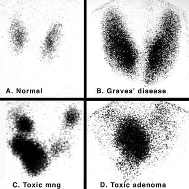
In hyperthyroidism, the elevated levels of T3 and T4 can cause a wide range of symptoms, such as rapid heartbeat, weight loss, heat intolerance, increased appetite, tremors, anxiety, and sleep disturbances. Some common causes of hyperthyroidism include Graves' disease, toxic adenoma, Plummer's disease (toxic multinodular goiter), and thyroiditis. Proper diagnosis and treatment are essential to manage the symptoms and prevent potential complications associated with this condition.
The thyroid gland is a major endocrine gland located in the neck, anterior to the trachea and extends from the lower third of the Adams apple to the suprasternal notch. It has two lateral lobes, connected by an isthmus, and sometimes a pyramidal lobe. This gland plays a crucial role in the metabolism, growth, and development of the human body through the production of thyroid hormones (triiodothyronine/T3 and thyroxine/T4) and calcitonin. The thyroid hormones regulate body temperature, heart rate, and the production of protein, while calcitonin helps in controlling calcium levels in the blood. The function of the thyroid gland is controlled by the hypothalamus and pituitary gland through the thyroid-stimulating hormone (TSH).
Iodide peroxidase, also known as iodide:hydrogen peroxide oxidoreductase, is an enzyme that belongs to the family of oxidoreductases. Specifically, it is a peroxidase that uses iodide as its physiological reducing substrate. This enzyme catalyzes the oxidation of iodide by hydrogen peroxide to produce iodine, which plays a crucial role in thyroid hormone biosynthesis.
The systematic name for this enzyme is iodide:hydrogen-peroxide oxidoreductase (iodinating). It is most commonly found in the thyroid gland, where it helps to produce and regulate thyroid hormones by facilitating the iodination of tyrosine residues on thyroglobulin, a protein produced by the thyroid gland.
Iodide peroxidase requires a heme cofactor for its enzymatic activity, which is responsible for the oxidation-reduction reactions it catalyzes. The enzyme's ability to iodinate tyrosine residues on thyroglobulin is essential for the production of triiodothyronine (T3) and thyroxine (T4), two critical hormones that regulate metabolism, growth, and development in mammals.
Carrier proteins, also known as transport proteins, are a type of protein that facilitates the movement of molecules across cell membranes. They are responsible for the selective and active transport of ions, sugars, amino acids, and other molecules from one side of the membrane to the other, against their concentration gradient. This process requires energy, usually in the form of ATP (adenosine triphosphate).
Carrier proteins have a specific binding site for the molecule they transport, and undergo conformational changes upon binding, which allows them to move the molecule across the membrane. Once the molecule has been transported, the carrier protein returns to its original conformation, ready to bind and transport another molecule.
Carrier proteins play a crucial role in maintaining the balance of ions and other molecules inside and outside of cells, and are essential for many physiological processes, including nerve impulse transmission, muscle contraction, and nutrient uptake.
Congenital hypothyroidism is a medical condition characterized by the partial or complete absence of thyroid hormone production in the baby's body at birth. The thyroid gland, which is located in the front of the neck, produces hormones that are essential for normal growth and development of the brain and body.
Congenital hypothyroidism can occur due to various reasons such as the absence or abnormal development of the thyroid gland, or a defect in the production or regulation of thyroid hormones. In some cases, it may be caused by genetic mutations that affect the development or function of the thyroid gland.
If left untreated, congenital hypothyroidism can lead to mental and physical retardation, growth problems, and other health issues. Therefore, it is important to diagnose and treat this condition as early as possible, usually within the first few weeks of life. Treatment typically involves replacing the missing thyroid hormones with synthetic medications, which are safe and effective when administered under a doctor's supervision.
Iodine isotopes are different forms of the chemical element iodine, which have different numbers of neutrons in their nuclei. Iodine has a total of 53 protons in its nucleus, and its stable isotope, iodine-127, has 74 neutrons, giving it a mass number of 127. However, there are also radioactive isotopes of iodine, which have different numbers of neutrons and are therefore unstable.
Radioactive isotopes of iodine emit radiation as they decay towards a stable state. For example, iodine-131 is a commonly used isotope in medical imaging and therapy, with a half-life of about 8 days. It decays by emitting beta particles and gamma rays, making it useful for treating thyroid cancer and other conditions that involve overactive thyroid glands.
Other radioactive iodine isotopes include iodine-123, which has a half-life of about 13 hours and is used in medical imaging, and iodine-125, which has a half-life of about 60 days and is used in brachytherapy (a type of radiation therapy that involves placing radioactive sources directly into or near tumors).
It's important to note that exposure to radioactive iodine isotopes can be harmful, especially if it occurs through inhalation or ingestion. This is because the iodine can accumulate in the thyroid gland and cause damage over time. Therefore, appropriate safety measures must be taken when handling or working with radioactive iodine isotopes.
Iodine is an essential trace element that is necessary for the production of thyroid hormones in the body. These hormones play crucial roles in various bodily functions, including growth and development, metabolism, and brain development during pregnancy and infancy. Iodine can be found in various foods such as seaweed, dairy products, and iodized salt. In a medical context, iodine is also used as an antiseptic to disinfect surfaces, wounds, and skin infections due to its ability to kill bacteria, viruses, and fungi.
Thyroid diseases are a group of conditions that affect the function and structure of the thyroid gland, a small butterfly-shaped endocrine gland located in the base of the neck. The thyroid gland produces hormones that regulate many vital functions in the body, including metabolism, growth, and development.
Thyroid diseases can be classified into two main categories: hypothyroidism and hyperthyroidism. Hypothyroidism occurs when the thyroid gland does not produce enough hormones, leading to symptoms such as fatigue, weight gain, cold intolerance, constipation, and depression. Hyperthyroidism, on the other hand, occurs when the thyroid gland produces too much hormone, resulting in symptoms such as weight loss, heat intolerance, rapid heart rate, tremors, and anxiety.
Other common thyroid diseases include:
1. Goiter: an enlargement of the thyroid gland that can be caused by iodine deficiency or autoimmune disorders.
2. Thyroid nodules: abnormal growths on the thyroid gland that can be benign or malignant.
3. Thyroid cancer: a malignant tumor of the thyroid gland that requires medical treatment.
4. Hashimoto's disease: an autoimmune disorder that causes chronic inflammation of the thyroid gland, leading to hypothyroidism.
5. Graves' disease: an autoimmune disorder that causes hyperthyroidism and can also lead to eye problems and skin changes.
Thyroid diseases are diagnosed through a combination of physical examination, medical history, blood tests, and imaging studies such as ultrasound or CT scan. Treatment options depend on the specific type and severity of the disease and may include medication, surgery, or radioactive iodine therapy.
Thyronines are a type of hormone that is produced and released by the thyroid gland. They are iodinated amino acids, specifically triiodothyronine (T3) and thyroxine (T4), that are essential for regulating the body's metabolic rate, growth, and development. These hormones play a crucial role in maintaining the body's energy balance, brain development, and overall health. They work by binding to specific receptors in cells throughout the body, where they help to regulate gene expression and various cellular processes. Disorders of thyronine production or function can lead to a variety of medical conditions, such as hypothyroidism or hyperthyroidism.
Protein binding, in the context of medical and biological sciences, refers to the interaction between a protein and another molecule (known as the ligand) that results in a stable complex. This process is often reversible and can be influenced by various factors such as pH, temperature, and concentration of the involved molecules.
In clinical chemistry, protein binding is particularly important when it comes to drugs, as many of them bind to proteins (especially albumin) in the bloodstream. The degree of protein binding can affect a drug's distribution, metabolism, and excretion, which in turn influence its therapeutic effectiveness and potential side effects.
Protein-bound drugs may be less available for interaction with their target tissues, as only the unbound or "free" fraction of the drug is active. Therefore, understanding protein binding can help optimize dosing regimens and minimize adverse reactions.
Radioimmunoassay (RIA) is a highly sensitive analytical technique used in clinical and research laboratories to measure concentrations of various substances, such as hormones, vitamins, drugs, or tumor markers, in biological samples like blood, urine, or tissues. The method relies on the specific interaction between an antibody and its corresponding antigen, combined with the use of radioisotopes to quantify the amount of bound antigen.
In a typical RIA procedure, a known quantity of a radiolabeled antigen (also called tracer) is added to a sample containing an unknown concentration of the same unlabeled antigen. The mixture is then incubated with a specific antibody that binds to the antigen. During the incubation period, the antibody forms complexes with both the radiolabeled and unlabeled antigens.
After the incubation, the unbound (free) radiolabeled antigen is separated from the antibody-antigen complexes, usually through a precipitation or separation step involving centrifugation, filtration, or chromatography. The amount of radioactivity in the pellet (containing the antibody-antigen complexes) is then measured using a gamma counter or other suitable radiation detection device.
The concentration of the unlabeled antigen in the sample can be determined by comparing the ratio of bound to free radiolabeled antigen in the sample to a standard curve generated from known concentrations of unlabeled antigen and their corresponding bound/free ratios. The higher the concentration of unlabeled antigen in the sample, the lower the amount of radiolabeled antigen that will bind to the antibody, resulting in a lower bound/free ratio.
Radioimmunoassays offer high sensitivity, specificity, and accuracy, making them valuable tools for detecting and quantifying low levels of various substances in biological samples. However, due to concerns about radiation safety and waste disposal, alternative non-isotopic immunoassay techniques like enzyme-linked immunosorbent assays (ELISAs) have become more popular in recent years.
Molecular sequence data refers to the specific arrangement of molecules, most commonly nucleotides in DNA or RNA, or amino acids in proteins, that make up a biological macromolecule. This data is generated through laboratory techniques such as sequencing, and provides information about the exact order of the constituent molecules. This data is crucial in various fields of biology, including genetics, evolution, and molecular biology, allowing for comparisons between different organisms, identification of genetic variations, and studies of gene function and regulation.
Tacrolimus binding proteins, also known as FK506 binding proteins (FKBPs), are a group of intracellular proteins that bind to the immunosuppressive drug tacrolimus (also known as FK506) and play a crucial role in its mechanism of action. Tacrolimus is primarily used in organ transplantation to prevent rejection of the transplanted organ.
FKBPs are a family of peptidyl-prolyl cis-trans isomerases (PPIases) that catalyze the conversion of proline residues from their cis to trans conformations in proteins, thereby regulating protein folding and function. FKBP12, a member of this family, has a high affinity for tacrolimus and forms a complex with it upon entry into the cell.
The formation of the tacrolimus-FKBP12 complex inhibits calcineurin, a serine/threonine phosphatase that plays a critical role in T-cell activation. Calcineurin inhibition prevents the dephosphorylation and nuclear translocation of the transcription factor NFAT (nuclear factor of activated T-cells), thereby blocking the expression of genes involved in T-cell activation, proliferation, and cytokine production.
In summary, tacrolimus binding proteins are intracellular proteins that bind to tacrolimus and inhibit calcineurin, leading to the suppression of T-cell activation and immune response, which is essential in organ transplantation and other immunological disorders.
Propylthiouracil is a medication that is primarily used to treat hyperthyroidism, a condition characterized by an overactive thyroid gland that produces too much thyroid hormone. The medication works by inhibiting the production of thyroid hormones in the body. It belongs to a class of drugs called antithyroid agents or thionamides.
In medical terms, propylthiouracil is defined as an antithyroid medication used to manage hyperthyroidism due to Graves' disease or toxic adenoma. It acts by inhibiting the synthesis of thyroid hormones, triiodothyronine (T3) and thyroxine (T4), in the thyroid gland. Propylthiouracil also reduces the peripheral conversion of T4 to T3. The medication is available as a tablet for oral administration and is typically prescribed at a starting dose of 100-150 mg three times daily, with adjustments made based on the patient's response and thyroid function tests.
It's important to note that propylthiouracil should be used under the close supervision of a healthcare provider due to potential side effects and risks associated with its use. Regular monitoring of thyroid function tests is necessary during treatment, and patients should promptly report any signs or symptoms of adverse reactions to their healthcare provider.
Thyroidectomy is a surgical procedure where all or part of the thyroid gland is removed. The thyroid gland is a butterfly-shaped endocrine gland located in the neck, responsible for producing hormones that regulate metabolism, growth, and development.
There are different types of thyroidectomy procedures, including:
1. Total thyroidectomy: Removal of the entire thyroid gland.
2. Partial (or subtotal) thyroidectomy: Removal of a portion of the thyroid gland.
3. Hemithyroidectomy: Removal of one lobe of the thyroid gland, often performed to treat benign solitary nodules or differentiated thyroid cancer.
Thyroidectomy may be recommended for various reasons, such as treating thyroid nodules, goiter, hyperthyroidism (overactive thyroid), or thyroid cancer. Potential risks and complications of the procedure include bleeding, infection, damage to nearby structures like the parathyroid glands and recurrent laryngeal nerve, and hypoparathyroidism or hypothyroidism due to removal of or damage to the parathyroid glands or thyroid gland, respectively. Close postoperative monitoring and management are essential to minimize these risks and ensure optimal patient outcomes.
Insulin-like growth factor binding proteins (IGFBPs) are a family of proteins that bind to and regulate the biological activity of insulin-like growth factors (IGFs), specifically IGF-1 and IGF-2. There are six distinct IGFBPs (IGFBP-1 to IGFBP-6) in humans, each with unique structural features, expression patterns, and functions.
The primary function of IGFBPs is to modulate the interaction between IGFs and their cell surface receptors, thereby controlling IGF-mediated intracellular signaling pathways involved in cell growth, differentiation, and survival. IGFBPs can either enhance or inhibit IGF actions depending on the specific context, such as cell type, subcellular localization, and presence of other binding partners.
In addition to their role in IGF regulation, some IGFBPs have IGF-independent functions, including direct interaction with cell surface receptors, modulation of extracellular matrix composition, and participation in cell migration and apoptosis. Dysregulation of IGFBP expression and function has been implicated in various pathological conditions, such as cancer, diabetes, and cardiovascular diseases.
An amino acid sequence is the specific order of amino acids in a protein or peptide molecule, formed by the linking of the amino group (-NH2) of one amino acid to the carboxyl group (-COOH) of another amino acid through a peptide bond. The sequence is determined by the genetic code and is unique to each type of protein or peptide. It plays a crucial role in determining the three-dimensional structure and function of proteins.
DNA-binding proteins are a type of protein that have the ability to bind to DNA (deoxyribonucleic acid), the genetic material of organisms. These proteins play crucial roles in various biological processes, such as regulation of gene expression, DNA replication, repair and recombination.
The binding of DNA-binding proteins to specific DNA sequences is mediated by non-covalent interactions, including electrostatic, hydrogen bonding, and van der Waals forces. The specificity of binding is determined by the recognition of particular nucleotide sequences or structural features of the DNA molecule.
DNA-binding proteins can be classified into several categories based on their structure and function, such as transcription factors, histones, and restriction enzymes. Transcription factors are a major class of DNA-binding proteins that regulate gene expression by binding to specific DNA sequences in the promoter region of genes and recruiting other proteins to modulate transcription. Histones are DNA-binding proteins that package DNA into nucleosomes, the basic unit of chromatin structure. Restriction enzymes are DNA-binding proteins that recognize and cleave specific DNA sequences, and are widely used in molecular biology research and biotechnology applications.
Methimazole is an anti-thyroid medication that is primarily used to treat hyperthyroidism, a condition in which the thyroid gland produces excessive amounts of thyroid hormones. It works by inhibiting the enzyme thyroperoxidase, which is essential for the production of thyroid hormones. By blocking this enzyme, methimazole reduces the amount of thyroid hormones produced by the thyroid gland, helping to restore normal thyroid function.
Methimazole is available in oral tablet form and is typically taken two to three times a day. Common side effects of methimazole include nausea, vomiting, skin rashes, and joint pain. In rare cases, it can cause more serious side effects such as liver damage or agranulocytosis (a severe decrease in white blood cell count).
It is important to note that methimazole should only be used under the close supervision of a healthcare provider, as regular monitoring of thyroid function and potential side effects is necessary. Additionally, it may take several weeks or months of treatment with methimazole before thyroid function returns to normal.
A base sequence in the context of molecular biology refers to the specific order of nucleotides in a DNA or RNA molecule. In DNA, these nucleotides are adenine (A), guanine (G), cytosine (C), and thymine (T). In RNA, uracil (U) takes the place of thymine. The base sequence contains genetic information that is transcribed into RNA and ultimately translated into proteins. It is the exact order of these bases that determines the genetic code and thus the function of the DNA or RNA molecule.
RNA-binding proteins (RBPs) are a class of proteins that selectively interact with RNA molecules to form ribonucleoprotein complexes. These proteins play crucial roles in the post-transcriptional regulation of gene expression, including pre-mRNA processing, mRNA stability, transport, localization, and translation. RBPs recognize specific RNA sequences or structures through their modular RNA-binding domains, which can be highly degenerate and allow for the recognition of a wide range of RNA targets. The interaction between RBPs and RNA is often dynamic and can be regulated by various post-translational modifications of the proteins or by environmental stimuli, allowing for fine-tuning of gene expression in response to changing cellular needs. Dysregulation of RBP function has been implicated in various human diseases, including neurological disorders and cancer.
Iopanoic acid is a contrast medium, specifically a radiocontrast agent, that is used during imaging examinations such as X-rays and CT scans to help improve the visibility of internal body structures. It works by blocking the absorption of X-rays in the digestive tract, making it possible to visualize the gastrointestinal tract more clearly on imaging studies. Iopanoic acid is typically given orally before the examination.
It's important to note that the use of iopanoic acid and other radiocontrast agents should be carefully weighed against the potential risks, as they can cause allergic reactions, kidney damage, and other complications in some individuals. Therefore, it is usually reserved for situations where the benefits of improved imaging outweigh these potential risks.
In the context of medical and biological sciences, a "binding site" refers to a specific location on a protein, molecule, or cell where another molecule can attach or bind. This binding interaction can lead to various functional changes in the original protein or molecule. The other molecule that binds to the binding site is often referred to as a ligand, which can be a small molecule, ion, or even another protein.
The binding between a ligand and its target binding site can be specific and selective, meaning that only certain ligands can bind to particular binding sites with high affinity. This specificity plays a crucial role in various biological processes, such as signal transduction, enzyme catalysis, or drug action.
In the case of drug development, understanding the location and properties of binding sites on target proteins is essential for designing drugs that can selectively bind to these sites and modulate protein function. This knowledge can help create more effective and safer therapeutic options for various diseases.
Fatty acid-binding proteins (FABPs) are a group of small intracellular proteins that play a crucial role in the transport and metabolism of fatty acids within cells. They are responsible for binding long-chain fatty acids, which are hydrophobic molecules, and facilitating their movement across the cell while protecting the cells from lipotoxicity.
FABPs are expressed in various tissues, including the heart, liver, muscle, and brain, with different isoforms found in specific organs. These proteins have a high affinity for long-chain fatty acids and can regulate their intracellular concentration by controlling the uptake, storage, and metabolism of these molecules.
FABPs also play a role in modulating cell signaling pathways that are involved in various physiological processes such as inflammation, differentiation, and apoptosis. Dysregulation of FABP expression and function has been implicated in several diseases, including diabetes, obesity, cancer, and neurodegenerative disorders.
In summary, fatty acid-binding proteins are essential intracellular proteins that facilitate the transport and metabolism of long-chain fatty acids while regulating cell signaling pathways.
Antithyroid agents are a class of medications that are used to treat hyperthyroidism, a condition in which the thyroid gland produces too much thyroid hormone. These medications work by inhibiting the production of thyroid hormones in the thyroid gland. There are several types of antithyroid agents available, including:
1. Propylthiouracil (PTU): This medication works by blocking the enzyme that is needed to produce thyroid hormones. It also reduces the conversion of thyroxine (T4) to triiodothyronine (T3), another thyroid hormone, in peripheral tissues.
2. Methimazole: This medication works similarly to propylthiouracil by blocking the enzyme that is needed to produce thyroid hormones. However, it does not affect the conversion of T4 to T3 in peripheral tissues.
3. Carbimazole: This medication is converted to methimazole in the body and works similarly to block the production of thyroid hormones.
Antithyroid agents are usually taken orally, and their effects on thyroid hormone production begin within a few hours after ingestion. However, it may take several weeks for patients to notice an improvement in their symptoms. These medications can have side effects, including rash, hives, and joint pain. In rare cases, they can cause liver damage or agranulocytosis, a condition in which the body does not produce enough white blood cells.
It is important to note that antithyroid agents do not cure hyperthyroidism; they only treat the symptoms by reducing thyroid hormone production. Therefore, patients may need to take these medications for several months or even years, depending on their individual circumstances. In some cases, surgery or radioactive iodine therapy may be recommended as alternative treatments for hyperthyroidism.
Thyroglobulin is a protein produced and used by the thyroid gland in the production of thyroid hormones, primarily thyroxine (T4) and triiodothyronine (T3). It is composed of two subunits, an alpha and a beta or gamma unit, which bind iodine atoms necessary for the synthesis of the thyroid hormones. Thyroglobulin is exclusively produced by the follicular cells of the thyroid gland.
In clinical practice, measuring thyroglobulin levels in the blood can be useful as a tumor marker for monitoring treatment and detecting recurrence of thyroid cancer, particularly in patients with differentiated thyroid cancer (papillary or follicular) who have had their thyroid gland removed. However, it is important to note that thyroglobulin is not specific to thyroid tissue and can be produced by some non-thyroidal cells under certain conditions, which may lead to false positive results in some cases.
Goiter is a medical term that refers to an enlarged thyroid gland. The thyroid gland is a small, butterfly-shaped gland located in the front of your neck below the larynx or voice box. It produces hormones that regulate your body's metabolism, growth, and development.
Goiter can vary in size and may be visible as a swelling at the base of the neck. It can be caused by several factors, including iodine deficiency, autoimmune disorders, thyroid cancer, pregnancy, or the use of certain medications. Depending on the underlying cause and the severity of the goiter, treatment options may include medication, surgery, or radioactive iodine therapy.
Thyroxine-binding globulin (TBG) is a glycoprotein found in human plasma that has a high affinity for binding thyroid hormones, specifically Thyroxine (T4) and Triiodothyronine (T3). It is produced by the liver and plays a crucial role in maintaining the balance of these hormones in the body. TBG binds to approximately 70-80% of circulating T4 and about 55% of circulating T3, acting as a transport protein that carries these hormones throughout the body. The amount of TBG in the blood can vary due to factors such as genetics, sex hormones, and certain medications, which can affect the levels of free (unbound) thyroid hormones and contribute to various thyroid-related disorders.
Thyrotropin-Releasing Hormone (TRH) is a tripeptide hormone that is produced and released by the hypothalamus in the brain. Its main function is to regulate the release of thyroid-stimulating hormone (TSH) from the anterior pituitary gland. TRH acts on the pituitary gland to stimulate the synthesis and secretion of TSH, which then stimulates the thyroid gland to produce and release thyroid hormones (triiodothyronine (T3) and thyroxine (T4)) into the bloodstream.
TRH is a tripeptide amino acid sequence with the structure of pGlu-His-Pro-NH2, and it is synthesized as a larger precursor molecule called preprothyrotropin-releasing hormone (preproTRH) in the hypothalamus. PreproTRH undergoes post-translational processing to produce TRH, which is then stored in secretory vesicles and released into the hypophyseal portal system, where it travels to the anterior pituitary gland and binds to TRH receptors on thyrotroph cells.
In addition to its role in regulating TSH release, TRH has been shown to have other physiological functions, including modulation of feeding behavior, body temperature, and neurotransmitter release. Dysregulation of the TRH-TSH axis can lead to various thyroid disorders, such as hypothyroidism or hyperthyroidism.
Messenger RNA (mRNA) is a type of RNA (ribonucleic acid) that carries genetic information copied from DNA in the form of a series of three-base code "words," each of which specifies a particular amino acid. This information is used by the cell's machinery to construct proteins, a process known as translation. After being transcribed from DNA, mRNA travels out of the nucleus to the ribosomes in the cytoplasm where protein synthesis occurs. Once the protein has been synthesized, the mRNA may be degraded and recycled. Post-transcriptional modifications can also occur to mRNA, such as alternative splicing and addition of a 5' cap and a poly(A) tail, which can affect its stability, localization, and translation efficiency.
Serum globulins are a group of proteins present in the liquid portion of blood, known as serum. They are produced by the immune system in response to foreign substances such as bacteria, viruses, and allergens. Serum globulins include several types of immunoglobulins (antibodies), complement components, and other proteins involved in the immune response.
The serum globulin level is often measured as part of a complete blood count (CBC) or a protein electrophoresis test. An elevated serum globulin level may indicate an ongoing infection, inflammation, or an autoimmune disorder. Conversely, a decreased level may suggest a liver or kidney disease, or a malnutrition condition. It is important to note that the interpretation of serum globulin levels should be done in conjunction with other laboratory and clinical findings.
Dialysis is a medical treatment that is used to remove waste and excess fluid from the blood when the kidneys are no longer able to perform these functions effectively. This life-sustaining procedure uses a specialized machine, called a dialyzer or artificial kidney, to filter the blood outside of the body and return clean, chemically balanced blood back into the body.
There are two main types of dialysis: hemodialysis and peritoneal dialysis.
1. Hemodialysis: In this method, a patient's blood is passed through an external filter (dialyzer) that removes waste products, toxins, and excess fluids. The cleaned blood is then returned to the body with the help of a specialized machine. Hemodialysis typically requires access to a large vein, often created by a surgical procedure called an arteriovenous (AV) fistula or graft. Hemodialysis sessions usually last for about 3-5 hours and are performed three times a week in a clinical setting, such as a dialysis center or hospital.
2. Peritoneal Dialysis: This method uses the lining of the patient's own abdomen (peritoneum) as a natural filter to clean the blood. A sterile dialysate solution is introduced into the peritoneal cavity via a permanently implanted catheter. The solution absorbs waste products and excess fluids from the blood vessels lining the peritoneum through a process called diffusion. After a dwell time, usually several hours, the used dialysate is drained out and replaced with fresh dialysate. This process is known as an exchange and is typically repeated multiple times throughout the day or night, depending on the specific type of peritoneal dialysis (continuous ambulatory peritoneal dialysis or automated peritoneal dialysis).
Both methods have their advantages and disadvantages, and the choice between them depends on various factors, such as a patient's overall health, lifestyle, and personal preferences. Dialysis is a life-saving treatment for people with end-stage kidney disease or severe kidney dysfunction, allowing them to maintain their quality of life and extend their lifespan until a kidney transplant becomes available or their kidney function improves.
Iodine radioisotopes are radioactive isotopes of the element iodine, which decays and emits radiation in the form of gamma rays. Some commonly used iodine radioisotopes include I-123, I-125, I-131. These radioisotopes have various medical applications such as in diagnostic imaging, therapy for thyroid disorders, and cancer treatment.
For example, I-131 is commonly used to treat hyperthyroidism and differentiated thyroid cancer due to its ability to destroy thyroid tissue. On the other hand, I-123 is often used in nuclear medicine scans of the thyroid gland because it emits gamma rays that can be detected by a gamma camera, allowing for detailed images of the gland's structure and function.
It is important to note that handling and administering radioisotopes require specialized training and safety precautions due to their radiation-emitting properties.
Myxedema is not a term used in modern medicine to describe a specific medical condition. However, historically, it was used to refer to the severe form of hypothyroidism, a condition characterized by an underactive thyroid gland that doesn't produce enough thyroid hormones. In hypothyroidism, various body functions slow down, which can lead to symptoms such as fatigue, weight gain, cold intolerance, constipation, and dry skin.
Myxedema specifically refers to the physical signs of severe hypothyroidism, including swelling (edema) and thickening of the skin, particularly around the face, hands, and feet, as well as a puffy appearance of the face. The term myxedema coma was used to describe a rare but life-threatening complication of long-standing, untreated hypothyroidism, characterized by altered mental status, hypothermia, and other systemic manifestations.
Nowadays, healthcare professionals use more precise medical terminology to describe these conditions, such as hypothyroidism or myxedematous edema, rather than the outdated term myxedema.
The liver is a large, solid organ located in the upper right portion of the abdomen, beneath the diaphragm and above the stomach. It plays a vital role in several bodily functions, including:
1. Metabolism: The liver helps to metabolize carbohydrates, fats, and proteins from the food we eat into energy and nutrients that our bodies can use.
2. Detoxification: The liver detoxifies harmful substances in the body by breaking them down into less toxic forms or excreting them through bile.
3. Synthesis: The liver synthesizes important proteins, such as albumin and clotting factors, that are necessary for proper bodily function.
4. Storage: The liver stores glucose, vitamins, and minerals that can be released when the body needs them.
5. Bile production: The liver produces bile, a digestive juice that helps to break down fats in the small intestine.
6. Immune function: The liver plays a role in the immune system by filtering out bacteria and other harmful substances from the blood.
Overall, the liver is an essential organ that plays a critical role in maintaining overall health and well-being.
Diiodothyronines are hormones that contain two iodine atoms and are produced by the thyroid gland. They are formed when thyroxine (T4), another thyroid hormone, is deiodinated. Diiodothyronines include T2 (3,5-diiodothyronine) and reverse T2 (3,3'-diiodothyronine). These hormones play a role in regulating metabolism and energy production in the body. However, their specific functions and mechanisms of action are not as well understood as those of thyroxine and triiodothyronine (T3), another important thyroid hormone.
Insulin-like Growth Factor Binding Protein 3 (IGFBP-3) is a protein that binds to and regulates the bioavailability and activity of Insulin-like Growth Factors (IGFs), specifically IGF-1 and IGF-2. It plays a crucial role in the growth, development, and homeostasis of various tissues and organs by modulating IGF signaling. IGFBP-3 is the most abundant IGF binding protein in circulation and has a longer half-life than IGFs, allowing it to act as a reservoir and transport protein for IGFs. Additionally, IGFBP-3 has been found to have IGF-independent functions, including roles in cell growth, differentiation, apoptosis, and tumor suppression.
Periplasmic binding proteins (PBPs) are a type of water-soluble protein found in the periplasmic space of gram-negative bacteria. They play a crucial role in the bacterial uptake of specific nutrients, such as amino acids, sugars, and ions, through a process known as active transport.
PBPs function by specifically binding to their target substrates in the extracellular environment and then shuttling them across the inner membrane into the cytoplasm. This is achieved through a complex series of interactions with other proteins, including transmembrane permeases and ATP-binding cassette (ABC) transporters.
The binding of PBPs to their substrates typically results in a conformational change that allows for the transport of the substrate across the inner membrane. Once inside the cytoplasm, the substrate can be used for various metabolic processes, such as energy production or biosynthesis.
PBPs are often used as targets for the development of new antibiotics, as they play a critical role in bacterial survival and virulence. Inhibiting their function can disrupt essential physiological processes and lead to bacterial death.
Serum albumin is the most abundant protein in human blood plasma, synthesized by the liver. It plays a crucial role in maintaining the oncotic pressure or colloid osmotic pressure of blood, which helps to regulate the fluid balance between the intravascular and extravascular spaces.
Serum albumin has a molecular weight of around 66 kDa and is composed of a single polypeptide chain. It contains several binding sites for various endogenous and exogenous substances, such as bilirubin, fatty acids, hormones, and drugs, facilitating their transport throughout the body. Additionally, albumin possesses antioxidant properties, protecting against oxidative damage.
Albumin levels in the blood are often used as a clinical indicator of liver function, nutritional status, and overall health. Low serum albumin levels may suggest liver disease, malnutrition, inflammation, or kidney dysfunction.
Calcium-binding proteins (CaBPs) are a diverse group of proteins that have the ability to bind calcium ions (Ca^2+^) with high affinity and specificity. They play crucial roles in various cellular processes, including signal transduction, muscle contraction, neurotransmitter release, and protection against oxidative stress.
The binding of calcium ions to these proteins induces conformational changes that can either activate or inhibit their functions. Some well-known CaBPs include calmodulin, troponin C, S100 proteins, and parvalbumins. These proteins are essential for maintaining calcium homeostasis within cells and for mediating the effects of calcium as a second messenger in various cellular signaling pathways.
 Thyroxine-binding proteins - Wikipedia
 Inherited thyroxine-binding globulin deficiency: MedlinePlus Genetics
 Thyroxine-Binding Globulin Deficiency: Overview, Molecular Biology of TBG, Etiology
Analysis of genetic variation in two human thyroxine-binding plasma proteins by immunodetection after isoelectric focusing -...
TBG blood test: MedlinePlus Medical Encyclopedia
MedlinePlus - Search Results for: LEVOTHYROXINE
 Thông tin thuốc của Thyroid tablets | MIMS Vietnam
 DailyMed - RABEPRAZOLE SODIUM tablet, delayed release
DailyMed - LEVOTHYROXINE SODIUM tablet
 RCSB PDB - 4ABW: Crystal Structure of Transthyretin in Complex With Ligand C-6
 Flashcards - Plasma Proteins & Enzymology S1M2
 Updates in the management of polyneuropathy of hereditary transthyretin amyloidosis (ATTR): Treating the condition head-on ...
Hypothyroidism - Causes, Symptoms, Diagnosis, Treatment - Health | RXharun
 Molecular chaperones and Transthyretin Amyloid Disease
 Cytomel (Liothyronine Sodium): Uses, Dosage, Side Effects, Interactions, Warning
 Answering your questions | Medical Laboratory Observer
 ttr Summary
Thyroid-Stimulating Hormone: Reference Range, Interpretation, Collection and Panels
Hypothyroidism Workup: Laboratory Studies, Imaging Studies, Screening
 Thyroid, porcine: Uses, Interactions, Mechanism of Action | DrugBank Online
 A Case of Hypothyroxinemia with Thyroxine-Binding-Globulin Deficiency
 FT3 - Fortress Diagnostics
 Thyroid Treatment Tip #10 - Parker Nutritional Healing Center
Endocrinology - Page 4 - Veterinary Sciences Tomorrow
 Thyroid Function and Disorders | Leaders in Pharmaceutical Business Intelligence (LPBI) Group
 Overview of Thyroid Function - Endocrine and Metabolic Disorders - MSD Manual Professional Edition
Transthyretin16
- 95%) of THs are thyroxine-binding globulin (TBG), transthyretin (TTR, or prealbumin), and albumin. (medscape.com)
- 2000. Assessing the role of ortho -substitution on polychlorinated biphenyl binding to transthyretin, a thyroxine transport protein. (cdc.gov)
- Interaction of organochlorine compounds with thyroid receptor, transthyretin, and thyroid-binding globulin. (cdc.gov)
- Transthyretin (TTR) is a homotetrameric serum and cerebrospinal fluid protein that transports thyroxine (T4) and retinol by binding to retinol binding protein. (rcsb.org)
- Transthyretin (TTR) is a protein present in human serum whose role is to transport thyroxine and retinol-binding proteins, and it is vital for behavior, cognition, nerve regeneration, and axonal growth. (cmelist.com)
- Transthyretin (TTR) is actually a protein present in human plasma as well as in cerebrospinal fluid. (projectsparadise.com)
- T4 is highly protein-bound (99.97%), with approximately 85% bound to thyroid-binding globulin (TBG), approximately 10% bound to transthyretin or thyroid-binding prealbumin, and the remainder bound loosely to albumin. (medscape.com)
- The transport proteins such as thyroxine-binding-globulin (TBG), albumin and transthyretin carry over 99% of circulating thyroid hormones. (sch.ac.kr)
- PFAS compete with the thyroid hormone thyroxin (T4 ) for binding to its distributor protein transthyretin (TTR). (bvsalud.org)
- Most thyroxine is bound to carrier proteins, such as thyroxine-binding globulin (TBG), transthyretin, and albumin in circulation, with only a tiny fraction (approximately 0.03%) present as free thyroxine (FT4)[3]. (myendoconsult.com)
- Following secretion by the thyroid gland, approximately 70% of circulating T4 and T3 are bound to thyroid-binding globulin (TBG), while 10% to 20% each are bound to transthyretin (TTR) and albumin, respectively. (mayocliniclabs.com)
- T4 & T3 are bound to plasma proteins which include thyroxine-binding globulin (TBG), transthyretin (TTR, formerly known as thyroxine-binding prealbumin, or TBPA) and albumin. (medquizzes.net)
- so as to increase solubility, the thyroid gland attaches to thyroid hormone binding proteins, thyroxin-binding globulin, and thyroxine-binding prealbumin (transthyretin). (drugsbanks.com)
- Immunogen Recombinant protein corresponding to the Human wild type Transthyretin. (agrisera.com)
- Background Transthyretin (TTR) , formerly known as Prealbumin, is in vivo involved in the binding and transportation of the Thyroxin hormone and retinol-binding protein. (agrisera.com)
- T3 has a faster onset of action as well as a shorter biological half-life, probably due to less plasma protein binding to thyroxine-binding globulin and transthyretin. (24hoursppc.org)
Manifesting thyroxine-binding1
- Okamoto H, Mori Y, Tani Y, Nakagomi Y, Sano T, Ohyama K, Saito H, Oiso Y. Molecular analysis of females manifesting thyroxine-binding globulin (TBG) deficiency: selective X-chromosome inactivation responsible for the difference between phenotype and genotype in TBG-deficient females. (medlineplus.gov)
Triiodothyronine12
- The thyroid hormones (THs)-thyroxine (T4) and 3,5,3'-triiodothyronine (T3)-circulate in blood by reversibly binding to carrier proteins. (medscape.com)
- TSH, in turn, is the physiologic stimulus for the synthesis and secretion of thyroid hormones, L-thyroxine (T 4 ) and L-triiodothyronine (T 3 ), by the thyroid gland. (nih.gov)
- Free thyroid hormone levels can be estimated by calculating the percentage of available thyroid hormone-binding sites (triiodothyronine [T3] resin uptake, or thyroid hormone binding ratio [THBR]) or by measuring the TBG concentration. (medscape.com)
- Once inside the follicle, most of the iodide is oxidized by the enzyme thyroid peroxidase (TPO) in a reaction that facilitates combination with a tyrosine molecule to ultimately form thyroxine (T4) and triiodothyronine (T3). (pharmaceuticalintelligence.com)
- Pituitary TSH regulates the secretion of the thyroid hormones T4 (thyroxine) and T3 (triiodothyronine). (pharmaceuticalintelligence.com)
- The Thyroid Profile consists of a battery of several tests for the measurement of thyroid function, including Total and Free Thyroxine, Total and Free Triiodothyronine, Thyroglobulin, Thyroglobulin Antibodies, Thyroid Peroxidase Antibodies, and Thyroid Stimulating Hormone. (cdc.gov)
- The t hyroid gland present in the anterior neck is responsible for the synthesis and secretion of thyroid hormones: thyroxine (T4) and triiodothyronine (T3). (myendoconsult.com)
- T3 circulates bound to carrier proteins (mainly TBG and albumin), with a small fraction (approximately 0.3%) existing as free triiodothyronine (FT3). (myendoconsult.com)
- Thyroxine (T4) and triiodothyronine (T3) are the 2 biologically active thyroid hormones. (mayocliniclabs.com)
- thyroxine ( T4 ) and triiodothyronine ( T3 ). (fieldsgynroboticsurgery.com)
- A T3 blood test measures both bound and free triiodothyronine. (fieldsgynroboticsurgery.com)
- Thyroxine-binding globulin (TBG), a glycoprotein produced in the liver, binds both thyroxine (T4) and triiodothyronine (T3) with high affinity. (selfdecodelabs.com)
Carrier proteins4
- 99.5%) to carrier proteins. (fortressdiagnostics.com)
- Furthermore, the concentrations of the carrier proteins are altered in many clinical conditions, such as pregnancy. (fortressdiagnostics.com)
- Trouble occurs when there are so many of these carrier proteins called TBG, these little taxicabs, that the normal free amount of T4 and T3 that should be floating around isn't. (synergyfixme.com)
- More than 99.9% of the T4 in blood is bound to carrier proteins, specially to thyroxine-binding globulin (TBG), and the free fraction of T4 (free T4, FT4) just accounts for ∼0.03% [ 1 ]. (ijbs.com)
Prealbumin1
- 13) Patients with this pattern have normal TSH and T4, low T3, high T3 uptake (a measure of the quantity of thyroxine-binding proteins such as prealbumin and albumin) and high TBG. (fullcirclefunction.com)
Albumin2
- the converse is true, ie, high capacity but low avidity, for TH binding to TTR and albumin. (medscape.com)
- 99% (T 4 ) to plasma proteins including thyroxine-binding globulin, thyroxine-binding pre-albumin and albumin. (mims.com)
Total thyroxine4
- We experienced a case that a man who had an abnormal thyroid function showed unexpectedly low concentrations of serum total thyroxine. (sch.ac.kr)
- The total thyroxine (T4), free thyroxine (FT4), and free thyroxine index (FTI) values are often used to keep track of treatment for hyperthyroidism . (fieldsgynroboticsurgery.com)
- Labs generally measure free T4 (FT4) levels, but they also may measure total thyroxine (T4) and T3 uptake (T3U). (fieldsgynroboticsurgery.com)
- New liquid chromatography tandem mass spectrometry (LC-MS/MS) methods were developed and applied to the measurement of total thyroxine (T4), total tri-iodothyronine (T3), total reverse tri-iodothyronine (rT3), vitamin D metabolites, and vitamin D binding protein (VDBP). (nist.gov)
Bloodstream4
- A thyroxine-binding protein is any of several transport proteins that bind thyroid hormone and carry it around the bloodstream. (wikipedia.org)
- Most of the time, these hormones circulate in the bloodstream attached to thyroxine-binding globulin and similar proteins. (medlineplus.gov)
- Probably transports thyroxine from the bloodstream to the brain. (xenbase.org)
- Free T3 and T4 are then released into the bloodstream, where they are bound to serum proteins for transport. (msdmanuals.com)
Thyroglobulin4
- Thyroid USP contains not less than (NLT) 0.17 percent and not more than (NMT) 0.23 percent iodine, and thyroglobulin contains not less than (NLT) 0.7 percent of organically bound iodine. (rxlist.com)
- Twenty-five mcg of liothyronine is equivalent to approximately 1 grain of desiccated thyroid or thyroglobulin and 0.1 mg of L-thyroxine. (rxlist.com)
- Each follicle is composed of a rim of simple cuboidal epithelial cells encircling a mass of colloidal storage protein named thyroglobulin. (myendoconsult.com)
- thyroglobulin (Tg) is a large (660 kDa) dimeric protein that consists of 2769 amino acids. (medquizzes.net)
Retinol binding1
- It functions like a transport protein, carrying thyroxin and holo-retinol binding protein. (projectsparadise.com)
Globulin deficiency10
- Inherited thyroxine-binding globulin deficiency is a genetic condition that typically does not cause any health problems. (medlineplus.gov)
- Researchers have identified two forms of inherited thyroxine-binding globulin deficiency: the complete form (TBG-CD), which results in a total loss of thyroxine-binding globulin, and the partial form (TBG-PD), which reduces the amount of this protein or alters its structure. (medlineplus.gov)
- Although inherited thyroxine-binding globulin deficiency does not cause any health problems, it can be mistaken for more serious thyroid disorders (such as hypothyroidism). (medlineplus.gov)
- Therefore, it is important to diagnose inherited thyroxine-binding globulin deficiency to avoid unnecessary treatments. (medlineplus.gov)
- The complete form of inherited thyroxine-binding globulin deficiency, TBG-CD, affects about 1 in 15,000 newborns worldwide. (medlineplus.gov)
- Inherited thyroxine-binding globulin deficiency results from mutations in the SERPINA7 gene. (medlineplus.gov)
- Researchers have also described non-inherited forms of thyroxine-binding globulin deficiency, which are more common than the inherited form. (medlineplus.gov)
- Non-inherited thyroxine-binding globulin deficiency can occur with a variety of illnesses and is a side effect of some medications. (medlineplus.gov)
- Inherited thyroxine-binding globulin deficiency has an X-linked pattern of inheritance. (medlineplus.gov)
- In males (who have only one X chromosome), a mutation in the only copy of the gene in each cell causes partial or complete inherited thyroxine-binding globulin deficiency. (medlineplus.gov)
Hormone24
- The TBG blood test measures the level of a protein that moves thyroid hormone throughout your body. (medlineplus.gov)
- T4 ( thyroxine ) is the main hormone produced by the thyroid gland. (nih.gov)
- This hormone nuclear receptor complex activates gene transcription and synthesis of messenger RNA and cytoplasmic proteins. (nih.gov)
- Thyroid hormone-binding protein. (xenbase.org)
- T3 binding to the thyroid hormone receptor (TR), changes the conformation of the TR, allowing it to bind to the retinoid X receptor (RXR) and form a coactivator complex. (drugbank.com)
- TBG is a major thyroid hormone transport protein in serum. (sch.ac.kr)
- In the cell, T3 binds to a nuclear receptor, resulting in transcription of specific thyroid hormone response genes. (pharmaceuticalintelligence.com)
- Free T4 measurements were primarily used for assessing thyroid function despite the technical difficulties in free thyroid hormone measurements owing to abnormal binding proteins, changes in binding protein concentrations, and the effects of drugs and illness on thyroid hormone binding. (pharmaceuticalintelligence.com)
- Thyroid hormone is required for normal brain and somatic tissue development in the fetus and neonate, and, in people of all ages, thyroid hormone regulates protein, carbohydrate, and fat metabolism. (msdmanuals.com)
- T4 is converted (in most tissues) to T3, the active form that binds to nuclear receptors, and to reverse T3 (rT3), an inactive form of thyroid hormone without metabolic activity. (msdmanuals.com)
- [7] [8] Activin, inhibin and a number of other structurally related proteins such as anti-Müllerian hormone , bone morphogenetic protein , and growth differentiation factor belong to the TGF-β protein superfamily . (wikidoc.org)
- Free thyroxine (free T4) tests are used to help evaluate thyroid function and diagnose thyroid diseases, including hyperthyroidism and hypothyroidism, usually after discovering that the thyroid stimulating hormone (TSH) level is abnormal. (privatemdlabs.com)
- Free T3 exerts its effects by binding to its cognate intracellular thyroid hormone receptors (TRs)[3]. (myendoconsult.com)
- Biologically, TBG-bound thyroid hormone serves as a hormone reservoir, and T4 serves as a prohormone for T3. (mayocliniclabs.com)
- Ultimately, T3, and to a much lesser degree T4, bind to the nuclear thyroid hormone receptor, altering gene expression patterns in a tissue-specific fashion. (mayocliniclabs.com)
- This results in tight regulation of thyroid hormone production and constant levels of FT4 and FT3 independent of the binding protein concentration. (mayocliniclabs.com)
- Plasma-binding proteins increase the pool of circulating hormone, delay hormone clearance, and may modulate hormone delivery to selected tissue sites. (medquizzes.net)
- Levothyroxine acts just like the endogenous thyroid hormone thyroxine (T4, a tetra-iodinated tyrosine derivative). (drugsbanks.com)
- Transport and binding to thyroid hormone receptors within the cytoplasm and nucleus then takes place. (drugsbanks.com)
- Es gibt drei verwandte Schilddrüsenhormone, die für den Stoffwechsel verantwortlich sind - Tricodid-Thyronin (L-T3), L-Thyroxin (L-T4) und thyrotrophe Hormone, die für den Start der T4-Produktion verantwortlich sind. (24hoursppc.org)
- Thyroid-binding globulin (TBG) is the protein that transports thyroid hormone through the blood. (fullcirclefunction.com)
- When thyroid hormone is bound to TBG, it is inactive and unavailable to tissues. (fullcirclefunction.com)
- Description: Description of target: Thyroid hormone-binding protein. (apoptosises.com)
- Serum 25(OH)D, vitamin D-binding protein (VDBP), parathyroid hormone (PTH) and calcium were measured, and 25(OH)D Bio was calculated based on VDBP levels. (medscape.com)
Concentrations3
- Results are independent of binding protein concentrations. (medscape.com)
- BAFF steady-state concentrations depend on B cells and also on the expression of BAFF-binding receptors. (wikidoc.org)
- Because TBG accounts for 76% of plasma protein thyroxine-binding activity, an increase or decrease in its circulating level alters total concentrations of T4 and T3 in blood, leading to potential confusion with true thyroid gland dysfunction. (selfdecodelabs.com)
Dialysis1
- METHODS: Serum FT4 was separated from protein-bound thyroxine with equilibrium dialysis (ED) following the recommended conditions in the Clinical and Laboratory Standards Institute C45-A guideline and the published RMP [23]. (cdc.gov)
High affinity4
- The primary transport protein is thyroxine-binding globulin (TBG), which has high affinity but low capacity for T3 and T4. (msdmanuals.com)
- Platelet factor-4 is a 70-amino acid protein that is released from the alpha-granules of activated platelets and binds with high affinity to heparin. (wikidoc.org)
- Donor-specific antibodies bind with high affinity to the vascular endothelium of graft and activate complement . (wikidoc.org)
- Although rT3 shares structural similarity with T3, it does not bind to TRs with high affinity and, therefore, does not exhibit any significant biological activity. (myendoconsult.com)
Human serum2
- A microplate chemiluminescence enzyme immunoassay (CLEIA) with high sensitivity, selectivity and reproducibility was developed for the determination of free thyroxine (FT4) in human serum. (ijbs.com)
- BACKGROUND: Accurate and reliable measurement of human serum free thyroxine (FT4) is critical for the diagnosis and treatment of thyroid diseases. (cdc.gov)
Affinity2
- Inherited or acquired variations in the concentration and/or affinity of these proteins may produce substantial changes in serum total TH levels measured by commercially available assays. (medscape.com)
- [5] TACI binds worst since its affinity is higher for a protein similar to BAFF, called a proliferation-inducing ligand (APRIL). (wikidoc.org)
Metabolism2
- The exact mechanism of action has not been fully elucidated, but it is known to enhance oxygen consumption by tissues, increase basal metabolic rate and metabolism of carbohydrate, lipids and proteins. (mims.com)
- The metabolic actions of thyroid hormones include augmentation of cellular respiration and thermogenesis, as well as metabolism of proteins, carbohydrates and lipids. (nih.gov)
Uptake2
- It will show that, actually, the T3 Uptake is low when the thyroid binding globulin, or TBGs are high. (synergyfixme.com)
- The T3 uptake test reports the percentage of TBG that is bound to thyroid hormones: a higher percentage of T3 uptake means more TBG bound to T4 or T3. (labtestsguide.com)
Autoantibodies1
- Results are independent of the concentration of the T4-binding proteins, the presence of molecular variants of these proteins, or circulating autoantibodies. (privatemdlabs.com)
Antibodies1
- Very rarely, and in the context of anti-TH antibodies in autoimmune thyroid disease, immunoglobulins also may bind TH. (medscape.com)
Abnormal1
- The FTI can help tell if abnormal amounts of T4 are present because of abnormal amounts of thyroxine-binding globulin. (fieldsgynroboticsurgery.com)
Receptors2
- [4] BAFF is the natural ligand of three unusual tumor necrosis factor receptors named BAFF-R (BR3), TACI (transmembrane activator and calcium modulator and cyclophilin ligand interactor), and BCMA (B-cell maturation antigen), all of which have differing binding affinities for it. (wikidoc.org)
- All these ligands act as homotrimers (i.e. three of the same molecule) interacting with homotrimeric receptors, [6] although BAFF has been known to be active as either a hetero- or homotrimer (can aggregate into 60-mer depending on the primary structure of the protein). (wikidoc.org)
Equilibrium3
- Approximately 0.3% of total serum T3 and 0.03% of total serum T4 are free and in equilibrium with bound hormones. (msdmanuals.com)
- Only the free hormones are biologically active, but bound and free fractions are in equilibrium. (mayocliniclabs.com)
- By contrast, TBG binds thyroid hormones very tightly and equilibrium dissociation is slow. (mayocliniclabs.com)
Hypothyroidism1
- The Total T3 test measures the bound(T3) and free forms of T3(FreeT3) levels in the blood to evaluate the thyroid functioning if it has underactive(Hypothyroidism) or overactive(Hyperthyroidism) performance. (labtestsguide.com)
Measures both free and bound2
- The total T3 test measures both free and bound T3 in your blood. (labtestsguide.com)
- A total T4 blood test measures both free and bound thyroxine. (fieldsgynroboticsurgery.com)
Gene7
- This gene provides instructions for making thyroxine-binding globulin. (medlineplus.gov)
- Some mutations in the SERPINA7 gene prevent the production of a functional protein, causing TBG-CD. (medlineplus.gov)
- In females (who have two X chromosomes), a mutation in one of the two copies of the gene in each cell reduces the amount of thyroxine-binding globulin. (medlineplus.gov)
- Thyroxine-binding globulin (TBG) is a 395-amino acid, 54kd polypeptide that is synthesized in the liver and encoded by a single gene copy. (medscape.com)
- B-cell activating factor ( BAFF ) also known as tumor necrosis factor ligand superfamily member 13B is a protein that in humans is encoded by the TNFSF13B gene . (wikidoc.org)
- Description: Description of target: The protein encoded by this gene is a cytokine produced primarily by monocytes and to a lesser extent by lymphocytes. (apoptosises.com)
- Vitamin D-binding protein (VDBP), encoded by the GC gene, binds to 85% to 90% circulating 25(OH)D and thereby regulates the vitamin D bioavailability. (medscape.com)
Blood is bound1
- Most of the thyroxine (T4) in the blood is bound to a protein called thyroxine-binding globulin. (fieldsgynroboticsurgery.com)
Transport2
- The main transport protein is thyroxine-binding globulin (TBG). (fortressdiagnostics.com)
- At the apical border, a second iodide transport protein called pendrin moves iodide into the colloid, where it is involved in hormono-genesis. (pharmaceuticalintelligence.com)
Transmembrane protein2
- It is expressed as a membrane-bound type II transmembrane protein [2] on various cell types including monocytes , dendritic cells and bone marrow stromal cells. (wikidoc.org)
- A transmembrane protein belonging to the tumor necrosis factor superfamily that specifically binds to CD27 ANTIGEN . (nih.gov)
Free9
- If TSH levels are above the reference range, the next step is measure free thyroxine (T4). (medscape.com)
- The vast majority of thyroid hormones are in the bound form, but it is generally more useful to measure the free form because it is the one that has a clinical effect. (labtestsguide.com)
- T3 has 2 forms: bound and free. (labtestsguide.com)
- Bound T3 is attached to a protein and free T3 is not attached to anything. (labtestsguide.com)
- order FRT4 / T4 (Thyroxine), Free, Serum. (mayocliniclabs.com)
- Free thyroxine affects tissue function in the body, but bound thyroxine does not. (fieldsgynroboticsurgery.com)
- Free thyroxine (FTI or FT4). (fieldsgynroboticsurgery.com)
- Free thyroxine (T4) can be measured directly (FT4) or calculated as the free thyroxine index (FTI). (fieldsgynroboticsurgery.com)
- The FTI tells how much free T4 is present compared to bound T4. (fieldsgynroboticsurgery.com)
Tyrosine1
- Structurally, T4 comprises four iodine atoms bound to a tyrosine-derived backbone. (myendoconsult.com)
Genes1
- [10] In addition, both complexes are derived from the same family of related genes and proteins but differ in their subunit composition. (wikidoc.org)
Glycoprotein1
- Human TSH is one of several glycoprotein hormones consisting of two non-covalently bound peptide chains: an a-chain, which is nearly identical in all, and a ß-chain, which is responsible for immunological and biological specificity. (cdc.gov)
Assay3
- The Access Total T3 assay is a competitive binding immunoenzymatic assay. (cdc.gov)
- For the environmental sediment sample, not all of the bioactivity observed in the TTR-binding assay could be assigned to the PFAS quantified using targeted chemical analyses. (bvsalud.org)
- A competitive assay has been utilized with horseradish peroxidase (HRP) labeled thyroxine analog in the chemiluminescence (CL) detection. (ijbs.com)
Tissue1
- Thyroxine-binding globulin is a protein that carries hormones made or used by the thyroid gland, which is a butterfly-shaped tissue in the lower neck. (medlineplus.gov)
Carries2
- More specifically, low TBG is suggested because this protein carries the majority of the serum TH. (medscape.com)
- TBG normally carries approximately 75% of bound thyroid hormones. (msdmanuals.com)