Tissue Adhesives
Cell Adhesion Molecules
Intercellular Adhesion Molecule-1
Focal Adhesions
Vascular Cell Adhesion Molecule-1
Bacterial Adhesion
Focal Adhesion Kinase 1
Focal Adhesion Protein-Tyrosine Kinases
Integrins
Neural Cell Adhesion Molecules
Cell Movement
Cell Adhesion Molecules, Neuronal
Fibronectins
E-Selectin
Paxillin
Cells, Cultured
Endothelium, Vascular
Cadherins
Antigens, CD18
Platelet Adhesiveness
Antigens, CD29
Lymphocyte Function-Associated Antigen-1
Vinculin
Integrin alpha4beta1
P-Selectin
Cell Aggregation
Neural Cell Adhesion Molecule L1
Signal Transduction
Evaluation of the performance of fertiloscopy in 160 consecutive infertile patients with no obvious pathology. (1/635)
We have defined fertiloscopy as the combination in one investigation of transvaginal hydropelviscopy, dye-test, optional salpingoscopy, and hysteroscopy, performed on an outpatient basis under local anaesthesia or neuroleptanalgesia. We have applied this approach in a routine manner to 160 infertile patients with no obvious pathology. Fertiloscopy was achieved in 154 patients (96.2%). In five patients visualization was not satisfactory because of technical problem or adhesions in the pouch of Douglas. We had one (0.6%) rectal injury, which was treated conservatively. Sixty patients (37.5%) had normal fertiloscopic examination. Endometriosis was discovered in 21 patients (13.1%) post-pelvic inflammatory disease (PID) lesions in 58 cases (36.2%), and subtle abnormalities in 15 cases (9.3%). Salpingoscopy was completed when post-PID lesions were encountered. In 39% of cases only partial examination was possible because of external tubal adhesions, but it was nevertheless sufficient to obtain a good view of the first one-third of the ampulla. In all, 74 patients (46.2%) were referred directly to in-vitro fertilization (IVF) procedures, and so avoided a further laparoscopy. Quality of imaging, accuracy of the pelvic examination in a physiological manner, and safety of the procedure are the main advantages of this minimally invasive technique. Selection of the patients for surgery is therefore enhanced, and indication for IVF is better balanced, avoiding the performance of extensive procedures in patients who should thus benefit from this less traumatic alternative. (+info)Hysteroscopic treatment of severe Asherman's syndrome and subsequent fertility. (2/635)
In a retrospective case report series, we evaluated the efficacy of hysteroscopic adhesiolysis in patients with severe Asherman's syndrome. In 31 patients with permanent severe adhesions, hysteroscopic treatment was performed. In all patients, uterine cavity with at least one free ostial area was restored after one (n = 16), two (n = 7), three (n = 7), and four (n = 1) surgical procedures. All previously amenorrhoeic patients (n = 16) had resumption of menses. Twenty-eight patients were followed-up with a mean time of 31 months (range 2-84). Fifteen pregnancies were obtained in 12 patients and the outcomes were the following: two first trimester missed abortions, three second trimester fetal losses, one second trimester termination of pregnancy for multiple fetal abnormalities and nine live births in nine different patients. Pregnancy rate after treatment was 12/28 (42.8%) and live birth rate was 9/28 (32.1%). In patients 35 years (P = 0. 01). Three patients were lost to follow-up and their results omitted. In nine patients with live births, one Caesarean hysterectomy for placenta accreta and one hypogastric arteries ligation for severe haemorrhage and placenta accreta were performed. Hysteroscopic treatment of severe Asherman's syndrome appeared to be effective for the reconstruction of a functional uterine cavity with a 42.8% pregnancy rate. However, these pregnancies were at risk for haemorrhage with abnormal placentation. (+info)Expanded polytetrafluoroethylene membrane for the prevention of peridural fibrosis after spinal surgery: an experimental study. (3/635)
One of the most common complications of lumbar spine surgery is peridural fibrosis, a fibroblastic invasion of the nerve roots and the peridural sac exposed at operation. Peridural fibrosis may produce symptoms similar to those the patient experienced preoperatively and, if another spinal operation is necessary, may increase the risk of injury at reexposure. In a controlled study in dogs, we assessed the use of expanded polytetrafluoroethylene (ePTFE) as a barrier to postoperative invasion of fibrous tissue into the laminectomy defect. In 14 dogs, a two-level laminectomy was done, at L4-L5 and L6-L7. In 12 dogs, an ePTFE membrane was placed directly over the dorsal surface of the laminectomy defect at L4-L5 and within the defect (over the surface of the dura) at L6-L7. No material was implanted in two dogs (controls). Tissue for histologic studies was obtained from the controls and from ten dogs with the membrane 12 weeks postoperatively. Two dogs with the membrane underwent reoperation. The study found that there was no peridural fibrosis in seven of the ten specimens in which the ePTFE membrane had been placed directly on the dorsal surface of the laminectomy defect, some peridural fibrosis in all specimens in which the membrane had been placed within the defect, and extensive fibrosis in controls. The ePTFE membrane created an excellent plane of dissection for reoperation. No foreign-body reactions to the membrane or membrane-related infections occurred. We conclude that the ePTFE spinal membrane, when properly implanted, is an effective barrier to postsurgical fibrous invasion of the vertebral canal. Clinical studies of use of this material in spinal surgery are warranted. (+info)Expanded polytetrafluoroethylene membrane for the prevention of peridural fibrosis after spinal surgery: a clinical study. (4/635)
Peridural fibrosis developing after laminectomy may cause pain that can necessitate reoperation. Many materials have been used as a barrier to invasion of fibrous tissue into the vertebral canal, but the ideal material has not been found. Various studies in animals have achieved favourable results with an expanded polytetrafluoroethylene (ePTFE) membrane. In a prospective, randomized study, we compared postoperative results in 33 patients who had an ePTFE membrane implanted to cover the defect caused by laminectomy during lumbar spine decompression with the results in 33 patients in whom no material was implanted. At operation, an ePTFE membrane was placed after the decompression procedure to cover the laminectomy defect completely. Systematic clinical and MRI follow-up evaluations of patients with and without the membrane were conducted 3, 6, 12, and 24 months postoperatively. The effect of ePTFE membrane implantation over laminectomy sites on postoperative peridural fibrosis, pain and neurological claudication was assessed. The ePTFE-membrane group had a significantly lower rate of epidural fibrosis on MRI (P<0.0001) and of clinical manifestations of radiculalgia (P = 0.002) compared with the no-material group. Epidural fibrosis that occurred in the ePTFE group was generally less extensive than that in the no-material group. There was no significant difference in the rate of postoperative claudication in the two groups. Significantly more seromas occurred in the ePTFE group (P = 0.0002). There were no infections or other complications in either group. The results showed that placement of an ePTFE spinal membrane over the laminectomy defect produced by lumbar spine surgery provided a physical barrier to invasion of fibrous tissue into the vertebral canal, and patients with the membrane had less postoperative radicular pain. (+info)Adhesion preventive effect of hyaluronic acid after intraperitoneal surgery in mice. (5/635)
Prevention of intraperitoneal adhesion after gynaecological surgery is essential for maintaining postoperative fertility. In this study, the adhesion prevention effect was examined of a hyaluronic acid (HA) solution obtained from the fermentation method and having a molecular weight of 1.9x10(6) with high viscosity. Laparotomy was conducted on female mice 7 weeks old, whose menstrual periods were synchronized by pregnant mare serum gonadotrophin (PMSG) to injure the uterine horn surface. Intraperitoneal adhesions were favourably formed in 91.7% of cases induced with iodine abrasion, compared with 50% induced by electrosurgery. Intraperitoneal administration of HA was evaluated for its effect on the prevention of adhesions made by iodine abrasion. Adhesion prevention effects of HA were observed at concentrations of 0.3, 0.5, 0.75 and 1.0%, among which the most pronounced effect was with the use of a 0.3% solution (92.3% of cases). Compared with the control group adhesion score of 2.0 +/- 0. 8, significant decreases in adhesion scores were observed at all concentrations. HA with a molecular weight of 1.9x10(6) was recognized to have a definitive prevention effect on postoperative adhesions in mice after laparotomy and is considered to be a prospective material for future clinical use. (+info)Nitric oxide modulation of focal adhesions in endothelial cells. (6/635)
A permissive role of nitric oxide (NO) in endothelial cell migration and angiogenesis promoted by vascular endothelial growth factor (VEGF), endothelin, and substance P has previously been established. The present studies were designed to examine the mechanism(s) involved in the NO effect on focal adhesions. Time-lapse videomicroscopy of human umbilical vein endothelial cells (HUVECs) plated on the silicone rubber substrate revealed that unstimulated cells were constantly remodeling the wrinkling pattern, indicative of changing tractional forces. Application of NO donors reversibly decreased the degree of wrinkling, consistent with the release of tractional forces exerted by focal adhesions and stress fibers. Morphometric and immunocytochemical analyses showed that NO inhibited adhesion and spreading of HUVECs and attenuated recruitment of paxillin to focal adhesions. NO also had a profound dose-dependent effect on the formation of stress fibers by HUVECs. De novo formation of focal adhesions in HUVECs was significantly diminished in the presence of NO donors. Migration of HUVECs showed an absolute requirement for the functional NO synthase. NO donors did not interfere with focal adhesion kinase recruitment to focal adhesions but affected the state of its tyrosine phosphorylation, as judged from the results of immunoprecipitation and immunoblotting experiments. Videomicroscopy of HUVECs presented with VEGF in a micropipette showed that the rate of cell migration was slowed down by NO synthase inhibition as well as by inhibition of tyrosine phosphorylation. Collectively, these data indicate that NO reversibly releases tractional forces exerted by spreading endothelial cells via interference with the de novo formation of focal adhesions, tyrosine phosphorylation of components of focal adhesion complexes, and assembly of stress fibers. (+info)How long does laparoscopic surgery really take? Lessons learned from 1000 operative laparoscopies. (7/635)
The purpose of this study was to assess the operating time of the most common gynaecological laparoscopic procedures. We analysed retrospectively 1000 consecutive operative laparoscopies on a procedure-by-procedure basis. Diagnostic laparoscopy and laparoscopic sterilization were specifically excluded from the analysis. The various laparoscopic procedures were grouped and analysed under six major categories. The average operating time for all cases was 76.9 min (range 10-400). In 38 cases (3.8%) the laparoscopic procedure was converted to laparotomy. The average operating time for treating ectopic pregnancy and tubal disease was approximately 60 min (range 13-240). Surgery for endometriosis and ovarian cysts averaged 72 min (range 10-240). Laparoscopic myomectomy and hysterectomy averaged 113 and 131 min respectively (range 25-400). Our results show that while the operating time for most operative laparoscopies is less than 75 min, the range of operating times is great. The relative lack of predictability in procedure times means that the efficient utilization of fixed theatre sessions is difficult. (+info)The role of neutrophils in the formation of peritoneal adhesions. (8/635)
The most common cause of intraperitoneal adhesions which may result in infertility and intestinal obstruction is previous abdominal surgery. Surgical trauma of the peritoneum in the absence of infection elicits a rapid and transient influx of polymorphonuclear leukocytes (PMN) into the peritoneal cavity. The role of neutrophils in intraperitoneal adhesion formation has not been studied. We aimed to study the effects of PMN counts and PMN functions on peritoneal adhesion formation. Forty peritoneal adhesion-induced rats were randomly divided into three groups; group I, receiving saline; group II, receiving cyclophosphamide; and group III, receiving granulocyte-macrophage colony-stimulating factor (GM-CSF). In all groups, peritoneal lavage was performed to determine PMN counts the day after adhesion induction. Blood neutrophil counts and neutrophil functions were also determined. Adhesions were evaluated blindly 14 days after the operation. Adhesion tissue samples were microscopically evaluated. Tissue hydroxyproline and collagen concentrations were measured. The neutrophil counts and phagocytosis significantly increased in group III and neutrophil counts decreased in group II (P < 0.05). The score of adhesion formation in group II was significantly less than that in groups I and III (P < 0.05). Hydroxyproline concentrations of adhesion tissue were significantly decreased in group II when compared with group III (P < 0.05). The present study shows that neutropenia lowers the degree of postoperative adhesion formation. It is concluded that PMN may have a role to play in modulating post-operative adhesion formation. (+info)Tissue adhesives, also known as surgical glues or tissue sealants, are medical devices used to approximate and hold together tissues or wounds in place of traditional sutures or staples. They work by creating a bond between the tissue surfaces, helping to promote healing and reduce the risk of infection. Tissue adhesives can be synthetic or biologically derived and are often used in various surgical procedures, including ophthalmic, dermatological, and pediatric surgeries. Some common types of tissue adhesives include cyanoacrylate-based glues, fibrin sealants, and collagen-based sealants.
Tissue adhesions, also known as scar tissue adhesions, are abnormal bands of fibrous tissue that form between two or more internal organs, or between organs and the walls of the chest or abdominal cavity. These adhesions can develop after surgery, infection, injury, radiation, or prolonged inflammation. The fibrous bands can cause pain, restrict movement of the organs, and potentially lead to complications such as bowel obstruction. Treatment options for tissue adhesions may include medication, physical therapy, or surgical intervention to remove the adhesions.
Cell adhesion refers to the binding of cells to extracellular matrices or to other cells, a process that is fundamental to the development, function, and maintenance of multicellular organisms. Cell adhesion is mediated by various cell surface receptors, such as integrins, cadherins, and immunoglobulin-like cell adhesion molecules (Ig-CAMs), which interact with specific ligands in the extracellular environment. These interactions lead to the formation of specialized junctions, such as tight junctions, adherens junctions, and desmosomes, that help to maintain tissue architecture and regulate various cellular processes, including proliferation, differentiation, migration, and survival. Disruptions in cell adhesion can contribute to a variety of diseases, including cancer, inflammation, and degenerative disorders.
Cell adhesion molecules (CAMs) are a type of protein found on the surface of cells that mediate the attachment or adhesion of cells to either other cells or to the extracellular matrix (ECM), which is the network of proteins and carbohydrates that provides structural and biochemical support to surrounding cells.
CAMs play crucial roles in various biological processes, including tissue development, differentiation, repair, and maintenance of tissue architecture and function. They are also involved in cell signaling, migration, and regulation of the immune response.
There are several types of CAMs, classified based on their structure and function, such as immunoglobulin-like CAMs (IgCAMs), cadherins, integrins, and selectins. Dysregulation of CAMs has been implicated in various diseases, including cancer, inflammation, and neurological disorders.
Intercellular Adhesion Molecule-1 (ICAM-1), also known as CD54, is a transmembrane glycoprotein expressed on the surface of various cell types including endothelial cells, fibroblasts, and immune cells. ICAM-1 plays a crucial role in the inflammatory response and the immune system by mediating the adhesion of leukocytes (white blood cells) to the endothelium, allowing them to migrate into surrounding tissues during an immune response or inflammation.
ICAM-1 contains five immunoglobulin-like domains in its extracellular region and binds to several integrins present on leukocytes, such as LFA-1 (lymphocyte function-associated antigen 1) and Mac-1 (macrophage-1 antigen). This interaction facilitates the firm adhesion of leukocytes to the endothelium, which is a critical step in the extravasation process.
In addition to its role in inflammation and immunity, ICAM-1 has been implicated in several pathological conditions, including atherosclerosis, cancer, and autoimmune diseases. Increased expression of ICAM-1 on endothelial cells is associated with the recruitment of immune cells to sites of injury or infection, making it an important target for therapeutic interventions in various inflammatory disorders.
Focal adhesions are specialized structures found in cells that act as points of attachment between the intracellular cytoskeleton and the extracellular matrix (ECM). They are composed of a complex network of proteins, including integrins, talin, vinculin, paxillin, and various others.
Focal adhesions play a crucial role in cellular processes such as adhesion, migration, differentiation, and signal transduction. They form when integrin receptors in the cell membrane bind to specific ligands within the ECM, leading to the clustering of these receptors and the recruitment of various adaptor and structural proteins. This results in the formation of a stable linkage between the cytoskeleton and the ECM, which helps maintain cell shape, provide mechanical stability, and facilitate communication between the intracellular and extracellular environments.
Focal adhesions are highly dynamic structures that can undergo rapid assembly and disassembly in response to various stimuli, allowing cells to adapt and respond to changes in their microenvironment. Dysregulation of focal adhesion dynamics has been implicated in several pathological conditions, including cancer metastasis, fibrosis, and impaired wound healing.
Vascular Cell Adhesion Molecule-1 (VCAM-1) is a glycoprotein expressed on the surface of endothelial cells that plays a crucial role in the inflammatory response. It is involved in the recruitment and adhesion of leukocytes to the site of inflammation. VCAM-1 interacts with integrins on the surface of leukocytes, particularly very late antigen-4 (VLA-4), to facilitate this adhesion process. This interaction leads to the activation of signaling pathways that promote the migration of leukocytes across the endothelial barrier and into the surrounding tissue, where they can contribute to the immune response and resolution of inflammation. Increased expression of VCAM-1 has been associated with various inflammatory diseases, including atherosclerosis, rheumatoid arthritis, and multiple sclerosis.
Bacterial adhesion is the initial and crucial step in the process of bacterial colonization, where bacteria attach themselves to a surface or tissue. This process involves specific interactions between bacterial adhesins (proteins, fimbriae, or pili) and host receptors (glycoproteins, glycolipids, or extracellular matrix components). The attachment can be either reversible or irreversible, depending on the strength of interaction. Bacterial adhesion is a significant factor in initiating biofilm formation, which can lead to various infectious diseases and medical device-associated infections.
Focal Adhesion Kinase 1 (FAK1), also known as Protein Tyrosine Kinase 2 (PTK2), is a cytoplasmic tyrosine kinase that plays a crucial role in cellular processes such as cell adhesion, migration, and survival. It is recruited to focal adhesions, which are specialized structures that form at the sites of integrin-mediated attachment of the cell to the extracellular matrix (ECM).
FAK1 becomes activated through autophosphorylation upon integrin clustering and ECM binding. Once activated, FAK1 can phosphorylate various downstream substrates, leading to the activation of several signaling pathways that regulate cell behavior. These pathways include the Ras/MAPK, PI3K/AKT, and JNK signaling cascades, which are involved in cell proliferation, survival, and motility.
FAK1 has been implicated in various physiological and pathological processes, including embryonic development, wound healing, angiogenesis, and tumorigenesis. Dysregulation of FAK1 signaling has been associated with several diseases, such as cancer, fibrosis, and neurological disorders. Therefore, FAK1 is considered a potential therapeutic target for the treatment of these conditions.
Focal adhesion protein-tyrosine kinases (FAKs) are a group of non-receptor tyrosine kinases that play crucial roles in the regulation of various cellular processes, including cell adhesion, migration, proliferation, and survival. They are primarily localized at focal adhesions, which are specialized structures formed at the sites of integrin-mediated attachment of cells to the extracellular matrix (ECM).
FAKs consist of two major domains: an N-terminal FERM (4.1 protein, ezrin, radixin, moesin) domain and a C-terminal kinase domain. The FERM domain is responsible for the interaction with various proteins, including integrins, growth factor receptors, and cytoskeletal components, while the kinase domain possesses enzymatic activity that phosphorylates tyrosine residues on target proteins.
FAKs are activated in response to various extracellular signals, such as ECM stiffness, growth factors, and integrin engagement. Once activated, FAKs initiate a cascade of intracellular signaling events that ultimately regulate cell behavior. Dysregulation of FAK signaling has been implicated in several pathological conditions, including cancer, fibrosis, and cardiovascular diseases.
In summary, focal adhesion protein-tyrosine kinases are essential regulators of cellular processes that localize to focal adhesions and modulate intracellular signaling pathways in response to extracellular cues.
Integrins are a type of cell-adhesion molecule that play a crucial role in cell-cell and cell-extracellular matrix (ECM) interactions. They are heterodimeric transmembrane receptors composed of non-covalently associated α and β subunits, which form more than 24 distinct integrin heterodimers in humans.
Integrins bind to specific ligands, such as ECM proteins (e.g., collagen, fibronectin, laminin), cell surface molecules, and soluble factors, through their extracellular domains. The intracellular domains of integrins interact with the cytoskeleton and various signaling proteins, allowing them to transduce signals from the ECM into the cell (outside-in signaling) and vice versa (inside-out signaling).
These molecular interactions are essential for numerous biological processes, including cell adhesion, migration, proliferation, differentiation, survival, and angiogenesis. Dysregulation of integrin function has been implicated in various pathological conditions, such as cancer, fibrosis, inflammation, and autoimmune diseases.
Neural Cell Adhesion Molecules (NCAMs) are a group of glycoproteins that play crucial roles in the development, function, and repair of the nervous system. They are located on the surface of neurons and other cells in the nervous system and mediate cell-cell recognition and adhesion. NCAMs are involved in various processes such as neuronal migration, axon guidance, synaptic plasticity, and nerve regeneration. They exist in different isoforms generated by alternative splicing, and their functions can be modulated by post-translational modifications like glycosylation. NCAMs have been implicated in several neurological disorders, including schizophrenia, Alzheimer's disease, and multiple sclerosis.
Cell movement, also known as cell motility, refers to the ability of cells to move independently and change their location within tissue or inside the body. This process is essential for various biological functions, including embryonic development, wound healing, immune responses, and cancer metastasis.
There are several types of cell movement, including:
1. **Crawling or mesenchymal migration:** Cells move by extending and retracting protrusions called pseudopodia or filopodia, which contain actin filaments. This type of movement is common in fibroblasts, immune cells, and cancer cells during tissue invasion and metastasis.
2. **Amoeboid migration:** Cells move by changing their shape and squeezing through tight spaces without forming protrusions. This type of movement is often observed in white blood cells (leukocytes) as they migrate through the body to fight infections.
3. **Pseudopodial extension:** Cells extend pseudopodia, which are temporary cytoplasmic projections containing actin filaments. These protrusions help the cell explore its environment and move forward.
4. **Bacterial flagellar motion:** Bacteria use a whip-like structure called a flagellum to propel themselves through their environment. The rotation of the flagellum is driven by a molecular motor in the bacterial cell membrane.
5. **Ciliary and ependymal movement:** Ciliated cells, such as those lining the respiratory tract and fallopian tubes, have hair-like structures called cilia that beat in coordinated waves to move fluids or mucus across the cell surface.
Cell movement is regulated by a complex interplay of signaling pathways, cytoskeletal rearrangements, and adhesion molecules, which enable cells to respond to environmental cues and navigate through tissues.
Cell adhesion molecules (CAMs) are a type of protein that mediates the attachment or binding of cells to their surrounding extracellular matrix or to other cells. Neuronal cell adhesion molecules (NCAMs) are a specific subtype of CAMs that are primarily expressed on neurons and play crucial roles in the development, maintenance, and function of the nervous system.
NCAMs are involved in various processes such as cell recognition, migration, differentiation, synaptic plasticity, and neural circuit formation. They can interact with other NCAMs or other types of CAMs to form homophilic or heterophilic bonds, respectively. The binding of NCAMs can activate intracellular signaling pathways that regulate various cellular responses.
NCAMs are classified into three major families based on their molecular structure: the immunoglobulin superfamily (Ig-CAMs), the cadherin family, and the integrin family. The Ig-CAMs include NCAM1 (also known as CD56), which is a glycoprotein with multiple extracellular Ig-like domains and intracellular signaling motifs. The cadherin family includes N-cadherin, which mediates calcium-dependent cell-cell adhesion. The integrin family includes integrins such as α5β1 and αVβ3, which mediate cell-matrix adhesion.
Abnormalities in NCAMs have been implicated in various neurological disorders, including schizophrenia, Alzheimer's disease, and autism spectrum disorder. Therefore, understanding the structure and function of NCAMs is essential for developing therapeutic strategies to treat these conditions.
Fibronectin is a high molecular weight glycoprotein that is found in many tissues and body fluids, including plasma, connective tissue, and the extracellular matrix. It is composed of two similar subunits that are held together by disulfide bonds. Fibronectin plays an important role in cell adhesion, migration, and differentiation by binding to various cell surface receptors, such as integrins, and other extracellular matrix components, such as collagen and heparan sulfate proteoglycans.
Fibronectin has several isoforms that are produced by alternative splicing of a single gene transcript. These isoforms differ in their biological activities and can be found in different tissues and developmental stages. Fibronectin is involved in various physiological processes, such as wound healing, tissue repair, and embryonic development, and has been implicated in several pathological conditions, including fibrosis, tumor metastasis, and thrombosis.
E-Selectin, also known as Endothelial Leukocyte Adhesion Molecule 1 (ELAM-1), is a type of cell adhesion molecule mainly expressed on the surface of endothelial cells in response to inflammatory cytokines. It plays a crucial role in the initial recruitment and attachment of leukocytes (white blood cells) to the site of inflammation or injury, facilitating their transendothelial migration into the surrounding tissue. E-Selectin recognizes specific carbohydrate structures on the surface of leukocytes, contributing to the specificity of this adhesive interaction during the inflammatory response.
Paxillin is a adaptor protein that plays a crucial role in the organization of signaling complexes at focal adhesions, which are specialized structures formed at sites of integrin-mediated cell attachment to the extracellular matrix. It contains multiple binding sites for various proteins involved in signal transduction, cytoskeletal organization, and cell adhesion. Paxillin has been implicated in several biological processes such as cell migration, proliferation, differentiation, and survival, and its dysregulation has been associated with the development of various diseases including cancer.
"Cells, cultured" is a medical term that refers to cells that have been removed from an organism and grown in controlled laboratory conditions outside of the body. This process is called cell culture and it allows scientists to study cells in a more controlled and accessible environment than they would have inside the body. Cultured cells can be derived from a variety of sources, including tissues, organs, or fluids from humans, animals, or cell lines that have been previously established in the laboratory.
Cell culture involves several steps, including isolation of the cells from the tissue, purification and characterization of the cells, and maintenance of the cells in appropriate growth conditions. The cells are typically grown in specialized media that contain nutrients, growth factors, and other components necessary for their survival and proliferation. Cultured cells can be used for a variety of purposes, including basic research, drug development and testing, and production of biological products such as vaccines and gene therapies.
It is important to note that cultured cells may behave differently than they do in the body, and results obtained from cell culture studies may not always translate directly to human physiology or disease. Therefore, it is essential to validate findings from cell culture experiments using additional models and ultimately in clinical trials involving human subjects.
The endothelium is a thin layer of simple squamous epithelial cells that lines the interior surface of blood vessels, lymphatic vessels, and heart chambers. The vascular endothelium, specifically, refers to the endothelial cells that line the blood vessels. These cells play a crucial role in maintaining vascular homeostasis by regulating vasomotor tone, coagulation, platelet activation, inflammation, and permeability of the vessel wall. They also contribute to the growth and repair of the vascular system and are involved in various pathological processes such as atherosclerosis, hypertension, and diabetes.
Cadherins are a type of cell adhesion molecule that play a crucial role in the development and maintenance of intercellular junctions. They are transmembrane proteins that mediate calcium-dependent homophilic binding between adjacent cells, meaning that they bind to identical cadherin molecules on neighboring cells.
There are several types of cadherins, including classical cadherins, desmosomal cadherins, and protocadherins, each with distinct functions and localization in tissues. Classical cadherins, also known as type I cadherins, are the most well-studied and are essential for the formation of adherens junctions, which help to maintain cell-to-cell contact and tissue architecture.
Desmosomal cadherins, on the other hand, are critical for the formation and maintenance of desmosomes, which are specialized intercellular junctions that provide mechanical strength and stability to tissues. Protocadherins are a diverse family of cadherin-related proteins that have been implicated in various developmental processes, including neuronal connectivity and tissue patterning.
Mutations in cadherin genes have been associated with several human diseases, including cancer, neurological disorders, and heart defects. Therefore, understanding the structure, function, and regulation of cadherins is essential for elucidating their roles in health and disease.
CD18 is a type of protein called an integrin that is found on the surface of many different types of cells in the human body, including white blood cells (leukocytes). It plays a crucial role in the immune system by helping these cells to migrate through blood vessel walls and into tissues where they can carry out their various functions, such as fighting infection and inflammation.
CD18 forms a complex with another protein called CD11b, and together they are known as Mac-1 or CR3 (complement receptor 3). This complex is involved in the recognition and binding of various molecules, including bacterial proteins and fragments of complement proteins, which help to trigger an immune response.
CD18 has been implicated in a number of diseases, including certain types of cancer, inflammatory bowel disease, and rheumatoid arthritis. Mutations in the gene that encodes CD18 can lead to a rare disorder called leukocyte adhesion deficiency (LAD) type 1, which is characterized by recurrent bacterial infections and impaired wound healing.
Platelet adhesiveness refers to the ability of platelets, which are small blood cells that help your body form clots to prevent excessive bleeding, to stick to other cells or surfaces. This process is crucial in hemostasis, the process of stopping bleeding after injury to a blood vessel.
When the endothelium (the lining of blood vessels) is damaged, subendothelial structures are exposed, which can trigger platelet adhesion. Platelets then change shape and release chemical signals that cause other platelets to clump together, forming a platelet plug. This plug helps to seal the damaged vessel and prevent further bleeding.
Platelet adhesiveness is influenced by several factors, including the presence of von Willebrand factor (vWF), a protein in the blood that helps platelets bind to damaged vessels, and the expression of glycoprotein receptors on the surface of platelets. Abnormalities in platelet adhesiveness can lead to bleeding disorders or thrombotic conditions.
CD29, also known as integrin β1, is a type of cell surface protein called an integrin that forms heterodimers with various α subunits to form different integrin receptors. These integrin receptors play important roles in various biological processes such as cell adhesion, migration, and signaling.
CD29/integrin β1 is widely expressed on many types of cells including leukocytes, endothelial cells, epithelial cells, and fibroblasts. It can bind to several extracellular matrix proteins such as collagen, laminin, and fibronectin, and mediate cell-matrix interactions. CD29/integrin β1 also participates in intracellular signaling pathways that regulate cell survival, proliferation, differentiation, and migration.
CD29/integrin β1 can function as an antigen, which is a molecule capable of inducing an immune response. Antibodies against CD29/integrin β1 have been found in some autoimmune diseases such as rheumatoid arthritis and systemic lupus erythematosus (SLE). These antibodies can contribute to the pathogenesis of these diseases by activating complement, inducing inflammation, and damaging tissues.
Therefore, CD29/integrin β1 is an important molecule in both physiological and pathological processes, and its functions as an antigen have been implicated in some autoimmune disorders.
Lymphocyte Function-Associated Antigen-1 (LFA-1) is a type of integrin, which is a family of cell surface proteins that are important for cell-cell adhesion and signal transduction. LFA-1 is composed of two subunits, called alpha-L (CD11a) and beta-2 (CD18), and it is widely expressed on various leukocytes, including T cells, B cells, and natural killer cells.
LFA-1 plays a crucial role in the immune system by mediating the adhesion of leukocytes to other cells, such as endothelial cells that line blood vessels, and extracellular matrix components. This adhesion is necessary for leukocyte migration from the bloodstream into tissues during inflammation or immune responses. LFA-1 also contributes to the activation of T cells and their interaction with antigen-presenting cells, such as dendritic cells and macrophages.
The binding of LFA-1 to its ligands, including intercellular adhesion molecule 1 (ICAM-1) and ICAM-2, triggers intracellular signaling pathways that regulate various cellular functions, such as cytoskeletal reorganization, gene expression, and cell survival. Dysregulation of LFA-1 function has been implicated in several immune-related diseases, including autoimmune disorders, inflammatory diseases, and cancer.
'Adhesiveness' is a term used in medicine and biology to describe the ability of two surfaces to stick or adhere to each other. In medical terms, it often refers to the property of tissues or cells to adhere to one another, as in the case of scar tissue formation where healing tissue adheres to adjacent structures.
In the context of microbiology, adhesiveness can refer to the ability of bacteria or other microorganisms to attach themselves to surfaces, such as medical devices or human tissues, which can lead to infection and other health problems. Adhesives used in medical devices, such as bandages or wound dressings, also have adhesiveness properties that allow them to stick to the skin or other surfaces.
Overall, adhesiveness is an important property in many areas of medicine and biology, with implications for wound healing, infection control, and the design and function of medical devices.
Vinculin is a protein found in many types of cells, including muscle and endothelial cells. It is primarily located at the sites of cell-cell and cell-matrix adhesions, where it plays important roles in cell adhesion, mechanotransduction, and cytoskeletal organization. Vinculin interacts with several other proteins, including actin, talin, and integrins, to form a complex network that helps regulate the connection between the extracellular matrix and the intracellular cytoskeleton. Mutations in the vinculin gene have been associated with certain inherited diseases, such as muscular dystrophy-cardiomyopathy syndrome.
Integrin α4β1, also known as Very Late Antigen-4 (VLA-4), is a heterodimeric transmembrane receptor protein composed of two subunits, α4 and β1. It is involved in various cellular activities such as adhesion, migration, and signaling. This integrin plays a crucial role in the immune system by mediating the interaction between leukocytes (white blood cells) and the endothelial cells that line blood vessels. The activation of Integrin α4β1 allows leukocytes to roll along and then firmly adhere to the endothelium, followed by their migration into surrounding tissues, particularly during inflammation and immune responses. Additionally, Integrin α4β1 also interacts with extracellular matrix proteins such as fibronectin and helps regulate cell survival, proliferation, and differentiation in various cell types.
P-Selectin is a type of cell adhesion molecule, specifically a member of the selectin family, that is involved in the inflammatory response. It is primarily expressed on the surface of activated platelets and endothelial cells. P-Selectin plays a crucial role in the initial interaction between leukocytes (white blood cells) and the vascular endothelium, which is an essential step in the recruitment of leukocytes to sites of inflammation or injury. This process helps to mediate the rolling and adhesion of leukocytes to the endothelial surface, facilitating their extravasation into the surrounding tissue. P-Selectin's function is regulated by its interaction with specific ligands on the surface of leukocytes, such as PSGL-1 (P-Selectin Glycoprotein Ligand-1).
Cell aggregation is the process by which individual cells come together and adhere to each other to form a group or cluster. This phenomenon can occur naturally during embryonic development, tissue repair, and wound healing, as well as in the formation of multicellular organisms such as slime molds. In some cases, cell aggregation may also be induced in the laboratory setting through the use of various techniques, including the use of cell culture surfaces that promote cell-to-cell adhesion or the addition of factors that stimulate the expression of adhesion molecules on the cell surface.
Cell aggregation can be influenced by a variety of factors, including the type and properties of the cells involved, as well as environmental conditions such as pH, temperature, and nutrient availability. The ability of cells to aggregate is often mediated by the presence of adhesion molecules on the cell surface, such as cadherins, integrins, and immunoglobulin-like cell adhesion molecules (Ig-CAMs). These molecules interact with each other and with extracellular matrix components to promote cell-to-cell adhesion and maintain the stability of the aggregate.
In some contexts, abnormal or excessive cell aggregation can contribute to the development of diseases such as cancer, fibrosis, and inflammatory disorders. For example, the aggregation of cancer cells can facilitate their invasion and metastasis, while the accumulation of fibrotic cells in tissues can lead to organ dysfunction and failure. Understanding the mechanisms that regulate cell aggregation is therefore an important area of research with potential implications for the development of new therapies and treatments for a variety of diseases.
Neural Cell Adhesion Molecule L1 (NCAM L1, or CD171) is a transmembrane glycoprotein involved in cell-cell adhesion and neuronal development. It belongs to the immunoglobulin superfamily and is widely expressed in the nervous system, playing crucial roles in various processes such as neurite outgrowth, axon guidance, fasciculation, migration, and synaptic plasticity. NCAM L1 can undergo alternative splicing, generating multiple isoforms with distinct functions. Its expression is not limited to the nervous system, as it has been found in other tissues like heart, muscle, and testis. Aberrant NCAM L1 regulation or function has been implicated in several neurological disorders, including schizophrenia, bipolar disorder, and Alzheimer's disease.
Signal transduction is the process by which a cell converts an extracellular signal, such as a hormone or neurotransmitter, into an intracellular response. This involves a series of molecular events that transmit the signal from the cell surface to the interior of the cell, ultimately resulting in changes in gene expression, protein activity, or metabolism.
The process typically begins with the binding of the extracellular signal to a receptor located on the cell membrane. This binding event activates the receptor, which then triggers a cascade of intracellular signaling molecules, such as second messengers, protein kinases, and ion channels. These molecules amplify and propagate the signal, ultimately leading to the activation or inhibition of specific cellular responses.
Signal transduction pathways are highly regulated and can be modulated by various factors, including other signaling molecules, post-translational modifications, and feedback mechanisms. Dysregulation of these pathways has been implicated in a variety of diseases, including cancer, diabetes, and neurological disorders.
Focal Adhesion Kinase 2 (FAK2), also known as Protein Tyrosine Kinase 2 beta (PTK2B), is a cytoplasmic tyrosine kinase that plays a crucial role in various cellular processes, including cell adhesion, migration, proliferation, and survival. FAK2 is structurally similar to Focal Adhesion Kinase 1 (FAK1 or PTK2A) but has distinct functions and expression patterns.
FAK2 contains several functional domains, such as an N-terminal FERM domain, a central kinase domain, a C-terminal focal adhesion targeting (FAT) domain, and proline-rich regions that interact with various signaling proteins. FAK2 is activated by autophosphorylation at the Y397 residue upon integrin clustering or growth factor receptor activation, which leads to the recruitment of downstream effectors and the initiation of intracellular signaling cascades.
FAK2 has been implicated in several pathological conditions, such as cancer, neurodegenerative diseases, and cardiovascular disorders. In cancer, FAK2 overexpression or hyperactivation promotes tumor cell survival, invasion, and metastasis, making it an attractive therapeutic target for anticancer therapy. However, the role of FAK2 in physiological processes is still not fully understood and requires further investigation.
 Adhesion (medicine)
Cell adhesion molecule
Cell adhesion
Differential adhesion hypothesis
William DeVries
Arabinogalactan protein
JAM2
Outline of cell biology
Cadherin-2
Paratenonitis
Oesophagostomum
Laparoscopy
Vitreomacular adhesion
Bowel resection
Carcinogenesis
Oral microbiology
Cysteine-rich secretory protein superfamily
Adhesion
Diaphragmatic rupture
Shoulder problem
Adhesive capsulitis of the shoulder
Leukocyte adhesion deficiency-1
Arginylglycylaspartic acid
Mario Rosemblatt
Adhesion G protein-coupled receptor
Adjustable gastric band
Hemothorax
Endosalpingiosis
Stress fiber
Tissue transglutaminase
ADHESIONS Messages for December, 2006: Scar Tissue from c-sections
 A novel cyclic RGD-containing peptide polymer improves serum-free adhesion of adipose tissue-derived mesenchymal stem cells to...
 In vitro degradation and characterization of sprayable polymer blends for tissue adhesion
 Adhesions | Abdominal Adhesions | MedlinePlus
 Scar tissues | Adhesions pain - Dr Anne Dempsey
 Cell type specific adhesion to surfaces functionalised by amine plasma polymers | Scientific Reports
 Leukocyte Adhesion Deficiency: Background, Pathophysiology, Epidemiology
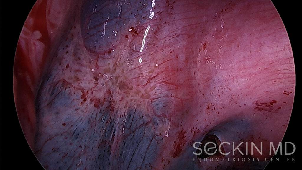
 Endometriosis adhesions: Symptoms, formation, and pictures
Adhesion (medicine) - Wikipedia
 Natural Remedy for Adhesion/Scar Tissue - LP2 - Uterine Adhesion
Nectin3 MGI Mouse Gene Detail - MGI:1930171 - nectin cell adhesion molecule 3
Cellular and matrix interactions of F4/80, an adhesion GPCR which defines murine tissue macrophages, in the normal and tumor...
Cell adhesion and mechanics as drivers of tissue organization and differentiation: local cues for large scale organization<...
 AMICUS Illustration of amicus,surgery,laparotomy,lysis,adhesions,midline,bowel,obstruction,scar,tissue,dissection,intestine...
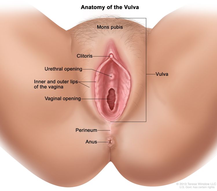
 Human Physiology/The female reproductive system - Wikibooks, open books for an open world
 April 2000 - Volume 105 - Issue 4 : Plastic and Reconstructive Surgery
 United Kingdom
Leukocyte Adhesion Deficiency: Background, Pathophysiology, Epidemiology
 Self-Organization in Biology | About | Elsevier
 SciELO - Brazil - Laparoscopic repair of congenital pleuroperitoneal hernia using a polypropylene mesh in a dog Laparoscopic...
Pelvic MRI for endometriosis: What to know
Structure and mechanism of the Nap adhesion complex from the human pathogen Mycoplasma genitalium | Nature Communications
Paratenonitis - Wikipedia
 References | BSI | Guidelines Library | Infection Control | CDC
Two neural-cell adhesion molecule(NCAM)-encoding genes in Xenopus laevis are expressed during development and in adult tissues ...
CNN.com - Transcripts
In Vitro Modeling of the Bipolar Disorder and Schizophrenia Using Patient-Derived Induced Pluripotent Stem Cells with Copy...
 A novel RGD-independent cell adhesion pathway mediated by fibronectin-bound tissue transglutaminase rescues cells from anoikis ...
 M protein mediated adhesion of M type 24 Streptococcus pyogenes stimulates release of interleukin-6 by HEp-2 tissue culture...
Scar tissue14
- I also have another friend who has had 10 C-sections, now she has some kind of Adhesions/scar tissue in her cervix adhereing to her adominal wall, but look how many surgerys she has had. (adhesions.org)
- An adhesion is a band of scar tissue that joins two internal body surfaces that are not usually connected. (wexfordphysiotherapy.com)
- Endometriosis adhesions are thick bands of scar tissue that can bind organs together. (medicalnewstoday.com)
- Adhesions are clumps of thick scar tissue on the inside of the body. (medicalnewstoday.com)
- The body usually forms scar tissue in response to inflammation and injuries. (medicalnewstoday.com)
- The body responds to this inflammation by forming scar tissue during the healing process. (medicalnewstoday.com)
- Large clumps of scar tissue may develop in the areas that endometriosis affects. (medicalnewstoday.com)
- The reason for this is that inflammation and scar tissue are normal parts of the healing process. (medicalnewstoday.com)
- They may be thought of as internal scar tissue that connects tissues not normally connected. (wikipedia.org)
- Used faithfully over time they can reduce scar tissue and adhesions. (earthclinic.com)
- Used regularly it has been known to heal even old scar tissue. (earthclinic.com)
- Abdominal adhesions are a type of scar tissue that causes your internal organs to "stick" together instead of move freely as they are intended to. (earthclinic.com)
- Women who have experienced major physical trauma (like severe bleeding, significant infection, severe scar tissue, surgical injury to nearby organs, uterine rupture, or hysterectomy) will need significant support as they recover. (ican-online.org)
- By increasing collagen production less scar tissue is formed at the damaged site. (nutrimedical.com)
Focal adhesi2
- RGD-independent cell adhesion to tTG-FN does not require transamidating activity, is mediated by the binding of tTG to cell-surface heparan sulfate chains, is dependent on the function of protein kinase Cα (PKCα) and leads to activation of the cell survival focal adhesion kinase (FAK). (ntu.ac.uk)
- Mechanistic studies showed that MWCNTs induced fibrogenesis of NHLFs through promoting expression and phosphorylation of focal adhesion kinase (FAK), while attenuating intracellular tension in the cells on stiff gels could increase MWCNTs uptake and thus elevate the induced fibrogenic responses. (cdc.gov)
Organs14
- Normally, internal tissues and organs have slippery surfaces so they can shift easily as the body moves. (medlineplus.gov)
- Adhesions cause tissues and organs to stick together. (medlineplus.gov)
- Organs or tissues within the body stick (adhere) to other internal surfaces. (wexfordphysiotherapy.com)
- Adhesions can affect the female reproductive organs (ovaries, fallopian tubes), the bowel, the area around the heart, the spine and the hand. (wexfordphysiotherapy.com)
- Adhesions are fibrous bands that form between tissues and organs, often as a result of injury during surgery. (wikipedia.org)
- While some adhesions do not cause problems, others may prevent muscle, nerve and other tissues and organs from moving freely, sometimes causing organs to become twisted or pulled from their normal positions. (wikipedia.org)
- The adhesions start to form within hours of surgery and may cause internal organs to attach to the surgical site or to other organs in the abdominal cavity. (wikipedia.org)
- Adhesion-related twisting and pulling of internal organs may result in complications such as abdominal pain or intestinal obstruction. (wikipedia.org)
- They are the same in that most of the reproductive organs of both sexes develop from similar embryonic tissue, meaning they are homologous. (wikibooks.org)
- In short, this is a known list of sex organs that evolve from the same tissues in a human life. (wikibooks.org)
- It also includes organs and tissues not connected to the reproductive system, such as the bladder and colon. (medicalnewstoday.com)
- Abdominal adhesions are bands of tissue that form between abdominal tissues and organs. (iffgd.org)
- Normally, internal tissues and organs have slippery surfaces, which allow them to shift easily as the body moves. (iffgd.org)
- Nobel Prize winner Alexis Carrel performed numerous experiments clearly showing that tissue explants, including connective tissue and heart tissue, could be cultured in vitro preserving their characteristics for prolonged periods of time [ 2 ] supporting the notion that entire organs could be cultured in vitro. (intechopen.com)
Bowel obstruction4
- As adhesions are likely to form after certain surgical procedures, open adhesiolysis may not be worthwhile, except to remedy serious problems such as bowel obstruction. (wexfordphysiotherapy.com)
- Small bowel obstruction (SBO) is a significant consequence of post-surgical adhesions. (wikipedia.org)
- Midline incision opened to expose adhesions and bowel obstruction. (medicalexhibits.com)
- Adhesions can cause pain, discomfort and even bowel obstruction. (earthclinic.com)
Cause more adhesions1
- In around 70 per cent of cases, the operation to remove the original adhesions will cause more adhesions to develop. (wexfordphysiotherapy.com)
Connective tissue4
- These are bands of dense connective tissue that may appear to tether pelvic structures. (medicalnewstoday.com)
- Peritendinous tissues become macroscopically thickened and new connective tissue adhesions occur. (wikipedia.org)
- Fibroblasts are present in connective tissue and are capable of forming collagen fibers. (nutrimedical.com)
- Stimulate tissue granulation and connective tissue projections, which are part of the healing process of wounds, ulcers or inflamed tissue. (nutrimedical.com)
Proteins9
- As demonstrated on LF fibroblasts, the resistance to trypsin was similar in serum-supplemented and serum-free media, i.e., medium without cell adhesion-mediating proteins. (nature.com)
- Based on all the results, the increased resistance to trypsinization of C2C12, LF, HaCaT, and VSMC cells on amine PPs can be explained most probably by a non-specific cell adhesion such as electrostatic interaction between the cells and amine groups on the material surface, rather than by the receptor-mediated adhesion through serum-derived proteins adsorbed on the PPs. (nature.com)
- Essential for infectivity is a transmembrane adhesion complex called Nap comprising proteins P110 and P140. (nature.com)
- Expression of stress proteins, adhesion molecules, and. (iifiir.org)
- Expression of stress proteins, adhesion molecul. (iifiir.org)
- Expression of stress proteins, adhesion molecules, and interleukin-8 in endothelial cells after preservation and reoxygenation. (iifiir.org)
- Apart from possible protective effects, allograft vasculopathy could be in part a consequence of the antigenic potential of heat shock proteins connected with effects caused by adhesion molecules and inflammatory cytokines. (iifiir.org)
- The two strains mediate adhesion by different mechanisms and, when combined, synergically induce the expression of Caco-2 tight junction proteins. (frontiersin.org)
- Sodium hydroxide of sufficient strength can hydrolyze proteins in tissues and can kill cells in tissues. (cdc.gov)
Develop adhesions1
- Up to 93 per cent of people who have abdominal surgery go on to develop adhesions. (wexfordphysiotherapy.com)
Cells20
- Quantifying the mechanics and growth of cells and tissues in 3D using high resolution computational models. (crossref.org)
- Our previously-obtained impressive results of highly increased C2C12 mouse myoblast adhesion to amine plasma polymers (PPs) motivated current detailed studies of cell resistance to trypsinization, cell proliferation, motility, and the rate of attachment carried out for fibroblasts (LF), keratinocytes (HaCaT), rat vascular smooth muscle cells (VSMC), and endothelial cells (HUVEC, HSVEC, and CPAE) on three different amine PPs. (nature.com)
- The increased cell adhesion was also confirmed for LF cells by an independent technique, single-cell force spectroscopy. (nature.com)
- Therefore, many laboratories try with great effort to develop resorbable tissue scaffolds that could support the patient´s cells. (nature.com)
- ECM provides mechanical support for cells and also determines the shape of tissue 9 . (nature.com)
- CD11a/CD18 (LFA-1) expressed on lymphocytes is known to play an important role in lymphocyte trafficking (adhesion to vascular endothelium), as well as interactions to antigen presenting cells (APC). (medscape.com)
- These 2 members mediate leukocyte adhesions to endothelial cells but they also serve as receptors for iC3b (inactivated C3b). (medscape.com)
- If this is allowed to happen, tissue repair cells such as macrophages, fibroblasts, and blood vessel cells penetrate into the fibrinous adhesion and lay down collagen and other matrix substances to form a permanent fibrous adhesion. (wikipedia.org)
- The human pathogen Mycoplasma genitalium , a member of the pneumoniae cluster of mycoplasmas, binds to eukaryotic cells by means of its adhesion complex, the Nap. (nature.com)
- These data suggest that lipoteichoic acid mediates reversible adhesion and that M protein is required for irreversible adhesion and for inducing release of interleukin-6 from HEp-2 cells. (tau.ac.il)
- Hasty, David L. / M protein mediated adhesion of M type 24 Streptococcus pyogenes stimulates release of interleukin-6 by HEp-2 tissue culture cells . (tau.ac.il)
- When co-cultured with human mesenchymal stem cells (hMSCs), the NMs exhibit excellent bioactivity in encouraging hMSCs adhesion. (jove.com)
- When co-cultured with human mesenchymal stem cells, the nano-matrix exhibited excellent bioactivity in encouraging cell adhesion. (jove.com)
- Hemoglobin is a protein found in red blood cells that plays a crucial role in transporting oxygen from the lungs to the body's tissues and carrying carbon dioxide, a waste product, from the tissues back to the lungs. (proprofs.com)
- The Cellular Potts Model and Biophysical Properties of Cells, Tissues and Morphogenesis. (maa.org)
- Modelling Development of Complex Tissues using Individual Viscoelastic Cells. (maa.org)
- In vitro characterization revealed that the strains tolerate gastric and bile challenges and display a great adhesion capacity to human intestinal cells. (frontiersin.org)
- Aim: To evaluate the adhesion of mouse bone marrow mesenchymal cells (MBMMC) on different titanium surfaces. (bvsalud.org)
- 2 (2007) observed that cell adhesion and proliferation, as well as the osteogenic differentiation of mouse mesenchymal stem cells (MSCs) to Ti discs were significantly similar to those on the plastic surface of the culture, indicating Ti as an excellent material for repairing hard tissue in the field of bone tissue engineering. (bvsalud.org)
- These properties determine the adhesion and behavior of cells in contact with the surface. (bvsalud.org)
Neural-cell adh1
- Human Neural cell adhesion molecule 1 (NCAM1) ELISA kit is Available at Gentaur Genprice with the fastest delivery. (joplink.net)
Leukocyte adhesion d18
- Leukocyte adhesion deficiency (LAD) is a rare primary immunodeficiency. (medscape.com)
- Thus the infections in patients with leukocyte adhesion deficiency act similarly as those observed in patients with neutropenia. (medscape.com)
- Labial ulceration from which Escherichia coli was cultured in an 8-month-old girl with leukocyte adhesion deficiency type 1 (LAD I). Note the thin bluish scar at the superior aspect of the labia from an earlier cellulitis. (medscape.com)
- This 3-year-old girl had leukocyte adhesion deficiency type I (LAD I) with complete absence of CD18 expression. (medscape.com)
- This 10-month-old patient with severe leukocyte adhesion deficiency type I (LAD I) developed a cervical adenitis caused by Klebsiella pneumoniae. (medscape.com)
- Leukocyte adhesion deficiency type I (LAD I) is a failure to express CD18, which composes the common ß 2 subunit of LFA1 family (ß2 integrins). (medscape.com)
- In milder forms of leukocyte adhesion deficiency I (1-30% expression of CD8), patients may survive to adulthood. (medscape.com)
- Patients with leukocyte adhesion deficiency II manifest the Bombay phenotype (ie, negative for O and H blood group antigens with potential production of anti-H antibody). (medscape.com)
- However, IgM and IgG serum levels are within the reference range in patients with leukocyte adhesion deficiency II. (medscape.com)
- Leukocyte adhesion deficiency II may be classified as one of the congenital disorders of glycosylation (CDG), a rapidly expanding group of metabolic syndromes with a wide symptomatology and severity. (medscape.com)
- Currently, 18 subtypes have been reported: 12 are type I (dysfunctional lipid-linked oligosaccharide precursor synthesis), and 6 are type II (dysfunctional trimming/processing of the protein-bound oligosaccharide), including leukocyte adhesion deficiency II (CDG-IIc). (medscape.com)
- Variants of leukocyte adhesion deficiency have also been reported, including fully expressed but nonfunctional CD18 and an E selectin that is expressed but rapidly cleaved from the cell surface (only present in soluble form). (medscape.com)
- Another reported type of leukocyte adhesion deficiency involves dysfunction in platelet aggregation in addition to a defect in leukocyte adhesion. (medscape.com)
- Thus, patients with this type of leukocyte adhesion deficiency manifest both severe bacterial infections and bleeding disorder. (medscape.com)
- This leukocyte adhesion deficiency variant is associated with defective expression of the Rap-1 activator CalDAG-GEFI. (medscape.com)
- Leukocyte adhesion deficiency results from an adhesion molecule defect that causes granulocyte and lymphocyte dysfunction and recurrent soft-tissue infections. (msdmanuals.com)
- Manifestations of leukocyte adhesion deficiency usually begin in infancy. (msdmanuals.com)
- Diagnosis of leukocyte adhesion deficiency is by detecting absence or severe deficiency of adhesive glycoproteins on the surface of WBCs using monoclonal antibodies (eg, anti-CD11, anti-CD18) and flow cytometry. (msdmanuals.com)
Uterus6
- OBGYN tells me usually the C-section is one of the least causes of Adhesions -because our Uterus is stretched so much that not much adheres afterwards. (adhesions.org)
- Adhesions can sometimes cause infertility in women by preventing fertilized eggs from reaching the uterus. (medlineplus.gov)
- Hysterosalpingography (an x-ray that views the inside of the uterus and fallopian tubes) may help diagnose adhesions inside the uterus or fallopian tubes. (wexfordphysiotherapy.com)
- Endometriosis causes the type of tissue that lines the uterus to grow in other places, such as on the fallopian tubes, ovaries, or bladder . (medicalnewstoday.com)
- Adhesions can also affect fertility, making it more difficult for an egg to travel to or implant in the uterus. (medicalnewstoday.com)
- Endometriosis is a condition in which tissue similar to the lining of the uterus grows outside of the uterus. (medicalnewstoday.com)
Fibrous1
- Adhesions may appear as thin sheets of tissue, similar to plastic wrap, or as thick fibrous bands. (wexfordphysiotherapy.com)
Extracellular5
- More than 30 adhesion GPCRs exist, utilizing large extracellular epidermal growth factor like domains to form multimeric signalosome-like structures. (ox.ac.uk)
- This project will explore the role of F4/80 in tumour-host interactions in vivo and identify cellular and extracellular binding partners of this adhesion GPCR. (ox.ac.uk)
- Specific association of tissue transglutaminase (tTG) with matrix fibronectin results in the formation of an extracellular complex (tTG-FN) with distinct adhesive and prosurvival characteristics. (ntu.ac.uk)
- The tTG-FN complex can maintain cell viability of tTG-null mouse dermal fibroblasts when apoptosis is induced by inhibition of RGD-dependent adhesion (anoikis), suggesting an extracellular survival role for tTG. (ntu.ac.uk)
- This formation helps deposition and adhesion of the extracellular matrix on the bone-implant interface. (bvsalud.org)
Infertility2
- Surgery inside the uterine cavity (e.g., suction dilation and curettage, myomectomy, endometrial ablation) may result in Asherman's syndrome (also known as intrauterine adhesions, intra uterine synechiae), a cause of infertility. (wikipedia.org)
- Adhesions can also cause infertility. (earthclinic.com)
Intestine3
- Further, we investigated the blends as tissue adhesives on two different porcine tissues (skin, and intestine). (umd.edu)
- A SBO may be caused when an adhesion pulls or kinks the small intestine and prevents the flow of content through the digestive tract. (wikipedia.org)
- Many obstructive events require surgery, however, to loosen or dissolve the offending adhesion(s) or to resect the affected small intestine. (wikipedia.org)
Pelvis2
- Adhesions can also form after inflammation in the abdomen or pelvis. (wexfordphysiotherapy.com)
- Pelvic adhesions are a form of abdominal adhesions in the pelvis. (wikipedia.org)
Surfaces3
- Adhesion formation post-surgery typically occurs when two injured surfaces are close to one another. (wikipedia.org)
- In the case of adhesive capsulitis of the shoulder (also known as frozen shoulder), adhesions grow between the shoulder joint surfaces, restricting motion. (wikipedia.org)
- Therefore, the present study aimed to evaluate the adhesion capacity of mouse bone marrow MSCs to smooth and plasmanitrided Ti surfaces in the cathodic cage configuration. (bvsalud.org)
Intra-abdominal1
- Abdominal adhesions (or intra-abdominal adhesions) are most commonly caused by abdominal surgical procedures. (wikipedia.org)
Inflammatory1
- Designed for excellent soft-tissue attachment and low inflammatory response. (nobelbiocare.com)
Molecules3
- Tissue resident and recruited macrophages also express adhesion molecules such as F4/80, a widely used biomarker and the founder member of a family of transmembrane G protein coupled receptors (GPCRs). (ox.ac.uk)
- Adhesion of macrophage cell lines from wild type and F4/80 knockout mice to purified matrix molecules, and complex 3D matrices, will be assessed, and binding sites mapped and downstream signalling examined. (ox.ac.uk)
- Briefly, IgSFs are adhesion molecules that bind to themselves (homophilic) or compatible IgSFs (heterophilic) across cell-cell junctions. (elifesciences.org)
Uterine1
- Adhesions are different than endometriosis implants, which grow and bleed in response to hormones in the same way as tissue from the uterine lining. (medicalnewstoday.com)
Regenerative medicine2
- For many years there is an urgent need in the field of regenerative medicine to develop replacements of non-functional tissues. (nature.com)
- Challenges in organ transplantation such as high organ demand and biocompatibility issues have led scientists in the field of tissue engineering and regenerative medicine to work on the use of scaffolds as an alternative to transplantation. (mdpi.com)
Cell16
- Niessen, Carien M. / Cell adhesion and mechanics as drivers of tissue organization and differentiation : local cues for large scale organization . (helsinki.fi)
- Wickstroem, SA & Niessen, CM 2018, ' Cell adhesion and mechanics as drivers of tissue organization and differentiation: local cues for large scale organization ', Current Opinion in Cell Biology , vol. 54, pp. 89-97. (helsinki.fi)
- tTG-FN supports RGD-independent cell adhesion of different cell types and the formation of distinctive RhoA-dependent focal adhesions following inhibition of integrin function by competitive RGD peptides and function blocking antiintegrin antibodies α5β1. (ntu.ac.uk)
- Association of tTG with its binding site on the 70 kda aminoterminal FN fragment does not support this cell adhesion process, which seems to involve the entire FN molecule. (ntu.ac.uk)
- We propose a novel RGD-independent cell adhesion mechanism that promotes cell survival when the anti-apoptotic role mediated by RGD-dependent integrin function is reduced as in tissue injury, which is consistent with the externalisation and binding of tTG to fibronectin following cell damage/stress. (ntu.ac.uk)
- Therefore, it has the potential to serve as an injectable scaffold to repair bone fractures by promoting cell adhesion and function at the target location. (jove.com)
- Two major thrusts in the last decade have dramatically impacted our understanding of biofilms: the utilization of the confocal laser scanning microscope to characterize biofilm ultrastructure, and an investigation of the genes involved in cell adhesion and biofilm formation. (cdc.gov)
- Cell and Tissue Biology (2022) 16 (4): 312. (aacrjournals.org)
- Most living tissues exhibit the specific stiffness, which has been known to have profound influence on cell behaviors, yet how the stiffness affects cellular responses to engineered nanomaterial s has not been elucidated. (cdc.gov)
- A 3-D Deformable Ellipsoidal Cell Model with Cell Adhesion and Signaling. (maa.org)
- How does a cancer cell push its way out of the surrounding tissue? (weizmann.ac.il)
- Weizmann Institute scientists propose a model regulating cell adhesion -- central to embryonic development, cellular movement. (weizmann.ac.il)
- Cell mechanics of adhesion, migration and dynamics. (gatech.edu)
- The term "adhesion" to the biomaterial refers to the most important phase, since the quality of it will influence morphology and the capacity of cell proliferation and differentiation 3 . (bvsalud.org)
- The functionality of the matured microvasculature networks was demonstrated through the enhancement of cell-cell adhesion, angiogenesis process, and. (lu.se)
- The functionality of the matured microvasculature networks was demonstrated through the enhancement of cell-cell adhesion, angiogenesis process, and perfusion tests with microparticles, FITC-dextran, and whole mouse blood. (lu.se)
Endometriosis14
- Although endometriosis is a risk factor for adhesions, it is not the only cause, and adhesions are not an inevitable result of endometriosis. (medicalnewstoday.com)
- In this article, we look at endometriosis adhesions in more detail, including how doctors diagnose and treat them. (medicalnewstoday.com)
- What are endometriosis adhesions? (medicalnewstoday.com)
- Advanced endometriosis can cause adhesions to form. (medicalnewstoday.com)
- Endometriosis is not the only condition that causes adhesions, however. (medicalnewstoday.com)
- Having endometriosis surgery can increase the risk of adhesions. (medicalnewstoday.com)
- Although many people with endometriosis adhesions report having pelvic pain, few studies have assessed the relationship between adhesions and pain. (medicalnewstoday.com)
- For example, there are experts who believe that the pain from endometriosis is different than the pain from adhesions. (medicalnewstoday.com)
- Adhesions, when they bind the bowels, can cause their own pain, which is separate and different from endometriosis pain. (medicalnewstoday.com)
- A 2009 study looked directly at the role of adhesions in endometriosis pain. (medicalnewstoday.com)
- People with stage 3 or 4 endometriosis are more likely to have adhesions . (medicalnewstoday.com)
- There is no consensus on what symptoms endometriosis adhesions cause, however, and research is ongoing. (medicalnewstoday.com)
- Surgery can remove endometriosis adhesions, which may help preserve fertility, reduce bleeding and pain, and prevent injuries to the ovaries. (medicalnewstoday.com)
- Endometriosis can cause additional adhesions to form even after surgery. (medicalnewstoday.com)
Abdomen1
- Almost everyone who has surgery on the abdomen gets adhesions. (medlineplus.gov)
Midline3
- Once you know about lysis and adhesions as they relate to laparotomy surgery, you can begin to understand midline. (medicalexhibits.com)
- Since midline and bowel are important components of Laparotomy with Lysis of Adhesions, adding bowel to the illustrations is important. (medicalexhibits.com)
- In fact, scar is usually the most common aspect of an illustration showing Laparotomy with Lysis of Adhesions, along with laparotomy, lysis, adhesions, midline, bowel and obstruction. (medicalexhibits.com)
Inflammation2
- According to the "classical paradigm" of adhesion formation, the pathogenesis starts with inflammation and activation of the coagulation system which causes fibrin deposits onto the damaged tissues. (wikipedia.org)
- In many cases, the production or activity of these enzymes are compromised because of inflammation following injury or infection, however, and the fibrinous adhesion persists. (wikipedia.org)
Occur4
- Adhesions can occur anywhere in the body. (medlineplus.gov)
- Abdominal adhesions also occur in 10 per cent of people who have never had surgery. (wexfordphysiotherapy.com)
- Adhesion-related disorder (ARD) is a group of symptoms that may occur as a result of adhesions. (wexfordphysiotherapy.com)
- Obstruction may occur 20 years or more after the initial surgical procedure, if a previously benign adhesion allows the small bowel to twist spontaneously around itself and obstruct. (wikipedia.org)
Symptoms3
- Typical adhesions form within the first few days after surgery, but symptoms can last for months or even years. (wexfordphysiotherapy.com)
- People with symptoms of adhesions may consider laparoscopic surgery. (wexfordphysiotherapy.com)
- The researchers identified an association between pain symptoms and the location and extent of the adhesions. (medicalnewstoday.com)
STIFFNESS1
- NHLFs were grown on polyacrylamide (PAAm) hydrogels with the stiffness comparable to that of human normal and fibrotic lung tissues, and treated with MWCNTs for various time. (cdc.gov)
Soft tissue2
- NobelPearl is designed to support a natural soft-tissue appearance. (nobelbiocare.com)
- Proven osseointegration and soft-tissue adhesion thanks to the hydrophilic sand-blasted, acid-etched Zerafil™ implant surface. (nobelbiocare.com)
Body's1
- Adhesions form as a natural part of the body's healing process after surgery in a similar way that a scar forms. (wikipedia.org)
Adipose Tissue1
- Here we demonstrate that a branched poly[Lys(Ser(i)-DL-Ala(m))] polymer functionalized with cyclic arginyl-glycyl-aspartate, when immobilized by simple adsorption to tissue culture plastic, surgical titanium alloy (Ti6Al4V), or Bio-Oss(®) bovine bone substitute, significantly accelerates serum-free adhesion and enhances seeding efficiency of human adipose tissue-derived MSCs. (nih.gov)
Complications3
- cause significantly higher post-operative complications (leaks, wound infections, haemorrhages) in people with adhesion-related perforations. (wexfordphysiotherapy.com)
- Although most adhesions are painless and do not cause complications, adhesions cause 60 to 70 per cent of small bowel obstructions in adults and are believed to contribute to the development of chronic pelvic pain. (wexfordphysiotherapy.com)
- Adhesions can cause serious complications, particularly when they are very large. (medicalnewstoday.com)
Aggregation2
- A more recent study suggested that the formation of "fibrinous" adhesions is preceded by the aggregation of cavity macrophages, that can act like extravascular platelets in the abdominal cavity. (wikipedia.org)
- adhesion and aggregation of platelets on damaged vessel 4. (proprofs.com)
Bone2
- In orthopaedic non‐load bearing devices or dental implants, the preferred materials can be resorbable ceramics chemically similar to the inorganic component of the bone tissue, such as hydroxyapatite 1 , 2 . (nature.com)
- Accordingly, the association of Ti implants with bone tissue culture may contribute to bone tissue regeneration 7 . (bvsalud.org)
Ovaries1
- For example, an endometrial adhesion can attach to the ovaries, cutting off the blood supply. (medicalnewstoday.com)
Adhesive2
- In previous work, blending low molecular weight (LMW) and high molecular weight (HMW) poly(lactide-co-caprolactone) (PLCL) yielded significantly higher adhesive strength to cardiac tissue than a clinical control. (umd.edu)
- Owing to their simple application, tunable degradation rates and mechanical properties, these blends have the potential to be used as a tissue adhesive on both dry and wet tissues. (umd.edu)
Scaffolds1
- Among different types of scaffolds, polymeric hydrogel scaffolds have received considerable attention because of their biocompatibility and structural similarity to native tissues. (mdpi.com)
Deficiency1
- One way is to carry out the transplantation, but the success of this strategy is complicated by immune reactions to allogeneic or xenogeneic grafts or deficiency of donor tissue. (nature.com)
Disorder2
- Adhesions can also lead to a complex set of problems called adhesion-related disorder (ARD). (wexfordphysiotherapy.com)
- This is an immune disorder that causes deep tissue infections. (clevelandclinic.org)
Diagnose2
- It is very difficult to diagnose the chronic pain of adhesions correctly. (wexfordphysiotherapy.com)
- Diagnostic tests such as blood tests, x-ray procedures, CT scans, MRIs and ultrasound will not diagnose adhesions. (wexfordphysiotherapy.com)
Tendon1
- Hypertrophic synovial tissue begins to invade and weaken the tendon, eventually leading to rupture. (medscape.com)
Extent2
- In body cavities such as the peritoneal, pericardial, and synovial cavities, a family of fibrinolytic enzymes may act to limit the extent of the initial fibrinous adhesion, and may even dissolve it. (wikipedia.org)
- A meta-analysis in 2012 came to the conclusion that there is only little evidence for the surgical principle that using less invasive techniques, introducing fewer foreign bodies, or causing less ischemia reduces the extent and severity of adhesions in pelvic surgery. (wikipedia.org)
Fibrin2
- The fibrin then connects the two adjacent structures where damage of the tissues occurred. (wikipedia.org)
- The fibrin acts like a glue to seal the injury and builds the fledgling adhesion, said at this point to be "fibrinous. (wikipedia.org)
Obstruction2
- According to statistics provided by the National Hospital Discharge Survey approximately 2,000 people die every year in the US from obstruction due to adhesions. (wikipedia.org)
- Along with bowel we can also focus on obstruction which also is important to Laparotomy with Lysis of Adhesions. (medicalexhibits.com)
Regeneration1
- equipment offers 7 specific settings to promote different levels of tissue healing and regeneration. (nutrimedical.com)
Protein3
- We investigated the contributions of lipoteichoic acid and M protein to reversible and irreversible adhesion of group A streptococci and the effects of such adhesion on release of interleukin-6. (tau.ac.il)
- Adequate protein plus vitamins A and E are important in helping to rebuild tissue. (ican-online.org)
- Collagen is the essential protein used to repair damaged tissue and to replace old tissue. (nutrimedical.com)
Abstract1
- article{2985125c-4fa1-47e4-9792-4bb6d8b1d6c4, abstract = {{Generating functional and perfusable micro-vascular networks is an important goal for the fabrication of large and three-dimensional tissues. (lu.se)
Severe2
Biomimetic1
- We developed a biomimetic nano-matrix with great promise to serve as a tissue engineering scaffold. (jove.com)
Macrophages2
Bands1
- Adhesions are bands of scar-like tissue. (medlineplus.gov)
Structures1
- The dye helps the technician identify internal body structures and tissues. (medicalnewstoday.com)

:quality(85)/cloudfront-us-east-1.images.arcpublishing.com/gray/FBU3K2LCRVEANIZFOA62DIA6WA.jpg)