Tooth Discoloration
Food Packaging
Color
Tooth Crown
Porcelain veneers: a challenging case. (1/76)
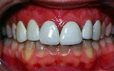
A patient in his early 20s with teeth badly discoloured by tetracycline was seeking treatment to improve his esthetics. Because retreatment and cost were important considerations, porcelain veneers were the treatment of choice. The challenge in this case was to mask the underlying tetracycline stain before the final cementation and thus gain more control over the final shade of the veneers. (+info)An evaluation of the changes in maxillary pulpal blood flow associated with orthognathic surgery. (2/76)
The objective of this study was to evaluate the use of the Laser Doppler Flowmeter (LDF) in the measurement of pulpal blood flow following orthognathic surgery and to conduct an initial study of the effects of a Le Fort I osteotomy on the pulpal blood flow of the maxillary central incisors. The design consisted of a preliminary prospective controlled consecutive clinical trial undertaken at the Orthodontic Clinic, University Dental Hospital NHS Trust, Wales, 1994. The study group consisted of 15 consecutive patients who were to receive a standard advancement Le Fort I osteotomy. Seven patients who were to undergo a mandibular advancement only acted as a control. A further 20 separate patients participated in a study for the assessment of measurement error. The blood flow in relative perfusion unit v. time, was measured using a Laser Doppler Flowmeter. Measurement error for flowmeter recordings with hand-held application and custom-made splint support showed no consistent difference or significant random variation between the two methods for holding the probe against the teeth (pooled S.D. of reproducibility 1/1 = 1.91/1.39 for custom splint location as opposed to 0.96/1.07 for hand-held/fixed bracket location). For the surgical patients under investigation no significant differences for maxillary pulpal blood flow were found in the control group (mandibular osteotomy) over time. However, in the maxillary osteotomy patients there was a tendency for an initial rise in the maxillary perfusion post-surgery as measured at the central incisor pulps, followed by an overall reduction at 6 months. As an example, the mean value for the upper right central showed a significant increase in blood flow during the immediate post-operative period (P < 0.05), but at 6 months after surgery demonstrated a statistically significant overall reduction in comparison with the presurgical reading (P < 0.001). The laser Doppler flowmeter is not an easy instrument to use in the clinical assessment of pulpal blood flow. However, it would appear from these longitudinal series of readings, taken over a 6-month period on 15 patients, that the maxillary perfusion recorded at the central incisor pulps may be permanently affected in many Le Fort I osteotomy patients. For patients that already have a prejudiced blood supply this could lead to devitalization and discoloration of incisors. It is not known if this affect on the perfusion of the pulp continues beyond 6 months post-surgery. (+info)The oral effects of smokeless tobacco. (3/76)
Smokeless tobacco use has increased rapidly in North America. This form of tobacco use has many oral effects including leukoplakia, oral cancer, loss of periodontal support (recession), and staining of teeth and composite restorations. Systemic effects such as nicotine dependence, transient hypertension and cardiovascular disease may also result from smokeless tobacco use. This paper aims to guide dental practitioners in identifying oral lesions that occur due to the use of smokeless tobacco and also offer guidelines on how to counsel patients who express a desire to stop using smokeless tobacco products. (+info)Unusual indelible enamel staining following fixed appliance treatment. (4/76)
Two cases are described of indelible enamel staining following fixed appliance therapy. The acquired pigmentation occurred in patients with an identifiable enamel defect prior to treatment. The interaction of factors to cause the staining is discussed and it's prevention in future cases highlighted. Subsequent restoration of the affected teeth is shown. (+info)Tooth discolouration and staining: a review of the literature. (5/76)
OBJECTIVE: To carry out an extensive review of the literature on tooth staining with particular regard to some of the more recent literature on the mechanisms of tooth staining involving mouthrinses. DESIGN: Comprehensive review of the literature over four decades. CONCLUSIONS: A knowledge of the aetiology of tooth staining is of importance to dental surgeons in order to enable a correct diagnosis to be made when examining a discoloured dentition and allows the dental practitioner to explain to the patient the exact nature of the condition. In some instances, the mechanism of staining may have an effect on the outcome of treatment and influence the treatment options the dentist will be able to offer to patients. (+info)Dental enamel formation and its impact on clinical dentistry. (6/76)
The nature of tooth enamel is of inherent interest to dental professionals. The current-day clinical practice of dentistry involves the prevention of enamel demineralization, the promotion of enamel remineralization, the restoration of cavitated enamel where demineralization has become irreversible, the vital bleaching of dental enamel that has become discolored, and the diagnosis and treatment of developmental enamel malformations, which can be caused by environmental or genetic factors. On a daily basis, dental health providers make diagnostic and treatment decisions that are influenced by their understanding of tooth formation. A systemic condition during tooth development, such as high fever, can produce a pattern of enamel defects in the dentition. Knowing the timing of tooth development permits estimates about the timing of the disturbance. The process of enamel maturation continues following tooth eruption, so that erupted teeth can become less susceptible to decay over time. Mutations in the genes encoding enamel proteins lead to amelogenesis imperfecta, a collection of inherited diseases having enamel malformations as the predominant phenotype. Defects in the amelogenin gene cause X-linked amelogenesis imperfecta, and genes encoding other enamel proteins are candidates for autosomal forms. Here we review our current understanding of dental enamel formation, and relate this information to clinical circumstances where this understanding may be particularly relevant. (+info)The use of QLF to quantify in vitro whitening in a product testing model. (7/76)
BACKGROUND: Professional and consumer interest in whitening products continues to increase against a background of both increased oral health awareness and demand for cosmetic procedures. In the current legal climate, few dentists are providing 'in-office' whitening treatments, and thus many patients turn to home-use products. The most common of these are the whitening toothpastes. Researchers are keen to quantify the effectiveness of such products through clinically relevant trials. AIM: Previous studies examining whitening products have employed a variety of stained substrates to monitor stain removal. This study aimed to quantify the removal of stain from human enamel using a new device, quantitative light-induced fluorescence (QLF). The experimental design follows that of a product-testing model. MATERIALS AND METHODS: A total of 11 previously extracted molar teeth were coated with transparent nail varnish leaving an exposed window of enamel. The sound, exposed enamel was subject to a staining regime of human saliva, chlorhexidine and tea. Each of the eleven teeth was subjected to serial exposures of a positive control (Bocasan), a negative control (water) and a test product (Yotuel toothpaste). Following each two-minute exposure QLF images of the teeth were taken (a total of 5 applications). Following completion of one test solution, the teeth were cleaned, re-stained and the procedure repeated with the next solution. QLF images were stored on a PC and analysed by a blinded single examiner. The deltaQ value at 5% threshold was reported. ANOVA and paired t-tests were used to analyse the data. RESULTS: The study confirmed the ability of QLF to longitudinally quantify stain reduction from human enamel. The reliability of the technique in relation to positive and negative test controls was proven. The positive control had a significantly (alpha = 0.05) higher stain removal efficacy than water (p = 0.023) and Yotuel (p = 0.046). Yotuel was more effective than water (p = 0.023). CONCLUSION: The research community, the practicing clinician and the consumer all require sound product evaluation data. The use of human enamel specimens may offer more relevant clinical data. QLF has been designed as an in vivo device. Further development of the technique should permit in vivo clinical whitening trials. (+info)Prevalence of black tooth stains and dental caries in Brazilian schoolchildren. (8/76)
This study evaluated the correlation between the presence of black extrinsic tooth stains and caries in Brazilian schoolchildren. The target population consisted of 263 children aged 6-12 years. Clinical examinations were performed by four dentists calibrated for the World Health Organization criteria for caries diagnosis. The c(2) test was used to compare the prevalence of caries between groups of children with and without black tooth stains. Spearman's correlation test was used to analyze the relationship between the presence of black stains and caries severity. Black stains were observed in 14.8% of the children. The number of children with caries-free permanent dentition was not statistically different between groups. The mean DMF-T was 1.46 +/- 1.39 for children with black stains and 2.42 +/- 2.09 for children without black stains. A negative correlation was observed between the presence (r = -0.16; p<0.05) and severity (r = -0.15; p<0.01) of black stains and DMF-T. (+info)Tooth discoloration, also known as tooth staining or tooth color change, refers to the darkening or staining of teeth. It can be categorized into two main types: extrinsic and intrinsic. Extrinsic discoloration occurs when the outer layer of the tooth (enamel) becomes stained due to exposure to colored substances such as coffee, tea, wine, tobacco, and certain foods. Intrinsic discoloration, on the other hand, occurs when the inner structure of the tooth (dentin) darkens or gets a yellowish tint due to factors like genetics, aging, trauma, or exposure to certain medications during tooth development. Tooth discoloration can also be caused by dental diseases or decay. It is important to note that while some forms of tooth discoloration are cosmetic concerns, others may indicate underlying oral health issues and should be evaluated by a dental professional.
A tooth is a hard, calcified structure found in the jaws (upper and lower) of many vertebrates and used for biting and chewing food. In humans, a typical tooth has a crown, one or more roots, and three layers: the enamel (the outermost layer, hardest substance in the body), the dentin (the layer beneath the enamel), and the pulp (the innermost layer, containing nerves and blood vessels). Teeth are essential for proper nutrition, speech, and aesthetics. There are different types of teeth, including incisors, canines, premolars, and molars, each designed for specific functions in the mouth.
I'm sorry for any confusion, but "Food Packaging" is not a medical term. It is a term used to describe the process and materials used to package food products to protect them from contamination, damage, and to provide information about the product. Medical definitions are typically related to diseases, conditions, treatments, or anatomical terms. If you have any questions related to medical terminology, I'd be happy to help with those!
In the context of medical terminology, 'color' is not defined specifically with a unique meaning. Instead, it generally refers to the characteristic or appearance of something, particularly in relation to the color that a person may observe visually. For instance, doctors may describe the color of a patient's skin, eyes, hair, or bodily fluids to help diagnose medical conditions or monitor their progression.
For example, jaundice is a yellowing of the skin and whites of the eyes that can indicate liver problems, while cyanosis refers to a bluish discoloration of the skin and mucous membranes due to insufficient oxygen in the blood. Similarly, doctors may describe the color of stool or urine to help diagnose digestive or kidney issues.
Therefore, 'color' is not a medical term with a specific definition but rather a general term used to describe various visual characteristics of the body and bodily fluids that can provide important diagnostic clues for healthcare professionals.
A tooth crown is a type of dental restoration that covers the entire visible portion of a tooth, restoring its shape, size, and strength. It is typically made of materials like porcelain, ceramic, or metal alloys and is custom-made to fit over the prepared tooth. The tooth crown is cemented in place and becomes the new outer surface of the tooth, protecting it from further damage or decay.
The process of getting a tooth crown usually involves two dental appointments. During the first appointment, the dentist prepares the tooth by removing any decay or damaged tissue and shaping the tooth to accommodate the crown. An impression is then taken of the prepared tooth and sent to a dental laboratory where the crown is fabricated. In the meantime, a temporary crown is placed over the prepared tooth to protect it until the permanent crown is ready. At the second appointment, the temporary crown is removed, and the permanent crown is cemented in place.
Tooth crowns are often recommended for several reasons, including:
* To restore a broken or fractured tooth
* To protect a weakened tooth from further damage or decay
* To support a large filling when there isn't enough natural tooth structure left
* To cover a dental implant
* To improve the appearance of a discolored or misshapen tooth
Overall, a tooth crown is an effective and long-lasting solution for restoring damaged or decayed teeth and improving oral health.
 Tooth discoloration
 Tooth Discoloration: Practice Essentials, Background, Pathophysiology
 Kingston upon thames dentists can provide helpful information to prevent discolouration of teeth - Mulberry Dental
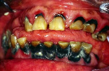
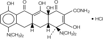
 Tetracycline teeth discoloration
 4 Best Dental Solutions for Brown Discoloration on Teeth - Allneedy
 Types of Tooth Discoloration - Memphis
 Oral Care Forums : Tooth Discoloration
 Tooth discoloration Archives - Ballwin Dental Care
 Suffer from tooth discoloration? Don't panic!
 baby teeth discoloration Archives - Dental Care Tips
 Tooth Discoloration Causes & Prevention - Clayton Individualized Dentistry
 What Causes Tooth Discoloration? | Parkside Family Dental
 These highlights do not include all the information needed to use MINOLIRA™ safely and effectively. See full prescribing...
 Why Teeth Discoloration Happens | Dr. Tracey Downtown Dental
 Teeth Whitening in Oradell | Professionally Remove Discoloration | Oradell Dentistry
 Common Foods and Drinks that Can Cause Tooth Discoloration
 Can I Prevent Tooth Discoloration? - Dr. Derek Wall Dentistry
 Teeth discolouration causes - Elite Dental Care
 Subjects: Tooth Discoloration - Digital Collections - National Library of Medicine Search Results
 The Most Common Causes of Teeth Discoloration - MOM News Daily
 Correcting Teeth Discolouration with Dental Veneers - Weightkut
 Risk of Dental Discoloration and Enamel Dysplasia in Children Exposed to Tetracycline and Its Derivatives - PubMed
 dental office in Vancouver WA - What Causes Teeth Discoloration | Thurston Oaks Dental
 Tooth Discoloration