Trichophyton
Tinea
Arthrodermataceae
Microsporum
Onychomycosis
Tinea Capitis
Tinea Pedis
Tinea Favosa
Antifungal Agents
Trichophytin
Nails
DNA, Ribosomal Spacer
Fungi
Lacquer
Naphthalenes
Mitosporic Fungi
Itraconazole
Deuteroporphyrins
Phylogenetic classification and species identification of dermatophyte strains based on DNA sequences of nuclear ribosomal internal transcribed spacer 1 regions. (1/337)
The mutual phylogenetic relationships of dermatophytes of the genera Trichophyton, Microsporum, and Epidermophyton were demonstrated by using internal transcribed spacer 1 (ITS1) region ribosomal DNA sequences. Trichophyton spp. and Microsporum spp. form a cluster in the phylogenetic tree with Epidermophyton floccosum as an outgroup, and within this cluster, all Trichophyton spp. except Trichophyton terrestre form a nested cluster (100% bootstrap support). Members of dermatophytes in the cluster of Trichophyton spp. were classified into three groups with ITS1 homologies, with each of them being a monophyletic cluster (100% bootstrap support). The Arthroderma vanbreuseghemii-Arthroderma simii group consists of A. vanbreuseghemii, A. simii, Trichophyton mentagrophytes isolates from humans, T. mentagrophytes var. quinckeanum, Trichophyton tonsurans, and Trichophyton schoenleinii. Arthroderma benhamiae, T. mentagrophytes var. erinacei, and Trichophyton verrucosum are members of the Arthroderma benhamiae group. Trichophyton rubrum and Trichophyton violaceum form the T. rubrum group. This suggests that these "species" of dermatophytes have been overclassified. The ITS1 sequences of 11 clinical isolates were also determined to identify the species, and all strains were successfully identified by comparison of their base sequences with those in the ITS1 DNA sequence database. (+info)Species identification and strain differentiation of dermatophyte fungi by analysis of ribosomal-DNA intergenic spacer regions. (2/337)
Restriction fragment length polymorphisms (RFLPs) identified in the ribosomal-DNA (rDNA) repeat were used for molecular strain differentiation of the dermatophyte fungus Trichophyton rubrum. The polymorphisms were detected by hybridization of EcoRI-digested T. rubrum genomic DNAs with a probe amplified from the small-subunit (18S) rDNA and adjacent internal transcribed spacer (ITS) regions. The rDNA RFLPs mapped to the nontranscribed spacer (NTS) region of the rDNA repeat and appeared similar to those caused by short repetitive sequences in the intergenic spacers of other fungi. Fourteen individual RFLP patterns (DNA types A to N) were recognized among 50 random clinical isolates of T. rubrum. A majority of strains (19 of 50 [38%]) were characterized by one RFLP pattern (DNA type A), and four types (DNA types A to D) accounted for 78% (39 of 50) of all strains. The remaining types (DNA types E to N) were represented by one or two isolates only. A rapid and simple method was also developed for molecular species identification of dermatophyte fungi. The contiguous ITS and 5.8S rDNA regions were amplified from 17 common dermatophyte species by using the universal primers ITS 1 and ITS 4. Digestion of the amplified ITS products with the restriction endonuclease MvaI produced unique and easily identifiable fragment patterns for a majority of species. However, some closely related taxon pairs, such as T. rubrum-T. soudanense and T. quinkeanum-T. schoenlenii could not be distinguished. We conclude that RFLP analysis of the NTS and ITS intergenic regions of the rDNA repeat is a valuable technique both for molecular strain differentiation of T. rubrum and for species identification of common dermatophyte fungi. (+info)The antifungal activity of mupirocin. (3/337)
The antibacterial agent mupirocin (pseudomonic acid A) is used as a topical agent in the treatment of superficial infections by Gram-positive bacteria, particularly Staphylococcus aureus. However, we demonstrate here that the compound also inhibits the growth of a number of pathogenic fungi in vitro, including a range of dermatophytes and Pityrosporum spp. It inhibited the incorporation of amino acids and precursors of RNA, but not that of acetate, by Trichophyton mentagrophytes. It also inhibited the isoleucyl-tRNA synthetase from Candida albicans, indicating a mechanism of action similar to that in bacteria. When administered topically, mupirocin was efficacious in a T. mentagrophytes ringworm model in guinea pigs. These results suggest that mupirocin could have clinical utility for superficial infections caused by dermatophytes. (+info)Molecular markers reveal exclusively clonal reproduction in Trichophyton rubrum. (4/337)
Genotypic variability among 96 Trichophyton rubrum strains which displayed different colony morphologies and were collected from four continents was investigated. Twelve markers representing 57 loci were analyzed by PCR fingerprinting, amplified fragment length polymorphism, and random amplified monomorphic DNA markers. Interestingly, none of the methods used revealed any DNA polymorphism, indicating a strictly clonal mode of reproduction and a strong adaptation to human skin. (+info)rRNA gene internal transcribed spacer 1 and 2 sequences of asexual, anthropophilic dermatophytes related to Trichophyton rubrum. (5/337)
The ribosomal region spanning the two internal transcribed spacer (ITS) regions and the 5.8S ribosomal DNA region was sequenced for asexual, anthropophilic dermatophyte species with morphological similarity to Trichophyton rubrum, as well as for members of the three previously delineated, related major clades in the T. mentagrophytes complex. Representative isolates of T. raubitschekii, T. fischeri, and T. kanei were found to have ITS sequences identical to that of T. rubrum. The ITS sequences of T. soudanense and T. megninii differed from that of T. rubrum by only a small number of base pairs. Their continued status as species, however, appears to meet criteria outlined in the population genetics-based cohesion species concept of A. R. Templeton. The ITS sequence of T. tonsurans differed from that of the biologically distinct T. equinum by only 1 bp, while the ITS sequence of the recently described species T. krajdenii had a sequence identical to that of T. mentagrophytes isolates related to the teleomorph Arthroderma vanbreuseghemii. (+info)Dermatophytosis: association between ABO blood groups and reactivity to the trichophytin. (6/337)
The authors investigated the relationship between dermatophytosis and ABO blood groups through blood typing, identification of isolated dermatophytes and specific cellular immune response of 40 individuals carriers of this mycosis. They verified that the fungus Trichophyton rubrum, isolated from 54.5% of the patients, was more frequent in individuals belonging to blood group A. The cellular immune response, evaluated through the trichophytin antigen, was positive in 25% of the studied patients; the presence of immediate reactions (30 minutes) was verified in 35%. The blood group distribution among patients with dermatophytosis and control groups was, respectively: 47.5% X 36% in group A, 40% X 50% in group O, 12. 5% X 11% in group B. Even though the authors have found a higher number of patients belonging to blood group A infected by T. rubrum, these results suggest that there is no statistical evidence that these individuals are more susceptible to dermatophytosis. (+info)Dermatophytosis caused by Trichophyton raubitschekii. Report of the first case in Sao Paulo, Brazil. (7/337)
The authors report the first case of dermatophytosis caused by Trichophyton raubitschekii in a patient from the State of Sao Paulo with Tinea corporis lesions localized on the buttocks. Culture on Sabouraud-agar with cycloheximide permitted the isolation and identification of the fungus, and the diagnosis was confirmed by Dr. Lynne Sigler, University of Alberta, Canada. Systemic treatment with fluconazole, 150 mg/week for 4 weeks, in combination with topical treatment with isoconazole initially yielded favorable results, with recurrence of the lesions after the medication was discontinued. This is the fifth case of this dermatophytosis published in the Brazilian medical literature. (+info)Antifungal susceptibility testing of dermatophytes: establishing a medium for inducing conidial growth and evaluation of susceptibility of clinical isolates. (8/337)
A standardized reference method for dermatophyte in vitro susceptibility testing is lacking. In a previous study, Norris et al. (H. A. Norris, B. E. Elewski, and M. A. Ghannoum, J. Am. Acad. Dermatol. 40(6, part 2):S9-S13) established the optimal medium and other growth variables. However, the earlier study did not address two issues: (i) selection of an optimal medium for conidial formation by dermatophytes and (ii) validation of the method with a large number of dermatophytes. The present study addresses these two points. To select which agar medium best supported conidial growth, representative isolates of dermatophytes were grown on different agars. Preliminary experiments showed that only oatmeal cereal agar supported the production of conidia by Trichophyton rubrum. We tested the abilities of 251 T. rubrum isolates to form conidia using three different cereal agars and potato dextrose agar. Overall, oatmeal cereal and rice agar media were comparable in their abilities to support T. rubrum conidial growth. Next, we used the oatmeal cereal agar for conidial formation along with the optimal conditions for dermatophyte susceptibility testing proposed by Norris et al. and determined the antifungal susceptibilities of 217 dermatophytes to fluconazole, griseofulvin, itraconazole, and terbinafine. Relative to the other agents tested, terbinafine possessed the highest antifungal activity against all of the dermatophytes. The mean +/- standard error of the mean MICs of fluconazole, itraconazole, terbinafine, and griseofulvin were 2.07 +/- 0.29, 0.13 +/- 0.01, 0.002 +/- 0.0003, and 0.71 +/- 0.05 microgram/ml, respectively. This study is the first step in the identification of optimal conditions that could be used for the standardization of the antifungal susceptibility testing method for dermatophytes. Inter- and intralaboratory agreement as well as clinical correlations need to be established. (+info)Trichophyton is a genus of fungi that are primarily responsible for causing various superficial and cutaneous infections in humans and animals. These infections, known as dermatophytoses or ringworm, typically involve the skin, hair, and nails. Some common examples of diseases caused by Trichophyton species include athlete's foot (T. rubrum), jock itch (T. mentagrophytes), and scalp ringworm (T. tonsurans).
The fungi in the Trichophyton genus are called keratinophilic, meaning they have a preference for keratin, a protein found in high concentrations in skin, hair, and nails. This characteristic allows them to thrive in these environments and cause infection. The specific species of Trichophyton involved in an infection will determine the clinical presentation and severity of the disease.
In summary, Trichophyton is a medical term referring to a group of fungi that can cause various skin, hair, and nail infections in humans and animals.
Tinea is a common fungal infection of the skin, also known as ringworm. It's called ringworm because of its characteristic red, circular, and often scaly rash with raised edges that can resemble a worm's shape. However, it has nothing to do with any kind of actual worm.
The fungi responsible for tinea infections belong to the genus Trichophyton, Microsporum, or Epidermophyton. These fungi thrive in warm, damp environments and can be contracted from infected people, animals, or contaminated soil. Common types of tinea infections include athlete's foot (tinea pedis), jock itch (tinea cruris), and ringworm of the scalp (tinea capitis).
Treatment for tinea typically involves antifungal medications, either topical or oral, depending on the location and severity of the infection. Proper hygiene and avoiding sharing personal items can help prevent the spread of this contagious condition.
Arthrodermataceae is a family of fungi that includes several medically important dermatophytes, which are fungi that can cause skin and nail infections known as tinea. Some notable genera within this family include:
1. Trichophyton: This genus contains several species that can cause various types of tinea infections, such as athlete's foot (tinea pedis), ringworm (tinea corporis), and jock itch (tinea cruris). Some species can also cause nail infections (tinea unguium or onychomycosis).
2. Microsporum: This genus includes some of the less common causes of tinea infections, such as tinea capitis (scalp ringworm) and tinea corporis.
3. Epidermophyton: This genus contains species that can cause tinea infections of the feet, hands, and nails.
These fungi primarily feed on keratin, a protein found in skin, hair, and nails, and typically invade dead or damaged tissue. Infections caused by Arthrodermataceae are usually treatable with antifungal medications, either topical or oral, depending on the severity and location of the infection.
Microsporum is a genus of fungi belonging to the family Arthrodermataceae. These fungi are known to cause various types of tinea (ringworm) infections in humans and animals. They are characterized by their ability to produce large, thick-walled macroconidia that are typically round to oval in shape.
The most common species of Microsporum that infect humans include M. canis, M. audouinii, and M. gypsum. These fungi are often found in soil and on the skin or fur of animals such as cats, dogs, and cattle. They can cause a variety of skin infections, including tinea capitis (scalp ringworm), tinea corporis (body ringworm), and tinea unguium (nail ringworm).
Microsporum infections are typically treated with topical or oral antifungal medications. Prevention measures include good personal hygiene, avoiding contact with infected animals, and prompt treatment of any fungal infections.
Onychomycosis is a medical term that refers to a fungal infection in the nails (both fingernails and toenails). This condition occurs when fungi, usually dermatophytes, invade the nail bed and cause damage to the nail plate. It can lead to symptoms such as discoloration, thickening, crumbling, and separation of the nail from the nail bed. Onychomycosis can be challenging to treat and may require long-term antifungal therapy, either topical or oral, or even removal of the infected nail in severe cases.
Dermatomycoses are a group of fungal infections that affect the skin, hair, and nails. These infections are caused by various types of fungi, including dermatophytes, yeasts, and molds. Dermatophyte infections, also known as tinea, are the most common type of dermatomycoses and can affect different areas of the body, such as the scalp (tinea capitis), beard (tinea barbae), body (tinea corporis), feet (tinea pedis or athlete's foot), hands (tinea manuum), and nails (tinea unguium or onychomycosis). Yeast infections, such as those caused by Candida albicans, can lead to conditions like candidal intertrigo, vulvovaginitis, and balanitis. Mold infections are less common but can cause skin disorders like scalded skin syndrome and phaeohyphomycosis. Dermatomycoses are typically treated with topical or oral antifungal medications.
Tinea capitis is a dermatophyte infection, primarily affecting the scalp and hair. It is commonly known as "ringworm of the scalp." The term "ringworm" is a misnomer because it has nothing to do with worms; instead, it refers to the ring-like appearance of the rash caused by these fungi.
The infection is more prevalent in children than adults and can spread through direct contact with an infected person or animal (like pets), or via contaminated objects such as combs, brushes, hats, etc. The causative agents are typically mold-like fungi called dermatophytes, which belong to the genera Microsporum or Trichophyton.
Symptoms of tinea capitis include itchiness, scaling, hair loss (in patches), and the presence of black dots on the scalp where broken hairs remain. In some cases, inflammation and pustules may occur. Diagnosis is usually confirmed through microscopic examination of hair or scale samples, and sometimes by culture.
Treatment typically involves oral antifungal medications like griseofulvin, terbinafine, itraconazole, or fluconazole for several weeks to ensure complete eradication of the fungus. Topical antifungals are often used in conjunction with oral therapy. Good hygiene practices and avoiding sharing personal items can help prevent transmission.
'Epidermophyton' is a genus of fungi that can cause skin and nail infections in humans. These types of infections are known as dermatophytoses or ringworm infections. The most common species that infect humans is Epidermophyton floccosum, which tends to cause infections of the feet (athlete's foot), nails, and groin (jock itch).
Epidermophyton fungi thrive on keratin, a protein found in skin, hair, and nails. They invade the dead outer layers of the skin or nails, causing inflammation, itching, scaling, and other symptoms. The infections can be spread through direct contact with an infected person or contaminated objects like towels, shoes, or floors.
To diagnose an Epidermophyton infection, a healthcare professional may collect a sample from the affected area and examine it under a microscope for the presence of fungal elements. The diagnosis can also be confirmed through culture methods, where the sample is grown on specialized media to identify the specific fungal species.
Treatment for Epidermophyton infections typically involves topical or oral antifungal medications, depending on the severity and location of the infection. Preventive measures such as keeping the skin clean and dry, avoiding sharing personal items, and wearing breathable footwear can help reduce the risk of contracting and spreading these types of infections.
Tinea Pedis, also known as athlete's foot, is a fungal infection that affects the skin on the feet, particularly between the toes. The causative agents are dermatophytes, which thrive in warm and damp environments. Common symptoms include itching, burning, cracked, blistered, or scaly skin, and sometimes painful peeling or cracking of the skin. It is contagious and can spread to other parts of the body or to other people through direct contact or via contaminated surfaces. Proper hygiene, keeping the feet dry, and using antifungal medications are common methods of preventing and treating this condition.
Griseofulvin is an antifungal medication used to treat various fungal infections, including those affecting the skin, hair, and nails. It works by inhibiting the growth of fungi, particularly dermatophytes, which cause these infections. Griseofulvin can be obtained through a prescription and is available in oral (by mouth) and topical (on the skin) forms.
The primary mechanism of action for griseofulvin involves binding to tubulin, a protein necessary for fungal cell division. This interaction disrupts the formation of microtubules, which are crucial for the fungal cell's structural integrity and growth. As a result, the fungi cannot grow and multiply, allowing the infected tissue to heal and the infection to resolve.
Common side effects associated with griseofulvin use include gastrointestinal symptoms (e.g., nausea, vomiting, diarrhea), headache, dizziness, and skin rashes. It is essential to follow the prescribing physician's instructions carefully when taking griseofulvin, as improper usage may lead to reduced effectiveness or increased risk of side effects.
It is important to note that griseofulvin has limited use in modern medicine due to the development of newer and more effective antifungal agents. However, it remains a valuable option for specific fungal infections, particularly those resistant to other treatments.
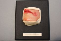
Tinea favosa, also known as "black dot ringworm," is a chronic and severe form of tinea capitis (ringworm of the scalp). It is caused by the fungus Trichophyton schoenleinii. The name "black dot" refers to the appearance of hair shafts that become broken off at the skin surface, leaving small black dots on the scalp.
The infection often affects children and can cause scaling, alopecia (hair loss), and formation of kerion (a severely inflamed and pustular lesion). The condition is highly contagious and can spread through contact with infected individuals or contaminated objects such as combs, brushes, hats, and towels.
Tinea favosa can be challenging to treat due to its chronic nature and the development of extensive scarring and permanent hair loss if left untreated. Treatment typically involves oral antifungal medications for an extended period, along with proper hygiene measures to prevent the spread of infection.
Foot dermatoses refer to various skin conditions that affect the feet. These can include inflammatory conditions like eczema and psoriasis, infectious diseases such as athlete's foot (tinea pedis), fungal infections, bacterial infections, viral infections (like plantar warts caused by HPV), and autoimmune blistering disorders. Additionally, contact dermatitis from irritants or allergens can also affect the feet. Proper diagnosis is essential to determine the best course of treatment for each specific condition.
Antifungal agents are a type of medication used to treat and prevent fungal infections. These agents work by targeting and disrupting the growth of fungi, which include yeasts, molds, and other types of fungi that can cause illness in humans.
There are several different classes of antifungal agents, including:
1. Azoles: These agents work by inhibiting the synthesis of ergosterol, a key component of fungal cell membranes. Examples of azole antifungals include fluconazole, itraconazole, and voriconazole.
2. Echinocandins: These agents target the fungal cell wall, disrupting its synthesis and leading to fungal cell death. Examples of echinocandins include caspofungin, micafungin, and anidulafungin.
3. Polyenes: These agents bind to ergosterol in the fungal cell membrane, creating pores that lead to fungal cell death. Examples of polyene antifungals include amphotericin B and nystatin.
4. Allylamines: These agents inhibit squalene epoxidase, a key enzyme in ergosterol synthesis. Examples of allylamine antifungals include terbinafine and naftifine.
5. Griseofulvin: This agent disrupts fungal cell division by binding to tubulin, a protein involved in fungal cell mitosis.
Antifungal agents can be administered topically, orally, or intravenously, depending on the severity and location of the infection. It is important to use antifungal agents only as directed by a healthcare professional, as misuse or overuse can lead to resistance and make treatment more difficult.
Trichophytin is not a medical condition or diagnosis, but rather a preparation used in skin tests to help identify whether someone has a sensitivity or allergic reaction to the fungus called Trichophyton. This fungus can cause various skin infections such as athlete's foot, ringworm, and jock itch. The trichophytin preparation contains antigens derived from the Trichophyton fungus, which are introduced into the skin to trigger an immune response. If a person is allergic or sensitive to the fungus, their body will mount a reaction, which can be observed and measured as part of the skin test. This information helps healthcare professionals diagnose and manage fungal infections and related allergies.
Mycological typing techniques are methods used to identify and classify fungi at the species or strain level, based on their unique biological characteristics. These techniques are often used in clinical laboratories to help diagnose fungal infections and determine the most effective treatment approaches.
There are several different mycological typing techniques that may be used, depending on the specific type of fungus being identified and the resources available in the laboratory. Some common methods include:
1. Phenotypic methods: These methods involve observing and measuring the physical characteristics of fungi, such as their growth patterns, colonial morphology, and microscopic features. Examples include macroscopic and microscopic examination, as well as biochemical tests to identify specific metabolic properties.
2. Genotypic methods: These methods involve analyzing the DNA or RNA of fungi to identify unique genetic sequences that can be used to distinguish between different species or strains. Examples include PCR-based methods, such as restriction fragment length polymorphism (RFLP) analysis and amplified fragment length polymorphism (AFLP) analysis, as well as sequencing-based methods, such as internal transcribed spacer (ITS) sequencing and multilocus sequence typing (MLST).
3. Proteotypic methods: These methods involve analyzing the proteins expressed by fungi to identify unique protein profiles that can be used to distinguish between different species or strains. Examples include matrix-assisted laser desorption/ionization time-of-flight mass spectrometry (MALDI-TOF MS) and liquid chromatography-mass spectrometry (LC-MS).
Mycological typing techniques are important tools for understanding the epidemiology of fungal infections, tracking outbreaks, and developing effective treatment strategies. By accurately identifying the specific fungi causing an infection, healthcare providers can tailor their treatments to target the most vulnerable aspects of the pathogen, improving patient outcomes and reducing the risk of drug resistance.
In the context of medical terminology, "nails" primarily refer to the keratinous plates that are found at the tips of fingers and toes. These specialized structures are part of the outermost layer of the skin (epidermis) and are formed by a type of cells called keratinocytes. The nails serve to protect the delicate underlying tissues from trauma, and they also aid in tasks such as picking up small objects or scratching itches.
The medical term for fingernails and toenails is "unguis," which comes from Latin. Each nail consists of several parts:
1. Nail plate: The visible part of the nail that is hard and flat, made up of keratin.
2. Nail bed: The skin beneath the nail plate to which the nail plate is attached; it supplies blood to the nail.
3. Matrix: The area where new cells are produced for the growth of the nail plate; located under the cuticle and extends slightly onto the finger or toe.
4. Lunula: The crescent-shaped white area at the base of the nail plate, which is the visible portion of the matrix.
5. Cuticle: The thin layer of skin that overlaps the nail plate and protects the underlying tissue from infection.
6. Eponychium: The fold of skin that surrounds and covers the nail plate; also known as the "proximal nail fold."
7. Hyponychium: The area of skin between the free edge of the nail plate and the fingertip or toe tip.
8. Perionychiun: The skin surrounding the nail on all sides.
Understanding the anatomy and medical aspects of nails is essential for healthcare professionals, as various conditions can affect nail health, such as fungal infections, ingrown nails, or tumors.
Fungal DNA refers to the genetic material present in fungi, which are a group of eukaryotic organisms that include microorganisms such as yeasts and molds, as well as larger organisms like mushrooms. The DNA of fungi, like that of all living organisms, is made up of nucleotides that are arranged in a double helix structure.
Fungal DNA contains the genetic information necessary for the growth, development, and reproduction of fungi. This includes the instructions for making proteins, which are essential for the structure and function of cells, as well as other important molecules such as enzymes and nucleic acids.
Studying fungal DNA can provide valuable insights into the biology and evolution of fungi, as well as their potential uses in medicine, agriculture, and industry. For example, researchers have used genetic engineering techniques to modify the DNA of fungi to produce drugs, biofuels, and other useful products. Additionally, understanding the genetic makeup of pathogenic fungi can help scientists develop new strategies for preventing and treating fungal infections.
The ribosomal spacer in DNA refers to the non-coding sequences of DNA that are located between the genes for ribosomal RNA (rRNA). These spacer regions are present in the DNA of organisms that have a nuclear genome, including humans and other animals, plants, and fungi.
In prokaryotic cells, such as bacteria, there are two ribosomal RNA genes, 16S and 23S, separated by a spacer region known as the intergenic spacer (IGS). In eukaryotic cells, there are multiple copies of ribosomal RNA genes arranged in clusters called nucleolar organizer regions (NORs), which are located on the short arms of several acrocentric chromosomes. Each cluster contains hundreds to thousands of copies of the 18S, 5.8S, and 28S rRNA genes, separated by non-transcribed spacer regions known as internal transcribed spacers (ITS) and external transcribed spacers (ETS).
The ribosomal spacer regions in DNA are often used as molecular markers for studying evolutionary relationships among organisms because they evolve more rapidly than the rRNA genes themselves. The sequences of these spacer regions can be compared among different species to infer their phylogenetic relationships and to estimate the time since they diverged from a common ancestor. Additionally, the length and composition of ribosomal spacers can vary between individuals within a species, making them useful for studying genetic diversity and population structure.
Fungi, in the context of medical definitions, are a group of eukaryotic organisms that include microorganisms such as yeasts and molds, as well as the more familiar mushrooms. The study of fungi is known as mycology.
Fungi can exist as unicellular organisms or as multicellular filamentous structures called hyphae. They are heterotrophs, which means they obtain their nutrients by decomposing organic matter or by living as parasites on other organisms. Some fungi can cause various diseases in humans, animals, and plants, known as mycoses. These infections range from superficial, localized skin infections to systemic, life-threatening invasive diseases.
Examples of fungal infections include athlete's foot (tinea pedis), ringworm (dermatophytosis), candidiasis (yeast infection), histoplasmosis, coccidioidomycosis, and aspergillosis. Fungal infections can be challenging to treat due to the limited number of antifungal drugs available and the potential for drug resistance.
I must clarify that "lacquer" does not have a specific medical definition. The term "lacquer" is commonly used in dermatology to describe a type of scale found on the skin, but it is not a formal medical term with a widely accepted definition. It's essential to provide more context or specify the field when seeking definitions to ensure accurate and helpful information.
Econazole is an antifungal medication used to treat various fungal infections of the skin, nails, and mucous membranes. It works by inhibiting the synthesis of ergosterol, a key component of fungal cell membranes, thereby weakening the cell membrane and increasing permeability, ultimately leading to fungal cell death.
Econazole is available in various formulations, including creams, lotions, powders, and tablets. It is commonly used to treat conditions such as athlete's foot, jock itch, ringworm, candidiasis (yeast infection), and other fungal skin infections.
It is important to follow the instructions of a healthcare provider when using econazole or any medication, and to report any side effects or concerns promptly.
Naphthalene is not typically referred to as a medical term, but it is a chemical compound with the formula C10H8. It is a white crystalline solid that is aromatic and volatile, and it is known for its distinctive mothball smell. In a medical context, naphthalene is primarily relevant as a potential toxin or irritant.
Naphthalene can be found in some chemical products, such as mothballs and toilet deodorant blocks. Exposure to high levels of naphthalene can cause symptoms such as nausea, vomiting, diarrhea, and headaches. Long-term exposure has been linked to anemia and damage to the liver and nervous system.
In addition, naphthalene is a known environmental pollutant that can be found in air, water, and soil. It is produced by the combustion of fossil fuels and is also released from some industrial processes. Naphthalene has been shown to have toxic effects on aquatic life and may pose a risk to human health if exposure levels are high enough.
Mitosporic fungi, also known as asexual fungi or anamorphic fungi, are a group of fungi that produce mitospores (also called conidia) during their asexual reproduction. Mitospores are produced from the tip of specialized hyphae called conidiophores and are used for dispersal and survival of the fungi in various environments. These fungi do not have a sexual reproductive stage or it has not been observed, making their taxonomic classification challenging. They are commonly found in soil, decaying organic matter, and water, and some of them can cause diseases in humans, animals, and plants. Examples of mitosporic fungi include Aspergillus, Penicillium, and Fusarium species.
Fungal spores are defined as the reproductive units of fungi that are produced by specialized structures called hyphae. These spores are typically single-celled and can exist in various shapes such as round, oval, or ellipsoidal. They are highly resistant to extreme environmental conditions like heat, cold, and dryness, which allows them to survive for long periods until they find a suitable environment to germinate and grow into a new fungal organism. Fungal spores can be found in the air, water, soil, and on various surfaces, making them easily dispersible and capable of causing infections in humans, animals, and plants.
Itraconazole is an antifungal medication used to treat various fungal infections, including blastomycosis, histoplasmosis, aspergillosis, and candidiasis. It works by inhibiting the synthesis of ergosterol, a vital component of fungal cell membranes, thereby disrupting the integrity and function of these membranes. Itraconazole is available in oral and intravenous forms for systemic use and as a topical solution or cream for localized fungal infections.
Medical Definition:
Itraconazole (i-tra-KON-a-zole): A synthetic triazole antifungal agent used to treat various fungal infections, such as blastomycosis, histoplasmosis, aspergillosis, and candidiasis. It inhibits the synthesis of ergosterol, a critical component of fungal cell membranes, leading to disruption of their integrity and function. Itraconazole is available in oral (capsule and solution) and intravenous forms for systemic use and as a topical solution or cream for localized fungal infections.
Miconazole is an antifungal medication used to treat various fungal infections, including those affecting the skin, mouth, and vagina. According to the Medical Subject Headings (MeSH) database maintained by the National Library of Medicine, miconazole is classified as an imidazole antifungal agent that works by inhibiting the synthesis of ergosterol, a key component of fungal cell membranes. By disrupting the structure and function of the fungal cell membrane, miconazole can help to kill or suppress the growth of fungi, providing therapeutic benefits in patients with fungal infections.
Miconazole is available in various formulations, including creams, ointments, powders, tablets, and vaginal suppositories, and is typically applied or administered topically or vaginally, depending on the site of infection. In some cases, miconazole may also be given intravenously for the treatment of severe systemic fungal infections.
As with any medication, miconazole can have side effects and potential drug interactions, so it is important to use it under the guidance of a healthcare professional. Common side effects of miconazole include skin irritation, redness, and itching at the application site, while more serious side effects may include allergic reactions, liver damage, or changes in heart rhythm. Patients should be sure to inform their healthcare provider of any other medications they are taking, as well as any medical conditions they have, before using miconazole.
Deuteroporphyrins are porphyrin derivatives that contain two carboxylic acid side chains. They are intermediates in the biosynthesis of heme and chlorophyll, which are essential molecules for biological processes such as oxygen transport and photosynthesis, respectively.
Deuteroporphyrins can be further classified into isomers based on the position of the carboxylic acid side chains. The most common isomer is deuteroporphyrin IX, which has the carboxylic acid side chains located at positions 1 and 2 relative to the pyrrole nitrogen atoms.
Deuteroporphyrins have been studied in various medical contexts, including as potential markers of porphyria, a group of metabolic disorders characterized by the accumulation of porphyrin precursors. Additionally, deuteroporphyrins and their derivatives have been investigated for their potential use in photodynamic therapy, a treatment modality that uses light-activated drugs to destroy cancer cells.
Malassezia is a genus of fungi (specifically, yeasts) that are commonly found on the skin surfaces of humans and other animals. They are part of the normal flora of the skin, but under certain conditions, they can cause various skin disorders such as dandruff, seborrheic dermatitis, pityriasis versicolor, and atopic dermatitis.
Malassezia species require lipids for growth, and they are able to break down the lipids present in human sebum into fatty acids, which can cause irritation and inflammation of the skin. Malassezia is also associated with fungal infections in people with weakened immune systems.
The genus Malassezia includes several species, such as M. furfur, M. globosa, M. restricta, M. sympodialis, and others. These species can be identified using various laboratory methods, including microscopy, culture, and molecular techniques.
 Trichophyton
Trichophyton erinacei
Trichophyton verrucosum
Trichophyton terrestre
Trichophyton concentricum
Trichophyton rubrum
Trichophyton tonsurans
Trichophyton interdigitale
Trichophyton mentagrophytes
Occupational hazards of human nail dust
List of fungi of South Africa - T
Tinea capitis
Myriodontium keratinophilum
Marine fungi
List of fungi of South Africa - E
Gene density
Rhoda Williams Benham
Tinea manuum
Tinea imbricata
Microsporum gallinae
List of types of tinea
Fungal folliculitis
Domenico Majocchi
List of fungi of South Africa - A
Arthrodermataceae
Two feet-one hand syndrome
Glossary of mycology
Favus
Tinea cruris
Venturicidin
Trichophyton - Wikipedia
 Trichophyton violaceum Sabouraud - MYA-839 | ATCC
 The Rise of Resistant Ringworm: Genomic Sequencing Confirms the First Two Reported U.S. Cases of Trichophyton indotineae |...
 Frontiers | TLR2−/− Mice Display Increased Clearance of Dermatophyte Trichophyton mentagrophytes in the Setting of Hyperglycemia
Table 2 - Sexually Transmitted Trichophyton mentagrophytes Genotype VII Infection among Men Who Have Sex with Men - Volume 29,...
Trichophyton rubrum DNA Strains in Patients with Onychomycosis with Persistent Mixed Infections Involving a Nondermatophyte...
Diagnosis of Trichophyton rubrum from Onychomycotic Nail Samples Using Polymerase Chain Reaction and Calcofluor White...
 Altmetric - Report of terbinafine resistant Trichophyton spp. in Italy: Clinical presentations, molecular identification,...
 Trichophyton tonsurans ringworm-A new public health problem
 Trichophyton mentagrophytes sebagai Agen Penyebab Dermatofitosis pad Kambing | Sunartatie | Jurnal Sain Veteriner
Emerging antimicrobial-resistant ringworm infections | CDC
 Trichophyton indotineae - Atlas of Clinical Fungi
Trichophyton mentagrophytes and trichophyton rubrum - wikidoc
Trichophyton schoenleinii - Atlas of Clinical Fungi
 Trichophyton rubrum complex detection kits - PCR KIT
 IgE specific la Trichophyton rubrum - Novus Medical
 What is the Difference Between Microsporum Trichophyton and Epidermophyton - Pediaa.Com
 Erythroderma combined with deeper dermal dermatophytosis due to Trichophyton rubrum in a patient with myasthenia gravis: first...
 Inhibition of the growth of the pathogenic fungus Trichophyton rubrum by M. azedarach leaves
Serine protease inhibitor with antifungal activity against Trichophyton rubrum from Para rubber leaves : Walailak University
 Medirabbit
 Mycology Update 2017 | PPT
 The extra-human occurrence of Trichophyton tonsurans var. sulfureum in a residential school.<...
Dexamethasone-induced flares of Trichophyton rubrummasquerading as docetaxel cutaneous toxicity: a case report | Cases Journal ...
 Folliculitis Organism-Specific Therapy: Specific Organisms and Therapeutic Regimens
 Unknown 27 | Mycology | University of Adelaide
 African Journal of Microbiology Research - trichophyton rubrum â€" the predominant etiological agent in human dermatophytoses...
 Opti-Cide Max for Animal Use - Drugs.com
 Sexually Transmitted Trichophyton mentagrophytes Genotype VII Infection among Men Who Have Sex with Men. | Emerg Infect Dis;29...
Tinea Corporis: Practice Essentials, Background, Pathophysiology
Mentagrophytes27
- 1939) Trichophyton vanbreuseghemii Rioux, Jarry & Juminer (1965) Trichophyton mentagrophytes (Family Arthrodermataceae, Genus Trichophyton) is capable of both mating and meiosis. (wikipedia.org)
- Trichophyton schoenleinii cause favus (tinea capitis),Trichophyton mentagrophytes var. (wikipedia.org)
- mentagrophytes and Trichophyton verrucosum cause kerion (violent reaction results from infection with an animal dermatophytes). (wikipedia.org)
- Initial testing at that time indicated that the patient had Trichophyton mentagrophytes, a common species of fungus closely related to T. indotineae . (cdc.gov)
- In the present study, we investigated the role of TLR2 during the development of experimental deep dermatophytosis in normal mice and mice with alloxan-induced diabetes mellitus, an experimental model of diabetes that exhibits a delay in the clearance of the dermatophyte, Trichophyton mentagrophytes (Tm). (frontiersin.org)
- Nenoff P , Schubert K , Jarsumbeck R , Uhrlaß S , Krüger C . Tinea genitalis profunda durch Trichophyton mentagrophytes nach Ägypten-Reise. (cdc.gov)
- Kupsch C , Czaika V , Deutsch C , Gräser Y . Trichophyton mentagrophytes -a new genotype of zoophilic dermatophyte causes sexually transmitted infections. (cdc.gov)
- Siopi M , Efstathiou I , Theodoropoulos K , Pournaras S , Meletiadis J . Molecular epidemiology and antifungal susceptibility of Trichophyton isolates in Greece: emergence of terbinafine-resistant Trichophyton mentagrophytes type VIII locally and globally. (cdc.gov)
- Klinger M , Theiler M , Bosshard PP . Epidemiological and clinical aspects of Trichophyton mentagrophytes / Trichophyton interdigitale infections in the Zurich area: a retrospective study using genotyping. (cdc.gov)
- Trichophyton mentagrophytes and T interdigitale genotypes are associated with particular geographic areas and clinical manifestations. (cdc.gov)
- Trichophyton indotineae (T. indotineae) , previously considered a subtype of Trichophyton mentagrophytes (T. mentagrophytes) genotype VIII, is now considered its own species of dermatophyte fungus. (cdc.gov)
- A 2023 study from France highlights 13 severe cases of ringworm caused by Trichophyton mentagrophytes , genotype VII (TMVII). (cdc.gov)
- Trichophyton mentagrophytes and trichophyton rubrum is an antifungal agent that is FDA approved for the diagnosis of type I hypersensitivity (i.e. allergy) to fungus. (wikidoc.org)
- There is limited information regarding Off-Label Guideline-Supported Use of Trichophyton mentagrophytes and trichophyton rubrum in adult patients. (wikidoc.org)
- There is limited information regarding Off-Label Non-Guideline-Supported Use of Trichophyton mentagrophytes and trichophyton rubrum in adult patients. (wikidoc.org)
- Trichophyton mentagrophytes tvar (gyűrűf rgek). (medirabbit.com)
- Transmission of dermatophytes , especially Trichophyton mentagrophytes genotype VII, during sexual intercourse has been recently reported. (bvsalud.org)
- The dermatophytes Trichophyton rubrum and Trichophyton mentagrophytes are the most common causative pathogens responsible for up to 90% of all cases [ 1 ]. (hindawi.com)
- Pustisek N, Skerlev M, Basta-Juzbasic A, Lipozencic J, Marinovic B, Bukvic-Mokos Z. Tinea incognito caused by trichophyton mentagrophytes -- a case report. (medscape.com)
- (1-2) As a consequence, a broad range of MIC values, 1-20 mcg/mL, were obtained for Trichophyton rubrum and Trichophyton mentagrophytes species. (guidelinecentral.com)
- One ex vivo study was conducted evaluating 8% ciclopirox against new and established Trichophyton rubrum and Trichophyton mentagrophytes infections in ovine hoof material. (guidelinecentral.com)
- Trichophyton mentagrophytes complex includes several different species and genotypes that are actually pretty difficult to differentiate just by looking under the microscope. (cdc.gov)
- This complex includes things like Trichophyton mentagrophytes (of course), Trichophyton interdigitale and erinacei . (cdc.gov)
- And also, within this complex, there are zoophilic strains, which means that they prefer animals (such as Trichophyton mentagrophytes and erinacei ). (cdc.gov)
- It was soon discovered that it was part of, actually, the Trichophyton mentagrophytes complex , but limitations in laboratory testing and sequencing really hindered the identification of Trichophyton indotineae until about 2020. (cdc.gov)
- The causative agent of mouse favus is T mentagrophytes var quinckeanum , also termed Trichophyton quinckeanum , which can cause favus in humans, although rarely. (medscape.com)
- Garcia-Sanchez MS, Pereiro M Jr, Pereiro MM, Toribio J. Favus due to Trichophyton mentagrophytes var. (medscape.com)
Microsporum12
- The main difference between Microsporum Trichophyton and Epidermophyton is that Microsporum is a zoophilic dermatophyte, and Trichophyton is both zoophilic and anthropophilic, whereas Epidermophyton is an anthropophilic dermatophyte. (pediaa.com)
- Microsporum, Trichophyton, and Epidermophyton are three types of dermatophytes that belong to the division Ascomycota . (pediaa.com)
- Microsporum, Trichophyton , and Epidermophyton are three types of fungi genera that are dermatophytes. (pediaa.com)
- Microsporum refers to a genus of fungi that causes tinea capitis, tinea corporis, ringworm, and other dermatophytoses (fungal infections of the skin), and Trichophyton refers to a genus of fungi of the family Moniliaceae that are parasitic in the skin and hair follicles, while Epidermophyton refers to a genus of fungus causing superficial and cutaneous mycoses, including E. floccosum. (pediaa.com)
- Microsporum is a zoophilic dermatophyte, Trichophyton is both zoophilic and anthropophilic while Epidermophyton is an anthropophilic dermatophyte. (pediaa.com)
- Furthermore, Microsporum contains both macroconidia and microconidia, and Trichophyton contains only microconidia, while Epidermophyton contains only macroconidia. (pediaa.com)
- In brief, Microsporum, Trichophyton , and Epodermophyton are three genera of dermatophytes that cause fungal infections in the superficial keratinized structures such as hair, nails, and the stratum corneum of the skin. (pediaa.com)
- The species of fungi that cause ringworm are Microsporum and Trichophyton. (proprofs.com)
- Therefore, the correct answer is Microsporum and Trichophyton. (proprofs.com)
- [2] They are typically of the Trichophyton , Microsporum , or Epidermophyton type. (wikipedia.org)
- Dermatophytes of the genera Trichophyton and Microsporum are the most common causative agents. (wikipedia.org)
- Rarely, T violaceum , Trichophyton verrucosum , Microsporum audouinii , Microsporum gallinae , M gypseum , and Microsporum canis have been linked with favus. (medscape.com)
Dermatophytes1
- Mycoses of the glabrous skin are predominantly triggered by dermatophytes, while members of the Trichophyton genus, including Trichophyton rubrum, are among the most commonly diagnosed fungal pathogens in Poland. (docksci.com)
Fungi7
- Trichophyton is a genus of fungi, which includes the parasitic varieties that cause tinea, including athlete's foot, ringworm, jock itch, and similar infections of the nail, beard, skin and scalp. (wikipedia.org)
- Trichophyton fungi are molds characterized by the development of both smooth-walled macro- and microconidia. (wikipedia.org)
- The Trichophyton rubrum species complex comprises commonly encountered dermatophytic fungi with a worldwide distribution. (kitpcr.com)
- This research goals to identify the effect of inhibitory potency in Aloe vera to fungi colonies Trichophyton rubrum and Candida albicans growth. (poltekkes-pontianak.ac.id)
- Method used in this study was Dillution Test for identifying fungi colonies growth )Trichophyton rubrum and Candida albicans). (poltekkes-pontianak.ac.id)
- Based on study, it is known that Aloe vera jelly was able to constrain fungi (Trichophyton rubrum and Candida albicans) improvement, but it could not capable to kill those fungi perfectly. (poltekkes-pontianak.ac.id)
- Karen Wu] Trichophyton is actually a genus of fungi that causes dermatophytosis. (cdc.gov)
Interdigitale2
- Kills odor-causing bacteria (Kills staphylococcus aureus, rhinovirus type 39, trichophyton interdigitale on fabric surfaces in 10 minutes). (safeway.com)
- And there's also anthropophilic strains, which means that they prefer people (such as Trichophyton interdigitale ). (cdc.gov)
Infection8
- Trichophyton concentricum causes "Malabar itch", a skin infection consisting of an eruption of a number of concentric rings of overlapping scales forming papulosquamous patches. (wikipedia.org)
- citation needed] Fungal folliculitis is a rare hair follicle infection induced overwhelmingly by Trichophyton, which can be spread zoonotically. (wikipedia.org)
- From research revealed that 251HbPI inhibits Trichophyton rubrum (dermatophyte infection in humans) growth, suggesting that 251HbPI inhibits T. rubrum serine protease, which is an important virulence factor secreted during host infection of T. rubrum . (wu.ac.th)
- BACKGROUND: The emerging outbreak of Trichophyton tonsurans infection among members of combat sports clubs in Japan during the last 4 years has become a serious public health problem. (thedoctorsdoctor.com)
- BACKGROUND: Tinea capitis, a fungal infection of the scalp, is of increasing public health importance, and Trichophyton tonsurans has become the primary causative agent in North America. (thedoctorsdoctor.com)
- Familial Cases of Trichophyton benhamiae Infection Transmitted from a Guinea Pig in Iran. (medscape.com)
- The infection is usually caused by the fungus Trichophyton . (msdmanuals.com)
- Favus, also termed tinea favosa, is a chronic inflammatory dermatophytic infection usually caused by Trichophyton schoenleinii . (medscape.com)
Indotineae4
- Trichophyton indotineae (T. indotineae). (cdc.gov)
- Trichophyton indotineae infections generally do not improve when treated with most topical antifungals or terbinafine. (cdc.gov)
- Dermatologists and public health officials in New York City have reported cases of severe ringworm caused by Trichophyton indotineae and fungal nail infections resistant to terbinafine. (cdc.gov)
- So the first case of Trichophyton indotineae in France was observed in 2017 and was laboratory-confirmed to be Trichophyton indotineae retrospectively. (cdc.gov)
Species4
- Macroconidia are absent or few in most species of Trichophyton . (pediaa.com)
- Trichophyton species of Arthroderma benhamiae - a new infectious agent in dermatology. (medscape.com)
- In vitro susceptibility testing methods for determining ciclopirox MIC values against the dermatophytic molds, including Trichophyton rubrum species, have not been standardized or validated. (guidelinecentral.com)
- Sarah Gregory] EID published two articles about a new strain of antifungal resistant Trichophyton species in France. (cdc.gov)
Dermatophyte1
- Trichophyton rubrum (T. rubrum) , a dermatophyte fungus, is the most common cause of fungal nail infections (onychomycosis) and ringworm worldwide. (cdc.gov)
Verrucosum1
- Infections with T rubrum or Trichophyton verrucosum may invade hairs and follicles. (medscape.com)
Caused by Trichophyton rubrum2
- Typically caused by Trichophyton rubrum , it manifests as perifollicular, granulomatous nodules typically in a distinct location, which is the lower two thirds of the leg in females, with an associated granulomatous reaction. (medscape.com)
- Abstract We report a case of a 34-year-old Polish Caucasian male who was diagnosed with tinea manuum caused by Trichophyton rubrum var. (docksci.com)
Fungus2
- Intradermal skin tests with Trichophyton extract are indicated for use in persons who are suspected of having Type I hypersensitivity (i.e. allergy) to the fungus . (wikidoc.org)
- Trichophyton tonsurans is an anthropophilic fungus causing inflammatory or chronic non-inflammatory finely scaling lesions of skin, nails and scalp. (edu.au)
Terbinafine2
- Report of terbinafine resistant Trichophyton spp. (altmetric.com)
- Trichophyton rubrum with terbinafine resistance-conferring mutations was isolated from two patients. (cdc.gov)
Dermatophytosis1
- In this article, we reported the first case of erythroderma combined with deeper dermal dermatophytosis due to Trichophyton rubrum ( T. rubrum ) in a patient with myasthenia gravis. (biomedcentral.com)
Resistant2
- Breakpoints to determine whether clinical isolates of Trichophyton rubrum are susceptible or resistant to ciclopirox have not been established. (guidelinecentral.com)
- We'll be discussing the detection of an antifungal- resistant Trichophyton strain in France. (cdc.gov)
Onychomycosis2
- The members of the Trichophyton rubrum complex are the most common agents of dermatomycoses, primarily causing tinea pedis, onychomycosis, tinea corporis, and tinea capitis. (kitpcr.com)
- Nenoff P, Mugge C, Hermann J, Keller U. Tinea faciei incognito due to Trichophyton rubrum as a result of autoinoculation from onychomycosis. (medscape.com)
Candida3
- Penelitian ini bertujuan untuk mengetahui pengaruh daya hambat jeli lidah buaya (Aloe vera) terhadap pertumbuhan koloni jamur Trichophyton rubrum dan Candida albicans. (poltekkes-pontianak.ac.id)
- Metode pada penelitian ini menggunakan Dillution Test terhadap pertumbuhan koloni jamur Trichophyton rubrum dan Candida albicans. (poltekkes-pontianak.ac.id)
- Berdasarkan dari penelitian dapat diketahui bahwa jeli lidah buaya mampu menghambat pertumbuhan jamur Trichophyton rubrum dan Candida albicans tetapi tidak sampai membunuh dengan sempurna. (poltekkes-pontianak.ac.id)
Violaceum2
- To download a certificate of analysis for Trichophyton violaceum Sabouraud ( MYA-839 ), enter the lot number exactly as it appears on your product label or packing slip. (atcc.org)
- The certificate of analysis for that lot of Trichophyton violaceum Sabouraud ( MYA-839 ) is not currently available online. (atcc.org)
Schoenleinii3
- Cecchi R, Paoli S, Giomi A, Rossetti R. Favus due to Trichophyton schoenleinii in a patient with metastatic bronchial carcinoma. (medscape.com)
- Tinea capitis favosa due to Trichophyton schoenleinii. (medscape.com)
- Raszeja-Kotelba B, Adamski Z, Pawlowicz A. A case of tinea favosa capitis caused by Trichophyton schoenleinii. (medscape.com)
Antifungal activity1
- 2016. Cloning and characterization of a putative gene encoding serine protease inhibitor (251Hbpi) with antifungal activity against Trichophyton rubrum from Hevea brasiliensis leaves. (wu.ac.th)
Raubitschekii2
- 1977) Trichophyton pedis M. Ota (1922) Trichophyton plurizoniforme (L. MacCarthy) Rippon (1988) Trichophyton pratense Szathmáry (1941) Trichophyton proliferans M.P. English & Stockdale (1968) Trichophyton pseudotonsurans Biguet, G. Cochet, Deblock, Andrieu & Duc (1960) Trichophyton purpureum H. Bang (1910) Trichophyton radicosum Catanei (1937) Trichophyton raubitschekii J. Kane, Salkin, Weitzman & Smitka (1981) Trichophyton rotundum L. MacCarthy (1925) Trichophyton sabouraudii R. Blanch. (wikipedia.org)
- raubitschekii or a urease-positive Trichophyton rubrum in Central Europe? (docksci.com)
Nail1
- Nail and skin scrapings were taken with Trichophyton rubrum isolated on fungal culture. (biomedcentral.com)
Trichophytosis1
- Typically, hair is not as heavily infected as in trichophytosis caused by Trichophyton tonsurans . (medscape.com)
Diagnosis3
- Initially, the rash was attributed to docetaxel cutaneous toxicity however a microbiological diagnosis of Trichophyton rubrum was subsequently made. (biomedcentral.com)
- Initially docetaxel cutaneous toxicity was suspected, however subsequently a microbiological diagnosis of Trichophyton rubrum was made. (biomedcentral.com)
- Therefore, some investigators have used antigen preparations, such as trichophyton, intended for the diagnosis and/or treatment of immediate type hypersensitivity reactions. (cdc.gov)
Ansari2
- 1939) Trichophyton brumptii (Langeron & Baeza) N. Ansari & Faghih (1951) Trichophyton candelabrum Listemann (1973) Trichophyton cerebriforme Sabour. (wikipedia.org)
- 1895) Trichophyton milochevitchii (Langeron & Baeza) N. Ansari & Faghih (1951) Trichophyton multicolor O. Magalh. (wikipedia.org)
Case2
- The first non-African case of Trichophyton rubrum var. (docksci.com)
- Tan J, Liu X, Gao Z, Yang H, Yang L, Wen H. A case of Tinea Faciei caused by Trichophyton benhamiae: first report in China. (medscape.com)


![Pure Pine Odor 10 Disinfectant, 1 gallon [01 P10 - 1G2] : WorldwideJanitor.com](https://cdn.worldwidejanitor.net/bmz_cache/c/cbccf4918715594b0c6207726043d62d.image.195x300.jpg)