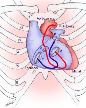
Tricuspid Valve
Tricuspid Valve Insufficiency
Tricuspid Valve Stenosis
Aortic Valve
Heart Valve Diseases
Ebstein Anomaly
Tricuspid Atresia
Tricuspid Valve Prolapse
Cardiac Valve Annuloplasty
Aortic Valve Stenosis
Heart Valve Prosthesis
Heart Valve Prosthesis Implantation
Endocarditis, Bacterial
Bioprosthesis
Echocardiography
Mitral Valve Insufficiency
Aortic Valve Insufficiency
Heart Septal Defects, Ventricular
Endocarditis
Chordae Tendineae
Echocardiography, Doppler, Color
Echocardiography, Transesophageal
Mitral Valve Stenosis
Mitral Valve Prolapse
Fetal Heart
Echocardiography, Doppler
Transposition of Great Vessels
Ventricular Function, Right
Echocardiography, Three-Dimensional
Ventricular Dysfunction, Right
Heart Valves
Carcinoid Heart Disease
Suture Techniques
Rheumatic Heart Disease
Cardiac Catheterization
Pacemaker, Artificial
Heart Defects, Congenital
Treatment Outcome
Pulmonary Valve Stenosis
Heart Neoplasms
Pulmonary Atresia
Venous Valves
Heart Septum
Fetal Diseases
Systolic Murmurs
Heart Ventricles
Pulmonary Artery
Postoperative Complications
Reoperation
Follow-Up Studies
Myxoma
Ultrasonography, Prenatal
Vena Cava, Superior
Heart Septal Defects, Atrial
Retrospective Studies
Wounds, Nonpenetrating
Heart Septal Defects
Mitral Valve Annuloplasty
Catheterization
Heart Sounds
Ventricular Septum
Vena Cava, Inferior
Phonocardiography
Papillary Muscles
Pulmonary Valve Insufficiency
Severity of Illness Index
Hypertension, Pulmonary
Tetralogy of Fallot
Holography
Prosthesis Failure
Blood Flow Velocity
Hemoptysis
Atrial Flutter
Blalock-Taussig Procedure
Hemodynamics
Equipment Failure
Effect of right atrial isthmus ablation on the occurrence of atrial fibrillation: observations in four patient groups having type I atrial flutter with or without associated atrial fibrillation. (1/579)
BACKGROUND: The goal of this study was to test the hypothesis that the occurrence of atrial fibrillation (AF), in at least some patients with coexisting type I atrial flutter (AFL), is based on macro-reentry around the tricuspid valve orifice, including the right atrial (RA) isthmus, by evaluation of AF recurrences after successful ablation of AFL. METHODS AND RESULTS: Eighty-two consecutive patients with type I AFL, with or without concomitant AF, underwent radiofrequency ablation (RFA) of the RA isthmus by an anatomical approach. The results were analyzed in 4 groups of patients: group 1 (only AFL; 29 patients), group 2 (AFL >AF; 22 patients), group 3 (AF >AFL; 15 patients), and group 4 (developing AFL while receiving class IC antiarrhythmic drug therapy for AF, the "class IC atrial flutter"; 16 patients). In all groups, RFA of type I AFL was performed with a high (>/=93%) procedural success rate. In group 1, only 2 patients (8%) had AF after (18+/-14 months) AFL ablation. These figures were 38% (20+/-14 months) and 86% (13+/-8 months) in groups 2 and 3, respectively. Group 4 patients (4+/-2 months) had a 73% freedom of AF recurrences with continuation of the class IC agent. CONCLUSIONS: The low incidence of new AF during long-term follow-up after RFA of type I AFL makes it unlikely that radiofrequency lesions promote the development of AF. The impact of isthmus ablation on AF recurrences differs according to the clinically predominant atrial arrhythmia and suggests a possible role of the RA isthmus in the occurrence of AF in some patients. Ablation of class IC atrial flutter in patients with therapy-resistant AF is a novel approach to management of this patient subset. Careful classification of AF patients plays a role in the selection of the site of ablation therapy. (+info)Malfunction of Bjork-Shiley valve prosthesis in tricuspid position. (2/579)
Eight months after triple valve replacement with Bjork-Shiley tilting disc valves a patient developed symptoms and signs suggesting malfunction of the prosthesis in the tricuspid position. This was confirmed by echocardiography and angiocardiography, and at operation thedisc of the prosthesis was found to be stuck half-open by fibrin and clot. A further 11 patients with the same tupe of prosthesis in the triscupid position were then studied by phonocardiography and echocardiography. In one of these the prosthesis was found to be stuck and this was confirmed by angiocardiography and surgery. These 2 cases are reported in detail and the findings in the other 10 are discussed. The implications of this high incidence of malfunction of the Bjork-Shiley prosthesis in the tricuspid position are considered. Echocardiography appears to be essential in the follow-up of such patients. (+info)Prenatal diagnosis of right ventricular outflow tract obstruction with intact ventricular septum, and detection of ventriculocoronary connections. (3/579)
OBJECTIVES: To determine the accuracy of prenatal diagnosis of pulmonary atresia and intact ventricular septum (PAIVS), and pulmonary stenosis, including prenatal detection of ventriculocoronary connections, to evaluate heart size during the prenatal period, and to evaluate the outcome. DESIGN AND PATIENTS: Medical records of 20 cases with prenatally diagnosed PAIVS and pulmonary stenosis were reviewed retrospectively. Prenatal and postnatal echocardiography were also reviewed and dimensions of the ventricles and vessels were measured retrespectively. RESULTS: Of 20 prenatal diagnoses (15 PAIVS and five pulmonary stenosis), 16 were confirmed as correct. One critical pulmonary stenosis case had been diagnosed as PAIVS prenatally; three had no confirmation. Eight pregnancies were terminated, three had no active treatment, and nine were treated; all survived. Of 13 assessed with ventriculocoronary connections prenatally, seven were diagnosed correctly (four with, three without ventriculocoronary connections), but one was falsely positive; five had no confirmation. The more prominent hypoplasia of the main pulmonary artery and the tricuspid valve annulus, and the sigmoid shape of the ductus arteriosus, seemed to be associated with the presence of ventriculocoronary connections. CONCLUSIONS: Current prenatal echocardiography can accurately diagnose right ventricular outflow tract obstruction and ventriculocoronary connections. Prenatal detection of this constellation of abnormalities aids in family counselling and decisions on postnatal management. (+info)Rate-dependent conduction block of the crista terminalis in patients with typical atrial flutter: influence on evaluation of cavotricuspid isthmus conduction block. (4/579)
BACKGROUND: The crista terminalis (CT) has been identified as the posterior boundary of typical atrial flutter (AFL) in the lateral wall (LW) of the right atrium (RA). To study conduction properties across the CT, rapid pacing was performed at both sides of the CT after bidirectional conduction block was achieved in the cavotricuspid isthmus by radiofrequency catheter ablation. METHODS AND RESULTS: In 22 patients (aged 61+/-7 years) with AFL (cycle length, 234+/-23 ms), CT was identified during AFL by double electrograms recorded between the LW and posterior wall (PW). After the ablation procedure, decremental pacing trains were delivered from 600 ms to 2-to-1 local capture at the LW and PW or coronary sinus ostium (CSO). At least 5 bipolar electrograms were recorded along the CT from the high to the low atrium next to the inferior vena cava. No double electrograms were recorded during sinus rhythm in that area. Complete transversal conduction block all along the CT (detected by the appearance of double electrograms at all recording sites and craniocaudal activation sequence on the side opposite to the pacing site) was observed in all patients during pacing from the PW or CSO (cycle length, 334+/-136 ms), but it was fixed in only 4 patients. During pacing from the LW, complete block appeared at a shorter pacing cycle length (281+/-125 ms; P<0.01) and was fixed in 2 patients. In 3 patients, complete block was not achieved. CONCLUSIONS: These data suggest the presence of rate-dependent transversal conduction block at the crista terminalis in patients with typical AFL. Block is usually observed at longer pacing cycle lengths with PW pacing than with LW pacing. This difference may be a critical determinant of the counterclockwise rotation of typical AFL. (+info)Partial cavotricuspid isthmus block before ablation in patients with typical atrial flutter. (5/579)
OBJECTIVES: The purpose of this study was to prospectively evaluate preexisting partial isthmus block in the context of an electrophysiologically directed linear ablation strategy for typical atrial flutter (AF). BACKGROUND: Double potentials (DPs) separated by an isoelectric interval have been recognized as markers of local block. However, the presence and significance of DPs in the cavotricuspid isthmus during AF before ablation have not been evaluated. METHODS: Thirty consecutive patients with AF (counterclockwise: 24, clockwise: 6) were studied during AF. Sequential withdrawal mapping was performed in the cavotricuspid isthmus from the tricuspid valve (TV) to the inferior vena cava (IVC) edge with electrograms coinciding with the center of the surface electrocardiographic plateau during counterclockwise AF or with the initial downslope of the positive flutter wave during clockwise AF. Atrial electrograms along this line were categorized as double, single or fractionated potentials (SPs or FPs). After demarcation of the zone of contiguous DPs, radiofrequency (RF) catheter ablation was performed during AF only at sites with SPs or FPs (other than DPs) on the mapped line. If isthmus conduction still persisted after AF termination, additional RF applications were delivered using the same electrophysiologic strategy of avoiding DPs with an isoelectric interval during low lateral right atrial pacing for filling in the gap of residual conduction. RESULTS: Before ablation, no DPs were recorded in the isthmus in 19 patients (63%); DPs were recorded only at the IVC edge in five patients, and only at the TV edge in one patient. A contiguous line of DPs extending through more than half the isthmus to the IVC edge was documented in five patients (17%: group DP). In group DP, AF was terminated with 1.4+/-0.5 applications (vs. 5.8+/-3.5 in the remaining patients: p < 0.01). Complete isthmus block was achieved with a total of 3.4+/-0.5 applications (vs. 12+/-6 in the remaining patients: p < 0.01). CONCLUSIONS: Seventeen percent of patients undergoing ablation of AF have preexisting partial isthmus block indicated by a large contiguous zone of DPs separated by an isoelectric interval. Electrophysiologically directed linear ablation avoiding confluent DPs can prevent unnecessary applications for effective cure of AF. (+info)Living anatomy of the atrioventricular junctions. A guide to electrophysiologic mapping. A Consensus Statement from the Cardiac Nomenclature Study Group, Working Group of Arrhythmias, European Society of Cardiology, and the Task Force on Cardiac Nomenclature from NASPE. (6/579)
Current nomenclature for the atrioventricular (AV) junctions derives from a surgically distorted view, placing the valvar rings and the triangle of Koch in a single plane with antero-posterior and right-left lateral coordinates. Within this convention, the aorta is considered to occupy an anterior position, although the mouth of the coronary sinus is shown as being posterior. Although this nomenclature has served its purpose for the description and treatment of arrhythmias dependent on accessory pathways and atrioventricular nodal reentry, it is less than satisfactory for the description of atrial and ventricular mapping. To correct these deficiencies, a consensus document has been prepared by experts from the Working Group of Arrhythmias of the European Society of Cardiology and the North American Society of Pacing and Electrophysiology. It proposes a new anatomically sound nomenclature that will be applicable to all chambers of the heart. In this report, we discuss its value for description of the AV junctions, establishing the principles of this new nomenclature. (+info)Persistent pulmonary hypertension of the newborn associated with pulmonary atresia and intact interventricular septum. (7/579)
Neonates with pulmonary atresia and intact interventricular septum (PAIVS) do not have pulmonary vascular disease secondary to their heart abnormality. Persistent pulmonary hypertension of the newborn has not been described in association with this condition. The case is reported of a female neonate born with PAIVS, who preoperatively had no clinical evidence or any risk factors for persistent pulmonary hypertension of the newborn, but whose postoperative course was highly suggestive of persistent pulmonary hypertension; necropsy confirmed the features of pulmonary vascular disease. (+info)Transesophageal echocardiographic imaging workshop: a basic transverse plane examination sequence. (8/579)
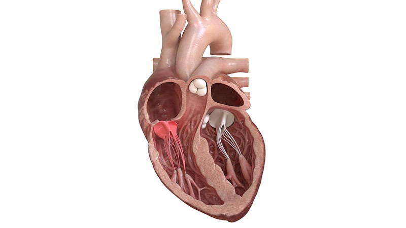
This workshop describes a 10-step sequence of transverse plane two-dimensional transesophageal echocardiographic views of the heart and great vessels that constitutes a basic standardized examination capable of being performed by a beginning practitioner. (+info)The tricuspid valve is the heart valve that separates the right atrium and the right ventricle in the human heart. It is called "tricuspid" because it has three leaflets or cusps, which are also referred to as flaps or segments. These cusps are named anterior, posterior, and septal. The tricuspid valve's function is to prevent the backflow of blood from the ventricle into the atrium during systole, ensuring unidirectional flow of blood through the heart.
Tricuspid valve insufficiency, also known as tricuspid regurgitation, is a cardiac condition in which the tricuspid valve located between the right atrium and right ventricle of the heart does not close properly, allowing blood to flow back into the right atrium during contraction of the right ventricle. This results in a portion of the blood being pumped inefficiently, which can lead to volume overload of the right side of the heart and potentially result in symptoms such as fatigue, weakness, shortness of breath, and fluid retention. The condition can be congenital or acquired, with common causes including dilated cardiomyopathy, infective endocarditis, rheumatic heart disease, and trauma.
Tricuspid valve stenosis is a cardiac condition characterized by the narrowing or stiffening of the tricuspid valve, which is located between the right atrium and right ventricle in the heart. This narrowing or stiffening restricts the normal flow of blood from the right atrium into the right ventricle, causing increased pressure in the right atrium and reduced blood flow to the lungs.
The tricuspid valve typically has three leaflets or cusps that open and close to regulate the flow of blood between the right atrium and right ventricle. In tricuspid valve stenosis, these leaflets become thickened, calcified, or fused together, leading to a reduced opening size and impaired function.
The most common causes of tricuspid valve stenosis include rheumatic heart disease, congenital heart defects, carcinoid syndrome, and infective endocarditis. Symptoms may include fatigue, shortness of breath, swelling in the legs and abdomen, and irregular heartbeats. Treatment options depend on the severity of the condition and underlying causes but may involve medications, surgical repair or replacement of the valve, or catheter-based procedures.
The aortic valve is the valve located between the left ventricle (the lower left chamber of the heart) and the aorta (the largest artery in the body, which carries oxygenated blood from the heart to the rest of the body). It is made up of three thin flaps or leaflets that open and close to regulate blood flow. During a heartbeat, the aortic valve opens to allow blood to be pumped out of the left ventricle into the aorta, and then closes to prevent blood from flowing back into the ventricle when it relaxes. Any abnormality or damage to this valve can lead to various cardiovascular conditions such as aortic stenosis, aortic regurgitation, or infective endocarditis.
The mitral valve, also known as the bicuspid valve, is a two-leaflet valve located between the left atrium and left ventricle in the heart. Its function is to ensure unidirectional flow of blood from the left atrium into the left ventricle during the cardiac cycle. The mitral valve consists of two leaflets (anterior and posterior), the chordae tendineae, papillary muscles, and the left atrial and ventricular myocardium. Dysfunction of the mitral valve can lead to various heart conditions such as mitral regurgitation or mitral stenosis.
Heart valve diseases are a group of conditions that affect the function of one or more of the heart's four valves (tricuspid, pulmonic, mitral, and aortic). These valves are responsible for controlling the direction and flow of blood through the heart. Heart valve diseases can cause the valves to become narrowed (stenosis), leaky (regurgitation or insufficiency), or improperly closed (prolapse), leading to disrupted blood flow within the heart and potentially causing symptoms such as shortness of breath, fatigue, chest pain, and irregular heart rhythms. The causes of heart valve diseases can include congenital defects, age-related degenerative changes, infections, rheumatic heart disease, and high blood pressure. Treatment options may include medications, surgical repair or replacement of the affected valve(s), or transcatheter procedures.
Ebstein anomaly is a congenital heart defect that affects the tricuspid valve, which is the valve between the right atrium and right ventricle of the heart. In Ebstein anomaly, the tricuspid valve is abnormally formed and positioned, causing it to leak blood back into the right atrium. This can lead to various symptoms such as shortness of breath, fatigue, and cyanosis (bluish discoloration of the skin). Treatment for Ebstein anomaly may include medication, surgery, or a combination of both. It is important to note that the severity of the condition can vary widely among individuals, and some people with Ebstein anomaly may require more intensive treatment than others.
Tricuspid atresia is a congenital heart defect where the tricuspid valve, which regulates blood flow between the right atrium and right ventricle, fails to develop properly. As a result, there is no direct pathway for blood to move from the right atrium to the right ventricle and then to the lungs for oxygenation.
In this condition, blood from the body returning to the heart enters the right atrium but cannot flow through the tricuspid valve into the right ventricle. Instead, it flows through an opening in the interatrial septum (atrial septal defect) into the left atrium and then into the left ventricle. The left ventricle pumps this blood to the body and a portion of it goes to the lungs via a patent ductus arteriosus or other collateral vessels.
Tricuspid atresia is often associated with other heart defects, such as transposition of the great arteries, pulmonary stenosis, or total anomalous pulmonary venous return. Symptoms can vary depending on the severity and associated defects but may include cyanosis (bluish discoloration of the skin), shortness of breath, fatigue, and poor growth. Treatment typically involves surgical interventions to create a path for blood to flow to the lungs and establish proper oxygenation.
Tricuspid valve prolapse is a cardiac condition where the tricuspid valve, located between the right atrium and right ventricle of the heart, doesn't close properly due to one or more of its leaflets (flaps) bulging or billowing into the right atrium during contraction of the right ventricle. This allows the backflow of blood from the right ventricle into the right atrium, known as tricuspid regurgitation. In some cases, tricuspid valve prolapse may not cause any symptoms and can be an incidental finding on echocardiography. However, if severe tricuspid regurgitation occurs, it can lead to right-sided heart failure, atrial arrhythmias, and other complications. The condition is often associated with mitral valve prolapse or other connective tissue disorders.
Cardiac valve annuloplasty is a surgical procedure that involves repairing and reinforcing the ring-like structure (annulus) surrounding the heart valves, primarily the mitral or tricuspid valves. This procedure is often performed to correct valve leaks or regurgitation caused by various conditions such as valve disease or dilated cardiomyopathy.
During the annuloplasty procedure, the surgeon typically uses an artificial ring-like device (annuloplasty ring) made of fabric, metal, or a combination of both to reshape and stabilize the damaged annulus. The ring is sewn in place, reducing the size of the valve opening and helping the valve leaflets to coapt properly, thereby preventing valve leaks and improving heart function.
Annuloplasty can be performed as a standalone procedure or in combination with other cardiac surgeries such as valve replacement or repair. The specific technique and approach may vary depending on the individual patient's needs and the surgeon's preference.
The pulmonary valve, also known as the pulmonic valve, is a semilunar valve located at the exit of the right ventricle of the heart and the beginning of the pulmonary artery. It has three cusps or leaflets that prevent the backflow of blood from the pulmonary artery into the right ventricle during ventricular diastole, ensuring unidirectional flow of blood towards the lungs for oxygenation.
Aortic valve stenosis is a cardiac condition characterized by the narrowing or stiffening of the aortic valve, which separates the left ventricle (the heart's main pumping chamber) from the aorta (the large artery that carries oxygen-rich blood to the rest of the body). This narrowing or stiffening prevents the aortic valve from opening fully, resulting in reduced blood flow from the left ventricle to the aorta and the rest of the body.
The narrowing can be caused by several factors, including congenital heart defects, calcification (hardening) of the aortic valve due to aging, or scarring of the valve due to rheumatic fever or other inflammatory conditions. As a result, the left ventricle must work harder to pump blood through the narrowed valve, which can lead to thickening and enlargement of the left ventricular muscle (left ventricular hypertrophy).
Symptoms of aortic valve stenosis may include chest pain or tightness, shortness of breath, fatigue, dizziness or fainting, and heart palpitations. Severe aortic valve stenosis can lead to serious complications such as heart failure, arrhythmias, or even sudden cardiac death. Treatment options may include medications to manage symptoms, lifestyle changes, or surgical intervention such as aortic valve replacement.
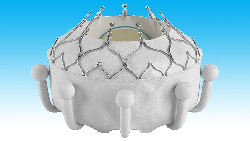
A heart valve prosthesis is a medical device that is implanted in the heart to replace a damaged or malfunctioning heart valve. The prosthetic valve can be made of biological tissue (such as from a pig or cow) or artificial materials (such as carbon or polyester). Its function is to allow for the proper directional flow of blood through the heart, opening and closing with each heartbeat to prevent backflow of blood.
There are several types of heart valve prostheses, including:
1. Mechanical valves: These are made entirely of artificial materials and have a longer lifespan than biological valves. However, they require the patient to take blood-thinning medication for the rest of their life to prevent blood clots from forming on the valve.
2. Bioprosthetic valves: These are made of biological tissue and typically last 10-15 years before needing replacement. They do not require the patient to take blood-thinning medication, but there is a higher risk of reoperation due to degeneration of the tissue over time.
3. Homografts or allografts: These are human heart valves that have been donated and preserved for transplantation. They have similar longevity to bioprosthetic valves and do not require blood-thinning medication.
4. Autografts: In this case, the patient's own pulmonary valve is removed and used to replace the damaged aortic valve. This procedure is called the Ross procedure and has excellent long-term results, but it requires advanced surgical skills and is not widely available.
The choice of heart valve prosthesis depends on various factors, including the patient's age, overall health, lifestyle, and personal preferences.
Heart valve prosthesis implantation is a surgical procedure where an artificial heart valve is inserted to replace a damaged or malfunctioning native heart valve. This can be necessary for patients with valvular heart disease, including stenosis (narrowing) or regurgitation (leaking), who do not respond to medical management and are at risk of heart failure or other complications.
There are two main types of artificial heart valves used in prosthesis implantation: mechanical valves and biological valves. Mechanical valves are made of synthetic materials, such as carbon and metal, and can last a long time but require lifelong anticoagulation therapy to prevent blood clots from forming. Biological valves, on the other hand, are made from animal or human tissue and typically do not require anticoagulation therapy but may have a limited lifespan and may need to be replaced in the future.
The decision to undergo heart valve prosthesis implantation is based on several factors, including the patient's age, overall health, type and severity of valvular disease, and personal preferences. The procedure can be performed through traditional open-heart surgery or minimally invasive techniques, such as robotic-assisted surgery or transcatheter aortic valve replacement (TAVR). Recovery time varies depending on the approach used and individual patient factors.
Bacterial endocarditis is a medical condition characterized by the inflammation and infection of the inner layer of the heart, known as the endocardium. This infection typically occurs when bacteria enter the bloodstream and attach themselves to damaged or abnormal heart valves or other parts of the endocardium. The bacteria can then multiply and cause the formation of vegetations, which are clusters of infected tissue that can further damage the heart valves and lead to serious complications such as heart failure, stroke, or even death if left untreated.
Bacterial endocarditis is a relatively uncommon but potentially life-threatening condition that requires prompt medical attention. Risk factors for developing bacterial endocarditis include pre-existing heart conditions such as congenital heart defects, artificial heart valves, previous history of endocarditis, or other conditions that damage the heart valves. Intravenous drug use is also a significant risk factor for this condition.
Symptoms of bacterial endocarditis may include fever, chills, fatigue, muscle and joint pain, shortness of breath, chest pain, and a new or changing heart murmur. Diagnosis typically involves a combination of medical history, physical examination, blood cultures, and imaging tests such as echocardiography. Treatment usually involves several weeks of intravenous antibiotics to eradicate the infection, and in some cases, surgical intervention may be necessary to repair or replace damaged heart valves.
A bioprosthesis is a type of medical implant that is made from biological materials, such as heart valves or tendons taken from animals (xenografts) or humans (allografts). These materials are processed and sterilized to be used in surgical procedures to replace damaged or diseased tissues in the body.
Bioprosthetic implants are often used in cardiac surgery, such as heart valve replacement, because they are less likely to cause an immune response than synthetic materials. However, they may have a limited lifespan due to calcification and degeneration of the biological tissue over time. Therefore, bioprosthetic implants may need to be replaced after several years.
Bioprostheses can also be used in other types of surgical procedures, such as ligament or tendon repair, where natural tissue is needed to restore function and mobility. These prostheses are designed to mimic the properties of native tissues and provide a more physiological solution than synthetic materials.
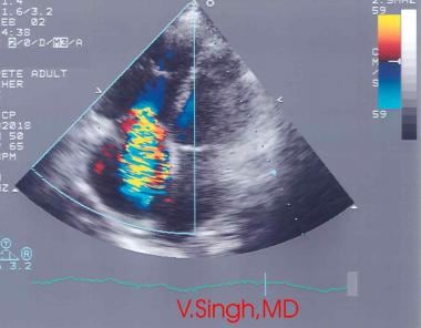
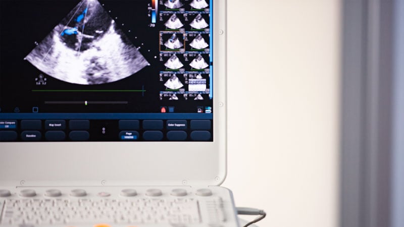
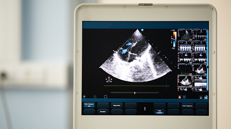
Echocardiography is a medical procedure that uses sound waves to produce detailed images of the heart's structure, function, and motion. It is a non-invasive test that can help diagnose various heart conditions, such as valve problems, heart muscle damage, blood clots, and congenital heart defects.
During an echocardiogram, a transducer (a device that sends and receives sound waves) is placed on the chest or passed through the esophagus to obtain images of the heart. The sound waves produced by the transducer bounce off the heart structures and return to the transducer, which then converts them into electrical signals that are processed to create images of the heart.
There are several types of echocardiograms, including:
* Transthoracic echocardiography (TTE): This is the most common type of echocardiogram and involves placing the transducer on the chest.
* Transesophageal echocardiography (TEE): This type of echocardiogram involves passing a specialized transducer through the esophagus to obtain images of the heart from a closer proximity.
* Stress echocardiography: This type of echocardiogram is performed during exercise or medication-induced stress to assess how the heart functions under stress.
* Doppler echocardiography: This type of echocardiogram uses sound waves to measure blood flow and velocity in the heart and blood vessels.
Echocardiography is a valuable tool for diagnosing and managing various heart conditions, as it provides detailed information about the structure and function of the heart. It is generally safe, non-invasive, and painless, making it a popular choice for doctors and patients alike.
Mitral valve insufficiency, also known as mitral regurgitation, is a cardiac condition in which the mitral valve located between the left atrium and left ventricle of the heart does not close properly, causing blood to flow backward into the atrium during contraction of the ventricle. This leads to an increased volume load on the left heart chamber and can result in symptoms such as shortness of breath, fatigue, and fluid retention. The condition can be caused by various factors including valve damage due to degenerative changes, infective endocarditis, rheumatic heart disease, or trauma. Treatment options include medication, mitral valve repair, or replacement surgery depending on the severity and underlying cause of the insufficiency.
Heart injuries, also known as cardiac injuries, refer to any damage or harm caused to the heart muscle, valves, or surrounding structures. This can result from various causes such as blunt trauma (e.g., car accidents, falls), penetrating trauma (e.g., gunshot wounds, stabbing), or medical conditions like heart attacks (myocardial infarction) and infections (e.g., myocarditis, endocarditis).
Some common types of heart injuries include:
1. Contusions: Bruising of the heart muscle due to blunt trauma.
2. Myocardial infarctions: Damage to the heart muscle caused by insufficient blood supply, often due to blocked coronary arteries.
3. Cardiac rupture: A rare but life-threatening condition where the heart muscle tears or breaks open, usually resulting from severe trauma or complications from a myocardial infarction.
4. Valvular damage: Disruption of the heart valves' function due to injury or infection, leading to leakage (regurgitation) or narrowing (stenosis).
5. Pericardial injuries: Damage to the pericardium, the sac surrounding the heart, which can result in fluid accumulation (pericardial effusion), inflammation (pericarditis), or tamponade (compression of the heart by excess fluid).
6. Arrhythmias: Irregular heart rhythms caused by damage to the heart's electrical conduction system.
Timely diagnosis and appropriate treatment are crucial for managing heart injuries, as they can lead to severe complications or even be fatal if left untreated.
Aortic valve insufficiency, also known as aortic regurgitation or aortic incompetence, is a cardiac condition in which the aortic valve does not close properly during the contraction phase of the heart cycle. This allows blood to flow back into the left ventricle from the aorta, instead of being pumped out to the rest of the body. As a result, the left ventricle must work harder to maintain adequate cardiac output, which can lead to left ventricular enlargement and heart failure over time if left untreated.
The aortic valve is a trileaflet valve that lies between the left ventricle and the aorta. During systole (the contraction phase of the heart cycle), the aortic valve opens to allow blood to be pumped out of the left ventricle into the aorta and then distributed to the rest of the body. During diastole (the relaxation phase of the heart cycle), the aortic valve closes to prevent blood from flowing back into the left ventricle.
Aortic valve insufficiency can be caused by various conditions, including congenital heart defects, infective endocarditis, rheumatic heart disease, Marfan syndrome, and trauma. Symptoms of aortic valve insufficiency may include shortness of breath, fatigue, chest pain, palpitations, and edema (swelling). Diagnosis is typically made through physical examination, echocardiography, and other imaging studies. Treatment options depend on the severity of the condition and may include medication, surgery to repair or replace the aortic valve, or a combination of both.
A ventricular septal defect (VSD) is a type of congenital heart defect that involves a hole in the wall separating the two lower chambers of the heart, the ventricles. This defect allows oxygenated blood from the left ventricle to mix with deoxygenated blood in the right ventricle, leading to inefficient oxygenation of the body's tissues. The size and location of the hole can vary, and symptoms may range from none to severe, depending on the size of the defect and the amount of blood that is able to shunt between the ventricles. Small VSDs may close on their own over time, while larger defects usually require medical intervention, such as medication or surgery, to prevent complications like pulmonary hypertension and heart failure.
Endocarditis is an inflammation of the inner layer of the heart chambers and heart valves, called the endocardium. This inflammation typically results from a bacterial or, less commonly, fungal infection that travels through the bloodstream and attaches to damaged areas of the heart.
There are two main types of endocarditis:
1. Acute Endocarditis: Develops quickly and can be severe, causing fever, chills, shortness of breath, fatigue, and heart murmurs. It may lead to serious complications like heart failure, embolism (blood clots that travel to other parts of the body), and damage to heart valves.
2. Subacute Endocarditis: Develops more slowly, often causing milder symptoms that can be mistaken for a cold or flu. Symptoms may include fatigue, weakness, fever, night sweats, weight loss, joint pain, and heart murmurs. Subacute endocarditis is more likely to affect people with previously damaged heart valves or congenital heart conditions.
Treatment usually involves several weeks of intravenous antibiotics or antifungal medications, depending on the cause of the infection. In some cases, surgery may be required to repair or replace damaged heart valves. Preventive measures include good oral hygiene and prompt treatment of infections, especially in individuals at a higher risk for endocarditis, such as those with congenital heart defects, artificial heart valves, or previous history of endocarditis.
The chordae tendineae are cord-like tendons that attach the heart's papillary muscles to the tricuspid and mitral valves in the heart. They play a crucial role in preventing the backflow of blood into the atria during ventricular contraction. The chordae tendineae ensure that the cusps of the atrioventricular valves close properly and maintain their shape during the cardiac cycle. Damage to these tendons can result in heart conditions such as mitral or tricuspid valve regurgitation.
Cardiac surgical procedures are operations that are performed on the heart or great vessels (the aorta and vena cava) by cardiothoracic surgeons. These surgeries are often complex and require a high level of skill and expertise. Some common reasons for cardiac surgical procedures include:
1. Coronary artery bypass grafting (CABG): This is a surgery to improve blood flow to the heart in patients with coronary artery disease. During the procedure, a healthy blood vessel from another part of the body is used to create a detour around the blocked or narrowed portion of the coronary artery.
2. Valve repair or replacement: The heart has four valves that control blood flow through and out of the heart. If one or more of these valves become damaged or diseased, they may need to be repaired or replaced. This can be done using artificial valves or valves from animal or human donors.
3. Aneurysm repair: An aneurysm is a weakened area in the wall of an artery that can bulge out and potentially rupture. If an aneurysm occurs in the aorta, it may require surgical repair to prevent rupture.
4. Heart transplantation: In some cases, heart failure may be so severe that a heart transplant is necessary. This involves removing the diseased heart and replacing it with a healthy donor heart.
5. Arrhythmia surgery: Certain types of abnormal heart rhythms (arrhythmias) may require surgical treatment. One such procedure is called the Maze procedure, which involves creating a pattern of scar tissue in the heart to disrupt the abnormal electrical signals that cause the arrhythmia.
6. Congenital heart defect repair: Some people are born with structural problems in their hearts that require surgical correction. These may include holes between the chambers of the heart or abnormal blood vessels.
Cardiac surgical procedures carry risks, including bleeding, infection, stroke, and death. However, for many patients, these surgeries can significantly improve their quality of life and longevity.
Echocardiography, Doppler, color is a type of ultrasound test that uses sound waves to create detailed moving images of the heart and its blood vessels. In this technique, color Doppler is used to visualize the direction and speed of blood flow through the heart and great vessels. The movement of the red blood cells causes a change in frequency of the reflected sound waves (Doppler shift), which can be used to calculate the velocity and direction of the blood flow. By adding color to the Doppler image, it becomes easier for the interpreting physician to understand the complex three-dimensional motion of blood through the heart. This test is often used to diagnose and monitor various heart conditions, including valve disorders, congenital heart defects, and cardiac muscle diseases.
Transesophageal echocardiography (TEE) is a type of echocardiogram, which is a medical test that uses sound waves to create detailed images of the heart. In TEE, a special probe containing a transducer is passed down the esophagus (the tube that connects the mouth to the stomach) to obtain views of the heart from behind. This allows for more detailed images of the heart structures and function compared to a standard echocardiogram, which uses a probe placed on the chest. TEE is often used in patients with poor image quality from a standard echocardiogram or when more detailed images are needed to diagnose or monitor certain heart conditions. It is typically performed by a trained cardiologist or sonographer under the direction of a cardiologist.
Mitral valve stenosis is a cardiac condition characterized by the narrowing or stiffening of the mitral valve, one of the four heart valves that regulate blood flow through the heart. This narrowing prevents the mitral valve from fully opening during diastole (relaxation phase of the heart cycle), leading to restricted flow of oxygenated blood from the left atrium into the left ventricle.
The narrowing or stiffening of the mitral valve can be caused by various factors, such as rheumatic heart disease, congenital heart defects, aging, or calcium deposits on the valve leaflets. As a result, the left atrium has to work harder to pump blood into the left ventricle, causing increased pressure in the left atrium and pulmonary veins. This can lead to symptoms such as shortness of breath, fatigue, coughing, and heart palpitations.
Mitral valve stenosis is typically diagnosed through a combination of medical history, physical examination, and imaging techniques like echocardiography or cardiac catheterization. Treatment options may include medications to manage symptoms and prevent complications, as well as surgical interventions such as mitral valve repair or replacement to alleviate the stenosis and improve heart function.
Mitral valve prolapse (MVP) is a heart condition where the mitral valve, which separates the left atrium and left ventricle in the heart, doesn't function properly. In MVP, one or both of the mitral valve flaps (known as leaflets) bulge or billow into the left atrium during the contraction of the left ventricle. This prolapse can cause a leakage of blood back into the atrium, known as mitral regurgitation. In many cases, MVP is asymptomatic and doesn't require treatment, but in some instances, it may lead to complications such as infective endocarditis or arrhythmias. The exact causes of MVP are not fully understood, but it can be associated with certain genetic factors, connective tissue disorders, and mitral valve abnormalities present at birth.
The fetal heart is the cardiovascular organ that develops in the growing fetus during pregnancy. It starts to form around 22 days after conception and continues to develop throughout the first trimester. By the end of the eighth week of gestation, the fetal heart has developed enough to pump blood throughout the body.
The fetal heart is similar in structure to the adult heart but has some differences. It is smaller and more compact, with a four-chambered structure that includes two atria and two ventricles. The fetal heart also has unique features such as the foramen ovale, which is a hole between the right and left atria that allows blood to bypass the lungs, and the ductus arteriosus, a blood vessel that connects the pulmonary artery to the aorta and diverts blood away from the lungs.
The fetal heart is responsible for pumping oxygenated blood from the placenta to the rest of the body and returning deoxygenated blood back to the placenta for re-oxygenation. The rate of the fetal heartbeat is faster than that of an adult, typically ranging from 120 to 160 beats per minute. Fetal heart rate monitoring is a common method used during pregnancy and childbirth to assess the health and well-being of the developing fetus.
Doppler echocardiography is a type of ultrasound test that uses high-frequency sound waves to produce detailed images of the heart and its blood vessels. It measures the direction and speed of blood flow in the heart and major blood vessels leading to and from the heart. This helps to evaluate various conditions such as valve problems, congenital heart defects, and heart muscle diseases.
In Doppler echocardiography, a small handheld device called a transducer is placed on the chest, which emits sound waves that bounce off the heart and blood vessels. The transducer then picks up the returning echoes, which are processed by a computer to create moving images of the heart.
The Doppler effect is used to measure the speed and direction of blood flow. This occurs when the frequency of the sound waves changes as they bounce off moving objects, such as red blood cells. By analyzing these changes, the ultrasound machine can calculate the velocity and direction of blood flow in different parts of the heart.
Doppler echocardiography is a non-invasive test that does not require any needles or dyes. It is generally safe and painless, although patients may experience some discomfort from the pressure applied by the transducer on the chest. The test usually takes about 30 to 60 minutes to complete.
Transposition of the Great Vessels is a congenital heart defect in which the two main vessels that carry blood from the heart to the rest of the body are switched in position. Normally, the aorta arises from the left ventricle and carries oxygenated blood to the body, while the pulmonary artery arises from the right ventricle and carries deoxygenated blood to the lungs. In transposition of the great vessels, the aorta arises from the right ventricle and the pulmonary artery arises from the left ventricle. This results in oxygen-poor blood being pumped to the body and oxygen-rich blood being recirculated back to the lungs, which can lead to serious health problems and is often fatal if not corrected through surgery soon after birth.
Right Ventricular Function refers to the ability of the right ventricle (RV) of the heart to receive and eject blood during the cardiac cycle. The right ventricle is one of the four chambers of the heart and is responsible for pumping deoxygenated blood from the body to the lungs for re-oxygenation.
Right ventricular function can be assessed by measuring various parameters such as:
1. Right Ventricular Ejection Fraction (RVEF): It is the percentage of blood that is ejected from the right ventricle during each heartbeat. A normal RVEF ranges from 45-75%.
2. Right Ventricular Systolic Function: It refers to the ability of the right ventricle to contract and eject blood during systole (contraction phase). This can be assessed by measuring the tricuspid annular plane systolic excursion (TAPSE) or tissue Doppler imaging.
3. Right Ventricular Diastolic Function: It refers to the ability of the right ventricle to relax and fill with blood during diastole (relaxation phase). This can be assessed by measuring the right ventricular inflow pattern, tricuspid valve E/A ratio, or deceleration time.
4. Right Ventricular Afterload: It refers to the pressure that the right ventricle must overcome to eject blood into the pulmonary artery. Increased afterload can impair right ventricular function.
Abnormalities in right ventricular function can lead to various cardiovascular conditions such as pulmonary hypertension, heart failure, and arrhythmias.
Three-dimensional echocardiography (3DE) is a type of cardiac ultrasound that uses advanced technologies to create a real-time, detailed 3D image of the heart. This imaging technique provides a more comprehensive view of the heart's structure and function compared to traditional 2D echocardiography. By visualizing the heart from multiple angles, 3DE can help physicians better assess complex cardiac conditions, plan treatments, and monitor their effectiveness.
In a 3DE examination, a transducer (a handheld device that emits and receives sound waves) is placed on the chest to capture ultrasound data. This data is then processed by specialized software to create a 3D model of the heart. The procedure is non-invasive and typically takes less than an hour to complete.
Three-dimensional echocardiography has several clinical applications, including:
1. Evaluation of cardiac morphology and function in congenital heart disease
2. Assessment of valvular structure and function, such as mitral or aortic valve regurgitation or stenosis
3. Guidance during interventional procedures like transcatheter aortic valve replacement (TAVR)
4. Quantification of left ventricular volumes, ejection fraction, and mass
5. Assessment of right ventricular size and function
6. Detection and monitoring of cardiac tumors or other masses
7. Pre-surgical planning for complex heart surgeries
Overall, 3DE offers a more accurate and detailed view of the heart, allowing healthcare providers to make informed decisions about patient care and improve outcomes.
Right ventricular dysfunction is a condition characterized by the impaired ability of the right ventricle (one of the two pumping chambers in the heart) to fill with blood during the diastolic phase or eject blood during the systolic phase. This results in reduced cardiac output from the right ventricle, which can lead to various complications such as fluid accumulation in the body, particularly in the abdomen and lower extremities, and ultimately congestive heart failure if left untreated.
Right ventricular dysfunction can be caused by various factors, including damage to the heart muscle due to a heart attack, high blood pressure in the lungs (pulmonary hypertension), chronic lung diseases, congenital heart defects, viral infections, and certain medications. Symptoms of right ventricular dysfunction may include shortness of breath, fatigue, swelling in the legs, ankles, or abdomen, and a decreased tolerance for physical activity.
Diagnosis of right ventricular dysfunction typically involves a combination of medical history, physical examination, imaging tests such as echocardiography, cardiac MRI, or CT scan, and other diagnostic procedures such as electrocardiogram (ECG) or cardiac catheterization. Treatment options depend on the underlying cause but may include medications to reduce fluid buildup, improve heart function, and manage symptoms, as well as lifestyle modifications such as reducing salt intake and increasing physical activity levels. In severe cases, more invasive treatments such as surgery or implantable devices like pacemakers or ventricular assist devices may be necessary.
Heart valves are specialized structures in the heart that ensure unidirectional flow of blood through its chambers during the cardiac cycle. There are four heart valves: the tricuspid valve and the mitral (bicuspid) valve, located between the atria and ventricles, and the pulmonic (pulmonary) valve and aortic valve, located between the ventricles and the major blood vessels leaving the heart.
The heart valves are composed of thin flaps of tissue called leaflets or cusps, which are supported by a fibrous ring. The aortic and pulmonic valves have three cusps each, while the tricuspid and mitral valves have three and two cusps, respectively.
The heart valves open and close in response to pressure differences across them, allowing blood to flow forward into the ventricles during diastole (filling phase) and preventing backflow of blood into the atria during systole (contraction phase). A properly functioning heart valve ensures efficient pumping of blood by the heart and maintains normal blood circulation throughout the body.
Carcinoid heart disease is a rare complication that occurs in some people with carcinoid tumors, which are slow-growing tumors that typically originate in the digestive tract. These tumors can release hormones and other substances into the bloodstream, which can cause various symptoms. In carcinoid heart disease, these substances cause fibrous plaques to form on the heart valves, leading to thickening and stiffening of the valve leaflets. This can result in leakage or obstruction of the heart valves, causing symptoms such as shortness of breath, fatigue, and fluid retention. Carcinoid heart disease is most commonly affects the tricuspid and pulmonary valves, which are located on the right side of the heart. If left untreated, carcinoid heart disease can lead to serious complications, including heart failure. Treatment typically involves a combination of medications to manage symptoms and control the growth of the tumor, as well as surgery to repair or replace damaged heart valves.
Suture techniques refer to the various methods used by surgeons to sew or stitch together tissues in the body after an injury, trauma, or surgical incision. The main goal of suturing is to approximate and hold the edges of the wound together, allowing for proper healing and minimizing scar formation.
There are several types of suture techniques, including:
1. Simple Interrupted Suture: This is one of the most basic suture techniques where the needle is passed through the tissue at a right angle, creating a loop that is then tightened to approximate the wound edges. Multiple stitches are placed along the length of the incision or wound.
2. Continuous Locking Suture: In this technique, the needle is passed continuously through the tissue in a zigzag pattern, with each stitch locking into the previous one. This creates a continuous line of sutures that provides strong tension and support to the wound edges.
3. Running Suture: Similar to the continuous locking suture, this technique involves passing the needle continuously through the tissue in a straight line. However, instead of locking each stitch, the needle is simply passed through the previous loop before being tightened. This creates a smooth and uninterrupted line of sutures that can be easily removed after healing.
4. Horizontal Mattress Suture: In this technique, two parallel stitches are placed horizontally across the wound edges, creating a "mattress" effect that provides additional support and tension to the wound. This is particularly useful in deep or irregularly shaped wounds.
5. Vertical Mattress Suture: Similar to the horizontal mattress suture, this technique involves placing two parallel stitches vertically across the wound edges. This creates a more pronounced "mattress" effect that can help reduce tension and minimize scarring.
6. Subcuticular Suture: In this technique, the needle is passed just below the surface of the skin, creating a smooth and barely visible line of sutures. This is particularly useful in cosmetic surgery or areas where minimizing scarring is important.
The choice of suture technique depends on various factors such as the location and size of the wound, the type of tissue involved, and the patient's individual needs and preferences. Proper suture placement and tension are crucial for optimal healing and aesthetic outcomes.
Rheumatic Heart Disease (RHD) is defined as a chronic heart condition caused by damage to the heart valves due to untreated or inadequately treated streptococcal throat infection (strep throat). The immune system's response to this infection can mistakenly attack and damage the heart tissue, leading to inflammation and scarring of the heart valves. This damage can result in narrowing, leakage, or abnormal functioning of the heart valves, which can further lead to complications such as heart failure, stroke, or infective endocarditis.
RHD is a preventable and treatable condition if detected early and managed effectively. It primarily affects children and young adults in developing countries where access to healthcare and antibiotics for strep throat infections may be limited. Long-term management of RHD typically involves medications, regular monitoring, and sometimes surgical intervention to repair or replace damaged heart valves.
Cardiac catheterization is a medical procedure used to diagnose and treat cardiovascular conditions. In this procedure, a thin, flexible tube called a catheter is inserted into a blood vessel in the arm or leg and threaded up to the heart. The catheter can be used to perform various diagnostic tests, such as measuring the pressure inside the heart chambers and assessing the function of the heart valves.
Cardiac catheterization can also be used to treat certain cardiovascular conditions, such as narrowed or blocked arteries. In these cases, a balloon or stent may be inserted through the catheter to open up the blood vessel and improve blood flow. This procedure is known as angioplasty or percutaneous coronary intervention (PCI).
Cardiac catheterization is typically performed in a hospital cardiac catheterization laboratory by a team of healthcare professionals, including cardiologists, radiologists, and nurses. The procedure may be done under local anesthesia with sedation or general anesthesia, depending on the individual patient's needs and preferences.
Overall, cardiac catheterization is a valuable tool in the diagnosis and treatment of various heart conditions, and it can help improve symptoms, reduce complications, and prolong life for many patients.
An artificial pacemaker is a medical device that uses electrical impulses to regulate the beating of the heart. It is typically used when the heart's natural pacemaker, the sinoatrial node, is not functioning properly and the heart rate is too slow or irregular. The pacemaker consists of a small generator that contains a battery and electronic circuits, which are connected to one or more electrodes that are placed in the heart.
The generator sends electrical signals through the electrodes to stimulate the heart muscle and cause it to contract, thereby maintaining a regular heart rhythm. Artificial pacemakers can be programmed to deliver electrical impulses at a specific rate or in response to the body's needs. They are typically implanted in the chest during a surgical procedure and can last for many years before needing to be replaced.
Artificial pacemakers are an effective treatment for various types of bradycardia, which is a heart rhythm disorder characterized by a slow heart rate. Pacemakers can significantly improve symptoms associated with bradycardia, such as fatigue, dizziness, shortness of breath, and fainting spells.
Congenital heart defects (CHDs) are structural abnormalities in the heart that are present at birth. They can affect any part of the heart's structure, including the walls of the heart, the valves inside the heart, and the major blood vessels that lead to and from the heart.
Congenital heart defects can range from mild to severe and can cause various symptoms depending on the type and severity of the defect. Some common symptoms of CHDs include cyanosis (a bluish tint to the skin, lips, and fingernails), shortness of breath, fatigue, poor feeding, and slow growth in infants and children.
There are many different types of congenital heart defects, including:
1. Septal defects: These are holes in the walls that separate the four chambers of the heart. The two most common septal defects are atrial septal defect (ASD) and ventricular septal defect (VSD).
2. Valve abnormalities: These include narrowed or leaky valves, which can affect blood flow through the heart.
3. Obstruction defects: These occur when blood flow is blocked or restricted due to narrowing or absence of a part of the heart's structure. Examples include pulmonary stenosis and coarctation of the aorta.
4. Cyanotic heart defects: These cause a lack of oxygen in the blood, leading to cyanosis. Examples include tetralogy of Fallot and transposition of the great arteries.
The causes of congenital heart defects are not fully understood, but genetic factors and environmental influences during pregnancy may play a role. Some CHDs can be detected before birth through prenatal testing, while others may not be diagnosed until after birth or later in childhood. Treatment for CHDs may include medication, surgery, or other interventions to improve blood flow and oxygenation of the body's tissues.
Treatment outcome is a term used to describe the result or effect of medical treatment on a patient's health status. It can be measured in various ways, such as through symptoms improvement, disease remission, reduced disability, improved quality of life, or survival rates. The treatment outcome helps healthcare providers evaluate the effectiveness of a particular treatment plan and make informed decisions about future care. It is also used in clinical research to compare the efficacy of different treatments and improve patient care.
Pulmonary Valve Stenosis is a cardiac condition where the pulmonary valve, located between the right ventricle and the pulmonary artery, has a narrowed opening. This stenosis (narrowing) can cause obstruction of blood flow from the right ventricle to the lungs. The narrowing can be caused by a fusion of the valve leaflets, thickened or calcified valve leaflets, or rarely, a dysplastic valve.
The severity of Pulmonary Valve Stenosis is classified based on the gradient pressure across the valve, which is measured during an echocardiogram. A mild stenosis has a gradient of less than 30 mmHg, moderate stenosis has a gradient between 30-59 mmHg, and severe stenosis has a gradient of 60 mmHg or higher.
Mild Pulmonary Valve Stenosis may not require treatment, while more severe cases may need to be treated with balloon valvuloplasty or surgical valve replacement. If left untreated, Pulmonary Valve Stenosis can lead to right ventricular hypertrophy, heart failure, and other complications.
The heart atria are the upper chambers of the heart that receive blood from the veins and deliver it to the lower chambers, or ventricles. There are two atria in the heart: the right atrium receives oxygen-poor blood from the body and pumps it into the right ventricle, which then sends it to the lungs to be oxygenated; and the left atrium receives oxygen-rich blood from the lungs and pumps it into the left ventricle, which then sends it out to the rest of the body. The atria contract before the ventricles during each heartbeat, helping to fill the ventricles with blood and prepare them for contraction.
Heart neoplasms are abnormal growths or tumors that develop within the heart tissue. They can be benign (noncancerous) or malignant (cancerous). Benign tumors, such as myxomas and rhabdomyomas, are typically slower growing and less likely to spread, but they can still cause serious complications if they obstruct blood flow or damage heart valves. Malignant tumors, such as angiosarcomas and rhabdomyosarcomas, are fast-growing and have a higher risk of spreading to other parts of the body. Symptoms of heart neoplasms can include shortness of breath, chest pain, fatigue, and irregular heart rhythms. Treatment options depend on the type, size, and location of the tumor, and may include surgery, radiation therapy, or chemotherapy.
Pulmonary atresia is a congenital heart defect where the pulmonary valve, which controls blood flow from the right ventricle to the lungs, doesn't form properly and instead of being open, there is a membranous obstruction or atresia. This results in an absence of communication between the right ventricle and the pulmonary artery.
The right ventricle is often small and underdeveloped due to this condition, and blood flow to the lungs can be severely limited. In some cases, there may be additional heart defects present, such as a ventricular septal defect (a hole between the two lower chambers of the heart) or patent ductus arteriosus (an abnormal connection between the pulmonary artery and the aorta).
Pulmonary atresia can range from mild to severe, and treatment options depend on the specific anatomy and physiology of each individual case. Treatment may include medications, catheter-based procedures, or open-heart surgery, and in some cases, a heart transplant may be necessary.
Venous valves are one-way flaps made of thin, flexible tissue that lie inside your veins. They allow blood to flow towards the heart but prevent it from flowing backward. These valves are especially important in the veins of the legs, where they help to counteract the force of gravity and ensure that blood flows back up to the heart. When venous valves become damaged or weakened, blood can pool in the veins, leading to conditions such as varicose veins or chronic venous insufficiency.
"Device Removal" in a medical context generally refers to the surgical or nonsurgical removal of a medical device that has been previously implanted in a patient's body. The purpose of removing the device may vary, depending on the individual case. Some common reasons for device removal include infection, malfunction, rejection, or when the device is no longer needed.
Examples of medical devices that may require removal include pacemakers, implantable cardioverter-defibrillators (ICDs), artificial joints, orthopedic hardware, breast implants, cochlear implants, and intrauterine devices (IUDs). The procedure for device removal will depend on the type of device, its location in the body, and the reason for its removal.
It is important to note that device removal carries certain risks, such as bleeding, infection, damage to surrounding tissues, or complications related to anesthesia. Therefore, the decision to remove a medical device should be made carefully, considering both the potential benefits and risks of the procedure.
The heart septum is the thick, muscular wall that divides the right and left sides of the heart. It consists of two main parts: the atrial septum, which separates the right and left atria (the upper chambers of the heart), and the ventricular septum, which separates the right and left ventricles (the lower chambers of the heart). A normal heart septum ensures that oxygen-rich blood from the lungs does not mix with oxygen-poor blood from the body. Any defect or abnormality in the heart septum is called a septal defect, which can lead to various congenital heart diseases.
Prosthesis design is a specialized field in medical device technology that involves creating and developing artificial substitutes to replace a missing body part, such as a limb, tooth, eye, or internal organ. The design process typically includes several stages: assessment of the patient's needs, selection of appropriate materials, creation of a prototype, testing and refinement, and final fabrication and fitting of the prosthesis.
The goal of prosthesis design is to create a device that functions as closely as possible to the natural body part it replaces, while also being comfortable, durable, and aesthetically pleasing for the patient. The design process may involve collaboration between medical professionals, engineers, and designers, and may take into account factors such as the patient's age, lifestyle, occupation, and overall health.
Prosthesis design can be highly complex, particularly for advanced devices such as robotic limbs or implantable organs. These devices often require sophisticated sensors, actuators, and control systems to mimic the natural functions of the body part they replace. As a result, prosthesis design is an active area of research and development in the medical field, with ongoing efforts to improve the functionality, comfort, and affordability of these devices for patients.
Fetal diseases are medical conditions or abnormalities that affect a fetus during pregnancy. These diseases can be caused by genetic factors, environmental influences, or a combination of both. They can range from mild to severe and may impact various organ systems in the developing fetus. Examples of fetal diseases include congenital heart defects, neural tube defects, chromosomal abnormalities such as Down syndrome, and infectious diseases such as toxoplasmosis or rubella. Fetal diseases can be diagnosed through prenatal testing, including ultrasound, amniocentesis, and chorionic villus sampling. Treatment options may include medication, surgery, or delivery of the fetus, depending on the nature and severity of the disease.
Systolic murmurs are heart sounds that occur during systole, which is the phase of the cardiac cycle when the ventricles contract and pump blood out to the body. These murmurs are often heard as blowing, whooshing, or rustling sounds, and they can vary in intensity, pitch, and duration.
Systolic murmurs can be caused by a variety of conditions, including valvular heart disease (such as stenosis or regurgitation), hypertrophic cardiomyopathy, mitral valve prolapse, and patent ductus arteriosus. In some cases, systolic murmurs may be innocent or functional, meaning that they are not associated with any underlying heart disease and are harmless.
The location, timing, and quality of the murmur can provide important clues about the underlying cause and severity of the condition. For example, a harsh, loud murmur heard best at the upper left sternal border may suggest aortic stenosis, while a high-pitched, blowing murmur heard best at the apex of the heart may indicate mitral regurgitation.
Overall, systolic murmurs are an important clinical sign that should be evaluated carefully in order to diagnose and manage any underlying heart conditions.
A pulmonary embolism (PE) is a medical condition that occurs when a blood clot, often formed in the deep veins of the legs (deep vein thrombosis), breaks off and travels to the lungs, blocking one or more pulmonary arteries. This blockage can lead to various symptoms such as shortness of breath, chest pain, rapid heart rate, and coughing up blood. In severe cases, it can cause life-threatening complications like low oxygen levels, hypotension, and even death if not promptly diagnosed and treated with anticoagulant medications or thrombolytic therapy to dissolve the clot.
The heart ventricles are the two lower chambers of the heart that receive blood from the atria and pump it to the lungs or the rest of the body. The right ventricle pumps deoxygenated blood to the lungs, while the left ventricle pumps oxygenated blood to the rest of the body. Both ventricles have thick, muscular walls to generate the pressure necessary to pump blood through the circulatory system.
The pulmonary artery is a large blood vessel that carries deoxygenated blood from the right ventricle of the heart to the lungs for oxygenation. It divides into two main branches, the right and left pulmonary arteries, which further divide into smaller vessels called arterioles, and then into a vast network of capillaries in the lungs where gas exchange occurs. The thin walls of these capillaries allow oxygen to diffuse into the blood and carbon dioxide to diffuse out, making the blood oxygen-rich before it is pumped back to the left side of the heart through the pulmonary veins. This process is crucial for maintaining proper oxygenation of the body's tissues and organs.
Postoperative complications refer to any unfavorable condition or event that occurs during the recovery period after a surgical procedure. These complications can vary in severity and may include, but are not limited to:
1. Infection: This can occur at the site of the incision or inside the body, such as pneumonia or urinary tract infection.
2. Bleeding: Excessive bleeding (hemorrhage) can lead to a drop in blood pressure and may require further surgical intervention.
3. Blood clots: These can form in the deep veins of the legs (deep vein thrombosis) and can potentially travel to the lungs (pulmonary embolism).
4. Wound dehiscence: This is when the surgical wound opens up, which can lead to infection and further complications.
5. Pulmonary issues: These include atelectasis (collapsed lung), pneumonia, or respiratory failure.
6. Cardiovascular problems: These include abnormal heart rhythms (arrhythmias), heart attack, or stroke.
7. Renal failure: This can occur due to various reasons such as dehydration, blood loss, or the use of certain medications.
8. Pain management issues: Inadequate pain control can lead to increased stress, anxiety, and decreased mobility.
9. Nausea and vomiting: These can be caused by anesthesia, opioid pain medication, or other factors.
10. Delirium: This is a state of confusion and disorientation that can occur in the elderly or those with certain medical conditions.
Prompt identification and management of these complications are crucial to ensure the best possible outcome for the patient.
A reoperation is a surgical procedure that is performed again on a patient who has already undergone a previous operation for the same or related condition. Reoperations may be required due to various reasons, such as inadequate initial treatment, disease recurrence, infection, or complications from the first surgery. The nature and complexity of a reoperation can vary widely depending on the specific circumstances, but it often carries higher risks and potential complications compared to the original operation.
Follow-up studies are a type of longitudinal research that involve repeated observations or measurements of the same variables over a period of time, in order to understand their long-term effects or outcomes. In medical context, follow-up studies are often used to evaluate the safety and efficacy of medical treatments, interventions, or procedures.
In a typical follow-up study, a group of individuals (called a cohort) who have received a particular treatment or intervention are identified and then followed over time through periodic assessments or data collection. The data collected may include information on clinical outcomes, adverse events, changes in symptoms or functional status, and other relevant measures.
The results of follow-up studies can provide important insights into the long-term benefits and risks of medical interventions, as well as help to identify factors that may influence treatment effectiveness or patient outcomes. However, it is important to note that follow-up studies can be subject to various biases and limitations, such as loss to follow-up, recall bias, and changes in clinical practice over time, which must be carefully considered when interpreting the results.
A myxoma is a type of benign (non-cancerous) tumor that develops in the heart, specifically in the heart's chambers or valves. It is the most common primary cardiac tumor in adults and typically affects the left atrium. Myxomas are composed of gelatinous, mucoid material and may have a stalk-like attachment to the endocardium (the inner lining of the heart).
Myxomas can vary in size and may cause symptoms such as shortness of breath, fatigue, chest pain, coughing, and fever. These symptoms are due to obstruction of blood flow within the heart or embolization (detachment and travel) of tumor fragments to other parts of the body. Surgical removal is usually required to treat myxomas, as they can lead to serious complications if left untreated.
Prenatal ultrasonography, also known as obstetric ultrasound, is a medical diagnostic procedure that uses high-frequency sound waves to create images of the developing fetus, placenta, and amniotic fluid inside the uterus. It is a non-invasive and painless test that is widely used during pregnancy to monitor the growth and development of the fetus, detect any potential abnormalities or complications, and determine the due date.
During the procedure, a transducer (a small handheld device) is placed on the mother's abdomen and moved around to capture images from different angles. The sound waves travel through the mother's body and bounce back off the fetus, producing echoes that are then converted into electrical signals and displayed as images on a screen.
Prenatal ultrasonography can be performed at various stages of pregnancy, including early pregnancy to confirm the pregnancy and detect the number of fetuses, mid-pregnancy to assess the growth and development of the fetus, and late pregnancy to evaluate the position of the fetus and determine if it is head down or breech. It can also be used to guide invasive procedures such as amniocentesis or chorionic villus sampling.
Overall, prenatal ultrasonography is a valuable tool in modern obstetrics that helps ensure the health and well-being of both the mother and the developing fetus.
The superior vena cava is a large vein that carries deoxygenated blood from the upper half of the body to the right atrium of the heart. It is formed by the union of the left and right brachiocephalic veins (also known as the internal jugular and subclavian veins) near the base of the neck. The superior vena cava runs posteriorly to the sternum and enters the upper right portion of the right atrium, just posterior to the opening of the inferior vena cava. It plays a crucial role in the circulatory system by allowing blood returning from the head, neck, upper limbs, and thorax to bypass the liver before entering the heart.
Atrial septal defect (ASD) is a type of congenital heart defect that involves the septum, which is the wall that separates the two upper chambers of the heart (atria). An ASD is a hole or abnormal opening in the atrial septum, allowing oxygen-rich blood to leak into the oxygen-poor blood chambers in the heart. This leads to an overload of blood in the right side of the heart, which can cause enlargement of the heart and increased work for the right ventricle.
ASDs can vary in size, and small defects may not cause any symptoms or require treatment. Larger defects, however, can result in symptoms such as shortness of breath, fatigue, and heart rhythm abnormalities. Over time, if left untreated, ASDs can lead to complications like pulmonary hypertension, atrial fibrillation, and stroke.
Treatment for ASD typically involves surgical closure of the defect or catheter-based procedures using devices to close the hole. The choice of treatment depends on factors such as the size and location of the defect, the patient's age and overall health, and the presence of any coexisting conditions.
Retrospective studies, also known as retrospective research or looking back studies, are a type of observational study that examines data from the past to draw conclusions about possible causal relationships between risk factors and outcomes. In these studies, researchers analyze existing records, medical charts, or previously collected data to test a hypothesis or answer a specific research question.
Retrospective studies can be useful for generating hypotheses and identifying trends, but they have limitations compared to prospective studies, which follow participants forward in time from exposure to outcome. Retrospective studies are subject to biases such as recall bias, selection bias, and information bias, which can affect the validity of the results. Therefore, retrospective studies should be interpreted with caution and used primarily to generate hypotheses for further testing in prospective studies.
Nonpenetrating wounds are a type of trauma or injury to the body that do not involve a break in the skin or underlying tissues. These wounds can result from blunt force trauma, such as being struck by an object or falling onto a hard surface. They can also result from crushing injuries, where significant force is applied to a body part, causing damage to internal structures without breaking the skin.
Nonpenetrating wounds can cause a range of injuries, including bruising, swelling, and damage to internal organs, muscles, bones, and other tissues. The severity of the injury depends on the force of the trauma, the location of the impact, and the individual's overall health and age.
While nonpenetrating wounds may not involve a break in the skin, they can still be serious and require medical attention. If you have experienced blunt force trauma or suspect a nonpenetrating wound, it is important to seek medical care to assess the extent of the injury and receive appropriate treatment.
A heart septal defect is a type of congenital heart defect, which means it is present at birth. It involves an abnormal opening in the septum, the wall that separates the two sides of the heart. This opening allows oxygen-rich blood to leak into the oxygen-poor blood chambers in the heart.
There are several types of heart septal defects, including:
1. Atrial Septal Defect (ASD): A hole in the atrial septum, the wall between the two upper chambers of the heart (the right and left atria).
2. Ventricular Septal Defect (VSD): A hole in the ventricular septum, the wall between the two lower chambers of the heart (the right and left ventricles).
3. Atrioventricular Septal Defect (AVSD): A combination of an ASD and a VSD, often accompanied by malformation of the mitral and/or tricuspid valves.
The severity of a heart septal defect depends on the size of the opening and its location in the septum. Small defects may cause no symptoms and may close on their own over time. Larger defects can lead to complications, such as heart failure, pulmonary hypertension, or infective endocarditis, and may require medical or surgical intervention.
Mitral valve annuloplasty is a surgical procedure that involves repairing and reinforcing the mitral valve in the heart, which helps control blood flow between the left atrium and left ventricle. The procedure typically aims to reduce the size of the mitral valve's dilated or stretched opening (annulus) by implanting a prosthetic ring or band around it. This reinforcement helps restore normal valve function, preventing regurgitation or backflow of blood into the atrium during heart contractions.
The procedure is often performed to treat mitral valve regurgitation, which can be caused by various factors such as age-related degenerative changes, infective endocarditis, rheumatic heart disease, or congenital abnormalities. Mitral valve annuloplasty may be done alone or in combination with other cardiac surgeries like mitral valve replacement or repair of the valve leaflets.
Catheterization is a medical procedure in which a catheter (a flexible tube) is inserted into the body to treat various medical conditions or for diagnostic purposes. The specific definition can vary depending on the area of medicine and the particular procedure being discussed. Here are some common types of catheterization:
1. Urinary catheterization: This involves inserting a catheter through the urethra into the bladder to drain urine. It is often performed to manage urinary retention, monitor urine output in critically ill patients, or assist with surgical procedures.
2. Cardiac catheterization: A procedure where a catheter is inserted into a blood vessel, usually in the groin or arm, and guided to the heart. This allows for various diagnostic tests and treatments, such as measuring pressures within the heart chambers, assessing blood flow, or performing angioplasty and stenting of narrowed coronary arteries.
3. Central venous catheterization: A catheter is inserted into a large vein, typically in the neck, chest, or groin, to administer medications, fluids, or nutrition, or to monitor central venous pressure.
4. Peritoneal dialysis catheterization: A catheter is placed into the abdominal cavity for individuals undergoing peritoneal dialysis, a type of kidney replacement therapy.
5. Neurological catheterization: In some cases, a catheter may be inserted into the cerebrospinal fluid space (lumbar puncture) or the brain's ventricular system (ventriculostomy) to diagnose or treat various neurological conditions.
These are just a few examples of catheterization procedures in medicine. The specific definition and purpose will depend on the medical context and the particular organ or body system involved.
Heart sounds are the noises generated by the beating heart and the movement of blood through it. They are caused by the vibration of the cardiac structures, such as the valves, walls, and blood vessels, during the cardiac cycle.
There are two normal heart sounds, often described as "lub-dub," that can be heard through a stethoscope. The first sound (S1) is caused by the closure of the mitral and tricuspid valves at the beginning of systole, when the ventricles contract to pump blood out to the body and lungs. The second sound (S2) is produced by the closure of the aortic and pulmonary valves at the end of systole, as the ventricles relax and the ventricular pressure decreases, allowing the valves to close.
Abnormal heart sounds, such as murmurs, clicks, or extra sounds (S3 or S4), may indicate cardiac disease or abnormalities in the structure or function of the heart. These sounds can be evaluated through a process called auscultation, which involves listening to the heart with a stethoscope and analyzing the intensity, pitch, quality, and timing of the sounds.
The ventricular septum is the thick, muscular wall that separates the left and right ventricles, which are the lower chambers of the heart. Its main function is to prevent the oxygen-rich blood in the left ventricle from mixing with the oxygen-poor blood in the right ventricle.
A congenital heart defect called a ventricular septal defect (VSD) can occur when there is an abnormal opening or hole in the ventricular septum, allowing blood to flow between the two ventricles. This can result in various symptoms and complications, depending on the size of the defect and the amount of blood that passes through it. VSDs are typically diagnosed and treated by pediatric cardiologists or cardiac surgeons.
The inferior vena cava (IVC) is the largest vein in the human body that carries deoxygenated blood from the lower extremities, pelvis, and abdomen to the right atrium of the heart. It is formed by the union of the left and right common iliac veins at the level of the fifth lumbar vertebra. The inferior vena cava is a retroperitoneal structure, meaning it lies behind the peritoneum, the lining that covers the abdominal cavity. It ascends through the posterior abdominal wall and passes through the central tendon of the diaphragm to enter the thoracic cavity.
The inferior vena cava is composed of three parts:
1. The infrarenal portion, which lies below the renal veins
2. The renal portion, which receives blood from the renal veins
3. The suprahepatic portion, which lies above the liver and receives blood from the hepatic veins before draining into the right atrium of the heart.
The inferior vena cava plays a crucial role in maintaining venous return to the heart and contributing to cardiovascular function.
Phonocardiography is a non-invasive medical procedure that involves the graphical representation and analysis of sounds produced by the heart. It uses a device called a phonocardiograph to record these sounds, which are then displayed as waveforms on a screen. The procedure is often used in conjunction with other diagnostic techniques, such as electrocardiography (ECG), to help diagnose various heart conditions, including valvular heart disease and heart murmurs.
During the procedure, a specialized microphone called a phonendoscope is placed on the chest wall over the area of the heart. The microphone picks up the sounds generated by the heart's movements, such as the closing and opening of the heart valves, and transmits them to the phonocardiograph. The phonocardiograph then converts these sounds into a visual representation, which can be analyzed for any abnormalities or irregularities in the heart's function.
Phonocardiography is a valuable tool for healthcare professionals, as it can provide important insights into the health and functioning of the heart. By analyzing the waveforms produced during phonocardiography, doctors can identify any potential issues with the heart's valves or other structures, which may require further investigation or treatment. Overall, phonocardiography is an essential component of modern cardiac diagnostics, helping to ensure that patients receive accurate and timely diagnoses for their heart conditions.
Papillary muscles are specialized muscle structures located in the heart, specifically in the ventricles (the lower chambers of the heart). They are attached to the tricuspid and mitral valves' leaflets via tendinous cords, also known as chordae tendineae. The main function of papillary muscles is to prevent the backflow of blood during contraction by providing tension to the valve leaflets through these tendinous cords.
There are two sets of papillary muscles in the heart:
1. Anterior and posterior papillary muscles in the left ventricle, which are attached to the mitral (bicuspid) valve.
2. Three smaller papillary muscles in the right ventricle, which are attached to the tricuspid valve.
These muscle structures play a crucial role in maintaining proper blood flow through the heart and ensuring efficient cardiac function.
Pulmonary Valve Insufficiency, also known as Pulmonary Regurgitation, is a cardiac condition in which the pulmonary valve located between the right ventricle and the pulmonary artery does not close properly. This leads to the backward leakage or regurgitation of blood from the pulmonary artery into the right ventricle during diastole, causing an increased volume load on the right ventricle.
The severity of Pulmonary Valve Insufficiency can vary from mild to severe and may be caused by congenital heart defects, infective endocarditis, Marfan syndrome, rheumatic heart disease, or as a result of aging, or following certain cardiac procedures such as pulmonary valvotomy or ventriculostomy.
Mild Pulmonary Valve Insufficiency may not cause any symptoms and may only require periodic monitoring. However, severe Pulmonary Valve Insufficiency can lead to right-sided heart failure, arrhythmias, and other complications if left untreated. Treatment options for Pulmonary Valve Insufficiency include medication, surgical repair or replacement of the pulmonary valve, or a combination of these approaches.
A Severity of Illness Index is a measurement tool used in healthcare to assess the severity of a patient's condition and the risk of mortality or other adverse outcomes. These indices typically take into account various physiological and clinical variables, such as vital signs, laboratory values, and co-morbidities, to generate a score that reflects the patient's overall illness severity.
Examples of Severity of Illness Indices include the Acute Physiology and Chronic Health Evaluation (APACHE) system, the Simplified Acute Physiology Score (SAPS), and the Mortality Probability Model (MPM). These indices are often used in critical care settings to guide clinical decision-making, inform prognosis, and compare outcomes across different patient populations.
It is important to note that while these indices can provide valuable information about a patient's condition, they should not be used as the sole basis for clinical decision-making. Rather, they should be considered in conjunction with other factors, such as the patient's overall clinical presentation, treatment preferences, and goals of care.
Pulmonary hypertension is a medical condition characterized by increased blood pressure in the pulmonary arteries, which are the blood vessels that carry blood from the right side of the heart to the lungs. This results in higher than normal pressures in the pulmonary circulation and can lead to various symptoms and complications.
Pulmonary hypertension is typically defined as a mean pulmonary artery pressure (mPAP) greater than or equal to 25 mmHg at rest, as measured by right heart catheterization. The World Health Organization (WHO) classifies pulmonary hypertension into five groups based on the underlying cause:
1. Pulmonary arterial hypertension (PAH): This group includes idiopathic PAH, heritable PAH, drug-induced PAH, and associated PAH due to conditions such as connective tissue diseases, HIV infection, portal hypertension, congenital heart disease, and schistosomiasis.
2. Pulmonary hypertension due to left heart disease: This group includes conditions that cause elevated left atrial pressure, such as left ventricular systolic or diastolic dysfunction, valvular heart disease, and congenital cardiovascular shunts.
3. Pulmonary hypertension due to lung diseases and/or hypoxia: This group includes chronic obstructive pulmonary disease (COPD), interstitial lung disease, sleep-disordered breathing, alveolar hypoventilation disorders, and high altitude exposure.
4. Chronic thromboembolic pulmonary hypertension (CTEPH): This group includes persistent obstruction of the pulmonary arteries due to organized thrombi or emboli.
5. Pulmonary hypertension with unclear and/or multifactorial mechanisms: This group includes hematologic disorders, systemic disorders, metabolic disorders, and other conditions that can cause pulmonary hypertension but do not fit into the previous groups.
Symptoms of pulmonary hypertension may include shortness of breath, fatigue, chest pain, lightheadedness, and syncope (fainting). Diagnosis typically involves a combination of medical history, physical examination, imaging studies, and invasive testing such as right heart catheterization. Treatment depends on the underlying cause but may include medications, oxygen therapy, pulmonary rehabilitation, and, in some cases, surgical intervention.
Tetralogy of Fallot is a congenital heart defect that consists of four components: ventricular septal defect (a hole between the lower chambers of the heart), pulmonary stenosis (narrowing of the pulmonary valve and outflow tract), overriding aorta (the aorta lies directly over the ventricular septal defect), and right ventricular hypertrophy (thickening of the right ventricular muscle). This condition results in insufficient oxygenation of the blood, leading to cyanosis (bluish discoloration of the skin and mucous membranes) and other symptoms such as shortness of breath, fatigue, and poor growth. Treatment typically involves surgical repair, which is usually performed during infancy or early childhood.
I'm sorry for any confusion, but "holography" is not a term that has a medical definition. Holography is a technique used to create 3D images, typically using laser light. It is commonly used in non-medical fields such as data storage, art, and security features on credit cards and banknotes. I'm here to help with medical information, so please feel free to ask me about any medical topics or questions you might have!
Prosthesis failure is a term used to describe a situation where a prosthetic device, such as an artificial joint or limb, has stopped functioning or failed to meet its intended purpose. This can be due to various reasons, including mechanical failure, infection, loosening of the device, or a reaction to the materials used in the prosthesis.
Mechanical failure can occur due to wear and tear, manufacturing defects, or improper use of the prosthetic device. Infection can also lead to prosthesis failure, particularly in cases where the prosthesis is implanted inside the body. The immune system may react to the presence of the foreign material, leading to inflammation and infection.
Loosening of the prosthesis can also cause it to fail over time, as the device becomes less stable and eventually stops working properly. Additionally, some people may have a reaction to the materials used in the prosthesis, leading to tissue damage or other complications that can result in prosthesis failure.
In general, prosthesis failure can lead to decreased mobility, pain, and the need for additional surgeries or treatments to correct the problem. It is important for individuals with prosthetic devices to follow their healthcare provider's instructions carefully to minimize the risk of prosthesis failure and ensure that the device continues to function properly over time.
Blood flow velocity is the speed at which blood travels through a specific part of the vascular system. It is typically measured in units of distance per time, such as centimeters per second (cm/s) or meters per second (m/s). Blood flow velocity can be affected by various factors, including cardiac output, vessel diameter, and viscosity of the blood. Measuring blood flow velocity is important in diagnosing and monitoring various medical conditions, such as heart disease, stroke, and peripheral vascular disease.
In the field of medicine, "time factors" refer to the duration of symptoms or time elapsed since the onset of a medical condition, which can have significant implications for diagnosis and treatment. Understanding time factors is crucial in determining the progression of a disease, evaluating the effectiveness of treatments, and making critical decisions regarding patient care.
For example, in stroke management, "time is brain," meaning that rapid intervention within a specific time frame (usually within 4.5 hours) is essential to administering tissue plasminogen activator (tPA), a clot-busting drug that can minimize brain damage and improve patient outcomes. Similarly, in trauma care, the "golden hour" concept emphasizes the importance of providing definitive care within the first 60 minutes after injury to increase survival rates and reduce morbidity.
Time factors also play a role in monitoring the progression of chronic conditions like diabetes or heart disease, where regular follow-ups and assessments help determine appropriate treatment adjustments and prevent complications. In infectious diseases, time factors are crucial for initiating antibiotic therapy and identifying potential outbreaks to control their spread.
Overall, "time factors" encompass the significance of recognizing and acting promptly in various medical scenarios to optimize patient outcomes and provide effective care.
The Ductus Arteriosus is a fetal blood vessel that connects the pulmonary trunk (the artery that carries blood from the heart to the lungs) and the aorta (the largest artery in the body, which carries oxygenated blood from the heart to the rest of the body). This vessel allows most of the blood from the right ventricle of the fetal heart to bypass the lungs, as the fetus receives oxygen through the placenta rather than breathing air.
After birth, with the first breaths, the blood oxygen level increases and the pressure in the lungs rises. As a result, the circulation in the newborn's body changes, and the Ductus Arteriosus is no longer needed. Within the first few days or weeks of life, this vessel usually closes spontaneously, turning into a fibrous cord called the Ligamentum Arteriosum.
Persistent Patency of the Ductus Arteriosus (PDA) occurs when the Ductus Arteriosus does not close after birth, which can lead to various complications such as heart failure and pulmonary hypertension. This condition is often seen in premature infants and may require medical intervention or surgical closure of the vessel.
Calcinosis is a medical condition characterized by the abnormal deposit of calcium salts in various tissues of the body, commonly under the skin or in the muscles and tendons. These calcium deposits can form hard lumps or nodules that can cause pain, inflammation, and restricted mobility. Calcinosis can occur as a complication of other medical conditions, such as autoimmune disorders, kidney disease, and hypercalcemia (high levels of calcium in the blood). In some cases, the cause of calcinosis may be unknown. Treatment for calcinosis depends on the underlying cause and may include medications to manage calcium levels, physical therapy, and surgical removal of large deposits.
Hemoptysis is the medical term for coughing up blood that originates from the lungs or lower respiratory tract. It can range in severity from streaks of blood mixed with mucus to large amounts of pure blood. Hemoptysis may be a sign of various underlying conditions, such as bronchitis, pneumonia, tuberculosis, cancer, or blood disorders. Immediate medical attention is required when hemoptysis occurs, especially if it's in significant quantities, to determine the cause and provide appropriate treatment.
Atrial flutter is a type of abnormal heart rhythm or arrhythmia that originates in the atria - the upper chambers of the heart. In atrial flutter, the atria beat too quickly, usually between 250 and 350 beats per minute, which is much faster than the normal resting rate of 60 to 100 beats per minute.
This rapid beating causes the atria to quiver or "flutter" instead of contracting effectively. As a result, blood may not be pumped efficiently into the ventricles - the lower chambers of the heart - which can lead to reduced cardiac output and symptoms such as palpitations, shortness of breath, fatigue, dizziness, or chest discomfort.
Atrial flutter is often caused by underlying heart conditions, such as coronary artery disease, hypertension, valvular heart disease, or congenital heart defects. It can also be a complication of cardiac surgery or other medical procedures. In some cases, atrial flutter may occur without any apparent underlying cause, which is known as lone atrial flutter.
Treatment for atrial flutter typically involves medications to control the heart rate and rhythm, electrical cardioversion to restore a normal heart rhythm, or catheter ablation to destroy the abnormal electrical pathways in the heart that are causing the arrhythmia. In some cases, surgical intervention may be necessary to treat atrial flutter.
The Blalock-Taussig (BT) procedure is a type of open-heart surgery used to treat cyanotic heart diseases in infants and children. It is primarily used as a palliative treatment for Tetralogy of Fallot, Truncus Arteriosus, and other complex congenital heart defects.
In this procedure, a connection is created between the subclavian artery (a major artery that branches off from the aorta) and the pulmonary artery (the blood vessel that carries oxygen-depleted blood from the heart to the lungs). This connection helps increase the amount of oxygen-rich blood flowing to the lungs, which in turn improves the oxygen saturation levels in the body.
The Blalock-Taussig procedure is typically performed as a temporary measure until a more definitive surgical repair can be carried out, usually when the child is older and has grown larger. The connection created during the BT procedure may be maintained using a synthetic tube (shunt) or by directly sewing the subclavian artery to the pulmonary artery.
The Blalock-Taussig procedure was first performed in 1945 by Drs. Alfred Blalock and Helen Taussig at Johns Hopkins Hospital, and it has since become a standard surgical technique for treating cyanotic heart diseases in infants and children.
Hemodynamics is the study of how blood flows through the cardiovascular system, including the heart and the vascular network. It examines various factors that affect blood flow, such as blood volume, viscosity, vessel length and diameter, and pressure differences between different parts of the circulatory system. Hemodynamics also considers the impact of various physiological and pathological conditions on these variables, and how they in turn influence the function of vital organs and systems in the body. It is a critical area of study in fields such as cardiology, anesthesiology, and critical care medicine.
A newborn infant is a baby who is within the first 28 days of life. This period is also referred to as the neonatal period. Newborns require specialized care and attention due to their immature bodily systems and increased vulnerability to various health issues. They are closely monitored for signs of well-being, growth, and development during this critical time.
Equipment failure is a term used in the medical field to describe the malfunction or breakdown of medical equipment, devices, or systems that are essential for patient care. This can include simple devices like syringes and thermometers, as well as complex machines such as ventilators, infusion pumps, and imaging equipment.
Equipment failure can have serious consequences for patients, including delayed or inappropriate treatment, injury, or even death. It is therefore essential that medical equipment is properly maintained, tested, and repaired to ensure its safe and effective operation.
There are many potential causes of equipment failure, including:
* Wear and tear from frequent use
* Inadequate cleaning or disinfection
* Improper handling or storage
* Power supply issues
* Software glitches or bugs
* Mechanical failures or defects
* Human error or misuse
To prevent equipment failure, healthcare facilities should have established policies and procedures for the acquisition, maintenance, and disposal of medical equipment. Staff should be trained in the proper use and handling of equipment, and regular inspections and testing should be performed to identify and address any potential issues before they lead to failure.
The endocardium is the innermost layer of tissue that lines the chambers of the heart and the valves between them. It is a thin, smooth membrane that is in contact with the blood within the heart. This layer helps to maintain the heart's internal environment, facilitates the smooth movement of blood through the heart, and provides a protective barrier against infection and other harmful substances. The endocardium is composed of simple squamous epithelial cells called endothelial cells, which are supported by a thin layer of connective tissue.
Cardiopulmonary bypass (CPB) is a medical procedure that temporarily takes over the functions of the heart and lungs during major heart surgery. It allows the surgeon to operate on a still, bloodless heart.
During CPB, the patient's blood is circulated outside the body with the help of a heart-lung machine. The machine pumps the blood through a oxygenator, where it is oxygenated and then returned to the body. This bypasses the heart and lungs, hence the name "cardiopulmonary bypass."
CPB involves several components, including a pump, oxygenator, heat exchanger, and tubing. The patient's blood is drained from the heart through cannulas (tubes) and passed through the oxygenator, where it is oxygenated and carbon dioxide is removed. The oxygenated blood is then warmed to body temperature in a heat exchanger before being pumped back into the body.
While on CPB, the patient's heart is stopped with the help of cardioplegia solution, which is infused directly into the coronary arteries. This helps to protect the heart muscle during surgery. The surgeon can then operate on a still and bloodless heart, allowing for more precise surgical repair.
After the surgery is complete, the patient is gradually weaned off CPB, and the heart is restarted with the help of electrical stimulation or medication. The patient's condition is closely monitored during this time to ensure that their heart and lungs are functioning properly.
While CPB has revolutionized heart surgery and allowed for more complex procedures to be performed, it is not without risks. These include bleeding, infection, stroke, kidney damage, and inflammation. However, with advances in technology and technique, the risks associated with CPB have been significantly reduced over time.
 Tricuspid valve
Tricuspid valve stenosis
Artificial heart valve
Cardiovascular disease in women
Minimally invasive cardiac surgery
Right atrioventricular orifice
Prolapse
Mitral regurgitation
Mitral valve prolapse
Heart valve
Superior vena cava
Aortic valve repair
Alden S. Gooch
Lupus
Torulaspora delbrueckii
Atrioventricular septal defect
Russell M. Nelson
Ventricle (heart)
Heart valve dysplasia
Systolic heart murmur
Tricuspid regurgitation
Heart murmur
Artificial cardiac pacemaker
Nonbacterial thrombotic endocarditis
Ebstein's anomaly
22q13 deletion syndrome
Systole
Angiostrongylus vasorum
Cardiac physiology
Tricuspid atresia
Anatomy of the human heart
Premature heart beat
Heart
Ventricular septal defect
Mitral stenosis
Tricuspid valve - Wikipedia
Tricuspid aortic valve aneurysm | Heart
 Tricuspid Valve Surgery Risk Predictor in the Works | MedPage Today
Carcinoid heart disease: successful tricuspid valve replacement. | Thorax
 WikiGenes - Tricuspid Valve
 Tricuspid Valve | Valve Conditions | Congenital Heart Valve Program | Programs | Boston Children's Hospital
 HeartWare inks $929m deal for Valtech Cardio's mitral and tricuspid valves - MassDevice
 Capnocytophaga canimorsus tricuspid valve endocarditis | Lund University Publications
Evaluation of rheumatic tricuspid valve stenosis by real-time three-dimensional echocardiography | Heart
Treatment of Mycobacterium abscessus subsp. massiliense Tricuspid Valve Endocarditis - Volume 21, Number 3-March 2015 -...
 Transcatheter edge-to-edge heart valve repair shows benefit for severe tricuspid regurgitation
 Surgeon Q&A: Rise of Tricuspid Valve Surgery with Dr. Patrick McCarthy
 Successful Debulking of Tricuspid Valve Vegetation Using Suction Filtration and Veno-Venous Bypass. - International Association...
 Aortic Valve Endocarditis: Comparing clinical outcomes in bicuspid versus tricuspid aortic valves - Cardiac Risk in the Young
 Compare and contrast tricuspid and mitral valve anatomy: interventional perspectives for transcatheter tricuspid valve...
Elsevier: Transcatheter Tricuspid Valve Intervention / Interventional Therapy For Pulmonary Embolism, An Issue of...
 Retrieval of a Leadless Transcatheter Pacemaker from the Tricuspid Valve Annulus: A Case Report - Authorea
 Trisol announces successful implants of its Transcatheter Tricuspid Valve in the US
Spontaneous Tricuspid Valve Chordal Rupture in Idiopathic Pulmonary Hypertension. - The Kennedy Institute of Rheumatology
View source for Tricuspid valve disease - wikidoc
Tricuspid Valve Anatomy: Overview, Gross Anatomy, Microscopic Anatomy
Tricuspid Valve Anatomy: Overview, Gross Anatomy, Microscopic Anatomy
 Tricuspid Valve Stenosis, Tricuspid Stenosis Treatment and Symptoms
 RCIN 919078 - The right ventricle and tricuspid valve
 Tricuspid Valve 03: PowerPoint Template of the Day
 Leaking Heart Valve Life Expectancy with and without Surgery
 Tricuspid annular peak systolic velocity (S') in children and young adults with pulmonary artery hypertension secondary to...
 Tricuspid Valve Disease: Understanding Symptoms, Causes, and Treatment - Longmore Clinic
 Tricuspid Valve Disease | Quick Answers Surgery | AccessSurgery | McGraw Hill Medical
Tricuspid Valve Disease : Causes - Symptoms- Diagnosis -Treatment - Usa-good | Clinic world
Regurgitation86
- The function of the valve is to allow blood to flow from the right atrium to the right ventricle during diastole, and to close to prevent backflow (regurgitation) from the right ventricle into the right atrium during right ventricular contraction (systole). (wikipedia.org)
- The tricuspid valve functions as a one-way valve that closes during ventricular systole to prevent regurgitation of blood from the right ventricle back into the right atrium. (wikipedia.org)
- The back flow of blood is also known as regression or tricuspid regurgitation. (wikipedia.org)
- Tricuspid regurgitation can result in increased ventricular preload because the blood refluxed back into the atrium is added to the volume of blood that must be pumped back into the ventricle during the next cycle of ventricular diastole. (wikipedia.org)
- The tricuspid valve can be affected by rheumatic fever, which can cause tricuspid stenosis or tricuspid regurgitation. (wikipedia.org)
- Congenital apical displacement of the tricuspid valve is called Ebstein's anomaly and typically causes significant tricuspid regurgitation. (wikipedia.org)
- Tricuspid regurgitation is common and is estimated to occur in 65-85% of the population. (wikipedia.org)
- In the Framingham Heart Study presence of any severity of tricuspid regurgitation, ranging from trace to above moderate was in 82% of men and in 85.7% of women. (wikipedia.org)
- Mild tricuspid regurgitation tends to be common, benign, and in structurally normal tricuspid valve apparatus can be considered a normal variant. (wikipedia.org)
- Moderate or severe tricuspid regurgitation is usually associated with tricuspid valve leaflet abnormalities and/or possibly annular dilation and is usually pathologic which can lead to irreversible damage of cardiac muscle and worse outcomes due to chronic prolonged right ventricular volume overload. (wikipedia.org)
- Diagram of tricuspid insufficiency/regurgitation. (wikipedia.org)
- Functionally, the papillary muscles contract just prior to the onset of right ventricular systole so as to increase tension in the chordae tendinae and maximize coaptation of the 3 cusps, thereby reducing regurgitation across the tricuspid valve. (medscape.com)
- Echocardiography showed severe mitral valve regurgitation associated with tricuspid valve regurgitation. (nih.gov)
- Simultaneous implantation of MitraClip devices in a patient with severe mitral and tricuspid valve regurgitation. (nih.gov)
- Trisol says its device is targeting an unmet clinical need for a transcatheter solution for tricuspid regurgitation. (medicaldevice-network.com)
- More than 1.6 million Americans have tricuspid regurgitation. (medicaldevice-network.com)
- The first two patients in the US have been implanted with Trisol's transcatheter tricuspid valve replacement, with both patients' tricuspid regurgitation being successfully reduced. (medicaldevice-network.com)
- Trisol will assess device feasibility via primary outcomes measures including device-related serious adverse events, procedural and technical success, and change in tricuspid regurgitation from baseline. (medicaldevice-network.com)
- The two patients who underwent the treatment are part of an estimated 15-participant enrolment, all of whom have moderate to severe tricuspid regurgitation. (medicaldevice-network.com)
- Tricuspid regurgitation occurs when the tricuspid valve - a flap of tissue between the right atrium and right ventricle - fails to close properly. (medicaldevice-network.com)
- Trisol says there is a significant unmet need for a transcatheter solution to tricuspid regurgitation. (medicaldevice-network.com)
- The University of Virginia Health System's Dr Scott Lim, who conducted the second case, said: "The patient had a gratifying result with essentially elimination of her tricuspid regurgitation, and rapid recovery, along with significant and rapid improvement in her symptoms. (medicaldevice-network.com)
- Dr. Shimon Eckhouse, Trisol's chairman, added: "There is a huge unmet need for a transcatheter solution to treat severe tricuspid regurgitation. (medicaldevice-network.com)
- SAN FRANCISCO , Sept. 28, 2019 / PRNewswire / -- Abbott (NYSE: ABT) today announced new data that suggest Abbott's investigational TriClip™ device may offer physicians an effective minimally invasive repair option for patients suffering from a leaky tricuspid valve, a condition also known as tricuspid regurgitation (TR). (mediaroom.com)
- TriClip builds upon the proven success of Abbott's clip-based MitraClip technology, which treats people with leaky mitral valves, or mitral regurgitation (MR). TriClip repairs the tricuspid valve without the need for an invasive surgical procedure and is delivered to the heart through the femoral vein, a blood vessel in the leg. (mediaroom.com)
- The resultant tricuspid regurgitation from percutaneous treatment is better tolerated than the insufficiency occurring during mitral valvuloplasty. (mdwiki.org)
- Tricuspid valve regurgitation, also known as tricuspid valve insufficiency, is a heart condition in which the tricuspid valve does not close properly, causing blood to flow backward into the right atrium. (longmoreclinic.org)
- Outcomes After Decompression of the Right Ventricle in Infants With Pulmonary Atresia With Intact Ventricular Septum Are Associated With Degree of Tricuspid Regurgitation: Results From the Congenital Catheterization Research Collaborative. (uams.edu)
- Cilingiroglu M, Marmagkiolis K. Percutanous management of tricuspid regurgitation: The "Achilles tendon" of transcatheter valve interventions. (uams.edu)
- This valve is thinner and more delicate than the mitral valve and a little tricuspid regurgitation is normal and more common than normal mitral regurgitation. (bhvs.org)
- What is tricuspid regurgitation? (bhvs.org)
- Tricuspid regurgitation is a condition where the valve between the right atrium and the right ventricle is leaking. (bhvs.org)
- In tricuspid regurgitation instead of all the blood exiting the heart the correct way via the pulmonary valve, some blood leaks backwards across the leaking tricuspid valve (which should be shut) back into the top chamber known as the right atrium. (bhvs.org)
- Mild tricuspid regurgitation is very common and should be considered normal. (bhvs.org)
- Moderate tricuspid regurgitation is also very common and often related to age-related changes. (bhvs.org)
- Severe tricuspid regurgitation can be caused by a stretch on the right side of the heart related to lung disease or a stretch on the top chamber of the heart known as the right atrium. (bhvs.org)
- How is tricuspid regurgitation detected? (bhvs.org)
- Tricuspid regurgitation is diagnosed following a cardiac ultrasound (echocardiogram). (bhvs.org)
- What are the symptoms of tricuspid regurgitation? (bhvs.org)
- Most people with tricuspid regurgitation will not experience any symptoms. (bhvs.org)
- Most people with tricuspid regurgitation will have an ECG and an echocardiogram. (bhvs.org)
- Most people with tricuspid regurgitation will not require any treatment, just regular monitoring. (bhvs.org)
- Technologies are evolving all the time and there are some emerging devices which may be able to treat severe tricuspid regurgitation via a keyhole approach. (bhvs.org)
- Most patients with tricuspid regurgitation will be encouraged to take regular gentle exercise but you should check this with your doctor. (bhvs.org)
- Patients with tricuspid regurgitation are also advised to take good care of their teeth and skin to prevent the risk of heart valve infection (endocarditis), which is a rare but serious condition. (bhvs.org)
- Since most of the TV pathologies are tricuspid regurgitation (TR) rather than tricuspid stenosis, in this article TV surgery applies mostly to TR. (clinicalultrasound.org)
- Tricuspid regurgitation (TR) adversely impacts both quality of life and long-term survival, which generates interest in therapeutic approaches to mitigate these effects. (elsevierpure.com)
- NEW ORLEANS - In the first pivotal randomized, controlled trial of a transcatheter device for the repair of severe tricuspid regurgitation, a large reduction in valve dysfunction was associated with substantial improvement in quality of life (QOL) persisting out of 1 year of follow-up, according to results of the TRILUMINATE trial. (medscape.com)
- One included patients with very severe tricuspid regurgitation enrolled in a single arm. (medscape.com)
- In the randomized portion of the study, 350 patients enrolled with severe tricuspid regurgitation underwent TEER with a clipping device and then were followed on the guideline-directed therapy (GDMT) for heart failure they were receiving at baseline. (medscape.com)
- This is a non-surgical tricuspid annuloplasty to treat functional tricuspid valve regurgitation, meaning regurgitation with intact valve leaflets. (nih.gov)
- The NHLBI is seeking statements of capability or interest from parties interested in collaborative research to further develop, evaluate or commercialize technologies for functional tricuspid valve regurgitation. (nih.gov)
- If there is noticed failure of one or probably more of 3 valve leaflets, then Tricuspid Valve Regurgitation is said to take place. (ingenierosdeprimera.com)
- Medical literature is said to refer tricuspid regurgitation as tricuspid insufficiency and tricuspid incompetence. (ingenierosdeprimera.com)
- Usually, Tricuspid regurgitation is caused due to heart disorders which enlarge right ventricle. (ingenierosdeprimera.com)
- With the enlarging of the right ventricle, the tricuspid valve is said to stretch, thus causing regurgitation. (ingenierosdeprimera.com)
- Pacemaker as well as defibrillator lead extraction might less frequently cause complications like severe regurgitation in the tricuspid valve. (ingenierosdeprimera.com)
- ECG or echocardiogram (heart echo) is regarded to be an important test to diagnose heart valve regurgitation. (ingenierosdeprimera.com)
- Either consequence may result in the recurrence of tricuspid regurgitation. (figshare.com)
- Echocardiography tests confirmed severe tricuspid valve regurgitation (TR) with right ventricle (RV), right atrium (RA), and annular dilation. (figshare.com)
- Tricuspid leaflet augmentation to address severe tethering in functional tricuspid regurgitation. (figshare.com)
- This webinar, at 5pm CET / 4pm GMT , will bring together an internationally-respected faculty to review the PASCAL platform focusing on the recently CE mark approved PASCAL Ace implant system for the treatment of mitral and tricuspid regurgitation. (ecrjournal.com)
- Conditions that we treat include aortic valve stenosis, mitral valve regurgitation, atrial septal defects, patent foramen ovale, and several others. (providence.org)
- The mitral valve can develop leakiness, called mitral regurgitation, which allows blood to be pumped backwards to the lungs with each heartbeat. (providence.org)
- Options for treating mitral valve regurgitation include open heart surgery, or catheter based mitral valve repair or replacement. (providence.org)
- Mitral valve repair with the MitraClip device is a minimally invasive procedure to treat certain types of mitral regurgitation. (providence.org)
- Degenerative mitral valve disease is the most common cause of mitral regurgitation. (mitralvalverepair.org)
- Dr. David H. Adams and his team offer patients a greater than 99 percent repair rate for patients with degenerative mitral valve regurgitation, and sets national benchmarks in safety. (mitralvalverepair.org)
- The idea of add-on tricuspid valve repair at the time of mitral valve surgery makes sense because severe tricuspid regurgitation (TR) is associated with a poor prognosis, and reoperation on the tricuspid valve has a high perioperative death rate . (medscape.com)
- Guidelines therefore recommend concomitant repair of mild or moderate tricuspid regurgitation with annular dilatation of 4.0 cm or more. (medscape.com)
- At the American Heart Association (AHA) Scientific Sessions 2021, James Gammie, MD, from the Cardiothoracic Surgical Trials Network (CTSN), presented results of a trial that randomly assigned 401 patients undergoing surgery for degenerative mitral regurgitation to add-on tricuspid valve annuloplasty or no tricuspid valve repair. (medscape.com)
- First, the reduction in the primary endpoint was driven by progression of tricuspid regurgitation, which is not a robust endpoint. (medscape.com)
- Background: Mitral PPM can be equated to residual mitral stenosis, which may halt the expected postoperative improvement of PH and concomitant functional tricuspid regurgitation (fTR). (unich.it)
- Aim of the present study is to evaluate the impact of mitral prosthesis-patient mismatch (PPM) on late tricuspid valve regurgitation and pulmonary hypertension (PH).Methods: A total of 210 patients undergoing isolated mitral valve replacement (MVR) were investigated. (unich.it)
- The case study describes the hospitalization of women with aortic valve stenosis and secondary regurgitation of the tricuspid valve. (upjs.sk)
- Figure 2 - ECHO picture of tricuspid valve regurgitation. (upjs.sk)
- Four years after OHT, he developed a severe tricuspid regurgitation and a PLE. (cmich.edu)
- Background Although recent studies contributed to an improved understanding of the prognosis of patients undergoing tricuspid valve (TV) surgery, the data are limited to certain causes of tricuspid regurgitation (TR) or types of surgery. (bmj.com)
- The three types of heart valve diseases are regurgitation, stenosis, and atresia. (nih.gov)
- Regurgitation, also known as backflow or insufficiency, happens when a valve does not seal tightly. (nih.gov)
- Regurgitation can happen if valve flaps are not the right size or shape, or if the valve opening is stretched. (nih.gov)
- You may be born with heart valve regurgitation or develop it over time. (nih.gov)
- Regurgitation is most often due to prolapse in the mitral valve. (nih.gov)
- This research protocol tests a new technique and devices that we have developed to treat functional mitral valve regurgitation, called transcatheter mitral valve cerclage annuloplasty, otherwise known as cerclage. (nih.gov)
- Functional mitral valve regurgitation is a condition caused by damaged heart muscle involving the left ventricle which results in mitral valve leakage. (nih.gov)
- The protocol has been changed to allow patients who have mitral valve regurgitation despite prior Mitra-Clip treatment, and to allow patients who have symptomatic heart failure with mild mitral regurgitation. (nih.gov)
Insufficiency6
- Tricuspid Valve Insufficiency" is a descriptor in the National Library of Medicine's controlled vocabulary thesaurus, MeSH (Medical Subject Headings) . (uams.edu)
- This graph shows the total number of publications written about "Tricuspid Valve Insufficiency" by people in UAMS Profiles by year, and whether "Tricuspid Valve Insufficiency" was a major or minor topic of these publications. (uams.edu)
- Below are the most recent publications written about "Tricuspid Valve Insufficiency" by people in Profiles over the past ten years. (uams.edu)
- Tricuspid Valve Insufficiency Diagnostic. (bookinghealth.com)
- Recurrent Protein-losing Enteropathy and Tricuspid Valve Insufficiency in a Transplanted Heart: A Causal Relationship? (cmich.edu)
- Dive into the research topics of 'Recurrent Protein-losing Enteropathy and Tricuspid Valve Insufficiency in a Transplanted Heart: A Causal Relationship? (cmich.edu)
Severe14
- Functional DNA variants at the NOTCH1 locus result in bicuspid aortic valve (BAV) and severe valve calcification. (nih.gov)
- The study involved 457 French Canadian patients with severe tricuspid AS. (nih.gov)
- In this study, rare functional variants were found in the NOTCH1 gene in a French Canadian population of patients with severe tricuspid AS. (nih.gov)
- A leaky tricuspid valve is a result of the valve's three leaflets not closing properly, and in severe cases this leaking can result in long-term complications such as atrial fibrillation, heart failure and, ultimately, death. (mediaroom.com)
- Despite advancements in catheter-based devices for treating aortic and mitral valve disease, there are currently no commercially approved minimally invasive devices on the market to treat a leaky tricuspid valve, leaving few options available to patients with moderate to severe TR who are at high risk of experiencing complications with open-heart surgery. (mediaroom.com)
- [1] In severe cases surgery, either in the form of a valvotomy , valve repair, of valve replacement , maybe done. (mdwiki.org)
- However, severe stenosis, or damage to other valves in the heart, may require surgical repair or replacement. (mdwiki.org)
- The only class I indication of TV surgery is for patients with severe TR undergoing left-sided valve surgery [ 2 ]. (clinicalultrasound.org)
- As the leads are extracted, damage can be severe to the valve, thus requiring open heart surgery for repairing the damage that has been caused. (ingenierosdeprimera.com)
- At times, the valve damage might go unnoticed, until severe symptoms are found. (ingenierosdeprimera.com)
- Severe leaks of these valves can lead to breathlessness, fatigue, swelling of the legs and abnormal heart rhythms, all of which result in an impaired quality of life for the patient and can adversely affect survival. (nicor.org.uk)
- Open heart surgery is a common treatment to fix the valves in the heart for more severe cases. (providence.org)
- Prosthetic valve thrombosis (PVT) is a severe and life-threatening complication of heart valve replacement. (johnshopkins.edu)
- Severe tricuspid valve stenosis secondary to pacemaker leads presentin" by Anita Krishnan, Achintya Moulick et al. (gwu.edu)
Stenosis15
- Frequent occurrence of pulmonary valve stenosis/atresia, especially if ventricular septum is intact. (cdc.gov)
- Anatomy - specify intracardiac anomalies, including the presence of ventricular septal defects, abnormally small right ventricle, pulmonary valve stenosis or atresia, transposition or malposition of the great arteries. (cdc.gov)
- Conclusions Compared with patients with TAV, patients with BAV with type A AD are younger, have less hypertension, more valve stenosis and previous AVR, higher maximal aortic dimension, worse aortic medial degeneration, high prevalence of aortic coarctation, and 1 out of 2 have known aortic dilatation prior to AD. (bmj.com)
- Tricuspid valve stenosis is a type of valvular heart disease in which there is narrowing of the tricuspid valve opening. (mdwiki.org)
- [1] It is often associated with mitral stenosis and aortic valve disease. (mdwiki.org)
- Tricuspid stenosis is rare, making up about 2.4% of tricuspid valve disease. (mdwiki.org)
- Tricuspid valve stenosis itself usually doesn't require treatment. (mdwiki.org)
- Aortic valve stenosis occurs when the aortic valve becomes diseased and narrowed. (providence.org)
- Options for treating aortic stenosis include aortic valve replacement with open heart surgery or with a catheter-based transcatheter aortic valve replacement (TAVR). (providence.org)
- The mitral valve can also become narrowed, called mitral stenosis, which can be treated with other transcatheter approaches. (providence.org)
- Figure 1 - ECHO picture of aortic valve stenosis. (upjs.sk)
- Patient with aortic valve stenosis documented by echocardiography accepted to the cardiology department because of evaluation of aortic stenosis severity. (upjs.sk)
- Stenosis describes a valve opening that is too small. (nih.gov)
- This condition and others that cause stenosis can make it difficult for blood to pass through the valve, so the heart has to work harder to pump enough blood to the body. (nih.gov)
- This can lead to a narrowing of the heart valve, called stenosis. (nih.gov)
Leaflets9
- The tricuspid valve usually has three cusps or leaflets, named the anterior, posterior, and septal cusps. (wikipedia.org)
- The right atrioventricular valve complex (the tricuspid valve) is made up of the 3 valve leaflets, the annulus, the supporting chordae tendineae, and the papillary muscles. (medscape.com)
- The device works by clipping together a portion of the leaflets of the tricuspid valve to reduce the backflow of blood, allowing the heart to pump blood more efficiently, and therefore relieving symptoms of TR and improving patient quality of life. (mediaroom.com)
- The tricuspid valve consists of three leaflets. (bhvs.org)
- Leads could become entangled within the valve leaflets or probably with time, adhere to any of the leaflets. (ingenierosdeprimera.com)
- Tricuspid valve leaflet augmentation has been introduced and widely used to overcome the malcoaptation of the tricuspid valve leaflets (1). (figshare.com)
- Background Accurate recognition of tricuspid valve (TV) leaflets by two-dimensional (2D) transthoracic echocardiography is hard because of variability in the intersection between the imaging aircraft and leaflets. (biomasswars.com)
- The anterior leaflet was depicted … When the ANT leaflet was because (14 situations) the aortic valve was noticeable in all situations and in 13 of 14 situations all three leaflets from the aortic valve had been also visualized. (biomasswars.com)
- With myxomatous degeneration, the valve leaflets are often thickened (arrows). (nih.gov)
Leaflet10
- The following image shows the AV valve leaflet and its attachment to the fibrous skeleton of the heart. (medscape.com)
- Surgery on hospital day 41 revealed a 2-cm nodule on each anterior and posterior leaflet and a 2-3 mm nodule on the septal leaflet of the tricuspid valve. (cdc.gov)
- The Jerusalem, Israel-based company's valve is made from a nitinol frame with a dome-shaped leaflet to help maintain correct blood flow direction. (medicaldevice-network.com)
- Trisol says its bi-leaflet design enables slower closing which preserves right ventricular function following valve replacement. (medicaldevice-network.com)
- Considering the leaflet tissue was insufficient in covering the valve area, it was decided to perform patch augmentation, artificial chordal support, and ring annuloplasty. (figshare.com)
- 2) Amirghofran AA, Nirooei E. Papillary Muscle Cross-Control Technique to Overcome Excessive Leaflet Tethering in Complex Tricuspid Valve Repair. (figshare.com)
- In a group of 16 pediatric patients, valve segmentation and modeling attains an accuracy (mean boundary displacement) of 0.8 ± 0.2 mm relative to manual tracing and shows consistency in annular and leaflet measurements. (harvard.edu)
- The valve leaflet is thickened and rounded. (nih.gov)
- The spongiosa of the valve leaflet is expanded with myxomatous material (arrow). (nih.gov)
- The valve leaflet has marked thickening due to fibromyxoid tissue in the subendocardium. (nih.gov)
Endocarditis18
- Infected valves can result in endocarditis in intravenous drug users. (wikipedia.org)
- Most prosthetic valve endocarditis cases have been fatal. (cdc.gov)
- massiliense native tricuspid valve endocarditis successfully treated with antimicrobial therapy and surgical debridement. (cdc.gov)
- Tricuspid valve endocarditis is mainly found in intravenous drug abusers and with splenic infarction in obstetric and gynecologic practice is rare. (uwi.edu)
- We report the case of a 37-year old woman with tricuspid infective endocarditis and splenic infarction after an elective abortion. (uwi.edu)
- AIMS: To compare early and late mortality of acute isolated tricuspid valve infective endocarditis (TVIE) treated with valve repair or replacement. (unige.it)
- METHODS: Patients who were surgically treated for TVIE from 1983 to 2018 were retrieved from the Italian Registry for Surgical Treatment of Valve and Prosthesis Infective Endocarditis. (unige.it)
- He had a background of complex congenital heart disease, including replacement of heart valves, and was treated for presumed infective endocarditis that was later confirmed by echocardiography. (nih.gov)
- 4. Chronic Q fever prosthetic valve endocarditis--an important cause of prosthetic valve dysfunction in Australia. (nih.gov)
- 5. Q fever tricuspid valve endocarditis. (nih.gov)
- 6. Infective endocarditis after transcatheter pulmonary valve replacement using the Melody valve: combined results of 3 prospective North American and European studies. (nih.gov)
- 7. Coxiella burnetii endocarditis on bioprosthetic aortic valve, with peripheral arterial embolism. (nih.gov)
- 9. Polymicrobial Q Fever and enterococcal aortic prosthetic valve endocarditis with aortic root abscess. (nih.gov)
- 10. Culture-negative polymicrobial chronic Q fever prosthetic valve infective endocarditis utilizing 16S ribosomal RNA polymerase chain reaction on explanted valvular tissue. (nih.gov)
- 13. Melody pulmonary valve bacterial endocarditis: experience in four pediatric patients and a review of the literature. (nih.gov)
- 15. [Late recurrence of endocarditis caused by Coxiella burnetii following prosthetic valve substitution and prolonged antibiotic treatment]. (nih.gov)
- 16. Q fever bioprosthetic aortic valve endocarditis (PVE) successfully treated with doxycycline monotherapy. (nih.gov)
- 18. Infective Endocarditis Risk After Percutaneous Pulmonary Valve Implantation With the Melody and Sapien Valves. (nih.gov)
Atresia10
- Tricuspid valve atresia is a structural heart defect characterized anatomically by a complete agenesis (failure of formation) of the tricuspid valve, leading to absence of a direct communication and blood flow from the right atrium to the right ventricle. (cdc.gov)
- Tricuspid valve atresia can be readily suspected prenatally but can be misdiagnosed and should be confirmed postnatally. (cdc.gov)
- Newborn screening via pulse oximetry - which is based on the detection of low blood oxygen saturation - is expected to detect most cases of tricuspid atresia. (cdc.gov)
- Look for extracardiac anomalies - tricuspid atresia can be associated with deletion 22q11 (5-10%), common trisomies, and other rarer conditions. (cdc.gov)
- Tricuspid atresia is a type of heart disease that is present at birth ( congenital heart disease ), in which the tricuspid heart valve is missing or abnormally developed. (medlineplus.gov)
- Tricuspid atresia is an uncommon form of congenital heart disease. (medlineplus.gov)
- In people with tricuspid atresia, the lungs receive blood either through a hole between the right and left ventricles (described above), or through maintenance of a fetal vessel called the ductus arteriosus. (medlineplus.gov)
- There is no known way to prevent tricuspid atresia. (medlineplus.gov)
- Pulmonary atresia and tricuspid atresia result when the pulmonary or tricuspid valve openings do not form correctly when the heart is developing. (nih.gov)
- Pulmonary atresia affects the pulmonary valve. (nih.gov)
Annuloplasty2
Cardiac9
- The atrial and ventricular masses, conduction system tissue, and support structure of the fibroelastic cardiac skeleton allow coordinated actions of the tricuspid valve. (medscape.com)
- Mayo Clinic care providers routinely use cardiac MRI and other imaging techniques to evaluate tricuspid valve disease and help determine the most appropriate treatment plan for you. (mayoclinic.org)
- A cardiac MRI is often used to diagnose tricuspid valve disease. (mayoclinic.org)
- An 18-year-old man with morphologic features characteristic of Coffin-Lowry syndrome was referred to our institution for valve disease surgery for worsening cardiac failure. (nih.gov)
- Preoperative cardiac magnetic resonance imaging (CMR) showed the tumor arising from the right ventricular free wall and involving the tricuspid valve apparatus. (duke.edu)
- Imaging the tricuspid valve (TV) using transesophageal echocardiography (TEE) seems more difficult than imaging other cardiac valves due to not only the complex valvular structure but also the anterior, far-field distance from the probe. (clinicalultrasound.org)
- According to SPER Market Research, the Tricuspid Valve Repair Market Outlook is primarily due to technological advancements, an increasing geriatric population, the prevalence of valvular cardiac defects, the strengthening of computational techniques to control changes in pressure, an increase in demand for minimally invasive heart surgeries, the risks associated with open heart surgery, and the lack of medical devices for treating tricuspid valves. (seosakti.com)
- The Providence Structural Heart Center (Valve Center) is a team of interventional cardiologists and cardiac surgeons with the aim of providing the latest, minimally invasive, catheter based techniques for the treatment of heart and heart valve disease. (providence.org)
- If it is possible our cardiac surgeons will try to fix the heart valves that you already have, although that isn't always possible or the best treatment option. (providence.org)
Pulmonic valve2
- The heart includes four valves: the tricuspid valve, the pulmonic valve, the mitral valve and the aortic valve. (providence.org)
- however, in rats it has a strong tendency to involve the ventricular surface of the mitral and aortic valves, and in mice the pulmonic valve is reported to be most commonly affected. (nih.gov)
Implantation7
- Mitral valve implantation with a biological valve and tricuspid annular plication with a ring was performed. (nih.gov)
- Numerous strategies, including edge-to-edge repair, annular reduction, spacers, caval valve implantation, and transcatheter tricuspid valve replacement form the basis of TTVI today. (elsevierpure.com)
- This decision was cemented when he read the first-in-human case report of transcatheter aortic valve implantation (TAVI) in Circulation by Alain Cribier in 2002. (ecrjournal.com)
- Figure 6 -Simple sutures implantation to the biological prosthesis of aortic valve. (upjs.sk)
- Figure 8 - Stitching of individual stitches during aortic valve prosthesis implantation. (upjs.sk)
- Figure 9 - Simple sutures implantation to the annulus of tricuspid valve. (upjs.sk)
- Figure 11 - Stitching of individual stitches during aortic tricuspid valve anuloplasty ring implantation. (upjs.sk)
Replacement25
- At Mayo Clinic, a team of cardiologists , cardiovascular surgeons and other care providers work together to provide coordinated, comprehensive care for people who need tricuspid valve repair or tricuspid valve replacement. (mayoclinic.org)
- People who come to Mayo Clinic for tricuspid valve repair or tricuspid valve replacement have access to state-of-the-art diagnostic and treatment facilities. (mayoclinic.org)
- Mayo Clinic cardiologists and cardiovascular surgeons have extensive experience in tricuspid valve repair and tricuspid valve replacement. (mayoclinic.org)
- Cardiologists evaluate people with tricuspid valve disease and determine whether tricuspid valve repair, tricuspid valve replacement or another treatment option is most appropriate. (mayoclinic.org)
- On the basis of hemodynamic findings, the cardiologist inferred that valve replacement would be of no value and recommended valvectomy alone. (cdc.gov)
- Previous aortic valve replacement (AVR) was more common in BAV (23% vs 6%, p=0.02). (bmj.com)
- The tumor was resected through a right atriotomy and required tricuspid valve replacement. (duke.edu)
- [1] The first tricuspid valve replacement was in 1966. (mdwiki.org)
- The treatment is usually by surgery (tricuspid valve replacement ) or percutaneous balloon valvuloplasty . (mdwiki.org)
- Ghobrial J, Reemtsen B, Levi DS, Aboulhosn J. Trans-apical systemic tricuspid valve-in-ring replacement. (uams.edu)
- All enrolled patients were considered to be at intermediate or greater risk for mortality with surgical replacement of the tricuspid valve, but Dr. Sorajja pointed out that surgery, which involves valve replacement, is not necessarily an alternative to valve repair. (medscape.com)
- The main focuses of clinical practice include coronary artery bypass grafting, modern procedures for heart valve reconstruction and replacement, treatment of diseases of the thoracic aorta, as well as treatment of heart failure by implanting an artificial heart. (bookinghealth.com)
- Price of heart valve replacement on India is much less than other countries, thus prompting patients from all over the world to visit here for further treatments. (ingenierosdeprimera.com)
- In such cases, there will be required valve replacement surgery. (ingenierosdeprimera.com)
- With Heart valve replacement surgery cost decreasing, the patients can get respite and become normal again. (ingenierosdeprimera.com)
- A Peek into the Future - Repair and Replacement of the Mitral and Tricuspid Valves with the Edward. (ecrjournal.com)
- Pipeline products for tricuspid transcatheter replacement from Edwards Lifesciences will be shown and briefly discussed. (ecrjournal.com)
- For some patients, transcatheter mitral valve replacement may be the best approach. (providence.org)
- Conventional therapy is surgical thrombectomy or valve replacement. (johnshopkins.edu)
- Mitral valve replacement should not be an option. (mitralvalverepair.org)
- The patient underwent aortic valve replacement by bioprosthesis and repair surgery of the tricuspid valve. (upjs.sk)
- Surgery (reparation/replacement) of aortic valve and surgery of tricuspid valve (reparation) indicated (AVR+TVr). (upjs.sk)
- His PLE improved after tricuspid valve replacement. (cmich.edu)
- It is now 2 years since his tricuspid valve replacement and he remains clinically free of ascites and peripheral edema with a normal serum albumin level. (cmich.edu)
- even current months after valve replacement, and late if knowledge of the incidence and prevalence they appeared thereafter. (who.int)
Pulmonary valve2
Surgical5
- It is very rare to consider intervention (either keyhole or surgical) on the tricuspid valve, if this is the only leaking heart valve you have. (bhvs.org)
- The therapeutic options include aortic surgery, coronary artery bypass grafting, transplantation surgery, surgical treatment of heart rhythm disorders (arrhythmias), minimally invasive surgery, surgical treatment of the heart valves, including reconstructive interventions. (bookinghealth.com)
- We review the presentation, etiology, diagnosis, and treatment of tricuspid PVT emphasizing a modified medical option as a safe, minimally invasive alternative to surgical intervention or conventional medical therapy for tricuspid valve thrombosis. (johnshopkins.edu)
- Mitral valve repair is the recommended surgical therapy for all prolapsing degenerative mitral valves. (mitralvalverepair.org)
- All prolapsing valves are repairable while using minimally invasive surgical techniques. (mitralvalverepair.org)
Lungs5
- This valve has three flaps that control the direction and flow of blood from the body to the lungs. (lahey.org)
- This valve controls blood flow from the right ventricle to the pulmonary artery, which carries blood to the lungs. (upmc.com)
- Normally, blood flows from the body into the right atrium, then through the tricuspid valve to the right ventricle and on to the lungs. (medlineplus.gov)
- Because of the problem with the tricuspid valve, blood ultimately cannot enter the lungs. (medlineplus.gov)
- When they are working well, your heart valves operate in a smooth sequence to direct blood efficiently through the heart to the lungs and rest of the body. (nih.gov)
Septal2
- The valve itself is slightly inclined to the vertical so that the margins of the valve are anterosuperior, inferior, and septal, and the cusps take their name from these attachment sites. (medscape.com)
- The tricuspid subvalvular apparatus consists of anterior, posterior, and septal papillary muscles and their true chordae tendineae. (medscape.com)
Prosthetic2
- We report the case of a 57 year old woman with a history of Ebstein anomaly who underwent successful treatment of tricuspid prosthetic valve thrombosis with intra-atrial infusion of very low dose recombinant tissue plasminogen activator (tPA). (johnshopkins.edu)
- His prosthetic tricuspid valve is functioning normally. (cmich.edu)
Annular dilatation1
- About 80% of significant TR are functional TR related to tricuspid annular dilatation, right heart remodeling often secondary to left-sided heart disorder [ 2 ]. (clinicalultrasound.org)
Valvular2
- Mayo doctors in the Valvular Heart Disease Clinic specialize in diagnosing and treating people with tricuspid valve disease and other heart valve diseases. (mayoclinic.org)
- People suffering from this particular valvular disorder are said to live without actually knowing about this malfunctioning tricuspid valve. (ingenierosdeprimera.com)
Clinical4
- Provides in-depth, clinical reviews on Tricuspid Valve Interventions , providing actionable insights for clinical practice. (elsevier.ca)
- This gap in therapeutic options led to the rapid evolution of transcatheter tricuspid valve intervention (TTVI), with a wide variety of approaches pursued and early results suggesting that TTVI improves clinical outcomes. (elsevierpure.com)
- We've been part of the pivotal clinical trials in heart valve disease and bring that knowledge to you. (upmc.com)
- The UPMC Center for Heart Valve Disease participates in a number of clinical trials designed to increase patient access to medical therapies. (upmc.com)
Annulus2
Bicuspid valve1
- Thus, functionally, the tricuspid valve acts more like a bicuspid valve . (medscape.com)
Patients7
- To date, ten patients have been implanted with the valve, with five occurring in the Israel-based pilot study. (medicaldevice-network.com)
- Due to the COVID-19 pandemic, the Tricuspid Valve Repair Market experienced a decline in growth as more emphasis was placed on covid infection and hospitals were unavailable for other patients. (seosakti.com)
- For enrollment in TRILUMINATE, patients were required to have at least an intermediate risk of morbidity or mortality from tricuspid valve surgery. (medscape.com)
- In conclusion, the annular detachment technique can be reliably and safely used in patients who need tricuspid valve patch augmentation, offering a secure and stable line of repair. (figshare.com)
- The Mitral Valve Repair Center at Mount Sinai offers patients the highest percentage of mitral valve repair available anywhere in the world. (mitralvalverepair.org)
- In HLHS patients, the tricuspid valve is the only functioning atrioventricular valve, and its competence is therefore critical. (harvard.edu)
- This work demonstrates the first automated strategy for segmentation, modeling, and morphometry of the tricuspid valve in transthoracic 3D echocardiographic (3DE) images of pediatric patients with HLHS. (harvard.edu)
Posterior1
- therefore, most of the tricuspid annular descent takes place along the margins of the anterior and posterior cusps. (medscape.com)
Intervention1
- Transcatheter Tricuspid Valve Intervention: The Forgotten Valve No More? (wisc.edu)
Heart's four valves2
Ventricular3
- After the atrioventricular (AV) endocardial cushions fuse, each atrioventricular orifice is surrounded by local proliferations of mesenchymal tissue, from which the AV valves form and are attached to the ventricular wall by muscular cords. (medscape.com)
- What valves are the atrial-ventricular (AV)? (unm.edu)
- The ventricular aspect of the tricuspid valve is displayed by viewing the heart from a position near its apex. (stanford.edu)
Functional1
- Proponents of add-on tricuspid repair will argue that even though there were no differences in functional status or quality of life, having worse TR may lead to problems in the future. (medscape.com)
Prosthesis2
- Cite this: Devices and tricuspid-valve prosthesis - Medscape - Jan 30, 2013. (medscape.com)
- 11. [Fever, negative blood culture findings and absence of response to antibiotic therapy in a patient after a second aortic valve prosthesis]. (nih.gov)
Repair8
- But If you are undergoing surgery on your mitral valve, and your tricuspid valve is also leaking, then the surgeon will usually also fix the tricuspid valve in a repair operation. (bhvs.org)
- Home Health and Fitness Tricuspid Valve Repair Market is projected to be worth around USD 180.75. (seosakti.com)
- Tricuspid Valve Repair Are You Crazy? (heartuniversity.org)
- At times, the valve can be severely damaged, such that it is noticed to be beyond repair. (ingenierosdeprimera.com)
- Adjustable Pericardial Lock Technique for Complex Mitral Valve Repair. (figshare.com)
- Prof Van Mieghem is also involved in the development and dispersion of mechanical circulatory support (MCS) devices as well as transcatheter mitral and tricuspid repair techniques. (ecrjournal.com)
- Dr. Adams and his team have been at the forefront of developing novel devices and approaches to facilitate mitral valve repair over the past decade. (mitralvalverepair.org)
- This clearly would not favor add-on tricuspid valve repair. (medscape.com)
Backflow4
- Backflow of blood from the RIGHT VENTRICLE into the RIGHT ATRIUM due to imperfect closure of the TRICUSPID VALVE. (uams.edu)
- The closure of the tricuspid valve prevents the backflow of blood from the ventricle to the atrium, ensuring that blood flows in the correct direction through the heart. (proprofs.com)
- Mitral valve prolapse does not always cause backflow. (nih.gov)
- In fact, most people who have mitral valve prolapse do not experience backflow and never have any related symptoms or problems. (nih.gov)
Ventricle8
- The tricuspid valve, or right atrioventricular valve, is on the right dorsal side of the mammalian heart, at the superior portion of the right ventricle. (wikipedia.org)
- The tricuspid valve is located between the right atrium and right ventricle and has a valve area of 4-6 cm 2 (see the following image and video). (medscape.com)
- Scholars@Duke publication: Resection of Carcinoid Tumor Metastatic to the Right Ventricle and Tricuspid Valve. (duke.edu)
- This valve normally has three flaps and regulates blood from the left ventricle to the aorta. (upmc.com)
- If the tricuspid valve does not open, the blood cannot flow from the right atrium to the right ventricle. (medlineplus.gov)
- The tricuspid valve is closed when the ventricle is in systole. (proprofs.com)
- The aortic valve lies between the left ventricle and the aorta. (nih.gov)
- The tricuspid valve lies between the right atrium and the right ventricle. (nih.gov)
Tissue4
- Finally, muscular tissue in the cords degenerates and is replaced by dense connective tissue with the valve itself covered by endocardium. (medscape.com)
- These valves are very durable and potentially last much longer than the tissue valves. (providence.org)
- These valves come from animals (typically either cows or pigs) because the tissue in these animals is similar to the valves in human hearts. (providence.org)
- Abundant fibromyxoid tissue is present within the subendocardium, giving the valve a pale, basophilic, mucoid appearance. (nih.gov)
Dissection1
- Objective Bicuspid aortic valve (BAV) is associated with a higher risk of type A aortic dissection (AD) compared with tricuspid aortic valve (TAV). (bmj.com)
Minimally3
- Mayo Clinic cardiologists and cardiovascular surgeons offer the latest tricuspid valve disease treatment options, including minimally invasive heart surgery and robot-assisted heart surgery. (mayoclinic.org)
- Cardiovascular and cardiothoracic surgeons at Mayo Clinic's campuses in Arizona, Florida and Minnesota use the most advanced techniques, including minimally invasive heart surgery and robot-assisted heart surgery to treat tricuspid valve disease. (mayoclinic.org)
- Providence Structural Heart specialists treat a variety of valve conditions with a focus on minimally invasive, transcatheter approaches. (providence.org)
Surgery4
- If you have other heart conditions besides tricuspid valve disease that require surgery, surgeons may do the procedures at the same time. (mayoclinic.org)
- We have seen the need for a safe and effective device to treat the tricuspid valve without open heart surgery, so we have focused efforts to support this patient population. (mediaroom.com)
- The primary composite endpoint at 1 year was a composite of death from any cause and/or tricuspid valve surgery, hospitalization for heart failure, and quality of life as measured with the Kansas City Cardiomyopathy questionnaire (KCCQ). (medscape.com)
- These procedures are performed when these valves have significant leaks and when open heart surgery is deemed unsuitable or too risky. (nicor.org.uk)