Tricuspid Valve Stenosis
Tricuspid Valve
Tricuspid Valve Insufficiency
Aortic Valve Stenosis
Pulmonary Valve Stenosis
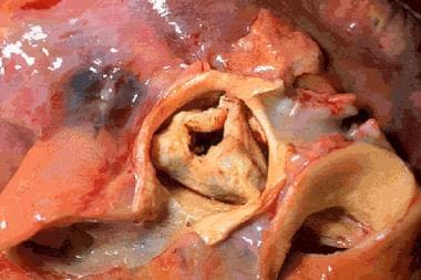
Aortic Valve
Heart Valve Diseases
Mitral Valve Stenosis
Ebstein Anomaly
Heart Valve Prosthesis Implantation
Tricuspid Atresia
Encyclopedias as Topic
Heart Murmurs
Primary right atrial angiosarcoma mimicking acute pericarditis, pulmonary embolism, and tricuspid stenosis. (1/29)
A 29 year old white man presented to the emergency room with new onset pleuritic chest pain and shortness of breath. He was initially diagnosed as having viral pericarditis and was treated with non-steroidal anti-inflammatory drugs. A few weeks later he developed recurrent chest pain with cough and haemoptysis. Chest radiography, cardiac examination, transthoracic and transoesophageal echocardiography pointed to a mass that arose from the posterior wall of the right atrium, not attached to the interatrial septum, which protruded into the lumen of the right atrium causing intermittent obstruction of inflow across the tricuspid valve. Contrast computed tomography of the chest showed a right atrial mass extending to the anterior chest wall. The lung fields were studded with numerous pulmonary nodules suggestive of metastases. A fine needle aspiration of the pulmonary nodule revealed histopathology consistent with spindle cell sarcoma thought to originate in the right atrium. Immunohistochemical stains confirmed that this was an angiosarcoma. There was no evidence of extracardiac origin of the tumour. The patient was treated with chemotherapy and radiation. This case highlights the clinical presentation, rapid and aggressive course of cardiac angiosarcomas, and the diagnostic modalities available for accurate diagnosis. (+info)Presence of oxidized low density lipoprotein in nonrheumatic stenotic aortic valves. (2/29)
The aim of the present study was to analyze if LDL particles trapped in stenotic aortic valve tissue undergo oxidative modification. Degenerative aortic stenosis affects >3% of the population >75 years of age in the Western world. Recent studies have revealed the presence of a chronic inflammatory process similar to what has been described in other degenerative diseases such as atherosclerosis. However, the underlying disease mechanisms of degenerative aortic stenosis still remain largely unknown. Six tricuspid stenotic valves, obtained at valve replacement, were compared with 3 control valves collected from hearts taken out during transplantation. The stenotic valves and the control valves were examined by immunohistochemistry, using antibodies against apoB, 4-hydroxynonenal-modified LDL, leukocytes, and HLA-DR. All valves were also stained with oil red O for neutral lipids. Extracellular neutral lipids were found in all stenotic valves, extending from the bases along the fibrosa layer. This lipid colocalized with apoB- and 4-hydroxynonenal-modified LDL immunoreactivity. 4-Hydroxynonenal-modified LDLs were present around calcium deposits, subendothelially, and in the deeper layer of the fibrosa. There was also a colocalization with macrophages, T lymphocytes, and HLA-DR expression. Control valves had a thin area of neutral lipid accumulation, a small amount of apoB, but no signs of inflammation. A distinct colocalization between oxidized LDLs, T-lymphocyte accumulation, and calcium deposits suggests that oxidized lipids may play a role in the disease process. (+info)Pacemaker lead related tricuspid stenosis: a report of two cases. (3/29)
Only four cases of tricuspid stenosis related to endocardial pacemaker leads have been reported. Two further cases associated with perforation of a tricuspid valve leaflet by a pacemaker lead are presented: a 46 year old woman and a 60 year old man. It is possible that tricuspid valve disease related to endocardial pacemaker and non-thoracotomy defibrillator leads is underrecognized. Diagnosis requires clinical suspicion and the use of Doppler echocardiography. Recent evidence of fibrosis affecting the tricuspid valve in hearts from patients who have had non-thoracotomy defibrillator implants suggests that this problem could be more common in the future. (+info)Thrombus on the tricuspid valve in a patient with primary antiphospholipid syndrome after implantation of an inferior vena cave filter. (4/29)
A 62-year-old woman with a history of pulmonary embolism and primary antiphospholipid syndrome (PAPS) with positivity for lupus anticoagulant was admitted to hospital with shortness of breath. A filter had been implanted in her inferior vena cava (IVC) 5 years previously. Emergency echocardiography revealed a lobulated, mobile echogenic mass on the tricuspid valve, and on pulmonary perfusion scintigraphy several apparently new defects were noted. Fibrinolytic therapy improved her symptoms and the pulmonary perfusion, then intravenous heparinization was continued for a further week. Repeat echocardiography performed on the 7th day of the admission showed complete disappearance of the mass, which was retrospectively diagnosed as a thrombus based on its resolution with fibrinolytic and anticoagulant therapies. (+info)THE PREOPERATIVE ASSESSMENT OF MULTIPLE VALVE DISEASE. (5/29)
Representative case histories are used to discuss the difficulties in preoperative assessment of patients with multiple valve disease and the dangers of correcting one lesion when two or more valves are seriously damaged. Errors fall into three broad categories: existing second valve disease (1) may not be suspected, (2) may be considered insignificant or (3) may be considered a consequence of the first.Recommendations are offered to minimize these errors. The four valves should be studied physiologically, no matter how "normal" the other three may appear to be clinically, whenever open-heart surgery is contemplated. In bivalvular disease angiographic methods are preferable to pressure studies, for data so obtained are not dependent on cardiac output. Mitral and tricuspid regurgitation can never be attributed with certainty to a more distal lesion but require direct examination at time of operation for assessment. (+info)Systemic lupus erythematosus complicated by tricuspid stenosis and regurgitation: successful treatment by valve transplantation. (6/29)
Clinical tricuspid stenosis has not previously been reported in patients with systemic lupus erythematosus (SLE). A 25 year old woman with active SLE presented with signs of severe right ventricular failure. Cardiac catheterisation confirmed the diagnosis of tricuspid stenosis and regurgitation together with mitral regurgitation. This patient underwent successful tricuspid and mitral valve replacement. (+info)The Inoue balloon for dilatation of the tricuspid valve: a modified over-the-wire approach. (7/29)
The Inoue balloon was used for dilatation of tricuspid stenosis in a 74 year old woman. The valve was reached by an over-the-wire approach with a 0.025 exchange length guide wire. The Inoue stylet would not reach the tricuspid orifice because the right atrium was so large. The Inoue balloon's special dilatation characteristics allowed good positioning at the tricuspid orifice. After dilatation to 27.5 mm, the pressure drop across the valve was reduced from 12 to 5 mm Hg. Further dilatation at 30 mm, however, created moderately severe tricuspid reflux without a further reduction of gradient. The Inoue balloon is suitable for dilatation of tricuspid stenosis but small increments in dilatation size may be required for optimal reduction in gradient without creating significant reflux. (+info)Aortic stenosis severity is not a risk factor for poststenotic dilatation of the ascending aorta. (8/29)
BACKGROUND: Dilatation of the ascending aorta in aortic stenosis may be partly explained by intrinsic wall structure changes, but the relative contribution of altered hemodynamics is unclear. The aim of this study was to assess the association between ascending aortic dimensions and valve stenosis severity. METHODS AND RESULTS: An analysis of echocardiographic examinations was conducted in 296 patients with aortic stenosis (179 males, mean age 71 years), 57 with bicuspid and 239 with tricuspid aortic valve, mean transaortic gradient 43+/-20 mmHg, and not more than moderate aortic regurgitation. Aortic dimensions at the level of annulus, sinuses of Valsalva, sinotubular junction and proximal ascending aorta were measured. Only height (p<0.001), degree of aortic regurgitation (p<0.01) and presence of bicuspid aortic valve (p<0.001) were independent predictors of ascending aortic dimensions. CONCLUSIONS: An independent association between aortic pressure gradients and proximal ascending aortic dimensions was not observed in patients with bicuspid or tricuspid aortic valve stenosis. Therefore, the poststenotic dilatation of the ascending aorta is not explained by aortic stenosis severity itself. Possible nonhemodynamic causes deserve detailed study at the time of diagnosis. (+info)Tricuspid valve stenosis is a cardiac condition characterized by the narrowing or stiffening of the tricuspid valve, which is located between the right atrium and right ventricle in the heart. This narrowing or stiffening restricts the normal flow of blood from the right atrium into the right ventricle, causing increased pressure in the right atrium and reduced blood flow to the lungs.
The tricuspid valve typically has three leaflets or cusps that open and close to regulate the flow of blood between the right atrium and right ventricle. In tricuspid valve stenosis, these leaflets become thickened, calcified, or fused together, leading to a reduced opening size and impaired function.
The most common causes of tricuspid valve stenosis include rheumatic heart disease, congenital heart defects, carcinoid syndrome, and infective endocarditis. Symptoms may include fatigue, shortness of breath, swelling in the legs and abdomen, and irregular heartbeats. Treatment options depend on the severity of the condition and underlying causes but may involve medications, surgical repair or replacement of the valve, or catheter-based procedures.
The tricuspid valve is the heart valve that separates the right atrium and the right ventricle in the human heart. It is called "tricuspid" because it has three leaflets or cusps, which are also referred to as flaps or segments. These cusps are named anterior, posterior, and septal. The tricuspid valve's function is to prevent the backflow of blood from the ventricle into the atrium during systole, ensuring unidirectional flow of blood through the heart.
Tricuspid valve insufficiency, also known as tricuspid regurgitation, is a cardiac condition in which the tricuspid valve located between the right atrium and right ventricle of the heart does not close properly, allowing blood to flow back into the right atrium during contraction of the right ventricle. This results in a portion of the blood being pumped inefficiently, which can lead to volume overload of the right side of the heart and potentially result in symptoms such as fatigue, weakness, shortness of breath, and fluid retention. The condition can be congenital or acquired, with common causes including dilated cardiomyopathy, infective endocarditis, rheumatic heart disease, and trauma.
Aortic valve stenosis is a cardiac condition characterized by the narrowing or stiffening of the aortic valve, which separates the left ventricle (the heart's main pumping chamber) from the aorta (the large artery that carries oxygen-rich blood to the rest of the body). This narrowing or stiffening prevents the aortic valve from opening fully, resulting in reduced blood flow from the left ventricle to the aorta and the rest of the body.
The narrowing can be caused by several factors, including congenital heart defects, calcification (hardening) of the aortic valve due to aging, or scarring of the valve due to rheumatic fever or other inflammatory conditions. As a result, the left ventricle must work harder to pump blood through the narrowed valve, which can lead to thickening and enlargement of the left ventricular muscle (left ventricular hypertrophy).
Symptoms of aortic valve stenosis may include chest pain or tightness, shortness of breath, fatigue, dizziness or fainting, and heart palpitations. Severe aortic valve stenosis can lead to serious complications such as heart failure, arrhythmias, or even sudden cardiac death. Treatment options may include medications to manage symptoms, lifestyle changes, or surgical intervention such as aortic valve replacement.
Pulmonary Valve Stenosis is a cardiac condition where the pulmonary valve, located between the right ventricle and the pulmonary artery, has a narrowed opening. This stenosis (narrowing) can cause obstruction of blood flow from the right ventricle to the lungs. The narrowing can be caused by a fusion of the valve leaflets, thickened or calcified valve leaflets, or rarely, a dysplastic valve.
The severity of Pulmonary Valve Stenosis is classified based on the gradient pressure across the valve, which is measured during an echocardiogram. A mild stenosis has a gradient of less than 30 mmHg, moderate stenosis has a gradient between 30-59 mmHg, and severe stenosis has a gradient of 60 mmHg or higher.
Mild Pulmonary Valve Stenosis may not require treatment, while more severe cases may need to be treated with balloon valvuloplasty or surgical valve replacement. If left untreated, Pulmonary Valve Stenosis can lead to right ventricular hypertrophy, heart failure, and other complications.
The aortic valve is the valve located between the left ventricle (the lower left chamber of the heart) and the aorta (the largest artery in the body, which carries oxygenated blood from the heart to the rest of the body). It is made up of three thin flaps or leaflets that open and close to regulate blood flow. During a heartbeat, the aortic valve opens to allow blood to be pumped out of the left ventricle into the aorta, and then closes to prevent blood from flowing back into the ventricle when it relaxes. Any abnormality or damage to this valve can lead to various cardiovascular conditions such as aortic stenosis, aortic regurgitation, or infective endocarditis.
The mitral valve, also known as the bicuspid valve, is a two-leaflet valve located between the left atrium and left ventricle in the heart. Its function is to ensure unidirectional flow of blood from the left atrium into the left ventricle during the cardiac cycle. The mitral valve consists of two leaflets (anterior and posterior), the chordae tendineae, papillary muscles, and the left atrial and ventricular myocardium. Dysfunction of the mitral valve can lead to various heart conditions such as mitral regurgitation or mitral stenosis.
Heart valve diseases are a group of conditions that affect the function of one or more of the heart's four valves (tricuspid, pulmonic, mitral, and aortic). These valves are responsible for controlling the direction and flow of blood through the heart. Heart valve diseases can cause the valves to become narrowed (stenosis), leaky (regurgitation or insufficiency), or improperly closed (prolapse), leading to disrupted blood flow within the heart and potentially causing symptoms such as shortness of breath, fatigue, chest pain, and irregular heart rhythms. The causes of heart valve diseases can include congenital defects, age-related degenerative changes, infections, rheumatic heart disease, and high blood pressure. Treatment options may include medications, surgical repair or replacement of the affected valve(s), or transcatheter procedures.
Mitral valve stenosis is a cardiac condition characterized by the narrowing or stiffening of the mitral valve, one of the four heart valves that regulate blood flow through the heart. This narrowing prevents the mitral valve from fully opening during diastole (relaxation phase of the heart cycle), leading to restricted flow of oxygenated blood from the left atrium into the left ventricle.
The narrowing or stiffening of the mitral valve can be caused by various factors, such as rheumatic heart disease, congenital heart defects, aging, or calcium deposits on the valve leaflets. As a result, the left atrium has to work harder to pump blood into the left ventricle, causing increased pressure in the left atrium and pulmonary veins. This can lead to symptoms such as shortness of breath, fatigue, coughing, and heart palpitations.
Mitral valve stenosis is typically diagnosed through a combination of medical history, physical examination, and imaging techniques like echocardiography or cardiac catheterization. Treatment options may include medications to manage symptoms and prevent complications, as well as surgical interventions such as mitral valve repair or replacement to alleviate the stenosis and improve heart function.
Ebstein anomaly is a congenital heart defect that affects the tricuspid valve, which is the valve between the right atrium and right ventricle of the heart. In Ebstein anomaly, the tricuspid valve is abnormally formed and positioned, causing it to leak blood back into the right atrium. This can lead to various symptoms such as shortness of breath, fatigue, and cyanosis (bluish discoloration of the skin). Treatment for Ebstein anomaly may include medication, surgery, or a combination of both. It is important to note that the severity of the condition can vary widely among individuals, and some people with Ebstein anomaly may require more intensive treatment than others.
Heart valve prosthesis implantation is a surgical procedure where an artificial heart valve is inserted to replace a damaged or malfunctioning native heart valve. This can be necessary for patients with valvular heart disease, including stenosis (narrowing) or regurgitation (leaking), who do not respond to medical management and are at risk of heart failure or other complications.
There are two main types of artificial heart valves used in prosthesis implantation: mechanical valves and biological valves. Mechanical valves are made of synthetic materials, such as carbon and metal, and can last a long time but require lifelong anticoagulation therapy to prevent blood clots from forming. Biological valves, on the other hand, are made from animal or human tissue and typically do not require anticoagulation therapy but may have a limited lifespan and may need to be replaced in the future.
The decision to undergo heart valve prosthesis implantation is based on several factors, including the patient's age, overall health, type and severity of valvular disease, and personal preferences. The procedure can be performed through traditional open-heart surgery or minimally invasive techniques, such as robotic-assisted surgery or transcatheter aortic valve replacement (TAVR). Recovery time varies depending on the approach used and individual patient factors.
The pulmonary valve, also known as the pulmonic valve, is a semilunar valve located at the exit of the right ventricle of the heart and the beginning of the pulmonary artery. It has three cusps or leaflets that prevent the backflow of blood from the pulmonary artery into the right ventricle during ventricular diastole, ensuring unidirectional flow of blood towards the lungs for oxygenation.
Tricuspid atresia is a congenital heart defect where the tricuspid valve, which regulates blood flow between the right atrium and right ventricle, fails to develop properly. As a result, there is no direct pathway for blood to move from the right atrium to the right ventricle and then to the lungs for oxygenation.
In this condition, blood from the body returning to the heart enters the right atrium but cannot flow through the tricuspid valve into the right ventricle. Instead, it flows through an opening in the interatrial septum (atrial septal defect) into the left atrium and then into the left ventricle. The left ventricle pumps this blood to the body and a portion of it goes to the lungs via a patent ductus arteriosus or other collateral vessels.
Tricuspid atresia is often associated with other heart defects, such as transposition of the great arteries, pulmonary stenosis, or total anomalous pulmonary venous return. Symptoms can vary depending on the severity and associated defects but may include cyanosis (bluish discoloration of the skin), shortness of breath, fatigue, and poor growth. Treatment typically involves surgical interventions to create a path for blood to flow to the lungs and establish proper oxygenation.
Heart auscultation is a medical procedure in which a healthcare professional uses a stethoscope to listen to the sounds produced by the heart. The process involves placing the stethoscope on various locations of the chest wall to hear different areas of the heart.
The sounds heard during auscultation are typically related to the opening and closing of the heart valves, as well as the turbulence created by blood flow through the heart chambers. These sounds can provide important clues about the structure and function of the heart, allowing healthcare professionals to diagnose various cardiovascular conditions such as heart murmurs, valvular disorders, and abnormal heart rhythms.
Heart auscultation is a key component of a physical examination and requires proper training and experience to interpret the findings accurately.
An encyclopedia is a comprehensive reference work containing articles on various topics, usually arranged in alphabetical order. In the context of medicine, a medical encyclopedia is a collection of articles that provide information about a wide range of medical topics, including diseases and conditions, treatments, tests, procedures, and anatomy and physiology. Medical encyclopedias may be published in print or electronic formats and are often used as a starting point for researching medical topics. They can provide reliable and accurate information on medical subjects, making them useful resources for healthcare professionals, students, and patients alike. Some well-known examples of medical encyclopedias include the Merck Manual and the Stedman's Medical Dictionary.
A heart murmur is an abnormal sound heard during a heartbeat, which is caused by turbulent blood flow through the heart. It is often described as a blowing, whooshing, or rasping noise. Heart murmurs can be innocent (harmless and not associated with any heart disease) or pathological (indicating an underlying heart condition). They are typically detected during routine physical examinations using a stethoscope. The classification of heart murmurs includes systolic, diastolic, continuous, and functional murmurs, based on the timing and auscultatory location. Various heart conditions, such as valvular disorders, congenital heart defects, or infections, can cause pathological heart murmurs. Further evaluation with diagnostic tests like echocardiography is often required to determine the underlying cause and appropriate treatment.