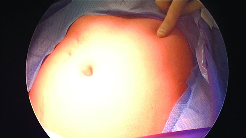
Umbilicus
Medicine in Art
Art
Vitelline Duct
Urachus
Abdominal Wall
Rectus Abdominis
Sister Mary Joseph's Nodule
Abdominal Muscles
Hernia, Umbilical
Laparoscopy
Cholecystectomy, Laparoscopic
Centella
Crassulaceae
Encyclopedias as Topic
Pentacyclic Triterpenes
Body piercing in the accident and emergency department. (1/151)
Recently an increasing number of patients with complications related to pierced body jewellery have been seen. Often removal of the jewellery is indicated. Removal of these items may also be required for radiological purposes. If the doctor is familiar with the opening mechanism of the item, removal is not usually difficult. Uninformed attempts at removal may cause unnecessary trauma and distress. In a survey of 28 accident and emergency doctors, only six were able accurately to describe the opening mechanisms of all three commonly used types of jewellery. Descriptions of the types of jewellery currently used are not available in the medical literature. The aim of this article is to familiarize doctors with the types of jewellery used, describe their opening mechanisms, and suggest techniques for their removal. The complications of body piercing and the indications for the removal of body jewellery are also outlined. (+info)Diphenhydramine disposition in the sheep maternal-placental-fetal unit: gestational age, plasma drug protein binding, and umbilical blood flow effects on clearance. (2/151)
The objective of this study was to examine the interrelationships between maternal and fetal plasma drug protein binding, umbilical blood flow (Q(um)), gestational age (GA), and maternal-fetal diphenhydramine (DPHM) clearances in chronically instrumented pregnant sheep. Maternal and fetal DPHM placental (CL(mf) and CL(fm), respectively) and nonplacental (CL(mo) and CL(fo), respectively) clearances and steady-state plasma protein binding were determined in 18 pregnant sheep at 124 to 140 days' gestation (term, approximately 145 days). The data demonstrated a highly significant fall of approximately 66% in CL(fm) and a decreasing trend in CL(fo) ( approximately 47%) over the GA range studied. However, no such relationships existed between GA and CL(mf) or CL(mo). Concomitant with this was a decrease in fetal DPHM plasma unbound fraction with GA, with no such change being evident in the mother. Both CL(mo) and CL(fo) were related to the respective DPHM plasma unbound fraction. A strong relationship also existed between fetal plasma unbound fraction and CL(fm). Thus, the decrease in fetal unbound fraction of DPHM during gestation could contribute to the fall in CL(fm), and possibly CL(fo). However, over the GA range studied, fetal DPHM free fraction decreased by approximately 47%, whereas CL(fm) fell by approximately 66%. Because fetal unbound fraction and CL(fm) are linearly related, the GA-associated fall in unbound fraction appears to be insufficient to account for the entire decline in CL(fm). In separate studies in pregnant sheep, we observed a approximately 40% fall in weight-normalized Q(um) between 125 and 137 days' gestation. Because CL(fm) for DPHM is similar to that of flow-limited compounds (e.g., ethanol, antipyrine), this decrease in Q(um) may also contribute to the GA-related fall in CL(fm). (+info)The Pitx2 protein in mouse development. (3/151)
The Rieger syndrome, an autosomal dominant disorder involving ocular, dental, and umbilical defects is caused by mutations in PITX2, a Bicoid-type homeobox protein. Mouse Pitx2 mRNA is expressed in eye, tooth and umbilicus consistent with the human Riegers phenotype. Moreover, Pitx2 is involved in the Nodal/Sonic hedgehog pathway that determines left/right polarity. In this report we demonstrate a 32-kDa polypeptide on Western blots of nuclear extracts from a rat pituitary cell line, using a Pitx2 specific antibody (designated P2R10). We describe also for the first time expression of the Pitx2 protein in mouse. Pitx2 protein immunostaining was detectable during the development of the eye, tooth, umbilicus, and also in the pituitary, heart, gut, and limb. We demonstrate for the first time directly that Pitx2 is asymmetrically expressed in early heart, gut, and lung development. (+info)Elimination of biliary stones through the urinary tract: a complication of the laparoscopic cholecystectomy. (4/151)
The introduction and popularization of laparoscopic cholecystectomy has been accompanied with a considerable increase in perforation of gallbladder during this procedure (10% - 32%), with the occurrence of intraperitoneal bile spillage and the consequent increase in the incidence of lost gallstones (0.2% - 20%). Recently the complications associated with these stones have been documented in the literature. We report a rare complication occurring in an 81-year-old woman who underwent laparoscopic cholecystectomy and developed cutaneous fistula to the umbilicus and elimination of biliary stones through the urinary tract. During the cholecystectomy, the gall bladder was perforated, and bile and gallstones were spilled into the peritoneal cavity. Two months after the initial procedure there was exteriorization of fistula through the umbilicus, with intermittent elimination of biliary stones. After eleven months, acute urinary retention occurred due to biliary stones in the bladder, which were removed by cystoscopy. We conclude that efforts should be concentrated on avoiding the spillage of stones during the surgery, and that no rules exist for indicating a laparotomy simply to retrieve these lost gallstones. (+info)A prospective, randomised trial of prophylactic antibiotics versus bag extraction in the prophylaxis of wound infection in laparoscopic cholecystectomy. (5/151)
Septic complications are rare following laparoscopic cholecystectomy if prophylactic antibiotics are given, as demonstrated in previous studies. Antibiotic treatment may be unnecessary and, therefore, undesirable, so we compared two forms of prophylaxis: a cephalosporin antibiotic and bag extraction of the dissected gallbladder. A total of 76 patients undergoing laparoscopic cholecystectomy were randomised to either receive an antibiotic or to have their gallbladder removed from the abdomen in a plastic bag. Complicated cases were excluded. There was a total of 6 wound infections (7.9%), 3 in each of the study groups. All these were associated with skin commensals. There were no other septic complications. Bacteriological studies grouped the organisms isolated from the bile and the wound as potential pathogens and likely commensals. A total of 10 potential pathogens were isolated, 9 of which were found in the group receiving antibiotics. We conclude that septic sequelae of uncomplicated laparoscopic cholecystectomy are uncommon, but clearly not entirely prevented by antibiotic or mechanical prophylaxis. Prophylactic antibiotics may not be required in uncomplicated laparoscopic cholecystectomy. Further study is warranted. (+info)Implantable insulin pumps: infections most likely due to seeding from skin flora determine severe outcomes of pump-pocket seromas. (6/151)
Complications at implantation site of implantable insulin pumps may lead to premature removal. To elucidate the origins and the outcomes of these local adverse events. We investigated seromas of the 'pump-pocket' that have been detected for an eight month-period during the follow-up of such-treated forty type 1 diabetic patients. At the start of study period, skin bacterial flora was sampled at umbilicus and groin, and isolated strains of Staphylococcus epidermidis were preserved in specific vials at -20 degrees C. Each time a seroma was detected at transcutaneous 45 days-refill of pump reservoir, it was sampled for bacterial cultures. Isolated strains of S. epidermidis from seroma were genetically compared to preserved strains of corresponding patients using Pulsed-Field Gel Electrophoresis (PFGE) after genomic restriction by SmaI. Among the ten seromas that occurred after a mean time of 9.9 months since implantation, S. epidermidis were isolated in five cases. Genetic comparison of isolated strains could be performed in three cases. Compared strains showed identical (in 2 cases) or closely related (in one case) PFGE profiles. While the five aseptic seromas resolved with rest, four infected cases required explantations after one to nineteen months in spite of antibiotic therapy and the fifth one persisted without impairment under long-term antibiotics. Our results suggest that seeding from the skin flora is a key-factor determining the severity of pump-pocket complications. We recommend that bacterial investigations of pump-pocket seromas should be systematically performed, while prophylactic measures might include antibiotic cover for each puncture of the pump-pocket. (+info)Recovery of anaerobic bacteria from 3 patients with infection at a pierced body site. (7/151)
We describe 3 adolescents who developed infections due to anaerobes at pierced body sites: the nipple, the umbilicus, and the nasal septum. Anaerobes (Prevotella intermedia and Peptostreptococcus anaerobius) were recovered from pure culture of specimens obtained from 1 patient with nipple infection and were mixed with aerobic bacteria in cultures of specimens obtained from 2 patients (Streptococcus aureus, Peptostreptococcus micros, and Prevotella melaninogenica were recovered from a patient with nasal septum infection, and Bacteroides fragilis and Enterococcus faecalis were recovered from a patient with umbilical infection). The infection resolved in all patients after removal of the ornaments and use of antimicrobial drug treatment. (+info)Significant abdominal wall hematoma from an umbilical port insertion. (8/151)
Laparoscopists consider the umbilical and ventral midline area to be "vascular safe." On occasion, however, the insertion of the first trocar at the umbilical port may result in severe abdominal wall hematoma. (+info)The umbilicus, also known as the navel, is the scar left on the abdominal wall after the removal of the umbilical cord in a newborn. The umbilical cord connects the developing fetus to the placenta in the uterus during pregnancy, providing essential nutrients and oxygen while removing waste products. After birth, the cord is clamped and cut, leaving behind a small stump that eventually dries up and falls off, leaving the umbilicus. In adults, it typically appears as a slight depression or dimple on the abdomen.
"Medicine in Art" is not a medical term per se, but rather a term used to describe the intersection and representation of medical themes, practices, or symbols in various art forms. It can include but is not limited to:
1. The depiction of medical scenes, practitioners, or patients in paintings, sculptures, or photographs.
2. The use of medical imagery such as X-rays, MRIs, or anatomical drawings in mixed media works.
3. The exploration of medical issues, diseases, or treatments in conceptual art.
4. The creation of art by artists with medical conditions, which can provide insight into their experiences.
5. The use of art therapy as a healing modality in medical settings.
This term is often used in the context of art history, visual culture, and medical humanities to analyze and understand the complex relationships between art, medicine, and society.
I am not aware of a specific medical definition for the term "art." In general, art refers to creative works that express or evoke emotions through meaning, symbolism, form, and/or color. This can include various forms such as visual arts (painting, sculpture, photography), performing arts (theater, music, dance), literary arts (poetry, novels), and more.
However, there is a field of study called medical humanities that explores the intersection between medicine and the humanities, including art. In this context, art can be used as a tool for healing, communication, reflection, and understanding in healthcare settings. For example, art therapy is a form of expressive therapy that uses creative activities like drawing, painting, or sculpting to help patients explore their emotions, improve their mental health, and enhance their well-being.
Therefore, while there may not be a specific medical definition for "art," it can have significant implications for healthcare and the human experience.
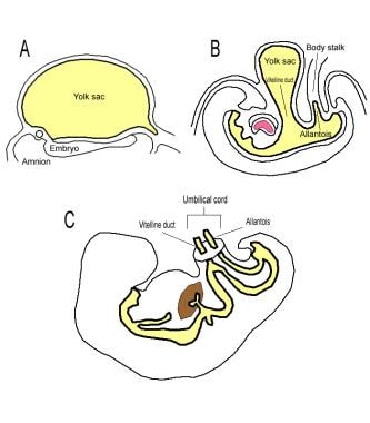
The Vitelline Duct, also known as the Yolk Stalk or the Omphalomesenteric Duct, is a vestigial structure in human embryonic development. It is a canal that connects the midgut of the developing fetus to the yolk sac, which provides nutrients during early stages of embryonic growth.
In normal development, this duct usually obliterates or closes off completely by the end of the 8th week of gestation. If it fails to do so, it can result in various congenital abnormalities. These may include Meckel's diverticulum (a pouch protruding from the wall of the intestine), omphalocele (a defect where the intestines and other organs protrude through the belly button), or persistent vitellointestinal duct, which can lead to infections and bowel obstructions.
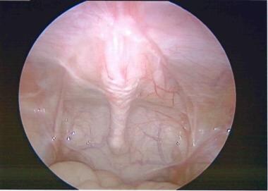
A laparoscope is a type of medical instrument called an endoscope, which is used to examine the interior of a body cavity or organ. Specifically, a laparoscope is a long, thin tube with a high-intensity light and a high-resolution camera attached to it. This device allows surgeons to view the abdominal cavity through small incisions, without having to make large, invasive cuts.
During a laparoscopic procedure, the surgeon will insert the laparoscope through a small incision in the abdomen, typically near the navel. The camera sends images back to a monitor, giving the surgeon a clear view of the organs and tissues inside the body. This allows for more precise and less invasive surgical procedures, often resulting in faster recovery times and fewer complications compared to traditional open surgery.
Laparoscopes are commonly used in a variety of surgical procedures, including:
1. Gynecological surgeries (e.g., hysterectomies, ovarian cyst removals)
2. Gallbladder removal (cholecystectomy)
3. Gastrointestinal surgeries (e.g., removing benign or malignant tumors)
4. Hernia repairs
5. Bariatric surgeries for weight loss (e.g., gastric bypass, sleeve gastrectomy)
While laparoscopes provide numerous benefits over open surgery, they still require specialized training and expertise to use effectively and safely.
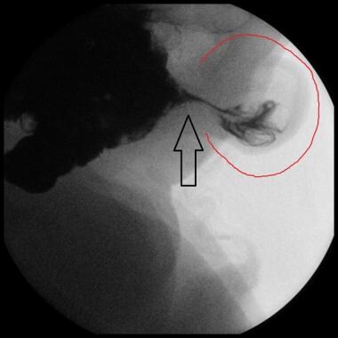
The urachus is a vestigial structure in humans, which is a fibrous cord that connects the umbilicus (navel or belly button) to the dome-shaped top of the bladder. In fetal development, the urachus is the passageway for urine to move from the developing bladder to the allantois, an outpouching of the hindgut that ultimately becomes part of the placenta.
After birth, the urachus usually obliterates and turns into a fibrous cord called the median umbilical ligament. However, in some cases, the urachus may not completely obliterate, leading to various congenital abnormalities such as urachal cysts, urachal sinuses, or urachal fistulas. These conditions can cause symptoms like lower abdominal pain, infection, and sometimes even sepsis if left untreated.
It's worth noting that the urachus is not a commonly discussed structure in routine medical practice, but it does have clinical significance in certain pediatric surgical cases and congenital anomalies.
The abdominal wall refers to the group of muscles, fascia (sheaths of connective tissue), and skin that make up the front and sides of the abdomen, extending from the thorax (chest) to the pelvis. It provides protection to the abdominal organs, supports the trunk, and allows for movement of the torso.
The main muscles of the anterior abdominal wall include:
1. Rectus sheaths (Rectus Abdominis): paired vertical muscles running from the pubic symphysis to the xiphoid process and costal cartilages of ribs 5-7.
2. External obliques: thin, irregular muscles that lie over the lower part of the abdomen and run diagonally downward and forward from the lower ribs to the iliac crest (pelvic bone) and pubic tubercle.
3. Internal obliques: thicker muscles that lie under the external obliques, running diagonally upward and forward from the iliac crest to the lower ribs.
4. Transverse abdominis: deepest of the abdominal muscles, lying horizontally across the abdomen, attaching from the lower ribs to the pelvis.
These muscles are interconnected by various layers of fascia and aponeuroses (flat, broad tendons), forming a complex structure that allows for both stability and mobility. The linea alba, a fibrous band, runs down the midline of the anterior abdominal wall, connecting the rectus sheaths.
Damage to the abdominal wall can occur due to trauma, surgery, or various medical conditions, which may require surgical intervention for repair.
The rectus abdominis is a paired, flat, and long muscle in the anterior (front) wall of the abdomen. It runs from the pubic symphysis (the joint where the two pubic bones meet in the front of the pelvis) to the xiphoid process (the lower end of the sternum or breastbone) and costal cartilages of the fifth, sixth, and seventh ribs.
The rectus abdominis is responsible for flexing the lumbar spine (lower back), which helps in bending forward or sitting up from a lying down position. It also contributes to maintaining proper posture and stabilizing the pelvis and spine. The muscle's visibility, especially in its lower portion, is often associated with a "six-pack" appearance in well-trained individuals.
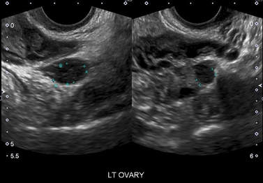
"Sister Mary Joseph's nodule" is a term used in medicine to describe a palpable (able to be felt) or visible nodule or lump that is located at the umbilicus (belly button). It is usually indicative of an underlying malignancy, most commonly originating from the stomach or ovaries. The presence of this nodule suggests a poor prognosis as it often indicates advanced stage cancer. The term was coined by Dr. Hamilton Bailey in honor of Sister Mary Joseph, who first recognized the association between umbilical nodules and internal malignancies during her work as a surgical nurse with Dr. William Mayo in the early 20th century.
The abdominal muscles, also known as the abdominals or abs, are a group of muscles in the anterior (front) wall of the abdominopelvic cavity. They play a crucial role in maintaining posture, supporting the trunk, and facilitating movement of the torso. The main abdominal muscles include:
1. Rectus Abdominis: These are the pair of long, flat muscles that run vertically along the middle of the anterior abdominal wall. They are often referred to as the "six-pack" muscles due to their visible, segmented appearance in well-trained individuals. The primary function of the rectus abdominis is to flex the spine, allowing for actions such as sitting up from a lying down position or performing a crunch exercise.
2. External Obliques: These are the largest and most superficial of the oblique muscles, located on the lateral (side) aspects of the abdominal wall. They run diagonally downward and forward from the lower ribs to the iliac crest (the upper part of the pelvis) and the pubic tubercle (a bony prominence at the front of the pelvis). The external obliques help rotate and flex the trunk, as well as assist in side-bending and exhalation.
3. Internal Obliques: These muscles lie deep to the external obliques and run diagonally downward and backward from the lower ribs to the iliac crest, pubic tubercle, and linea alba (the strong band of connective tissue that runs vertically along the midline of the abdomen). The internal obliques help rotate and flex the trunk, as well as assist in forced exhalation and increasing intra-abdominal pressure during actions such as coughing or lifting heavy objects.
4. Transversus Abdominis: This is the deepest of the abdominal muscles, located inner to both the internal obliques and the rectus sheath (a strong, fibrous covering that surrounds the rectus abdominis). The transversus abdominis runs horizontally around the abdomen, attaching to the lower six ribs, the thoracolumbar fascia (a broad sheet of connective tissue spanning from the lower back to the pelvis), and the pubic crest (the front part of the pelvic bone). The transversus abdominis helps maintain core stability by compressing the abdominal contents and increasing intra-abdominal pressure.
Together, these muscles form the muscular "corset" of the abdomen, providing support, stability, and flexibility to the trunk. They also play a crucial role in respiration, posture, and various movements such as bending, twisting, and lifting.
The abdomen refers to the portion of the body that lies between the thorax (chest) and the pelvis. It is a musculo-fascial cavity containing the digestive, urinary, and reproductive organs. The abdominal cavity is divided into several regions and quadrants for medical description and examination purposes. These include the upper and lower abdomen, as well as nine quadrants formed by the intersection of the midline and a horizontal line drawn at the level of the umbilicus (navel).
The major organs located within the abdominal cavity include:
1. Stomach - muscular organ responsible for initial digestion of food
2. Small intestine - long, coiled tube where most nutrient absorption occurs
3. Large intestine - consists of the colon and rectum; absorbs water and stores waste products
4. Liver - largest internal organ, involved in protein synthesis, detoxification, and metabolism
5. Pancreas - secretes digestive enzymes and hormones such as insulin
6. Spleen - filters blood and removes old red blood cells
7. Kidneys - pair of organs responsible for filtering waste products from the blood and producing urine
8. Adrenal glands - sit atop each kidney, produce hormones that regulate metabolism, immune response, and stress response
The abdomen is an essential part of the human body, playing a crucial role in digestion, absorption, and elimination of food and waste materials, as well as various metabolic processes.
An umbilical hernia is a type of hernia that occurs at the umbilicus, or belly button. It results from a protrusion of abdominal contents through a weakened area in the abdominal wall surrounding the navel. This condition is common in newborns and infants, especially premature babies, due to incomplete closure of the abdominal muscles during development.
In most cases, umbilical hernias in children close on their own by age 3-4 or by the time they reach school age. However, if the hernia is still present after this age, surgical intervention may be required to prevent potential complications such as incarceration (where the herniated tissue becomes trapped and cannot be pushed back in) or strangulation (where the blood supply to the herniated tissue is cut off, leading to tissue death).
Adults can also develop umbilical hernias, often as a result of increased pressure in the abdomen due to obesity, pregnancy, heavy lifting, or persistent coughing. Umbilical hernias in adults are generally more likely to require surgical repair due to the higher risk of complications.
Laparoscopy is a surgical procedure that involves the insertion of a laparoscope, which is a thin tube with a light and camera attached to it, through small incisions in the abdomen. This allows the surgeon to view the internal organs without making large incisions. It's commonly used to diagnose and treat various conditions such as endometriosis, ovarian cysts, infertility, and appendicitis. The advantages of laparoscopy over traditional open surgery include smaller incisions, less pain, shorter hospital stays, and quicker recovery times.
Laparoscopic cholecystectomy is a surgical procedure to remove the gallbladder using a laparoscope, a thin tube with a camera, which allows the surgeon to view the internal structures on a video monitor. The surgery is performed through several small incisions in the abdomen, rather than a single large incision used in open cholecystectomy. This approach results in less postoperative pain, fewer complications, and shorter recovery time compared to open cholecystectomy.
The procedure is typically indicated for symptomatic gallstones or chronic inflammation of the gallbladder (cholecystitis), which can cause severe abdominal pain, nausea, vomiting, and fever. Laparoscopic cholecystectomy has become the standard of care for gallbladder removal due to its minimally invasive nature and excellent outcomes.
I'm not a medical professional, but the term "History, Ancient" is not a medical term per se. However, in a broader context, it could refer to the study of ancient medical practices, theories, and beliefs that existed in civilizations prior to the Middle Ages or Classical Antiquity. This might include the examination of ancient texts, artifacts, and archaeological evidence to understand how illnesses were treated and viewed in these historical periods. It forms an essential part of the evolution of medical knowledge and practices over time.
"Centella" is the common name for a plant species known scientifically as *Centella asiatica*, also referred to as gotu kola. It is a herb that has been used in traditional medicine in various cultures, including Ayurvedic and Chinese medicine, for its potential health benefits. The active compounds in centella include saponins called asiaticoside, madecassoside, and madasiatic acid, which are believed to have anti-inflammatory, antioxidant, and wound-healing properties.
Centella has been studied for its potential effects on various conditions, such as anxiety, cognitive function, and skin health. However, more research is needed to confirm these potential benefits and establish recommended dosages and safety guidelines. As with any supplement or medication, it's important to consult with a healthcare provider before using centella to ensure that it's appropriate for your individual health needs and to avoid potential interactions with other medications.
Crassulaceae is a family of succulent plants, also known as stonecrops or orpines. These plants are characterized by their thick, fleshy leaves that store water, allowing them to survive in dry environments. They are native to various parts of the world, including Europe, Africa, and Asia. Some common examples of Crassulaceae include Sedum species (such as Sedum spectabile and Sedum telephium), Sempervivum species (also known as hens and chicks), and Echeveria species. These plants are often grown as ornamentals for their attractive foliage and flowers.
An encyclopedia is a comprehensive reference work containing articles on various topics, usually arranged in alphabetical order. In the context of medicine, a medical encyclopedia is a collection of articles that provide information about a wide range of medical topics, including diseases and conditions, treatments, tests, procedures, and anatomy and physiology. Medical encyclopedias may be published in print or electronic formats and are often used as a starting point for researching medical topics. They can provide reliable and accurate information on medical subjects, making them useful resources for healthcare professionals, students, and patients alike. Some well-known examples of medical encyclopedias include the Merck Manual and the Stedman's Medical Dictionary.
Pentacyclic triterpenes are a type of natural compounds that are characterized by their structure, which consists of five cyclic rings made up of 30 carbon atoms. They are formed from squalene through a series of enzymatic reactions and can be found in various plants, as well as some animals and marine organisms.
Pentacyclic triterpenes have been studied for their potential medicinal properties, including anti-inflammatory, antiviral, and antitumor activities. Some examples of pentacyclic triterpenes include oleanolic acid, ursolic acid, and betulinic acid, which are found in a variety of fruits, vegetables, and herbs.
It's worth noting that while there is a growing body of research on the potential health benefits of pentacyclic triterpenes, more studies are needed to fully understand their mechanisms of action and therapeutic potential.
East Asian traditional medicine (ETAM) refers to the traditional medical systems that have been practiced in China, Japan, Korea, and other countries in this region for centuries. The most well-known forms of ETAM are Traditional Chinese Medicine (TCM), Kampo (Japanese traditional medicine), and Korean traditional medicine (KTM).
TCM is a comprehensive medical system that includes acupuncture, moxibustion, herbal medicine, dietary therapy, tuina (Chinese massage), and qigong (breathing exercises) among its modalities. TCM is based on the concept of balancing the flow of qi (vital energy) through a system of channels or meridians in the body.
Kampo is a Japanese adaptation of Chinese medicine that emphasizes the use of herbal formulas to treat illness and maintain health. Kampo practitioners often prescribe individualized herbal formulas based on the patient's unique pattern of symptoms, which are determined through careful diagnosis and examination.
KTM is a traditional Korean medical system that combines elements of Chinese and Japanese medicine with indigenous Korean practices. KTM includes acupuncture, moxibustion, herbal medicine, cupping, and various forms of manual therapy.
While ETAM has been practiced for centuries and has a rich cultural heritage, it is important to note that its safety and efficacy have not always been rigorously studied using modern scientific methods. As such, it is essential to consult with a qualified healthcare provider before pursuing any form of traditional medicine.
 Umbilicus
Umbilicus (plant)
Umbilicus intermedius
Umbilicus horizontalis
Umbilicus oppositifolius
Umbilicus schmidtii
Umbilicus (mollusc)
Umbilicus rupestris
Umbilicus chrysanthus
Umbilicus luteus
Umbilicus chloranthus
Umbilicus (reference point)
Umbilicus urbis Romae
Glyphipterix umbilici
Puccinia umbilici
Diaphanidae
Abyssochrysos bicinctus
Navel
Euthema
Flora of Malta
Mirachelus corbis
List of wort plants
Asymptoceras
Allognathus hispanicus
Glossary of bird terms
Semisulcospira libertina
Cotyledon chrysantha
Calliostoma annulatum
Notodiscus hookeri
Anania (foraminifera)
Umbilicus - Wikipedia
Umbilicus rupestris - Wikipedia
 Disorders of the Umbilicus: Practice Essentials, Anatomy, Pathophysiology
 Cotyledon Umbilicus throat symptoms - ABC Homeopathy
 Umbilicus rupestris; Navelwort
 Umbilicus Undulatus - The Official Masterworks Broadway Site
Brian Knep :: Flower Umbilicus
 Umbilicus Desidero - Buy, watch, or rent from the Microsoft Store
 Improving the Result of the Umbilicus | Abdominioplasty
 Location of the transverse colon in relationship to the umbilicus: implications for laparoscopic techniques.
 Novel navel operation Technique optimizes post-tummy tuck umbilicus | theaestheticguide.com
Galerie Sultana - Umbilicus
 CREATION OF 'NEW' UMBILICUS
![Umbilicus [Urzas Saga] -
Tacoma Games](data:image/png;base64,iVBORw0KGgoAAAANSUhEUgAAABAAAAANCAMAAACXZR4WAAABR1BMVEVHcEx/bSJ5fH1uUySur68fKy52YkunpqcWKCEiHiulo6TCwcG6ubq2JB3Q1NS4ubkxLC19OzyufB2uZyFwbm/Ly8vr6+uTQxgAAABtIySedxmQah6gkxiDaWaMgSe/u7y1cx+npqfNzc3Fw8S4t7dvIh23uLihbBa8gBTMzc6IKyM3YTPNKiBZEgzu2gC2s7PGxcWnISKrlpWysLGMUU+wqam6RB5jMijEvr65KyXakRibJx2hnZ2kg4EZFyaWeHY3RRqsURl+UhjSugglMiQrT1gYFBlaXx0pr93+8yv66QCeXTmpYCB5CBPkMR+YPSLt3iTGZl96Kywha4l6ST9KGCAPoc5lki4fXXkLTGgthZ+lZVlhRB6dLjA9osRCdmUtgI94OTVAXGLAuRs7Lx2tpAZwhlR4dRpvbBp8fBdiJCrlwQBNjpI/i5KyAAAAJXRSTlMAQDpWtBgv+iILW/a5+1c/dojb2uPSzdYzy8uVxmix6faB3eTPC5TDUwAAAM5JREFUCJljYGBg4OTiUGJR5lHkYgADNjFuNXYednZNNQsmsICAiCi7poGtfVCWlqUcI0iEQ81YS1XXLiPZ3SlAhoGBVUrcxNvczTXJy9MzU4sXKOBjxqseo+2Rpuell26tw8rAaehnru0QFZeTmu2c4qjBzCCtoWPnnhDsEebq4sarYSXEoGBl5qQXER+p7aKbGGgtzMjA7K/jrO4QHqqramtqJAiyVdLQMTY6xMTIWFNVnw/sDHVfG3t9GwtTAxZ+qNsl5LlVWGSZOME8ANK1ITIp02C3AAAAAElFTkSuQmCC) Umbilicus [Urza's Saga] -
Tacoma Games
Umbilicus [Urza's Saga] -
Tacoma Games
 Umbilicus II - CG Sculpture and Jewelry
 Umbilicus urbis Romae
Go to wpid-clusius-umbilicus.jpg
 umbilicus- Meaning in Bengali - HinKhoj English Bengali Dictionary
 Cotyledon umbilicus in omeopatia Archivi - An Eco-sustainable World
 ICD-10-CM Diagnosis Code L03.316 - Cellulitis of umbilicus
Disorders of the Umbilicus: Background, History of the Procedure, Problem
 Umbilicus rupestris (Salisb.) Dandy - Global Pollen Project - Global Pollen Project
 Live Action Mafia • View topic - If umbilicus self-harm iris, receive.
 "umbilicus strike of lightning in the void"- Yehonatan Koenig | THE STOREFRONT...
 Time to Explore the Umbilicus: New Belly Button Biodiversity Website - Your Wild Life
UMBILICUS (CANNIBAL CORPSE, DEICIDE) Streams Retro Rockin' New Single "I, Human" | Truth Media
 Acid Archives - Page 12 of 13 - Urban Jungle - Plant Nursery in Norwich, Norfolk and Beccles, Suffolk.
Green Archives - Page 7 of 8 - Urban Jungle - Plant Nursery in Norwich, Norfolk and Beccles, Suffolk.
 Ruptured Bladder Archives - The Horse
Rupestris5
- Umbilicus rupestris , the navelwort , [1] penny-pies or wall pennywort , is a fleshy, perennial, edible flowering plant in the stonecrop family Crassulaceae in the genus Umbilicus so named for its umbilicate ( navel -like) leaves. (wikipedia.org)
- Umbilicus rupestris in bloom in Nazaré , Portugal . (wikipedia.org)
- Umbilicus rupestris is not the same "Pennywort" as the one used in Asian medicine, which is the unrelated Asiatic Pennywort, Centella asiatica . (wikipedia.org)
- The photographer's identification Umbilicus rupestris has not been reviewed. (berkeley.edu)
- Umbilicus rupestris The navelwort or penny-pies or wall pennywort (Umbilicus rupestris (Salisb. (antropocene.it)
Navel7
- Umbilicus may refer to: The navel or belly button Umbilicus (mollusc), a feature of gastropod, Nautilus and Ammonite shell anatomy Umbilicus (plant), a genus of over ninety species of perennial flowering plants Umbilicus urbis Romae, the designated center of the city of Rome from which and to which all distances in Rome and the Roman Empire were measured Umbilicus (reference point), a central point used to plan an Ancient Roman city. (wikipedia.org)
- Umbilicus mundi, or "the world's navel", a Greek artifact Umbilicus, an American rock band that includes members of death metal bands. (wikipedia.org)
- Both the name "navelwort" and the scientific name Umbilicus come from the round shape of the leaves, which have a navel-like depression in the center. (wikipedia.org)
- The original navel is then sutured onto the u-flap, which has been defattened with the sutures being buried, and the umbilicus being secured. (theaestheticguide.com)
- point to your umbilicus (navel). (educationworld.com)
- Having an umbilicus, or navel. (yourdictionary.com)
- Shaped or depressed like an umbilicus, or navel. (yourdictionary.com)
Abdominal4
- [ 2 ] An understanding of the anatomy and embryology of the abdominal wall and umbilicus is important for identifying and properly treating these conditions. (medscape.com)
- Omphalocele and gastroschisis, which are common abdominal wall defects associated with the umbilicus, are discussed further elsewhere (see Pediatric Omphalocele and Gastroschisis ). (medscape.com)
- After elevation of the abdominal flap, two percutaneous horizontal mattress sutures of 3-0 vicryl tack the center of the umbilicus superiorly and inferiorly to the fascia. (drchasan.com)
- The original umbilicus is tacked to the abdominal fascia to shorten the pedicle. (theaestheticguide.com)
Natural umbilicus2
- Spontaneous closure of these hernias and preservation of the appearance of the natural umbilicus were recognized. (medscape.com)
- Closure is by suture of the flap apex only, creating scaring that resembles a natural umbilicus. (nih.gov)
Desidero2
- When I heard about his short, Umbilicus Desidero , I knew I had to see it even if it wasn't something that I would typically cover at PopHorror. (mikeandsophia.com)
- Review of "Umbilicus Desidero" by The Headless Critic Watching Movies - Without Your Head Umbilicus Desidero - 2019 Production by: Launch Over You should live every day like it's your last because you never know when your life will be forever changed. (mikeandsophia.com)
Cotyledon Umbilicus1
- Below are the main rubriks (i.e strongest indications or symptoms) of Cotyledon Umbilicus in traditional homeopathic usage , not approved by the FDA. (abchomeopathy.com)
Base of the umbilicus1
- This fixes the base of the umbilicus and preserves the lateral blood supply. (drchasan.com)
Species1
- Contemplate how many species live in our umbilici. (yourwildlife.org)
Lungs1
- The predominant malformations in both the 100 and 50ppm groups were of the heart with some malformations of the retina, eye, umbilicus, lungs, ribs and vertebrae. (cdc.gov)
Laparoscopic2
Incision1
- In resource-constrained settings where there is paucity of needed equipment and cost is prohibitive, a method utilizing fewer instruments will be useful.Aim:This study aims to describe a method of primary trocar introduction that utilizes any available port.Methods:A supra- or infra-umbilical incision is made into an everted tubular umbilicus. (bvsalud.org)
Flap1
- An inverted u-flap is created as the surgeon conducts defattening of the umbilicus and the u-shaped flap. (theaestheticguide.com)
Anatomy1
- Unusual umbilical anatomy, such as a single umbilical artery or abnormal position of the umbilicus, may be associated with other congenital anomalies or syndromes. (medscape.com)
Procedure1
- Toronto A new procedure is designed to optimize the umbilicus in combination with other procedures. (theaestheticguide.com)
Surgical1
- The embryology of the umbilicus and the developmental basis for surgical abnormalities has been well described for more than 100 years. (medscape.com)
Endometriosis1
- Primary cutaneous endometriosis of the umbilicus. (bvsalud.org)
Technique2
- The technique described allows for a predictable and natural appearing umbilicus in the patient population that has a stretched out belly button and very little subcutaneous fat. (drchasan.com)
- I routinely use this technique in those patients who have an elongated umbilicus. (drchasan.com)
Relationships1
- MEASUREMENTS AND MAIN RESULTS: The relative relationships of the transverse colon and umbilicus were compared with age, height, weight, and body mass index (BMI = kg/m2) using multiple regression analysis. (duke.edu)
Birth1
- A stark contrast is observed between the physiologic importance of the umbilicus during development and its importance after birth. (medscape.com)
Title1
- This disambiguation page lists articles associated with the title Umbilicus. (wikipedia.org)
Made1
- The lifelike patented umbilicus made of soft, realistic material can be attached and detached easily, and offers venous and arterial access to practice blood withdrawal or infusion of fluids. (ambu.com)
Center1
- An off-center hole, the umbilicus , never grows over, always revealing the under layer. (blep.com)
Surgery1
- One of the more challenging cosmetic surgery goals is to obtain depth of the umbilicus in the thin multiparious woman when performing abdominoplasty. (drchasan.com)
Dictionary1
- Look up umbilicus in Wiktionary, the free dictionary. (wikipedia.org)
Blood1
- During development, the umbilicus functions as a channel that allows blood flow between the placenta and fetus. (medscape.com)
Similar1
- translation in Bengali for umbilicus with similar and opposite words. (hinkhojdictionary.com)
Position1
- He maintains a 10 cm to 12 cm position from the suprapubic area to the umbilicus. (theaestheticguide.com)
Skin2
- Masses of the umbilicus may be related to lesions of the skin, embryologic remnants, or an umbilical hernia. (medscape.com)
- The umbilicus is opened by creating 4 isosceles triangular skin flaps. (nih.gov)
Site1
- The suturing is done within the new umbilical site with the sutures half-buried in the umbilicus. (theaestheticguide.com)