Ventricular Fibrillation
Atrial Fibrillation
Electric Countershock
Heart Arrest
Electrocardiography
Anti-Arrhythmia Agents
Cardiopulmonary Resuscitation
Arrhythmias, Cardiac
Tachycardia, Ventricular
Defibrillators
Heart Conduction System
Defibrillators, Implantable
Electrophysiologic Techniques, Cardiac
Cardiac Pacing, Artificial
Catheter Ablation
Amiodarone
Death, Sudden, Cardiac
Body Surface Potential Mapping
Brugada Syndrome
Cardiac Complexes, Premature
Dogs
Refractory Period, Electrophysiological
Tachycardia
Pulmonary Veins
Heart Massage
Pericardium
Heart Ventricles
Swine
Treatment Outcome
Flecainide
Out-of-Hospital Cardiac Arrest
Myocardial Ischemia
Action Potentials
Death, Sudden
Atrial Flutter
Electrocardiography, Ambulatory
Warfarin
Follow-Up Studies
Models, Cardiovascular
Myocardial Infarction
Bundle-Branch Block
Lidocaine
Epicardial Mapping
Emergency Medical Services
Ventricular Premature Complexes
Voltage-Sensitive Dye Imaging
Prospective Studies
NAV1.5 Voltage-Gated Sodium Channel
Syncope
Pacemaker, Artificial
Disopyramide
Hypothermia, Induced
Wolff-Parkinson-White Syndrome
Myocardial Reperfusion Injury
Electrophysiology
Propafenone
Risk Factors
Fourier Analysis
Electric Injuries
Myocardium
Disease Models, Animal
Signal Processing, Computer-Assisted
Hypokalemia
Purkinje Fibers
Commotio Cordis
Predictive Value of Tests
Cardiac Electrophysiology
Retrospective Studies
Vagus Nerve Stimulation
Tachycardia, Supraventricular
Thromboembolism
Equipment Failure
Digitalis Glycosides
Hemodynamics
Bretylium Compounds
Heart Diseases
Stroke
Sick Sinus Syndrome
Electrodes
Adrenergic beta-Antagonists
Survival Rate
Quinidine
Coronary Disease
Long QT Syndrome
Risk Assessment
Atrial Premature Complexes
Heart Block
Myocardial Reperfusion
Incidence
Sodium Channels
Tocainide
Anesthesia
Heart Failure
Mexiletine
Sodium Channel Blockers
Prognosis
Rabbits
Echocardiography
Papillary Muscles
Survival Analysis
Bradycardia
beta-Alanine
Disease Susceptibility
Optics and Photonics
Sydnones
Stroke Volume
Ventricular Function, Left
Epinephrine
Autonomic Nervous System
Stellate Ganglion
Atrioventricular Node
Coronary Care Units
Cardiomyopathies
Electrodes, Implanted
Torsades de Pointes
Emergency Medical Technicians
Ergonovine
Random Allocation
International Normalized Ratio
Embolism
Sus scrofa
Aconitine
Postoperative Complications
Bepridil
Echocardiography, Transesophageal
Ventricular Dysfunction, Left
Amplifiers, Electronic
Bethanidine
Chronic Disease
Isoproterenol
Atrioventricular Block
Electrophysiological Phenomena
Chi-Square Distribution
Differential Threshold
Propranolol
Emergencies
Heart Arrest, Induced
Age Factors
Logistic Models
Jervell-Lange Nielsen Syndrome
Monitoring, Physiologic
Guinea Pigs
Arrhythmogenic Right Ventricular Dysplasia
Myocytes, Cardiac
Multivariate Analysis
Feasibility Studies
Myocardial Stunning
Analysis of Variance
Connexin 43
Tachycardia, Ectopic Atrial
Cardiomyopathy, Hypertrophic
Digoxin
Parasympathetic Fibers, Postganglionic
Telemetry
Vagus Nerve
Hospital Mortality
Arrhythmia, Sinus
Coronary Angiography
Propanolamines
Cohort Studies
Site of myocardial infarction. A determinant of the cardiovascular changes induced in the cat by coronary occlusion. (1/1728)
The influence of site of acute myocardial infarction on heart rate, blood pressure, cardiac output, total peripheral resistance (TPR), cardiac rhythm, and mortality was determined in 58 anesthetized cats by occlusion of either the left anterior descending (LAD), left circumflex or right coronary artery. LAD occlusion resulted in immediate decrease in cardiac output, heart rate, and blood pressure, an increase in TPR, and cardiac rhythm changes including premature ventricular beats, ventricular tachycardia, and occasionally ventricular fibrillation. The decrease in cardiac output and increase in TPR persisted in the cats surviving a ventricular arrhythmia. In contrast, right coronary occlusion resulted in a considerably smaller decrease in cardiac output. TPR did not increase, atrioventricular condition disturbances were common, and sinus bradycardia and hypotension persisted in the cats recovering from an arrhythmia. Left circumflex ligation resulted in cardiovascular changes intermediate between those produced by occlusion of the LAD or the right coronary artery. Mortality was similar in each of the three groups. We studied the coronary artery anatomy in 12 cats. In 10, the blood supply to the sinus node was from the right coronary artery and in 2, from the left circumflex coronary artery. The atrioventricular node artery arose from the right in 9 cats, and from the left circumflex in 3. The right coronary artery was dominant in 9 cats and the left in 3. In conclusion, the site of experimental coronary occlusion in cats is a major determinant of the hemodynamic and cardiac rhythm changes occurring after acute myocardial infarction. The cardiovascular responses evoked by ligation are related in part to the anatomical distribution of the occluded artery. (+info)Mechanism linking T-wave alternans to the genesis of cardiac fibrillation. (2/1728)
BACKGROUND: Although T-wave alternans has been closely associated with vulnerability to ventricular arrhythmias, the cellular processes underlying T-wave alternans and their role, if any, in the mechanism of reentry remain unclear. METHODS AND RESULTS: -T-wave alternans on the surface ECG was elicited in 8 Langendorff-perfused guinea pig hearts during fixed-rate pacing while action potentials were recorded simultaneously from 128 epicardial sites with voltage-sensitive dyes. Alternans of the repolarization phase of the action potential was observed above a critical threshold heart rate (HR) (209+/-46 bpm) that was significantly lower (by 57+/-36 bpm) than the HR threshold for alternation of action potential depolarization. The magnitude (range, 2.7 to 47.0 mV) and HR threshold (range, 171 to 272 bpm) of repolarization alternans varied substantially between cells across the epicardial surface. T-wave alternans on the surface ECG was explained primarily by beat-to-beat alternation in the time course of cellular repolarization. Above a critical HR, membrane repolarization alternated with the opposite phase between neighboring cells (ie, discordant alternans), creating large spatial gradients of repolarization. In the presence of discordant alternans, a small acceleration of pacing cycle length produced a characteristic sequence of events: (1) unidirectional block of an impulse propagating against steep gradients of repolarization, (2) reentrant propagation, and (3) the initiation of ventricular fibrillation. CONCLUSIONS: Repolarization alternans at the level of the single cell accounts for T-wave alternans on the surface ECG. Discordant alternans produces spatial gradients of repolarization of sufficient magnitude to cause unidirectional block and reentrant ventricular fibrillation. These data establish a mechanism linking T-wave alternans of the ECG to the pathogenesis of sudden cardiac death. (+info)Electrocardiographic measures of ventricular repolarisation dispersion in patients with coronary artery disease susceptible to ventricular fibrillation. (3/1728)
OBJECTIVE: To study electrocardiographic measures of ventricular repolarisation dispersion in patients prone to ventricular fibrillation compared with controls matched for the extent of coronary heart disease and the use of beta blockers. DESIGN: A case-control study. SETTING: Cardiovascular laboratory of a tertiary referral centre. PATIENTS: Fifty patients with documented ventricular fibrillation not associated with acute myocardial infarction, and their controls matched for sex, age, number of diseased coronary vessels, left ventricular ejection fraction, previous myocardial infarction and its location, and the use of beta blockers. MAIN OUTCOME MEASURES: Electrocardiographic measures of QT, JT, and Tend interval dispersions in a 12 lead electrocardiogram. RESULTS: The ventricular fibrillation patients compared to controls showed increased mean (SD) QTapex dispersion (53 (18) ms v 44 (18) ms, respectively; p < 0.01) and mean (SD) Tend dispersion (46 (17) ms v 38 (15) ms, respectively; p < 0.05). CONCLUSIONS: Increased QTapex and Tend dispersions are associated with a susceptibility to ventricular fibrillation even when the extent of the coronary heart disease and use of beta blockers are taken into consideration. However, because of a considerable overlap between the groups, measures of QT dispersion assessed from a 12 lead electrocardiogram do not provide clinically useful information for identification of patients at risk of sudden cardiac death. (+info)Differential effects of defibrillation on systemic and cardiac sympathetic activity. (4/1728)
OBJECTIVE: To assess the effect of defibrillation shocks on cardiac and circulating catecholamines. DESIGN: Prospective examination of myocardial catecholamine balance during dc shock by simultaneous determination of arterial and coronary sinus plasma concentrations. Internal countershocks (10-34 J) were applied in 30 patients after initiation of ventricular fibrillation for a routine implantable cardioverter defibrillator test. Another 10 patients were externally cardioverted (50-360 J) for atrial fibrillation. MAIN OUTCOME MEASURES: Transcardiac noradrenaline, adrenaline, and lactate gradients immediately after the shock. RESULTS: After internal shock, arterial noradrenaline increased from a mean (SD) of 263 (128) pg/ml at baseline to 370 (148) pg/ml (p = 0.001), while coronary sinus noradrenaline fell from 448 (292) to 363 (216) pg/ml (p = 0.01), reflecting a shift from cardiac net release to net uptake. After external shock delivery, there was a similar increase in arterial noradrenaline, from 260 (112) to 459 (200) pg/ml (p = 0.03), while coronary sinus noradrenaline remained unchanged. Systemic adrenaline increased 11-fold after external shock (p = 0.01), outlasting the threefold rise following internal shock (p = 0.001). In both groups, a negative transmyocardial adrenaline gradient at baseline decreased further, indicating enhanced myocardial uptake. Cardiac lactate production occurred after ventricular fibrillation and internal shock, but not after external cardioversion, so the neurohumoral changes resulted from the defibrillation process and not from alterations in oxidative metabolism. CONCLUSIONS: A dc shock induces marked systemic sympathoadrenal and sympathoneuronal activation, but attenuates cardiac sympathetic activity. This might promote the transient myocardial depression observed after electrical discharge to the heart. (+info)Mechanisms of isoflurane-induced myocardial preconditioning in rabbits. (5/1728)
BACKGROUND: Isoflurane has cardioprotective effects that mimic the ischemic preconditioning phenomenon. Because adenosine triphosphate-sensitive potassium channels and adenosine receptors are implicated in ischemic preconditioning, the authors wanted to determine whether the preconditioning effect of isoflurane is mediated through these pathways. METHODS: Myocardial infarct size was measured in seven groups of propofol-anesthetized rabbits, each subjected to 30 min of anterolateral coronary occlusion followed by 3 h of reperfusion. Groups differed only in the pretreatments given, and controls received no pretreatment. An ischemia-preconditioned group was pretreated with 5 min of coronary occlusion and 15 min of reperfusion. An isoflurane-preconditioned group was pretreated with 15 min end-tidal isoflurane, 1.1%, and then 15 min of washout. An isoflurane-plus-glyburide group was administered 0.33 mg/kg glyburide intravenously before isoflurane pretreatment. An isoflurane plus 8-(p-sulfophenyl)-theophylline (SPT) group received 7.5 mg/kg SPT intravenously before isoflurane. Additional groups were administered identical doses of glyburide or SPT, but they were not pretreated with isoflurane. Infarct size and area at risk were defined by staining. Data were analyzed by analysis of variance or covariance. RESULTS: Infarct size, expressed as a percentage of the area at risk (IS:AR) was 30.2+/-11% (SD) in controls. Ischemic preconditioning and isoflurane preexposure reduced myocardial infarct size significantly, to 8.3+/-5% and 13.4+/-8.2% (P<0.05), respectively. Both glyburide and SPT pretreatment eliminated the preconditioning-like effect of isoflurane (IS:AR = 30.0+/-9.1% and 29.2+/-12.6%, respectively; P = not significant). Neither glyburide nor SPF alone increased infarct size (IS:AR = 33.9+/-7.6% and 31.8+/-12.7%, respectively; P = not significant). CONCLUSIONS: Glyburide and SPT abolished the preconditioning-like effects of isoflurane but did not increase infarct size when administered in the absence of isoflurane. Isoflurane-induced preconditioning and ischemia-induced preconditioning share similar mechanisms, which include activation of adenosine triphosphate-sensitive potassium channels and adenosine receptors. (+info)Percutaneous transluminal coronary angioplasty, alone or in combination with urokinase therapy, during acute myocardial infarction. (6/1728)
To investigate the effect of pre-treatment of a thrombus with a low dose of urokinase on establishing patency in a persistent infarct-related artery (IRA) during direct percutaneous coronary angioplasty (PTCA), the frequency of acute restenosis during direct PTCA, alone, or in combination with the intracoronary administration of urokinase, was examined in a consecutive nonrandomized series of patients with acute myocardial infarction (AMI). Two hundred and seventy-two successful PTCA patients (residual stenosis <50%) were divided into 2 groups: 88 patients received pre-treatment with intracoronary urokinase following PTCA (combination group); 184 received only direct PTCA without thrombolytic therapy (PTCA group). In the present study, after achievement of a residual stenosis of less than 50%, IRA was visualized every 15 min to assess the frequency of acute restenosis, which was defined as an acute progression of IRA with more than 75% restenosis after initially successful PTCA. In the patients with a large coronary thrombus, the frequency (times) of acute restenosis was significantly lower in the combination group than in the PTCA group (0.98+/-0.19 vs 2.92+/-0.32, p<0.0001). On the other hand, in the patients with a small coronary thrombus, the frequency of acute restenosis showed no difference in either group. The present study indicates that in patients with AMI, PTCA combined with pre-treatment of a low dose of urokinase is much more effective than PTCA alone, especially for those patients who have a large coronary thrombus. (+info)Atrial fibrillation detection and R-wave synchronization by Metrix implantable atrial defibrillator: implications for long-term efficacy and safety. The Metrix Investigators. (7/1728)
BACKGROUND: The long-term efficacy of atrial fibrillation (AF) detection and R-wave synchronization are critical safety requirements for the development of an implantable atrial defibrillator (IAD) for treatment of AF. METHODS AND RESULTS: The long-term efficacy of the Metrix IAD for AF detection and R-wave synchronization was tested in 51 patients. The mean duration of follow-up was 259+/-138 days (72 to 613 days). AF detection tests were performed 2240 times during observed operation with 100% specificity and 92.3% sensitivity for differentiation between sinus rhythm and AF; 2219 episodes and their electrograms stored in the device during AF detection were analyzed. The positive predictive value of the AF detection algorithm was 97.4% (lower 95% confidence limit [CL], 94.5%) in the out-of-hospital setting. A total of 242 435 R waves were analyzed for R-wave synchronization. Of these, 49% were marked for synchronized shock delivery, 82% of sinus rhythm and 36% of AF R waves, respectively. All shock markers were properly synchronized and within the R wave (overall synchronization accuracy, 100%; lower 95% CL, 99.999%). Overall, 3719 shocks have been delivered via the IAD with no instance of unsynchronized shock delivery or any episode of proarrhythmia. The observed proarrhythmic risk was 0%, with an estimated maximum proarrhythmic risk of 0.084% per shock (95% upper CL). CONCLUSIONS: The Metrix IAD can appropriately detect AF with a high specificity and sensitivity and reliably synchronize within a suitable R wave for shock delivery to minimize the risk of ventricular proarrhythmia. (+info)Intracoronary flecainide induces ST alternans and reentrant arrhythmia on intact canine heart: A role of 4-aminopyridine-sensitive current. (8/1728)
BACKGROUND: The electrical alternans shown on an ST segment, ST alternans, is known as one of the most important predictors of ventricular fibrillation (VF). It has also been reported that sodium channel inhibition changes action potential configuration, especially on the repolarization phase. Thus, the sodium channel blocker may produce ST alternans and trigger reentrant arrhythmia. METHODS AND RESULTS: A sodium channel blocker (disopyramide, lidocaine, or flecainide) was infused selectively into the left anterior descending coronary artery in anesthetized, open-chest dogs. Sixty unipolar electrograms were simultaneously recorded from the entire cardiac surface of the heart. The amplitude of ST alternans (STa) was determined as the difference in the ST-segment magnitude between 2 consecutive electrograms. We accepted the greatest STa among 60 leads for evaluation. High-dose flecainide (100 microg. kg-1. min-1) increased STa and evoked a spontaneous VF. The STa in high-dose flecainide loading (8.7+/-3.4 mV; mean+/-SEM) was significantly greater than that in disopyramide or lidocaine (0. 9+/-0.4 and 0.8+/-0.2 mV, P<0.05). Treatment of 4-aminopyridine (4-AP) suppressed the increase in STa and the occurrence of VF evoked by flecainide, while E4031 or verapamil did not inhibit those. CONCLUSIONS: Flecainide caused the ST alternans that was closely correlated to the occurrence of VF. Because the ST alternans was suppressed by 4-AP treatment, a 4-AP-sensitive current such as Ito or Isus may play an important role on this phenomenon. (+info)Ventricular Fibrillation (VF) is a type of cardiac arrhythmia, which is an abnormal heart rhythm. In VF, the ventricles, which are the lower chambers of the heart, beat in a rapid and unorganized manner. This results in the heart being unable to pump blood effectively to the rest of the body, leading to immediate circulatory collapse and cardiac arrest if not treated promptly. It is often caused by underlying heart conditions such as coronary artery disease, structural heart problems, or electrolyte imbalances. VF is a medical emergency that requires immediate defibrillation to restore a normal heart rhythm.
Atrial fibrillation (A-tre-al fi-bru-la'shun) is a type of abnormal heart rhythm characterized by rapid and irregular beating of the atria, the upper chambers of the heart. In this condition, the electrical signals that coordinate heartbeats don't function properly, causing the atria to quiver instead of contracting effectively. As a result, blood may not be pumped efficiently into the ventricles, which can lead to blood clots, stroke, and other complications. Atrial fibrillation is a common type of arrhythmia and can cause symptoms such as palpitations, shortness of breath, fatigue, and dizziness. It can be caused by various factors, including heart disease, high blood pressure, age, and genetics. Treatment options include medications, electrical cardioversion, and surgical procedures to restore normal heart rhythm.
Electric countershock, also known as defibrillation, is a medical procedure that uses an electric current to restore normal heart rhythm in certain types of cardiac arrhythmias, such as ventricular fibrillation or pulseless ventricular tachycardia. The procedure involves delivering a therapeutic dose of electrical energy to the heart through electrodes placed on the chest wall or directly on the heart. This electric current helps to depolarize a large number of cardiac cells simultaneously, which can help to interrupt the abnormal electrical activity in the heart and allow the normal conduction system to regain control and restore a normal rhythm. Electric countershock is typically delivered using an automated external defibrillator (AED) or a manual defibrillator, and it is a critical component of advanced cardiac life support (ACLS).
Cardiac arrest, also known as heart arrest, is a medical condition where the heart suddenly stops beating or functioning properly. This results in the cessation of blood flow to the rest of the body, including the brain, leading to loss of consciousness and pulse. Cardiac arrest is often caused by electrical disturbances in the heart that disrupt its normal rhythm, known as arrhythmias. If not treated immediately with cardiopulmonary resuscitation (CPR) and defibrillation, it can lead to death or permanent brain damage due to lack of oxygen supply. It's important to note that a heart attack is different from cardiac arrest; a heart attack occurs when blood flow to a part of the heart is blocked, often by a clot, causing damage to the heart muscle, but the heart continues to beat. However, a heart attack can sometimes trigger a cardiac arrest.
Electrocardiography (ECG or EKG) is a medical procedure that records the electrical activity of the heart. It provides a graphic representation of the electrical changes that occur during each heartbeat. The resulting tracing, called an electrocardiogram, can reveal information about the heart's rate and rhythm, as well as any damage to its cells or abnormalities in its conduction system.
During an ECG, small electrodes are placed on the skin of the chest, arms, and legs. These electrodes detect the electrical signals produced by the heart and transmit them to a machine that amplifies and records them. The procedure is non-invasive, painless, and quick, usually taking only a few minutes.
ECGs are commonly used to diagnose and monitor various heart conditions, including arrhythmias, coronary artery disease, heart attacks, and electrolyte imbalances. They can also be used to evaluate the effectiveness of certain medications or treatments.
Anti-arrhythmia agents are a class of medications used to treat abnormal heart rhythms or arrhythmias. These drugs work by modifying the electrical activity of the heart to restore and maintain a normal heart rhythm. There are several types of anti-arrhythmia agents, including:
1. Sodium channel blockers: These drugs slow down the conduction of electrical signals in the heart, which helps to reduce rapid or irregular heartbeats. Examples include flecainide, propafenone, and quinidine.
2. Beta-blockers: These medications work by blocking the effects of adrenaline on the heart, which helps to slow down the heart rate and reduce the force of heart contractions. Examples include metoprolol, atenolol, and esmolol.
3. Calcium channel blockers: These drugs block the entry of calcium into heart muscle cells, which helps to slow down the heart rate and reduce the force of heart contractions. Examples include verapamil and diltiazem.
4. Potassium channel blockers: These medications work by prolonging the duration of the heart's electrical cycle, which helps to prevent abnormal rhythms. Examples include amiodarone and sotalol.
5. Digoxin: This drug increases the force of heart contractions and slows down the heart rate, which can help to restore a normal rhythm in certain types of arrhythmias.
It's important to note that anti-arrhythmia agents can have significant side effects and should only be prescribed by a healthcare professional who has experience in managing arrhythmias. Close monitoring is necessary to ensure the medication is working effectively and not causing any adverse effects.
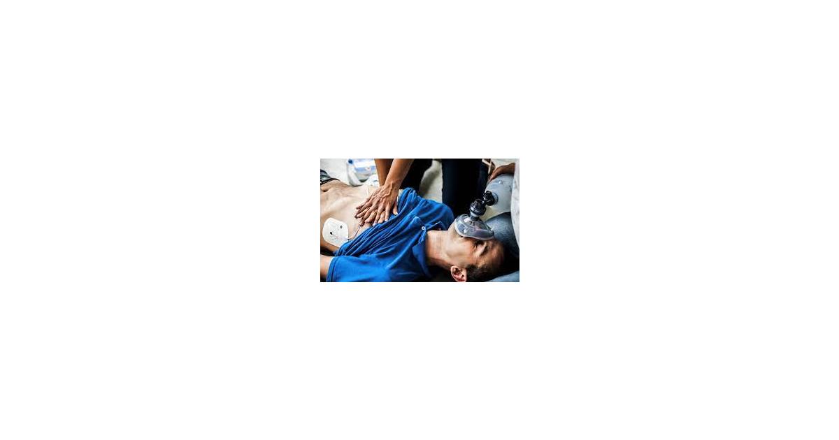
Cardiopulmonary resuscitation (CPR) is a lifesaving procedure that is performed when someone's breathing or heartbeat has stopped. It involves a series of steps that are designed to manually pump blood through the body and maintain the flow of oxygen to the brain until advanced medical treatment can be provided.
CPR typically involves a combination of chest compressions and rescue breaths, which are delivered in a specific rhythm and frequency. The goal is to maintain circulation and oxygenation of vital organs, particularly the brain, until advanced life support measures such as defibrillation or medication can be administered.
Chest compressions are used to manually pump blood through the heart and into the rest of the body. This is typically done by placing both hands on the lower half of the chest and pressing down with enough force to compress the chest by about 2 inches. The compressions should be delivered at a rate of at least 100-120 compressions per minute.
Rescue breaths are used to provide oxygen to the lungs and maintain oxygenation of the body's tissues. This is typically done by pinching the nose shut, creating a seal around the person's mouth with your own, and blowing in enough air to make the chest rise. The breath should be delivered over about one second, and this process should be repeated until the person begins to breathe on their own or advanced medical help arrives.
CPR can be performed by trained laypeople as well as healthcare professionals. It is an important skill that can help save lives in emergency situations where a person's breathing or heartbeat has stopped.
Cardiac arrhythmias are abnormal heart rhythms that result from disturbances in the electrical conduction system of the heart. The heart's normal rhythm is controlled by an electrical signal that originates in the sinoatrial (SA) node, located in the right atrium. This signal travels through the atrioventricular (AV) node and into the ventricles, causing them to contract and pump blood throughout the body.
An arrhythmia occurs when there is a disruption in this electrical pathway or when the heart's natural pacemaker produces an abnormal rhythm. This can cause the heart to beat too fast (tachycardia), too slow (bradycardia), or irregularly.
There are several types of cardiac arrhythmias, including:
1. Atrial fibrillation: A rapid and irregular heartbeat that starts in the atria (the upper chambers of the heart).
2. Atrial flutter: A rapid but regular heartbeat that starts in the atria.
3. Supraventricular tachycardia (SVT): A rapid heartbeat that starts above the ventricles, usually in the atria or AV node.
4. Ventricular tachycardia: A rapid and potentially life-threatening heart rhythm that originates in the ventricles.
5. Ventricular fibrillation: A chaotic and disorganized electrical activity in the ventricles, which can be fatal if not treated immediately.
6. Heart block: A delay or interruption in the conduction of electrical signals from the atria to the ventricles.
Cardiac arrhythmias can cause various symptoms, such as palpitations, dizziness, shortness of breath, chest pain, and fatigue. In some cases, they may not cause any symptoms and go unnoticed. However, if left untreated, certain types of arrhythmias can lead to serious complications, including stroke, heart failure, or even sudden cardiac death.
Treatment for cardiac arrhythmias depends on the type, severity, and underlying causes. Options may include lifestyle changes, medications, cardioversion (electrical shock therapy), catheter ablation, implantable devices such as pacemakers or defibrillators, and surgery. It is essential to consult a healthcare professional for proper evaluation and management of cardiac arrhythmias.
Ventricular Tachycardia (VT) is a rapid heart rhythm that originates from the ventricles, the lower chambers of the heart. It is defined as three or more consecutive ventricular beats at a rate of 120 beats per minute or greater in a resting adult. This abnormal heart rhythm can cause the heart to pump less effectively, leading to inadequate blood flow to the body and potentially life-threatening conditions such as hypotension, shock, or cardiac arrest.
VT can be classified into three types based on its duration, hemodynamic stability, and response to treatment:
1. Non-sustained VT (NSVT): It lasts for less than 30 seconds and is usually well tolerated without causing significant symptoms or hemodynamic instability.
2. Sustained VT (SVT): It lasts for more than 30 seconds, causes symptoms such as palpitations, dizziness, shortness of breath, or chest pain, and may lead to hemodynamic instability.
3. Pulseless VT: It is a type of sustained VT that does not produce a pulse, blood pressure, or adequate cardiac output, requiring immediate electrical cardioversion or defibrillation to restore a normal heart rhythm.
VT can occur in people with various underlying heart conditions such as coronary artery disease, cardiomyopathy, valvular heart disease, congenital heart defects, and electrolyte imbalances. It can also be triggered by certain medications, substance abuse, or electrical abnormalities in the heart. Prompt diagnosis and treatment of VT are crucial to prevent complications and improve outcomes.
A defibrillator is a medical device that delivers a therapeutic dose of electrical energy to the heart. The aim of the treatment is to restore the normal rhythm of the heart in cases where it has started to beat irregularly, or in a chaotic and unsynchronized manner, which can be life-threatening.
There are two main types of defibrillators: external and implantable. External defibrillators are typically used in emergency situations and are often found in public places such as airports, casinos, and sports arenas. These devices have pads that are placed on the chest of the patient, and they deliver an electrical shock to the heart through the chest wall.
Implantable cardioverter-defibrillators (ICDs) are small devices that are implanted in the chest of patients who are at risk of sudden cardiac death due to life-threatening arrhythmias. ICDs constantly monitor the heart's rhythm and deliver an electrical shock if they detect a dangerous arrhythmia, such as ventricular fibrillation or ventricular tachycardia.
Defibrillators are important medical devices that can save lives in emergency situations. They are often used in conjunction with other treatments, such as medications and cardiac procedures, to manage heart conditions and prevent sudden cardiac death.
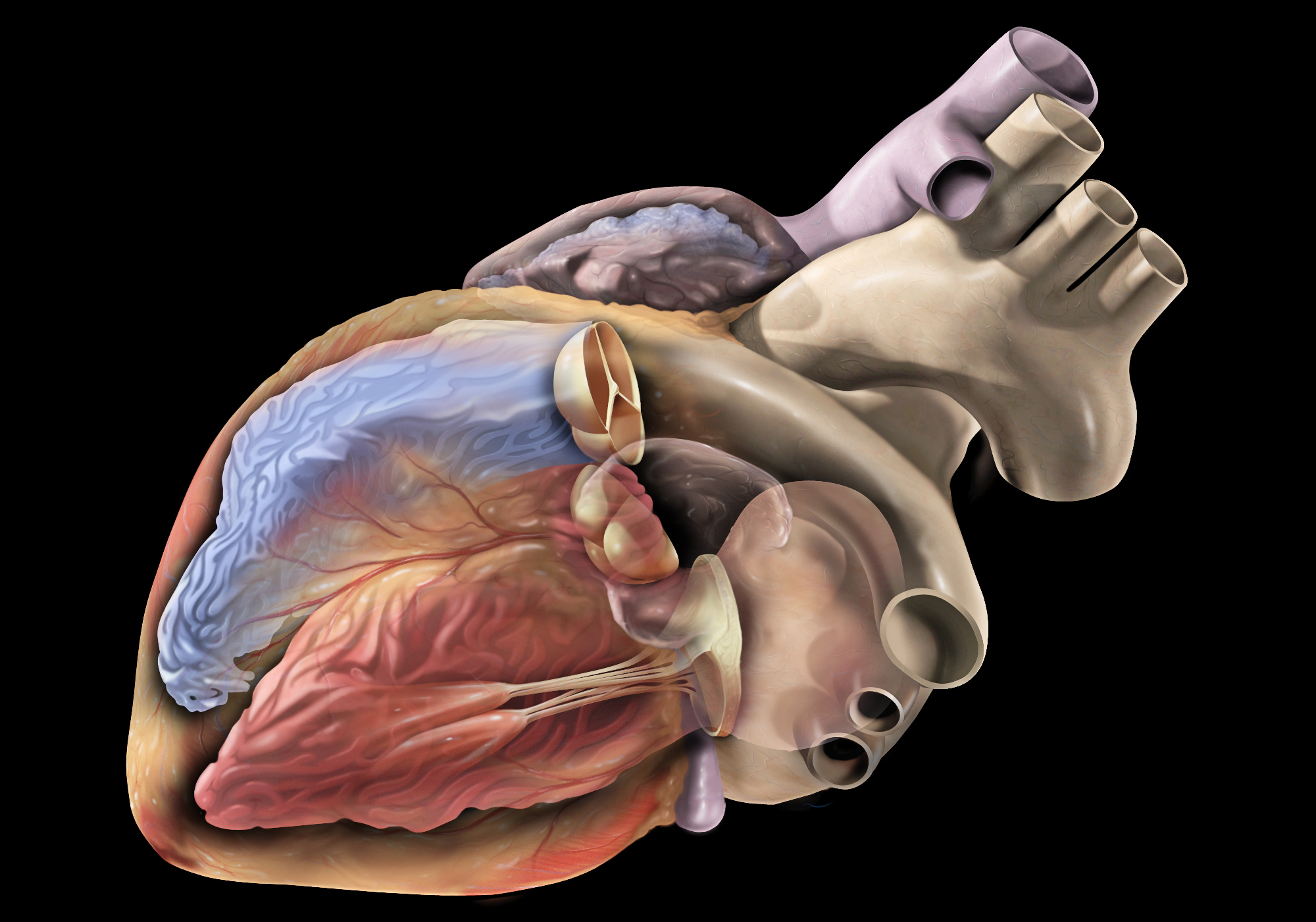
The heart conduction system is a group of specialized cardiac muscle cells that generate and conduct electrical impulses to coordinate the contraction of the heart chambers. The main components of the heart conduction system include:
1. Sinoatrial (SA) node: Also known as the sinus node, it is located in the right atrium near the entrance of the superior vena cava and functions as the primary pacemaker of the heart. It sets the heart rate by generating electrical impulses at regular intervals.
2. Atrioventricular (AV) node: Located in the interatrial septum, near the opening of the coronary sinus, it serves as a relay station for electrical signals between the atria and ventricles. The AV node delays the transmission of impulses to allow the atria to contract before the ventricles.
3. Bundle of His: A bundle of specialized cardiac muscle fibers that conducts electrical impulses from the AV node to the ventricles. It divides into two main branches, the right and left bundle branches, which further divide into smaller Purkinje fibers.
4. Right and left bundle branches: These are extensions of the Bundle of His that transmit electrical impulses to the respective right and left ventricular myocardium. They consist of specialized conducting tissue with large diameters and minimal resistance, allowing for rapid conduction of electrical signals.
5. Purkinje fibers: Fine, branching fibers that arise from the bundle branches and spread throughout the ventricular myocardium. They are responsible for transmitting electrical impulses to the working cardiac muscle cells, triggering coordinated ventricular contraction.
In summary, the heart conduction system is a complex network of specialized muscle cells responsible for generating and conducting electrical signals that coordinate the contraction of the atria and ventricles, ensuring efficient blood flow throughout the body.
An implantable defibrillator is a medical device that is surgically placed inside the chest to continuously monitor the heart's rhythm and deliver electrical shocks to restore a normal heartbeat when it detects a life-threatening arrhythmia, such as ventricular fibrillation or ventricular tachycardia.
The device consists of a small generator that is implanted in the upper chest, along with one or more electrode leads that are threaded through veins and positioned in the heart's chambers. The generator contains a battery and a microcomputer that constantly monitors the heart's electrical activity and detects any abnormal rhythms.
When an arrhythmia is detected, the defibrillator delivers an electrical shock to the heart to restore a normal rhythm. This can be done automatically by the device or manually by a healthcare provider using an external programmer.
Implantable defibrillators are typically recommended for people who have a high risk of sudden cardiac death due to a history of heart attacks, heart failure, or inherited heart conditions that affect the heart's electrical system. They can significantly reduce the risk of sudden cardiac death and improve quality of life for those at risk.
Electrophysiologic techniques, cardiac, refer to medical procedures used to study the electrical activities and conduction systems of the heart. These techniques involve the insertion of electrode catheters into the heart through blood vessels under fluoroscopic guidance to record and stimulate electrical signals. The information obtained from these studies can help diagnose and evaluate various cardiac arrhythmias, determine the optimal treatment strategy, and assess the effectiveness of therapies such as ablation or implantable devices.
The electrophysiologic study (EPS) is a type of cardiac electrophysiologic technique that involves the measurement of electrical signals from different regions of the heart to evaluate its conduction system's function. The procedure can help identify the location of abnormal electrical pathways responsible for arrhythmias and determine the optimal treatment strategy, such as catheter ablation or medication therapy.
Cardiac electrophysiologic techniques are also used in device implantation procedures, such as pacemaker or defibrillator implantation, to ensure proper placement and function of the devices. These techniques can help program and test the devices to optimize their settings for each patient's needs.
In summary, cardiac electrophysiologic techniques are medical procedures used to study and manipulate the electrical activities of the heart, helping diagnose and treat various arrhythmias and other cardiac conditions.
Artificial cardiac pacing is a medical procedure that involves the use of an artificial device to regulate and stimulate the contraction of the heart muscle. This is often necessary when the heart's natural pacemaker, the sinoatrial node, is not functioning properly and the heart is beating too slowly or irregularly.
The artificial pacemaker consists of a small generator that produces electrical impulses and leads that are positioned in the heart to transmit the impulses. The generator is typically implanted just under the skin in the chest, while the leads are inserted into the heart through a vein.
There are different types of artificial cardiac pacing systems, including single-chamber pacemakers, which stimulate either the right atrium or right ventricle, and dual-chamber pacemakers, which stimulate both chambers of the heart. Some pacemakers also have additional features that allow them to respond to changes in the body's needs, such as during exercise or sleep.
Artificial cardiac pacing is a safe and effective treatment for many people with abnormal heart rhythms, and it can significantly improve their quality of life and longevity.
Catheter ablation is a medical procedure in which specific areas of heart tissue that are causing arrhythmias (irregular heartbeats) are destroyed or ablated using heat energy (radiofrequency ablation), cold energy (cryoablation), or other methods. The procedure involves threading one or more catheters through the blood vessels to the heart, where the tip of the catheter can be used to selectively destroy the problematic tissue. Catheter ablation is often used to treat atrial fibrillation, atrial flutter, and other types of arrhythmias that originate in the heart's upper chambers (atria). It may also be used to treat certain types of arrhythmias that originate in the heart's lower chambers (ventricles), such as ventricular tachycardia.
The goal of catheter ablation is to eliminate or reduce the frequency and severity of arrhythmias, thereby improving symptoms and quality of life. In some cases, it may also help to reduce the risk of stroke and other complications associated with arrhythmias. Catheter ablation is typically performed by a specialist in heart rhythm disorders (electrophysiologist) in a hospital or outpatient setting under local anesthesia and sedation. The procedure can take several hours to complete, depending on the complexity of the arrhythmia being treated.
It's important to note that while catheter ablation is generally safe and effective, it does carry some risks, such as bleeding, infection, damage to nearby structures, and the possibility of recurrent arrhythmias. Patients should discuss the potential benefits and risks of the procedure with their healthcare provider before making a decision about treatment.
Amiodarone is a Class III antiarrhythmic medication used to treat and prevent various types of irregular heart rhythms (arrhythmias). It works by stabilizing the electrical activity of the heart and slowing down the nerve impulses in the heart tissue. Amiodarone is available in oral tablet and injection forms.
The medical definition of 'Amiodarone' is:
A benzofuran derivative with Class III antiarrhythmic properties, used for the treatment of ventricular arrhythmias. It has a relatively slow onset of action and is therefore not useful in acute situations. Additionally, it has negative inotropic effects and may exacerbate heart failure. The most serious adverse effect is pulmonary fibrosis, which occurs in approximately 1-2% of patients. Other important side effects include corneal microdeposits, hepatotoxicity, thyroid dysfunction, and photosensitivity. Amiodarone has a very long half-life (approximately 50 days) due to its extensive tissue distribution. It is metabolized by the liver and excreted in bile and urine.
Sources:
1. UpToDate - Amiodarone use in adults: Indications, dosing, and adverse effects.
2. Micromedex - Amiodarone.
3. Drugs.com - Amiodarone.
Sudden cardiac death (SCD) is a sudden, unexpected natural death caused by the cessation of cardiac activity. It is often caused by cardiac arrhythmias, particularly ventricular fibrillation, and is often associated with underlying heart disease, although it can occur in people with no known heart condition. SCD is typically defined as a natural death due to cardiac causes that occurs within one hour of the onset of symptoms, or if the individual was last seen alive in a normal state of health, it can be defined as occurring within 24 hours.
It's important to note that sudden cardiac arrest (SCA) is different from SCD, although they are related. SCA refers to the sudden cessation of cardiac activity, which if not treated immediately can lead to SCD.
Body Surface Potential Mapping (BSPM) is a non-invasive medical technique used to record and analyze the electrical activity of the heart from the surface of the body. It involves placing multiple electrodes on the skin of the chest, back, and limbs to measure the potential differences between these points during each heartbeat. This information is then used to create a detailed, visual representation of the electrical activation pattern of the heart, which can help in the diagnosis and evaluation of various cardiac disorders such as arrhythmias, myocardial infarction, and ventricular hypertrophy.
The BSPM technique provides high-resolution spatial and temporal information about the cardiac electrical activity, making it a valuable tool for both clinical and research purposes. It can help identify the origin and spread of abnormal electrical signals in the heart, which is crucial for determining appropriate treatment strategies. Overall, Body Surface Potential Mapping is an important diagnostic modality that offers unique insights into the electrical functioning of the heart.
Brugada Syndrome is a genetic disorder characterized by abnormal electrocardiogram (ECG) findings and an increased risk of sudden cardiac death. It is typically caused by a mutation in the SCN5A gene, which encodes for a sodium channel protein in the heart. This mutation can lead to abnormal ion transport in the heart cells, causing changes in the electrical activity of the heart that can trigger dangerous arrhythmias.
The ECG findings associated with Brugada Syndrome include a distinct pattern of ST-segment elevation in the right precordial leads (V1-V3), which can appear spontaneously or be induced by certain medications. The syndrome is often classified into two types based on the presence or absence of symptoms:
* Type 1 Brugada Syndrome: This type is characterized by a coved-type ST-segment elevation of at least 2 mm in height in at least one right precordial lead, with a negative T wave. This pattern must be present to make the diagnosis, and it should not be transient or induced by any medication or condition. Type 1 Brugada Syndrome is associated with a higher risk of sudden cardiac death.
* Type 2 Brugada Syndrome: This type is characterized by a saddleback-type ST-segment elevation of at least 2 mm in height in at least one right precordial lead, with a positive or biphasic T wave. The ST segment should return to the baseline level or below within 0.08 seconds after the J point (the junction between the QRS complex and the ST segment). Type 2 Brugada Syndrome is associated with a lower risk of sudden cardiac death compared to Type 1, but it can still pose a significant risk in some individuals.
Brugada Syndrome can affect people of any age, gender, or ethnicity, although it is more commonly diagnosed in middle-aged men of Asian descent. The syndrome can be inherited in an autosomal dominant manner, meaning that a child has a 50% chance of inheriting the mutation from a parent who carries the gene. However, not all individuals with the genetic mutation will develop symptoms or have abnormal ECG findings.
Treatment for Brugada Syndrome typically involves implanting a cardioverter-defibrillator (ICD) to prevent sudden cardiac death. Medications such as quinidine or isoproterenol may also be used to reduce the risk of arrhythmias. Lifestyle modifications, such as avoiding alcohol and certain medications that can trigger arrhythmias, may also be recommended.
Premature cardiac complexes, also known as premature heartbeats or premature ventricular contractions (PVCs), refer to extra or early heartbeats that originate in the lower chambers of the heart (the ventricles). These extra beats disrupt the normal rhythm and sequence of heartbeats, causing the heart to beat earlier than expected.
Premature cardiac complexes can occur in healthy individuals as well as those with heart disease. They are usually harmless and do not cause any symptoms, but in some cases, they may cause palpitations, skipped beats, or a fluttering sensation in the chest. In rare cases, frequent premature cardiac complexes can lead to more serious heart rhythm disorders or decreased heart function.
The diagnosis of premature cardiac complexes is usually made through an electrocardiogram (ECG) or Holter monitoring, which records the electrical activity of the heart over a period of time. Treatment is typically not necessary unless the premature complexes are frequent, symptomatic, or associated with underlying heart disease. In such cases, medications, cardioversion, or catheter ablation may be recommended.
The heart atria are the upper chambers of the heart that receive blood from the veins and deliver it to the lower chambers, or ventricles. There are two atria in the heart: the right atrium receives oxygen-poor blood from the body and pumps it into the right ventricle, which then sends it to the lungs to be oxygenated; and the left atrium receives oxygen-rich blood from the lungs and pumps it into the left ventricle, which then sends it out to the rest of the body. The atria contract before the ventricles during each heartbeat, helping to fill the ventricles with blood and prepare them for contraction.
I believe there might be a misunderstanding in your question. "Dogs" is not a medical term or condition. It is the common name for a domesticated carnivore of the family Canidae, specifically the genus Canis, which includes wolves, foxes, and other extant and extinct species of mammals. Dogs are often kept as pets and companions, and they have been bred in a wide variety of forms and sizes for different purposes, such as hunting, herding, guarding, assisting police and military forces, and providing companionship and emotional support.
If you meant to ask about a specific medical condition or term related to dogs, please provide more context so I can give you an accurate answer.
Resuscitation is a medical term that refers to the process of reversing cardiopulmonary arrest or preventing further deterioration of someone in cardiac or respiratory arrest. It involves a series of interventions aimed at restoring spontaneous blood circulation and breathing, thereby preventing or minimizing tissue damage due to lack of oxygen.
The most common form of resuscitation is cardiopulmonary resuscitation (CPR), which combines chest compressions to manually pump blood through the body with rescue breaths to provide oxygen to the lungs. In a hospital setting, more advanced techniques such as defibrillation, medication administration, and intubation may also be used as part of the resuscitation process.
The goal of resuscitation is to stabilize the patient's condition and prevent further harm while treating the underlying cause of the arrest. Successful resuscitation can lead to a full recovery or, in some cases, result in varying degrees of neurological impairment depending on the severity and duration of the cardiac or respiratory arrest.
The refractory period, electrophysiological, refers to the time interval during which a cardiac or neural cell is unable to respond to a new stimulus immediately after an action potential has been generated. This period is divided into two phases: the absolute refractory period and the relative refractory period.
During the absolute refractory period, the cell cannot be re-stimulated, regardless of the strength of the stimulus, due to the rapid inactivation of voltage-gated sodium channels that are responsible for the rapid depolarization during an action potential. This phase is crucial for maintaining the unidirectional conduction of electrical impulses and preventing the occurrence of re-entry circuits, which can lead to life-threatening arrhythmias in the heart or hyperexcitability in neural tissue.
The relative refractory period follows the absolute refractory period and is characterized by a reduced excitability of the cell. During this phase, a stronger than normal stimulus is required to elicit an action potential due to the slower recovery of voltage-gated sodium channels and the partial activation of potassium channels, which promote repolarization. The duration of both the absolute and relative refractory periods varies depending on the cell type, its physiological state, and other factors such as temperature and pH.
In summary, the electrophysiological refractory period is a fundamental property of excitable cells that ensures proper electrical signaling and prevents uncontrolled excitation or re-entry circuits.
Tachycardia is a medical term that refers to an abnormally rapid heart rate, often defined as a heart rate greater than 100 beats per minute in adults. It can occur in either the atria (upper chambers) or ventricles (lower chambers) of the heart. Different types of tachycardia include supraventricular tachycardia (SVT), atrial fibrillation, atrial flutter, and ventricular tachycardia.
Tachycardia can cause various symptoms such as palpitations, shortness of breath, dizziness, lightheadedness, chest discomfort, or syncope (fainting). In some cases, tachycardia may not cause any symptoms and may only be detected during a routine physical examination or medical test.
The underlying causes of tachycardia can vary widely, including heart disease, electrolyte imbalances, medications, illicit drug use, alcohol abuse, smoking, stress, anxiety, and other medical conditions. In some cases, the cause may be unknown. Treatment for tachycardia depends on the underlying cause, type, severity, and duration of the arrhythmia.
Pulmonary veins are blood vessels that carry oxygenated blood from the lungs to the left atrium of the heart. There are four pulmonary veins in total, two from each lung, and they are the only veins in the body that carry oxygen-rich blood. The oxygenated blood from the pulmonary veins is then pumped by the left ventricle to the rest of the body through the aorta. Any blockage or damage to the pulmonary veins can lead to various cardiopulmonary conditions, such as pulmonary hypertension and congestive heart failure.
Heart massage, also known as cardiac massage or chest compression, is a medical procedure that involves applying pressure to the chest in order to manually pump blood through the heart and maintain circulation when the heart has stopped or is not functioning effectively. This is a critical component of cardiopulmonary resuscitation (CPR) and is typically performed during a cardiac arrest to help restore proper blood flow to vital organs and tissues.
During heart massage, the rescuer places their hands on the lower half of the victim's chest, typically at the center, and presses down with the heel of one or both hands. The recommended compression depth for adults is at least 2 inches (5 cm) and should be performed at a rate of 100-120 compressions per minute. It is essential to minimize interruptions in chest compressions and ensure that they are deep and fast enough to maintain adequate blood flow.
Heart massage can also be performed surgically during specific medical procedures, such as open-heart surgery or extracorporeal membrane oxygenation (ECMO). In these cases, the surgeon directly compresses the heart using their hands or specialized instruments. This technique is called a "surgical heart massage" or "direct cardiac compression."
It's important to note that heart massage should only be performed by trained individuals, as improper techniques can cause harm and potentially worsen the patient's condition.
In the field of medicine, "time factors" refer to the duration of symptoms or time elapsed since the onset of a medical condition, which can have significant implications for diagnosis and treatment. Understanding time factors is crucial in determining the progression of a disease, evaluating the effectiveness of treatments, and making critical decisions regarding patient care.
For example, in stroke management, "time is brain," meaning that rapid intervention within a specific time frame (usually within 4.5 hours) is essential to administering tissue plasminogen activator (tPA), a clot-busting drug that can minimize brain damage and improve patient outcomes. Similarly, in trauma care, the "golden hour" concept emphasizes the importance of providing definitive care within the first 60 minutes after injury to increase survival rates and reduce morbidity.
Time factors also play a role in monitoring the progression of chronic conditions like diabetes or heart disease, where regular follow-ups and assessments help determine appropriate treatment adjustments and prevent complications. In infectious diseases, time factors are crucial for initiating antibiotic therapy and identifying potential outbreaks to control their spread.
Overall, "time factors" encompass the significance of recognizing and acting promptly in various medical scenarios to optimize patient outcomes and provide effective care.
The pericardium is the double-walled sac that surrounds the heart. It has an outer fibrous layer and an inner serous layer, which further divides into two parts: the parietal layer lining the fibrous pericardium and the visceral layer (epicardium) closely adhering to the heart surface.
The space between these two layers is filled with a small amount of lubricating serous fluid, allowing for smooth movement of the heart within the pericardial cavity. The pericardium provides protection, support, and helps maintain the heart's normal position within the chest while reducing friction during heart contractions.
Heart rate is the number of heartbeats per unit of time, often expressed as beats per minute (bpm). It can vary significantly depending on factors such as age, physical fitness, emotions, and overall health status. A resting heart rate between 60-100 bpm is generally considered normal for adults, but athletes and individuals with high levels of physical fitness may have a resting heart rate below 60 bpm due to their enhanced cardiovascular efficiency. Monitoring heart rate can provide valuable insights into an individual's health status, exercise intensity, and response to various treatments or interventions.
The heart ventricles are the two lower chambers of the heart that receive blood from the atria and pump it to the lungs or the rest of the body. The right ventricle pumps deoxygenated blood to the lungs, while the left ventricle pumps oxygenated blood to the rest of the body. Both ventricles have thick, muscular walls to generate the pressure necessary to pump blood through the circulatory system.
Procainamide is an antiarrhythmic medication used to treat various types of irregular heart rhythms (arrhythmias), such as atrial fibrillation, atrial flutter, and ventricular tachycardia. It works by prolonging the duration of the cardiac action potential and decreasing the slope of the phase 0 depolarization, which helps to stabilize the heart's electrical activity and restore a normal rhythm.
Procainamide is classified as a Class Ia antiarrhythmic drug, according to the Vaughan Williams classification system. It primarily affects the fast sodium channels in the heart muscle cells, reducing their availability during depolarization. This results in a decreased rate of impulse generation and conduction velocity, which can help to suppress abnormal rhythms.
The medication is available as an oral formulation (procainamide hydrochloride) and as an injectable solution for intravenous use. Common side effects of procainamide include nausea, vomiting, diarrhea, headache, and dizziness. Procainamide can also cause a lupus-like syndrome, characterized by joint pain, skin rashes, and other autoimmune symptoms, in some patients who take the medication for an extended period.
It is essential to monitor procainamide levels in the blood during treatment to ensure that the drug is within the therapeutic range and to minimize the risk of adverse effects. Healthcare providers should also regularly assess patients' renal function, as procainamide and its active metabolite, N-acetylprocainamide (NAPA), are primarily excreted by the kidneys.
The endocardium is the innermost layer of tissue that lines the chambers of the heart and the valves between them. It is a thin, smooth membrane that is in contact with the blood within the heart. This layer helps to maintain the heart's internal environment, facilitates the smooth movement of blood through the heart, and provides a protective barrier against infection and other harmful substances. The endocardium is composed of simple squamous epithelial cells called endothelial cells, which are supported by a thin layer of connective tissue.
"Swine" is a common term used to refer to even-toed ungulates of the family Suidae, including domestic pigs and wild boars. However, in a medical context, "swine" often appears in the phrase "swine flu," which is a strain of influenza virus that typically infects pigs but can also cause illness in humans. The 2009 H1N1 pandemic was caused by a new strain of swine-origin influenza A virus, which was commonly referred to as "swine flu." It's important to note that this virus is not transmitted through eating cooked pork products; it spreads from person to person, mainly through respiratory droplets produced when an infected person coughs or sneezes.
In medical terms, the heart is a muscular organ located in the thoracic cavity that functions as a pump to circulate blood throughout the body. It's responsible for delivering oxygen and nutrients to the tissues and removing carbon dioxide and other wastes. The human heart is divided into four chambers: two atria on the top and two ventricles on the bottom. The right side of the heart receives deoxygenated blood from the body and pumps it to the lungs, while the left side receives oxygenated blood from the lungs and pumps it out to the rest of the body. The heart's rhythmic contractions and relaxations are regulated by a complex electrical conduction system.
Treatment outcome is a term used to describe the result or effect of medical treatment on a patient's health status. It can be measured in various ways, such as through symptoms improvement, disease remission, reduced disability, improved quality of life, or survival rates. The treatment outcome helps healthcare providers evaluate the effectiveness of a particular treatment plan and make informed decisions about future care. It is also used in clinical research to compare the efficacy of different treatments and improve patient care.
Flecainide is an antiarrhythmic medication used to regularize abnormal heart rhythms, specifically certain types of irregular heartbeats called ventricular arrhythmias and paroxysmal atrial tachycardia/atrial fibrillation. It works by blocking sodium channels in the heart, which helps to slow down the conduction of electrical signals and reduces the likelihood of erratic heart rhythms.
Flecainide is available in oral forms such as tablets or capsules and is typically prescribed under the supervision of a healthcare professional experienced in managing heart rhythm disorders. It's important to note that flecainide can have serious side effects, including increasing the risk of dangerous arrhythmias in some patients, so it should only be used under close medical monitoring.
This definition is for informational purposes only and should not be considered a substitute for professional medical advice, diagnosis, or treatment. If you have any questions about your medications or health conditions, please consult with your healthcare provider.
Out-of-hospital cardiac arrest (OHCA) is a medical condition where the heart suddenly and unexpectedly stops functioning outside of a hospital setting, leading to the cessation of blood circulation and breathing. This results in immediate unconsciousness and can be caused by various factors such as electrical disturbances in the heart, severe trauma, or suffocation. It is a serious emergency that requires immediate cardiopulmonary resuscitation (CPR) and advanced life support measures to restore spontaneous circulation and improve survival outcomes.
Myocardial ischemia is a condition in which the blood supply to the heart muscle (myocardium) is reduced or blocked, leading to insufficient oxygen delivery and potential damage to the heart tissue. This reduction in blood flow typically results from the buildup of fatty deposits, called plaques, in the coronary arteries that supply the heart with oxygen-rich blood. The plaques can rupture or become unstable, causing the formation of blood clots that obstruct the artery and limit blood flow.
Myocardial ischemia may manifest as chest pain (angina pectoris), shortness of breath, fatigue, or irregular heartbeats (arrhythmias). In severe cases, it can lead to myocardial infarction (heart attack) if the oxygen supply is significantly reduced or cut off completely, causing permanent damage or death of the heart muscle. Early diagnosis and treatment of myocardial ischemia are crucial for preventing further complications and improving patient outcomes.
An action potential is a brief electrical signal that travels along the membrane of a nerve cell (neuron) or muscle cell. It is initiated by a rapid, localized change in the permeability of the cell membrane to specific ions, such as sodium and potassium, resulting in a rapid influx of sodium ions and a subsequent efflux of potassium ions. This ion movement causes a brief reversal of the electrical potential across the membrane, which is known as depolarization. The action potential then propagates along the cell membrane as a wave, allowing the electrical signal to be transmitted over long distances within the body. Action potentials play a crucial role in the communication and functioning of the nervous system and muscle tissue.
Sudden death is a term used to describe a situation where a person dies abruptly and unexpectedly, often within minutes to hours of the onset of symptoms. It is typically caused by cardiac or respiratory arrest, which can be brought on by various medical conditions such as heart disease, stroke, severe infections, drug overdose, or trauma. In some cases, the exact cause of sudden death may remain unknown even after a thorough post-mortem examination.
It is important to note that sudden death should not be confused with "sudden cardiac death," which specifically refers to deaths caused by the abrupt loss of heart function (cardiac arrest). Sudden cardiac death is often related to underlying heart conditions such as coronary artery disease, cardiomyopathy, or electrical abnormalities in the heart.
Atrial flutter is a type of abnormal heart rhythm or arrhythmia that originates in the atria - the upper chambers of the heart. In atrial flutter, the atria beat too quickly, usually between 250 and 350 beats per minute, which is much faster than the normal resting rate of 60 to 100 beats per minute.
This rapid beating causes the atria to quiver or "flutter" instead of contracting effectively. As a result, blood may not be pumped efficiently into the ventricles - the lower chambers of the heart - which can lead to reduced cardiac output and symptoms such as palpitations, shortness of breath, fatigue, dizziness, or chest discomfort.
Atrial flutter is often caused by underlying heart conditions, such as coronary artery disease, hypertension, valvular heart disease, or congenital heart defects. It can also be a complication of cardiac surgery or other medical procedures. In some cases, atrial flutter may occur without any apparent underlying cause, which is known as lone atrial flutter.
Treatment for atrial flutter typically involves medications to control the heart rate and rhythm, electrical cardioversion to restore a normal heart rhythm, or catheter ablation to destroy the abnormal electrical pathways in the heart that are causing the arrhythmia. In some cases, surgical intervention may be necessary to treat atrial flutter.
Ambulatory electrocardiography, also known as ambulatory ECG or Holter monitoring, is a non-invasive method of recording the electrical activity of the heart over an extended period of time (typically 24 hours or more) while the patient goes about their daily activities. The device used to record the ECG is called a Holter monitor, which consists of a small, portable recorder that is attached to the patient's chest with electrodes.
The recorded data provides information on any abnormalities in the heart's rhythm or electrical activity during different stages of activity and rest, allowing healthcare providers to diagnose and evaluate various cardiac conditions such as arrhythmias, ischemia, and infarction. The ability to monitor the heart's activity over an extended period while the patient performs their normal activities provides valuable information that may not be captured during a standard ECG, which only records the heart's electrical activity for a few seconds.
In summary, ambulatory electrocardiography is a diagnostic tool used to evaluate the electrical activity of the heart over an extended period, allowing healthcare providers to diagnose and manage various cardiac conditions.
Warfarin is a anticoagulant medication that works by inhibiting the vitamin K-dependent activation of several coagulation factors (factors II, VII, IX, and X). This results in prolonged clotting times and reduced thrombus formation. It is commonly used to prevent and treat blood clots in conditions such as atrial fibrillation, deep vein thrombosis, and pulmonary embolism. Warfarin is also known by its brand names Coumadin and Jantoven.
It's important to note that warfarin has a narrow therapeutic index, meaning that the difference between an effective dose and a toxic one is small. Therefore, it requires careful monitoring of the patient's coagulation status through regular blood tests (INR) to ensure that the dosage is appropriate and to minimize the risk of bleeding complications.
Follow-up studies are a type of longitudinal research that involve repeated observations or measurements of the same variables over a period of time, in order to understand their long-term effects or outcomes. In medical context, follow-up studies are often used to evaluate the safety and efficacy of medical treatments, interventions, or procedures.
In a typical follow-up study, a group of individuals (called a cohort) who have received a particular treatment or intervention are identified and then followed over time through periodic assessments or data collection. The data collected may include information on clinical outcomes, adverse events, changes in symptoms or functional status, and other relevant measures.
The results of follow-up studies can provide important insights into the long-term benefits and risks of medical interventions, as well as help to identify factors that may influence treatment effectiveness or patient outcomes. However, it is important to note that follow-up studies can be subject to various biases and limitations, such as loss to follow-up, recall bias, and changes in clinical practice over time, which must be carefully considered when interpreting the results.
Cardiovascular models are simplified representations or simulations of the human cardiovascular system used in medical research, education, and training. These models can be physical, computational, or mathematical and are designed to replicate various aspects of the heart, blood vessels, and blood flow. They can help researchers study the structure and function of the cardiovascular system, test new treatments and interventions, and train healthcare professionals in diagnostic and therapeutic techniques.
Physical cardiovascular models may include artificial hearts, blood vessels, or circulation systems made from materials such as plastic, rubber, or silicone. These models can be used to study the mechanics of heart valves, the effects of different surgical procedures, or the impact of various medical devices on blood flow.
Computational and mathematical cardiovascular models use algorithms and equations to simulate the behavior of the cardiovascular system. These models may range from simple representations of a single heart chamber to complex simulations of the entire circulatory system. They can be used to study the electrical activity of the heart, the biomechanics of blood flow, or the distribution of drugs in the body.
Overall, cardiovascular models play an essential role in advancing our understanding of the human body and improving patient care.
Myocardial infarction (MI), also known as a heart attack, is a medical condition characterized by the death of a segment of heart muscle (myocardium) due to the interruption of its blood supply. This interruption is most commonly caused by the blockage of a coronary artery by a blood clot formed on the top of an atherosclerotic plaque, which is a buildup of cholesterol and other substances in the inner lining of the artery.
The lack of oxygen and nutrients supply to the heart muscle tissue results in damage or death of the cardiac cells, causing the affected area to become necrotic. The extent and severity of the MI depend on the size of the affected area, the duration of the occlusion, and the presence of collateral circulation.
Symptoms of a myocardial infarction may include chest pain or discomfort, shortness of breath, nausea, lightheadedness, and sweating. Immediate medical attention is necessary to restore blood flow to the affected area and prevent further damage to the heart muscle. Treatment options for MI include medications, such as thrombolytics, antiplatelet agents, and pain relievers, as well as procedures such as percutaneous coronary intervention (PCI) or coronary artery bypass grafting (CABG).
Recurrence, in a medical context, refers to the return of symptoms or signs of a disease after a period of improvement or remission. It indicates that the condition has not been fully eradicated and may require further treatment. Recurrence is often used to describe situations where a disease such as cancer comes back after initial treatment, but it can also apply to other medical conditions. The likelihood of recurrence varies depending on the type of disease and individual patient factors.
Anticoagulants are a class of medications that work to prevent the formation of blood clots in the body. They do this by inhibiting the coagulation cascade, which is a series of chemical reactions that lead to the formation of a clot. Anticoagulants can be given orally, intravenously, or subcutaneously, depending on the specific drug and the individual patient's needs.
There are several different types of anticoagulants, including:
1. Heparin: This is a naturally occurring anticoagulant that is often used in hospitalized patients who require immediate anticoagulation. It works by activating an enzyme called antithrombin III, which inhibits the formation of clots.
2. Low molecular weight heparin (LMWH): LMWH is a form of heparin that has been broken down into smaller molecules. It has a longer half-life than standard heparin and can be given once or twice daily by subcutaneous injection.
3. Direct oral anticoagulants (DOACs): These are newer oral anticoagulants that work by directly inhibiting specific clotting factors in the coagulation cascade. Examples include apixaban, rivaroxaban, and dabigatran.
4. Vitamin K antagonists: These are older oral anticoagulants that work by inhibiting the action of vitamin K, which is necessary for the formation of clotting factors. Warfarin is an example of a vitamin K antagonist.
Anticoagulants are used to prevent and treat a variety of conditions, including deep vein thrombosis (DVT), pulmonary embolism (PE), atrial fibrillation, and prosthetic heart valve thrombosis. It is important to note that anticoagulants can increase the risk of bleeding, so they must be used with caution and regular monitoring of blood clotting times may be required.
Bundle-branch block (BBB) is a type of conduction delay or block in the heart's electrical system that affects the way electrical impulses travel through the ventricles (the lower chambers of the heart). In BBB, one of the two main bundle branches that conduct electrical impulses to the ventricles is partially or completely blocked, causing a delay in the contraction of one of the ventricles.
There are two types of bundle-branch block: right bundle-branch block (RBBB) and left bundle-branch block (LBBB). In RBBB, the right bundle branch is affected, while in LBBB, the left bundle branch is affected. The symptoms and severity of BBB can vary depending on the underlying cause and the presence of other heart conditions.
In some cases, BBB may not cause any noticeable symptoms and may only be detected during a routine electrocardiogram (ECG). However, if BBB occurs along with other heart conditions such as coronary artery disease, heart failure, or cardiomyopathy, it can increase the risk of serious complications such as arrhythmias, syncope, and even sudden cardiac death.
Treatment for bundle-branch block depends on the underlying cause and the severity of the condition. In some cases, no treatment may be necessary, while in others, medications, pacemakers, or other treatments may be recommended to manage symptoms and prevent complications.
Lidocaine is a type of local anesthetic that numbs painful areas and is used to prevent pain during certain medical procedures. It works by blocking the nerves that transmit pain signals to the brain. In addition to its use as an anesthetic, lidocaine can also be used to treat irregular heart rates and relieve itching caused by allergic reactions or skin conditions such as eczema.
Lidocaine is available in various forms, including creams, gels, ointments, sprays, solutions, and injectable preparations. It can be applied directly to the skin or mucous membranes, or it can be administered by injection into a muscle or vein. The specific dosage and method of administration will depend on the reason for its use and the individual patient's medical history and current health status.
Like all medications, lidocaine can have side effects, including allergic reactions, numbness that lasts too long, and in rare cases, heart problems or seizures. It is important to follow the instructions of a healthcare provider carefully when using lidocaine to minimize the risk of adverse effects.
Epicardial mapping is a medical procedure used to create a detailed map of the electrical activity on the surface of the heart (epicardium). This technique is often used during electrophysiology studies to help diagnose and locate the source of abnormal heart rhythms, such as ventricular tachycardia or atrial fibrillation.
During epicardial mapping, a specialist (usually an electrophysiologist) will introduce a catheter through a vein or artery, which is then guided to the heart. Once in position, electrodes on the tip of the catheter record electrical signals from the heart's surface. These signals are used to create a detailed map of the heart's electrical activity, allowing the specialist to identify areas with abnormal electrical patterns.
This information can be crucial for determining the best course of treatment, such as targeted ablation therapy to eliminate the source of the arrhythmia. Epicardial mapping is typically performed in an electrophysiology lab or cardiac catheterization laboratory under fluoroscopy guidance, and it requires expertise in both cardiovascular medicine and interventional techniques.
Emergency Medical Services (EMS) is a system that provides immediate and urgent medical care, transportation, and treatment to patients who are experiencing an acute illness or injury that poses an immediate threat to their health, safety, or life. EMS is typically composed of trained professionals, such as emergency medical technicians (EMTs), paramedics, and first responders, who work together to assess a patient's condition, administer appropriate medical interventions, and transport the patient to a hospital or other medical facility for further treatment.
The goal of EMS is to quickly and effectively stabilize patients in emergency situations, prevent further injury or illness, and ensure that they receive timely and appropriate medical care. This may involve providing basic life support (BLS) measures such as cardiopulmonary resuscitation (CPR), controlling bleeding, and managing airway obstructions, as well as more advanced interventions such as administering medications, establishing intravenous lines, and performing emergency procedures like intubation or defibrillation.
EMS systems are typically organized and managed at the local or regional level, with coordination and oversight provided by public health agencies, hospitals, and other healthcare organizations. EMS providers may work for private companies, non-profit organizations, or government agencies, and they may be dispatched to emergencies via 911 or other emergency response systems.
In summary, Emergency Medical Services (EMS) is a critical component of the healthcare system that provides urgent medical care and transportation to patients who are experiencing acute illnesses or injuries. EMS professionals work together to quickly assess, stabilize, and transport patients to appropriate medical facilities for further treatment.
Ventricular Premature Complexes (VPCs), also known as Ventricular Extrasystoles or Premature Ventricular Contractions (PVCs), are extra heartbeats that originate in the ventricles, the lower chambers of the heart. These premature beats disrupt the normal sequence of electrical impulses in the heart and cause the ventricles to contract earlier than they should.
VPCs can result in a noticeable "skipped" or "extra" beat sensation, often followed by a stronger beat as the heart returns to its regular rhythm. They may occur occasionally in healthy individuals with no underlying heart condition, but frequent VPCs could indicate an underlying issue such as heart disease, electrolyte imbalance, or digitalis toxicity. In some cases, VPCs can be harmless and require no treatment; however, if they are frequent or associated with structural heart problems, further evaluation and management may be necessary to prevent potential complications like reduced cardiac output or heart failure.
Sotalol is a non-selective beta blocker and class III antiarrhythmic drug. It works by blocking the action of certain natural substances in your body, such as adrenaline, on the heart. This helps to decrease the heart's workload, slow the heart rate, and regulate certain types of irregular heartbeats (such as atrial fibrillation).
Sotalol is used to treat various types of irregular heartbeats (atrial fibrillation/flutter, ventricular tachycardia) and may also be used to help maintain a normal heart rhythm after a heart attack. It is important to note that Sotalol should only be prescribed by a healthcare professional who has experience in treating heart rhythm disorders.
This medical definition is based on the information provided by the National Library of Medicine (NLM).
Voltage-sensitive dye imaging (VSDI) is not a medical definition itself, but it is a technique used in the field of physiology and neuroscience to measure the electrical activity of cells, particularly excitable cells such as neurons and cardiac myocytes. Here's a brief explanation:
Voltage-sensitive dyes are fluorescent or luminescent molecules that change their optical properties in response to changes in membrane potential. When these dyes bind to the cell membrane, they can report on the electrical activity of the cell by changing their emission intensity, polarization, or lifetime depending on the voltage across the membrane.
VSDI is a technique that uses these voltage-sensitive dyes to measure changes in membrane potential in a population of cells or even in an entire organ. By illuminating the sample with light and measuring the emitted fluorescence or luminescence, researchers can visualize and quantify the electrical activity of cells in real-time.
VSDI has many applications in basic research, including studying the electrical properties of neurons, mapping neural circuits, investigating the mechanisms of excitation-contraction coupling in cardiac myocytes, and developing new drugs that target ion channels. However, it is not a commonly used clinical technique due to its limitations, such as the need for specialized equipment, the potential for phototoxicity, and the difficulty of interpreting signals from complex tissues.
Prospective studies, also known as longitudinal studies, are a type of cohort study in which data is collected forward in time, following a group of individuals who share a common characteristic or exposure over a period of time. The researchers clearly define the study population and exposure of interest at the beginning of the study and follow up with the participants to determine the outcomes that develop over time. This type of study design allows for the investigation of causal relationships between exposures and outcomes, as well as the identification of risk factors and the estimation of disease incidence rates. Prospective studies are particularly useful in epidemiology and medical research when studying diseases with long latency periods or rare outcomes.
NAV1.5, also known as SCN5A, is a specific type of voltage-gated sodium channel found in the heart muscle cells (cardiomyocytes). These channels play a crucial role in the generation and transmission of electrical signals that coordinate the contraction of the heart.
More specifically, NAV1.5 channels are responsible for the rapid influx of sodium ions into cardiomyocytes during the initial phase of the action potential, which is the electrical excitation of the cell. This rapid influx of sodium ions helps to initiate and propagate the action potential throughout the heart muscle, allowing for coordinated contraction and proper heart function.
Mutations in the SCN5A gene, which encodes the NAV1.5 channel, have been associated with various cardiac arrhythmias, including long QT syndrome, Brugada syndrome, and familial atrial fibrillation, among others. These genetic disorders can lead to abnormal heart rhythms, syncope, and in some cases, sudden cardiac death.
The atrial appendage, also known as the left atrial appendage (LAA), is a small, ear-shaped structure that is located on the upper left chamber of the heart (left atrium). It has a unique muscular structure and plays a role in the normal functioning of the heart. However, it is best known for its association with atrial fibrillation, a common type of irregular heart rhythm. In people with atrial fibrillation, blood clots can form in the LAA, which can then travel to other parts of the body and cause strokes. For this reason, one treatment option for atrial fibrillation is to close off or remove the LAA to reduce the risk of stroke.
Syncope is a medical term defined as a transient, temporary loss of consciousness and postural tone due to reduced blood flow to the brain. It's often caused by a drop in blood pressure, which can be brought on by various factors such as dehydration, emotional stress, prolonged standing, or certain medical conditions like heart diseases, arrhythmias, or neurological disorders.
During a syncope episode, an individual may experience warning signs such as lightheadedness, dizziness, blurred vision, or nausea before losing consciousness. These episodes usually last only a few minutes and are followed by a rapid, full recovery. However, if left untreated or undiagnosed, recurrent syncope can lead to severe injuries from falls or even life-threatening conditions related to the underlying cause.
An artificial pacemaker is a medical device that uses electrical impulses to regulate the beating of the heart. It is typically used when the heart's natural pacemaker, the sinoatrial node, is not functioning properly and the heart rate is too slow or irregular. The pacemaker consists of a small generator that contains a battery and electronic circuits, which are connected to one or more electrodes that are placed in the heart.
The generator sends electrical signals through the electrodes to stimulate the heart muscle and cause it to contract, thereby maintaining a regular heart rhythm. Artificial pacemakers can be programmed to deliver electrical impulses at a specific rate or in response to the body's needs. They are typically implanted in the chest during a surgical procedure and can last for many years before needing to be replaced.
Artificial pacemakers are an effective treatment for various types of bradycardia, which is a heart rhythm disorder characterized by a slow heart rate. Pacemakers can significantly improve symptoms associated with bradycardia, such as fatigue, dizziness, shortness of breath, and fainting spells.
Disopyramide is an antiarrhythmic medication that is primarily used to treat certain types of irregular heart rhythms (arrhythmias), such as ventricular tachycardia and atrial fibrillation. It works by blocking the activity of sodium channels in the heart, which helps to slow down and regulate the heart rate.
Disopyramide is available in immediate-release and extended-release forms, and it may be taken orally as a tablet or capsule. Common side effects of this medication include dry mouth, blurred vision, constipation, and difficulty urinating. More serious side effects can include dizziness, fainting, irregular heartbeat, and allergic reactions.
It is important to take disopyramide exactly as directed by a healthcare provider, as improper use or dosing can lead to serious complications. Additionally, individuals with certain medical conditions, such as heart failure, kidney disease, or myasthenia gravis, may not be able to safely take this medication.
Induced hypothermia is a medically controlled lowering of the core body temperature to around 89.6-93.2°F (32-34°C) for therapeutic purposes. It is intentionally induced to reduce the metabolic rate and oxygen demand of organs, thereby offering protection during periods of low blood flow or inadequate oxygenation, such as during cardiac bypass surgery, severe trauma, or after a cardiac arrest. The deliberate induction and maintenance of hypothermia can help minimize tissue damage and improve outcomes in specific clinical scenarios. Once the risk has passed, the body temperature is gradually rewarmed to normal levels under controlled conditions.
Wolff-Parkinson-White (WPW) Syndrome is a heart condition characterized by the presence of an accessory pathway or abnormal electrical connection between the atria (the upper chambers of the heart) and ventricles (the lower chambers of the heart). This accessory pathway allows electrical impulses to bypass the normal conduction system, leading to a shorter PR interval and a "delta wave" on the electrocardiogram (ECG), which is the hallmark of WPW Syndrome.
Individuals with WPW Syndrome may experience no symptoms or may have palpitations, rapid heartbeat (tachycardia), or episodes of atrial fibrillation. In some cases, WPW Syndrome can lead to more serious heart rhythm disturbances and may require treatment, such as medication, catheter ablation, or in rare cases, surgery.
It is important to note that not all individuals with WPW Syndrome will experience symptoms or complications, and many people with this condition can lead normal, active lives with appropriate monitoring and management.
Myocardial reperfusion injury is a pathological process that occurs when blood flow is restored to the heart muscle (myocardium) after a period of ischemia or reduced oxygen supply, such as during a myocardial infarction (heart attack). The restoration of blood flow, although necessary to salvage the dying tissue, can itself cause further damage to the heart muscle. This paradoxical phenomenon is known as myocardial reperfusion injury.
The mechanisms behind myocardial reperfusion injury are complex and involve several processes, including:
1. Oxidative stress: The sudden influx of oxygen into the previously ischemic tissue leads to an overproduction of reactive oxygen species (ROS), which can damage cellular structures, such as proteins, lipids, and DNA.
2. Calcium overload: During reperfusion, there is an increase in calcium influx into the cardiomyocytes (heart muscle cells). This elevated intracellular calcium level can disrupt normal cellular functions, leading to further damage.
3. Inflammation: Reperfusion triggers an immune response, with the recruitment of inflammatory cells, such as neutrophils and monocytes, to the site of injury. These cells release cytokines and other mediators that can exacerbate tissue damage.
4. Mitochondrial dysfunction: The restoration of blood flow can cause mitochondria, the powerhouses of the cell, to malfunction, leading to the release of pro-apoptotic factors and contributing to cell death.
5. Vasoconstriction and microvascular obstruction: During reperfusion, there may be vasoconstriction of the small blood vessels (microvasculature) in the heart, which can further limit blood flow and contribute to tissue damage.
Myocardial reperfusion injury is a significant concern because it can negate some of the benefits of early reperfusion therapy, such as thrombolysis or primary percutaneous coronary intervention (PCI), used to treat acute myocardial infarction. Strategies to minimize myocardial reperfusion injury are an area of active research and include pharmacological interventions, ischemic preconditioning, and remote ischemic conditioning.
Electrophysiology is a branch of medicine that deals with the electrical activities of the body, particularly the heart. In a medical context, electrophysiology studies (EPS) are performed to assess abnormal heart rhythms (arrhythmias) and to evaluate the effectiveness of certain treatments, such as medication or pacemakers.
During an EPS, electrode catheters are inserted into the heart through blood vessels in the groin or neck. These catheters can record the electrical activity of the heart and stimulate it to help identify the source of the arrhythmia. The information gathered during the study can help doctors determine the best course of treatment for each patient.
In addition to cardiac electrophysiology, there are also other subspecialties within electrophysiology, such as neuromuscular electrophysiology, which deals with the electrical activity of the nervous system and muscles.
Propafenone is an antiarrhythmic medication used to treat certain types of irregular heartbeats (such as atrial fibrillation, paroxysmal supraventricular tachycardia). It works by blocking certain electrical signals in the heart to help it beat regularly. Propafenone belongs to a class of drugs known as Class IC antiarrhythmics.
It is important to note that this definition provides an overview of what propafenone is and how it is used, but it does not cover all possible uses, precautions, side effects, and interactions related to the drug. For more detailed information about propafenone, including its specific indications, contraindications, and potential adverse effects, consult a reliable medical reference or speak with a healthcare professional.
Medical Definition:
"Risk factors" are any attribute, characteristic or exposure of an individual that increases the likelihood of developing a disease or injury. They can be divided into modifiable and non-modifiable risk factors. Modifiable risk factors are those that can be changed through lifestyle choices or medical treatment, while non-modifiable risk factors are inherent traits such as age, gender, or genetic predisposition. Examples of modifiable risk factors include smoking, alcohol consumption, physical inactivity, and unhealthy diet, while non-modifiable risk factors include age, sex, and family history. It is important to note that having a risk factor does not guarantee that a person will develop the disease, but rather indicates an increased susceptibility.
I'm sorry for any confusion, but Fourier Analysis is not a medical term. It is a mathematical concept used in various scientific fields, including physics, engineering, and signal processing.
Fourier Analysis is a method to decompose functions into sinusoidal components (sines and cosines) of different frequencies. This allows for the representation of a function or a signal as a sum of these frequency components. It's particularly useful in analyzing periodic functions, understanding signals, and solving partial differential equations.
If you have any medical terms you would like me to define, please let me know!
Electric injuries refer to damage to the body caused by exposure to electrical energy. This can occur when a person comes into contact with an electrical source, such as a power line or outlet, and the electrical current passes through the body. The severity of the injury depends on various factors, including the voltage and amperage of the electrical current, the duration of exposure, and the path the current takes through the body.
Electric injuries can cause a range of symptoms and complications, including burns, cardiac arrest, muscle damage, nerve damage, and fractures or dislocations (if the victim is thrown by the electrical shock). In some cases, electric injuries can be fatal. Treatment typically involves supportive care to stabilize the patient's vital signs, as well as specific interventions to address any complications that may have arisen as a result of the injury. Prevention measures include following safety guidelines when working with electricity and being aware of potential electrical hazards in one's environment.
The myocardium is the middle layer of the heart wall, composed of specialized cardiac muscle cells that are responsible for pumping blood throughout the body. It forms the thickest part of the heart wall and is divided into two sections: the left ventricle, which pumps oxygenated blood to the rest of the body, and the right ventricle, which pumps deoxygenated blood to the lungs.
The myocardium contains several types of cells, including cardiac muscle fibers, connective tissue, nerves, and blood vessels. The muscle fibers are arranged in a highly organized pattern that allows them to contract in a coordinated manner, generating the force necessary to pump blood through the heart and circulatory system.
Damage to the myocardium can occur due to various factors such as ischemia (reduced blood flow), infection, inflammation, or genetic disorders. This damage can lead to several cardiac conditions, including heart failure, arrhythmias, and cardiomyopathy.
Perfusion, in medical terms, refers to the process of circulating blood through the body's organs and tissues to deliver oxygen and nutrients and remove waste products. It is a measure of the delivery of adequate blood flow to specific areas or tissues in the body. Perfusion can be assessed using various methods, including imaging techniques like computed tomography (CT) scans, magnetic resonance imaging (MRI), and perfusion scintigraphy.
Perfusion is critical for maintaining proper organ function and overall health. When perfusion is impaired or inadequate, it can lead to tissue hypoxia, acidosis, and cell death, which can result in organ dysfunction or failure. Conditions that can affect perfusion include cardiovascular disease, shock, trauma, and certain surgical procedures.
Animal disease models are specialized animals, typically rodents such as mice or rats, that have been genetically engineered or exposed to certain conditions to develop symptoms and physiological changes similar to those seen in human diseases. These models are used in medical research to study the pathophysiology of diseases, identify potential therapeutic targets, test drug efficacy and safety, and understand disease mechanisms.
The genetic modifications can include knockout or knock-in mutations, transgenic expression of specific genes, or RNA interference techniques. The animals may also be exposed to environmental factors such as chemicals, radiation, or infectious agents to induce the disease state.
Examples of animal disease models include:
1. Mouse models of cancer: Genetically engineered mice that develop various types of tumors, allowing researchers to study cancer initiation, progression, and metastasis.
2. Alzheimer's disease models: Transgenic mice expressing mutant human genes associated with Alzheimer's disease, which exhibit amyloid plaque formation and cognitive decline.
3. Diabetes models: Obese and diabetic mouse strains like the NOD (non-obese diabetic) or db/db mice, used to study the development of type 1 and type 2 diabetes, respectively.
4. Cardiovascular disease models: Atherosclerosis-prone mice, such as ApoE-deficient or LDLR-deficient mice, that develop plaque buildup in their arteries when fed a high-fat diet.
5. Inflammatory bowel disease models: Mice with genetic mutations affecting intestinal barrier function and immune response, such as IL-10 knockout or SAMP1/YitFc mice, which develop colitis.
Animal disease models are essential tools in preclinical research, but it is important to recognize their limitations. Differences between species can affect the translatability of results from animal studies to human patients. Therefore, researchers must carefully consider the choice of model and interpret findings cautiously when applying them to human diseases.
A syndrome, in medical terms, is a set of symptoms that collectively indicate or characterize a disease, disorder, or underlying pathological process. It's essentially a collection of signs and/or symptoms that frequently occur together and can suggest a particular cause or condition, even though the exact physiological mechanisms might not be fully understood.
For example, Down syndrome is characterized by specific physical features, cognitive delays, and other developmental issues resulting from an extra copy of chromosome 21. Similarly, metabolic syndromes like diabetes mellitus type 2 involve a group of risk factors such as obesity, high blood pressure, high blood sugar, and abnormal cholesterol or triglyceride levels that collectively increase the risk of heart disease, stroke, and diabetes.
It's important to note that a syndrome is not a specific diagnosis; rather, it's a pattern of symptoms that can help guide further diagnostic evaluation and management.
Computer-assisted signal processing is a medical term that refers to the use of computer algorithms and software to analyze, interpret, and extract meaningful information from biological signals. These signals can include physiological data such as electrocardiogram (ECG) waves, electromyography (EMG) signals, electroencephalography (EEG) readings, or medical images.
The goal of computer-assisted signal processing is to automate the analysis of these complex signals and extract relevant features that can be used for diagnostic, monitoring, or therapeutic purposes. This process typically involves several steps, including:
1. Signal acquisition: Collecting raw data from sensors or medical devices.
2. Preprocessing: Cleaning and filtering the data to remove noise and artifacts.
3. Feature extraction: Identifying and quantifying relevant features in the signal, such as peaks, troughs, or patterns.
4. Analysis: Applying statistical or machine learning algorithms to interpret the extracted features and make predictions about the underlying physiological state.
5. Visualization: Presenting the results in a clear and intuitive way for clinicians to review and use.
Computer-assisted signal processing has numerous applications in healthcare, including:
* Diagnosing and monitoring cardiac arrhythmias or other heart conditions using ECG signals.
* Assessing muscle activity and function using EMG signals.
* Monitoring brain activity and diagnosing neurological disorders using EEG readings.
* Analyzing medical images to detect abnormalities, such as tumors or fractures.
Overall, computer-assisted signal processing is a powerful tool for improving the accuracy and efficiency of medical diagnosis and monitoring, enabling clinicians to make more informed decisions about patient care.
Hypokalemia is a medical condition characterized by abnormally low potassium levels in the blood, specifically when the concentration falls below 3.5 milliequivalents per liter (mEq/L). Potassium is an essential electrolyte that helps regulate heart function, nerve signals, and muscle contractions.
Hypokalemia can result from various factors, including inadequate potassium intake, increased potassium loss through the urine or gastrointestinal tract, or shifts of potassium between body compartments. Common causes include diuretic use, vomiting, diarrhea, certain medications, kidney diseases, and hormonal imbalances.
Mild hypokalemia may not cause noticeable symptoms but can still affect the proper functioning of muscles and nerves. More severe cases can lead to muscle weakness, fatigue, cramps, paralysis, heart rhythm abnormalities, and in rare instances, respiratory failure or cardiac arrest. Treatment typically involves addressing the underlying cause and replenishing potassium levels through oral or intravenous (IV) supplementation, depending on the severity of the condition.
Purkinje fibers are specialized cardiac muscle fibers that are located in the subendocardial region of the inner ventricular walls of the heart. They play a crucial role in the electrical conduction system of the heart, transmitting electrical impulses from the bundle branches to the ventricular myocardium, which enables the coordinated contraction of the ventricles during each heartbeat.
These fibers have a unique structure that allows for rapid and efficient conduction of electrical signals. They are larger in diameter than regular cardiac muscle fibers, have fewer branching points, and possess more numerous mitochondria and a richer blood supply. These features enable Purkinje fibers to conduct electrical impulses at faster speeds, ensuring that the ventricles contract simultaneously and forcefully, promoting efficient pumping of blood throughout the body.
Commotio cordis is a medical condition that results from a sudden, violent blow to the chest, which can cause the heart to stop beating (cardiac arrest). It usually occurs in young, healthy individuals during sports activities. The impact disrupts the electrical activity of the heart without causing any structural damage to the organ. This disruption can lead to ventricular fibrillation, an abnormal heart rhythm that prevents the heart from pumping blood effectively. If not treated promptly with defibrillation, commotio cordis can result in death within minutes.
The Predictive Value of Tests, specifically the Positive Predictive Value (PPV) and Negative Predictive Value (NPV), are measures used in diagnostic tests to determine the probability that a positive or negative test result is correct.
Positive Predictive Value (PPV) is the proportion of patients with a positive test result who actually have the disease. It is calculated as the number of true positives divided by the total number of positive results (true positives + false positives). A higher PPV indicates that a positive test result is more likely to be a true positive, and therefore the disease is more likely to be present.
Negative Predictive Value (NPV) is the proportion of patients with a negative test result who do not have the disease. It is calculated as the number of true negatives divided by the total number of negative results (true negatives + false negatives). A higher NPV indicates that a negative test result is more likely to be a true negative, and therefore the disease is less likely to be present.
The predictive value of tests depends on the prevalence of the disease in the population being tested, as well as the sensitivity and specificity of the test. A test with high sensitivity and specificity will generally have higher predictive values than a test with low sensitivity and specificity. However, even a highly sensitive and specific test can have low predictive values if the prevalence of the disease is low in the population being tested.
Cardiac electrophysiology is a branch of medicine that deals with the study and understanding of the electrical activities of the heart. It involves the diagnosis and treatment of various heart rhythm disorders (arrhythmias) such as bradycardia (slow heart rate), tachycardia (fast heart rate), atrial fibrillation, atrial flutter, ventricular fibrillation, and other rhythm abnormalities.
Cardiac electrophysiologists use various diagnostic tests, including electrocardiograms (ECGs), Holter monitors, event monitors, and invasive procedures such as electrophysiology studies (EPS) and catheter ablation to evaluate and treat heart rhythm disorders. The goal of treatment is to restore a normal heart rhythm and prevent complications associated with arrhythmias, such as stroke or heart failure.
Retrospective studies, also known as retrospective research or looking back studies, are a type of observational study that examines data from the past to draw conclusions about possible causal relationships between risk factors and outcomes. In these studies, researchers analyze existing records, medical charts, or previously collected data to test a hypothesis or answer a specific research question.
Retrospective studies can be useful for generating hypotheses and identifying trends, but they have limitations compared to prospective studies, which follow participants forward in time from exposure to outcome. Retrospective studies are subject to biases such as recall bias, selection bias, and information bias, which can affect the validity of the results. Therefore, retrospective studies should be interpreted with caution and used primarily to generate hypotheses for further testing in prospective studies.
Vagus nerve stimulation (VNS) is a medical treatment that involves the use of a device to send electrical signals to the vagus nerve, which is a key part of the body's autonomic nervous system. The autonomic nervous system controls various automatic functions of the body, such as heart rate and digestion.
In VNS, a small generator is implanted in the chest, and thin wires are routed under the skin to the vagus nerve in the neck. The generator is programmed to send electrical signals to the vagus nerve at regular intervals. These signals can help regulate certain body functions and have been found to be effective in treating a number of conditions, including epilepsy and depression.
The exact mechanism by which VNS works is not fully understood, but it is thought to affect the release of neurotransmitters, chemicals that transmit signals in the brain. This can help reduce seizure activity in people with epilepsy and improve mood and other symptoms in people with depression.
VNS is typically used as a last resort for people who have not responded to other treatments. It is generally considered safe, but like any medical procedure, it does carry some risks, such as infection, bleeding, and damage to the vagus nerve or surrounding tissues.
Supraventricular tachycardia (SVT) is a rapid heart rhythm that originates above the ventricles (the lower chambers of the heart). This type of tachycardia includes atrial tachycardia, atrioventricular nodal reentrant tachycardia (AVNRT), and atrioventricular reentrant tachycardia (AVRT). SVT usually causes a rapid heartbeat that starts and stops suddenly, and may not cause any other symptoms. However, some people may experience palpitations, shortness of breath, chest discomfort, dizziness, or fainting. SVT is typically diagnosed through an electrocardiogram (ECG) or Holter monitor, and can be treated with medications, cardioversion, or catheter ablation.
Coronary vasospasm refers to a sudden constriction (narrowing) of the coronary arteries, which supply oxygenated blood to the heart muscle. This constriction can reduce or block blood flow, leading to symptoms such as chest pain (angina) or, in severe cases, a heart attack (myocardial infarction). Coronary vasospasm can occur spontaneously or be triggered by various factors, including stress, smoking, and certain medications. It is also associated with conditions such as coronary artery disease and variant angina. Prolonged or recurrent vasospasms can cause damage to the heart muscle and increase the risk of cardiovascular events.
Thromboembolism is a medical condition that refers to the obstruction of a blood vessel by a thrombus (blood clot) that has formed elsewhere in the body and then been transported by the bloodstream to a narrower vessel, where it becomes lodged. This process can occur in various parts of the body, leading to different types of thromboembolisms:
1. Deep Vein Thrombosis (DVT): A thrombus forms in the deep veins, usually in the legs or pelvis, and then breaks off and travels to the lungs, causing a pulmonary embolism.
2. Pulmonary Embolism (PE): A thrombus formed elsewhere, often in the deep veins of the legs, dislodges and travels to the lungs, blocking one or more pulmonary arteries. This can lead to shortness of breath, chest pain, and potentially life-threatening complications if not treated promptly.
3. Cerebral Embolism: A thrombus formed in another part of the body, such as the heart or carotid artery, dislodges and travels to the brain, causing a stroke or transient ischemic attack (TIA).
4. Arterial Thromboembolism: A thrombus forms in an artery and breaks off, traveling to another part of the body and blocking blood flow to an organ or tissue, leading to potential damage or loss of function. Examples include mesenteric ischemia (intestinal damage due to blocked blood flow) and retinal artery occlusion (vision loss due to blocked blood flow in the eye).
Prevention, early detection, and appropriate treatment are crucial for managing thromboembolism and reducing the risk of severe complications.
Equipment failure is a term used in the medical field to describe the malfunction or breakdown of medical equipment, devices, or systems that are essential for patient care. This can include simple devices like syringes and thermometers, as well as complex machines such as ventilators, infusion pumps, and imaging equipment.
Equipment failure can have serious consequences for patients, including delayed or inappropriate treatment, injury, or even death. It is therefore essential that medical equipment is properly maintained, tested, and repaired to ensure its safe and effective operation.
There are many potential causes of equipment failure, including:
* Wear and tear from frequent use
* Inadequate cleaning or disinfection
* Improper handling or storage
* Power supply issues
* Software glitches or bugs
* Mechanical failures or defects
* Human error or misuse
To prevent equipment failure, healthcare facilities should have established policies and procedures for the acquisition, maintenance, and disposal of medical equipment. Staff should be trained in the proper use and handling of equipment, and regular inspections and testing should be performed to identify and address any potential issues before they lead to failure.
Digitalis glycosides are a type of cardiac glycoside that are derived from the foxglove plant (Digitalis purpurea) and related species. These compounds have a steroidal structure with a lactone ring attached to the molecule, which is responsible for their positive inotropic effects on the heart.
The two main digitalis glycosides used clinically are digoxin and digitoxin. They work by inhibiting the sodium-potassium pump in cardiac muscle cells, leading to an increase in intracellular calcium levels and a subsequent enhancement of myocardial contractility. This makes them useful in the treatment of heart failure and atrial arrhythmias such as atrial fibrillation.
However, digitalis glycosides have a narrow therapeutic index, meaning that there is only a small difference between their therapeutic and toxic doses. Therefore, they must be administered with caution and patients should be closely monitored for signs of toxicity such as nausea, vomiting, visual disturbances, and cardiac arrhythmias.
Hemodynamics is the study of how blood flows through the cardiovascular system, including the heart and the vascular network. It examines various factors that affect blood flow, such as blood volume, viscosity, vessel length and diameter, and pressure differences between different parts of the circulatory system. Hemodynamics also considers the impact of various physiological and pathological conditions on these variables, and how they in turn influence the function of vital organs and systems in the body. It is a critical area of study in fields such as cardiology, anesthesiology, and critical care medicine.
Bretylium compounds are a class of medications that are primarily used in the management of life-threatening cardiac arrhythmias (abnormal heart rhythms). Bretylium tosylate is the most commonly used formulation. It works by stabilizing the membranes of certain types of heart cells, which can help to prevent or stop ventricular fibrillation and other dangerous arrhythmias.
Bretylium compounds are typically administered intravenously in a hospital setting under close medical supervision. They may be used in conjunction with other medications and treatments for the management of cardiac emergencies. It's important to note that bretylium compounds have a narrow therapeutic index, which means that the difference between an effective dose and a toxic one is relatively small. Therefore, they should only be administered by healthcare professionals who are experienced in their use.
Like all medications, bretylium compounds can cause side effects, including but not limited to:
- Increased heart rate
- Low blood pressure
- Nausea and vomiting
- Dizziness or lightheadedness
- Headache
- Tremors or muscle twitching
- Changes in mental status or behavior
Healthcare providers will monitor patients closely for any signs of adverse reactions while they are receiving bretylium compounds.
Heart disease is a broad term for a class of diseases that involve the heart or blood vessels. It's often used to refer to conditions that include:
1. Coronary artery disease (CAD): This is the most common type of heart disease. It occurs when the arteries that supply blood to the heart become hardened and narrowed due to the buildup of cholesterol and other substances, which can lead to chest pain (angina), shortness of breath, or a heart attack.
2. Heart failure: This condition occurs when the heart is unable to pump blood efficiently to meet the body's needs. It can be caused by various conditions, including coronary artery disease, high blood pressure, and cardiomyopathy.
3. Arrhythmias: These are abnormal heart rhythms, which can be too fast, too slow, or irregular. They can lead to symptoms such as palpitations, dizziness, and fainting.
4. Valvular heart disease: This involves damage to one or more of the heart's four valves, which control blood flow through the heart. Damage can be caused by various conditions, including infection, rheumatic fever, and aging.
5. Cardiomyopathy: This is a disease of the heart muscle that makes it harder for the heart to pump blood efficiently. It can be caused by various factors, including genetics, viral infections, and drug abuse.
6. Pericardial disease: This involves inflammation or other problems with the sac surrounding the heart (pericardium). It can cause chest pain and other symptoms.
7. Congenital heart defects: These are heart conditions that are present at birth, such as a hole in the heart or abnormal blood vessels. They can range from mild to severe and may require medical intervention.
8. Heart infections: The heart can become infected by bacteria, viruses, or parasites, leading to various symptoms and complications.
It's important to note that many factors can contribute to the development of heart disease, including genetics, lifestyle choices, and certain medical conditions. Regular check-ups and a healthy lifestyle can help reduce the risk of developing heart disease.
Paroxysmal Tachycardia is a type of arrhythmia (abnormal heart rhythm) characterized by rapid and abrupt onset and offset of episodes of tachycardia, which are faster than normal heart rates. The term "paroxysmal" refers to the sudden and recurring nature of these episodes.
Paroxysmal Tachycardia can occur in various parts of the heart, including the atria (small upper chambers) or ventricles (larger lower chambers). The two most common types are Atrial Paroxysmal Tachycardia (APT) and Ventricular Paroxysmal Tachycardia (VPT).
APT is more common and typically results in a rapid heart rate of 100-250 beats per minute. It usually begins and ends suddenly, lasting for seconds to hours. APT can cause symptoms such as palpitations, lightheadedness, shortness of breath, chest discomfort, or anxiety.
VPT is less common but more serious because it involves the ventricles, which are responsible for pumping blood to the rest of the body. VPT can lead to decreased cardiac output and potentially life-threatening conditions such as syncope (fainting) or even cardiac arrest.
Treatment options for Paroxysmal Tachycardia depend on the underlying cause, severity, and frequency of symptoms. These may include lifestyle modifications, medications, cardioversion (electrical shock to restore normal rhythm), catheter ablation (destroying problematic heart tissue), or implantable devices such as pacemakers or defibrillators.
Atrial function in a medical context refers to the role and performance of the two upper chambers of the heart, known as the atria. The main functions of the atria are to receive blood from the veins and help pump it into the ventricles, which are the lower pumping chambers of the heart.
The atria contract in response to electrical signals generated by the sinoatrial node, which is the heart's natural pacemaker. This contraction helps to fill the ventricles with blood before they contract and pump blood out to the rest of the body. Atrial function can be assessed through various diagnostic tests, such as echocardiograms or electrocardiograms (ECGs), which can help identify any abnormalities in atrial structure or electrical activity that may affect heart function.
A stroke, also known as cerebrovascular accident (CVA), is a serious medical condition that occurs when the blood supply to part of the brain is interrupted or reduced, leading to deprivation of oxygen and nutrients to brain cells. This can result in the death of brain tissue and cause permanent damage or temporary impairment to cognitive functions, speech, memory, movement, and other body functions controlled by the affected area of the brain.
Strokes can be caused by either a blockage in an artery that supplies blood to the brain (ischemic stroke) or the rupture of a blood vessel in the brain (hemorrhagic stroke). A transient ischemic attack (TIA), also known as a "mini-stroke," is a temporary disruption of blood flow to the brain that lasts only a few minutes and does not cause permanent damage.
Symptoms of a stroke may include sudden weakness or numbness in the face, arm, or leg; difficulty speaking or understanding speech; vision problems; loss of balance or coordination; severe headache with no known cause; and confusion or disorientation. Immediate medical attention is crucial for stroke patients to receive appropriate treatment and prevent long-term complications.
Sick Sinus Syndrome (SSS) is a term used to describe a group of abnormal heart rhythm disturbances that originates in the sinoatrial node (the natural pacemaker of the heart). This syndrome is characterized by impaired functioning of the sinoatrial node, resulting in various abnormalities such as sinus bradycardia (abnormally slow heart rate), sinus arrest (complete cessation of sinus node activity), and/or sinoatrial exit block (failure of the electrical impulse to leave the sinus node and spread to the atria).
People with SSS may experience symptoms such as palpitations, dizziness, fatigue, shortness of breath, or syncope (fainting) due to inadequate blood supply to the brain caused by slow heart rate. The diagnosis of SSS is typically made based on the patient's symptoms and the results of an electrocardiogram (ECG), Holter monitoring, or event recorder that shows evidence of abnormal sinus node function. Treatment options for SSS may include lifestyle modifications, medications, or implantation of a pacemaker to regulate the heart rate.
Left atrial function refers to the role and performance of the left atrium in the heart. The left atrium is the upper chamber on the left side of the heart that receives oxygenated blood from the lungs via the pulmonary veins and then contracts to help pump it into the left ventricle, which is the lower chamber that pumps blood out to the rest of the body.
The main functions of the left atrium include:
1. Receiving oxygen-rich blood from the lungs: The left atrium receives oxygenated blood from the pulmonary veins and acts as a reservoir for this blood before it is pumped into the left ventricle.
2. Contracting to help pump blood into the left ventricle: During atrial contraction, also known as atrial kick, the left atrium contracts and helps push blood into the left ventricle, increasing the amount of blood that can be ejected with each heartbeat.
3. Relaxing to receive more blood: Between heartbeats, the left atrium relaxes and fills up with more oxygenated blood from the lungs.
4. Contributing to heart rate regulation: The left atrium contains specialized cells called pacemaker cells that can help regulate the heart rate by initiating electrical impulses that trigger heart contractions.
Left atrial function is crucial for maintaining efficient cardiac output and overall cardiovascular health. Various conditions, such as heart failure, atrial fibrillation, and hypertension, can negatively impact left atrial function and contribute to the development of complications like stroke and reduced exercise tolerance.
Ventricular function, in the context of cardiac medicine, refers to the ability of the heart's ventricles (the lower chambers) to fill with blood during the diastole phase and eject blood during the systole phase. The ventricles are primarily responsible for pumping oxygenated blood out to the body (left ventricle) and deoxygenated blood to the lungs (right ventricle).
There are several ways to assess ventricular function, including:
1. Ejection Fraction (EF): This is the most commonly used measure of ventricular function. It represents the percentage of blood that is ejected from the ventricle during each heartbeat. A normal left ventricular ejection fraction is typically between 55% and 70%.
2. Fractional Shortening (FS): This is another measure of ventricular function, which calculates the change in size of the ventricle during contraction as a percentage of the original size. A normal FS for the left ventricle is typically between 25% and 45%.
3. Stroke Volume (SV): This refers to the amount of blood that is pumped out of the ventricle with each heartbeat. SV is calculated by multiplying the ejection fraction by the end-diastolic volume (the amount of blood in the ventricle at the end of diastole).
4. Cardiac Output (CO): This is the total amount of blood that the heart pumps in one minute. It is calculated by multiplying the stroke volume by the heart rate.
Impaired ventricular function can lead to various cardiovascular conditions, such as heart failure, cardiomyopathy, and valvular heart disease. Assessing ventricular function is crucial for diagnosing these conditions, monitoring treatment response, and guiding clinical decision-making.
An electrode is a medical device that can conduct electrical currents and is used to transmit or receive electrical signals, often in the context of medical procedures or treatments. In a medical setting, electrodes may be used for a variety of purposes, such as:
1. Recording electrical activity in the body: Electrodes can be attached to the skin or inserted into body tissues to measure electrical signals produced by the heart, brain, muscles, or nerves. This information can be used to diagnose medical conditions, monitor the effectiveness of treatments, or guide medical procedures.
2. Stimulating nerve or muscle activity: Electrodes can be used to deliver electrical impulses to nerves or muscles, which can help to restore function or alleviate symptoms in people with certain medical conditions. For example, electrodes may be used to stimulate the nerves that control bladder function in people with spinal cord injuries, or to stimulate muscles in people with muscle weakness or paralysis.
3. Administering treatments: Electrodes can also be used to deliver therapeutic treatments, such as transcranial magnetic stimulation (TMS) for depression or deep brain stimulation (DBS) for movement disorders like Parkinson's disease. In these procedures, electrodes are implanted in specific areas of the brain and connected to a device that generates electrical impulses, which can help to regulate abnormal brain activity and improve symptoms.
Overall, electrodes play an important role in many medical procedures and treatments, allowing healthcare professionals to diagnose and treat a wide range of conditions that affect the body's electrical systems.
Adrenergic beta-antagonists, also known as beta blockers, are a class of medications that block the effects of adrenaline and noradrenaline (also known as epinephrine and norepinephrine) on beta-adrenergic receptors. These receptors are found in various tissues throughout the body, including the heart, lungs, and blood vessels.
Beta blockers work by binding to these receptors and preventing the activation of certain signaling pathways that lead to increased heart rate, force of heart contractions, and relaxation of blood vessels. As a result, beta blockers can lower blood pressure, reduce heart rate, and decrease the workload on the heart.
Beta blockers are used to treat a variety of medical conditions, including hypertension (high blood pressure), angina (chest pain), heart failure, irregular heart rhythms, migraines, and certain anxiety disorders. Some common examples of beta blockers include metoprolol, atenolol, propranolol, and bisoprolol.
It is important to note that while beta blockers can have many benefits, they can also cause side effects such as fatigue, dizziness, and shortness of breath. Additionally, sudden discontinuation of beta blocker therapy can lead to rebound hypertension or worsening chest pain. Therefore, it is important to follow the dosing instructions provided by a healthcare provider carefully when taking these medications.
Medical survival rate is a statistical measure used to determine the percentage of patients who are still alive for a specific period of time after their diagnosis or treatment for a certain condition or disease. It is often expressed as a five-year survival rate, which refers to the proportion of people who are alive five years after their diagnosis. Survival rates can be affected by many factors, including the stage of the disease at diagnosis, the patient's age and overall health, the effectiveness of treatment, and other health conditions that the patient may have. It is important to note that survival rates are statistical estimates and do not necessarily predict an individual patient's prognosis.
Quinidine is a Class IA antiarrhythmic medication that is primarily used to treat and prevent various types of cardiac arrhythmias (abnormal heart rhythms). It works by blocking the rapid sodium channels in the heart, which helps to slow down the conduction of electrical signals within the heart and stabilize its rhythm.
Quinidine is derived from the bark of the Cinchona tree and has been used for centuries as a treatment for malaria. However, its antiarrhythmic properties were discovered later, and it became an important medication in cardiology.
In addition to its use in treating arrhythmias, quinidine may also be used off-label for other indications such as the treatment of nocturnal leg cramps or myasthenia gravis. It is available in various forms, including tablets and injectable solutions.
It's important to note that quinidine has a narrow therapeutic index, meaning that there is only a small difference between an effective dose and a toxic one. Therefore, it must be carefully monitored to ensure that the patient is receiving a safe and effective dose. Common side effects of quinidine include gastrointestinal symptoms such as nausea, vomiting, and diarrhea, as well as visual disturbances, headache, and dizziness. More serious side effects can include QT prolongation, which can lead to dangerous arrhythmias, and hypersensitivity reactions.
Coronary circulation refers to the circulation of blood in the coronary vessels, which supply oxygenated blood to the heart muscle (myocardium) and drain deoxygenated blood from it. The coronary circulation system includes two main coronary arteries - the left main coronary artery and the right coronary artery - that branch off from the aorta just above the aortic valve. These arteries further divide into smaller branches, which supply blood to different regions of the heart muscle.
The left main coronary artery divides into two branches: the left anterior descending (LAD) artery and the left circumflex (LCx) artery. The LAD supplies blood to the front and sides of the heart, while the LCx supplies blood to the back and sides of the heart. The right coronary artery supplies blood to the lower part of the heart, including the right ventricle and the bottom portion of the left ventricle.
The veins that drain the heart muscle include the great cardiac vein, the middle cardiac vein, and the small cardiac vein, which merge to form the coronary sinus. The coronary sinus empties into the right atrium, allowing deoxygenated blood to enter the right side of the heart and be pumped to the lungs for oxygenation.
Coronary circulation is essential for maintaining the health and function of the heart muscle, as it provides the necessary oxygen and nutrients required for proper contraction and relaxation of the myocardium. Any disruption or blockage in the coronary circulation system can lead to serious consequences, such as angina, heart attack, or even death.
Coronary artery disease, often simply referred to as coronary disease, is a condition in which the blood vessels that supply oxygen-rich blood to the heart become narrowed or blocked due to the buildup of fatty deposits called plaques. This can lead to chest pain (angina), shortness of breath, or in severe cases, a heart attack.
The medical definition of coronary artery disease is:
A condition characterized by the accumulation of atheromatous plaques in the walls of the coronary arteries, leading to decreased blood flow and oxygen supply to the myocardium (heart muscle). This can result in symptoms such as angina pectoris, shortness of breath, or arrhythmias, and may ultimately lead to myocardial infarction (heart attack) or heart failure.
Risk factors for coronary artery disease include age, smoking, high blood pressure, high cholesterol, diabetes, obesity, physical inactivity, and a family history of the condition. Lifestyle changes such as quitting smoking, exercising regularly, eating a healthy diet, and managing stress can help reduce the risk of developing coronary artery disease. Medical treatments may include medications to control blood pressure, cholesterol levels, or irregular heart rhythms, as well as procedures such as angioplasty or bypass surgery to improve blood flow to the heart.
Long QT syndrome (LQTS) is a cardiac electrical disorder characterized by a prolonged QT interval on the electrocardiogram (ECG), which can potentially trigger rapid, chaotic heartbeats known as ventricular tachyarrhythmias, such as torsades de pointes. These arrhythmias can be life-threatening and lead to syncope (fainting) or sudden cardiac death. LQTS is often congenital but may also be acquired due to certain medications, medical conditions, or electrolyte imbalances. It's essential to identify and manage LQTS promptly to reduce the risk of severe complications.
Risk assessment in the medical context refers to the process of identifying, evaluating, and prioritizing risks to patients, healthcare workers, or the community related to healthcare delivery. It involves determining the likelihood and potential impact of adverse events or hazards, such as infectious diseases, medication errors, or medical devices failures, and implementing measures to mitigate or manage those risks. The goal of risk assessment is to promote safe and high-quality care by identifying areas for improvement and taking action to minimize harm.
Atrial premature complexes (APCs or APCTs) are extra heartbeats that originate in the atria, which are the upper chambers of the heart. These early beats disrupt the normal rhythm and cause a premature contraction before the next scheduled beat. APCs can sometimes be felt as a "skipped" beat or palpitation. They are usually benign and do not require treatment unless they occur frequently or are associated with underlying heart disease.
Heart block is a cardiac condition characterized by the interruption of electrical impulse transmission from the atria (the upper chambers of the heart) to the ventricles (the lower chambers of the heart). This disruption can lead to abnormal heart rhythms, including bradycardia (a slower-than-normal heart rate), and in severe cases, can cause the heart to stop beating altogether. Heart block is typically caused by damage to the heart's electrical conduction system due to various factors such as aging, heart disease, or certain medications.
There are three types of heart block: first-degree, second-degree, and third-degree (also known as complete heart block). Each type has distinct electrocardiogram (ECG) findings and symptoms. Treatment for heart block depends on the severity of the condition and may include monitoring, medication, or implantation of a pacemaker to regulate the heart's electrical activity.
Myocardial reperfusion is the restoration of blood flow to the heart muscle (myocardium), usually after a period of ischemia or reduced oxygen supply, such as during a myocardial infarction (heart attack). This can be achieved through various medical interventions, including thrombolytic therapy, percutaneous coronary intervention (PCI), or coronary artery bypass surgery (CABG). The goal of myocardial reperfusion is to salvage the jeopardized myocardium, preserve cardiac function, and reduce the risk of complications like heart failure or arrhythmias. However, it's important to note that while reperfusion is crucial for treating ischemic heart disease, it can also lead to additional injury to the heart muscle, known as reperfusion injury.
In epidemiology, the incidence of a disease is defined as the number of new cases of that disease within a specific population over a certain period of time. It is typically expressed as a rate, with the number of new cases in the numerator and the size of the population at risk in the denominator. Incidence provides information about the risk of developing a disease during a given time period and can be used to compare disease rates between different populations or to monitor trends in disease occurrence over time.
Sodium channels are specialized protein structures that are embedded in the membranes of excitable cells, such as nerve and muscle cells. They play a crucial role in the generation and transmission of electrical signals in these cells. Sodium channels are responsible for the rapid influx of sodium ions into the cell during the initial phase of an action potential, which is the electrical signal that travels along the membrane of a neuron or muscle fiber. This sudden influx of sodium ions causes the membrane potential to rapidly reverse, leading to the depolarization of the cell. After the action potential, the sodium channels close and become inactivated, preventing further entry of sodium ions and helping to restore the resting membrane potential.
Sodium channels are composed of a large alpha subunit and one or two smaller beta subunits. The alpha subunit forms the ion-conducting pore, while the beta subunits play a role in modulating the function and stability of the channel. Mutations in sodium channel genes have been associated with various inherited diseases, including certain forms of epilepsy, cardiac arrhythmias, and muscle disorders.
Tocainide is an antiarrhythmic medication, which means it's used to treat irregular heart rhythms. It works by stabilizing the electrical activity of the heart and helping to restore a normal heartbeat. Tocainide belongs to a class of medications called Class Ib antiarrhythmics.
It is important to note that tocainide has been associated with serious side effects, including an increased risk of potentially life-threatening heart rhythm abnormalities, and its use is generally reserved for situations where other treatments have not been effective. It's also worth mentioning that tocainide is no longer commonly used due to these safety concerns and has been largely replaced by newer antiarrhythmic medications with more favorable side effect profiles.
Anesthesia is a medical term that refers to the loss of sensation or awareness, usually induced by the administration of various drugs. It is commonly used during surgical procedures to prevent pain and discomfort. There are several types of anesthesia, including:
1. General anesthesia: This type of anesthesia causes a complete loss of consciousness and is typically used for major surgeries.
2. Regional anesthesia: This type of anesthesia numbs a specific area of the body, such as an arm or leg, while the patient remains conscious.
3. Local anesthesia: This type of anesthesia numbs a small area of the body, such as a cut or wound, and is typically used for minor procedures.
Anesthesia can be administered through various routes, including injection, inhalation, or topical application. The choice of anesthesia depends on several factors, including the type and duration of the procedure, the patient's medical history, and their overall health. Anesthesiologists are medical professionals who specialize in administering anesthesia and monitoring patients during surgical procedures to ensure their safety and comfort.
Heart failure is a pathophysiological state in which the heart is unable to pump sufficient blood to meet the metabolic demands of the body or do so only at the expense of elevated filling pressures. It can be caused by various cardiac disorders, including coronary artery disease, hypertension, valvular heart disease, cardiomyopathy, and arrhythmias. Symptoms may include shortness of breath, fatigue, and fluid retention. Heart failure is often classified based on the ejection fraction (EF), which is the percentage of blood that is pumped out of the left ventricle during each contraction. A reduced EF (less than 40%) is indicative of heart failure with reduced ejection fraction (HFrEF), while a preserved EF (greater than or equal to 50%) is indicative of heart failure with preserved ejection fraction (HFpEF). There is also a category of heart failure with mid-range ejection fraction (HFmrEF) for those with an EF between 40-49%.
Mexiletine is defined as an antiarrhythmic agent, classified as a Class IB medication. It works by blocking sodium channels in the heart, which helps to stabilize cardiac membranes and reduces the rate of firing of cardiac cells. This makes it useful for treating certain types of irregular heart rhythms (ventricular arrhythmias).
Mexiletine is also known to have analgesic properties and is sometimes used off-label for the treatment of neuropathic pain. It is available in oral form, and its use should be under the close supervision of a healthcare provider due to its potential side effects, which can include gastrointestinal symptoms, dizziness, tremors, and cardiac arrhythmias.
Sodium channel blockers are a class of medications that work by blocking sodium channels in the heart, which prevents the rapid influx of sodium ions into the cells during depolarization. This action slows down the rate of impulse generation and propagation in the heart, which in turn decreases the heart rate and prolongs the refractory period.
Sodium channel blockers are primarily used to treat cardiac arrhythmias, including atrial fibrillation, atrial flutter, and ventricular tachycardia. They may also be used to treat certain types of neuropathic pain. Examples of sodium channel blockers include Class I antiarrhythmics such as flecainide, propafenone, lidocaine, and mexiletine.
It's important to note that sodium channel blockers can have potential side effects, including proarrhythmia (i.e., the development of new arrhythmias or worsening of existing ones), negative inotropy (decreased contractility of the heart muscle), and cardiac conduction abnormalities. Therefore, these medications should be used with caution and under the close supervision of a healthcare provider.
Prognosis is a medical term that refers to the prediction of the likely outcome or course of a disease, including the chances of recovery or recurrence, based on the patient's symptoms, medical history, physical examination, and diagnostic tests. It is an important aspect of clinical decision-making and patient communication, as it helps doctors and patients make informed decisions about treatment options, set realistic expectations, and plan for future care.
Prognosis can be expressed in various ways, such as percentages, categories (e.g., good, fair, poor), or survival rates, depending on the nature of the disease and the available evidence. However, it is important to note that prognosis is not an exact science and may vary depending on individual factors, such as age, overall health status, and response to treatment. Therefore, it should be used as a guide rather than a definitive forecast.
I believe there may be some confusion in your question. "Rabbits" is a common name used to refer to the Lagomorpha species, particularly members of the family Leporidae. They are small mammals known for their long ears, strong legs, and quick reproduction.
However, if you're referring to "rabbits" in a medical context, there is a term called "rabbit syndrome," which is a rare movement disorder characterized by repetitive, involuntary movements of the fingers, resembling those of a rabbit chewing. It is also known as "finger-chewing chorea." This condition is usually associated with certain medications, particularly antipsychotics, and typically resolves when the medication is stopped or adjusted.
Echocardiography is a medical procedure that uses sound waves to produce detailed images of the heart's structure, function, and motion. It is a non-invasive test that can help diagnose various heart conditions, such as valve problems, heart muscle damage, blood clots, and congenital heart defects.
During an echocardiogram, a transducer (a device that sends and receives sound waves) is placed on the chest or passed through the esophagus to obtain images of the heart. The sound waves produced by the transducer bounce off the heart structures and return to the transducer, which then converts them into electrical signals that are processed to create images of the heart.
There are several types of echocardiograms, including:
* Transthoracic echocardiography (TTE): This is the most common type of echocardiogram and involves placing the transducer on the chest.
* Transesophageal echocardiography (TEE): This type of echocardiogram involves passing a specialized transducer through the esophagus to obtain images of the heart from a closer proximity.
* Stress echocardiography: This type of echocardiogram is performed during exercise or medication-induced stress to assess how the heart functions under stress.
* Doppler echocardiography: This type of echocardiogram uses sound waves to measure blood flow and velocity in the heart and blood vessels.
Echocardiography is a valuable tool for diagnosing and managing various heart conditions, as it provides detailed information about the structure and function of the heart. It is generally safe, non-invasive, and painless, making it a popular choice for doctors and patients alike.
Papillary muscles are specialized muscle structures located in the heart, specifically in the ventricles (the lower chambers of the heart). They are attached to the tricuspid and mitral valves' leaflets via tendinous cords, also known as chordae tendineae. The main function of papillary muscles is to prevent the backflow of blood during contraction by providing tension to the valve leaflets through these tendinous cords.
There are two sets of papillary muscles in the heart:
1. Anterior and posterior papillary muscles in the left ventricle, which are attached to the mitral (bicuspid) valve.
2. Three smaller papillary muscles in the right ventricle, which are attached to the tricuspid valve.
These muscle structures play a crucial role in maintaining proper blood flow through the heart and ensuring efficient cardiac function.
Survival analysis is a branch of statistics that deals with the analysis of time to event data. It is used to estimate the time it takes for a certain event of interest to occur, such as death, disease recurrence, or treatment failure. The event of interest is called the "failure" event, and survival analysis estimates the probability of not experiencing the failure event until a certain point in time, also known as the "survival" probability.
Survival analysis can provide important information about the effectiveness of treatments, the prognosis of patients, and the identification of risk factors associated with the event of interest. It can handle censored data, which is common in medical research where some participants may drop out or be lost to follow-up before the event of interest occurs.
Survival analysis typically involves estimating the survival function, which describes the probability of surviving beyond a certain time point, as well as hazard functions, which describe the instantaneous rate of failure at a given time point. Other important concepts in survival analysis include median survival times, restricted mean survival times, and various statistical tests to compare survival curves between groups.
Bradycardia is a medical term that refers to an abnormally slow heart rate, typically defined as a resting heart rate of less than 60 beats per minute in adults. While some people, particularly well-trained athletes, may have a naturally low resting heart rate, bradycardia can also be a sign of an underlying health problem.
There are several potential causes of bradycardia, including:
* Damage to the heart's electrical conduction system, such as from heart disease or aging
* Certain medications, including beta blockers, calcium channel blockers, and digoxin
* Hypothyroidism (underactive thyroid gland)
* Sleep apnea
* Infection of the heart (endocarditis or myocarditis)
* Infiltrative diseases such as amyloidosis or sarcoidosis
Symptoms of bradycardia can vary depending on the severity and underlying cause. Some people with bradycardia may not experience any symptoms, while others may feel weak, fatigued, dizzy, or short of breath. In severe cases, bradycardia can lead to fainting, confusion, or even cardiac arrest.
Treatment for bradycardia depends on the underlying cause. If a medication is causing the slow heart rate, adjusting the dosage or switching to a different medication may help. In other cases, a pacemaker may be necessary to regulate the heart's rhythm. It is important to seek medical attention if you experience symptoms of bradycardia, as it can be a sign of a serious underlying condition.
Cardiac surgical procedures are operations that are performed on the heart or great vessels (the aorta and vena cava) by cardiothoracic surgeons. These surgeries are often complex and require a high level of skill and expertise. Some common reasons for cardiac surgical procedures include:
1. Coronary artery bypass grafting (CABG): This is a surgery to improve blood flow to the heart in patients with coronary artery disease. During the procedure, a healthy blood vessel from another part of the body is used to create a detour around the blocked or narrowed portion of the coronary artery.
2. Valve repair or replacement: The heart has four valves that control blood flow through and out of the heart. If one or more of these valves become damaged or diseased, they may need to be repaired or replaced. This can be done using artificial valves or valves from animal or human donors.
3. Aneurysm repair: An aneurysm is a weakened area in the wall of an artery that can bulge out and potentially rupture. If an aneurysm occurs in the aorta, it may require surgical repair to prevent rupture.
4. Heart transplantation: In some cases, heart failure may be so severe that a heart transplant is necessary. This involves removing the diseased heart and replacing it with a healthy donor heart.
5. Arrhythmia surgery: Certain types of abnormal heart rhythms (arrhythmias) may require surgical treatment. One such procedure is called the Maze procedure, which involves creating a pattern of scar tissue in the heart to disrupt the abnormal electrical signals that cause the arrhythmia.
6. Congenital heart defect repair: Some people are born with structural problems in their hearts that require surgical correction. These may include holes between the chambers of the heart or abnormal blood vessels.
Cardiac surgical procedures carry risks, including bleeding, infection, stroke, and death. However, for many patients, these surgeries can significantly improve their quality of life and longevity.
Electric stimulation, also known as electrical nerve stimulation or neuromuscular electrical stimulation, is a therapeutic treatment that uses low-voltage electrical currents to stimulate nerves and muscles. It is often used to help manage pain, promote healing, and improve muscle strength and mobility. The electrical impulses can be delivered through electrodes placed on the skin or directly implanted into the body.
In a medical context, electric stimulation may be used for various purposes such as:
1. Pain management: Electric stimulation can help to block pain signals from reaching the brain and promote the release of endorphins, which are natural painkillers produced by the body.
2. Muscle rehabilitation: Electric stimulation can help to strengthen muscles that have become weak due to injury, illness, or surgery. It can also help to prevent muscle atrophy and improve range of motion.
3. Wound healing: Electric stimulation can promote tissue growth and help to speed up the healing process in wounds, ulcers, and other types of injuries.
4. Urinary incontinence: Electric stimulation can be used to strengthen the muscles that control urination and reduce symptoms of urinary incontinence.
5. Migraine prevention: Electric stimulation can be used as a preventive treatment for migraines by applying electrical impulses to specific nerves in the head and neck.
It is important to note that electric stimulation should only be administered under the guidance of a qualified healthcare professional, as improper use can cause harm or discomfort.
Verapamil is a calcium channel blocker medication that is primarily used to treat hypertension (high blood pressure), angina (chest pain), and certain types of cardiac arrhythmias (irregular heart rhyats). It works by relaxing the smooth muscle cells in the walls of blood vessels, which causes them to dilate or widen, reducing the resistance to blood flow and thereby lowering blood pressure. Verapamil also slows down the conduction of electrical signals within the heart, which can help to regulate the heart rate and rhythm.
In addition to its cardiovascular effects, verapamil is sometimes used off-label for the treatment of other conditions such as migraine headaches, Raynaud's phenomenon, and certain types of tremors. It is available in various forms, including immediate-release tablets, extended-release capsules, and intravenous (IV) injection.
It is important to note that verapamil can interact with other medications, so it is essential to inform your healthcare provider about all the drugs you are taking before starting this medication. Additionally, verapamil should be used with caution in people with certain medical conditions, such as heart failure, liver disease, and low blood pressure.
Beta-alanine is a non-essential amino acid, which means that it is not required in the diet because the body can produce it from other amino acids. It is produced in the liver and is also found in some foods such as meat, poultry, and fish.
Beta-alanine plays a role in the production of carnosine, a dipeptide molecule that helps to regulate muscle pH and improve muscle function during high-intensity exercise. When muscles contract during intense exercise, they produce hydrogen ions, which can cause the muscle pH to decrease (become more acidic), leading to fatigue and reduced muscle function. Carnosine acts as a buffer against this acidity, helping to maintain optimal muscle pH levels and improve performance during high-intensity exercise.
Beta-alanine supplements have been shown to increase carnosine levels in muscles, which may lead to improved athletic performance, particularly in activities that require short bursts of intense effort, such as weightlifting or sprinting. However, more research is needed to fully understand the effects and potential benefits of beta-alanine supplementation.
It's important to note that while beta-alanine supplements are generally considered safe for most people, they can cause a tingling sensation in the skin (paresthesia) when taken in high doses. This is a harmless side effect and typically subsides within an hour or so of taking the supplement.
An ambulance is a vehicle specifically equipped to provide emergency medical care and transportation to sick or injured individuals. The term "ambulance" generally refers to the vehicle itself, as well as the medical services provided within it.
The primary function of an ambulance is to quickly transport patients to a hospital or other medical facility where they can receive further treatment. However, many ambulances are also staffed with trained medical professionals, such as paramedics and emergency medical technicians (EMTs), who can provide basic life support and advanced life support during transportation.
Ambulances may be equipped with a variety of medical equipment, including stretchers, oxygen tanks, heart monitors, defibrillators, and medication to treat various medical emergencies. Some ambulances may also have specialized equipment for transporting patients with specific needs, such as bariatric patients or those requiring critical care.
There are several types of ambulances, including:
1. Ground Ambulance: These are the most common type of ambulance and are designed to travel on roads and highways. They can range from basic transport vans to advanced mobile intensive care units (MICUs).
2. Air Ambulance: These are helicopters or fixed-wing aircraft that are used to transport patients over long distances or in remote areas where ground transportation is not feasible.
3. Water Ambulance: These are specialized boats or ships that are used to transport patients in coastal or aquatic environments, such as offshore oil rigs or cruise ships.
4. Bariatric Ambulance: These are specially designed ambulances that can accommodate patients who weigh over 300 pounds (136 kg). They typically have reinforced floors and walls, wider doors, and specialized lifting equipment to safely move the patient.
5. Critical Care Ambulance: These are advanced mobile intensive care units that are staffed with critical care nurses and paramedics. They are equipped with sophisticated medical equipment, such as ventilators and monitoring devices, to provide critical care during transportation.
Myocardial contraction refers to the rhythmic and forceful shortening of heart muscle cells (myocytes) in the myocardium, which is the muscular wall of the heart. This process is initiated by electrical signals generated by the sinoatrial node, causing a wave of depolarization that spreads throughout the heart.
During myocardial contraction, calcium ions flow into the myocytes, triggering the interaction between actin and myosin filaments, which are the contractile proteins in the muscle cells. This interaction causes the myofilaments to slide past each other, resulting in the shortening of the sarcomeres (the functional units of muscle contraction) and ultimately leading to the contraction of the heart muscle.
Myocardial contraction is essential for pumping blood throughout the body and maintaining adequate circulation to vital organs. Any impairment in myocardial contractility can lead to various cardiac disorders, such as heart failure, cardiomyopathy, and arrhythmias.
Equipment design, in the medical context, refers to the process of creating and developing medical equipment and devices, such as surgical instruments, diagnostic machines, or assistive technologies. This process involves several stages, including:
1. Identifying user needs and requirements
2. Concept development and brainstorming
3. Prototyping and testing
4. Design for manufacturing and assembly
5. Safety and regulatory compliance
6. Verification and validation
7. Training and support
The goal of equipment design is to create safe, effective, and efficient medical devices that meet the needs of healthcare providers and patients while complying with relevant regulations and standards. The design process typically involves a multidisciplinary team of engineers, clinicians, designers, and researchers who work together to develop innovative solutions that improve patient care and outcomes.
Blood pressure is the force exerted by circulating blood on the walls of the blood vessels. It is measured in millimeters of mercury (mmHg) and is given as two figures:
1. Systolic pressure: This is the pressure when the heart pushes blood out into the arteries.
2. Diastolic pressure: This is the pressure when the heart rests between beats, allowing it to fill with blood.
Normal blood pressure for adults is typically around 120/80 mmHg, although this can vary slightly depending on age, sex, and other factors. High blood pressure (hypertension) is generally considered to be a reading of 130/80 mmHg or higher, while low blood pressure (hypotension) is usually defined as a reading below 90/60 mmHg. It's important to note that blood pressure can fluctuate throughout the day and may be affected by factors such as stress, physical activity, and medication use.
Disease susceptibility, also known as genetic predisposition or genetic susceptibility, refers to the increased likelihood or risk of developing a particular disease due to inheriting specific genetic variations or mutations. These genetic factors can make an individual more vulnerable to certain diseases compared to those who do not have these genetic changes.
It is important to note that having a genetic predisposition does not guarantee that a person will definitely develop the disease. Other factors, such as environmental exposures, lifestyle choices, and additional genetic variations, can influence whether or not the disease will manifest. In some cases, early detection and intervention may help reduce the risk or delay the onset of the disease in individuals with a known genetic susceptibility.
I'm sorry for any confusion, but "Optics and Photonics" is a broad field that encompasses various scientific and engineering disciplines. It doesn't have a specific medical definition, as it's not a term typically used in a medical context.
However, I can provide a general definition:
Optics is the study of light and its interactions with matter. This includes how light is produced, controlled, transmitted, and detected. It involves phenomena such as reflection, refraction, diffraction, and interference.
Photonics, on the other hand, is a branch of optics that deals with the generation, detection, and manipulation of individual photons, the basic units of light. Photonics is often applied to technologies such as lasers, fiber optics, and optical communications.
In a medical context, these fields might be used in various diagnostic and therapeutic applications, such as endoscopes, ophthalmic devices, laser surgery, and imaging technologies like MRI and CT scans. But the terms "Optics" and "Photonics" themselves are not medical conditions or treatments.
I'm sorry for any confusion, but "Sydnones" is not a recognized term in medical terminology. It's possible that there may be a misunderstanding or a typo in the term you're looking for. If you meant "Sympathomimetics," these are medications that stimulate the sympathetic nervous system, leading to effects such as increased heart rate, blood pressure, and respiratory rate.
If you had something different in mind or if there's a specific medical concept you'd like me to explain, please provide more details, and I would be happy to help.
Stroke volume is a term used in cardiovascular physiology and medicine. It refers to the amount of blood that is pumped out of the left ventricle of the heart during each contraction (systole). Specifically, it is the difference between the volume of blood in the left ventricle at the end of diastole (when the ventricle is filled with blood) and the volume at the end of systole (when the ventricle has contracted and ejected its contents into the aorta).
Stroke volume is an important measure of heart function, as it reflects the ability of the heart to pump blood effectively to the rest of the body. A low stroke volume may indicate that the heart is not pumping efficiently, while a high stroke volume may suggest that the heart is working too hard. Stroke volume can be affected by various factors, including heart disease, high blood pressure, and physical fitness level.
The formula for calculating stroke volume is:
Stroke Volume = End-Diastolic Volume - End-Systolic Volume
Where end-diastolic volume (EDV) is the volume of blood in the left ventricle at the end of diastole, and end-systolic volume (ESV) is the volume of blood in the left ventricle at the end of systole.
Left ventricular function refers to the ability of the left ventricle (the heart's lower-left chamber) to contract and relax, thereby filling with and ejecting blood. The left ventricle is responsible for pumping oxygenated blood to the rest of the body. Its function is evaluated by measuring several parameters, including:
1. Ejection fraction (EF): This is the percentage of blood that is pumped out of the left ventricle with each heartbeat. A normal ejection fraction ranges from 55% to 70%.
2. Stroke volume (SV): The amount of blood pumped by the left ventricle in one contraction. A typical SV is about 70 mL/beat.
3. Cardiac output (CO): The total volume of blood that the left ventricle pumps per minute, calculated as the product of stroke volume and heart rate. Normal CO ranges from 4 to 8 L/minute.
Assessment of left ventricular function is crucial in diagnosing and monitoring various cardiovascular conditions such as heart failure, coronary artery disease, valvular heart diseases, and cardiomyopathies.
Epinephrine, also known as adrenaline, is a hormone and a neurotransmitter that is produced in the body. It is released by the adrenal glands in response to stress or excitement, and it prepares the body for the "fight or flight" response. Epinephrine works by binding to specific receptors in the body, which causes a variety of physiological effects, including increased heart rate and blood pressure, improved muscle strength and alertness, and narrowing of the blood vessels in the skin and intestines. It is also used as a medication to treat various medical conditions, such as anaphylaxis (a severe allergic reaction), cardiac arrest, and low blood pressure.
The Autonomic Nervous System (ANS) is a part of the peripheral nervous system that operates largely below the level of consciousness and controls visceral functions. It is divided into two main subdivisions: the sympathetic and parasympathetic nervous systems, which generally have opposing effects and maintain homeostasis in the body.
The Sympathetic Nervous System (SNS) prepares the body for stressful or emergency situations, often referred to as the "fight or flight" response. It increases heart rate, blood pressure, respiratory rate, and metabolic rate, while also decreasing digestive activity. This response helps the body respond quickly to perceived threats.
The Parasympathetic Nervous System (PNS), on the other hand, promotes the "rest and digest" state, allowing the body to conserve energy and restore itself after the stress response has subsided. It decreases heart rate, blood pressure, and respiratory rate, while increasing digestive activity and promoting relaxation.
These two systems work together to maintain balance in the body by adjusting various functions based on internal and external demands. Disorders of the Autonomic Nervous System can lead to a variety of symptoms, such as orthostatic hypotension, gastroparesis, and cardiac arrhythmias, among others.
The Stellate Ganglion is a part of the sympathetic nervous system. It's a collection of nerve cells (a ganglion) located in the neck, more specifically at the level of the sixth and seventh cervical vertebrae. The stellate ganglion is formed by the fusion of the inferior cervical ganglion and the first thoracic ganglion.
This ganglion plays a crucial role in the body's "fight or flight" response, providing sympathetic innervation to the head, neck, upper extremities, and heart. It's responsible for various functions including regulation of blood flow, sweat gland activity, and contributing to the sensory innervation of the head and neck.
Stellate ganglion block is a medical procedure used to diagnose or treat certain conditions like pain disorders, by injecting local anesthetic near the stellate ganglion to numb the area and interrupt nerve signals.
The atrioventricular (AV) node is a critical part of the electrical conduction system of the heart. It is a small cluster of specialized cardiac muscle cells located in the lower interatrial septum, near the opening of the coronary sinus. The AV node receives electrical impulses from the sinoatrial node (the heart's natural pacemaker) via the internodal pathways and delays their transmission for a brief period before transmitting them to the bundle of His and then to the ventricles. This delay allows the atria to contract and empty their contents into the ventricles before the ventricles themselves contract, ensuring efficient pumping of blood throughout the body.
The AV node plays an essential role in maintaining a normal heart rhythm, as it can also function as a backup pacemaker if the sinoatrial node fails to generate impulses. However, certain heart conditions or medications can affect the AV node's function and lead to abnormal heart rhythms, such as atrioventricular block or atrial tachycardia.
Coronary Care Units (CCUs) are specialized hospital wards that provide intensive care to patients with severe, life-threatening heart conditions. These units are equipped with advanced monitoring and treatment technologies to continuously monitor a patient's cardiac function and provide immediate medical interventions when necessary. Common conditions treated in CCUs include acute myocardial infarction (heart attack), unstable angina, cardiac arrhythmias, and heart failure. The primary goal of a CCU is to stabilize the patient's condition, prevent further complications, and facilitate recovery.
Cardiomyopathies are a group of diseases that affect the heart muscle, leading to mechanical and/or electrical dysfunction. The American Heart Association (AHA) defines cardiomyopathies as "a heterogeneous group of diseases of the myocardium associated with mechanical and/or electrical dysfunction that usually (but not always) exhibit inappropriate ventricular hypertrophy or dilatation and frequently lead to heart failure."
There are several types of cardiomyopathies, including:
1. Dilated cardiomyopathy (DCM): This is the most common type of cardiomyopathy, characterized by an enlarged left ventricle and impaired systolic function, leading to heart failure.
2. Hypertrophic cardiomyopathy (HCM): In this type, there is abnormal thickening of the heart muscle, particularly in the septum between the two ventricles, which can obstruct blood flow and increase the risk of arrhythmias.
3. Restrictive cardiomyopathy (RCM): This is a rare form of cardiomyopathy characterized by stiffness of the heart muscle, impaired relaxation, and diastolic dysfunction, leading to reduced filling of the ventricles and heart failure.
4. Arrhythmogenic right ventricular cardiomyopathy (ARVC): In this type, there is replacement of the normal heart muscle with fatty or fibrous tissue, primarily affecting the right ventricle, which can lead to arrhythmias and sudden cardiac death.
5. Unclassified cardiomyopathies: These are conditions that do not fit into any of the above categories but still significantly affect the heart muscle and function.
Cardiomyopathies can be caused by genetic factors, acquired conditions (e.g., infections, toxins, or autoimmune disorders), or a combination of both. The diagnosis typically involves a comprehensive evaluation, including medical history, physical examination, electrocardiogram (ECG), echocardiography, cardiac magnetic resonance imaging (MRI), and sometimes genetic testing. Treatment depends on the type and severity of the condition but may include medications, lifestyle modifications, implantable devices, or even heart transplantation in severe cases.
Implanted electrodes are medical devices that are surgically placed inside the body to interface directly with nerves, neurons, or other electrically excitable tissue for various therapeutic purposes. These electrodes can be used to stimulate or record electrical activity from specific areas of the body, depending on their design and application.
There are several types of implanted electrodes, including:
1. Deep Brain Stimulation (DBS) electrodes: These are placed deep within the brain to treat movement disorders such as Parkinson's disease, essential tremor, and dystonia. DBS electrodes deliver electrical impulses that modulate abnormal neural activity in targeted brain regions.
2. Spinal Cord Stimulation (SCS) electrodes: These are implanted along the spinal cord to treat chronic pain syndromes. SCS electrodes emit low-level electrical pulses that interfere with pain signals traveling to the brain, providing relief for patients.
3. Cochlear Implant electrodes: These are surgically inserted into the cochlea of the inner ear to restore hearing in individuals with severe to profound hearing loss. The electrodes stimulate the auditory nerve directly, bypassing damaged hair cells within the cochlea.
4. Retinal Implant electrodes: These are implanted in the retina to treat certain forms of blindness caused by degenerative eye diseases like retinitis pigmentosa. The electrodes convert visual information from a camera into electrical signals, which stimulate remaining retinal cells and transmit the information to the brain via the optic nerve.
5. Sacral Nerve Stimulation (SNS) electrodes: These are placed near the sacral nerves in the lower back to treat urinary or fecal incontinence and overactive bladder syndrome. SNS electrodes deliver electrical impulses that regulate the function of the affected muscles and nerves.
6. Vagus Nerve Stimulation (VNS) electrodes: These are wrapped around the vagus nerve in the neck to treat epilepsy and depression. VNS electrodes provide intermittent electrical stimulation to the vagus nerve, which has connections to various regions of the brain involved in these conditions.
Overall, implanted electrodes serve as a crucial component in many neuromodulation therapies, offering an effective treatment option for numerous neurological and sensory disorders.
Torsades de Pointes is a type of polymorphic ventricular tachycardia, characterized by a distinct pattern on the electrocardiogram (ECG) where the QRS complexes appear to twist around the isoelectric line. This condition is often associated with a prolonged QT interval, which can be congenital or acquired due to various factors such as medications, electrolyte imbalances, or heart diseases. Torsades de Pointes can degenerate into ventricular fibrillation, leading to sudden cardiac death if not promptly treated.
Emergency Medical Technicians (EMTs) are healthcare professionals who provide emergency medical services to critically ill or injured individuals. They are trained to assess a patient's condition, manage respiratory, cardiac, and trauma emergencies, and administer basic life support care. EMTs may also perform emergency procedures such as spinal immobilization, automated external defibrillation, and administer medications under certain circumstances.
EMTs typically work in ambulances, fire departments, hospitals, and other emergency medical settings. They must be able to work in high-stress situations, make quick decisions, and communicate effectively with other healthcare providers. EMTs are required to obtain certification and maintain continuing education to ensure they are up-to-date on the latest practices and protocols in emergency medicine.
Ergonovine is a medication that belongs to a class of drugs called ergot alkaloids. It is derived from the ergot fungus and is used in medical settings as a uterotonic agent, which means it causes the uterus to contract. Ergonovine is often used after childbirth to help the uterus return to its normal size and reduce bleeding.
Ergonovine works by binding to specific receptors in the smooth muscle of the uterus, causing it to contract. It has a potent effect on the uterus and can also cause vasoconstriction (narrowing of blood vessels) in other parts of the body. This is why ergonovine is sometimes used to treat severe bleeding caused by conditions such as uterine fibroids or ectopic pregnancy.
Like other ergot alkaloids, ergonovine can have serious side effects if not used carefully. It should be administered under the close supervision of a healthcare provider and should not be used in women with certain medical conditions, such as high blood pressure or heart disease. Ergonovine can also interact with other medications, so it's important to inform your healthcare provider of all medications you are taking before receiving this drug.
"Random allocation," also known as "random assignment" or "randomization," is a process used in clinical trials and other research studies to distribute participants into different intervention groups (such as experimental group vs. control group) in a way that minimizes selection bias and ensures the groups are comparable at the start of the study.
In random allocation, each participant has an equal chance of being assigned to any group, and the assignment is typically made using a computer-generated randomization schedule or other objective methods. This process helps to ensure that any differences between the groups are due to the intervention being tested rather than pre-existing differences in the participants' characteristics.
The International Normalized Ratio (INR) is a standardized measurement of the prothrombin time (PT), which is the time it takes for blood to clot. The INR is used to monitor and regulate the effects of anticoagulant medications, such as warfarin, that affect the blood's ability to clot.
The INR is calculated by dividing the patient's PT by a control value (the PT of normal, healthy blood), raised to the power of a sensitivity factor called the International Sensitivity Index (ISI). The ISI is specific to the thromboplastin reagent used in the PT assay.
The INR provides a consistent and comparable way to monitor anticoagulation therapy across different laboratories, regardless of the thromboplastin reagent used. This helps ensure that patients receive appropriate doses of anticoagulant medications and reduces the risk of bleeding or clotting complications.
In general, an INR range of 2.0 to 3.0 is recommended for most people taking anticoagulants for conditions such as atrial fibrillation, deep vein thrombosis, or pulmonary embolism. However, the target INR range may vary depending on individual patient factors and medical indications.
Coronary vessels refer to the network of blood vessels that supply oxygenated blood and nutrients to the heart muscle, also known as the myocardium. The two main coronary arteries are the left main coronary artery and the right coronary artery.
The left main coronary artery branches off into the left anterior descending artery (LAD) and the left circumflex artery (LCx). The LAD supplies blood to the front of the heart, while the LCx supplies blood to the side and back of the heart.
The right coronary artery supplies blood to the right lower part of the heart, including the right atrium and ventricle, as well as the back of the heart.
Coronary vessel disease (CVD) occurs when these vessels become narrowed or blocked due to the buildup of plaque, leading to reduced blood flow to the heart muscle. This can result in chest pain, shortness of breath, or a heart attack.
An embolism is a medical condition that occurs when a substance, such as a blood clot or an air bubble, blocks a blood vessel. This can happen in any part of the body, but it is particularly dangerous when it affects the brain (causing a stroke) or the lungs (causing a pulmonary embolism). Embolisms can cause serious harm by preventing oxygen and nutrients from reaching the tissues and organs that need them. They are often the result of underlying medical conditions, such as heart disease or deep vein thrombosis, and may require immediate medical attention to prevent further complications.
'Sus scrofa' is the scientific name for the wild boar, a species of suid that is native to much of Eurasia and North Africa. It is not a medical term or concept. If you have any questions related to medical terminology or health-related topics, I would be happy to help with those instead!
Aconitine is a toxic alkaloid compound that can be found in various plants of the Aconitum genus, also known as monkshood or wolf's bane. It is a highly poisonous substance that can cause serious medical symptoms, including numbness, tingling, and paralysis of the muscles, as well as potentially life-threatening cardiac arrhythmias and seizures. Aconitine works by binding to sodium channels in nerve cells, causing them to become overactive and leading to the release of large amounts of neurotransmitters.
In medical contexts, aconitine is not used as a therapeutic agent due to its high toxicity. However, it has been studied for its potential medicinal properties, such as its analgesic and anti-inflammatory effects. Despite these potential benefits, the risks associated with using aconitine as a medicine far outweigh any possible advantages, and it is not considered a viable treatment option.
Postoperative complications refer to any unfavorable condition or event that occurs during the recovery period after a surgical procedure. These complications can vary in severity and may include, but are not limited to:
1. Infection: This can occur at the site of the incision or inside the body, such as pneumonia or urinary tract infection.
2. Bleeding: Excessive bleeding (hemorrhage) can lead to a drop in blood pressure and may require further surgical intervention.
3. Blood clots: These can form in the deep veins of the legs (deep vein thrombosis) and can potentially travel to the lungs (pulmonary embolism).
4. Wound dehiscence: This is when the surgical wound opens up, which can lead to infection and further complications.
5. Pulmonary issues: These include atelectasis (collapsed lung), pneumonia, or respiratory failure.
6. Cardiovascular problems: These include abnormal heart rhythms (arrhythmias), heart attack, or stroke.
7. Renal failure: This can occur due to various reasons such as dehydration, blood loss, or the use of certain medications.
8. Pain management issues: Inadequate pain control can lead to increased stress, anxiety, and decreased mobility.
9. Nausea and vomiting: These can be caused by anesthesia, opioid pain medication, or other factors.
10. Delirium: This is a state of confusion and disorientation that can occur in the elderly or those with certain medical conditions.
Prompt identification and management of these complications are crucial to ensure the best possible outcome for the patient.
Bepridil is a calcium channel blocker medication that is used to treat angina (chest pain) and certain types of irregular heart rhythms. It works by relaxing the blood vessels and increasing the supply of oxygen and blood to the heart.
Here is the medical definition of Bepridil:
Bepridil is a non-dihydropyridine calcium channel blocker that selectively inhibits the L-type calcium channels in cardiac and smooth muscle cells, resulting in vasodilation, negative inotropic and chronotropic effects on the heart. It is used in the management of chronic stable angina pectoris and certain types of arrhythmias. The most common side effects include dizziness, headache, nausea, and constipation. Bepridil has a negative inotropic effect and should be used with caution in patients with heart failure or reduced left ventricular function. It is also metabolized by the cytochrome P450 system and can interact with other medications that are metabolized by this pathway.
Transesophageal echocardiography (TEE) is a type of echocardiogram, which is a medical test that uses sound waves to create detailed images of the heart. In TEE, a special probe containing a transducer is passed down the esophagus (the tube that connects the mouth to the stomach) to obtain views of the heart from behind. This allows for more detailed images of the heart structures and function compared to a standard echocardiogram, which uses a probe placed on the chest. TEE is often used in patients with poor image quality from a standard echocardiogram or when more detailed images are needed to diagnose or monitor certain heart conditions. It is typically performed by a trained cardiologist or sonographer under the direction of a cardiologist.
Left ventricular dysfunction (LVD) is a condition characterized by the impaired ability of the left ventricle of the heart to pump blood efficiently during contraction. The left ventricle is one of the four chambers of the heart and is responsible for pumping oxygenated blood to the rest of the body.
LVD can be caused by various underlying conditions, such as coronary artery disease, cardiomyopathy, valvular heart disease, or hypertension. These conditions can lead to structural changes in the left ventricle, including remodeling, hypertrophy, and dilation, which ultimately impair its contractile function.
The severity of LVD is often assessed by measuring the ejection fraction (EF), which is the percentage of blood that is pumped out of the left ventricle during each contraction. A normal EF ranges from 55% to 70%, while an EF below 40% is indicative of LVD.
LVD can lead to various symptoms, such as shortness of breath, fatigue, fluid retention, and decreased exercise tolerance. It can also increase the risk of complications, such as heart failure, arrhythmias, and cardiac arrest. Treatment for LVD typically involves managing the underlying cause, along with medications to improve contractility, reduce fluid buildup, and control heart rate. In severe cases, devices such as implantable cardioverter-defibrillators (ICDs) or left ventricular assist devices (LVADs) may be required.
An electronic amplifier is a device that increases the power of an electrical signal. It does this by taking a small input signal and producing a larger output signal while maintaining the same or similar signal shape. Amplifiers are used in various applications, such as audio systems, radio communications, and medical equipment.
In medical terminology, electronic amplifiers can be found in different diagnostic and therapeutic devices. For example, they are used in electrocardiogram (ECG) machines to amplify the small electrical signals generated by the heart, making them strong enough to be recorded and analyzed. Similarly, in electromyography (EMG) tests, electronic amplifiers are used to amplify the weak electrical signals produced by muscles.
In addition, electronic amplifiers play a crucial role in neurostimulation devices such as cochlear implants, which require amplification of electrical signals to stimulate the auditory nerve and restore hearing in individuals with severe hearing loss. Overall, electronic amplifiers are essential components in many medical applications that involve the detection, measurement, or manipulation of weak electrical signals.
Bethanidine is a non-cardioselective, moderately potent, short-acting antihypertensive drug. It belongs to the class of medications known as ganglionic blockers, which work by blocking the action of certain nerves in the body, leading to a decrease in blood pressure.
Bethanidine is used to treat high blood pressure and has been used in the management of symptoms associated with congestive heart failure. However, its use has declined over the years due to the availability of safer and more effective antihypertensive medications.
Like other ganglionic blockers, bethanidine can cause side effects such as dry mouth, blurred vision, constipation, difficulty urinating, dizziness, and weakness. It should be used with caution in patients with certain medical conditions, including kidney or liver disease, narrow-angle glaucoma, and bladder neck obstruction.
It is important to note that bethanidine is not commonly used in clinical practice today due to its potential for serious side effects and the availability of safer alternatives.
Canrenone is a synthetic steroid hormone that is used primarily as a diuretic to treat high blood pressure and edema (fluid retention) associated with heart, kidney, or liver disease. It works by increasing the amount of salt and water that the kidneys remove from the blood, which helps to reduce fluid buildup in the body. Canrenone is also known as a "aldosterone antagonist" because it blocks the action of aldosterone, a hormone that regulates sodium and potassium balance in the body.
Canrenone is not available as a standalone medication in many countries, but is instead found in combination with other medications such as spironolactone. It is important to note that canrenone and other aldosterone antagonists may increase potassium levels in the blood, so regular monitoring of potassium levels is necessary during treatment.
Here are some medical resources where you can find more information about Canrenone:
* National Library of Medicine's MedlinePlus:
* Drugs.com:
* Mayo Clinic:
Cryosurgery is a medical procedure that uses extreme cold, such as liquid nitrogen or argon gas, to destroy abnormal or unwanted tissue. The intense cold causes the water inside the cells to freeze and form ice crystals, which can rupture the cell membrane and cause the cells to die. Cryosurgery is often used to treat a variety of conditions including skin growths such as warts and tumors, precancerous lesions, and some types of cancer. The procedure is typically performed in a doctor's office or outpatient setting and may require local anesthesia.
A chronic disease is a long-term medical condition that often progresses slowly over a period of years and requires ongoing management and care. These diseases are typically not fully curable, but symptoms can be managed to improve quality of life. Common chronic diseases include heart disease, stroke, cancer, diabetes, arthritis, and COPD (chronic obstructive pulmonary disease). They are often associated with advanced age, although they can also affect children and younger adults. Chronic diseases can have significant impacts on individuals' physical, emotional, and social well-being, as well as on healthcare systems and society at large.
Isoproterenol is a medication that belongs to a class of drugs called beta-adrenergic agonists. Medically, it is defined as a synthetic catecholamine with both alpha and beta adrenergic receptor stimulating properties. It is primarily used as a bronchodilator to treat conditions such as asthma and chronic obstructive pulmonary disease (COPD) by relaxing the smooth muscles in the airways, thereby improving breathing.
Isoproterenol can also be used in the treatment of bradycardia (abnormally slow heart rate), cardiac arrest, and heart blocks by increasing the heart rate and contractility. However, due to its non-selective beta-agonist activity, it may cause various side effects such as tremors, palpitations, and increased blood pressure. Its use is now limited due to the availability of more selective and safer medications.
Atrioventricular (AV) block is a disorder of the electrical conduction system of the heart that causes a delay or interruption in the transmission of electrical signals from the atria (the upper chambers of the heart) to the ventricles (the lower chambers of the heart). This results in an abnormal heart rhythm, also known as an arrhythmia.
There are three degrees of AV block:
1. First-degree AV block: In this type of AV block, there is a delay in the conduction of electrical signals from the atria to the ventricles, but all signals are eventually conducted. This condition may not cause any symptoms and is often discovered during a routine electrocardiogram (ECG).
2. Second-degree AV block: In this type of AV block, some electrical signals from the atria are not conducted to the ventricles. There are two types of second-degree AV block: Mobitz type I and Mobitz type II. Mobitz type I is characterized by a progressive prolongation of the PR interval (the time between the electrical activation of the atria and ventricles) until a QRS complex (which represents the electrical activation of the ventricles) is dropped. Mobitz type II is characterized by a constant PR interval with occasional non-conducted P waves.
3. Third-degree AV block: In this type of AV block, no electrical signals are conducted from the atria to the ventricles. The atria and ventricles beat independently of each other, resulting in a slow heart rate (bradycardia) and an irregular rhythm. This condition can be life-threatening if not treated promptly.
The causes of AV block include aging, heart disease, medications, and certain medical conditions such as hypothyroidism and Lyme disease. Treatment depends on the severity of the condition and may include medication, a pacemaker, or surgery.
Electrophysiological phenomena refer to the electrical properties and activities of biological tissues, cells, or organ systems, particularly in relation to nerve and muscle function. These phenomena can be studied using various techniques such as electrocardiography (ECG), electromyography (EMG), and electroencephalography (EEG).
In the context of cardiology, electrophysiological phenomena are often used to describe the electrical activity of the heart. The ECG is a non-invasive test that measures the electrical activity of the heart as it contracts and relaxes. By analyzing the patterns of electrical activity, doctors can diagnose various heart conditions such as arrhythmias, myocardial infarction, and electrolyte imbalances.
In neurology, electrophysiological phenomena are used to study the electrical activity of the brain. The EEG is a non-invasive test that measures the electrical activity of the brain through sensors placed on the scalp. By analyzing the patterns of electrical activity, doctors can diagnose various neurological conditions such as epilepsy, sleep disorders, and brain injuries.
Overall, electrophysiological phenomena are an important tool in medical diagnostics and research, providing valuable insights into the function of various organ systems.
The Chi-square distribution is a continuous probability distribution that is often used in statistical hypothesis testing. It is the distribution of a sum of squares of k independent standard normal random variables. The resulting quantity follows a chi-square distribution with k degrees of freedom, denoted as χ²(k).
The probability density function (pdf) of the Chi-square distribution with k degrees of freedom is given by:
f(x; k) = (1/ (2^(k/2) * Γ(k/2))) \* x^((k/2)-1) \* e^(-x/2), for x > 0 and 0, otherwise.
Where Γ(k/2) is the gamma function evaluated at k/2. The mean and variance of a Chi-square distribution with k degrees of freedom are k and 2k, respectively.
The Chi-square distribution has various applications in statistical inference, including testing goodness-of-fit, homogeneity of variances, and independence in contingency tables.
Methylguanidine is not typically referred to as a medical term, but it is a chemical compound that can be found in various biological samples. It is a product of protein breakdown and is commonly elevated in the context of renal insufficiency or failure. Therefore, methylguanidine may be mentioned in medical reports related to kidney function.
The ICD-10-CM (International Classification of Diseases, Tenth Revision, Clinical Modification) code for "Elevated blood urea nitrogen and/or creatinine" is E87.6, which could include elevated methylguanidine levels as a consequence of renal dysfunction.
In summary, while methylguanidine itself is not a medical term, it can be relevant in the context of medical diagnostics and reports related to kidney function.
The Differential Threshold, also known as the Just Noticeable Difference (JND), is the minimum change in a stimulus that can be detected or perceived as different from another stimulus by an average human observer. It is a fundamental concept in psychophysics, which deals with the relationship between physical stimuli and the sensations and perceptions they produce.
The differential threshold is typically measured using methods such as the method of limits or the method of constant stimuli, in which the intensity of a stimulus is gradually increased or decreased until the observer can reliably detect a difference. The difference between the original stimulus and the barely detectable difference is then taken as the differential threshold.
The differential threshold can vary depending on a number of factors, including the type of stimulus (e.g., visual, auditory, tactile), the intensity of the original stimulus, the observer's attention and expectations, and individual differences in sensory sensitivity. Understanding the differential threshold is important for many applications, such as designing sensory aids for people with hearing or vision impairments, optimizing the design of multimedia systems, and developing more effective methods for detecting subtle changes in physiological signals.
The mitral valve, also known as the bicuspid valve, is a two-leaflet valve located between the left atrium and left ventricle in the heart. Its function is to ensure unidirectional flow of blood from the left atrium into the left ventricle during the cardiac cycle. The mitral valve consists of two leaflets (anterior and posterior), the chordae tendineae, papillary muscles, and the left atrial and ventricular myocardium. Dysfunction of the mitral valve can lead to various heart conditions such as mitral regurgitation or mitral stenosis.
Propranolol is a medication that belongs to a class of drugs called beta blockers. Medically, it is defined as a non-selective beta blocker, which means it blocks the effects of both epinephrine (adrenaline) and norepinephrine (noradrenaline) on the heart and other organs. These effects include reducing heart rate, contractility, and conduction velocity, leading to decreased oxygen demand by the myocardium. Propranolol is used in the management of various conditions such as hypertension, angina pectoris, arrhythmias, essential tremor, anxiety disorders, and infants with congenital heart defects. It may also be used to prevent migraines and reduce the risk of future heart attacks. As with any medication, it should be taken under the supervision of a healthcare provider due to potential side effects and contraindications.
An emergency is a sudden, unexpected situation that requires immediate medical attention to prevent serious harm, permanent disability, or death. Emergencies can include severe injuries, trauma, cardiac arrest, stroke, difficulty breathing, severe allergic reactions, and other life-threatening conditions. In such situations, prompt medical intervention is necessary to stabilize the patient's condition, diagnose the underlying problem, and provide appropriate treatment.
Emergency medical services (EMS) are responsible for providing emergency care to patients outside of a hospital setting, such as in the home, workplace, or public place. EMS personnel include emergency medical technicians (EMTs), paramedics, and other first responders who are trained to assess a patient's condition, provide basic life support, and transport the patient to a hospital for further treatment.
In a hospital setting, an emergency department (ED) is a specialized unit that provides immediate care to patients with acute illnesses or injuries. ED staff includes physicians, nurses, and other healthcare professionals who are trained to handle a wide range of medical emergencies. The ED is equipped with advanced medical technology and resources to provide prompt diagnosis and treatment for critically ill or injured patients.
Overall, the goal of emergency medical care is to stabilize the patient's condition, prevent further harm, and provide timely and effective treatment to improve outcomes and save lives.
Induced heart arrest, also known as controlled cardiac arrest or planned cardiac arrest, is a deliberate medical intervention where cardiac activity is temporarily stopped through the use of medications or electrical disruption. This procedure is typically carried out during a surgical procedure, such as open-heart surgery, where the heart needs to be stilled to allow surgeons to work on it safely.
The most common method used to induce heart arrest is by administering a medication called potassium chloride, which stops the heart's electrical activity. Alternatively, an electrical shock may be delivered to the heart to achieve the same effect. Once the procedure is complete, the heart can be restarted using various resuscitation techniques, such as defibrillation or medication administration.
It's important to note that induced heart arrest is a carefully monitored and controlled medical procedure carried out by trained healthcare professionals in a hospital setting. It should not be confused with sudden cardiac arrest, which is an unexpected and often unpredictable event that occurs outside of a medical setting.
Blood circulation, also known as cardiovascular circulation, refers to the process by which blood is pumped by the heart and circulated throughout the body through a network of blood vessels, including arteries, veins, and capillaries. This process ensures that oxygen and nutrients are delivered to cells and tissues, while waste products and carbon dioxide are removed.
The circulation of blood can be divided into two main parts: the pulmonary circulation and the systemic circulation. The pulmonary circulation involves the movement of blood between the heart and the lungs, where it picks up oxygen and releases carbon dioxide. The systemic circulation refers to the movement of blood between the heart and the rest of the body, delivering oxygen and nutrients to cells and tissues while picking up waste products for removal.
The heart plays a central role in blood circulation, acting as a pump that contracts and relaxes to move blood through the body. The contraction of the heart's left ventricle pushes oxygenated blood into the aorta, which then branches off into smaller arteries that carry blood throughout the body. The blood then flows through capillaries, where it exchanges oxygen and nutrients for waste products and carbon dioxide with surrounding cells and tissues. The deoxygenated blood is then collected in veins, which merge together to form larger vessels that eventually return the blood back to the heart's right atrium. From there, the blood is pumped into the lungs to pick up oxygen and release carbon dioxide, completing the cycle of blood circulation.
"Age factors" refer to the effects, changes, or differences that age can have on various aspects of health, disease, and medical care. These factors can encompass a wide range of issues, including:
1. Physiological changes: As people age, their bodies undergo numerous physical changes that can affect how they respond to medications, illnesses, and medical procedures. For example, older adults may be more sensitive to certain drugs or have weaker immune systems, making them more susceptible to infections.
2. Chronic conditions: Age is a significant risk factor for many chronic diseases, such as heart disease, diabetes, cancer, and arthritis. As a result, age-related medical issues are common and can impact treatment decisions and outcomes.
3. Cognitive decline: Aging can also lead to cognitive changes, including memory loss and decreased decision-making abilities. These changes can affect a person's ability to understand and comply with medical instructions, leading to potential complications in their care.
4. Functional limitations: Older adults may experience physical limitations that impact their mobility, strength, and balance, increasing the risk of falls and other injuries. These limitations can also make it more challenging for them to perform daily activities, such as bathing, dressing, or cooking.
5. Social determinants: Age-related factors, such as social isolation, poverty, and lack of access to transportation, can impact a person's ability to obtain necessary medical care and affect their overall health outcomes.
Understanding age factors is critical for healthcare providers to deliver high-quality, patient-centered care that addresses the unique needs and challenges of older adults. By taking these factors into account, healthcare providers can develop personalized treatment plans that consider a person's age, physical condition, cognitive abilities, and social circumstances.
Logistic models, specifically logistic regression models, are a type of statistical analysis used in medical and epidemiological research to identify the relationship between the risk of a certain health outcome or disease (dependent variable) and one or more independent variables, such as demographic factors, exposure variables, or other clinical measurements.
In contrast to linear regression models, logistic regression models are used when the dependent variable is binary or dichotomous in nature, meaning it can only take on two values, such as "disease present" or "disease absent." The model uses a logistic function to estimate the probability of the outcome based on the independent variables.
Logistic regression models are useful for identifying risk factors and estimating the strength of associations between exposures and health outcomes, adjusting for potential confounders, and predicting the probability of an outcome given certain values of the independent variables. They can also be used to develop clinical prediction rules or scores that can aid in decision-making and patient care.
Jervell-Lange Nielsen Syndrome (JLNS) is a rare inherited disorder characterized by the combination of congenital deafness and prolongation of the QT interval on an electrocardiogram (ECG), which can lead to life-threatening cardiac arrhythmias. It is caused by mutations in the KCNQ1 or KCNE1 genes, which are responsible for the potassium ion channels in the heart that help maintain a regular heart rhythm.
There are two types of JLNS: type 1 and type 2. Type 1 is characterized by profound congenital deafness and severe, life-threatening cardiac arrhythmias, while type 2 has less severe hearing loss and fewer cardiac complications. The syndrome can be diagnosed through genetic testing and ECG monitoring. Treatment typically involves the use of beta blockers to regulate heart rhythm, as well as the implementation of measures to manage the risk of sudden death, such as the implantation of a pacemaker or defibrillator.
Diacetyl is a volatile, yellow-green liquid that is a byproduct of fermentation and is used as a butter flavoring in foods. The chemical formula for diacetyl is CH3COCH3. It has a buttery or creamy taste and is often added to microwave popcorn, margarine, and other processed foods to give them a buttery flavor.
Diacetyl can also be found in some alcoholic beverages, such as beer and wine, where it is produced naturally during fermentation. In high concentrations, diacetyl can have a strong, unpleasant odor and taste.
There has been concern about the potential health effects of diacetyl, particularly for workers in factories that manufacture artificial butter flavorings. Some studies have suggested that exposure to diacetyl may increase the risk of developing lung disease, including bronchiolitis obliterans, a serious and sometimes fatal condition characterized by scarring and narrowing of the airways in the lungs. However, more research is needed to fully understand the health effects of diacetyl and to determine safe levels of exposure.
Physiological monitoring is the continuous or intermittent observation and measurement of various body functions or parameters in a patient, with the aim of evaluating their health status, identifying any abnormalities or changes, and guiding clinical decision-making and treatment. This may involve the use of specialized medical equipment, such as cardiac monitors, pulse oximeters, blood pressure monitors, and capnographs, among others. The data collected through physiological monitoring can help healthcare professionals assess the effectiveness of treatments, detect complications early, and make timely adjustments to patient care plans.
I must clarify that the term "Guinea Pigs" is not typically used in medical definitions. However, in colloquial or informal language, it may refer to people who are used as the first to try out a new medical treatment or drug. This is known as being a "test subject" or "in a clinical trial."
In the field of scientific research, particularly in studies involving animals, guinea pigs are small rodents that are often used as experimental subjects due to their size, cost-effectiveness, and ease of handling. They are not actually pigs from Guinea, despite their name's origins being unclear. However, they do not exactly fit the description of being used in human medical experiments.
Arrhythmogenic Right Ventricular Dysplasia (ARVD) is a rare cardiac condition characterized by the replacement of the normal heart muscle tissue in the right ventricle with fatty and fibrous tissues. This can lead to abnormal heart rhythms (arrhythmias), particularly during exercise or emotional stress.
The condition can be inherited and is often associated with genetic mutations that affect the desmosomes, which are protein structures that help connect heart muscle cells together. These mutations can weaken the heart muscle and make it more prone to arrhythmias and heart failure over time.
Symptoms of ARVD may include palpitations, chest pain, shortness of breath, dizziness, or fainting, especially during exercise. In some cases, the condition may not cause any symptoms and may only be discovered during a routine medical exam or evaluation for another condition.
Diagnosis of ARVD typically involves a combination of clinical evaluation, imaging tests such as echocardiography or magnetic resonance imaging (MRI), and electrophysiological testing to assess heart rhythm abnormalities. Treatment may include medications to control arrhythmias, implantable devices such as pacemakers or defibrillators, and lifestyle modifications such as avoiding strenuous exercise. In severe cases, a heart transplant may be necessary.
Cardiac myocytes are the muscle cells that make up the heart muscle, also known as the myocardium. These specialized cells are responsible for contracting and relaxing in a coordinated manner to pump blood throughout the body. They differ from skeletal muscle cells in several ways, including their ability to generate their own electrical impulses, which allows the heart to function as an independent rhythmical pump. Cardiac myocytes contain sarcomeres, the contractile units of the muscle, and are connected to each other by intercalated discs that help coordinate contraction and ensure the synchronous beating of the heart.
Multivariate analysis is a statistical method used to examine the relationship between multiple independent variables and a dependent variable. It allows for the simultaneous examination of the effects of two or more independent variables on an outcome, while controlling for the effects of other variables in the model. This technique can be used to identify patterns, associations, and interactions among multiple variables, and is commonly used in medical research to understand complex health outcomes and disease processes. Examples of multivariate analysis methods include multiple regression, factor analysis, cluster analysis, and discriminant analysis.
A feasibility study is a preliminary investigation or analysis conducted to determine the viability of a proposed project, program, or product. In the medical field, feasibility studies are often conducted before implementing new treatments, procedures, equipment, or facilities. These studies help to assess the practicality and effectiveness of the proposed intervention, as well as its potential benefits and risks.
Feasibility studies in healthcare typically involve several steps:
1. Problem identification: Clearly define the problem that the proposed project, program, or product aims to address.
2. Objectives setting: Establish specific, measurable, achievable, relevant, and time-bound (SMART) objectives for the study.
3. Literature review: Conduct a thorough review of existing research and best practices related to the proposed intervention.
4. Methodology development: Design a methodology for data collection and analysis that will help answer the research questions and achieve the study's objectives.
5. Resource assessment: Evaluate the availability and adequacy of resources, including personnel, time, and finances, required to carry out the proposed intervention.
6. Risk assessment: Identify potential risks and challenges associated with the implementation of the proposed intervention and develop strategies to mitigate them.
7. Cost-benefit analysis: Estimate the costs and benefits of the proposed intervention, including direct and indirect costs, as well as short-term and long-term benefits.
8. Stakeholder engagement: Engage relevant stakeholders, such as patients, healthcare providers, administrators, and policymakers, to gather their input and support for the proposed intervention.
9. Decision-making: Based on the findings of the feasibility study, make an informed decision about whether or not to proceed with the proposed project, program, or product.
Feasibility studies are essential in healthcare as they help ensure that resources are allocated efficiently and effectively, and that interventions are evidence-based, safe, and beneficial for patients.
Myocardial stunning is a condition in cardiovascular medicine where the heart muscle (myocardium) temporarily loses its ability to contract effectively after being exposed to a brief, severe episode of ischemia (restriction of blood supply) or reperfusion injury (damage that occurs when blood flow is restored to an organ or tissue after a period of ischemia). This results in a reduction in the heart's pumping function, which can be detected using imaging techniques such as echocardiography.
The stunning phenomenon is believed to be caused by complex biochemical and cellular processes that occur during ischemia-reperfusion injury, including the generation of free radicals, calcium overload, inflammation, and activation of various signaling pathways. These changes can lead to the dysfunction of contractile proteins, mitochondrial damage, and altered gene expression in cardiomyocytes (heart muscle cells).
Myocardial stunning is often observed following procedures such as coronary angioplasty or bypass surgery, where blood flow is temporarily interrupted and then restored to the heart. It can also occur during episodes of unstable angina, acute myocardial infarction, or cardiac arrest. Although the stunning itself is usually reversible within a few days to several weeks, it may contribute to short-term hemodynamic instability and increased risk of adverse events such as heart failure, arrhythmias, or even death.
Management of myocardial stunning typically involves supportive care, optimizing hemodynamics, and addressing any underlying conditions that may have contributed to the ischemic episode. In some cases, medications like inotropes or vasopressors might be used to support cardiac function temporarily. Preventive strategies, such as maintaining adequate blood pressure, heart rate, and oxygenation during procedures, can help reduce the risk of myocardial stunning.
Analysis of Variance (ANOVA) is a statistical technique used to compare the means of two or more groups and determine whether there are any significant differences between them. It is a way to analyze the variance in a dataset to determine whether the variability between groups is greater than the variability within groups, which can indicate that the groups are significantly different from one another.
ANOVA is based on the concept of partitioning the total variance in a dataset into two components: variance due to differences between group means (also known as "between-group variance") and variance due to differences within each group (also known as "within-group variance"). By comparing these two sources of variance, ANOVA can help researchers determine whether any observed differences between groups are statistically significant, or whether they could have occurred by chance.
ANOVA is a widely used technique in many areas of research, including biology, psychology, engineering, and business. It is often used to compare the means of two or more experimental groups, such as a treatment group and a control group, to determine whether the treatment had a significant effect. ANOVA can also be used to compare the means of different populations or subgroups within a population, to identify any differences that may exist between them.
Connexin 43 is a protein that forms gap junctions, which are specialized channels that allow for the direct communication and transport of small molecules between adjacent cells. Connexin 43 is widely expressed in many tissues, including the heart, brain, and various types of epithelial and connective tissues. In the heart, connexin 43 plays a crucial role in electrical conduction and coordination of contraction between cardiac muscle cells. Mutations in the gene that encodes connexin 43 have been associated with several human diseases, including certain types of cardiac arrhythmias and skin disorders.
Tachycardia is a heart rate that is faster than normal when resting. In adults, a normal resting heart rate is typically between 60 and 100 beats per minute (bpm). Tachycardia is generally considered to be a heart rate of more than 100 bpm.
Ectopic atrial tachycardia (EAT) is a type of supraventricular tachycardia (SVT), which means that the abnormal rapid heartbeats originate in the atria, the upper chambers of the heart. EAT is caused by an ectopic focus, or an abnormal electrical focus outside of the sinoatrial node (the heart's natural pacemaker). This ectopic focus can be located in one of the pulmonary veins or in other atrial tissue.
EAT may present with symptoms such as palpitations, lightheadedness, shortness of breath, chest discomfort, or syncope (fainting). In some cases, EAT may not cause any symptoms and can be an incidental finding on an electrocardiogram (ECG) or Holter monitor.
The diagnosis of EAT is typically made based on the ECG findings, which show a regular narrow QRS complex tachycardia with P waves that are inverted in the inferior leads and often dissociated from the QRS complexes. Treatment options for EAT include observation, pharmacologic therapy, cardioversion, or catheter ablation.
Hypertrophic cardiomyopathy (HCM) is a genetic disorder characterized by the thickening of the heart muscle, specifically the ventricles (the lower chambers of the heart that pump blood out to the body). This thickening can make it harder for the heart to pump blood effectively, which can lead to symptoms such as shortness of breath, chest pain, and fatigue. In some cases, HCM can also cause abnormal heart rhythms (arrhythmias) and may increase the risk of sudden cardiac death.
The thickening of the heart muscle in HCM is caused by an overgrowth of the cells that make up the heart muscle, known as cardiomyocytes. This overgrowth can be caused by mutations in any one of several genes that encode proteins involved in the structure and function of the heart muscle. These genetic mutations are usually inherited from a parent, but they can also occur spontaneously in an individual with no family history of the disorder.
HCM is typically diagnosed using echocardiography (a type of ultrasound that uses sound waves to create images of the heart) and other diagnostic tests such as electrocardiogram (ECG) and cardiac magnetic resonance imaging (MRI). Treatment for HCM may include medications to help manage symptoms, lifestyle modifications, and in some cases, surgical procedures or implantable devices to help prevent or treat arrhythmias.
Digoxin is a medication that belongs to a class of drugs called cardiac glycosides. It is used to treat various heart conditions, such as heart failure and atrial fibrillation, by helping the heart beat stronger and more regularly. Digoxin works by inhibiting the sodium-potassium pump in heart muscle cells, which leads to an increase in intracellular calcium and a strengthening of heart contractions. It is important to monitor digoxin levels closely, as too much can lead to toxicity and serious side effects.
Equipment safety in a medical context refers to the measures taken to ensure that medical equipment is free from potential harm or risks to patients, healthcare providers, and others who may come into contact with the equipment. This includes:
1. Designing and manufacturing the equipment to meet safety standards and regulations.
2. Properly maintaining and inspecting the equipment to ensure it remains safe over time.
3. Providing proper training for healthcare providers on how to use the equipment safely.
4. Implementing safeguards, such as alarms and warnings, to alert users of potential hazards.
5. Conducting regular risk assessments to identify and address any potential safety concerns.
6. Reporting and investigating any incidents or accidents involving the equipment to determine their cause and prevent future occurrences.
Hypothermia is a medically defined condition where the core body temperature drops below 35°C (95°F). It is often associated with exposure to cold environments, but can also occur in cases of severe illness, injury, or immersion in cold water. Symptoms may include shivering, confusion, slowed heart rate and breathing, and if not treated promptly, can lead to unconsciousness, cardiac arrest, and even death.
Parasympathetic fibers, postganglionic, refer to the portion of the parasympathetic nervous system's peripheral nerves that arise from ganglia (clusters of neurons) located near or within the target organs. These postganglionic fibers are responsible for transmitting signals from the ganglia to the effector organs such as glands, smooth muscles, and heart, instructing them to carry out specific functions.
The parasympathetic nervous system is one of the two subdivisions of the autonomic nervous system (the other being the sympathetic nervous system). Its primary role is to conserve energy and maintain homeostasis during rest or digestion. The preganglionic fibers originate in the brainstem and sacral spinal cord, synapsing in the ganglia located near or within the target organs. Upon receiving signals from the preganglionic fibers, the postganglionic fibers release neurotransmitters like acetylcholine to activate muscarinic receptors on the effector organ, leading to responses such as decreased heart rate, increased gastrointestinal motility and secretion, and contraction of the urinary bladder.
Telemetry is the automated measurement and wireless transmission of data from remote or inaccessible sources to receiving stations for monitoring and analysis. In a medical context, telemetry is often used to monitor patients' vital signs such as heart rate, blood pressure, oxygen levels, and other important physiological parameters continuously and remotely. This technology allows healthcare providers to track patients' conditions over time, detect any abnormalities or trends, and make informed decisions about their care, even when they are not physically present with the patient. Telemetry is commonly used in hospitals, clinics, and research settings to monitor patients during procedures, after surgery, or during extended stays in intensive care units.
The vagus nerve, also known as the 10th cranial nerve (CN X), is the longest of the cranial nerves and extends from the brainstem to the abdomen. It has both sensory and motor functions and plays a crucial role in regulating various bodily functions such as heart rate, digestion, respiratory rate, speech, and sweating, among others.
The vagus nerve is responsible for carrying sensory information from the internal organs to the brain, and it also sends motor signals from the brain to the muscles of the throat and voice box, as well as to the heart, lungs, and digestive tract. The vagus nerve helps regulate the body's involuntary responses, such as controlling heart rate and blood pressure, promoting relaxation, and reducing inflammation.
Dysfunction in the vagus nerve can lead to various medical conditions, including gastroparesis, chronic pain, and autonomic nervous system disorders. Vagus nerve stimulation (VNS) is a therapeutic intervention that involves delivering electrical impulses to the vagus nerve to treat conditions such as epilepsy, depression, and migraine headaches.
Hospital mortality is a term used to describe the number or rate of deaths that occur in a hospital setting during a specific period. It is often used as a measure of the quality of healthcare provided by a hospital, as a higher hospital mortality rate may indicate poorer care or more complex cases being treated. However, it's important to note that hospital mortality rates can be influenced by many factors, including the severity of illness of the patients being treated, patient demographics, and the availability of resources and specialized care. Therefore, hospital mortality rates should be interpreted with caution and in the context of other quality metrics.
Sinus arrhythmia is a type of heart rhythm disorder (arrhythmia) where the normal rhythm generated by the sinus node in the heart varies in rate or pattern. The sinus node is the natural pacemaker of the heart and usually sets a steady pace for heartbeats. However, in sinus arrhythmia, the heart rate may speed up or slow down abnormally during breathing in (inspiration) or breathing out (expiration).
When the heart rate increases during inspiration, it is called "inspiratory sinus arrhythmia," and when the heart rate decreases during expiration, it is called "expiratory sinus arrhythmia." Most people experience a mild form of inspiratory sinus arrhythmia, which is considered normal, especially in children and young adults.
However, if the variation in heart rate is significant or accompanied by symptoms such as palpitations, dizziness, shortness of breath, or chest discomfort, it may require medical evaluation and treatment. Sinus arrhythmia can be caused by various factors, including lung disease, heart disease, electrolyte imbalances, or the use of certain medications.
Coronary angiography is a medical procedure that uses X-ray imaging to visualize the coronary arteries, which supply blood to the heart muscle. During the procedure, a thin, flexible catheter is inserted into an artery in the arm or groin and threaded through the blood vessels to the heart. A contrast dye is then injected through the catheter, and X-ray images are taken as the dye flows through the coronary arteries. These images can help doctors diagnose and treat various heart conditions, such as blockages or narrowing of the arteries, that can lead to chest pain or heart attacks. It is also known as coronary arteriography or cardiac catheterization.
Hemorrhage is defined in the medical context as an excessive loss of blood from the circulatory system, which can occur due to various reasons such as injury, surgery, or underlying health conditions that affect blood clotting or the integrity of blood vessels. The bleeding may be internal, external, visible, or concealed, and it can vary in severity from minor to life-threatening, depending on the location and extent of the bleeding. Hemorrhage is a serious medical emergency that requires immediate attention and treatment to prevent further blood loss, organ damage, and potential death.
Propanolamines are a class of pharmaceutical compounds that contain a propan-2-olamine functional group, which is a secondary amine formed by the replacement of one hydrogen atom in an ammonia molecule with a propan-2-ol group. They are commonly used as decongestants and bronchodilators in medical treatments.
Examples of propanolamines include:
* Phenylephrine: a decongestant used to relieve nasal congestion.
* Pseudoephedrine: a decongestant and stimulant used to treat nasal congestion and sinus pressure.
* Ephedrine: a bronchodilator, decongestant, and stimulant used to treat asthma, nasal congestion, and low blood pressure.
It is important to note that propanolamines can have side effects such as increased heart rate, elevated blood pressure, and insomnia, so they should be used with caution and under the supervision of a healthcare professional.
A cohort study is a type of observational study in which a group of individuals who share a common characteristic or exposure are followed up over time to determine the incidence of a specific outcome or outcomes. The cohort, or group, is defined based on the exposure status (e.g., exposed vs. unexposed) and then monitored prospectively to assess for the development of new health events or conditions.
Cohort studies can be either prospective or retrospective in design. In a prospective cohort study, participants are enrolled and followed forward in time from the beginning of the study. In contrast, in a retrospective cohort study, researchers identify a cohort that has already been assembled through medical records, insurance claims, or other sources and then look back in time to assess exposure status and health outcomes.
Cohort studies are useful for establishing causality between an exposure and an outcome because they allow researchers to observe the temporal relationship between the two. They can also provide information on the incidence of a disease or condition in different populations, which can be used to inform public health policy and interventions. However, cohort studies can be expensive and time-consuming to conduct, and they may be subject to bias if participants are not representative of the population or if there is loss to follow-up.
Advanced Cardiac Life Support (ACLS) is a set of clinical guidelines and protocols used by healthcare providers to manage and treat cardiopulmonary emergencies, such as cardiac arrest, stroke, and other life-threatening conditions. It is an advanced level of care that builds upon Basic Life Support (BLS) skills and includes the use of medications, electrical therapies, and specialized monitoring techniques.
ACLS certification courses typically cover topics such as airway management, electrocardiogram (ECG) interpretation, pharmacology, rhythm recognition, and team dynamics. The goal of ACLS is to provide a systematic approach to assessing, diagnosing, and treating patients in critical situations, with the ultimate aim of improving outcomes and increasing survival rates.
ACLS protocols are regularly updated by professional organizations such as the American Heart Association (AHA) and the European Resuscitation Council (ERC), based on the latest scientific research and evidence-based practices. Healthcare providers who work in critical care settings, such as emergency departments, intensive care units, and cardiac catheterization labs, are often required to maintain ACLS certification through regular training and recertification.
 Ventricular fibrillation
Primary ventricular fibrillation
GATA5
Fibrillation
Telehealth
History of cardiopulmonary resuscitation
Cardiac arrest
Danny Kassap
Tarlochan Singh Kler
Alan Garfinkel
Long QT syndrome
Deaths in July 2011
Deaths in December 1990
Michel Haïssaguerre
Arrhythmia
Electrical injury
Ashman phenomenon
Sotalol
Photoplethysmogram
Cardiology
Cameron Health
Electrocardiography
Neurocardiology
Police vehicles equipped with automated external defibrillators in North America
Walter Pritchard
Brugada syndrome
The Prize (1963 film)
Carotid sinus
Cardiac stimulant
Fredrick Arthur Willius
 Ventricular Fibrillation Differential Diagnoses
 Ventricular fibrillation: MedlinePlus Medical Encyclopedia
Ventricular fibrillation - Wikipedia
Ventricular Fibrillation Differential Diagnoses
 Ventricular Fibrillation
 Atrial Fibrillation vs. Ventricular Fibrillation
 Can Tasers® directly cause ventricular fibrillation? | Office of Justice Programs
 Arrhythmia-Ventricular Fibrillation and Pulseless Ventricular Tachycardia
 Idiopathic ventricular fibrillation: the ongoing quest for diagnostic refinement - PubMed
 Premature Ventricular Complexes as a Trigger for Ventricular Fibrillation | Revista Española de Cardiología
Left ventricular beat to beat performance in atrial fibrillation: dependence on contractility, preload, and afterload | Heart
 Ventricular Fibrillation (VF) - Cardiovascular Disorders - MSD Manual Professional Edition
 BET 4. INTRAVENOUS DILTIAZEM FOR THE CONTROL OF VENTRICULAR RATE FOR PATIENTS WITH RECENT ONSET ATRIAL FIBRILLATION AND LEFT...
 Dronedarone for the control of ventricular rate in permanent atrial fibrillation: the Efficacy and safety of dRonedArone for...
Ventricular Fibrillation Differential Diagnoses
 Life threatening arrhythmias: Risk factors, causes, and treatment
 Ventricular Fibrillation : Medical Animation
 Boston Ventricular Fibrillation induction | Cardiocases
 Publication: Influence of left ventricular remodeling on atrial fibrillation recurrence and …
 Subjects: Ventricular Fibrillation -- physiopathology - Digital Collections - National Library of Medicine Search Results
 Arrythmia Conditions - Emory Arrhythmia Center - Emory Healthcare
 Control of ventricular rate in patients with atrial fibrillation who do not have heart failure: Pharmacologic therapy
NLM Classification 2015 Edition . NLM Technical Bulletin. 2015 May-Jun
 Catheter Ablation of Ventricular Fibrillation. | Card Electrophysiol Clin;14(4): 729-742, 2022 12. | MEDLINE
 PC, 75y/o F with
atrial fibrillation with
rapid ventricular response.…
PC, 75y/o F with
atrial fibrillation with
rapid ventricular response.…
 ESC 365 - Amiodarone and ventricular fibrillation amplitude of spectral area in patients with out-of-hospital cardiac arrest....
Spatio-temporal Organization During Ventricular Fibrillation in the Human Heart. - Oxford Neuroscience
Arrhythmias11
- Recently described syndromes such as the Brugada Syndrome may give clues to the underlying mechanism of ventricular arrhythmias. (wikipedia.org)
- 2017 AHA/ACC/HRS Guideline for management of patients with ventricular arrhythmias and the prevention of sudden cardiac death: a report of the American College of Cardiology/American Heart Association Task Force on clinical practice guidelines and the Heart Rhythm Society [published correction appears in J Am Coll Cardiol. (medlineplus.gov)
- Predicting Malignant Ventricular Arrhythmias Using Real-Time Remote Monitoring. (harvard.edu)
- Investigator-reported ventricular arrhythmias and mortality in heart failure with mildly reduced or preserved ejection fraction. (harvard.edu)
- Ventricular arrhythmias after fibroma resection: Are patients still at risk? (harvard.edu)
- Further theoretical concerns were highlighted by Shenasa and colleagues5 who studied patients undergoing electrophysiological studies for the investigation of both AF and ventricular arrhythmias. (bestbets.org)
- Families of patients with life-threatening arrhythmias, such as ventricular fibrillation, must be competent in bystander CPR and must be aware of the need for early defibrillation. (medscape.com)
- Ventricular arrhythmias and sudden death in children. (medscape.com)
- Ventricular arrhythmias: when to worry. (medscape.com)
- The class I and III agents are all effective in reducing ventricular premature beats but often cause side effects and may exacerbate serious arrhythmias in 5-20% of patients. (health.am)
- Solidarity Entities arrhythmias of Homes and Day Centres of ventricular fibrillation the Solidarity Network and ventricular tachycardia in a patient, and is able to treat them through defibrillation, the application of electrical therapy which stops the arrhythmia, allowing the heart to reestablish an effective rhythm. (who.int)
Arrhythmia6
- Ventricular fibrillation (VF) is a severely abnormal heart rhythm ( arrhythmia ) that is life threatening. (medlineplus.gov)
- Ventricular fibrillation (VF) is a life-threatening cardiac arrhythmia in which the coordinated contraction of the ventricular myocardium is replaced by high-frequency, disorganized excitation, resulting in [the effective] failure of the heart to pump blood. (medscape.com)
- Ventricular fibrillation (VF) is a type of ventricular arrhythmia characterised by rapid, irregular, ineffective and uncoordinated ventricular activation with no mechanical effect. (easymbbs.com)
- In patients with long QT syndrome, Brugada syndrome, arrhythmogenic right ventricular dysplasia, familial dilated or hypertrophic cardiomyopathy, and other inherited arrhythmia disorders, family members should be evaluated for the presence of the disease. (medscape.com)
- Introduction: Ventricular tachycardia (VT) is a potentially life-threatening cardiac arrhythmia characterized by rapid and abnormal electrical impulses originating in the ventricles. (madeformedical.com)
- ABSTRACT: Implantable cardiac vagal nerve stimulators are a promising treatment for ventricular arrhythmia in patients with heart failure. (ox.ac.uk)
Sudden cardi4
- For related topics, see Ventricular Fibrillation in Emergency Medicine , Sudden Cardiac Death , Hypertrophic Cardiomyopathy , and Pediatric Ventricular Fibrillation . (medscape.com)
- Ventricular fibrillation is the main mechanism of sudden cardiac death, but the source of its spontaneous initiation has not been mapped. (nih.gov)
- Here, we report clinical and functional characterization of a novel TNNC1 mutation, A31S, identified in a pediatric HCM proband with multiple episodes of ventricular fibrillation and aborted sudden cardiac death. (elsevierpure.com)
- Hellemans, A. & Skotarek, M. & Mampaey, G. & Smets, P., (2023) "Electrocardiography-documented sudden cardiac death due to ventricular fibrillation in a young cat without echocardiographic evidence of severe structural heart disease", Vlaams Diergeneeskundig Tijdschrift 92(4), 177-184. (ugent.be)
Myocardial infarction5
- citation needed] Idiopathic ventricular fibrillation occurs with a reputed incidence of approximately 1% of all cases of out-of-hospital arrest, as well as 3-9% of the cases of ventricular fibrillation unrelated to myocardial infarction, and 14% of all ventricular fibrillation resuscitations in patients under the age of 40. (wikipedia.org)
- Circulating linoleic acid at the time of myocardial infarction and risk of primary ventricular fibrillation. (harvard.edu)
- Ventricular tachycardia is a frequent complication of acute myocardial infarction and dilated cardiomyopathy but may occur in chronic coronary disease, hypertrophic cardiomyopathy, mitral valve prolapse, myocarditis, and in most other forms of myocardial disease. (health.am)
- CONTEXT: The incidence and timing of sustained ventricular tachycardia or fibrillation (VT/VF) and its impact on outcomes in patients with ST-elevation myocardial infarction (STEMI) undergoing primary percutaneous coronary intervention (PCI) are poorly understood. (wustl.edu)
- INTRODUCTION: A reliable electrocardiographic predictor of ventricular fibrillation (VF) in patients with ST elevation myocardial infarction (STEMI) is lacking so far. (lu.se)
Idiopathic5
- It is also notable that ventricular fibrillation occurs where there is no discernible heart pathology or other evident cause, the so-called idiopathic ventricular fibrillation. (wikipedia.org)
- It follows then that, on the basis of the fact that ventricular fibrillation itself is common, idiopathic ventricular fibrillation accounts for an appreciable mortality. (wikipedia.org)
- 16 patients were investigated by electrography and radiofrequency ablation after resuscitation from recurrent idiopathic ventricular fibrillation. (nih.gov)
- Idiopathic ventricular fibrillation Ventricular fibrillation causes uncoordinated quivering of the ventricle with no useful contractions. (msdmanuals.com)
- If comprehensive electrocardiographic, imaging, and provocative testing do not identify any such causative disorder, the patient is considered to have idiopathic ventricular fibrillation. (msdmanuals.com)
Tachycardia and ventricular2
- Ventricular tachycardia and ventricular fibrillation in a young population. (medscape.com)
- Examples are ventricular tachycardia and ventricular fibrillation. (medlineplus.gov)
Human ventricular fibrillation1
- Evidence for multiple mechanisms in human ventricular fibrillation. (ox.ac.uk)
Irregular2
- Ventricular fibrillation (VF) is due to multiple wavelet reentrant electrical activity and is manifested on electrocardiogram (ECG) by ultrarapid baseline undulations that are irregular in timing and morphology. (msdmanuals.com)
- These fibrillatory waves lead to an irregular rapid ventricular response. (cyberounds.com)
Defibrillation2
- The ventricular muscle twitches randomly rather than contracting in a coordinated fashion (from the apex of the heart to the outflow of the ventricles), and so the ventricles fail to pump blood around the body - because of this, it is classified as a cardiac arrest rhythm, and patients in V-fib should be treated with cardiopulmonary resuscitation (CPR) and prompt defibrillation. (wikipedia.org)
- Activation during ventricular defibrillation in open-chest dogs. (bvsalud.org)
Patients11
- Left untreated, ventricular fibrillation is rapidly fatal as the vital organs of the body, including the heart, are starved of oxygen, and as a result patients in this rhythm will not be conscious or responsive to stimuli. (wikipedia.org)
- Coronary artery disease (CAD) is the single most common etiologic factor predisposing patients to ventricular fibrillation (VF). (medscape.com)
- Triggers of ventricular fibrillation originated from various locations within the Purkinje system in 12 patients and from the ordinary myocardial muscle in four. (nih.gov)
- The accuracy of mapping was confirmed by acute elimination of triggers by radiofrequency delivery, and there was no recurrence of ventricular fibrillation in 14 patients. (nih.gov)
- PATIENTS--16 patients undergoing cardioversion of atrial fibrillation in whom sinus rhythm had persisted for 24 hours or more. (bmj.com)
- BACKGROUND: We describe implementation, evaluate performance, and report outcomes from the first program serving an entire metropolitan area designed to rapidly deliver extracorporeal membrane oxygenation (ECMO)-facilitated resuscitation to patients with refractory ventricular fibrillation/ventricular tachycardia (VF/VT) out-of-hospital cardiac arrest (OHCA). (healthpartners.com)
- BestBets: Which is the best calcium channel blocker for controlling the ventricular rate in patients with atrial fibrillation? (bestbets.org)
- Question and Methods: A cluster-randomized open-label trial evaluating survival in adult patients with refractory ventricular fibrillation during OHCA treated by paramedics with standard. (emottawablog.com)
- In nonacute settings, most patients with ventricular tachycardia have known or easily detectable cardiac disease, and the finding of ventricular tachycardia is an unfavorable prognostic sign. (health.am)
- Incidence of and outcomes associated with ventricular tachycardia or fibrillation in patients undergoing primary percutaneous coronary intervention. (wustl.edu)
- The aim of this study was to test if terminal T-wave inversion (TTWI) at admission ECG (before reperfusion) can serve as a predictor of ventricular fibrillation during reperfusion (rVF) in patients with anterior STEMI undergoing primary PCI. (lu.se)
Hypertrophic3
- Hypertrophic Cardiomyopathy Hypertrophic cardiomyopathy is a congenital or acquired disorder characterized by marked ventricular hypertrophy with diastolic dysfunction (eg, due to valvular aortic stenosis, coarctation. (msdmanuals.com)
- If the underlying condition is mitral prolapse, hypertrophic cardiomyopathy, left ventricular hypertrophy, or coronary disease - or if the QT interval is prolonged - ß-blocker therapy is appropriate. (health.am)
- A hypertrophic cardiomyopathy characterized by apical hypertrophy, typically LEFT VENTRICULAR HYPERTROPHY (described as "ace of spades" sign), and deep T-wave inversions in EKG. (bvsalud.org)
Syncope2
- Such asynchronous ventricular quivering or fibrillation prevents any effective cardiac output and results in unconsciousness (SYNCOPE). (harvard.edu)
- Brugada Syndrome Brugada syndrome is an inherited channelopathy causing an increased risk of ventricular tachycardia (VT) and ventricular fibrillation (VF) leading to syncope and sudden death. (msdmanuals.com)
Hypertrophy2
- Apart from localized concentric left ventricular septal hypertrophy, echocardiography was normal. (ugent.be)
- A transient left ventricular apical hypertrophy is sometimes associated with TAKOTSUBO CARDIOMYOPATHY. (bvsalud.org)
Cardioversion3
- Atrial and ventricular function after cardioversion of atrial fibrillation. (bmj.com)
- OBJECTIVE--Previous studies on atrial recovery after cardioversion of atrial fibrillation have not taken into account new knowledge about the pathophysiology of transmitral and transtricuspid flow velocity patterns. (bmj.com)
- CONCLUSIONS--This study indicates that, in addition to the previously known electromechanical dissociation of atrial recovery that exists after cardioversion of atrial fibrillation, there may also be a transient deterioration of ventricular function modulating the transvalvar inflow velocity recordings. (bmj.com)
Bradycardia1
- ATP1A3-Encoded Sodium-Potassium ATPase Subunit Alpha 3 D801N Variant Is Associated With Shortened QT Interval and Predisposition to Ventricular Fibrillation Preceded by Bradycardia. (harvard.edu)
Shocks3
- Scholars@Duke publication: Ventricular fibrillation threshold for AC shocks of long duration, in dogs with normal acid-base state. (duke.edu)
- A technician was called to read the data off my ICD and it was concluded that Atrial Fibrillation (AF) was the cause of my 50 shocks and that my device had shocked me inappropriately as when you heart is beating fast enough the device can get tricked between VT and AF. (hearts4heart.org.au)
- Evidence of complete cessation and regeneration of ventricular fibrillation after unsuccessful shocks. (bvsalud.org)
Recurrent1
- She was hospitalised and experienced recurrent ventricular fibrillation. (nih.gov)
Respiratory2
Occur3
- Ambulatory electrocardiographic monitoring or monitoring during graded exercise may reveal more frequent and complex ventricular premature beats than occur in a single routine ECG. (health.am)
- Sudden death occurs more frequently (presumably as a result of ventricular fibrillation ) when ventricular premature beats occur in the presence of organic heart disease but not in individuals with no known cardiac disease. (health.am)
- Ventricular fibrillation can occur at currents of 50-100 mA. (medscape.com)
Atrioventricular1
- 60 or atrioventricular block] or tachydysrhythmias [ventricular tachycardia/fibrillation or atrial tachycardia with 2:1 block]) (1-4). (cdc.gov)
Premature5
- Ventricular premature beats are characterized by wide QRS complexes that differ in morphology from the patient's normal beats. (health.am)
- An increased frequency of ventricular premature beats during exercise is associated with a higher risk of cardiovascular mortality, though there is no evidence that specific therapy has a role. (health.am)
- Jouven X et al: Long-term outcome in asymptomatic men with exercise-induced premature ventricular depolarizations. (health.am)
- Ventricular tachycardia is defined as three or more consecutive ventricular premature beats. (health.am)
- Analysis showed sinus rhythm interrupted by 192 single ventricular premature complexes, a ventricular couplet, two incidents of ventricular bigeminy and two runs of ventricular tachycardia (VT). (ugent.be)
Rhythm3
- Ventricular fibrillation (V-fib or VF) is an abnormal heart rhythm in which the ventricles of the heart quiver. (wikipedia.org)
- The initial rhythm is ventricular fibrillation. (emottawablog.com)
- an electronic device that administers an electric shock of preset voltage to the heart through the chest wall in an attempt to restore the normal rhythm of the heart during ventricular fibrillation. (wordinn.com)
Ectopy3
- Frolkis JP et al: Frequent ventricular ectopy after exercise as a predictor of death. (health.am)
- However, ventricular ectopy was observed on electrocardiography (ECG) during echocardiography. (ugent.be)
- In this case report, it is shown that VF due to ventricular ectopy is a possible cause of SCD even in asymptomatic cats with only mild echocardiographic signs of structural heart disease. (ugent.be)
Myocarditis1
- Ventricular fibrillation is also seen in those with cardiomyopathy, myocarditis, and other heart pathologies. (wikipedia.org)
Pathophysiology1
- The relevance of this is that theories of the underlying pathophysiology and electrophysiology must account for the occurrence of fibrillation in the apparent "healthy" heart. (wikipedia.org)
Atrial Flutter1
- People who have atrial flutter also often have atrial fibrillation at other times. (mayoclinic.org)
Resuscitation1
- For patient education resources, see Heart Health Center, as well as Atrial Fibrillation (A fib) and Cardiopulmonary Resuscitation (CPR). (medscape.com)
Asynchronous1
- citation needed] Ventricular fibrillation has been described as "chaotic asynchronous fractionated activity of the heart" (Moe et al. (wikipedia.org)
Treatment2
- treatment by stopping fibrillation of heart muscles (usually by electric shock delivered by a defibrillator). (wordinn.com)
- This treatment is most often used for people with atrial fibrillation that comes and goes. (medlineplus.gov)
Frequency1
- Ventricular fibrillation (VF) continues to be proposed to become taken care of by localized high-frequency sources. (ap26113.com)
Flattens1
- Using a Langendorff perfused rat heart, we show that the acetylcholine analogue carbamylcholine raises ventricular fibrillation threshold (VFT) and flattens the electrical restitution curve. (ox.ac.uk)
Initially2
- Ventricular fibrillation is initially found in about 10% of people with cardiac arrest. (wikipedia.org)
- This pattern is coarse initially but becomes finer as ventricular disorganization increases. (medscape.com)
Coronary1
- Ventricular fibrillation (VF) occurs in a variety of clinical situations but is most often associated with coronary artery disease (CAD). (medscape.com)
Left2
- Predictive value of global longitudinal strain by left ventricular ejection fraction. (harvard.edu)
- In most cases, it is the muscle in the wall of the left ventricle that thickens, but sometimes the right ventricular wall is also affected. (news-medical.net)
Heart's2
- Dr. Kusumoto says atrial fibrillation decreases the heart's blood pumping efficiency and puts a patient at higher risk for blood clots, heart failure, and stroke. (mayoclinic.org)
- The disordered arrangement of the heart cells and the thickened ventricular wall may interfere with the heart's electrical signaling system, which can cause the organ to beat rapidly or irregularly. (news-medical.net)
Outcomes1
- Out-of-hospital ventricular fibrillation in children and adolescents: causes and outcomes. (medscape.com)
Cardiomyopathy1
- Phase distribution during ventricular tachycardia (VT) and ventricular fibrillation (VF) in an ischemic cardiomyopathy Tandutinib patient. (ap26113.com)
Threshold1
- We investigated whether a stable analogue of acetylcholine could raise ventricular fibrillation threshold (VFT), and whether this was dependent on NO generation and/or muscarinic/nicotinic receptor stimulation. (ox.ac.uk)
Hearts3
- Ventricular fibrillation most commonly occurs within diseased hearts, and, in the vast majority of cases, is a manifestation of underlying ischemic heart disease. (wikipedia.org)
- The aim of this study was to map electrical activity from the entire ventricular epicardium of human hearts to establish the relative roles of these mechanisms in sustaining early human VF. (ox.ac.uk)
- Ventricular tachycardia may not cause serious problems in people with otherwise healthy hearts. (mayoclinic.org)
Cardiopulmonary1
- This is cardioplegic ventricular fibrillation, occurring as the heart is stopped during cardiopulmonary bypass for open heart surgery. (ecgguru.com)
Absence1
- failure of the ventricles of the heart to contract (usually caused by ventricular fibrillation) with consequent absence of the heart beat leading to oxygen lack and eventually to death. (wordinn.com)
Episodes1
- Ventricular tachycardia episodes may be brief and last only a couple of seconds without causing harm. (mayoclinic.org)