Cardiac-Gated Imaging Techniques
Cardiac-Gated Single-Photon Emission Computer-Assisted Tomography
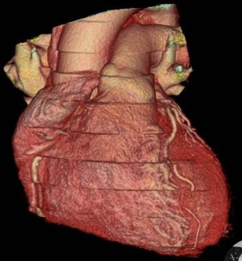
Cardiac-gated imaging techniques are medical diagnostic procedures that involve synchronizing the acquisition of data to the electrical activity of the heart. This is typically achieved by using an electrocardiogram (ECG) to trigger the imaging process at specific points during the cardiac cycle, such as at the R-wave of the ECG signal which corresponds to the peak of ventricular contraction.
Cardiac-gated imaging techniques are used in various medical imaging modalities including computed tomography (CT), magnetic resonance imaging (MRI) and positron emission tomography (PET) scans, among others. These techniques help to minimize motion artifacts caused by the contraction of the heart and improve the overall quality of the images obtained, allowing for more accurate diagnosis and treatment planning.
In cardiac CT and MRI, cardiac-gated imaging is used to assess various aspects of cardiac anatomy and function, such as the size and shape of the heart chambers, the motion of the heart walls, and the presence and severity of coronary artery disease. In PET scans, cardiac-gating can be used to improve the accuracy of myocardial perfusion imaging, which is used to assess blood flow to the heart muscle.
Overall, cardiac-gated imaging techniques are essential tools in the diagnosis and management of various cardiovascular diseases, providing valuable information about the structure and function of the heart that can help guide clinical decision-making and improve patient outcomes.
Cardiac-gated single-photon emission computed tomography (SPECT) is a medical imaging technique used to evaluate the function and perfusion of the heart. In this procedure, a small amount of radioactive tracer is injected into the patient's bloodstream and is taken up by the heart muscle. The gamma rays emitted by the tracer are then detected by a specialized camera that rotates around the patient's chest, capturing multiple images from different angles.
The "cardiac-gated" component of the name refers to the fact that the imaging is synchronized with the patient's electrocardiogram (ECG) signal, allowing for the acquisition of images only during specific phases of the cardiac cycle. This helps to reduce motion artifacts caused by the beating heart and provides more accurate information about the distribution of blood flow within the heart muscle.
The "single-photon emission" part of the name refers to the type of radioactive tracer used in the procedure, which emits single photons that can be detected by the gamma camera. This is different from positron emission tomography (PET) scans, which use a different type of radioactive tracer that emits positrons that then annihilate with electrons to produce pairs of gamma rays.
Overall, cardiac-gated SPECT is a valuable tool for diagnosing and evaluating various heart conditions, including coronary artery disease, cardiomyopathy, and heart failure.
 Magnetisk resonanstomografi cine. Medicinsk sök. Bilder


![HealthLine of Northern Colorado - September 2009 - [PDF Document]](https://static.fdocuments.in/doc/1200x630/568bf2481a28ab8933961a22/healthline-of-northern-colorado-september-2009.jpg?t=1696304570)