Hydroxysteroid Dehydrogenases
17-Hydroxysteroid Dehydrogenases
20-Hydroxysteroid Dehydrogenases
3-alpha-Hydroxysteroid Dehydrogenase (B-Specific)
11-beta-Hydroxysteroid Dehydrogenase Type 2
11-beta-Hydroxysteroid Dehydrogenase Type 1
11-beta-Hydroxysteroid Dehydrogenases
Estradiol Dehydrogenases
Sulfotransferases
Cortisone
Steroid 17-alpha-Hydroxylase
Alcohol Oxidoreductases
Steroids
NAD
Hydrocortisone
L-Lactate Dehydrogenase
Testosterone
Androsterone
Liver
Alcohol Dehydrogenase
Glyceraldehyde-3-Phosphate Dehydrogenases
20-alpha-Hydroxysteroid Dehydrogenase
Aldehyde Dehydrogenase
Glutamate Dehydrogenase
Glucosephosphate Dehydrogenase
Malate Dehydrogenase
Isocitrate Dehydrogenase
Phosphoadenosine Phosphosulfate
Arylsulfotransferase
Ketosteroids
Dihydrolipoamide Dehydrogenase
NADP
Carbohydrate Dehydrogenases
Succinate Dehydrogenase
L-Iditol 2-Dehydrogenase
Dehydroepiandrosterone
Glycerolphosphate Dehydrogenase
Substrate Specificity
Glucose 1-Dehydrogenase
Molecular Sequence Data
Ketoglutarate Dehydrogenase Complex
Glucose Dehydrogenases
Phosphogluconate Dehydrogenase
Sugar Alcohol Dehydrogenases
Stereoisomerism
NADH Dehydrogenase
IMP Dehydrogenase
Amino Acid Sequence
Gene Expression Regulation, Enzymologic
Isoenzymes
Formate Dehydrogenases
Acyl-CoA Dehydrogenase
Xanthine Dehydrogenase
3-Methyl-2-Oxobutanoate Dehydrogenase (Lipoamide)
Hydroxybutyrate Dehydrogenase
Base Sequence
Pyruvate Dehydrogenase (Lipoamide)
3-Hydroxyacyl CoA Dehydrogenases
Oxidoreductases
Dihydrouracil Dehydrogenase (NADP)
Uridine Diphosphate Glucose Dehydrogenase
Catalysis
Luteinizing hormone inhibits conversion of pregnenolone to progesterone in luteal cells from rats on day 19 of pregnancy. (1/633)
We have previously reported that intrabursal ovarian administration of LH at the end of pregnancy in rats induces a decrease in luteal progesterone (P4) synthesis and an increase in P4 metabolism. However, whether this local luteolytic effect of LH is exerted directly on luteal cells or on other structures, such as follicular or stromal cells, to modify luteal function is unknown. The aim of the present study was to determine the effect of LH on isolated luteal cells obtained on Day 19 of pregnancy. Incubation of luteal cells with 1, 10, 100, or 1000 ng/ml of ovine LH (oLH) for 6 h did not modify basal P4 production. The addition to the culture medium of 22(R)-hydroxycholesterol (22R-HC, 10 microgram/ml), a membrane-permeable P4 precursor, or pregnenolone (10(-2) microM) induced a significant increase in P4 accumulation in the medium in relation to the control value. When luteal cells were preincubated for 2 h with oLH, a significant (p < 0.01) reduction in the 22R-HC- or pregnenolone-stimulated P4 accumulation was observed. Incubation of luteal cells with dibutyryl cAMP (1 mM, a cAMP analogue) plus isobutylmethylxanthine (1 mM, a phosphodiesterase inhibitor) also inhibited pregnenolone-stimulated P4 accumulation. Incubation with an inositol triphosphate synthesis inhibitor, neomycin (1 mM), or an inhibitor of intracellular Ca2+ mobilization, (8,9-N, N-diethylamino)octyl-3,4,5-trimethoxybenzoate (1 mM), did not prevent the decrease in pregnenolone-stimulated P4 secretion induced by oLH. It was concluded that the luteolytic action of LH in late pregnancy is due, at least in part, to a direct action on the luteal cells and that an increase in intracellular cAMP level might mediate this effect. (+info)Luteinization and proteolysis in ovarian follicles of Meishan and Large White gilts during the preovulatory period. (2/633)
This experiment was conducted to determine why follicles luteinize faster in the Meishan breed than in the Large White breed of pig. Follicles were recovered during the late follicular phase from ovaries of both breeds before and after administration of hCG given to mimic the LH surge. First, the patterns of cholesterol transporters (high and low density lipoproteins: HDL and LDL) were compared. Cholesterol transporters detected in follicular fluid consisted of HDL only. Similar amounts of Apolipoprotein A-I were found in all samples. There was no obvious breed effect on minor lipoproteins found in the HDL-rich fraction, and this pattern was altered similarly by hCG in the two breeds. The LDL-rich samples of serum from both breeds contained similar amounts of protein. Second, three steroidogenic enzymes, adrenodoxin, 17 alpha-hydroxylase-lyase (P450(17) alpha) and 3 beta-hydroxysteroid-dehydrogenase (3 beta-HSD) were detected by immunohistochemistry and quantified by image analysis on sections of the two largest follicles. Before hCG treatment, theca interna cells demonstrated immunoreactivities for adrenodoxin (strong), P450(17) alpha and 3 beta-HSD (very strong), whereas granulosa cells displayed immunoreactivities for adrenodoxin only. After hCG treatment, the localization of the enzymes was unchanged but the staining intensity of adrenodoxin on granulosa cells and 3 beta-HSD on theca cells increased (P < 0.01 and P < 0.05, respectively). Breed effects were detected for the amounts of adrenodoxin in theca cells (Meishan > Large White; P < 0.05) and of 17 alpha-hydroxylase (Large White > Meishan, P < 0.01). Breed x treatment interactions were never detected. Finally, gelatinases, plasminogen activator, plasminogen activator inhibitor, tissue inhibitors of metalloproteases (TIMP-1 and TIMP-2) were visualized by direct or reverse zymography or western blotting. Whatever the stage relative to LH administration, follicular fluid from Large White gilts contained more TIMP-1, and TIMP-2 (P < 0.02 and P < 0.01, respectively). No breed effect was detected for the amounts of gelatinases and plasminogen activator inhibitor 1. However, for these parameters, a significant breed x time interaction was obvious, as the Meishan follicles had a greater response to hCG (P < 0.01). Since proteolysis plays a key role in the bioavailability of growth factors such as insulin-like growth factor 1, fibroblast growth factor and transforming growth factor beta, which have the ability to alter gonadotrophin-induced progesterone production in pigs, the differences observed in its control in the present study may explain, at least in part, the different patterns of luteinization observed in Meishan and Large White follicles. (+info)Opposing changes in 3alpha-hydroxysteroid dehydrogenase oxidative and reductive activities in rat leydig cells during pubertal development. (3/633)
The enzyme 3alpha-hydroxysteroid dehydrogenase (3alpha-HSD) has an important role in androgen metabolism, catalyzing the interconversion of dihydrotestosterone (DHT) and 5alpha-androstane-3alpha,17beta-diol (3alpha-DIOL). The net direction of this interconversion will affect the amount of biologically active ligand available for androgen receptor binding. We hypothesize that in Leydig cells, differential expression of 3alpha-HSD enzymes favoring one of the two directions is a mechanism by which DHT levels are controlled. In order to characterize 3alpha-HSD in rat Leydig cells, the following properties were analyzed: rates of oxidation (3alpha-DIOL to DHT) and reduction (DHT to 3alpha-DIOL) and preference for the cofactors NADP(H) and NAD(H) (i.e., the oxidized and reduced forms of both pyridine nucleotides) in Leydig cells isolated on Days 21, 35, and 90 postpartum. Levels of 3alpha-HSD protein were measured by immunoblotting using an antibody directed against the liver type of the enzyme. Levels of 3alpha-HSD protein and rates of reduction were highest on Day 21 and lowest on Day 90. The opposite was true for the rate of 3alpha-HSD oxidation, which was barely detectable on Day 21 and highest on Day 90 (59.08 +/- 6.35 pmol/min per 10(6) cells, mean +/- SE). Therefore, the level of 3alpha-HSD protein detectable by liver enzyme was consistent with reduction but not with oxidation. There was a clear partitioning of NADP(H)-dependent activity into the cytosolic fraction of Leydig cells, whereas on Days 35 and 90, Leydig cells also contained a microsomal NAD(H)-activated 3alpha-HSD. We conclude that 1) the cytosolic 3alpha-HSD in Leydig cells on Day 21 behaves as a unidirectional NADPH-dependent reductase; 2) by Day 35, a microsomal NAD(H)-dependent enzyme activity is present and may account for predominance of 3alpha-HSD oxidation over reduction and the resultant high capacity of Leydig cells on Day 90 to synthesize DHT from 3alpha-DIOL. (+info)Expression of 3beta-hydroxysteroid dehydrogenase type I and type VI isoforms in the mouse testis during development. (4/633)
Six isoforms of the enzyme 3beta-hydroxysteroid dehydrogenase (3betaHSD) have been identified in the mouse, each the product of a distinct gene. Two of these isoforms (type I and type VI) are detectable in the adult testis but changes in their expression during development are unknown. In this study we have examined changes in testicular expression and localization of mRNA encoding the type I and type VI isoforms of 3betaHSD. Total 3betaHSD (type I plus type VI) mRNA was measured by reverse transcription-polymerase chain reaction and showed a peak of expression at day 5 after birth followed by a decline and then a further rise after day 10 that continued up to adulthood. When each isoform was measured individually it was clear that the type I isoform was expressed at all ages from embryonic day 13 to adulthood. In contrast, the type VI isoform was only expressed at significant levels during fetal life on embryonic day 13 and then not again until after day 10 postnatally. Expression of the type VI isoform mRNA increased markedly after day 10 so that by adulthood it was the predominant 3betaHSD isoform present in the testis. Closer examination of the timing of type VI expression showed that the isoform mRNA was first detectable at a significant level on day 11. In-situ hybridization confirmed that the type I isoform is the only one expressed in the fetal/neonatal animal and showed that expression was limited to the interstitial tissue. In the adult, both type I and type VI expression was within the interstitial tissue. The timing of 3betaHSD type VI mRNA expression suggests, strongly, that this isoform is expressed only by adult-type Leydig cells in the mouse testis and that this development starts shortly before day 11. The limited expression of the type VI isoform means that it will be a useful marker in studies of adult Leydig cell development. (+info)Molecular cloning and characterization of hemolymph 3-dehydroecdysone 3beta-reductase from the cotton leafworm, Spodoptera littoralis. A new member of the third superfamily of oxidoreductases. (5/633)
The primary product of the prothoracic glands of last instar larvae of Spodoptera littoralis is 3-dehydroecdysone (3DE). After secretion, 3DE is reduced to ecdysone by 3DE 3beta-reductase in the hemolymph. We have previously purified and characterized 3DE 3beta-reductase from the hemolymph of S. littoralis. In this study, cDNA clones encoding the enzyme were obtained by reverse transcription-polymerase chain reaction, employing primers based on the amino acid sequences, in conjunction with 5'- and 3'-rapid amplification of cDNA ends. Multiple polyadenylation signals and AT-rich elements were found in the 3'-untranslated region, suggesting that this region may have a role in regulation of expression of the gene. Conceptual translation and amino acid sequence analysis suggest that 3DE 3beta-reductase from S. littoralis is a new member of the third superfamily of oxidoreductases. Northern analysis shows that 3DE 3beta-reductase mRNA transcripts are widely distributed, but are differentially expressed, in some tissues. The developmental profile of the mRNA revealed that the gene encoding 3DE 3beta-reductase is only transcribed in the second half of the last larval instar and that this fluctuation in expression accounts for the change in the enzyme activity during the instar. Southern analysis indicates that the 3DE 3beta-reductase is encoded by a single gene, which probably contains at least one intron. (+info)An inborn error of bile acid synthesis (3beta-hydroxy-delta5-C27-steroid dehydrogenase deficiency) presenting as malabsorption leading to rickets. (6/633)
Deficiency of 3beta-hydroxy-delta5-C27-steroid dehydrogenase (3beta-HSDH), the enzyme that catalyses the second reaction in the principal pathway for the synthesis of bile acids, has been reported to present with prolonged neonatal jaundice with the biopsy features of neonatal hepatitis. It has also been shown to present between the ages of 4 and 46 months with jaundice, hepatosplenomegaly, and steatorrhoea (a clinical picture resembling progressive familial intrahepatic cholestasis). This paper reports two children with 3beta-HSDH deficiency who developed rickets during infancy and did not develop clinically evident liver disease until the age of 3 years. Bile acid replacement resulted in considerable clinical and biochemical improvement. The importance of thorough investigation of fat soluble vitamin deficiencies in infancy is emphasised. (+info)Dynamics of periovulatory steroidogenesis in the rhesus monkey follicle after ovarian stimulation. (7/633)
The temporal relationships and regulation of events in the primate follicle during the periovulatory interval are poorly understood. This study was designed to elucidate the dynamics of steroid synthesis in the macaque follicle during ovarian stimulation cycles in which serum/follicular fluid aspirates were collected at precise intervals before (0 h) and after (up to 36 h) administration of the ovulatory human chorionic gonadotrophin (HCG) bolus. Serum concentrations of progesterone increased (P < 0.05) within 30 min, and follicular fluid progesterone concentrations were elevated 180-fold within 12 h, of HCG injection, and remained elevated until the time of ovulation. In contrast, 17beta-oestradiol concentrations increased initially, but then declined (P < 0.05) by 36 h post-HCG. Acute incubation of granulosa cells with and without steroidogenic substrates demonstrated that: (i) 3beta-hydroxysteroid dehydrogenase and aromatase activities were present in equivalent amounts before and after HCG; whereas (ii) P450 side-chain cleavage activity increased (P < 0.05) within 12 h of HCG; and (iii) exogenous low-density lipoprotein and cholesterol were not utilized for steroidogenesis. This model should be useful for further studies on ovulation and luteinization in primates, and enable elucidation of the local actions of progesterone and other steroids at specific time points during the periovulatory interval. (+info)Paracrine glucocorticoid activity produced by mouse thymic epithelial cells. (8/633)
Previous data have suggested that glucocorticoids (GCs) are involved in the differentiation of thymocytes into mature T cells. In this report we demonstrate that the mouse thymic epithelial cells (TEC) express the cytochrome P450 hydroxylases Cyp11A1, Cyp21, and Cyp11B1. These enzymes, in combination with 3beta-hydroxysteroid dehydrogenase (3betaHSD), convert cholesterol into corticosterone, the major GC in rodents. In addition, when TEC were cocultured with 'reporter cells' containing the glucocorticoid receptor (GR) and a GR-dependent reporter gene, a specific induction of reporter gene activity was observed. Induction of reporter gene activity was blocked when the TEC and reporter cells were incubated in the presence of the Cyp11B1 inhibitor metyrapone or the 3betaHSD inhibitor trilostane, as well as by the GR antagonist RU486. Coculturing of TEC with thymocytes induced apoptosis in the latter, which was partially blocked by the enzyme inhibitors and RU486. We conclude that TEC secrete a GC hormone activity and suggest a paracrine role for this in thymocyte development. (+info)Hydroxysteroid dehydrogenases (HSDs) are a group of enzymes that play a crucial role in steroid hormone metabolism. They catalyze the oxidation and reduction reactions of hydroxyl groups on the steroid molecule, which can lead to the activation or inactivation of steroid hormones. HSDs are involved in the conversion of various steroids, including sex steroids (e.g., androgens, estrogens) and corticosteroids (e.g., cortisol, cortisone). These enzymes can be found in different tissues throughout the body, and their activity is regulated by various factors, such as hormones, growth factors, and cytokines. Dysregulation of HSDs has been implicated in several diseases, including cancer, diabetes, and cardiovascular disease.
3-Hydroxysteroid dehydrogenases (3-HSDs) are a group of enzymes that play a crucial role in steroid hormone biosynthesis. These enzymes catalyze the conversion of 3-beta-hydroxy steroids to 3-keto steroids, which is an essential step in the production of various steroid hormones, including progesterone, cortisol, aldosterone, and sex hormones such as testosterone and estradiol.
There are several isoforms of 3-HSDs that are expressed in different tissues and have distinct substrate specificities. For instance, 3-HSD type I is primarily found in the ovary and adrenal gland, where it catalyzes the conversion of pregnenolone to progesterone and 17-hydroxyprogesterone to 17-hydroxycortisol. On the other hand, 3-HSD type II is mainly expressed in the testes, adrenal gland, and placenta, where it catalyzes the conversion of dehydroepiandrosterone (DHEA) to androstenedione and androstenedione to testosterone.
Defects in 3-HSDs can lead to various genetic disorders that affect steroid hormone production and metabolism, resulting in a range of clinical manifestations such as adrenal insufficiency, ambiguous genitalia, and sexual development disorders.
17-Hydroxysteroid dehydrogenases (17-HSDs) are a group of enzymes that play a crucial role in steroid hormone biosynthesis. They are involved in the conversion of 17-ketosteroids to 17-hydroxy steroids or vice versa, by adding or removing a hydroxyl group (–OH) at the 17th carbon atom of the steroid molecule. This conversion is essential for the production of various steroid hormones, including cortisol, aldosterone, and sex hormones such as estrogen and testosterone.
There are several isoforms of 17-HSDs, each with distinct substrate specificities, tissue distributions, and functions:
1. 17-HSD type 1 (17-HSD1): This isoform primarily catalyzes the conversion of estrone (E1) to estradiol (E2), an active form of estrogen. It is mainly expressed in the ovary, breast, and adipose tissue.
2. 17-HSD type 2 (17-HSD2): This isoform catalyzes the reverse reaction, converting estradiol (E2) to estrone (E1). It is primarily expressed in the placenta, prostate, and breast tissue.
3. 17-HSD type 3 (17-HSD3): This isoform is responsible for the conversion of androstenedione to testosterone, an essential step in male sex hormone biosynthesis. It is predominantly expressed in the testis and adrenal gland.
4. 17-HSD type 4 (17-HSD4): This isoform catalyzes the conversion of dehydroepiandrosterone (DHEA) to androstenedione, an intermediate step in steroid hormone biosynthesis. It is primarily expressed in the placenta.
5. 17-HSD type 5 (17-HSD5): This isoform catalyzes the conversion of cortisone to cortisol, a critical step in glucocorticoid biosynthesis. It is predominantly expressed in the adrenal gland and liver.
6. 17-HSD type 6 (17-HSD6): This isoform catalyzes the conversion of androstenedione to testosterone, similar to 17-HSD3. However, it has a different substrate specificity and is primarily expressed in the ovary.
7. 17-HSD type 7 (17-HSD7): This isoform catalyzes the conversion of estrone (E1) to estradiol (E2), similar to 17-HSD1. However, it has a different substrate specificity and is primarily expressed in the ovary.
8. 17-HSD type 8 (17-HSD8): This isoform catalyzes the conversion of DHEA to androstenedione, similar to 17-HSD4. However, it has a different substrate specificity and is primarily expressed in the testis.
9. 17-HSD type 9 (17-HSD9): This isoform catalyzes the conversion of estrone (E1) to estradiol (E2), similar to 17-HSD1. However, it has a different substrate specificity and is primarily expressed in the placenta.
10. 17-HSD type 10 (17-HSD10): This isoform catalyzes the conversion of DHEA to androstenedione, similar to 17-HSD4. However, it has a different substrate specificity and is primarily expressed in the testis.
11. 17-HSD type 11 (17-HSD11): This isoform catalyzes the conversion of estrone (E1) to estradiol (E2), similar to 17-HSD1. However, it has a different substrate specificity and is primarily expressed in the placenta.
12. 17-HSD type 12 (17-HSD12): This isoform catalyzes the conversion of DHEA to androstenedione, similar to 17-HSD4. However, it has a different substrate specificity and is primarily expressed in the testis.
13. 17-HSD type 13 (17-HSD13): This isoform catalyzes the conversion of estrone (E1) to estradiol (E2), similar to 17-HSD1. However, it has a different substrate specificity and is primarily expressed in the placenta.
14. 17-HSD type 14 (17-HSD14): This isoform catalyzes the conversion of DHEA to androstenedione, similar to 17-HSD4. However, it has a different substrate specificity and is primarily expressed in the testis.
15. 17-HSD type 15 (17-HSD15): This isoform catalyzes the conversion of estrone (E1) to estradiol (E2), similar to 17-HSD1. However, it has a different substrate specificity and is primarily expressed in the placenta.
16. 17-HSD type 16 (17-HSD16): This isoform catalyzes the conversion of DHEA to androstenedione, similar to 17-HSD4. However, it has a different substrate specificity and is primarily expressed in the testis.
17. 17-HSD type 17 (17-HSD17): This isoform catalyzes the conversion of estrone (E1) to estradiol (E2), similar to 17-HSD1. However, it has a different substrate specificity and is primarily expressed in the placenta.
18. 17-HSD type 18 (17-HSD18): This isoform catalyzes the conversion of DHEA to androstenedione, similar to 17-HSD4. However, it has a different substrate specificity and is primarily expressed in the testis.
19. 17-HSD type 19 (17-HSD19): This isoform catalyzes the conversion of estrone (E1) to estradiol (E2), similar to 17-HSD1. However, it has a different substrate specificity and is primarily expressed in the placenta.
20. 17-HSD type 20 (17-HSD20): This isoform catalyzes the conversion of DHEA to androstenedione, similar to 17-HSD4. However, it has a different substrate specificity and is primarily expressed in the testis.
21. 17-HSD type 21 (17-HSD21): This isoform catalyzes the conversion of estrone (E1) to estradiol (E2), similar to 17-HSD1. However, it has a different substrate specificity and is primarily expressed in the placenta.
22. 17-HSD type 22 (17-HSD22): This isoform catalyzes the conversion of DHEA to androstenedione, similar to 17-HSD4. However, it has a different substrate specificity and is primarily expressed in the testis.
23. 17-HSD type 23 (17-HSD23): This isoform catalyzes the conversion of estrone (E1) to estradiol (E2), similar to 17-HSD1. However, it has a different substrate specificity and is primarily expressed in the placenta.
24. 17-HSD type 24 (17-HSD24): This isoform catalyzes the conversion of DHEA to androstenedione, similar to 17-HSD4. However, it has a different substrate specificity and is primarily expressed in the testis.
25. 17-HSD type 25 (17-HSD25): This isoform catalyzes the conversion of estrone (E1) to estradiol (E2), similar to 17-HSD1. However, it has a different substrate specificity and is primarily expressed in the placenta.
26. 17-HSD type 26 (17-HSD26): This isoform catalyzes the conversion of DHEA to androstenedione, similar to 17-HSD4. However
20-Hydroxysteroid Dehydrogenases (20-HSDs) are a group of enzymes that play a crucial role in the metabolism of steroid hormones. These enzymes catalyze the conversion of steroid hormone precursors to their active forms by adding or removing a hydroxyl group at the 20th carbon position of the steroid molecule.
There are several isoforms of 20-HSDs, each with distinct tissue distribution and substrate specificity. The most well-known isoforms include 20-HSD type I and II, which have opposing functions in regulating the activity of cortisol, a glucocorticoid hormone produced by the adrenal gland.
Type I 20-HSD, primarily found in the liver and adipose tissue, converts inactive cortisone to its active form, cortisol. In contrast, type II 20-HSD, expressed mainly in the kidney, brain, and immune cells, catalyzes the reverse reaction, converting cortisol back to cortisone.
Dysregulation of 20-HSDs has been implicated in various medical conditions, such as metabolic disorders, inflammatory diseases, and cancers. Therefore, understanding the function and regulation of these enzymes is essential for developing targeted therapies for these conditions.
11-Beta-Hydroxysteroid Dehydrogenase Type 2 (11β-HSD2) is an enzyme that plays a crucial role in the regulation of steroid hormones, particularly cortisol and aldosterone. It is primarily found in tissues such as the kidneys, colon, and salivary glands.
The main function of 11β-HSD2 is to convert active cortisol into inactive cortisone, which helps to prevent excessive mineralocorticoid receptor activation by cortisol. This is important because cortisol can bind to and activate mineralocorticoid receptors, leading to increased sodium reabsorption and potassium excretion in the kidneys, as well as other effects on blood pressure and electrolyte balance.
By converting cortisol to cortisone, 11β-HSD2 helps to protect mineralocorticoid receptors from being overstimulated by cortisol, allowing aldosterone to bind and activate these receptors instead. This is important for maintaining normal blood pressure and electrolyte balance.
Deficiencies or mutations in the 11β-HSD2 enzyme can lead to a condition called apparent mineralocorticoid excess (AME), which is characterized by high blood pressure, low potassium levels, and increased sodium reabsorption in the kidneys. This occurs because cortisol is able to bind to and activate mineralocorticoid receptors in the absence of 11β-HSD2 activity.
11-Beta-Hydroxysteroid Dehydrogenase Type 1 (11β-HSD1) is an enzyme that plays a crucial role in the metabolism of steroid hormones, particularly cortisol, in the body. Cortisol is a glucocorticoid hormone produced by the adrenal glands that helps regulate various physiological processes such as metabolism, immune response, and stress response.
11β-HSD1 is primarily expressed in liver, fat, and muscle tissues, where it catalyzes the conversion of cortisone to cortisol. Cortisone is a biologically inactive form of cortisol that is produced when cortisol levels are high, and it needs to be converted back to cortisol for the hormone to exert its effects.
By increasing the availability of active cortisol in these tissues, 11β-HSD1 has been implicated in several metabolic disorders, including obesity, insulin resistance, and type 2 diabetes. Inhibitors of 11β-HSD1 are currently being investigated as potential therapeutic agents for the treatment of these conditions.
11-Beta-Hydroxysteroid dehydrogenases (11-β-HSDs) are a group of enzymes that play a crucial role in the metabolism of steroid hormones, particularly cortisol and cortisone, which belong to the class of glucocorticoids. These enzymes exist in two isoforms: 11-β-HSD1 and 11-β-HSD2.
1. 11-β-HSD1: This isoform is primarily located within the liver, adipose tissue, and various other peripheral tissues. It functions as a NADPH-dependent reductase, converting inactive cortisone to its active form, cortisol. This enzyme helps regulate glucocorticoid action in peripheral tissues, influencing glucose and lipid metabolism, insulin sensitivity, and inflammation.
2. 11-β-HSD2: This isoform is predominantly found in mineralocorticoid target tissues such as the kidneys, colon, and salivary glands. It functions as a NAD+-dependent dehydrogenase, converting active cortisol to its inactive form, cortisone. By doing so, it protects the mineralocorticoid receptor from being overstimulated by cortisol, ensuring aldosterone specifically binds and activates this receptor to maintain proper electrolyte and fluid balance.
Dysregulation of 11-β-HSDs has been implicated in several disease states, including metabolic syndrome, type 2 diabetes, hypertension, and psychiatric disorders. Therefore, understanding the function and regulation of these enzymes is essential for developing novel therapeutic strategies to treat related conditions.
Estradiol dehydrogenases are a group of enzymes that are involved in the metabolism of estradiols, which are steroid hormones that play important roles in the development and maintenance of female reproductive system and secondary sexual characteristics. These enzymes catalyze the oxidation or reduction reactions of estradiols, converting them to other forms of steroid hormones.
There are two main types of estradiol dehydrogenases: 1) 3-alpha-hydroxysteroid dehydrogenase (3-alpha HSD), which catalyzes the conversion of estradi-17-beta to estrone, and 2) 17-beta-hydroxysteroid dehydrogenase (17-beta HSD), which catalyzes the reverse reaction, converting estrone back to estradiol.
These enzymes are widely distributed in various tissues, including the ovaries, placenta, liver, and adipose tissue, and play important roles in regulating the levels of estradiols in the body. Abnormalities in the activity of these enzymes have been associated with several medical conditions, such as hormone-dependent cancers, polycystic ovary syndrome, and hirsutism.
Sulfotransferases (STs) are a group of enzymes that play a crucial role in the process of sulfoconjugation, which is the transfer of a sulfo group (-SO3H) from a donor molecule to an acceptor molecule. These enzymes are widely distributed in nature and are found in various organisms, including humans.
In humans, STs are involved in the metabolism and detoxification of numerous xenobiotics, such as drugs, food additives, and environmental pollutants, as well as endogenous compounds, such as hormones, neurotransmitters, and lipids. The sulfoconjugation reaction catalyzed by STs can increase the water solubility of these compounds, facilitating their excretion from the body.
STs can be classified into several families based on their sequence similarity and cofactor specificity. The largest family of STs is the cytosolic sulfotransferases, which use 3'-phosphoadenosine 5'-phosphosulfate (PAPS) as a cofactor to transfer the sulfo group to various acceptor molecules, including phenols, alcohols, amines, and steroids.
Abnormalities in ST activity have been implicated in several diseases, such as cancer, cardiovascular disease, and neurological disorders. Therefore, understanding the function and regulation of STs is essential for developing new therapeutic strategies to treat these conditions.
Cortisone is a type of corticosteroid hormone that is produced naturally in the body by the adrenal gland. It is released in response to stress and helps to regulate metabolism, reduce inflammation, and suppress the immune system. Cortisone can also be synthetically produced and is often used as a medication to treat a variety of conditions such as arthritis, asthma, and skin disorders. It works by mimicking the effects of the natural hormone in the body and reducing inflammation and suppressing the immune system. Cortisone can be administered through various routes, including oral, injectable, topical, and inhalational.
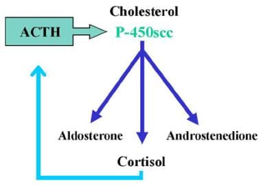
Steroid 17-alpha-hydroxylase, also known as CYP17A1, is a cytochrome P450 enzyme that plays a crucial role in steroid hormone biosynthesis. It is located in the endoplasmic reticulum of cells in the adrenal glands and gonads. This enzyme catalyzes the 17-alpha-hydroxylation and subsequent lyase cleavage of pregnenolone and progesterone, converting them into dehydroepiandrosterone (DHEA) and androstenedione, respectively. These steroid intermediates are essential for the biosynthesis of both glucocorticoids and sex steroids, including cortisol, aldosterone, estrogens, and testosterone.
Defects in the CYP17A1 gene can lead to several disorders, such as congenital adrenal hyperplasia (CAH) due to 17-alpha-hydroxylase deficiency, which is characterized by decreased production of cortisol and sex steroids and increased mineralocorticoid levels. This condition results in sexual infantilism, electrolyte imbalances, and hypertension.
Alcohol oxidoreductases are a class of enzymes that catalyze the oxidation of alcohols to aldehydes or ketones, while reducing nicotinamide adenine dinucleotide (NAD+) to NADH. These enzymes play an important role in the metabolism of alcohols and other organic compounds in living organisms.
The most well-known example of an alcohol oxidoreductase is alcohol dehydrogenase (ADH), which is responsible for the oxidation of ethanol to acetaldehyde in the liver during the metabolism of alcoholic beverages. Other examples include aldehyde dehydrogenases (ALDH) and sorbitol dehydrogenase (SDH).
These enzymes are important targets for the development of drugs used to treat alcohol use disorder, as inhibiting their activity can help to reduce the rate of ethanol metabolism and the severity of its effects on the body.
Steroids, also known as corticosteroids, are a type of hormone that the adrenal gland produces in your body. They have many functions, such as controlling the balance of salt and water in your body and helping to reduce inflammation. Steroids can also be synthetically produced and used as medications to treat a variety of conditions, including allergies, asthma, skin conditions, and autoimmune disorders.
Steroid medications are available in various forms, such as oral pills, injections, creams, and inhalers. They work by mimicking the effects of natural hormones produced by your body, reducing inflammation and suppressing the immune system's response to prevent or reduce symptoms. However, long-term use of steroids can have significant side effects, including weight gain, high blood pressure, osteoporosis, and increased risk of infections.
It is important to note that anabolic steroids are a different class of drugs that are sometimes abused for their muscle-building properties. These steroids are synthetic versions of the male hormone testosterone and can have serious health consequences when taken in large doses or without medical supervision.
NAD (Nicotinamide Adenine Dinucleotide) is a coenzyme found in all living cells. It plays an essential role in cellular metabolism, particularly in redox reactions, where it acts as an electron carrier. NAD exists in two forms: NAD+, which accepts electrons and becomes reduced to NADH. This pairing of NAD+/NADH is involved in many fundamental biological processes such as generating energy in the form of ATP during cellular respiration, and serving as a critical cofactor for various enzymes that regulate cellular functions like DNA repair, gene expression, and cell death.
Maintaining optimal levels of NAD+/NADH is crucial for overall health and longevity, as it declines with age and in certain disease states. Therefore, strategies to boost NAD+ levels are being actively researched for their potential therapeutic benefits in various conditions such as aging, neurodegenerative disorders, and metabolic diseases.
Hydrocortisone is a synthetic glucocorticoid, which is a class of steroid hormones. It is identical to the naturally occurring cortisol, a hormone produced by the adrenal gland that helps regulate metabolism and helps your body respond to stress. Hydrocortisone has anti-inflammatory effects and is used to treat various inflammatory conditions such as allergies, skin disorders, and autoimmune diseases. It works by suppressing the immune system's response to reduce swelling, redness, itching, and other symptoms caused by inflammation.
Hydrocortisone is available in different forms, including oral tablets, topical creams, lotions, gels, and ointments, as well as injectable solutions. The specific use and dosage depend on the condition being treated and the individual patient's medical history and current health status.
As with any medication, hydrocortisone can have side effects, especially when used in high doses or for extended periods. Common side effects include increased appetite, weight gain, mood changes, insomnia, and skin thinning. Long-term use of hydrocortisone may also increase the risk of developing osteoporosis, diabetes, cataracts, and other health problems. Therefore, it is essential to follow your healthcare provider's instructions carefully when using this medication.
L-Lactate Dehydrogenase (LDH) is an enzyme found in various tissues within the body, including the heart, liver, kidneys, muscles, and brain. It plays a crucial role in the process of energy production, particularly during anaerobic conditions when oxygen levels are low.
In the presence of the coenzyme NADH, LDH catalyzes the conversion of pyruvate to lactate, generating NAD+ as a byproduct. Conversely, in the presence of NAD+, LDH can convert lactate back to pyruvate using NADH. This reversible reaction is essential for maintaining the balance between lactate and pyruvate levels within cells.
Elevated blood levels of LDH may indicate tissue damage or injury, as this enzyme can be released into the circulation following cellular breakdown. As a result, LDH is often used as a nonspecific biomarker for various medical conditions, such as myocardial infarction (heart attack), liver disease, muscle damage, and certain types of cancer. However, it's important to note that an isolated increase in LDH does not necessarily pinpoint the exact location or cause of tissue damage, and further diagnostic tests are usually required for confirmation.
Testosterone is a steroid hormone that belongs to androsten class of hormones. It is primarily secreted by the Leydig cells in the testes of males and, to a lesser extent, by the ovaries and adrenal glands in females. Testosterone is the main male sex hormone and anabolic steroid. It plays a key role in the development of masculine characteristics, such as body hair and muscle mass, and contributes to bone density, fat distribution, red cell production, and sex drive. In females, testosterone contributes to sexual desire and bone health. Testosterone is synthesized from cholesterol and its production is regulated by luteinizing hormone (LH) and follicle-stimulating hormone (FSH).
In the context of medicine and pharmacology, "kinetics" refers to the study of how a drug moves throughout the body, including its absorption, distribution, metabolism, and excretion (often abbreviated as ADME). This field is called "pharmacokinetics."
1. Absorption: This is the process of a drug moving from its site of administration into the bloodstream. Factors such as the route of administration (e.g., oral, intravenous, etc.), formulation, and individual physiological differences can affect absorption.
2. Distribution: Once a drug is in the bloodstream, it gets distributed throughout the body to various tissues and organs. This process is influenced by factors like blood flow, protein binding, and lipid solubility of the drug.
3. Metabolism: Drugs are often chemically modified in the body, typically in the liver, through processes known as metabolism. These changes can lead to the formation of active or inactive metabolites, which may then be further distributed, excreted, or undergo additional metabolic transformations.
4. Excretion: This is the process by which drugs and their metabolites are eliminated from the body, primarily through the kidneys (urine) and the liver (bile).
Understanding the kinetics of a drug is crucial for determining its optimal dosing regimen, potential interactions with other medications or foods, and any necessary adjustments for special populations like pediatric or geriatric patients, or those with impaired renal or hepatic function.
Androsterone is a weak androgen and an endogenous steroid hormone. It's produced in the liver from dehydroepiandrosterone (DHEA) and is converted into androstenedione, another weak androgen. Androsterone is excreted in urine as a major metabolite of testosterone. It plays a role in male sexual development and function, although its effects are much weaker than those of testosterone. In clinical contexts, androsterone levels may be measured to help diagnose certain hormonal disorders or to monitor hormone therapy.
The liver is a large, solid organ located in the upper right portion of the abdomen, beneath the diaphragm and above the stomach. It plays a vital role in several bodily functions, including:
1. Metabolism: The liver helps to metabolize carbohydrates, fats, and proteins from the food we eat into energy and nutrients that our bodies can use.
2. Detoxification: The liver detoxifies harmful substances in the body by breaking them down into less toxic forms or excreting them through bile.
3. Synthesis: The liver synthesizes important proteins, such as albumin and clotting factors, that are necessary for proper bodily function.
4. Storage: The liver stores glucose, vitamins, and minerals that can be released when the body needs them.
5. Bile production: The liver produces bile, a digestive juice that helps to break down fats in the small intestine.
6. Immune function: The liver plays a role in the immune system by filtering out bacteria and other harmful substances from the blood.
Overall, the liver is an essential organ that plays a critical role in maintaining overall health and well-being.
Alcohol dehydrogenase (ADH) is a group of enzymes responsible for catalyzing the oxidation of alcohols to aldehydes or ketones, and reducing equivalents such as NAD+ to NADH. In humans, ADH plays a crucial role in the metabolism of ethanol, converting it into acetaldehyde, which is then further metabolized by aldehyde dehydrogenase (ALDH) into acetate. This process helps to detoxify and eliminate ethanol from the body. Additionally, ADH enzymes are also involved in the metabolism of other alcohols, such as methanol and ethylene glycol, which can be toxic if allowed to accumulate in the body.
Glyceraldehyde-3-phosphate dehydrogenase (GAPDH) is an enzyme that plays a crucial role in the metabolic pathway of glycolysis. Its primary function is to convert glyceraldehyde-3-phosphate (a triose sugar phosphate) into D-glycerate 1,3-bisphosphate, while also converting nicotinamide adenine dinucleotide (NAD+) into its reduced form NADH. This reaction is essential for the production of energy in the form of adenosine triphosphate (ATP) during cellular respiration. GAPDH has also been implicated in various non-metabolic processes, including DNA replication, repair, and transcription regulation, due to its ability to interact with different proteins and nucleic acids.
20-α-Hydroxysteroid Dehydrogenase (20-α-HSD) is an enzyme that catalyzes the conversion of steroids, specifically the oxidation of 20α-hydroxysteroids to 20-keto steroids. This enzyme plays a crucial role in the metabolism and regulation of steroid hormones, such as corticosteroids and progestogens.
In the adrenal gland, 20-α-HSD is involved in the biosynthesis and interconversion of various corticosteroids, including cortisol, cortisone, and aldosterone. By catalyzing the conversion of cortisol to cortisone or vice versa, this enzyme helps maintain a balance between active and inactive forms of these hormones, which is essential for proper physiological functioning.
In the reproductive system, 20-α-HSD is involved in the metabolism of progestogens, such as progesterone and its derivatives. This enzyme can convert active forms of progestogens into their inactive counterparts, thereby regulating their levels and activity within the body.
Dysregulation or mutations in 20-α-HSD have been implicated in several medical conditions, including adrenal insufficiency, congenital adrenal hyperplasia, and certain reproductive disorders.
Aldehyde dehydrogenase (ALDH) is a class of enzymes that play a crucial role in the metabolism of alcohol and other aldehydes in the body. These enzymes catalyze the oxidation of aldehydes to carboxylic acids, using nicotinamide adenine dinucleotide (NAD+) as a cofactor.
There are several isoforms of ALDH found in different tissues throughout the body, with varying substrate specificities and kinetic properties. The most well-known function of ALDH is its role in alcohol metabolism, where it converts the toxic aldehyde intermediate acetaldehyde to acetate, which can then be further metabolized or excreted.
Deficiencies in ALDH activity have been linked to a number of clinical conditions, including alcohol flush reaction, alcohol-induced liver disease, and certain types of cancer. Additionally, increased ALDH activity has been associated with chemotherapy resistance in some cancer cells.
Glutamate Dehydrogenase (GLDH or GDH) is a mitochondrial enzyme that plays a crucial role in the metabolism of amino acids, particularly within liver and kidney tissues. It catalyzes the reversible oxidative deamination of glutamate to alpha-ketoglutarate, which links amino acid metabolism with the citric acid cycle and energy production. This enzyme is significant in clinical settings as its levels in blood serum can be used as a diagnostic marker for diseases that damage liver or kidney cells, since these cells release GLDH into the bloodstream upon damage.
Glyceraldehyde-3-phosphate dehydrogenase (GAPDH), also known as Glucosephosphate Dehydrogenase, is an enzyme that plays a crucial role in cellular metabolism, particularly in the glycolytic pathway. It catalyzes the conversion of glyceraldehyde 3-phosphate (G3P) to 1,3-bisphosphoglycerate (1,3-BPG), while also converting nicotinamide adenine dinucleotide (NAD+) to its reduced form NADH. This reaction is essential for the production of energy in the form of adenosine triphosphate (ATP) during cellular respiration. GAPDH has been widely used as a housekeeping gene in molecular biology research due to its consistent expression across various tissues and cells, although recent studies have shown that its expression can vary under certain conditions.
Malate Dehydrogenase (MDH) is an enzyme that plays a crucial role in the Krebs cycle, also known as the citric acid cycle or tricarboxylic acid (TCA) cycle. It catalyzes the reversible oxidation of malate to oxaloacetate, while simultaneously reducing NAD+ to NADH. This reaction is essential for energy production in the form of ATP and NADH within the cell.
There are two main types of Malate Dehydrogenase:
1. NAD-dependent Malate Dehydrogenase (MDH1): Found primarily in the cytoplasm, this isoform plays a role in the malate-aspartate shuttle, which helps transfer reducing equivalents between the cytoplasm and mitochondria.
2. FAD-dependent Malate Dehydrogenase (MDH2): Located within the mitochondrial matrix, this isoform is involved in the Krebs cycle for energy production.
Abnormal levels of Malate Dehydrogenase enzyme can be indicative of certain medical conditions or diseases, such as myocardial infarction (heart attack), muscle damage, or various types of cancer. Therefore, MDH enzyme activity is often assessed in diagnostic tests to help identify and monitor these health issues.
Isocitrate Dehydrogenase (IDH) is an enzyme that catalyzes the oxidative decarboxylation of isocitrate to α-ketoglutarate in the presence of NAD+ or NADP+, producing NADH or NADPH respectively. This reaction occurs in the citric acid cycle, also known as the Krebs cycle or tricarboxylic acid (TCA) cycle, which is a crucial metabolic pathway in the cell's energy production and biosynthesis of various molecules. There are three isoforms of IDH found in humans: IDH1 located in the cytosol, IDH2 in the mitochondrial matrix, and IDH3 within the mitochondria. Mutations in IDH1 and IDH2 have been associated with several types of cancer, such as gliomas and acute myeloid leukemia (AML), leading to abnormal accumulation of 2-hydroxyglutarate, which can contribute to tumorigenesis.
Phosphoadenosine phosphosulfate (PAPS) is not exactly a medical term, but a biochemical term. However, it is often referred to in the context of medical and biological research.
PAPS is a crucial molecule in the metabolism of living organisms and serves as the primary donor of sulfate groups in the process of sulfonation, which is a type of enzymatic modification that adds a sulfate group to various substrates such as proteoglycans, hormones, neurotransmitters, and xenobiotics. This process plays an essential role in several biological processes, including detoxification, signal transduction, and cell-cell recognition.
Therefore, PAPS is a critical molecule for maintaining proper physiological functions in the body, and its dysregulation has been implicated in various diseases, such as cancer, inflammation, and neurodevelopmental disorders.
Arylsulfotransferases (ASTs) are a group of enzymes that play a role in the detoxification of xenobiotics and endogenous compounds by catalyzing the transfer of a sulfuryl group from a donor, such as 3'-phosphoadenosine-5'-phosphosulfate (PAPS), to an acceptor aromatic molecule. This results in the formation of a sulfate ester, which can then be excreted from the body. ASTs are found in various tissues, including the liver, kidney, and intestine, and are involved in the metabolism of numerous drugs, hormones, and neurotransmitters. Defects in ASTs have been associated with certain genetic disorders, such as aromatic L-amino acid decarboxylase deficiency and disorders of steroid sulfation.
Ketosteroids are a type of steroid compound that contain a ketone functional group in their chemical structure. They are derived from cholesterol and are present in both animal and plant tissues. Some ketosteroids are produced endogenously, while others can be introduced exogenously through the diet or medication.
Endogenous ketosteroids include steroid hormones such as testosterone, estradiol, and cortisol, which contain a ketone group in their structure. Exogenous ketosteroids can be found in certain medications, such as those used to treat hormonal imbalances or inflammation.
Ketosteroids have been studied for their potential therapeutic uses, including as anti-inflammatory agents and for the treatment of hormone-related disorders. However, more research is needed to fully understand their mechanisms of action and potential benefits.
Dihydrolipoamide dehydrogenase (DHLD) is an enzyme that plays a crucial role in several important metabolic pathways in the human body, including the citric acid cycle and the catabolism of certain amino acids. DHLD is a component of multi-enzyme complexes, such as the pyruvate dehydrogenase complex (PDC) and the alpha-ketoglutarate dehydrogenase complex (KGDC).
The primary function of DHLD is to catalyze the oxidation of dihydrolipoamide, a reduced form of lipoamide, back to its oxidized state (lipoamide) while simultaneously reducing NAD+ to NADH. This reaction is essential for the continued functioning of the PDC and KGDC, as dihydrolipoamide is a cofactor for these enzyme complexes.
Deficiencies in DHLD can lead to serious metabolic disorders, such as maple syrup urine disease (MSUD) and riboflavin-responsive multiple acyl-CoA dehydrogenase deficiency (RR-MADD). These conditions can result in neurological symptoms, developmental delays, and metabolic acidosis, among other complications. Treatment typically involves dietary modifications, supplementation with specific nutrients, and, in some cases, enzyme replacement therapy.
NADP (Nicotinamide Adenine Dinucleotide Phosphate) is a coenzyme that plays a crucial role as an electron carrier in various redox reactions in the human body. It exists in two forms: NADP+, which functions as an oxidizing agent and accepts electrons, and NADPH, which serves as a reducing agent and donates electrons.
NADPH is particularly important in anabolic processes, such as lipid and nucleotide synthesis, where it provides the necessary reducing equivalents to drive these reactions forward. It also plays a critical role in maintaining the cellular redox balance by participating in antioxidant defense mechanisms that neutralize harmful reactive oxygen species (ROS).
In addition, NADP is involved in various metabolic pathways, including the pentose phosphate pathway and the Calvin cycle in photosynthesis. Overall, NADP and its reduced form, NADPH, are essential molecules for maintaining proper cellular function and energy homeostasis.
Carbohydrate dehydrogenases are a group of enzymes that catalyze the oxidation of carbohydrates, including sugars and sugar alcohols. These enzymes play a crucial role in cellular metabolism by helping to convert these molecules into forms that can be used for energy or as building blocks for other biological compounds.
During the oxidation process, carbohydrate dehydrogenases remove hydrogen atoms from the carbohydrate substrate and transfer them to an electron acceptor, such as NAD+ or FAD. This results in the formation of a ketone or aldehyde group on the carbohydrate molecule and the reduction of the electron acceptor to NADH or FADH2.
Carbohydrate dehydrogenases are classified into several subgroups based on their substrate specificity, cofactor requirements, and other factors. Some examples include glucose dehydrogenase, galactose dehydrogenase, and sorbitol dehydrogenase.
These enzymes have important applications in various fields, including biotechnology, medicine, and industry. For example, they can be used to detect or quantify specific carbohydrates in biological samples, or to produce valuable chemical compounds through the oxidation of renewable resources such as plant-derived sugars.
Succinate dehydrogenase (SDH) is an enzyme complex that plays a crucial role in the process of cellular respiration, specifically in the citric acid cycle (also known as the Krebs cycle) and the electron transport chain. It is located in the inner mitochondrial membrane of eukaryotic cells.
SDH catalyzes the oxidation of succinate to fumarate, converting it into a molecule of fadaquate in the process. During this reaction, two electrons are transferred from succinate to the FAD cofactor within the SDH enzyme complex, reducing it to FADH2. These electrons are then passed on to ubiquinone (CoQ), which is a mobile electron carrier in the electron transport chain, leading to the generation of ATP, the main energy currency of the cell.
SDH is also known as mitochondrial complex II because it is the second complex in the electron transport chain. Mutations in the genes encoding SDH subunits or associated proteins have been linked to various human diseases, including hereditary paragangliomas, pheochromocytomas, gastrointestinal stromal tumors (GISTs), and some forms of neurodegenerative disorders.
L-Iditol 2-Dehydrogenase is an enzyme that catalyzes the chemical reaction between L-iditol and NAD+ to produce L-sorbose and NADH + H+. This enzyme plays a role in the metabolism of sugars, specifically in the conversion of L-iditol to L-sorbose in various organisms, including bacteria and fungi. The reaction catalyzed by this enzyme is part of the polyol pathway, which is involved in the regulation of osmotic pressure and other cellular processes.
Dehydroepiandrosterone (DHEA) is a steroid hormone produced by the adrenal glands. It serves as a precursor to other hormones, including androgens such as testosterone and estrogens such as estradiol. DHEA levels typically peak during early adulthood and then gradually decline with age.
DHEA has been studied for its potential effects on various health conditions, including aging, cognitive function, sexual dysfunction, and certain chronic diseases. However, the evidence supporting its use for these purposes is generally limited and inconclusive. As with any supplement or medication, it's important to consult with a healthcare provider before taking DHEA to ensure safety and effectiveness.
Glycerol-3-phosphate dehydrogenase (GPD) is an enzyme that plays a crucial role in the metabolism of glucose and lipids. It catalyzes the conversion of dihydroxyacetone phosphate (DHAP) to glycerol-3-phosphate (G3P), which is a key intermediate in the synthesis of triglycerides, phospholipids, and other glycerophospholipids.
There are two main forms of GPD: a cytoplasmic form (GPD1) and a mitochondrial form (GPD2). The cytoplasmic form is involved in the production of NADH, which is used in various metabolic processes, while the mitochondrial form is involved in the production of ATP, the main energy currency of the cell.
Deficiencies or mutations in GPD can lead to a variety of metabolic disorders, including glycerol kinase deficiency and congenital muscular dystrophy. Elevated levels of GPD have been observed in certain types of cancer, suggesting that it may play a role in tumor growth and progression.
Substrate specificity in the context of medical biochemistry and enzymology refers to the ability of an enzyme to selectively bind and catalyze a chemical reaction with a particular substrate (or a group of similar substrates) while discriminating against other molecules that are not substrates. This specificity arises from the three-dimensional structure of the enzyme, which has evolved to match the shape, charge distribution, and functional groups of its physiological substrate(s).
Substrate specificity is a fundamental property of enzymes that enables them to carry out highly selective chemical transformations in the complex cellular environment. The active site of an enzyme, where the catalysis takes place, has a unique conformation that complements the shape and charge distribution of its substrate(s). This ensures efficient recognition, binding, and conversion of the substrate into the desired product while minimizing unwanted side reactions with other molecules.
Substrate specificity can be categorized as:
1. Absolute specificity: An enzyme that can only act on a single substrate or a very narrow group of structurally related substrates, showing no activity towards any other molecule.
2. Group specificity: An enzyme that prefers to act on a particular functional group or class of compounds but can still accommodate minor structural variations within the substrate.
3. Broad or promiscuous specificity: An enzyme that can act on a wide range of structurally diverse substrates, albeit with varying catalytic efficiencies.
Understanding substrate specificity is crucial for elucidating enzymatic mechanisms, designing drugs that target specific enzymes or pathways, and developing biotechnological applications that rely on the controlled manipulation of enzyme activities.
Glucose 1-Dehydrogenase (G1DH) is an enzyme that catalyzes the oxidation of β-D-glucose into D-glucono-1,5-lactone and reduces the cofactor NAD+ into NADH. This reaction plays a role in various biological processes, including glucose sensing and detoxification of reactive carbonyl species. G1DH is found in many organisms, including humans, and has several isoforms with different properties and functions.
Molecular sequence data refers to the specific arrangement of molecules, most commonly nucleotides in DNA or RNA, or amino acids in proteins, that make up a biological macromolecule. This data is generated through laboratory techniques such as sequencing, and provides information about the exact order of the constituent molecules. This data is crucial in various fields of biology, including genetics, evolution, and molecular biology, allowing for comparisons between different organisms, identification of genetic variations, and studies of gene function and regulation.
The Ketoglutarate Dehydrogenase Complex (KGDC or α-KGDH) is a multi-enzyme complex that plays a crucial role in the Krebs cycle, also known as the citric acid cycle. It is located within the mitochondrial matrix of eukaryotic cells and functions to catalyze the oxidative decarboxylation of α-ketoglutarate into succinyl-CoA, thereby connecting the Krebs cycle to the electron transport chain for energy production.
The KGDC is composed of three distinct enzymes:
1. α-Ketoglutarate dehydrogenase (E1): This enzyme catalyzes the decarboxylation and oxidation of α-ketoglutarate to form a thioester intermediate with lipoamide, which is bound to the E2 component.
2. Dihydrolipoyl succinyltransferase (E2): This enzyme facilitates the transfer of the acetyl group from the lipoamide cofactor to CoA, forming succinyl-CoA and regenerating oxidized lipoamide.
3. Dihydrolipoyl dehydrogenase (E3): The final enzyme in the complex catalyzes the reoxidation of reduced lipoamide back to its disulfide form, using FAD as a cofactor and transferring electrons to NAD+, forming NADH.
The KGDC is subject to regulation by several mechanisms, including phosphorylation-dephosphorylation reactions that can inhibit or activate the complex, respectively. Dysfunction of this enzyme complex has been implicated in various diseases, such as neurodegenerative disorders and cancer.
Aldehyde oxidoreductases are a class of enzymes that catalyze the oxidation of aldehydes to carboxylic acids using NAD+ or FAD as cofactors. They play a crucial role in the detoxification of aldehydes generated from various metabolic processes, such as lipid peroxidation and alcohol metabolism. These enzymes are widely distributed in nature and have been identified in bacteria, yeast, plants, and animals.
The oxidation reaction catalyzed by aldehyde oxidoreductases involves the transfer of electrons from the aldehyde substrate to the cofactor, resulting in the formation of a carboxylic acid and reduced NAD+ or FAD. The enzymes are classified into several families based on their sequence similarity and cofactor specificity.
One of the most well-known members of this family is alcohol dehydrogenase (ADH), which catalyzes the oxidation of alcohols to aldehydes or ketones as part of the alcohol metabolism pathway. Another important member is aldehyde dehydrogenase (ALDH), which further oxidizes the aldehydes generated by ADH to carboxylic acids, thereby preventing the accumulation of toxic aldehydes in the body.
Deficiencies in ALDH enzymes have been linked to several human diseases, including alcoholism and certain types of cancer. Therefore, understanding the structure and function of aldehyde oxidoreductases is essential for developing new therapeutic strategies to treat these conditions.
Glucose dehydrogenases (GDHs) are a group of enzymes that catalyze the oxidation of glucose to generate gluconic acid or glucuronic acid. This reaction involves the transfer of electrons from glucose to an electron acceptor, most commonly nicotinamide adenine dinucleotide (NAD+) or phenazine methosulfate (PMS).
GDHs are widely distributed in nature and can be found in various organisms, including bacteria, fungi, plants, and animals. They play important roles in different biological processes, such as glucose metabolism, energy production, and detoxification of harmful substances. Based on their cofactor specificity, GDHs can be classified into two main types: NAD(P)-dependent GDHs and PQQ-dependent GDHs.
NAD(P)-dependent GDHs use NAD+ or NADP+ as a cofactor to oxidize glucose to glucono-1,5-lactone, which is then hydrolyzed to gluconic acid by an accompanying enzyme. These GDHs are involved in various metabolic pathways, such as the Entner-Doudoroff pathway and the oxidative pentose phosphate pathway.
PQQ-dependent GDHs, on the other hand, use pyrroloquinoline quinone (PQQ) as a cofactor to catalyze the oxidation of glucose to gluconic acid directly. These GDHs are typically found in bacteria and play a role in energy production and detoxification.
Overall, glucose dehydrogenases are essential enzymes that contribute to the maintenance of glucose homeostasis and energy balance in living organisms.
Phosphogluconate dehydrogenase (PGD) is an enzyme that plays a crucial role in the pentose phosphate pathway, which is a metabolic pathway that supplies reducing energy to cells by converting glucose into ribose-5-phosphate and NADPH.
PGD catalyzes the third step of this pathway, in which 6-phosphogluconate is converted into ribulose-5-phosphate, with the concurrent reduction of NADP+ to NADPH. This reaction is essential for the generation of NADPH, which serves as a reducing agent in various cellular processes, including fatty acid synthesis and antioxidant defense.
Deficiencies in PGD can lead to several metabolic disorders, such as congenital nonspherocytic hemolytic anemia, which is characterized by the premature destruction of red blood cells due to a defect in the pentose phosphate pathway.
Sugar alcohol dehydrogenases (SADHs) are a group of enzymes that catalyze the interconversion between sugar alcohols and sugars, which involves the gain or loss of a pair of electrons, typically in the form of NAD(P)+/NAD(P)H. These enzymes play a crucial role in the metabolism of sugar alcohols, which are commonly found in various plants and some microorganisms.
Sugar alcohols, also known as polyols, are reduced forms of sugars that contain one or more hydroxyl groups instead of aldehyde or ketone groups. Examples of sugar alcohols include sorbitol, mannitol, xylitol, and erythritol. SADHs can interconvert these sugar alcohols to their corresponding sugars through a redox reaction that involves the transfer of hydrogen atoms.
The reaction catalyzed by SADHs is typically represented as follows:
R-CH(OH)-CH2OH + NAD(P)+ ↔ R-CO-CH2OH + NAD(P)H + H+
where R represents a carbon chain, and CH(OH)-CH2OH and CO-CH2OH represent the sugar alcohol and sugar forms, respectively.
SADHs are widely distributed in nature and have been found in various organisms, including bacteria, fungi, plants, and animals. These enzymes have attracted significant interest in biotechnology due to their potential applications in the production of sugar alcohols and other value-added products. Additionally, SADHs have been studied as targets for developing novel antimicrobial agents, as inhibiting these enzymes can disrupt the metabolism of certain pathogens that rely on sugar alcohols for growth and survival.
Stereoisomerism is a type of isomerism (structural arrangement of atoms) in which molecules have the same molecular formula and sequence of bonded atoms, but differ in the three-dimensional orientation of their atoms in space. This occurs when the molecule contains asymmetric carbon atoms or other rigid structures that prevent free rotation, leading to distinct spatial arrangements of groups of atoms around a central point. Stereoisomers can have different chemical and physical properties, such as optical activity, boiling points, and reactivities, due to differences in their shape and the way they interact with other molecules.
There are two main types of stereoisomerism: enantiomers (mirror-image isomers) and diastereomers (non-mirror-image isomers). Enantiomers are pairs of stereoisomers that are mirror images of each other, but cannot be superimposed on one another. Diastereomers, on the other hand, are non-mirror-image stereoisomers that have different physical and chemical properties.
Stereoisomerism is an important concept in chemistry and biology, as it can affect the biological activity of molecules, such as drugs and natural products. For example, some enantiomers of a drug may be active, while others are inactive or even toxic. Therefore, understanding stereoisomerism is crucial for designing and synthesizing effective and safe drugs.
Acyl-CoA dehydrogenases are a group of enzymes that play a crucial role in the body's energy production process. They are responsible for catalyzing the oxidation of various fatty acids, which are broken down into smaller molecules called acyl-CoAs in the body.
More specifically, acyl-CoA dehydrogenases facilitate the removal of electrons from the acyl-CoA molecules, which are then transferred to coenzyme Q10 and eventually to the electron transport chain. This process generates energy in the form of ATP, which is used by cells throughout the body for various functions.
There are several different types of acyl-CoA dehydrogenases, each responsible for oxidizing a specific type of acyl-CoA molecule. These include:
* Very long-chain acyl-CoA dehydrogenase (VLCAD), which oxidizes acyl-CoAs with 12 to 20 carbon atoms
* Long-chain acyl-CoA dehydrogenase (LCAD), which oxidizes acyl-CoAs with 14 to 20 carbon atoms
* Medium-chain acyl-CoA dehydrogenase (MCAD), which oxidizes acyl-CoAs with 6 to 12 carbon atoms
* Short-chain acyl-CoA dehydrogenase (SCAD), which oxidizes acyl-CoAs with 4 to 8 carbon atoms
* Isovaleryl-CoA dehydrogenase, which oxidizes isovaleryl-CoA, a specific type of branched-chain acyl-CoA molecule
Deficiencies in these enzymes can lead to various metabolic disorders, such as medium-chain acyl-CoA dehydrogenase deficiency (MCADD) or long-chain acyl-CoA dehydrogenase deficiency (LCADD), which can cause symptoms such as hypoglycemia, muscle weakness, and developmental delays.
NADH dehydrogenase, also known as Complex I, is an enzyme complex in the electron transport chain located in the inner mitochondrial membrane. It catalyzes the oxidation of NADH to NAD+ and the reduction of coenzyme Q to ubiquinol, playing a crucial role in cellular respiration and energy production. The reaction involves the transfer of electrons from NADH to coenzyme Q, which contributes to the generation of a proton gradient across the membrane, ultimately leading to ATP synthesis. Defects in NADH dehydrogenase can result in various mitochondrial diseases and disorders.
Inosine Monophosphate Dehydrogenase (IMDH or IMPDH) is an enzyme that is involved in the de novo biosynthesis of guanine nucleotides. It catalyzes the conversion of inosine monophosphate (IMP) to xanthosine monophosphate (XMP), which is the rate-limiting step in the synthesis of guanosine triphosphate (GTP).
There are two isoforms of IMPDH, type I and type II, which are encoded by separate genes. Type I IMPDH is expressed in most tissues, while type II IMPDH is primarily expressed in lymphocytes and other cells involved in the immune response. Inhibitors of IMPDH have been developed as immunosuppressive drugs to prevent rejection of transplanted organs. Defects in the gene encoding IMPDH type II have been associated with retinal degeneration and hearing loss.
An amino acid sequence is the specific order of amino acids in a protein or peptide molecule, formed by the linking of the amino group (-NH2) of one amino acid to the carboxyl group (-COOH) of another amino acid through a peptide bond. The sequence is determined by the genetic code and is unique to each type of protein or peptide. It plays a crucial role in determining the three-dimensional structure and function of proteins.
Lactate dehydrogenases (LDH) are a group of intracellular enzymes found in nearly all human cells, particularly in the heart, liver, kidneys, muscles, and brain. They play a crucial role in energy production during anaerobic metabolism, converting pyruvate to lactate while regenerating NAD+ from NADH. LDH exists as multiple isoenzymes (LDH-1 to LDH-5) in the body, each with distinct distributions and functions.
An elevated level of LDH in the blood may indicate tissue damage or injury, as these enzymes are released into the circulation following cellular destruction. Therefore, measuring LDH levels is a common diagnostic tool to assess various medical conditions, such as myocardial infarction (heart attack), liver disease, muscle damage, and some types of cancer. However, an isolated increase in LDH may not be specific enough for a definitive diagnosis, and additional tests are usually required for confirmation.
Gene expression regulation, enzymologic refers to the biochemical processes and mechanisms that control the transcription and translation of specific genes into functional proteins or enzymes. This regulation is achieved through various enzymatic activities that can either activate or repress gene expression at different levels, such as chromatin remodeling, transcription factor activation, mRNA processing, and protein degradation.
Enzymologic regulation of gene expression involves the action of specific enzymes that catalyze chemical reactions involved in these processes. For example, histone-modifying enzymes can alter the structure of chromatin to make genes more or less accessible for transcription, while RNA polymerase and its associated factors are responsible for transcribing DNA into mRNA. Additionally, various enzymes are involved in post-transcriptional modifications of mRNA, such as splicing, capping, and tailing, which can affect the stability and translation of the transcript.
Overall, the enzymologic regulation of gene expression is a complex and dynamic process that allows cells to respond to changes in their environment and maintain proper physiological function.
Isoenzymes, also known as isoforms, are multiple forms of an enzyme that catalyze the same chemical reaction but differ in their amino acid sequence, structure, and/or kinetic properties. They are encoded by different genes or alternative splicing of the same gene. Isoenzymes can be found in various tissues and organs, and they play a crucial role in biological processes such as metabolism, detoxification, and cell signaling. Measurement of isoenzyme levels in body fluids (such as blood) can provide valuable diagnostic information for certain medical conditions, including tissue damage, inflammation, and various diseases.
Formate dehydrogenases (FDH) are a group of enzymes that catalyze the oxidation of formic acid (formate) to carbon dioxide and hydrogen or to carbon dioxide and water, depending on the type of FDH. The reaction is as follows:
Formic acid + Coenzyme Q (or NAD+) -> Carbon dioxide + H2 (or H2O) + Reduced coenzyme Q (or NADH)
FDHs are widely distributed in nature and can be found in various organisms, including bacteria, archaea, and eukaryotes. They play a crucial role in the metabolism of many microorganisms that use formate as an electron donor for energy conservation or as a carbon source for growth. In addition to their biological significance, FDHs have attracted much interest as biocatalysts for various industrial applications, such as the production of hydrogen, reduction of CO2, and detoxification of formic acid in animal feed.
FDHs can be classified into two main types based on their cofactor specificity: NAD-dependent FDHs and quinone-dependent FDHs. NAD-dependent FDHs use nicotinamide adenine dinucleotide (NAD+) as a cofactor, while quinone-dependent FDHs use menaquinone or ubiquinone as a cofactor. Both types of FDHs have a similar reaction mechanism that involves the transfer of a hydride ion from formate to the cofactor and the release of carbon dioxide.
FDHs are composed of two subunits: a small subunit containing one or two [4Fe-4S] clusters and a large subunit containing a molybdenum cofactor (Moco) and one or two [2Fe-2S] clusters. Moco is a complex prosthetic group that consists of a pterin ring, a dithiolene group, and a molybdenum atom coordinated to three ligands: a sulfur atom from the dithiolene group, a terminal oxygen atom from a mononucleotide, and a serine residue. The molybdenum center can adopt different oxidation states (+4, +5, or +6) during the catalytic cycle, allowing for the transfer of electrons and the activation of formate.
FDHs have various applications in biotechnology and industry, such as the production of hydrogen gas, the removal of nitrate from wastewater, and the synthesis of fine chemicals. The high selectivity and efficiency of FDHs make them attractive catalysts for these processes, which require mild reaction conditions and low energy inputs. However, the stability and activity of FDHs are often limited by their sensitivity to oxygen and other inhibitors, which can affect their performance in industrial settings. Therefore, efforts have been made to improve the properties of FDHs through protein engineering, genetic modification, and immobilization techniques.
Acyl-CoA dehydrogenase is a group of enzymes that play a crucial role in the body's energy production process. Specifically, they are involved in the breakdown of fatty acids within the cells.
More technically, acyl-CoA dehydrogenases catalyze the removal of electrons from the thiol group of acyl-CoAs, forming a trans-double bond and generating FADH2. This reaction is the first step in each cycle of fatty acid beta-oxidation, which occurs in the mitochondria of cells.
There are several different types of acyl-CoA dehydrogenases, each specific to breaking down different lengths of fatty acids. For example, very long-chain acyl-CoA dehydrogenase (VLCAD) is responsible for breaking down longer chain fatty acids, while medium-chain acyl-CoA dehydrogenase (MCAD) breaks down medium-length chains.
Deficiencies in these enzymes can lead to various metabolic disorders, such as MCAD deficiency or LC-FAOD (long-chain fatty acid oxidation disorders), which can cause symptoms like vomiting, lethargy, and muscle weakness, especially during periods of fasting or illness.
Xanthine dehydrogenase (XDH) is an enzyme involved in the metabolism of purines, which are nitrogen-containing compounds that form part of DNA and RNA. Specifically, XDH helps to break down xanthine and hypoxanthine into uric acid, a waste product that is excreted in the urine.
XDH can exist in two interconvertible forms: a dehydrogenase form (XDH) and an oxidase form (XO). In its dehydrogenase form, XDH uses NAD+ as an electron acceptor to convert xanthine into uric acid. However, when XDH is converted to its oxidase form (XO), it can use molecular oxygen as an electron acceptor instead, producing superoxide and hydrogen peroxide as byproducts. These reactive oxygen species can contribute to oxidative stress and tissue damage in the body.
Abnormal levels or activity of XDH have been implicated in various diseases, including gout, cardiovascular disease, and neurodegenerative disorders.
Succinic semialdehyde dehydrogenase, also known as hydroxybutyrate dehydrogenase (EC 1.2.1.16), is an enzyme involved in the metabolism of the inhibitory neurotransmitter gamma-aminobutyric acid (GABA). This enzyme catalyzes the oxidation of succinic semialdehyde to succinate, which is a key step in the GABA degradation pathway.
Deficiency in this enzyme can lead to an accumulation of succinic semialdehyde and its downstream metabolite, gamma-hydroxybutyric acid (GHB), resulting in neurological symptoms such as developmental delay, hypotonia, seizures, and movement disorders. GHB is a naturally occurring neurotransmitter and also a recreational drug known as "Grievous Bodily Harm" or "Liquid Ecstasy."
The gene that encodes for succinic semialdehyde dehydrogenase is located on chromosome 6 (6p22.3) and has been identified as ALDH5A1. Mutations in this gene can lead to succinic semialdehyde dehydrogenase deficiency, which is an autosomal recessive disorder.
A base sequence in the context of molecular biology refers to the specific order of nucleotides in a DNA or RNA molecule. In DNA, these nucleotides are adenine (A), guanine (G), cytosine (C), and thymine (T). In RNA, uracil (U) takes the place of thymine. The base sequence contains genetic information that is transcribed into RNA and ultimately translated into proteins. It is the exact order of these bases that determines the genetic code and thus the function of the DNA or RNA molecule.
3-Hydroxyacyl CoA Dehydrogenases (3-HADs) are a group of enzymes that play a crucial role in the beta-oxidation of fatty acids. These enzymes catalyze the third step of the beta-oxidation process, which involves the oxidation of 3-hydroxyacyl CoA to 3-ketoacyl CoA. This reaction is an essential part of the energy-generating process that occurs in the mitochondria of cells and allows for the breakdown of fatty acids into smaller molecules, which can then be used to produce ATP, the primary source of cellular energy.
There are several different isoforms of 3-HADs, each with specific substrate preferences and tissue distributions. The most well-known isoform is the mitochondrial 3-hydroxyacyl CoA dehydrogenase (M3HD), which is involved in the oxidation of medium and long-chain fatty acids. Other isoforms include the short-chain 3-hydroxyacyl CoA dehydrogenase (SCHAD) and the long-chain 3-hydroxyacyl CoA dehydrogenase (LCHAD), which are involved in the oxidation of shorter and longer chain fatty acids, respectively.
Deficiencies in 3-HADs can lead to serious metabolic disorders, such as 3-hydroxyacyl-CoA dehydrogenase deficiency (3-HAD deficiency), which is characterized by the accumulation of toxic levels of 3-hydroxyacyl CoAs in the body. Symptoms of this disorder can include hypoglycemia, muscle weakness, cardiomyopathy, and developmental delays. Early diagnosis and treatment of 3-HAD deficiency are essential to prevent serious complications and improve outcomes for affected individuals.
Ketone oxidoreductases are a group of enzymes that catalyze the conversion of ketones to corresponding alcohols or vice versa, through the process of reduction or oxidation. These enzymes play an essential role in various metabolic pathways and biochemical reactions within living organisms.
In the context of medical research and diagnostics, ketone oxidoreductases have gained attention for their potential applications in the development of biosensors to detect and monitor blood ketone levels, particularly in patients with diabetes. Elevated levels of ketones in the blood (known as ketonemia) can indicate a serious complication called diabetic ketoacidosis, which requires prompt medical attention.
One example of a ketone oxidoreductase is the enzyme known as d-beta-hydroxybutyrate dehydrogenase (d-BDH), which catalyzes the conversion of d-beta-hydroxybutyrate to acetoacetate. This reaction is part of the metabolic pathway that breaks down fatty acids for energy production, and it becomes particularly important during periods of low carbohydrate availability or insulin deficiency, as seen in diabetes.
Understanding the function and regulation of ketone oxidoreductases can provide valuable insights into the pathophysiology of metabolic disorders like diabetes and contribute to the development of novel therapeutic strategies for their management.
Oxidoreductases are a class of enzymes that catalyze oxidation-reduction reactions, which involve the transfer of electrons from one molecule (the reductant) to another (the oxidant). These enzymes play a crucial role in various biological processes, including energy production, metabolism, and detoxification.
The oxidoreductase-catalyzed reaction typically involves the donation of electrons from a reducing agent (donor) to an oxidizing agent (acceptor), often through the transfer of hydrogen atoms or hydride ions. The enzyme itself does not undergo any permanent chemical change during this process, but rather acts as a catalyst to lower the activation energy required for the reaction to occur.
Oxidoreductases are classified and named based on the type of electron donor or acceptor involved in the reaction. For example, oxidoreductases that act on the CH-OH group of donors are called dehydrogenases, while those that act on the aldehyde or ketone groups are called oxidases. Other examples include reductases, peroxidases, and catalases.
Understanding the function and regulation of oxidoreductases is important for understanding various physiological processes and developing therapeutic strategies for diseases associated with impaired redox homeostasis, such as cancer, neurodegenerative disorders, and cardiovascular disease.
In the context of medicine and biology, sulfates are ions or compounds that contain the sulfate group (SO4−2). Sulfate is a polyatomic anion with the structure of a sphere. It consists of a central sulfur atom surrounded by four oxygen atoms in a tetrahedral arrangement.
Sulfates can be found in various biological molecules, such as glycosaminoglycans and proteoglycans, which are important components of connective tissue and the extracellular matrix. Sulfate groups play a crucial role in these molecules by providing negative charges that help maintain the structural integrity and hydration of tissues.
In addition to their biological roles, sulfates can also be found in various medications and pharmaceutical compounds. For example, some laxatives contain sulfate salts, such as magnesium sulfate (Epsom salt) or sodium sulfate, which work by increasing the water content in the intestines and promoting bowel movements.
It is important to note that exposure to high levels of sulfates can be harmful to human health, particularly in the form of sulfur dioxide (SO2), a common air pollutant produced by burning fossil fuels. Prolonged exposure to SO2 can cause respiratory problems and exacerbate existing lung conditions.
Uridine Diphosphate (UDP) Glucose Dehydrogenase is an enzyme that plays a role in carbohydrate metabolism. Its systematic name is UDP-glucose:NAD+ oxidoreductase, and it catalyzes the following chemical reaction:
UDP-glucose + NAD+ -> UDP-glucuronate + NADH + H+
This enzyme helps convert UDP-glucose into UDP-glucuronate, which is a crucial component in the biosynthesis of various substances in the body, such as glycosaminoglycans and other glyconjugates. The reaction also results in the reduction of NAD+ to NADH, which is an essential coenzyme in numerous metabolic processes.
UDP-glucose dehydrogenase is widely distributed in various tissues, including the liver, kidney, and intestine. Deficiencies or mutations in this enzyme can lead to several metabolic disorders, such as glucosuria and hypermethioninemia.
Catalysis is the process of increasing the rate of a chemical reaction by adding a substance known as a catalyst, which remains unchanged at the end of the reaction. A catalyst lowers the activation energy required for the reaction to occur, thereby allowing the reaction to proceed more quickly and efficiently. This can be particularly important in biological systems, where enzymes act as catalysts to speed up metabolic reactions that are essential for life.
Glucose-6-Phosphate Dehydrogenase (G6PD) deficiency is a genetic disorder that affects the normal functioning of an enzyme called G6PD. This enzyme is found in red blood cells and plays a crucial role in protecting them from damage.
In people with G6PD deficiency, the enzyme's activity is reduced or absent, making their red blood cells more susceptible to damage and destruction, particularly when they are exposed to certain triggers such as certain medications, infections, or foods. This can lead to a condition called hemolysis, where the red blood cells break down prematurely, leading to anemia, jaundice, and in severe cases, kidney failure.
G6PD deficiency is typically inherited from one's parents in an X-linked recessive pattern, meaning that males are more likely to be affected than females. While there is no cure for G6PD deficiency, avoiding triggers and managing symptoms can help prevent complications.
 3β-Hydroxysteroid dehydrogenase
3-Hydroxysteroid dehydrogenase
3α-Hydroxysteroid dehydrogenase
3(or 17)a-hydroxysteroid dehydrogenase
Congenital adrenal hyperplasia due to 3β-hydroxysteroid dehydrogenase deficiency
17β-Hydroxysteroid dehydrogenase III deficiency
17β-Hydroxysteroid dehydrogenase
20alpha-hydroxysteroid dehydrogenase
3alpha-hydroxysteroid dehydrogenase (B-specific)
3alpha(17beta)-hydroxysteroid dehydrogenase (NAD+)
3alpha(or 20beta)-hydroxysteroid dehydrogenase
3alpha-hydroxysteroid dehydrogenase (A-specific)
AKR1C2
Indanol dehydrogenase
HSD17B3
Genetic history of the Arab world
3β-Dihydroprogesterone
3α-Dihydroprogesterone
5α-Pregnan-17α-ol-3,20-dione
AKR1C1
Erysimum crepidifolium
AKR1C3
Erysimum
3β-Androstenol
3-Keto-5α-abiraterone
Progesterone
Short-chain dehydrogenase
Disorders of sex development
Trilostane
12beta-hydroxysteroid dehydrogenase
3β-Hydroxysteroid dehydrogenase - Wikipedia
 3-Beta-Hydroxysteroid Dehydrogenase Deficiency: Background, Pathophysiology, Etiology
Development of hormone-dependent prostate cancer models for the evaluation of inhibitors of 17β-hydroxysteroid dehydrogenase...
3-Beta-Hydroxysteroid Dehydrogenase Deficiency Differential Diagnoses
3-Beta-Hydroxysteroid Dehydrogenase Deficiency - Naqlafshk.com
Structure-function relationships of 3 beta-hydroxysteroid dehydrogenases involved in bile acid metabolism. - Immunology
 Adrenal hyperplasia, congenital, due to 3-beta-hydroxysteroid dehydrogenase deficiency - Bioarray
 What does hydroxysteroid dehydrogenase do? - Richardvigilantebooks.com
 Congenital adrenal hyperplasia due to 3-beta-hydroxysteroid dehydrogenase deficiency - Global Genes
 Human HSD17b14(17-Beta-Hydroxysteroid Dehydrogenase Type 14) ELISA Kit - Sugar Glider University
![NSDHL NAD(P) dependent steroid dehydrogenase-like [Homo sapiens (human)] - Gene - NCBI](data:image/png;base64,iVBORw0KGgoAAAANSUhEUgAAABAAAAAQCAYAAAAf8/9hAAAB1ElEQVQ4jaWSPWgTcRjGf/ehuWhobO2JxGJRY3taTTRV2yoqSpW6iIWO4iAoUsRBioNDKUWKLU7i4KA4OfhVREQnETRia03k7IdiS0LaQYKJQg3mLtfc30GySNUDn/V5nx/vy/vAf0pqad3db2xquiBJku93s2Tb2eEHdw1rTcsxol23sObTjN7oIp9KVmaU9kMdTxcLAyiqGtA0bfms+XKQULSdQG2EmnUx0q9ughAA8p/CFW0IN3Sv0vUI5p2zIMpUrd5JeP/Jii//80ZJUlrb9lyV8qn3zI5dB8A4MoBWtcITAKBmZe3eRmPzccYf9uIUsyzx6zQd7fMMAIjFdgxpkuPy4clFANbu6qa6fouybXtznxeAoqoBn0/zz5kvBqVQ5DBasJ5gXaPnDQAWFpwCkiwLZekyAMp2wTPAsqy5d8nEZcIHThPQo7jlIua9854BibdvekqKX8PouARAOn6F+c8pT4Bc7svz6U8f77O1cwDVV439PcPU4yHw8AUhhDPyOn4OfWOMuuZfBZp41INTLACorhC2/Jc2zsxMX8vl8lMcPBUHFL5mnpEZGa748sS42esKYS8WLtl2NjE22s/6fScIhtr48W2S5O0zIFwvp3vST6Z+myCvkaonAAAAAElFTkSuQmCC) NSDHL NAD(P) dependent steroid dehydrogenase-like [Homo sapiens (human)] - Gene - NCBI
NSDHL NAD(P) dependent steroid dehydrogenase-like [Homo sapiens (human)] - Gene - NCBI
 MedlinePlus: Genetic Conditions: 0-9
MSMEG 2566 MSMEG 2566 3-alpha-(Or 20-beta)-hydroxysteroid dehydrogenase (EC 1.1.1.53)Mycobacterium smegmatis (strain ATCC...
Evaluation of Molecular Characteristics and Steroid Metabolomics in a Large Cohort of Children with 3β-Hydroxysteroid...
Table 2 - Foodborne Origin and Local and Global Spread of Staphylococcus saprophyticus Causing Human Urinary Tract Infections -...
AKR1C3 | Cancer Genetics Web
Polycystic Ovarian Syndrome Differential Diagnoses
 STX2171, a 17β-hydroxysteroid dehydrogenase type 3 inhibitor, is efficacious in vivo in a novel hormone-dependent prostate...
Avhandlingar.se: SHORT-CHAIN DEHYDROGENASES REDUCTASES
 Volume 95 Issue 6 | Biology of Reproduction
 17βHSD-3 enzyme deficiency due to novel mutations in the HSD17B3 gene diagnosed in a neonate
 Bilirubin Oxidase from Myrothecium verrucaria lyophilized powder, 15-65 units/mg protein | 80619-01-8
MedlinePlus - Search Results for: Hypoplastic male external genitalia
 Steroid Hormone Pathway
KEGG T01002: 77337
![Babu BG[au] - Search Results - PubMed](data:image/png;base64,iVBORw0KGgoAAAANSUhEUgAAABAAAAAQCAMAAAAoLQ9TAAAARVBMVEVHcEwoU45gYmYAUpQAUpRPYGVgYmZLXnJgYmYAUZUAUpRJXnIAUpQAUpRgYmYAUpRgYmZgYmZhYmYAUpQAUpQAUpRgYmaDiPJuAAAAFXRSTlMADOJ+6QewGO8/uTRqtH7GdFJ11p1bCL3TAAAAZUlEQVQYlV2PVw7AIAxDTeney7n/UcsoldX3E+VJOAboEi7MBpHWMs1ADlG8u7UYWauwyZFeRQVPOhG2o+aiwhByJxUx91Jxhje3iJSqGfHuLKI0+0TpXvY1twCOPlFh5pa/++MB0vIOBm+1zaoAAAAASUVORK5CYII=) Babu BG[au] - Search Results - PubMed
Babu BG[au] - Search Results - PubMed
 Cancers | Free Full-Text | Pilot Multi-Omic Analysis of Human Bile from Benign and Malignant Biliary Strictures: A Machine...
 Sequence search - BRENDA Enzyme Database
SGN Unigene - Show All Stored BLAST Hits - Sol Genomics Network
 Expanded Carrier Screening | Thermo Fisher Scientific - US
Deficiency40
- 3-Beta-hydroxysteroid dehydrogenase (3BHSD) deficiency is a rare form of congenital adrenal hyperplasia that results in decreased production of all three groups of adrenal steroids: mineralocorticoids, glucocorticoids, and sex steroids. (medscape.com)
- Although first described in male infants with ambiguous genitalia and severe salt wasting, 3-beta-hydroxysteroid dehydrogenase deficiency also occurs in 46,XX female infants (who may have mild clitoromegaly), as well as in older patients who present with a milder or so-called late-onset variant. (medscape.com)
- A variety of mutations in the HSD3B2 gene affect the activity of this enzyme, resulting in the extremely variable, phenotypic presentations of 3-beta-hydroxysteroid dehydrogenase deficiency. (medscape.com)
- New insight into the molecular basis of 3beta-hydroxysteroid dehydrogenase deficiency: identification of eight mutations in the HSD3B2 gene eleven patients from seven new families and comparison of the functional properties of twenty-five mutant enzym. (medscape.com)
- Subramaniam P, Clayton PT, Portmann BC, Mieli-Vergani G, Hadzic N. Variable clinical spectrum of the most common inborn error of bile acid metabolism--3beta-hydroxy-Delta 5-C27-steroid dehydrogenase deficiency. (medscape.com)
- Simard J, Moisan AM, Morel Y. Congenital adrenal hyperplasia due to 3beta-hydroxysteroid dehydrogenase/Delta(5)-Delta(4) isomerase deficiency. (medscape.com)
- Persistent testicular delta5-isomerase-3beta-hydroxysteroid dehydrogenase (delta5-3beta-HSD) deficiency in the delta5-3beta-HSD form of congenital adrenal hyperplasia. (medscape.com)
- 3alpha-Hydroxysteroid dehydrogenase type III deficiency: a novel mechanism for hirsutism. (medscape.com)
- Delayed diagnosis of congenital adrenal hyperplasia with salt wasting due to type II 3beta-hydroxysteroid dehydrogenase deficiency. (medscape.com)
- Nordenström A, Forest MG, Wedell A. A case of 3beta-hydroxysteroid dehydrogenase type II (HSD3B2) deficiency picked up by neonatal screening for 21-hydroxylase deficiency: difficulties and delay in etiologic diagnosis. (medscape.com)
- Jeandron DD, Sahakitrungruang T. A novel homozygous Q334X mutation in the HSD3B2 gene causing classic 3ß-hydroxysteroid dehydrogenase deficiency: an unexpected diagnosis after a positive newborn screen for 21-hydroxylase deficiency. (medscape.com)
- Detection and functional characterization of the novel missense mutation Y254D in type II 3 beta-hydroxysteroid dehydrogenase (3 beta HSD) gene of a female patient with nonsalt-losing 3 beta HSD deficiency. (medscape.com)
- The need for replacement therapy varies in late-onset (nonclassic) 3-beta-hydroxysteroid dehydrogenase deficiency and depends on the severity of the defect. (naqlafshk.com)
- It is caused by deficiency of 11β-hydroxysteroid dehydrogenase, which results in a defect of the peripheral metabolism of cortisol to cortisone. (richardvigilantebooks.com)
- 17-beta hydroxysteroid dehydrogenase 3 deficiency is a condition that affects male sexual development. (richardvigilantebooks.com)
- A rare form of congenital adrenal hyperplasia (CAH) due to 3-beta-hydroxysteroid dehydrogenase (HSD3B2) deficiency and characterized by salt-wasting and non-salt wasting CAH with a wide variety of symptoms including glucocorticoid and mineralocorticoid deficiencies in both sexes. (globalgenes.org)
- Deficiency of 3β-hydroxysteroid dehydrogenase 2 (3βHSD2) causes a very rare form of congenital adrenal hyperplasia (CAH) known as 3βHSD2 deficiency, which is a consequence of biallelic HSD3B2 gene defects. (eurospe.org)
- Knowledge of comprehensive steroid metabolome patterns in 3βHSD2 deficiency is scarce. (eurospe.org)
- We aimed to investigate phenotypical, molecular, and biochemical characteristics, as well as the genotype-phenotype relationship in patients with 3βHSD2 deficiency. (eurospe.org)
- We evaluated steroid hormone profiles in individuals with homozygous and heterozygous HSD3B2 gene defects, mutation-negative 'functional 3βHSD2 deficiency', and patients with 21-hydroxylase deficiency (21-OHD). (eurospe.org)
- Children with homozygous 3βHSD2 deficiency ( n =31), individuals with heterozygous 3βHSD2 deficiency ( n =31), children with classical 21-OHD ( n =57), functional 3βHSD2 deficiency ( n =18), and healthy controls ( n =172). (eurospe.org)
- There was a strong genotype-phenotype-steroid metabolome correlation in patients with 3βHSD2 deficiency. (eurospe.org)
- The plasma ratio of (17OH-Pregnenolone+Pregnenolone+DHEA)/(17OH-Progesterone+Progesterone+Androstenedione+ Cortisol) was superior to (17OH-Pregnenolone/Cortisol) to discriminate 3βHSD2 deficiency from the other groups. (eurospe.org)
- Heterozygote carriers and functional 3βHSD2 deficiency patients showed higher Δ5-to-Δ4 steroids than controls. (eurospe.org)
- 11-oxygenated androgens were significantly lower in patients with 3βHSD2 deficiency. (eurospe.org)
- This largest pediatric cohort of patients with 3βHSD2 deficiency will be of importance to gain a deeper understanding of the steroid metabolome and underlying molecular pathogenesis of 3βHSD2 deficiency as well as the disease-specific complications of specific molecular genetic variants. (eurospe.org)
- 17-β-Hydroxysteroid dehydrogenase type 3 (17βHSD-3) is present almost exclusively in the testes, and converts androstenedione (A) to testosterone (T). 17βHSD-3 deficiency is rare. (degruyter.com)
- The most frequent presentation of 17 HSD-3 deficiency is a 46,XY individual with female external genitalia, labial fusion and a blind ending vagina, with or without clitoromegaly. (degruyter.com)
- A low testosterone/androstenedion (T/A) ratio is suggestive of 17βHSD-3 deficiency, and such diagnosis can be confirmed with molecular genetic studies. (degruyter.com)
- T/A ratio was 0.26 and the diagnosis was 17βHSD-3 deficiency, which was confirmed by the evidence of compound heterozygousity novel frameshift mutations in exon 9 and 10 of HSD17B3 gene. (degruyter.com)
- 4. Rosler A. 17 beta-hydroxysteroid dehydrogenase3 deficiency in the Mediterranean population. (degruyter.com)
- Phenotypic variability in 17beta-hydroxysteroid dehydrogenase-3 deficiency and diagnostic pitfalls. (degruyter.com)
- Familial male pseudohermaphroditism with gynaecomastia due to 17 betahydroxysteroid dehydrogenase deficiency. (degruyter.com)
- 17betahydroxysteroid dehydrogenase 3 deficiency in a male pseudohermaphrodite. (degruyter.com)
- 17p-Hydroxysteroid dehydrogenase type 3 deficiency as a result of a homozygous 7 base pair deletion in 17 β HSD3 gene. (degruyter.com)
- 11. George MM, New MI, Ten S, Sultan C, Bhangoo A. The clinical and molecular heterogeneity of 17βHSD-3 enzyme deficiency. (degruyter.com)
- 12. Bertelloni S, Dati E, Hiort O. Diagnosis of 17 beta-hydroxysteroid dehydrogenase deficiency. (degruyter.com)
- Molecular genetics and pathophysiology of 17 beta-hydroxysteroid dehydrogenase 3 deficiency. (degruyter.com)
- Hypoglycemia due to 3β-Hydroxysteroid Dehydrogenase type II Deficiency in a Newborn. (nih.gov)
- The differential diagnosis is made by the marked accumulation of the steroid above the enzymatic block: 17-OHP for 21-OH deficiency, 11-deoxycortisol for 11-OH deficiency and pregnelonone and other Δ 5 precursors for 3β-HSD deficiency. (health.am)
3beta-hydroxysteroi2
- Carboxyl-terminal mutations in 3beta-hydroxysteroid dehydrogenase type II cause severe salt-wasting congenital adrenal hyperplasia. (medscape.com)
- Mutations in the type II 3beta-hydroxysteroid dehydrogenase (HSD3B2) gene can cause premature pubarche in girls. (medscape.com)
Isomerase4
- 3β-Hydroxysteroid dehydrogenase/Δ5-4 isomerase (3β-HSD) (EC 1.1.1.145) is an enzyme that catalyzes the biosynthesis of the steroid progesterone from pregnenolone, 17α-hydroxyprogesterone from 17α-hydroxypregnenolone, and androstenedione from dehydroepiandrosterone (DHEA) in the adrenal gland. (wikipedia.org)
- 3β-HSD is also known as delta Δ5-4-isomerase, which catalyzes the oxidative conversion of Δ5-3β-hydroxysteroids to the Δ4-3-keto configuration and is, therefore, essential for the biosynthesis of all classes of hormonal steroids, namely progesterone, glucocorticoids, mineralocorticoids, androgens, and estrogens. (wikipedia.org)
- The enzymatic defects causing female virilization involve 3β-hydroxysteroid dehydrogenase Δ5-Δ4 isomerase (3β-HSD), P 450 C21 hydroxylase (21-OH), and P 450 C11 hydroxylase (11-OH). (health.am)
- The enzyme 3 beta-hydroxysteroid dehydrogenase/5-ene-4-ene isomerase (3 beta-HSD) catalyses the oxidation and isomerisation of 5-ene-3 beta-hydroxypregnene and 5-ene-hydroxyandrostene steroid precursors into the corresponding 4-ene-ketosteroids necessary for the formation of all classes of steroid hormones. (embl.de)
Nonclassic1
- Older patients with mild defects in 3-beta-hydroxysteroid dehydrogenase activity (late-onset or nonclassic variant) may present with premature pubic hair development, hirsutism, irregular menstrual cycles or primary amenorrhea. (medscape.com)
Adrenal6
- The type I isoenzyme is expressed in placenta and peripheral tissues, whereas the type II 3β-HSD isoenzyme is expressed in the adrenal gland, ovary, and testis. (wikipedia.org)
- This condition is due to defects in type II 3-beta-hydroxysteroid dehydrogenase, an enzyme that occurs almost exclusively in the gonads and adrenal glands. (medscape.com)
- Anatomically, the adrenal gland can be divided into three zones, (1) the zona glomerulosa, which predominately produces mineralocorticoid, (2) the zona fasciculata, which predominately produces glucocorticoid, and (3) the zona reticularis, which predominantly produces androgens. (medscape.com)
- Normal adrenal steroid biosynthesis results in 3 products: mineralocorticoid (aldosterone), glucocorticoids (cortisol), and androgens (androstenedione). (medscape.com)
- A CBC count, serum biochemical analyses, urinalysis, ACTH stimulation test, and ultrasonographic evaluation of the adrenal glands were performed in each dog 1, 3 to 4, 6 to 7, 12 to 16, and 24 to 28 weeks after initiation of treatment. (avma.org)
- DHEA-S is a C-19 (19 carbon atoms) steroid hormones and 1 of 3 androgens (DHEA, DHEAS, androstenedione) secreted by the adrenal gland. (medscape.com)
Androgens1
- 17 beta-Hydroxysteroid dehydrogenase (17 beta-HSD) controls the last step in the formation of all androgens and all estrogens. (richardvigilantebooks.com)
Testosterone3
- The 3β-HSD complex is responsible for the conversion of: Pregnenolone to progesterone 17α-Hydroxypregnenolone to 17α-hydroxyprogesterone DHEA to androstenedione Androstenediol to testosterone Androstadienol to androstadienone 3β-HSD belongs to the family of oxidoreductases, to be specific, those acting on the CH-OH group with NAD+ or NADP+ as acceptor. (wikipedia.org)
- 17β-HSD Type 3 (17β-HSD3) has been seen to be over-expressed in prostate cancer, and catalyses the reduction of androstenedione (Adione) to testosterone (T), which stimulates prostate tumour growth. (bath.ac.uk)
- 17β-HSD type 3 (17β-HSD3) catalyses the reduction of the weakly androgenic androstenedione (adione) to testosterone, suggesting that specific inhibitors of 17β-HSD3 may have a role in the treatment of hormone-dependent prostate cancer and benign prostate hyperplasia. (ox.ac.uk)
Inhibitor3
- STX2171, a 17β-hydroxysteroid dehydrogenase type 3 inhibitor, is efficacious in vivo in a novel hormone-dependent prostate cancer model. (ox.ac.uk)
- Objective -To determine the efficacy of trilostane, a 3β-hydroxysteroid dehydrogenase inhibitor, in dogs with pituitary-dependent hyperadrenocorticism (PDH). (avma.org)
- A therapeutic alternative is trilostane, a competitive inhibitor of 3,hydroxysteroid dehydrogenase and its efficacy has been shown to be similar to that of mitotane. (vin.com)
Metabolism3
- Structure-function relationships of 3 beta-hydroxysteroid dehydrogenases involved in bile acid metabolism. (ox.ac.uk)
- Hydroxysteroid (17β) dehydrogenases (HSD17Bs) form an enzyme family characterized by their ability to catalyze reactions in steroid and lipid metabolism. (richardvigilantebooks.com)
- Short-chain dehydrogenases/reductases (SDRs) constitute a universal superfamily of functionally heterogeneous proteins and participate in the metabolism of steroids, prostaglandins, retinoids, aliphatic alcohols, and xenobiotics. (avhandlingar.se)
Cortisol3
- 11β-Hydroxysteroid dehydrogenase (HSD-11β or 11β-HSD) enzymes catalyze the conversion of inert 11 keto-products (cortisone) to active cortisol, or vice versa, thus regulating the access of glucocorticoids to the steroid receptors. (richardvigilantebooks.com)
- Licorice influences cortisol production by inhibiting the enzyme 11-beta-hydroxysteroid dehydrogenase. (richardvigilantebooks.com)
- [ 3 ] The diagnosis of CAH depends on the demonstration of inadequate production of cortisol and/or aldosterone in the presence of accumulation of excess concentrations of precursor hormones. (medscape.com)
HSD3B22
- In humans, there are two 3β-HSD isozymes encoded by the HSD3B1 and HSD3B2 genes. (wikipedia.org)
- Humans express two 3β-HSD isozymes, HSD3B1 (type I) and HSD3B2 (type II). (wikipedia.org)
Testicular2
- Male pseudohermaphroditism caused by mutations of testicular 17-β-hydroxysteroid dehydrogenase 3. (degruyter.com)
- Administration of POS extract reversed the ACR-induced epididymides weight loss with improved semen quality and count, ameliorated the ACR-decreased testicular lesion scoring, testicular oxidative stress, testicular degeneration, Leydig cell apoptosis and the dysregulated PCNA and Caspase-3 expression in a dose-dependent manner. (biomedcentral.com)
Type2
- 5% of wild-type 3βHSD2 activity in vitro were associated with non-salt losing clinical phenotype. (eurospe.org)
- 8. Rosler A, Silverstein S, Abeliovich D. A (R80Q) mutation in 17 betahydroxysteroid dehydrogenase type 3 gene among Arabs of Israel is associated with pseudohermaphroditism in males and normal asymptomatic females. (degruyter.com)
Steroids3
- 17β-Hydroxysteroid dehydrogenases (17β-HSDs) are responsible for the pre-receptor reduction/oxidation of steroids at the 17-position into active/inactive hormones, and the 15 known enzymes vary in their substrate specificity, localisation, and directional activity. (bath.ac.uk)
- Patients with classic salt-losing 3-beta-hydroxysteroid dehydrogenase require initial replacement of glucocorticoids and mineralocorticoid, plus the addition of sex steroids at appropriate, pubertal age. (naqlafshk.com)
- 17β-Hydroxysteroid dehydrogenases (17β-HSDs) catalyse the 17-position reduction/oxidation of steroids. (ox.ac.uk)
Oxidoreductases3
- Hydroxysteroid dehydrogenases (HSDs) or oxidoreductases catalyze the interconversion of alcohol and carbonyl functions in a position- and stereospecific manner on the steroid nucleus and side chain, using oxidized (+) or reduced (H) nicotinamide adenine dinucleotide, NAD(H), or NADP(H) as cofactors. (richardvigilantebooks.com)
- Hydroxysteroid dehydrogenases (HSDs) are a group of alcohol oxidoreductases that catalyze the dehydrogenation of hydroxysteroids. (richardvigilantebooks.com)
- Short-chain dehydrogenases/reductases (SDRs) constitute a large family of NAD(P)(H)-dependent oxidoreductases, sharing sequence motifs and displaying similar mechanisms. (researchgate.net)
19941
- 1994 Mar. 78(3):561-7. (medscape.com)
Alcohol4
- In pea (Pisum sativum), the short-chain alcohol dehydrogenase-like protein (SAD) gene family consists of at least three members (SAD-A, -B, and -C). The SAD genes are transiently expressed in plants after short exposures to ultraviolet-B radiation, which in turn leads to formation of SAD protein in leaf and stem tissue upon prolonged irradiation. (avhandlingar.se)
- COG1028: Dehydrogenases with different specificities (related to short-chain alcohol d. (cornell.edu)
- The MDR superfamily with ~350-residue subunits contains the classical liver alcohol dehydrogenase (ADH), quinone reductase, leukotriene B4 dehydrogenase and many more forms. (researchgate.net)
- Zinc binding to the peptide replica and analogs to residues 93-115 of horse liver alcohol dehydrogenase (ADH) was examined by competition of the peptides and the chromophoric chelator 4-(2- pyridylazo)resorcinol for zinc and X-ray absorption fine structure analysis of the zinc ligands. (researchgate.net)
HSD17B31
- The HSD17B3 gene provides instructions for making an enzyme called 17-beta hydroxysteroid dehydrogenase 3. (richardvigilantebooks.com)
Diagnosis1
- Generally, the medical team has a final diagnosis and has determined the etiology of the intersex state by the end of 3 weeks. (health.am)
Compounds1
- Omega-3 rich oil and an interesting amino acids profile were detected in the seeds as well as the presence of a high content of polyphenolic compounds, including two structurally new compounds (phenylpropane derivatives) which were isolated and structurally characterized. (ac.be)
Genes1
- Studies of 3 beta-hydroxysteroid dehydrogenase genes in infants and children manifesting premature pubarche and increased adrenocorticotropin-stimulated delta 5-steroid levels. (medscape.com)
20231
- 3(3): 20220164, 2023 Jun. (bvsalud.org)
Superfamily1
- Short-chain dehydrogenases/reductases (SDR) constitute a superfamily of proteins catalysing reactions of a wide range of substrates in all life forms. (avhandlingar.se)
Reactions1
- Human steroidogenesis, showing reactions of 3β-HSD near-left in green box. (wikipedia.org)
Beta4
- What does 11 beta hydroxysteroid dehydrogenase do? (richardvigilantebooks.com)
- The enzyme 11-beta-hydroxysteroid dehydrogenase (EC 1.1. (richardvigilantebooks.com)
- What does 17 beta hydroxysteroid dehydrogenase do? (richardvigilantebooks.com)
- How does 17-beta hydroxysteroid dehydrogenase 3 affect male sexual development? (richardvigilantebooks.com)