Aromatase Inhibitors
Triazoles
Nitriles
Fadrozole
Androstenedione
Estrogens
Androstatrienes
Estradiol
Testosterone
Estrone
Antineoplastic Agents, Hormonal
Tamoxifen
Neoplasms, Hormone-Dependent
Receptors, Estrogen
Estrogen Antagonists
Ovary
Gynecomastia
Postmenopause
Granulosa Cells
Testolactone
Androstadienes
Estrogen Receptor alpha
Gene Expression Regulation, Enzymologic
Androgens
17-Hydroxysteroid Dehydrogenases
Sex Differentiation
Placenta
Estrogen Receptor beta
Estrogen Receptor Modulators
Steroid 17-alpha-Hydroxylase
RNA, Messenger
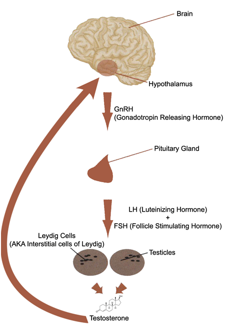
Follicle Stimulating Hormone
Cyclic CMP
Feminization
Steroidogenic Factor 1
Enzyme Inhibitors
Steroids
Receptors, Progesterone
Endometriosis
Testis
Ovarian Follicle
Dihydrotestosterone
Cholestenone 5 alpha-Reductase
Choriocarcinoma
Chemotherapy, Adjuvant
Selective Estrogen Receptor Modulators
Progesterone
Cholesterol Side-Chain Cleavage Enzyme
Follicular Fluid
Reverse Transcriptase Polymerase Chain Reaction
Microsomes
Hermaphroditic Organisms
Luciferases, Renilla
Promoter Regions, Genetic
Luteinizing Hormone
Gonadal Steroid Hormones
Veratrum
Breast
Immunohistochemistry
Gonadotropins
Atrazine
Pregnancy
Endocrine Disruptors
Leydig Cells
Sertoli Cells
Leydig Cell Tumor
Sex Characteristics
Oxidoreductases
Theca Cells
Receptors, Gonadotropin
Brain
Base Sequence
Virilism
Follicular Phase
Receptors, LH
Batrachoidiformes
Coturnix
Estradiol Dehydrogenases
Receptors, FSH
Cells, Cultured
Megestrol Acetate
Enzyme Induction
Receptors, Androgen
Gene Expression Regulation, Neoplastic
Perciformes
Stromal Cells
Endometrium
3-Oxo-5-alpha-Steroid 4-Dehydrogenase
Fushi Tarazu Transcription Factors
Cytochrome P-450 Enzyme System
Finches
Puberty, Precocious
Papio anubis
Ovotesticular Disorders of Sex Development
Molecular Sequence Data
Receptors, Estradiol
Follicular Atresia
Drug Resistance, Neoplasm
Dose-Response Relationship, Drug
Chorionic Gonadotropin
Exons
Clinical Trials as Topic
Endocrine System
Gene Expression
Dinoprostone
In Situ Hybridization
Co-Repressor Proteins
MCF-7 Cells
Dehydroepiandrosterone
Adrenocortical Carcinoma
Adipose Tissue
Follicle Stimulating Hormone, Human
Granulosa Cell Tumor
Tumor Cells, Cultured
Vitellogenins
Mammary Neoplasms, Experimental
Anticarcinogenic Agents
Hypothalamus
Blotting, Western
Gonadal Hormones
Bucladesine
17-alpha-Hydroxyprogesterone
Hot Flashes
Inhibins
Constitutional genetic variation at the human aromatase gene (Cyp19) and breast cancer risk. (1/1297)
The activity of the aromatase enzyme, which converts androgens into oestrogens and has a major role in regulating oestrogen levels in the breast, is thought to be a contributing factor in the development of breast cancer. We undertook this study to assess the role of constitutional genetic variation in the human aromatase gene (Cyp19) in the development of this disease. Our genotyping of 348 cases with breast cancer and 145 controls (all Caucasian women) for a published tetranucleotide repeat polymorphism at intron 4 of the Cyp19 gene revealed the presence of six common and two rare alleles. Contingency table analysis revealed a significant difference in allelic distribution between cases and controls (chi2 5df = 13.52, P = 0.019). The allele measuring 171 bp was over-represented in cases; of 14 individuals homozygous for this allele, 13 were cases. These individuals had a higher incidence of cancer in family members and an earlier age at diagnosis than other cases. In sequencing Cyp19's coding exons and regulatory regions, we discovered a perfect association between a silent polymorphism (G-->A at Val80) and the high-risk genotype. Our conclusion is that constitutional genetic variation at the Cyp19 locus is associated with the risk of developing breast cancer, with the 171-bp allele serving as the high-risk allele. (+info)Endometriosis: a dysfunction and disease of the archimetra. (2/1297)
Endometriosis is considered primarily a disease of the endometrial-subendometrial unit or archimetra. The clinical picture of endometriosis characterises this disease as a hyperactivation of genuine archimetrial functions such as proliferation, inflammatory defence and peristalsis. While the aetiology of the disease remains to be elucidated, a key event appears to consist in the local production of extraovarian oestrogen by a pathological expression of the P450 aromatase. The starting event may consist in a hyperactivity of the endometrial inflammatory defence, a hyperactivity of the endometrial oxytocin/oxytocin receptor system or in the pathological expression of the P450 aromatase system itself. Regardless of which of these levels the starting event is localized in, they influence each other on both the level of the archimetra and the endometriotic lesions. Locally elevated oestrogen levels inevitably up-regulate the endometrial oxytocin mRNA and increased levels of oxytocin result in uterine hyperperistalsis, increased transtubal seeding of endometrial tissue fragments and finally subfertility and infertility by impairment of the uterine mechanism of rapid and sustained sperm transport. Locally increased levels of oestrogen lead, on both the level of the endometrial-subendometrial unit and the endometriotic lesion, to processes of hyperproliferation. These processes result, on the level of the uterus, in an infiltrative growth of elements of the archimetra into the neometra and, on the level of the endometriotic lesion, in infiltrative endometriosis. There is circumstantial evidence that trauma might be an important initial event that induces the specific biochemical and cellular responses of the archimetra. This model is able to explain both the pleiomorphic appearance of endometriosis and the, up until now, enigmatic infertility associated with mild and moderate endometriosis. (+info)The aromatase inactivator 4-hydroxyandrostenedione (4-OH-A) inhibits tamoxifen metabolism by rat hepatic cytochrome P-450 3A: potential for drug-drug interaction of tamoxifen and 4-OH-A in combined anti-breast cancer therapy. (3/1297)
Tamoxifen (tam), an anti-breast cancer agent, is metabolized into tam-N-oxide by the hepatic flavin-containing monooxygenase and into N-desmethyl- and 4-hydroxy-tam by cytochrome P-450s (CYPs). Additionally, tam is metabolically activated by hepatic CYP3A, forming a reactive intermediate that binds covalently to proteins. Tam and 4-hydroxyandrostenedione (4-OH-A) are currently used to treat breast cancer, and it has been contemplated that 4-OH-A be given concurrently with tam to contravene potential tumor resistance to tam. Because alterations in tam metabolism may influence its therapeutic efficacy, the effect of 4-OH-A on tam metabolism was examined. Incubation of tam with liver microsomes from phenobarbital-treated rats, in the presence of 4-OH-A (10-100 microM), resulted in marked inhibition of tam-N-demethylation and tam covalent binding and in decreased tam-N-oxide accumulation; however, there was no inhibition of the formation of 4-hydroxy-tam and of 3,4-dihydroxytamoxifen. These findings indicate that 4-OH-A inhibits CYP3A, but not P-450(s) that catalyze tam 4-hydroxylation. The diminished tam-N-oxide accumulation could be due to decreased N-oxide formation and/or due to increased N-oxide reduction. Incubation of tam-N-oxide with liver microsomes containing heat-inactivated flavin-containing monooxygenase demonstrated that 4-OH-A increases the accumulation of tam, possibly by diminishing its P-450-mediated metabolism. Kinetic studies indicate that 4-OH-A is a competitive inhibitor of CYP3A, but not a time-dependent inactivator. Consequently, the concurrent treatment of tam and 4-OH-A may result in increased tam half-life and thus could potentiate the therapeutic efficacy of tam and diminish the potential side effects of tam by inhibiting its covalent binding to proteins and possibly to DNA. (+info)The mechanism of action of epidermal growth factor and transforming growth factor alpha on aromatase activity in granulosa cells from polycystic ovaries. (4/1297)
We investigated aromatization and the mechanism of action of epidermal growth factor (EGF) and transforming growth factor alpha (TGFalpha) on oestradiol biosynthesis in freshly prepared granulosa cells from polycystic ovaries. Freshly prepared granulosa cells from polycystic ovaries incubated for only 3 h under basal conditions secreted significantly (P< 0.001) greater amounts of oestradiol-17beta than that of granulosa cells from normal ovaries. 8-Bromo-cyclic adenosine monophosphate (8-Br-cAMP), but not follicle stimulating hormone (FSH) or luteinizing hormone (LH), further enhanced this activity. Both EGF and TGFalpha inhibited gonadotrophinor 8-Br-cAMP-stimulated, but not basal, oestradiol production. LH receptor (LHR) binding, estimated by immunolabelling the bound LH, was significantly (P< 0.001) reduced in granulosa cells from polycystic ovaries when compared with cells from normal ovaries. EGF or TGFalpha significantly reduced the binding in cultured cells from all patient groups (P< 0.05). More interestingly, a further increase of the inhibitory effect was seen in granulosa cells from polycystic ovaries (P < 0.001). In conclusion, granulosa cells from polycystic ovaries contain high levels of basal aromatase activity in vitro, which is probably inherited from the in-vivo condition. EGF and TGFalpha suppress oestradiol synthesis at a step beyond the production of cAMP and also LHR binding with more effect in granulosa cells from polycystic ovaries. (+info)Effect of labor induction on the expression of oxytocin receptor, cytochrome P450 aromatase, and estradiol receptor in the reproductive tract of the late-pregnant ewe. (5/1297)
In this study, we investigated the timing of changes in aromatase, estradiol receptor, and oxytocin receptor expression in ovine uterine and placental tissues before parturition. Labor was induced by betamethasone injection into the fetus on Days 130-132 of pregnancy. Tissue samples were collected at injection and then every 14 h until labor (56 h) from four ewes at each time point. Samples were analyzed for aromatase, estradiol receptor, and oxytocin receptor expression by in situ hybridization; for oxytocin binding to its receptor using a specific antagonist; and for estradiol receptor quantitation by immunocytochemistry. Aromatase mRNA expression increased by 14 h postinjection (p < 0.02) in the fetal villi and remained high until labor. Expression of estradiol and oxytocin receptor mRNAs was unchanged in myometrium but increased in the endometrial luminal epithelium by 28 h (p < 0.05) and remained high until labor. Estradiol receptor protein concentration increased modestly at labor while oxytocin receptor binding in the luminal epithelium changed in parallel to the mRNA concentration. IN CONCLUSION: 1) induction of aromatase may facilitate the expression of endometrial estradiol and oxytocin receptors in the placentome, 2) changes in endometrial rather than myometrial oxytocin receptor may be important in inducing parturition, and 3) the transcription of estradiol receptor and oxytocin receptor in the uterine epithelium are positively correlated during parturition. (+info)A 500-bp region, approximately 40 kb upstream of the human CYP19 (aromatase) gene, mediates placenta-specific expression in transgenic mice. (6/1297)
In humans, aromatase P450 (product of CYP19 gene), which catalyzes conversion of C19 steroids to estrogens, is expressed in a number of tissues, including ovary, adipose, and syncytiotrophoblast of the placenta. The 5' untranslated regions of CYP19 mRNA transcripts in these tissues are encoded by different tissue-specific first exons, which are spliced onto a common site just upstream of the translation initiation site in exon II. In placenta, the 5' untranslated region of CYP19 mRNA transcripts is encoded by exon I.1, which lies approximately 40 kb upstream of exon II. To map genomic sequences required for placenta-specific CYP19 expression, fusion genes containing 2,400 and 501 bp of placenta-specific exon I.1 5' flanking DNA linked to the human growth hormone gene (hGH), as reporter, were introduced into transgenic mice. Expression of CYP19(I.1):hGH fusion genes containing as little as 501 bp of 5' flanking DNA was placenta-specific and developmentally regulated. Furthermore, transgene expression occurred specifically in the labyrinthine trophoblast of the mouse placenta, which contains syncytial cells that may be analogous to the human syncytiotrophoblast. We show that a relatively small segment of DNA (approximately 500 bp) >40 kb upstream of the protein coding region of a human gene is able to direct expression in an appropriate tissue- and cell-specific manner in transgenic mice. These findings suggest that 5' flanking DNA within 501 bp of exon I.1 of the human CYP19 gene contains cis-acting elements that bind placenta-specific transcription factors that are conserved between humans and mice. (+info)Intrafollicular content of luteinizing hormone receptor, alpha-inhibin, and aromatase in relation to follicular growth, estrous cycle stage, and oocyte competence for in vitro maturation in the mare. (7/1297)
The intrafollicular content of LH receptor, alpha-inhibin, and aromatase are known good indicators of follicular status. We investigated the amounts of these proteins in granulosa and cumulus cells in relation to oocyte competence for in vitro maturation, follicular growth, and estrous cycle stage in the mare. Follicular punctures were performed 34 h after an injection of crude equine gonadotropins, either during the follicular phase, at the end of the follicular phase, or during the luteal phase. The cumulus-oocyte complex, granulosa cells, and follicular fluid of follicles larger than 5 mm were collected. The nuclear stage of the oocytes after in vitro culture was determined microscopically. Granulosa and cumulus cell amounts of LH receptor, alpha-inhibin, and aromatase were assessed by the semiquantitative Western blot method and image analysis. Follicular fluids were assayed for progesterone (P4) and estradiol-17beta (E2). The three factors were expressed in mural granulosa and cumulus cells from all follicles from the gonadotropin-independent growth period until the preovulatory stage. Considering all the follicles punctured, the amounts of LH receptor and alpha-inhibin in granulosa cells were not different for the three physiological stages studied. The amounts of aromatase in granulosa cells, as well as the E2:P4 ratios, were higher for follicles punctured during the follicular phase than for the two other groups (p < 0.05). Considering the data from the three groups, the E2:P4 ratio and the LH receptor and aromatase contents, but not alpha-inhibin, in granulosa cells increased with an increase in follicular diameter (p < 0.01). The E2:P4 ratios and the amounts of LH receptor, alpha-inhibin, and aromatase in granulosa cells were lower in follicles 5-9 mm in diameter than in larger ones (p < 0.05). In cumulus cells, the amounts of the three factors were different neither between the three groups nor between the follicular diameters. Although we could not establish any obvious relationship to oocyte competence for in vitro maturation, the influence of the follicle diameter on the content of LH receptors, alpha-inhibin, and aromatase in granulosa cells was similar to the influence of follicle diameter on oocyte competence. Therefore, one can hypothesize that, in the mare, there is a link between the acquisition of oocyte competence and the expression of these factors in the follicular cells. (+info)Dynamics of periovulatory steroidogenesis in the rhesus monkey follicle after ovarian stimulation. (8/1297)
The temporal relationships and regulation of events in the primate follicle during the periovulatory interval are poorly understood. This study was designed to elucidate the dynamics of steroid synthesis in the macaque follicle during ovarian stimulation cycles in which serum/follicular fluid aspirates were collected at precise intervals before (0 h) and after (up to 36 h) administration of the ovulatory human chorionic gonadotrophin (HCG) bolus. Serum concentrations of progesterone increased (P < 0.05) within 30 min, and follicular fluid progesterone concentrations were elevated 180-fold within 12 h, of HCG injection, and remained elevated until the time of ovulation. In contrast, 17beta-oestradiol concentrations increased initially, but then declined (P < 0.05) by 36 h post-HCG. Acute incubation of granulosa cells with and without steroidogenic substrates demonstrated that: (i) 3beta-hydroxysteroid dehydrogenase and aromatase activities were present in equivalent amounts before and after HCG; whereas (ii) P450 side-chain cleavage activity increased (P < 0.05) within 12 h of HCG; and (iii) exogenous low-density lipoprotein and cholesterol were not utilized for steroidogenesis. This model should be useful for further studies on ovulation and luteinization in primates, and enable elucidation of the local actions of progesterone and other steroids at specific time points during the periovulatory interval. (+info)Aromatase inhibitors (AIs) are a class of drugs that are primarily used in the treatment of hormone-sensitive breast cancer in postmenopausal women. They work by inhibiting the enzyme aromatase, which is responsible for converting androgens into estrogens. By blocking this conversion, AIs decrease the amount of estrogen in the body, thereby depriving hormone-sensitive breast cancer cells of the estrogen they need to grow and multiply.
There are three main types of aromatase inhibitors:
1. Letrozole (Femara) - a non-steroidal AI that is taken orally once a day.
2. Anastrozole (Arimidex) - another non-steroidal AI that is also taken orally once a day.
3. Exemestane (Aromasin) - a steroidal AI that is taken orally once a day.
In addition to their use in breast cancer treatment, AIs are also sometimes used off-label for the treatment of estrogen-dependent conditions such as endometriosis and uterine fibroids. However, it's important to note that the use of aromatase inhibitors can have significant side effects, including hot flashes, joint pain, and bone loss, so they should only be used under the close supervision of a healthcare provider.
Triazoles are a class of antifungal medications that have broad-spectrum activity against various fungi, including yeasts, molds, and dermatophytes. They work by inhibiting the synthesis of ergosterol, an essential component of fungal cell membranes, leading to increased permeability and disruption of fungal growth. Triazoles are commonly used in both systemic and topical formulations for the treatment of various fungal infections, such as candidiasis, aspergillosis, cryptococcosis, and dermatophytoses. Some examples of triazole antifungals include fluconazole, itraconazole, voriconazole, and posaconazole.
Nitriles, in a medical context, refer to a class of organic compounds that contain a cyano group (-CN) bonded to a carbon atom. They are widely used in the chemical industry and can be found in various materials, including certain plastics and rubber products.
In some cases, nitriles can pose health risks if ingested, inhaled, or come into contact with the skin. Short-term exposure to high levels of nitriles can cause irritation to the eyes, nose, throat, and respiratory tract. Prolonged or repeated exposure may lead to more severe health effects, such as damage to the nervous system, liver, and kidneys.
However, it's worth noting that the medical use of nitriles is not very common. Some nitrile gloves are used in healthcare settings due to their resistance to many chemicals and because they can provide a better barrier against infectious materials compared to latex or vinyl gloves. But beyond this application, nitriles themselves are not typically used as medications or therapeutic agents.
Fadrozole is a non-steroidal aromatase inhibitor drug that is used in the treatment of breast cancer. Aromatase inhibitors work by blocking the production of estrogen, which some types of breast cancer cells need to grow. By reducing the amount of estrogen in the body, fadrozole can help slow or stop the growth of these cancer cells.
Fadrozole is typically used as a treatment for postmenopausal women with hormone receptor-positive breast cancer. It may be used as a first-line therapy or after other treatments have failed. The drug is administered orally, and the typical dosage is 1-2 mg per day.
Like all medications, fadrozole can cause side effects, including hot flashes, nausea, vomiting, and joint pain. In some cases, it may also cause more serious side effects such as liver damage or an increased risk of bone fractures. Patients taking fadrozole should be monitored closely by their healthcare provider to ensure that the drug is working effectively and to manage any side effects that may occur.
Androstenedione is a steroid hormone produced by the adrenal glands, ovaries, and testes. It is a precursor to both male and female sex hormones, including testosterone and estrogen. In the adrenal glands, it is produced from cholesterol through a series of biochemical reactions involving several enzymes. Androstenedione can also be converted into other steroid hormones, such as dehydroepiandrosterone (DHEA) and estrone.
In the body, androstenedione plays an important role in the development and maintenance of secondary sexual characteristics, such as facial hair and a deep voice in men, and breast development and menstrual cycles in women. It also contributes to bone density, muscle mass, and overall physical strength.
Androstenedione is available as a dietary supplement and has been marketed as a way to boost athletic performance and increase muscle mass. However, its effectiveness for these purposes is not supported by scientific evidence, and it may have harmful side effects when taken in high doses or for extended periods of time. Additionally, the use of androstenedione as a dietary supplement is banned by many sports organizations, including the International Olympic Committee and the National Collegiate Athletic Association.
Estrogens are a group of steroid hormones that are primarily responsible for the development and regulation of female sexual characteristics and reproductive functions. They are also present in lower levels in males. The main estrogen hormone is estradiol, which plays a key role in promoting the growth and development of the female reproductive system, including the uterus, fallopian tubes, and breasts. Estrogens also help regulate the menstrual cycle, maintain bone density, and have important effects on the cardiovascular system, skin, hair, and cognitive function.
Estrogens are produced primarily by the ovaries in women, but they can also be produced in smaller amounts by the adrenal glands and fat cells. In men, estrogens are produced from the conversion of testosterone, the primary male sex hormone, through a process called aromatization.
Estrogen levels vary throughout a woman's life, with higher levels during reproductive years and lower levels after menopause. Estrogen therapy is sometimes used to treat symptoms of menopause, such as hot flashes and vaginal dryness, or to prevent osteoporosis in postmenopausal women. However, estrogen therapy also carries risks, including an increased risk of certain cancers, blood clots, and stroke, so it is typically recommended only for women who have a high risk of these conditions.
Androstatrienes are a class of steroidal compounds that contain a 1,2-dehydrogenated A-ring in their chemical structure. They are named after androstane, which is the reduced form of testosterone, by replacing two hydrogen atoms with a double bond between the first and second carbon atoms in the A-ring.
Androstatrienes do not have any significant medical relevance on their own, but some compounds that contain an androstadiene structure may have biological activity. For example, certain androstadienedione derivatives have been investigated for their potential as progestins or as inhibitors of 5α-reductase, an enzyme involved in the conversion of testosterone to dihydrotestosterone.
It is worth noting that some androstadiene compounds may be produced endogenously in the human body, while others may be synthesized in the laboratory for research or therapeutic purposes. However, it is important to note that some androstadienes are also found in certain anabolic-androgenic steroids (AAS) and can be used as markers of AAS use in drug testing.
Estradiol is a type of estrogen, which is a female sex hormone. It is the most potent and dominant form of estrogen in humans. Estradiol plays a crucial role in the development and maintenance of secondary sexual characteristics in women, such as breast development and regulation of the menstrual cycle. It also helps maintain bone density, protect the lining of the uterus, and is involved in cognition and mood regulation.
Estradiol is produced primarily by the ovaries, but it can also be synthesized in smaller amounts by the adrenal glands and fat cells. In men, estradiol is produced from testosterone through a process called aromatization. Abnormal levels of estradiol can contribute to various health issues, such as hormonal imbalances, infertility, osteoporosis, and certain types of cancer.
Breast neoplasms refer to abnormal growths in the breast tissue that can be benign or malignant. Benign breast neoplasms are non-cancerous tumors or growths, while malignant breast neoplasms are cancerous tumors that can invade surrounding tissues and spread to other parts of the body.
Breast neoplasms can arise from different types of cells in the breast, including milk ducts, milk sacs (lobules), or connective tissue. The most common type of breast cancer is ductal carcinoma, which starts in the milk ducts and can spread to other parts of the breast and nearby structures.
Breast neoplasms are usually detected through screening methods such as mammography, ultrasound, or MRI, or through self-examination or clinical examination. Treatment options for breast neoplasms depend on several factors, including the type and stage of the tumor, the patient's age and overall health, and personal preferences. Treatment may include surgery, radiation therapy, chemotherapy, hormone therapy, or targeted therapy.
Aminoglutethimide is a medication that is primarily used to treat hormone-sensitive cancers such as breast cancer and prostate cancer. It works by blocking the production of certain hormones in the body, including estrogen and cortisol. Aminoglutethimide is an inhibitor of steroid synthesis, specifically targeting the enzymes involved in the conversion of cholesterol to steroid hormones.
The medication is available in oral form and is typically taken 2-3 times a day. Common side effects include drowsiness, dizziness, dry mouth, skin rash, and changes in appetite or weight. More serious side effects may include liver damage, severe allergic reactions, and changes in heart rhythm.
It's important to note that aminoglutethimide can interact with other medications, so it's crucial to inform your healthcare provider about all the drugs you are currently taking before starting this medication. Additionally, regular monitoring of liver function and hormone levels may be necessary during treatment with aminoglutethimide.
Testosterone is a steroid hormone that belongs to androsten class of hormones. It is primarily secreted by the Leydig cells in the testes of males and, to a lesser extent, by the ovaries and adrenal glands in females. Testosterone is the main male sex hormone and anabolic steroid. It plays a key role in the development of masculine characteristics, such as body hair and muscle mass, and contributes to bone density, fat distribution, red cell production, and sex drive. In females, testosterone contributes to sexual desire and bone health. Testosterone is synthesized from cholesterol and its production is regulated by luteinizing hormone (LH) and follicle-stimulating hormone (FSH).
Estrone is a type of estrogen, which is a female sex hormone. It's one of the three major naturally occurring estrogens in women, along with estradiol and estriol. Estrone is weaker than estradiol but has a longer half-life, meaning it remains active in the body for a longer period of time.
Estrone is produced primarily in the ovaries, adrenal glands, and fat tissue. In postmenopausal women, when the ovaries stop producing estradiol, estrone becomes the dominant form of estrogen. It plays a role in maintaining bone density, regulating the menstrual cycle, and supporting the development and maintenance of female sexual characteristics.
Like other forms of estrogen, estrone can also have effects on various tissues throughout the body, including the brain, heart, and breast tissue. Abnormal levels of estrone, either too high or too low, can contribute to a variety of health issues, such as osteoporosis, menstrual irregularities, and increased risk of certain types of cancer.
Antineoplastic agents, hormonal, are a class of drugs used to treat cancers that are sensitive to hormones. These agents work by interfering with the production or action of hormones in the body. They can be used to slow down or stop the growth of cancer cells and may also help to relieve symptoms caused by the spread of cancer.
Hormonal therapies can work in one of two ways: they can either block the production of hormones or prevent their action on cancer cells. For example, some hormonal therapies work by blocking the action of estrogen or testosterone, which are hormones that can stimulate the growth of certain types of cancer cells.
Examples of hormonal agents used to treat cancer include:
* Aromatase inhibitors (such as letrozole, anastrozole, and exemestane), which block the production of estrogen in postmenopausal women
* Selective estrogen receptor modulators (such as tamoxifen and raloxifene), which block the action of estrogen on cancer cells
* Luteinizing hormone-releasing hormone agonists (such as leuprolide, goserelin, and triptorelin), which block the production of testosterone in men
* Antiandrogens (such as bicalutamide, flutamide, and enzalutamide), which block the action of testosterone on cancer cells
Hormonal therapies are often used in combination with other treatments, such as surgery or radiation therapy. They may be used to shrink tumors before surgery, to kill any remaining cancer cells after surgery, or to help control the spread of cancer that cannot be removed by surgery. Hormonal therapies can also be used to relieve symptoms and improve quality of life in people with advanced cancer.
It's important to note that hormonal therapies are not effective for all types of cancer. They are most commonly used to treat breast, prostate, and endometrial cancers, which are known to be sensitive to hormones. Hormonal therapies may also be used to treat other types of cancer in certain situations.
Like all medications, hormonal therapies can have side effects. These can vary depending on the specific drug and the individual person. Common side effects of hormonal therapies include hot flashes, fatigue, mood changes, and sexual dysfunction. Some hormonal therapies can also cause more serious side effects, such as an increased risk of osteoporosis or blood clots. It's important to discuss the potential risks and benefits of hormonal therapy with a healthcare provider before starting treatment.
Tamoxifen is a selective estrogen receptor modulator (SERM) medication that is primarily used in the treatment and prevention of breast cancer. It works by blocking the action of estrogen in the body, particularly in breast tissue. This can help to stop or slow the growth of hormone-sensitive tumors.
Tamoxifen has been approved by the U.S. Food and Drug Administration (FDA) for use in both men and women. It is often used as a part of adjuvant therapy, which is treatment given after surgery to reduce the risk of cancer recurrence. Tamoxifen may also be used to treat metastatic breast cancer that has spread to other parts of the body.
Common side effects of tamoxifen include hot flashes, vaginal discharge, and changes in mood or vision. Less commonly, tamoxifen can increase the risk of blood clots, stroke, and endometrial cancer (cancer of the lining of the uterus). However, for many women with breast cancer, the benefits of taking tamoxifen outweigh the risks.
It's important to note that while tamoxifen can be an effective treatment option for some types of breast cancer, it is not appropriate for all patients. A healthcare professional will consider a variety of factors when determining whether tamoxifen is the right choice for an individual patient.
Hormone-dependent neoplasms are a type of tumor that requires the presence of specific hormones to grow and multiply. These neoplasms have receptors on their cell surfaces that bind to the hormones, leading to the activation of signaling pathways that promote cell division and growth.
Examples of hormone-dependent neoplasms include breast cancer, prostate cancer, and endometrial cancer. In breast cancer, for instance, estrogen and/or progesterone can bind to their respective receptors on the surface of cancer cells, leading to the activation of signaling pathways that promote tumor growth. Similarly, in prostate cancer, androgens such as testosterone can bind to androgen receptors on the surface of cancer cells, promoting cell division and tumor growth.
Hormone-dependent neoplasms are often treated with hormonal therapies that aim to reduce or block the production of the relevant hormones or interfere with their ability to bind to their respective receptors. This can help slow down or stop the growth of the tumor and improve outcomes for patients.
Estrogen receptors (ERs) are a type of nuclear receptor protein that are expressed in various tissues and cells throughout the body. They play a critical role in the regulation of gene expression and cellular responses to the hormone estrogen. There are two main subtypes of ERs, ERα and ERβ, which have distinct molecular structures, expression patterns, and functions.
ERs function as transcription factors that bind to specific DNA sequences called estrogen response elements (EREs) in the promoter regions of target genes. When estrogen binds to the ER, it causes a conformational change in the receptor that allows it to recruit co-activator proteins and initiate transcription of the target gene. This process can lead to a variety of cellular responses, including changes in cell growth, differentiation, and metabolism.
Estrogen receptors are involved in a wide range of physiological processes, including the development and maintenance of female reproductive tissues, bone homeostasis, cardiovascular function, and cognitive function. They have also been implicated in various pathological conditions, such as breast cancer, endometrial cancer, and osteoporosis. As a result, ERs are an important target for therapeutic interventions in these diseases.
Estrogen antagonists, also known as antiestrogens, are a class of drugs that block the effects of estrogen in the body. They work by binding to estrogen receptors and preventing the natural estrogen from attaching to them. This results in the inhibition of estrogen-mediated activities in various tissues, including breast and uterine tissue.
There are two main types of estrogen antagonists: selective estrogen receptor modulators (SERMs) and pure estrogen receptor downregulators (PERDS), also known as estrogen receptor downregulators (ERDs). SERMs, such as tamoxifen and raloxifene, can act as estrogen agonists or antagonists depending on the tissue type. For example, they may block the effects of estrogen in breast tissue while acting as an estrogen agonist in bone tissue, helping to prevent osteoporosis.
PERDS, such as fulvestrant, are pure estrogen receptor antagonists and do not have any estrogen-like activity. They are used primarily for the treatment of hormone receptor-positive breast cancer in postmenopausal women.
Overall, estrogen antagonists play an important role in the management of hormone receptor-positive breast cancer and other conditions where inhibiting estrogen activity is beneficial.
An ovary is a part of the female reproductive system in which ova or eggs are produced through the process of oogenesis. They are a pair of solid, almond-shaped structures located one on each side of the uterus within the pelvic cavity. Each ovary measures about 3 to 5 centimeters in length and weighs around 14 grams.
The ovaries have two main functions: endocrine (hormonal) function and reproductive function. They produce and release eggs (ovulation) responsible for potential fertilization and development of an embryo/fetus during pregnancy. Additionally, they are essential in the production of female sex hormones, primarily estrogen and progesterone, which regulate menstrual cycles, sexual development, and reproduction.
During each menstrual cycle, a mature egg is released from one of the ovaries into the fallopian tube, where it may be fertilized by sperm. If not fertilized, the egg, along with the uterine lining, will be shed, leading to menstruation.
Gynecomastia is a medical term that refers to the benign enlargement of the glandular tissue in male breasts, usually caused by an imbalance of the hormones estrogen and testosterone. It's important to note that gynecomastia is not the same as having excess fat in the breast area, which is called pseudogynecomastia.
Gynecomastia can occur during infancy, puberty, or old age due to natural hormonal changes. Certain medications, medical conditions, and recreational drugs can also cause gynecomastia by affecting hormone levels in the body. In some cases, the exact cause of gynecomastia may remain unknown.
Mild cases of gynecomastia may not require treatment, but severe or persistent cases may be treated with medication or surgery to remove excess breast tissue. It's essential to consult a healthcare professional for an accurate diagnosis and appropriate treatment options if you suspect you have gynecomastia.
Postmenopause is a stage in a woman's life that follows 12 months after her last menstrual period (menopause) has occurred. During this stage, the ovaries no longer release eggs and produce lower levels of estrogen and progesterone hormones. The reduced levels of these hormones can lead to various physical changes and symptoms, such as hot flashes, vaginal dryness, and mood changes. Postmenopause is also associated with an increased risk of certain health conditions, including osteoporosis and heart disease. It's important for women in postmenopause to maintain a healthy lifestyle, including regular exercise, a balanced diet, and routine medical check-ups to monitor their overall health and manage any potential risks.
Granulosa cells are specialized cells that surround and enclose the developing egg cells (oocytes) in the ovaries. They play a crucial role in the growth, development, and maturation of the follicles (the fluid-filled sacs containing the oocytes) by providing essential nutrients and hormones.
Granulosa cells are responsible for producing estrogen, which supports the development of the endometrium during the menstrual cycle in preparation for a potential pregnancy. They also produce inhibin and activin, two hormones that regulate the function of the pituitary gland and its secretion of follicle-stimulating hormone (FSH) and luteinizing hormone (LH).
These cells are critical for female reproductive health and fertility. Abnormalities in granulosa cell function can lead to various reproductive disorders, such as polycystic ovary syndrome (PCOS), premature ovarian failure, and infertility.
Testolactone is a medication that is primarily used in the treatment of breast cancer. It is an oral steroidal aromatase inhibitor, which means it works by blocking the enzyme aromatase, thereby preventing the conversion of androgens into estrogens. This helps to reduce the amount of estrogen in the body, which can slow or stop the growth of certain types of breast cancer cells that need estrogen to grow.
Testolactone is not as commonly used as other aromatase inhibitors such as letrozole, anastrozole, and exemestane, but it may be prescribed in certain cases where these medications are not suitable or have not been effective. It is important to note that testolactone can have side effects, including nausea, vomiting, diarrhea, skin rash, and changes in liver function tests. As with any medication, it should only be taken under the supervision of a healthcare provider.
Androstadienes are a class of steroid hormones that are derived from androstenedione, which is a weak male sex hormone. Androstadienes include various compounds such as androstadiene-3,17-dione and androstanedione, which are intermediate products in the biosynthesis of more potent androgens like testosterone and dihydrotestosterone.
Androstadienes are present in both males and females but are found in higher concentrations in men. They can be detected in various bodily fluids, including blood, urine, sweat, and semen. In addition to their role in steroid hormone synthesis, androstadienes have been studied for their potential use as biomarkers of physiological processes and disease states.
It's worth noting that androstadienes are sometimes referred to as "androstenes" in the literature, although this term can also refer to other related compounds.
Estrogen Receptor alpha (ERα) is a type of nuclear receptor protein that is activated by the hormone estrogen. It is encoded by the gene ESR1 and is primarily expressed in the cells of the reproductive system, breast, bone, liver, heart, and brain tissue.
When estrogen binds to ERα, it causes a conformational change in the receptor, which allows it to dimerize and translocate to the nucleus. Once in the nucleus, ERα functions as a transcription factor, binding to specific DNA sequences called estrogen response elements (EREs) and regulating the expression of target genes.
ERα plays important roles in various physiological processes, including the development and maintenance of female reproductive organs, bone homeostasis, and lipid metabolism. It is also a critical factor in the growth and progression of certain types of breast cancer, making ERα status an important consideration in the diagnosis and treatment of this disease.
Gene expression regulation, enzymologic refers to the biochemical processes and mechanisms that control the transcription and translation of specific genes into functional proteins or enzymes. This regulation is achieved through various enzymatic activities that can either activate or repress gene expression at different levels, such as chromatin remodeling, transcription factor activation, mRNA processing, and protein degradation.
Enzymologic regulation of gene expression involves the action of specific enzymes that catalyze chemical reactions involved in these processes. For example, histone-modifying enzymes can alter the structure of chromatin to make genes more or less accessible for transcription, while RNA polymerase and its associated factors are responsible for transcribing DNA into mRNA. Additionally, various enzymes are involved in post-transcriptional modifications of mRNA, such as splicing, capping, and tailing, which can affect the stability and translation of the transcript.
Overall, the enzymologic regulation of gene expression is a complex and dynamic process that allows cells to respond to changes in their environment and maintain proper physiological function.
Androgens are a class of hormones that are primarily responsible for the development and maintenance of male sexual characteristics and reproductive function. Testosterone is the most well-known androgen, but other androgens include dehydroepiandrosterone (DHEA), androstenedione, and dihydrotestosterone (DHT).
Androgens are produced primarily by the testes in men and the ovaries in women, although small amounts are also produced by the adrenal glands in both sexes. They play a critical role in the development of male secondary sexual characteristics during puberty, such as the growth of facial hair, deepening of the voice, and increased muscle mass.
In addition to their role in sexual development and function, androgens also have important effects on bone density, mood, and cognitive function. Abnormal levels of androgens can contribute to a variety of medical conditions, including infertility, erectile dysfunction, acne, hirsutism (excessive hair growth), and prostate cancer.
Gonads are the reproductive organs that produce gametes (sex cells) and sex hormones. In males, the gonads are the testes, which produce sperm and testosterone. In females, the gonads are the ovaries, which produce eggs and estrogen and progesterone. The development, function, and regulation of the gonads are crucial for reproductive health and fertility.
17-Hydroxysteroid dehydrogenases (17-HSDs) are a group of enzymes that play a crucial role in steroid hormone biosynthesis. They are involved in the conversion of 17-ketosteroids to 17-hydroxy steroids or vice versa, by adding or removing a hydroxyl group (–OH) at the 17th carbon atom of the steroid molecule. This conversion is essential for the production of various steroid hormones, including cortisol, aldosterone, and sex hormones such as estrogen and testosterone.
There are several isoforms of 17-HSDs, each with distinct substrate specificities, tissue distributions, and functions:
1. 17-HSD type 1 (17-HSD1): This isoform primarily catalyzes the conversion of estrone (E1) to estradiol (E2), an active form of estrogen. It is mainly expressed in the ovary, breast, and adipose tissue.
2. 17-HSD type 2 (17-HSD2): This isoform catalyzes the reverse reaction, converting estradiol (E2) to estrone (E1). It is primarily expressed in the placenta, prostate, and breast tissue.
3. 17-HSD type 3 (17-HSD3): This isoform is responsible for the conversion of androstenedione to testosterone, an essential step in male sex hormone biosynthesis. It is predominantly expressed in the testis and adrenal gland.
4. 17-HSD type 4 (17-HSD4): This isoform catalyzes the conversion of dehydroepiandrosterone (DHEA) to androstenedione, an intermediate step in steroid hormone biosynthesis. It is primarily expressed in the placenta.
5. 17-HSD type 5 (17-HSD5): This isoform catalyzes the conversion of cortisone to cortisol, a critical step in glucocorticoid biosynthesis. It is predominantly expressed in the adrenal gland and liver.
6. 17-HSD type 6 (17-HSD6): This isoform catalyzes the conversion of androstenedione to testosterone, similar to 17-HSD3. However, it has a different substrate specificity and is primarily expressed in the ovary.
7. 17-HSD type 7 (17-HSD7): This isoform catalyzes the conversion of estrone (E1) to estradiol (E2), similar to 17-HSD1. However, it has a different substrate specificity and is primarily expressed in the ovary.
8. 17-HSD type 8 (17-HSD8): This isoform catalyzes the conversion of DHEA to androstenedione, similar to 17-HSD4. However, it has a different substrate specificity and is primarily expressed in the testis.
9. 17-HSD type 9 (17-HSD9): This isoform catalyzes the conversion of estrone (E1) to estradiol (E2), similar to 17-HSD1. However, it has a different substrate specificity and is primarily expressed in the placenta.
10. 17-HSD type 10 (17-HSD10): This isoform catalyzes the conversion of DHEA to androstenedione, similar to 17-HSD4. However, it has a different substrate specificity and is primarily expressed in the testis.
11. 17-HSD type 11 (17-HSD11): This isoform catalyzes the conversion of estrone (E1) to estradiol (E2), similar to 17-HSD1. However, it has a different substrate specificity and is primarily expressed in the placenta.
12. 17-HSD type 12 (17-HSD12): This isoform catalyzes the conversion of DHEA to androstenedione, similar to 17-HSD4. However, it has a different substrate specificity and is primarily expressed in the testis.
13. 17-HSD type 13 (17-HSD13): This isoform catalyzes the conversion of estrone (E1) to estradiol (E2), similar to 17-HSD1. However, it has a different substrate specificity and is primarily expressed in the placenta.
14. 17-HSD type 14 (17-HSD14): This isoform catalyzes the conversion of DHEA to androstenedione, similar to 17-HSD4. However, it has a different substrate specificity and is primarily expressed in the testis.
15. 17-HSD type 15 (17-HSD15): This isoform catalyzes the conversion of estrone (E1) to estradiol (E2), similar to 17-HSD1. However, it has a different substrate specificity and is primarily expressed in the placenta.
16. 17-HSD type 16 (17-HSD16): This isoform catalyzes the conversion of DHEA to androstenedione, similar to 17-HSD4. However, it has a different substrate specificity and is primarily expressed in the testis.
17. 17-HSD type 17 (17-HSD17): This isoform catalyzes the conversion of estrone (E1) to estradiol (E2), similar to 17-HSD1. However, it has a different substrate specificity and is primarily expressed in the placenta.
18. 17-HSD type 18 (17-HSD18): This isoform catalyzes the conversion of DHEA to androstenedione, similar to 17-HSD4. However, it has a different substrate specificity and is primarily expressed in the testis.
19. 17-HSD type 19 (17-HSD19): This isoform catalyzes the conversion of estrone (E1) to estradiol (E2), similar to 17-HSD1. However, it has a different substrate specificity and is primarily expressed in the placenta.
20. 17-HSD type 20 (17-HSD20): This isoform catalyzes the conversion of DHEA to androstenedione, similar to 17-HSD4. However, it has a different substrate specificity and is primarily expressed in the testis.
21. 17-HSD type 21 (17-HSD21): This isoform catalyzes the conversion of estrone (E1) to estradiol (E2), similar to 17-HSD1. However, it has a different substrate specificity and is primarily expressed in the placenta.
22. 17-HSD type 22 (17-HSD22): This isoform catalyzes the conversion of DHEA to androstenedione, similar to 17-HSD4. However, it has a different substrate specificity and is primarily expressed in the testis.
23. 17-HSD type 23 (17-HSD23): This isoform catalyzes the conversion of estrone (E1) to estradiol (E2), similar to 17-HSD1. However, it has a different substrate specificity and is primarily expressed in the placenta.
24. 17-HSD type 24 (17-HSD24): This isoform catalyzes the conversion of DHEA to androstenedione, similar to 17-HSD4. However, it has a different substrate specificity and is primarily expressed in the testis.
25. 17-HSD type 25 (17-HSD25): This isoform catalyzes the conversion of estrone (E1) to estradiol (E2), similar to 17-HSD1. However, it has a different substrate specificity and is primarily expressed in the placenta.
26. 17-HSD type 26 (17-HSD26): This isoform catalyzes the conversion of DHEA to androstenedione, similar to 17-HSD4. However
"Sex differentiation" is a term used in the field of medicine, specifically in reproductive endocrinology and genetics. It refers to the biological development of sexual characteristics that distinguish males from females. This process is regulated by hormones and genetic factors.
There are two main stages of sex differentiation: genetic sex determination and gonadal sex differentiation. Genetic sex determination occurs at fertilization, where the combination of X and Y chromosomes determines the sex of the individual (typically, XX = female and XY = male). Gonadal sex differentiation then takes place during fetal development, where the genetic sex signals the development of either ovaries or testes.
Once the gonads are formed, they produce hormones that drive further sexual differentiation, leading to the development of internal reproductive structures (such as the uterus and fallopian tubes in females, and the vas deferens and seminal vesicles in males) and external genitalia.
It's important to note that while sex differentiation is typically categorized as male or female, there are individuals who may have variations in their sexual development, leading to intersex conditions. These variations can occur at any stage of the sex differentiation process and can result in a range of physical characteristics that do not fit neatly into male or female categories.
The placenta is an organ that develops in the uterus during pregnancy and provides oxygen and nutrients to the growing baby through the umbilical cord. It also removes waste products from the baby's blood. The placenta attaches to the wall of the uterus, and the baby's side of the placenta contains many tiny blood vessels that connect to the baby's circulatory system. This allows for the exchange of oxygen, nutrients, and waste between the mother's and baby's blood. After the baby is born, the placenta is usually expelled from the uterus in a process called afterbirth.
Estrogen Receptor beta (ER-β) is a protein that is encoded by the gene ESR2 in humans. It belongs to the family of nuclear receptors, which are transcription factors that regulate gene expression in response to hormonal signals. ER-β is one of two main estrogen receptors, the other being Estrogen Receptor alpha (ER-α), and it plays an important role in mediating the effects of estrogens in various tissues, including the breast, uterus, bone, brain, and cardiovascular system.
Estrogens are steroid hormones that play a critical role in the development and maintenance of female reproductive and sexual function. They also have important functions in other tissues, such as maintaining bone density and promoting cognitive function. ER-β is widely expressed in many tissues, including those outside of the reproductive system, suggesting that it may have diverse physiological roles beyond estrogen-mediated reproduction.
ER-β has been shown to have both overlapping and distinct functions from ER-α, and its expression patterns differ between tissues. For example, in the breast, ER-β is expressed at higher levels in normal tissue compared to cancerous tissue, suggesting that it may play a protective role against breast cancer development. In contrast, in the uterus, ER-β has been shown to have anti-proliferative effects and may protect against endometrial cancer.
Overall, ER-β is an important mediator of estrogen signaling and has diverse physiological roles in various tissues. Understanding its functions and regulation may provide insights into the development of novel therapies for a range of diseases, including cancer, osteoporosis, and cardiovascular disease.
Estrogen receptor modulators (ERMs) are a class of medications that act on the estrogen receptors in the body. They can have mixed estrogenic and anti-estrogenic effects, depending on the target tissue. In some tissues, ERMs behave as estrogen agonists, activating the estrogen receptor and mimicking the effects of estrogen. In other tissues, they act as estrogen antagonists, blocking the effects of estrogen.
ERMs are often used in hormone replacement therapy and to treat certain types of breast cancer. Tamoxifen is a well-known example of an ERM that is commonly used to treat estrogen receptor-positive (ER+) breast cancer. It works by blocking the effects of estrogen on cancer cells, thereby slowing or stopping the growth of the tumor. Other examples of ERMs include raloxifene and toremifene.
While ERMs can be effective in treating certain conditions, they can also have side effects, including an increased risk of blood clots, hot flashes, and mood changes. It is important for individuals taking ERMs to be monitored by a healthcare provider to manage any potential side effects and ensure that the medication is working effectively.
Steroid 17-alpha-hydroxylase, also known as CYP17A1, is a cytochrome P450 enzyme that plays a crucial role in steroid hormone biosynthesis. It is located in the endoplasmic reticulum of cells in the adrenal glands and gonads. This enzyme catalyzes the 17-alpha-hydroxylation and subsequent lyase cleavage of pregnenolone and progesterone, converting them into dehydroepiandrosterone (DHEA) and androstenedione, respectively. These steroid intermediates are essential for the biosynthesis of both glucocorticoids and sex steroids, including cortisol, aldosterone, estrogens, and testosterone.
Defects in the CYP17A1 gene can lead to several disorders, such as congenital adrenal hyperplasia (CAH) due to 17-alpha-hydroxylase deficiency, which is characterized by decreased production of cortisol and sex steroids and increased mineralocorticoid levels. This condition results in sexual infantilism, electrolyte imbalances, and hypertension.
Messenger RNA (mRNA) is a type of RNA (ribonucleic acid) that carries genetic information copied from DNA in the form of a series of three-base code "words," each of which specifies a particular amino acid. This information is used by the cell's machinery to construct proteins, a process known as translation. After being transcribed from DNA, mRNA travels out of the nucleus to the ribosomes in the cytoplasm where protein synthesis occurs. Once the protein has been synthesized, the mRNA may be degraded and recycled. Post-transcriptional modifications can also occur to mRNA, such as alternative splicing and addition of a 5' cap and a poly(A) tail, which can affect its stability, localization, and translation efficiency.
Follicle-Stimulating Hormone (FSH) is a glycoprotein hormone secreted and released by the anterior pituitary gland. In females, it promotes the growth and development of ovarian follicles in the ovary, which ultimately leads to the maturation and release of an egg (ovulation). In males, FSH stimulates the testes to produce sperm. It works in conjunction with luteinizing hormone (LH) to regulate reproductive processes. The secretion of FSH is controlled by the hypothalamic-pituitary-gonadal axis and its release is influenced by the levels of gonadotropin-releasing hormone (GnRH), estrogen, inhibin, and androgens.
I'm sorry for any confusion, but "Cyclic CMP" is not a standard medical term or abbreviation that I am familiar with. It appears to be related to biochemistry, specifically in the context of cyclic nucleotides. However, I would recommend consulting a reliable biochemistry or molecular biology resource for a precise definition and further information.
Cyclic nucleotides are important second messengers in cells, and they include molecules like cAMP (cyclic adenosine monophosphate) and cGMP (cyclic guanosine monophosphate). If "Cyclic CMP" refers to a cyclic nucleotide, it would most likely be referring to cyclic cytidine monophosphate. However, the use of this term in the medical field is not widespread or well-known.
Feminization is a process or condition in which typically male characteristics are diminished or absent, and female characteristics become more prominent. This term is often used in the context of transgender health to describe hormone therapy that helps individuals align their physical appearance with their gender identity. The goal of feminizing hormone therapy is to promote the development of secondary sexual characteristics such as breast development, softer skin, reduced muscle mass and body hair, and fat redistribution to create a more typically female body shape. It's important to note that every individual's experience with feminization is unique, and the specific changes experienced may vary depending on factors such as age, genetics, and the duration of hormone therapy.
Steroidogenic Factor 1 (SF-1 or NR5A1) is a nuclear receptor protein that functions as a transcription factor, playing a crucial role in the development and regulation of the endocrine system. It is involved in the differentiation and maintenance of steroidogenic tissues such as the adrenal glands, gonads (ovaries and testes), and the hypothalamus and pituitary glands in the brain.
SF-1 regulates the expression of genes that are essential for steroid hormone biosynthesis, including enzymes involved in the production of cortisol, aldosterone, and sex steroids (androgens, estrogens). Mutations in the SF-1 gene can lead to various disorders related to sexual development, adrenal function, and fertility.
In summary, Steroidogenic Factor 1 is a critical transcription factor that regulates the development and function of steroidogenic tissues and the biosynthesis of steroid hormones.
Enzyme inhibitors are substances that bind to an enzyme and decrease its activity, preventing it from catalyzing a chemical reaction in the body. They can work by several mechanisms, including blocking the active site where the substrate binds, or binding to another site on the enzyme to change its shape and prevent substrate binding. Enzyme inhibitors are often used as drugs to treat various medical conditions, such as high blood pressure, abnormal heart rhythms, and bacterial infections. They can also be found naturally in some foods and plants, and can be used in research to understand enzyme function and regulation.
Steroids, also known as corticosteroids, are a type of hormone that the adrenal gland produces in your body. They have many functions, such as controlling the balance of salt and water in your body and helping to reduce inflammation. Steroids can also be synthetically produced and used as medications to treat a variety of conditions, including allergies, asthma, skin conditions, and autoimmune disorders.
Steroid medications are available in various forms, such as oral pills, injections, creams, and inhalers. They work by mimicking the effects of natural hormones produced by your body, reducing inflammation and suppressing the immune system's response to prevent or reduce symptoms. However, long-term use of steroids can have significant side effects, including weight gain, high blood pressure, osteoporosis, and increased risk of infections.
It is important to note that anabolic steroids are a different class of drugs that are sometimes abused for their muscle-building properties. These steroids are synthetic versions of the male hormone testosterone and can have serious health consequences when taken in large doses or without medical supervision.
Progesterone receptors (PRs) are a type of nuclear receptor proteins that are expressed in the nucleus of certain cells and play a crucial role in the regulation of various physiological processes, including the menstrual cycle, embryo implantation, and maintenance of pregnancy. These receptors bind to the steroid hormone progesterone, which is produced primarily in the ovaries during the second half of the menstrual cycle and during pregnancy.
Once progesterone binds to the PRs, it triggers a series of molecular events that lead to changes in gene expression, ultimately resulting in the modulation of various cellular functions. Progesterone receptors exist in two main isoforms, PR-A and PR-B, which differ in their size, structure, and transcriptional activity. Both isoforms are expressed in a variety of tissues, including the female reproductive tract, breast, brain, and bone.
Abnormalities in progesterone receptor expression or function have been implicated in several pathological conditions, such as uterine fibroids, endometriosis, breast cancer, and osteoporosis. Therefore, understanding the molecular mechanisms underlying PR signaling is essential for developing novel therapeutic strategies to treat these disorders.
Endometriosis is a medical condition in which tissue similar to the lining of the uterus (endometrium) grows outside the uterine cavity, most commonly on the ovaries, fallopian tubes, and the pelvic peritoneum. This misplaced endometrial tissue continues to act as it would inside the uterus, thickening, breaking down, and bleeding with each menstrual cycle. However, because it is outside the uterus, this blood and tissue have no way to exit the body and can lead to inflammation, scarring, and the formation of adhesions (tissue bands that bind organs together).
The symptoms of endometriosis may include pelvic pain, heavy menstrual periods, painful intercourse, and infertility. The exact cause of endometriosis is not known, but several theories have been proposed, including retrograde menstruation (the backflow of menstrual blood through the fallopian tubes into the pelvic cavity), genetic factors, and immune system dysfunction.
Endometriosis can be diagnosed through a combination of methods, such as medical history, physical examination, imaging tests like ultrasound or MRI, and laparoscopic surgery with tissue biopsy. Treatment options for endometriosis include pain management, hormonal therapies, and surgical intervention to remove the misplaced endometrial tissue. In severe cases, a hysterectomy (removal of the uterus) may be recommended, but this is typically considered a last resort due to its impact on fertility and quality of life.
The testis, also known as the testicle, is a male reproductive organ that is part of the endocrine system. It is located in the scrotum, outside of the abdominal cavity. The main function of the testis is to produce sperm and testosterone, the primary male sex hormone.
The testis is composed of many tiny tubules called seminiferous tubules, where sperm are produced. These tubules are surrounded by a network of blood vessels, nerves, and supportive tissues. The sperm then travel through a series of ducts to the epididymis, where they mature and become capable of fertilization.
Testosterone is produced in the Leydig cells, which are located in the interstitial tissue between the seminiferous tubules. Testosterone plays a crucial role in the development and maintenance of male secondary sexual characteristics, such as facial hair, deep voice, and muscle mass. It also supports sperm production and sexual function.
Abnormalities in testicular function can lead to infertility, hormonal imbalances, and other health problems. Regular self-examinations and medical check-ups are recommended for early detection and treatment of any potential issues.
The preoptic area (POA) is a region within the anterior hypothalamus of the brain. It is named for its location near the optic chiasm, where the optic nerves cross. The preoptic area is involved in various functions, including body temperature regulation, sexual behavior, and sleep-wake regulation.
The preoptic area contains several groups of neurons that are sensitive to changes in temperature and are responsible for generating heat through shivering or non-shivering thermogenesis. It also contains neurons that release inhibitory neurotransmitters such as GABA and galanin, which help regulate arousal and sleep.
Additionally, the preoptic area has been implicated in the regulation of sexual behavior, particularly in males. Certain populations of neurons within the preoptic area are involved in the expression of male sexual behavior, such as mounting and intromission.
Overall, the preoptic area is a critical region for the regulation of various physiological and behavioral functions, making it an important area of study in neuroscience research.
An ovarian follicle is a fluid-filled sac in the ovary that contains an immature egg or ovum (oocyte). It's a part of the female reproductive system and plays a crucial role in the process of ovulation.
Ovarian follicles start developing in the ovaries during fetal development, but only a small number of them will mature and release an egg during a woman's reproductive years. The maturation process is stimulated by hormones like follicle-stimulating hormone (FSH) and luteinizing hormone (LH).
There are different types of ovarian follicles, including primordial, primary, secondary, and tertiary or Graafian follicles. The Graafian follicle is the mature follicle that ruptures during ovulation to release the egg into the fallopian tube, where it may be fertilized by sperm.
It's important to note that abnormal growth or development of ovarian follicles can lead to conditions like polycystic ovary syndrome (PCOS) and ovarian cancer.
Dihydrotestosterone (DHT) is a sex hormone and androgen that plays a critical role in the development and maintenance of male characteristics, such as facial hair, deep voice, and muscle mass. It is synthesized from testosterone through the action of the enzyme 5-alpha reductase. DHT is essential for the normal development of the male genitalia during fetal development and for the maturation of the sexual organs at puberty.
In addition to its role in sexual development, DHT also contributes to the growth of hair follicles, the health of the prostate gland, and the maintenance of bone density. However, an excess of DHT has been linked to certain medical conditions, such as benign prostatic hyperplasia (BPH) and androgenetic alopecia (male pattern baldness).
DHT exerts its effects by binding to androgen receptors in various tissues throughout the body. Once bound, DHT triggers a series of cellular responses that regulate gene expression and influence the growth and differentiation of cells. In some cases, these responses can lead to unwanted side effects, such as hair loss or prostate enlargement.
Medications that block the action of 5-alpha reductase, such as finasteride and dutasteride, are sometimes used to treat conditions associated with excess DHT production. These drugs work by reducing the amount of DHT available to bind to androgen receptors, thereby alleviating symptoms and slowing disease progression.
In summary, dihydrotestosterone is a potent sex hormone that plays a critical role in male sexual development and function. While it is essential for normal growth and development, an excess of DHT has been linked to certain medical conditions, such as BPH and androgenetic alopecia. Medications that block the action of 5-alpha reductase are sometimes used to treat these conditions by reducing the amount of DHT available to bind to androgen receptors.
Cholestenone 5 alpha-reductase is an enzyme that plays a role in the conversion of cholesterol and other steroid hormones in the body. Specifically, it catalyzes the reduction of 5,7-dihydroxycholest-4-en-3-one (also known as cholestenone) to 5α-androstan-3α,17β-diol, which is a precursor to the male sex hormone testosterone.
This enzyme is found in various tissues throughout the body, including the prostate gland, skin, and liver. In the prostate gland, 5 alpha-reductase helps regulate the growth and function of the gland by converting testosterone to dihydrotestosterone (DHT), a more potent form of the hormone.
Inhibitors of 5 alpha-reductase are sometimes used as medications to treat conditions such as benign prostatic hyperplasia (BPH) and male pattern baldness, as reducing DHT levels can help alleviate symptoms associated with these conditions.
Choriocarcinoma is a rapidly growing and invasive type of gestational trophoblastic disease (GTD), which are abnormal growths that develop in the tissues that are supposed to become the placenta during pregnancy. It occurs when a malignant tumor develops from trophoblast cells, which are normally found in the developing embryo and help to form the placenta.
Choriocarcinoma can occur after any type of pregnancy, including normal pregnancies, molar pregnancies (a rare mass that forms inside the uterus after conception), or ectopic pregnancies (when a fertilized egg implants outside the uterus). It is characterized by the presence of both trophoblastic and cancerous cells, which can produce human chorionic gonadotropin (hCG) hormone.
Choriocarcinoma can spread quickly to other parts of the body, such as the lungs, liver, brain, or vagina, through the bloodstream. It is important to diagnose and treat choriocarcinoma early to prevent serious complications and improve the chances of a successful treatment outcome. Treatment typically involves surgery, chemotherapy, or radiation therapy.
Simazine is a herbicide, specifically a triazine compound. According to the medical definitions provided by MedlinePlus, a service of the US National Library of Medicine, simazine is used to control broadleaf weeds and grasses in various settings such as agriculture (for crops like fruits, vegetables, nuts, and grains), residential areas, and golf courses. It works by inhibiting photosynthesis in plants.
Exposure to simazine can occur through skin contact, ingestion, or inhalation. Potential health effects of exposure may include irritation to the eyes, skin, and respiratory tract. Ingesting large amounts can cause nausea, vomiting, diarrhea, and abdominal pain. Chronic exposure has been linked to neurological symptoms like headaches, dizziness, and decreased coordination. However, it's important to note that the general population's exposure to simazine is usually low, and significant health effects are unlikely under normal circumstances.
As with any chemical substance, individual sensitivity and susceptibility can vary, so if you suspect exposure or experience symptoms, it's advisable to consult a healthcare professional for proper evaluation and treatment.
3-Hydroxysteroid dehydrogenases (3-HSDs) are a group of enzymes that play a crucial role in steroid hormone biosynthesis. These enzymes catalyze the conversion of 3-beta-hydroxy steroids to 3-keto steroids, which is an essential step in the production of various steroid hormones, including progesterone, cortisol, aldosterone, and sex hormones such as testosterone and estradiol.
There are several isoforms of 3-HSDs that are expressed in different tissues and have distinct substrate specificities. For instance, 3-HSD type I is primarily found in the ovary and adrenal gland, where it catalyzes the conversion of pregnenolone to progesterone and 17-hydroxyprogesterone to 17-hydroxycortisol. On the other hand, 3-HSD type II is mainly expressed in the testes, adrenal gland, and placenta, where it catalyzes the conversion of dehydroepiandrosterone (DHEA) to androstenedione and androstenedione to testosterone.
Defects in 3-HSDs can lead to various genetic disorders that affect steroid hormone production and metabolism, resulting in a range of clinical manifestations such as adrenal insufficiency, ambiguous genitalia, and sexual development disorders.
Adjuvant chemotherapy is a medical treatment that is given in addition to the primary therapy, such as surgery or radiation, to increase the chances of a cure or to reduce the risk of recurrence in patients with cancer. It involves the use of chemicals (chemotherapeutic agents) to destroy any remaining cancer cells that may not have been removed by the primary treatment. This type of chemotherapy is typically given after the main treatment has been completed, and its goal is to kill any residual cancer cells that may be present in the body and reduce the risk of the cancer coming back. The specific drugs used and the duration of treatment will depend on the type and stage of cancer being treated.
Selective estrogen receptor modulators (SERMs) are a class of medications that act as either agonists or antagonists on the estrogen receptors in different tissues of the body. They selectively bind to estrogen receptors and can have opposite effects depending on the target tissue. In some tissues, such as bone and liver, SERMs behave like estrogens and stimulate estrogen receptors, promoting bone formation and reducing cholesterol levels. In contrast, in other tissues, such as breast and uterus, SERMs block the effects of estrogen, acting as estrogen antagonists and preventing the growth of hormone-sensitive tumors.
Examples of SERMs include:
* Tamoxifen: used for the prevention and treatment of breast cancer in both pre- and postmenopausal women.
* Raloxifene: used for the prevention and treatment of osteoporosis in postmenopausal women, as well as for reducing the risk of invasive breast cancer in high-risk postmenopausal women.
* Toremifene: used for the treatment of metastatic breast cancer in postmenopausal women with estrogen receptor-positive tumors.
* Lasofoxifene: used for the prevention and treatment of osteoporosis in postmenopausal women, as well as reducing the risk of invasive breast cancer in high-risk postmenopausal women.
It is important to note that SERMs can have side effects, including hot flashes, vaginal dryness, and an increased risk of blood clots. The choice of a specific SERM depends on the individual patient's needs, medical history, and potential risks.
Progesterone is a steroid hormone that is primarily produced in the ovaries during the menstrual cycle and in pregnancy. It plays an essential role in preparing the uterus for implantation of a fertilized egg and maintaining the early stages of pregnancy. Progesterone works to thicken the lining of the uterus, creating a nurturing environment for the developing embryo.
During the menstrual cycle, progesterone is produced by the corpus luteum, a temporary structure formed in the ovary after an egg has been released from a follicle during ovulation. If pregnancy does not occur, the levels of progesterone will decrease, leading to the shedding of the uterine lining and menstruation.
In addition to its reproductive functions, progesterone also has various other effects on the body, such as helping to regulate the immune system, supporting bone health, and potentially influencing mood and cognition. Progesterone can be administered medically in the form of oral pills, intramuscular injections, or vaginal suppositories for various purposes, including hormone replacement therapy, contraception, and managing certain gynecological conditions.
The Cholesterol Side-Chain Cleavage Enzyme, also known as Steroidogenic Acute Regulatory (StAR) protein or P450scc, is a complex enzymatic system that plays a crucial role in the production of steroid hormones. It is located in the inner mitochondrial membrane of steroid-producing cells, such as those found in the adrenal glands, gonads, and placenta.
The Cholesterol Side-Chain Cleavage Enzyme is responsible for converting cholesterol into pregnenolone, which is the first step in the biosynthesis of all steroid hormones, including cortisol, aldosterone, sex hormones, and vitamin D. This enzymatic complex consists of two components: a flavoprotein called NADPH-cytochrome P450 oxidoreductase, which provides electrons for the reaction, and a cytochrome P450 protein called CYP11A1, which catalyzes the actual cleavage of the cholesterol side chain.
Defects in the Cholesterol Side-Chain Cleavage Enzyme can lead to various genetic disorders, such as congenital lipoid adrenal hyperplasia (CLAH), a rare autosomal recessive disorder characterized by impaired steroidogenesis and accumulation of cholesteryl esters in the adrenal glands and gonads.
Follicular fluid is the fluid that accumulates within the follicle (a small sac or cyst) in the ovary where an egg matures. This fluid contains various chemicals, hormones, and proteins that support the growth and development of the egg cell. It also contains metabolic waste products and other substances from the granulosa cells (the cells that surround the egg cell within the follicle). Follicular fluid is often analyzed in fertility treatments and studies as it can provide valuable information about the health and viability of the egg cell.
Reverse Transcriptase Polymerase Chain Reaction (RT-PCR) is a laboratory technique used in molecular biology to amplify and detect specific DNA sequences. This technique is particularly useful for the detection and quantification of RNA viruses, as well as for the analysis of gene expression.
The process involves two main steps: reverse transcription and polymerase chain reaction (PCR). In the first step, reverse transcriptase enzyme is used to convert RNA into complementary DNA (cDNA) by reading the template provided by the RNA molecule. This cDNA then serves as a template for the PCR amplification step.
In the second step, the PCR reaction uses two primers that flank the target DNA sequence and a thermostable polymerase enzyme to repeatedly copy the targeted cDNA sequence. The reaction mixture is heated and cooled in cycles, allowing the primers to anneal to the template, and the polymerase to extend the new strand. This results in exponential amplification of the target DNA sequence, making it possible to detect even small amounts of RNA or cDNA.
RT-PCR is a sensitive and specific technique that has many applications in medical research and diagnostics, including the detection of viruses such as HIV, hepatitis C virus, and SARS-CoV-2 (the virus that causes COVID-19). It can also be used to study gene expression, identify genetic mutations, and diagnose genetic disorders.
Microsomes are subcellular membranous vesicles that are obtained as a byproduct during the preparation of cellular homogenates. They are not naturally occurring structures within the cell, but rather formed due to fragmentation of the endoplasmic reticulum (ER) during laboratory procedures. Microsomes are widely used in various research and scientific studies, particularly in the fields of biochemistry and pharmacology.
Microsomes are rich in enzymes, including the cytochrome P450 system, which is involved in the metabolism of drugs, toxins, and other xenobiotics. These enzymes play a crucial role in detoxifying foreign substances and eliminating them from the body. As such, microsomes serve as an essential tool for studying drug metabolism, toxicity, and interactions, allowing researchers to better understand and predict the effects of various compounds on living organisms.
A hermaphroditic organism is one that has both male and female reproductive structures in the same individual. This means that the organism has both ovaries and testes, or their equivalents, and can produce both sperm and eggs. Hermaphroditism is most commonly found in plants, but it also occurs in some animals, including certain species of snails, slugs, worms, and fish.
It's important to note that true hermaphroditism is different from intersex conditions, which refer to individuals who may have physical or genetic features that do not fit typical binary notions of male or female bodies. Intersex people may have physical characteristics that are not typically associated with male or female anatomy, or they may have chromosomal variations that do not fit the typical pattern of XX (female) or XY (male).
In medical terminology, hermaphroditism is sometimes referred to as "true hermaphroditism" to distinguish it from intersex conditions. However, the term "hermaphrodite" has fallen out of favor in modern medical and social contexts because it is often considered stigmatizing and misleading. Instead, many professionals prefer to use terms like "intersex" or "disorders of sex development" (DSD) to describe individuals with atypical sexual anatomy or chromosomal patterns.
Luciferases are enzymes that catalyze light-emitting reactions. They are named after the phenomenon of luciferin, a generic term for the light-emitting compound, being oxidized by the enzyme luciferase in fireflies. The reaction produces oxyluciferin, carbon dioxide, and a large amount of energy, which is released as light.
Renilla luciferase, specifically, is a type of luciferase that comes from the sea pansy, Renilla reniformis. It catalyzes the oxidation of coelenterazine, a substrate derived from green algae, to produce coelenteramide, carbon dioxide, and light. The reaction takes place in the presence of oxygen and magnesium ions.
Renilla luciferase is widely used as a reporter gene in molecular biology research. A reporter gene is a gene that produces a protein that can be easily detected and measured, allowing researchers to monitor the activity of other genes or regulatory elements in a cell. In this case, when the Renilla luciferase gene is introduced into cells, the amount of light emitted by the enzyme reflects the level of expression of the gene of interest.
Promoter regions in genetics refer to specific DNA sequences located near the transcription start site of a gene. They serve as binding sites for RNA polymerase and various transcription factors that regulate the initiation of gene transcription. These regulatory elements help control the rate of transcription and, therefore, the level of gene expression. Promoter regions can be composed of different types of sequences, such as the TATA box and CAAT box, and their organization and composition can vary between different genes and species.
Luteinizing Hormone (LH) is a glycoprotein hormone, which is primarily produced and released by the anterior pituitary gland. In women, a surge of LH triggers ovulation, the release of an egg from the ovaries during the menstrual cycle. During pregnancy, LH stimulates the corpus luteum to produce progesterone. In men, LH stimulates the testes to produce testosterone. It plays a crucial role in sexual development, reproduction, and maintaining the reproductive system.
Gonadal steroid hormones, also known as gonadal sex steroids, are hormones that are produced and released by the gonads (i.e., ovaries in women and testes in men). These hormones play a critical role in the development and maintenance of secondary sexual characteristics, reproductive function, and overall health.
The three main classes of gonadal steroid hormones are:
1. Androgens: These are male sex hormones that are primarily produced by the testes but also produced in smaller amounts by the ovaries and adrenal glands. The most well-known androgen is testosterone, which plays a key role in the development of male secondary sexual characteristics such as facial hair, deepening of the voice, and increased muscle mass.
2. Estrogens: These are female sex hormones that are primarily produced by the ovaries but also produced in smaller amounts by the adrenal glands. The most well-known estrogen is estradiol, which plays a key role in the development of female secondary sexual characteristics such as breast development and the menstrual cycle.
3. Progestogens: These are hormones that are produced by the ovaries during the second half of the menstrual cycle and play a key role in preparing the uterus for pregnancy. The most well-known progestogen is progesterone, which also plays a role in maintaining pregnancy and regulating the menstrual cycle.
Gonadal steroid hormones can have significant effects on various physiological processes, including bone density, cognitive function, mood, and sexual behavior. Disorders of gonadal steroid hormone production or action can lead to a range of health problems, including infertility, osteoporosis, and sexual dysfunction.
"Veratrum" is a genus of plants that are part of the Melanthiaceae family, also known as hellebore. These plants contain various alkaloids with pharmacological properties and have been used in traditional medicine for their therapeutic effects. However, they can also be highly toxic if not used properly.
In a medical context, "Veratrum" may refer to the medicinal preparations made from these plants, which have been used historically to treat various conditions such as hypertension, heart failure, and gastrointestinal disorders. However, due to their narrow therapeutic index and potential for serious side effects, they are not commonly used in modern medicine.
It's worth noting that the term "Veratrum" is primarily a botanical designation, and its medical use is relatively limited. If you have any specific questions about the medicinal or toxicological properties of Veratrum plants, it would be best to consult with a healthcare professional or a trained medical herbalist.
The breast is the upper ventral region of the human body in females, which contains the mammary gland. The main function of the breast is to provide nutrition to infants through the production and secretion of milk, a process known as lactation. The breast is composed of fibrous connective tissue, adipose (fatty) tissue, and the mammary gland, which is made up of 15-20 lobes that are arranged in a radial pattern. Each lobe contains many smaller lobules, where milk is produced during lactation. The milk is then transported through a network of ducts to the nipple, where it can be expressed by the infant.
In addition to its role in lactation, the breast also has important endocrine and psychological functions. It contains receptors for hormones such as estrogen and progesterone, which play a key role in sexual development and reproduction. The breast is also a source of sexual pleasure and can be an important symbol of femininity and motherhood.
It's worth noting that males also have breast tissue, although it is usually less developed than in females. Male breast tissue consists mainly of adipose tissue and does not typically contain functional mammary glands. However, some men may develop enlarged breast tissue due to conditions such as gynecomastia, which can be caused by hormonal imbalances or certain medications.
Antineoplastic agents are a class of drugs used to treat malignant neoplasms or cancer. These agents work by inhibiting the growth and proliferation of cancer cells, either by killing them or preventing their division and replication. Antineoplastic agents can be classified based on their mechanism of action, such as alkylating agents, antimetabolites, topoisomerase inhibitors, mitotic inhibitors, and targeted therapy agents.
Alkylating agents work by adding alkyl groups to DNA, which can cause cross-linking of DNA strands and ultimately lead to cell death. Antimetabolites interfere with the metabolic processes necessary for DNA synthesis and replication, while topoisomerase inhibitors prevent the relaxation of supercoiled DNA during replication. Mitotic inhibitors disrupt the normal functioning of the mitotic spindle, which is essential for cell division. Targeted therapy agents are designed to target specific molecular abnormalities in cancer cells, such as mutated oncogenes or dysregulated signaling pathways.
It's important to note that antineoplastic agents can also affect normal cells and tissues, leading to various side effects such as nausea, vomiting, hair loss, and myelosuppression (suppression of bone marrow function). Therefore, the use of these drugs requires careful monitoring and management of their potential adverse effects.
Immunohistochemistry (IHC) is a technique used in pathology and laboratory medicine to identify specific proteins or antigens in tissue sections. It combines the principles of immunology and histology to detect the presence and location of these target molecules within cells and tissues. This technique utilizes antibodies that are specific to the protein or antigen of interest, which are then tagged with a detection system such as a chromogen or fluorophore. The stained tissue sections can be examined under a microscope, allowing for the visualization and analysis of the distribution and expression patterns of the target molecule in the context of the tissue architecture. Immunohistochemistry is widely used in diagnostic pathology to help identify various diseases, including cancer, infectious diseases, and immune-mediated disorders.
Testicular hormones, also known as androgens, are a type of sex hormone primarily produced in the testes of males. The most important and well-known androgen is testosterone, which plays a crucial role in the development of male reproductive system and secondary sexual characteristics. Testosterone is responsible for the growth and maintenance of male sex organs, such as the testes and prostate, and it also promotes the development of secondary sexual characteristics like facial hair, deep voice, and muscle mass.
Testicular hormones are produced and regulated by a feedback system involving the hypothalamus and pituitary gland in the brain. The hypothalamus produces gonadotropin-releasing hormone (GnRH), which stimulates the pituitary gland to release follicle-stimulating hormone (FSH) and luteinizing hormone (LH). LH stimulates the testes to produce testosterone, while FSH works together with testosterone to promote sperm production.
In addition to their role in male sexual development and function, testicular hormones also have important effects on other bodily functions, such as bone density, muscle mass, red blood cell production, mood, and cognitive function.
Gonadotropins are hormones that stimulate the gonads (sex glands) to produce sex steroids and gametes (sex cells). In humans, there are two main types of gonadotropins: follicle-stimulating hormone (FSH) and luteinizing hormone (LH), which are produced and released by the anterior pituitary gland.
FSH plays a crucial role in the development and maturation of ovarian follicles in females and sperm production in males. LH triggers ovulation in females, causing the release of a mature egg from the ovary, and stimulates testosterone production in males.
Gonadotropins are often used in medical treatments to stimulate the gonads, such as in infertility therapies where FSH and LH are administered to induce ovulation or increase sperm production.
Atrazine is a herbicide that is widely used to control broadleaf and grassy weeds in crops such as corn, sorghum, and sugarcane. It belongs to a class of chemicals called triazines. Atrazine works by inhibiting the photosynthesis process in plants, which ultimately leads to their death.
Here is the medical definition of Atrazine:
Atrazine: A selective systemic herbicide used for pre- and postemergence control of broadleaf weeds and grasses in corn, sorghum, sugarcane, and other crops. It acts by inhibiting photosynthesis in susceptible plants. Exposure to atrazine can occur through skin or eye contact, ingestion, or inhalation during its use or after its application. Short-term exposure to high levels of atrazine can cause irritation to the skin, eyes, and mucous membranes, while long-term exposure has been linked to reproductive effects in both humans and animals. It is classified as a possible human carcinogen by the International Agency for Research on Cancer (IARC).
Pregnancy is a physiological state or condition where a fertilized egg (zygote) successfully implants and grows in the uterus of a woman, leading to the development of an embryo and finally a fetus. This process typically spans approximately 40 weeks, divided into three trimesters, and culminates in childbirth. Throughout this period, numerous hormonal and physical changes occur to support the growing offspring, including uterine enlargement, breast development, and various maternal adaptations to ensure the fetus's optimal growth and well-being.
Endocrine disruptors are defined as exogenous (external) substances or mixtures that interfere with the way hormones work in the body, leading to negative health effects. They can mimic, block, or alter the normal synthesis, secretion, transport, binding, action, or elimination of natural hormones in the body responsible for maintaining homeostasis, reproduction, development, and/or behavior.
Endocrine disruptors can be found in various sources, including industrial chemicals, pesticides, pharmaceuticals, and personal care products. They have been linked to a range of health problems, such as cancer, reproductive issues, developmental disorders, neurological impairments, and immune system dysfunction.
Examples of endocrine disruptors include bisphenol A (BPA), phthalates, dioxins, polychlorinated biphenyls (PCBs), perfluoroalkyl substances (PFAS), and certain pesticides like dichlorodiphenyltrichloroethane (DDT) and vinclozolin.
It is important to note that endocrine disruptors can have effects at very low doses, and their impact may depend on the timing of exposure, particularly during critical windows of development such as fetal growth and early childhood.
Leydig cells, also known as interstitial cells of Leydig or interstitial cell-stroma, are cells in the testes that produce and release testosterone and other androgens into the bloodstream. They are located in the seminiferous tubules of the testis, near the blood vessels, and are named after Franz Leydig, the German physiologist who discovered them in 1850.
Leydig cells contain cholesterol esters, which serve as precursors for the synthesis of testosterone. They respond to luteinizing hormone (LH) released by the anterior pituitary gland, which stimulates the production and release of testosterone. Testosterone is essential for the development and maintenance of male secondary sexual characteristics, such as facial hair, deep voice, and muscle mass. It also plays a role in sperm production and bone density.
In addition to their endocrine function, Leydig cells have been shown to have non-hormonal functions, including phagocytosis, antigen presentation, and immune regulation. However, these functions are not as well understood as their hormonal roles.
Sertoli cells, also known as sustentacular cells or nurse cells, are specialized cells in the seminiferous tubules of the testis in mammals. They play a crucial role in supporting and nurturing the development of sperm cells (spermatogenesis). Sertoli cells create a microenvironment within the seminiferous tubules that facilitates the differentiation, maturation, and survival of germ cells.
These cells have several essential functions:
1. Blood-testis barrier formation: Sertoli cells form tight junctions with each other, creating a physical barrier called the blood-testis barrier, which separates the seminiferous tubules into basal and adluminal compartments. This barrier protects the developing sperm cells from the immune system and provides an isolated environment for their maturation.
2. Nutrition and support: Sertoli cells provide essential nutrients and growth factors to germ cells, ensuring their proper development and survival. They also engulf and digest residual bodies, which are byproducts of spermatid differentiation.
3. Phagocytosis: Sertoli cells have phagocytic properties, allowing them to remove debris and dead cells within the seminiferous tubules.
4. Hormone metabolism: Sertoli cells express receptors for various hormones, such as follicle-stimulating hormone (FSH), testosterone, and estradiol. They play a role in regulating hormonal signaling within the testis by metabolizing these hormones or producing inhibins, which modulate FSH secretion from the pituitary gland.
5. Regulation of spermatogenesis: Sertoli cells produce and secrete various proteins and growth factors that influence germ cell development and proliferation. They also control the release of mature sperm cells into the epididymis through a process called spermiation.
Sexual behavior in animals refers to a variety of behaviors related to reproduction and mating that occur between members of the same species. These behaviors can include courtship displays, mating rituals, and various physical acts. The specific forms of sexual behavior displayed by a given species are influenced by a combination of genetic, hormonal, and environmental factors.
In some animals, sexual behavior is closely tied to reproductive cycles and may only occur during certain times of the year or under specific conditions. In other species, sexual behavior may be more frequent and less closely tied to reproduction, serving instead as a means of social bonding or communication.
It's important to note that while humans are animals, the term "sexual behavior" is often used in a more specific sense to refer to sexual activities between human beings. The study of sexual behavior in animals is an important area of research within the field of animal behavior and can provide insights into the evolutionary origins of human sexual behavior as well as the underlying mechanisms that drive it.
A Leydig cell tumor is a rare type of sex cord-stromal tumor that arises from the Leydig cells (interstitial cells) of the testis in males or ovarian tissue in females. These cells are responsible for producing androgens, particularly testosterone.
Leydig cell tumors can occur at any age but are most common in middle-aged to older men. In women, they are extremely rare and usually found in postmenopausal women. Most Leydig cell tumors are benign (noncancerous), but about 10% can be malignant (cancerous) and have the potential to spread to other parts of the body.
Symptoms of a Leydig cell tumor may include:
* A painless testicular or ovarian mass
* Gynecomastia (enlargement of breast tissue in men) due to increased estrogen production
* Early puberty in children
* Decreased libido and erectile dysfunction in men
* Irregular menstrual cycles in women
Diagnosis is usually made through imaging tests such as ultrasound, CT scan, or MRI, followed by a biopsy to confirm the presence of a Leydig cell tumor. Treatment typically involves surgical removal of the tumor, and additional therapies such as radiation therapy or chemotherapy may be recommended for malignant tumors. Regular follow-up is necessary to monitor for recurrence.
"Sex characteristics" refer to the anatomical, chromosomal, and genetic features that define males and females. These include both primary sex characteristics (such as reproductive organs like ovaries or testes) and secondary sex characteristics (such as breasts or facial hair) that typically develop during puberty. Sex characteristics are primarily determined by the presence of either X or Y chromosomes, with XX individuals usually developing as females and XY individuals usually developing as males, although variations and exceptions to this rule do occur.
Oxidoreductases are a class of enzymes that catalyze oxidation-reduction reactions, which involve the transfer of electrons from one molecule (the reductant) to another (the oxidant). These enzymes play a crucial role in various biological processes, including energy production, metabolism, and detoxification.
The oxidoreductase-catalyzed reaction typically involves the donation of electrons from a reducing agent (donor) to an oxidizing agent (acceptor), often through the transfer of hydrogen atoms or hydride ions. The enzyme itself does not undergo any permanent chemical change during this process, but rather acts as a catalyst to lower the activation energy required for the reaction to occur.
Oxidoreductases are classified and named based on the type of electron donor or acceptor involved in the reaction. For example, oxidoreductases that act on the CH-OH group of donors are called dehydrogenases, while those that act on the aldehyde or ketone groups are called oxidases. Other examples include reductases, peroxidases, and catalases.
Understanding the function and regulation of oxidoreductases is important for understanding various physiological processes and developing therapeutic strategies for diseases associated with impaired redox homeostasis, such as cancer, neurodegenerative disorders, and cardiovascular disease.
Theca cells are specialized cells that are part of the follicle where the egg matures in the ovary. They are located in the outer layer of the follicle and play an important role in producing hormones necessary for the growth and development of the follicle and the egg within it. Specifically, they produce androgens, such as testosterone, which are then converted into estrogens by another type of cells in the follicle called granulosa cells. These hormones help to thicken the lining of the uterus in preparation for a possible pregnancy. In some cases, theca cells can become overactive and produce too much testosterone, leading to conditions such as polycystic ovary syndrome (PCOS).
Gonadotropin receptors are specialized protein molecules found on the surface of certain cells in the body. They play a crucial role in regulating the functions of the reproductive system.
More specifically, gonadotropin receptors are found on the surface of cells in the gonads (ovaries and testes) and respond to two hormones produced by the pituitary gland: follicle-stimulating hormone (FSH) and luteinizing hormone (LH). These hormones are called gonadotropins because they stimulate the gonads.
When FSH or LH binds to its specific receptor on the target cell, it triggers a series of intracellular signals that ultimately lead to various physiological responses. For example, in the ovaries, FSH binds to its receptor on granulosa cells and stimulates the production of estrogen, which is essential for follicle development and ovulation. LH, on the other hand, binds to its receptor on theca cells and granulosa cells, triggering the final stages of follicle development and ovulation, as well as the production of progesterone, which supports pregnancy.
In the testes, FSH binds to its receptor on Sertoli cells and stimulates spermatogenesis (the production of sperm), while LH binds to its receptor on Leydig cells and stimulates the production of testosterone, which is necessary for male sexual development and function.
Abnormalities in gonadotropin receptors have been linked to various reproductive disorders, such as polycystic ovary syndrome (PCOS) and precocious puberty.
A cell line that is derived from tumor cells and has been adapted to grow in culture. These cell lines are often used in research to study the characteristics of cancer cells, including their growth patterns, genetic changes, and responses to various treatments. They can be established from many different types of tumors, such as carcinomas, sarcomas, and leukemias. Once established, these cell lines can be grown and maintained indefinitely in the laboratory, allowing researchers to conduct experiments and studies that would not be feasible using primary tumor cells. It is important to note that tumor cell lines may not always accurately represent the behavior of the original tumor, as they can undergo genetic changes during their time in culture.
The brain is the central organ of the nervous system, responsible for receiving and processing sensory information, regulating vital functions, and controlling behavior, movement, and cognition. It is divided into several distinct regions, each with specific functions:
1. Cerebrum: The largest part of the brain, responsible for higher cognitive functions such as thinking, learning, memory, language, and perception. It is divided into two hemispheres, each controlling the opposite side of the body.
2. Cerebellum: Located at the back of the brain, it is responsible for coordinating muscle movements, maintaining balance, and fine-tuning motor skills.
3. Brainstem: Connects the cerebrum and cerebellum to the spinal cord, controlling vital functions such as breathing, heart rate, and blood pressure. It also serves as a relay center for sensory information and motor commands between the brain and the rest of the body.
4. Diencephalon: A region that includes the thalamus (a major sensory relay station) and hypothalamus (regulates hormones, temperature, hunger, thirst, and sleep).
5. Limbic system: A group of structures involved in emotional processing, memory formation, and motivation, including the hippocampus, amygdala, and cingulate gyrus.
The brain is composed of billions of interconnected neurons that communicate through electrical and chemical signals. It is protected by the skull and surrounded by three layers of membranes called meninges, as well as cerebrospinal fluid that provides cushioning and nutrients.
A base sequence in the context of molecular biology refers to the specific order of nucleotides in a DNA or RNA molecule. In DNA, these nucleotides are adenine (A), guanine (G), cytosine (C), and thymine (T). In RNA, uracil (U) takes the place of thymine. The base sequence contains genetic information that is transcribed into RNA and ultimately translated into proteins. It is the exact order of these bases that determines the genetic code and thus the function of the DNA or RNA molecule.
Ovariectomy is a surgical procedure in which one or both ovaries are removed. It is also known as "ovary removal" or "oophorectomy." This procedure is often performed as a treatment for various medical conditions, including ovarian cancer, endometriosis, uterine fibroids, and pelvic pain. Ovariectomy can also be part of a larger surgical procedure called an hysterectomy, in which the uterus is also removed.
In some cases, an ovariectomy may be performed as a preventative measure for individuals at high risk of developing ovarian cancer. This is known as a prophylactic ovariectomy. After an ovariectomy, a person will no longer have menstrual periods and will be unable to become pregnant naturally. Hormone replacement therapy may be recommended in some cases to help manage symptoms associated with the loss of hormones produced by the ovaries.
Arthralgia is a medical term that refers to pain in the joints. It does not involve inflammation, which would be referred to as arthritis. The pain can range from mild to severe and may occur in one or multiple joints. Arthralgia can have various causes, including injuries, infections, degenerative conditions, or systemic diseases. In some cases, the underlying cause of arthralgia remains unknown. Treatment typically focuses on managing the pain and addressing the underlying condition if it can be identified.
Virilism is a condition that results from excessive exposure to androgens (male hormones) such as testosterone. It can occur in both males and females, but it is more noticeable in women and children. In females, virilism can cause various masculinizing features like excess body hair, deepened voice, enlarged clitoris, and irregular menstrual cycles. In children, it can lead to premature puberty and growth abnormalities. Virilism is often caused by conditions that involve the adrenal glands or ovaries, including tumors, congenital adrenal hyperplasia, and certain medications.
The follicular phase is a term used in reproductive endocrinology, which refers to the first part of the menstrual cycle. This phase begins on the first day of menstruation and lasts until ovulation. During this phase, several follicles in the ovaries begin to mature under the influence of follicle-stimulating hormone (FSH) released by the pituitary gland.
Typically, one follicle becomes dominant and continues to mature, while the others regress. The dominant follicle produces increasing amounts of estrogen, which causes the lining of the uterus to thicken in preparation for a possible pregnancy. The follicular phase can vary in length, but on average it lasts about 14 days.
It's important to note that the length and characteristics of the follicular phase can provide valuable information in diagnosing various reproductive disorders, such as polycystic ovary syndrome (PCOS) or thyroid dysfunction.
Luteinizing Hormone (LH) receptors are specialized protein structures found on the surface of certain cells in the body. They play a crucial role in the endocrine system by binding to specific hormones, such as Luteinizing Hormone, and triggering a series of intracellular events that ultimately lead to changes in cell function.
In particular, LH receptors are found on the cells of the ovaries and testes. In females, when LH binds to its receptor in the ovary, it stimulates ovulation and the development of the corpus luteum, which produces progesterone. In males, LH (also known as Interstitial Cell-Stimulating Hormone in this context) binding to its receptor on testicular Leydig cells triggers the production of testosterone.
Therefore, LH receptors are essential for reproductive processes and the maintenance of secondary sexual characteristics.
Batrachoidiformes is an order of primarily marine ray-finned fish that includes the genera Batrachoides, Halophryne, Porichthys, and Thalassophryne. These fish are characterized by having a stout body, large head, and strong, bony mouthparts. They are often called "toadfish" due to their warty skin and toad-like appearance. Some species have the ability to produce sounds, which they use for communication and mating. They are found in tropical and subtropical waters of the Atlantic and Pacific Oceans, as well as in the Mediterranean Sea.
Adrenal cortex neoplasms refer to abnormal growths (tumors) in the adrenal gland's outer layer, known as the adrenal cortex. These neoplasms can be benign or malignant (cancerous). Benign tumors are called adrenal adenomas, while cancerous tumors are called adrenocortical carcinomas.
Adrenal cortex neoplasms can produce various hormones, leading to different clinical presentations. For instance, they may cause Cushing's syndrome (characterized by excessive cortisol production), Conn's syndrome (caused by aldosterone excess), or virilization (due to androgen excess). Some tumors may not produce any hormones and are discovered incidentally during imaging studies for unrelated conditions.
The diagnosis of adrenal cortex neoplasms typically involves a combination of imaging techniques, such as CT or MRI scans, and hormonal assessments to determine if the tumor is functional or non-functional. In some cases, a biopsy may be necessary to confirm the diagnosis and differentiate between benign and malignant tumors. Treatment options depend on the type, size, location, and hormonal activity of the neoplasm and may include surgical excision, radiation therapy, chemotherapy, or a combination of these approaches.
"Coturnix" is a genus of birds that includes several species of quails. The most common species is the Common Quail (Coturnix coturnix), which is also known as the European Quail or the Eurasian Quail. This small ground-dwelling bird is found throughout Europe, Asia, and parts of Africa, and it is known for its distinctive call and its migratory habits. Other species in the genus Coturnix include the Rain Quail (Coturnix coromandelica), the Stubble Quail (Coturnix pectoralis), and the Harlequin Quail (Coturnix delegorguei). These birds are all similar in appearance and behavior, with small, round bodies, short wings, and strong legs that are adapted for running and scratching in leaf litter. They are also known for their cryptic coloration, which helps them blend in with their surroundings and avoid predators. Quails are popular game birds and are also kept as pets and for ornamental purposes in some parts of the world.
Uterine diseases refer to a range of medical conditions that affect the uterus, which is the reproductive organ in females where fetal development occurs. These diseases can be categorized into structural abnormalities, infectious diseases, and functional disorders. Here are some examples:
1. Structural abnormalities: These include congenital malformations such as septate uterus or bicornuate uterus, as well as acquired conditions like endometrial polyps, fibroids (benign tumors of the muscular wall), and adenomyosis (where the endometrial tissue grows into the muscular wall).
2. Infectious diseases: The uterus can be affected by various infections, including bacterial, viral, fungal, or parasitic agents. Examples include pelvic inflammatory disease (PID), tuberculosis, and candidiasis.
3. Functional disorders: These are conditions that affect the normal functioning of the uterus without any apparent structural abnormalities or infections. Examples include dysmenorrhea (painful periods), menorrhagia (heavy periods), and endometriosis (where the endometrial tissue grows outside the uterus).
4. Malignant diseases: Uterine cancer, including endometrial cancer and cervical cancer, are significant health concerns for women.
5. Other conditions: Miscarriage, ectopic pregnancy, and infertility can also be considered as uterine diseases since they involve the abnormal functioning or structural issues of the uterus.
"Sex determination processes" refer to the series of genetic and biological events that occur during embryonic and fetal development which lead to the development of male or female physical characteristics. In humans, this process is typically determined by the presence or absence of a Y chromosome in the fertilized egg. If the egg has a Y chromosome, it will develop into a male (genetically XY) and if it does not have a Y chromosome, it will develop into a female (genetically XX).
The sex determination process involves the activation and repression of specific genes on the sex chromosomes, which direct the development of the gonads (ovaries or testes) and the production of hormones that influence the development of secondary sexual characteristics. This includes the development of internal and external genitalia, as well as other sex-specific physical traits.
It is important to note that while sex is typically determined by genetics and biology, gender identity is a separate construct that can be self-identified and may not align with an individual's biological sex.
Estradiol dehydrogenases are a group of enzymes that are involved in the metabolism of estradiols, which are steroid hormones that play important roles in the development and maintenance of female reproductive system and secondary sexual characteristics. These enzymes catalyze the oxidation or reduction reactions of estradiols, converting them to other forms of steroid hormones.
There are two main types of estradiol dehydrogenases: 1) 3-alpha-hydroxysteroid dehydrogenase (3-alpha HSD), which catalyzes the conversion of estradi-17-beta to estrone, and 2) 17-beta-hydroxysteroid dehydrogenase (17-beta HSD), which catalyzes the reverse reaction, converting estrone back to estradiol.
These enzymes are widely distributed in various tissues, including the ovaries, placenta, liver, and adipose tissue, and play important roles in regulating the levels of estradiols in the body. Abnormalities in the activity of these enzymes have been associated with several medical conditions, such as hormone-dependent cancers, polycystic ovary syndrome, and hirsutism.
Alligators and crocodiles are large, semi-aquatic reptiles belonging to the order Crocodylia. They are characterized by a long, broad snout, powerful tail, and sharp teeth designed for grabbing and holding onto prey. Alligators and crocodiles are similar in appearance but can be distinguished by their snouts: alligators have a wider, U-shaped snout, while crocodiles have a more V-shaped snout.
Alligators (family Alligatoridae) are native to the United States and China, with two living species: the American alligator (Alligator mississippiensis) and the Chinese alligator (Alligator sinensis). They prefer freshwater habitats such as rivers, lakes, and marshes.
Crocodiles (family Crocodylidae) are found in tropical regions around the world, including Africa, Asia, Australia, and the Americas. There are 14 species of crocodiles, including the Nile crocodile (Crocodylus niloticus), the Saltwater crocodile (Crocodylus porosus), and the American crocodile (Crocodylus acutus). Crocodiles can tolerate both freshwater and saltwater environments.
Both alligators and crocodiles are apex predators, feeding on a variety of animals such as fish, birds, and mammals. They are known for their powerful bite force and have been reported to take down large prey, including deer and cattle. Alligators and crocodiles play an important role in maintaining the balance of their ecosystems by controlling populations of other animals and helping to keep waterways clean.
While alligators and crocodiles are often feared due to their size and predatory nature, they are also threatened by habitat loss, pollution, and hunting. Several species are considered endangered or vulnerable, and conservation efforts are underway to protect them and their habitats.
Follicle-stimulating hormone (FSH) receptors are specialized protein structures found on the surface of specific cells in the body. They play a crucial role in the endocrine system, particularly in the regulation of reproduction and development.
FSH receptors are primarily located on the granulosa cells that surround and support the developing eggs (oocytes) within the ovarian follicles in females. In males, these receptors can be found on the Sertoli cells in the seminiferous tubules of the testes.
When FSH, a glycoprotein hormone secreted by the anterior pituitary gland, binds to its specific receptor, it triggers a series of intracellular signaling events that ultimately lead to various physiological responses. In females, FSH receptor activation stimulates follicle growth, estrogen production, and oocyte maturation. In males, FSH receptor signaling supports spermatogenesis, the process of sperm cell development within the testes.
In summary, FSH receptors are essential components in the hormonal regulation of reproduction and development, mediating the actions of follicle-stimulating hormone on target cells in both females and males.
Trialkyltin compounds are a category of organotin (oceanic) chemicals, characterized by the presence of three alkyl groups bonded to a tin atom. The general formula for these compounds is (CnH2n+1)3Sn, where n represents the number of carbon atoms in each alkyl group.
These compounds have been used in various industrial applications such as biocides, heat stabilizers, and PVC plasticizers. However, due to their high toxicity, environmental persistence, and potential bioaccumulation, their use has been restricted or banned in many countries.
Examples of trialkyltin compounds include tributyltin (TBT) and triphenyltin (TPT). TBT was widely used as an antifouling agent in marine paints to prevent the growth of barnacles, algae, and other organisms on ship hulls. However, due to its detrimental effects on marine life, particularly on shellfish and mollusks, its use has been largely phased out.
Trialkyltin compounds can have toxic effects on both aquatic and terrestrial organisms, including humans. They can cause neurological damage, impaired immune function, reproductive issues, and developmental abnormalities in various species.
"Cells, cultured" is a medical term that refers to cells that have been removed from an organism and grown in controlled laboratory conditions outside of the body. This process is called cell culture and it allows scientists to study cells in a more controlled and accessible environment than they would have inside the body. Cultured cells can be derived from a variety of sources, including tissues, organs, or fluids from humans, animals, or cell lines that have been previously established in the laboratory.
Cell culture involves several steps, including isolation of the cells from the tissue, purification and characterization of the cells, and maintenance of the cells in appropriate growth conditions. The cells are typically grown in specialized media that contain nutrients, growth factors, and other components necessary for their survival and proliferation. Cultured cells can be used for a variety of purposes, including basic research, drug development and testing, and production of biological products such as vaccines and gene therapies.
It is important to note that cultured cells may behave differently than they do in the body, and results obtained from cell culture studies may not always translate directly to human physiology or disease. Therefore, it is essential to validate findings from cell culture experiments using additional models and ultimately in clinical trials involving human subjects.
Megestrol acetate is a synthetic progestin, which is a hormone that acts like progesterone in the body. It is used to treat various conditions such as endometrial cancer, breast cancer, and anorexia associated with AIDS. It works by blocking the action of certain hormones in the body, which can slow or stop the growth of some types of cancer cells. In addition, megestrol acetate can help increase appetite and weight gain in people with HIV/AIDS.
The medication is available in various forms, including tablets and oral suspension, and its use should be under the supervision of a healthcare professional who will determine the appropriate dosage based on the patient's medical condition and response to treatment. Common side effects of megestrol acetate include nausea, vomiting, diarrhea, gas, headache, dizziness, and changes in mood or sex drive.
Orchiectomy is a surgical procedure where one or both of the testicles are removed. It is also known as castration. This procedure can be performed for various reasons, including the treatment of testicular cancer, prostate cancer, or other conditions that may affect the testicles. It can also be done to reduce levels of male hormones in the body, such as in the case of transgender women undergoing gender affirming surgery. The specific medical definition may vary slightly depending on the context and the extent of the procedure.
Enzyme induction is a process by which the activity or expression of an enzyme is increased in response to some stimulus, such as a drug, hormone, or other environmental factor. This can occur through several mechanisms, including increasing the transcription of the enzyme's gene, stabilizing the mRNA that encodes the enzyme, or increasing the translation of the mRNA into protein.
In some cases, enzyme induction can be a beneficial process, such as when it helps the body to metabolize and clear drugs more quickly. However, in other cases, enzyme induction can have negative consequences, such as when it leads to the increased metabolism of important endogenous compounds or the activation of harmful procarcinogens.
Enzyme induction is an important concept in pharmacology and toxicology, as it can affect the efficacy and safety of drugs and other xenobiotics. It is also relevant to the study of drug interactions, as the induction of one enzyme by a drug can lead to altered metabolism and effects of another drug that is metabolized by the same enzyme.
Androgen receptors (ARs) are a type of nuclear receptor protein that are expressed in various tissues throughout the body. They play a critical role in the development and maintenance of male sexual characteristics and reproductive function. ARs are activated by binding to androgens, which are steroid hormones such as testosterone and dihydrotestosterone (DHT). Once activated, ARs function as transcription factors that regulate gene expression, ultimately leading to various cellular responses.
In the context of medical definitions, androgen receptors can be defined as follows:
Androgen receptors are a type of nuclear receptor protein that bind to androgens, such as testosterone and dihydrotestosterone, and mediate their effects on gene expression in various tissues. They play critical roles in the development and maintenance of male sexual characteristics and reproductive function, and are involved in the pathogenesis of several medical conditions, including prostate cancer, benign prostatic hyperplasia, and androgen deficiency syndromes.
Neoplastic gene expression regulation refers to the processes that control the production of proteins and other molecules from genes in neoplastic cells, or cells that are part of a tumor or cancer. In a normal cell, gene expression is tightly regulated to ensure that the right genes are turned on or off at the right time. However, in cancer cells, this regulation can be disrupted, leading to the overexpression or underexpression of certain genes.
Neoplastic gene expression regulation can be affected by a variety of factors, including genetic mutations, epigenetic changes, and signals from the tumor microenvironment. These changes can lead to the activation of oncogenes (genes that promote cancer growth and development) or the inactivation of tumor suppressor genes (genes that prevent cancer).
Understanding neoplastic gene expression regulation is important for developing new therapies for cancer, as targeting specific genes or pathways involved in this process can help to inhibit cancer growth and progression.
I'm sorry for any confusion, but "Perciformes" is not a medical term. It is a term used in the field of biology, specifically in taxonomy and ichthyology (the study of fish). Perciformes is an order of ray-finned bony fishes that includes over 10,000 species, making it the largest order of vertebrates. Examples of fish within this order include perch, sea bass, sunfish, and tuna.
Stromal cells, also known as stromal/stroma cells, are a type of cell found in various tissues and organs throughout the body. They are often referred to as the "connective tissue" or "supporting framework" of an organ because they play a crucial role in maintaining the structure and function of the tissue. Stromal cells include fibroblasts, adipocytes (fat cells), and various types of progenitor/stem cells. They produce and maintain the extracellular matrix, which is the non-cellular component of tissues that provides structural support and biochemical cues for other cells. Stromal cells also interact with immune cells and participate in the regulation of the immune response. In some contexts, "stromal cells" can also refer to cells found in the microenvironment of tumors, which can influence cancer growth and progression.
The endometrium is the innermost layer of the uterus, which lines the uterine cavity and has a critical role in the menstrual cycle and pregnancy. It is composed of glands and blood vessels that undergo cyclic changes under the influence of hormones, primarily estrogen and progesterone. During the menstrual cycle, the endometrium thickens in preparation for a potential pregnancy. If fertilization does not occur, it will break down and be shed, resulting in menstruation. In contrast, if implantation takes place, the endometrium provides essential nutrients to support the developing embryo and placenta throughout pregnancy.
3-Oxo-5-alpha-steroid 4-dehydrogenase is an enzyme that plays a role in steroid metabolism. It is involved in the conversion of certain steroids into others by removing hydrogen atoms and adding oxygen to create double bonds in the steroid molecule. Specifically, this enzyme catalyzes the dehydrogenation of 3-oxo-5-alpha-steroids at the 4th position, which results in the formation of a 4,5-double bond.
The enzyme is found in various tissues throughout the body and is involved in the metabolism of several important steroid hormones, including cortisol, aldosterone, and androgens. It helps to regulate the levels of these hormones in the body by converting them into their active or inactive forms as needed.
Deficiencies or mutations in the 3-oxo-5-alpha-steroid 4-dehydrogenase enzyme can lead to various medical conditions, such as congenital adrenal hyperplasia, which is characterized by abnormal hormone levels and development of sexual characteristics.
Fushi Tarazu (FTZ) transcription factors are a family of proteins that regulate gene expression during development in various organisms, including insects and mammals. The name "Fushi Tarazu" comes from the phenotype observed in Drosophila melanogaster (fruit fly) mutants, which have segmentation defects resembling a "broken rosary bead" or "incomplete abdomen."
FTZ transcription factors contain a zinc finger DNA-binding domain and are involved in the regulation of homeotic genes, which control body pattern formation during development. They play crucial roles in establishing and maintaining proper segmentation and regional identity along the anterior-posterior axis of the organism. In mammals, FTZ transcription factors have been implicated in various processes, including neurogenesis, adipogenesis, and energy metabolism.
The Cytochrome P-450 (CYP450) enzyme system is a group of enzymes found primarily in the liver, but also in other organs such as the intestines, lungs, and skin. These enzymes play a crucial role in the metabolism and biotransformation of various substances, including drugs, environmental toxins, and endogenous compounds like hormones and fatty acids.
The name "Cytochrome P-450" refers to the unique property of these enzymes to bind to carbon monoxide (CO) and form a complex that absorbs light at a wavelength of 450 nm, which can be detected spectrophotometrically.
The CYP450 enzyme system is involved in Phase I metabolism of xenobiotics, where it catalyzes oxidation reactions such as hydroxylation, dealkylation, and epoxidation. These reactions introduce functional groups into the substrate molecule, which can then undergo further modifications by other enzymes during Phase II metabolism.
There are several families and subfamilies of CYP450 enzymes, each with distinct substrate specificities and functions. Some of the most important CYP450 enzymes include:
1. CYP3A4: This is the most abundant CYP450 enzyme in the human liver and is involved in the metabolism of approximately 50% of all drugs. It also metabolizes various endogenous compounds like steroids, bile acids, and vitamin D.
2. CYP2D6: This enzyme is responsible for the metabolism of many psychotropic drugs, including antidepressants, antipsychotics, and beta-blockers. It also metabolizes some endogenous compounds like dopamine and serotonin.
3. CYP2C9: This enzyme plays a significant role in the metabolism of warfarin, phenytoin, and nonsteroidal anti-inflammatory drugs (NSAIDs).
4. CYP2C19: This enzyme is involved in the metabolism of proton pump inhibitors, antidepressants, and clopidogrel.
5. CYP2E1: This enzyme metabolizes various xenobiotics like alcohol, acetaminophen, and carbon tetrachloride, as well as some endogenous compounds like fatty acids and prostaglandins.
Genetic polymorphisms in CYP450 enzymes can significantly affect drug metabolism and response, leading to interindividual variability in drug efficacy and toxicity. Understanding the role of CYP450 enzymes in drug metabolism is crucial for optimizing pharmacotherapy and minimizing adverse effects.
I'm sorry for any confusion, but "finches" generally refers to a group of small-sized songbirds that belong to the family Fringillidae. They are not a medical term and do not have a medical definition. Finches are commonly kept as pets and are known for their melodious songs and vibrant colors. If you have any medical questions or terms, I'd be happy to help clarify those for you!
Precocious puberty is a medical condition where the onset of sexual maturation occurs at an unusually early age, typically before the age of 8 in girls and before the age of 9 in boys. It is characterized by the development of secondary sexual characteristics such as breast development or growth of facial hair, as well as the start of menstruation in girls. This condition can be caused by various factors including central nervous system abnormalities, genetic disorders, or exposure to certain hormones. Early diagnosis and treatment are important to prevent potential negative effects on growth, bone health, and psychosocial development.
"Papio anubis" is the scientific name for the Olive Baboon, which is a species of Old World monkey found in savannas, open woodlands, and hills in East Africa. The term "Papio" refers to the genus of baboons, while "anubis" is the specific name for this particular species.
The Olive Baboon is named for its distinctive olive-gray fur, which can vary in color depending on the subspecies. They have a distinct dog-like face with a pink or red area around their mouths and noses. Adult males typically have a large, rough cheek pad on either side of their faces, which they use to display dominance during social interactions.
Olive Baboons are highly social animals that live in large troops consisting of several adult males, females, and their offspring. They have a complex social hierarchy based on age, size, and rank, and engage in various behaviors such as grooming, playing, and communication to maintain social bonds.
While "Papio anubis" is a medical or scientific term, it is not typically used in clinical settings. However, understanding the behavior and ecology of primates like Olive Baboons can provide valuable insights into human evolution, behavior, and disease transmission.
Ovotesticular Disorders of Sex Development (OT-DSD), also known as true gonadal intersex, are rare conditions where the individual has both ovarian and testicular tissue in their gonads. This condition is characterized by the presence of both ovarian and testicular structures in the same person, which can be found in various combinations and locations within the body.
Individuals with OT-DSD may have varying degrees of development of internal reproductive organs (such as the uterus, fallopian tubes, or vas deferens) and external genitalia that may not clearly fit typical definitions of male or female. The chromosomal patterns in these individuals can also vary, with 46,XX, 46,XY, or mosaic karyotypes (a combination of both).
The diagnosis of OT-DSD is typically made during infancy, adolescence, or adulthood, depending on the individual's presentation. Treatment usually involves surgical management of the gonads and genitalia, hormone replacement therapy, and psychological support for the person and their family. The ultimate goal is to help the individual establish a gender identity that aligns with their personal sense of self while ensuring their physical health and well-being.
Molecular sequence data refers to the specific arrangement of molecules, most commonly nucleotides in DNA or RNA, or amino acids in proteins, that make up a biological macromolecule. This data is generated through laboratory techniques such as sequencing, and provides information about the exact order of the constituent molecules. This data is crucial in various fields of biology, including genetics, evolution, and molecular biology, allowing for comparisons between different organisms, identification of genetic variations, and studies of gene function and regulation.
Estradiol receptors are a type of nuclear receptor protein that are activated by the hormone 17-β estradiol, which is a form of estrogen. These receptors are found in various tissues throughout the body, including the breasts, uterus, ovaries, prostate, and brain.
There are two main types of estradiol receptors, known as ERα and ERβ. Once activated by estradiol, these receptors function as transcription factors, binding to specific DNA sequences in the nucleus of cells and regulating the expression of target genes. This process plays a critical role in the development and maintenance of female sex characteristics, as well as in various physiological processes such as bone metabolism, cognitive function, and cardiovascular health.
Abnormalities in estradiol receptor signaling have been implicated in several diseases, including breast and endometrial cancers, osteoporosis, and neurological disorders. As a result, estradiol receptors are an important target for the development of therapies aimed at treating these conditions.
Cyprinidae is a family of fish that includes carps, minnows, and barbs. It is the largest family of freshwater fish, with over 2,400 species found worldwide, particularly in Asia and Europe. These fish are characterized by their lack of teeth on the roof of their mouths and have a single dorsal fin. Some members of this family are economically important as food fish or for aquarium trade.
Follicular atresia is a physiological process that occurs in the ovary, where follicles (fluid-filled sacs containing immature eggs or oocytes) undergo degeneration and disappearance. This process begins after the primordial follicle stage and continues throughout a woman's reproductive years. At birth, a female has approximately 1 to 2 million primordial follicles, but only about 400 of these will mature and release an egg during her lifetime. The rest undergo atresia, which is a natural process that helps regulate the number of available eggs and maintain hormonal balance within the body.
The exact mechanisms that trigger follicular atresia are not fully understood, but it is believed to be influenced by various factors such as hormonal imbalances, oxidative stress, and apoptosis (programmed cell death). In some cases, accelerated or excessive follicular atresia can lead to infertility or early menopause.
Drug resistance in neoplasms (also known as cancer drug resistance) refers to the ability of cancer cells to withstand the effects of chemotherapeutic agents or medications designed to kill or inhibit the growth of cancer cells. This can occur due to various mechanisms, including changes in the cancer cell's genetic makeup, alterations in drug targets, increased activity of drug efflux pumps, and activation of survival pathways.
Drug resistance can be intrinsic (present at the beginning of treatment) or acquired (developed during the course of treatment). It is a significant challenge in cancer therapy as it often leads to reduced treatment effectiveness, disease progression, and poor patient outcomes. Strategies to overcome drug resistance include the use of combination therapies, development of new drugs that target different mechanisms, and personalized medicine approaches that consider individual patient and tumor characteristics.
A dose-response relationship in the context of drugs refers to the changes in the effects or symptoms that occur as the dose of a drug is increased or decreased. Generally, as the dose of a drug is increased, the severity or intensity of its effects also increases. Conversely, as the dose is decreased, the effects of the drug become less severe or may disappear altogether.
The dose-response relationship is an important concept in pharmacology and toxicology because it helps to establish the safe and effective dosage range for a drug. By understanding how changes in the dose of a drug affect its therapeutic and adverse effects, healthcare providers can optimize treatment plans for their patients while minimizing the risk of harm.
The dose-response relationship is typically depicted as a curve that shows the relationship between the dose of a drug and its effect. The shape of the curve may vary depending on the drug and the specific effect being measured. Some drugs may have a steep dose-response curve, meaning that small changes in the dose can result in large differences in the effect. Other drugs may have a more gradual dose-response curve, where larger changes in the dose are needed to produce significant effects.
In addition to helping establish safe and effective dosages, the dose-response relationship is also used to evaluate the potential therapeutic benefits and risks of new drugs during clinical trials. By systematically testing different doses of a drug in controlled studies, researchers can identify the optimal dosage range for the drug and assess its safety and efficacy.
Chorionic Gonadotropin (hCG) is a hormone that is produced during pregnancy. It is produced by the placenta after implantation of the fertilized egg in the uterus. The main function of hCG is to prevent the disintegration of the corpus luteum, which is a temporary endocrine structure that forms in the ovary after ovulation and produces progesterone during early pregnancy. Progesterone is essential for maintaining the lining of the uterus and supporting the pregnancy.
hCG can be detected in the blood or urine as early as 10 days after conception, and its levels continue to rise throughout the first trimester of pregnancy. In addition to its role in maintaining pregnancy, hCG is also used as a clinical marker for pregnancy and to monitor certain medical conditions such as gestational trophoblastic diseases.
Exons are the coding regions of DNA that remain in the mature, processed mRNA after the removal of non-coding intronic sequences during RNA splicing. These exons contain the information necessary to encode proteins, as they specify the sequence of amino acids within a polypeptide chain. The arrangement and order of exons can vary between different genes and even between different versions of the same gene (alternative splicing), allowing for the generation of multiple protein isoforms from a single gene. This complexity in exon structure and usage significantly contributes to the diversity and functionality of the proteome.
Clinical trials are research studies that involve human participants and are designed to evaluate the safety and efficacy of new medical treatments, drugs, devices, or behavioral interventions. The purpose of clinical trials is to determine whether a new intervention is safe, effective, and beneficial for patients, as well as to compare it with currently available treatments. Clinical trials follow a series of phases, each with specific goals and criteria, before a new intervention can be approved by regulatory authorities for widespread use.
Clinical trials are conducted according to a protocol, which is a detailed plan that outlines the study's objectives, design, methodology, statistical analysis, and ethical considerations. The protocol is developed and reviewed by a team of medical experts, statisticians, and ethicists, and it must be approved by an institutional review board (IRB) before the trial can begin.
Participation in clinical trials is voluntary, and participants must provide informed consent before enrolling in the study. Informed consent involves providing potential participants with detailed information about the study's purpose, procedures, risks, benefits, and alternatives, as well as their rights as research subjects. Participants can withdraw from the study at any time without penalty or loss of benefits to which they are entitled.
Clinical trials are essential for advancing medical knowledge and improving patient care. They help researchers identify new treatments, diagnostic tools, and prevention strategies that can benefit patients and improve public health. However, clinical trials also pose potential risks to participants, including adverse effects from experimental interventions, time commitment, and inconvenience. Therefore, it is important for researchers to carefully design and conduct clinical trials to minimize risks and ensure that the benefits outweigh the risks.
Pargyline is an antihypertensive drug and a irreversible monoamine oxidase inhibitor (MAOI) of type B. It works by blocking the breakdown of certain chemicals in the brain, such as neurotransmitters, which can help improve mood and behavior in people with depression.
Pargyline is not commonly used as a first-line treatment for depression due to its potential for serious side effects, including interactions with certain foods and medications that can lead to dangerously high blood pressure. It is also associated with a risk of serotonin syndrome when taken with selective serotonin reuptake inhibitors (SSRIs) or other drugs that increase serotonin levels in the brain.
Pargyline is available only through a prescription and should be used under the close supervision of a healthcare provider.
The endocrine system is a complex network of glands and organs that produce, store, and secrete hormones. It plays a crucial role in regulating various functions and processes in the body, including metabolism, growth and development, tissue function, sexual function, reproduction, sleep, and mood.
The major endocrine glands include:
1. Pituitary gland: located at the base of the brain, it is often referred to as the "master gland" because it controls other glands' functions. It produces and releases several hormones that regulate growth, development, and reproduction.
2. Thyroid gland: located in the neck, it produces hormones that regulate metabolism, growth, and development.
3. Parathyroid glands: located near the thyroid gland, they produce parathyroid hormone, which regulates calcium levels in the blood.
4. Adrenal glands: located on top of the kidneys, they produce hormones that regulate stress response, metabolism, and blood pressure.
5. Pancreas: located in the abdomen, it produces hormones such as insulin and glucagon that regulate blood sugar levels.
6. Sex glands (ovaries and testes): they produce sex hormones such as estrogen, progesterone, and testosterone that regulate sexual development and reproduction.
7. Pineal gland: located in the brain, it produces melatonin, a hormone that regulates sleep-wake cycles.
The endocrine system works closely with the nervous system to maintain homeostasis or balance in the body's internal environment. Hormones are chemical messengers that travel through the bloodstream to target cells or organs, where they bind to specific receptors and elicit a response. Disorders of the endocrine system can result from overproduction or underproduction of hormones, leading to various health problems such as diabetes, thyroid disorders, growth disorders, and sexual dysfunction.
D-Aspartic acid is an optical isomer of aspartic acid, a naturally occurring amino acid. Unlike L-aspartic acid, which is involved in protein synthesis, D-aspartic acid is primarily found in the nervous and endocrine tissues, where it plays roles in neurotransmission and hormone production. Specifically, D-aspartic acid has been shown to stimulate the release of hormones such as luteinizing hormone, follicle-stimulating hormone, and growth hormone from the pituitary gland, as well as testosterone from the testes.
D-Aspartic acid is available as a dietary supplement and has been marketed for its potential to increase testosterone levels and enhance athletic performance. However, more research is needed to confirm these effects and establish safe and effective dosages. It's important to consult with a healthcare provider before starting any new supplement regimen.
Gene expression is the process by which the information encoded in a gene is used to synthesize a functional gene product, such as a protein or RNA molecule. This process involves several steps: transcription, RNA processing, and translation. During transcription, the genetic information in DNA is copied into a complementary RNA molecule, known as messenger RNA (mRNA). The mRNA then undergoes RNA processing, which includes adding a cap and tail to the mRNA and splicing out non-coding regions called introns. The resulting mature mRNA is then translated into a protein on ribosomes in the cytoplasm through the process of translation.
The regulation of gene expression is a complex and highly controlled process that allows cells to respond to changes in their environment, such as growth factors, hormones, and stress signals. This regulation can occur at various stages of gene expression, including transcriptional activation or repression, RNA processing, mRNA stability, and translation. Dysregulation of gene expression has been implicated in many diseases, including cancer, genetic disorders, and neurological conditions.
Dinoprostone is a prostaglandin E2 analog used in medical practice for the induction of labor and ripening of the cervix in pregnant women. It is available in various forms, including vaginal suppositories, gel, and tablets. Dinoprostone works by stimulating the contraction of uterine muscles and promoting cervical dilation, which helps in facilitating a successful delivery.
It's important to note that dinoprostone should only be administered under the supervision of a healthcare professional, as its use is associated with certain risks and side effects, including uterine hyperstimulation, fetal distress, and maternal infection. The dosage and duration of treatment are carefully monitored to minimize these risks and ensure the safety of both the mother and the baby.
I'm sorry for any confusion, but "Sea Bream" is a common name for several species of fish that are often consumed as food, particularly in coastal regions where they are found. It is not a medical term or concept.
Sea breams belong to the family Sparidae and are marine fish that inhabit temperate and tropical waters worldwide. They are characterized by their laterally compressed bodies and large scales. Some common examples of sea bream include the red seabream (Pagrus major), black seabream (Spondyliosoma cantharus), and the gilthead seabream (Sparus aurata).
While there may be medical studies examining the health benefits or risks associated with consuming sea bream, the term itself does not have a specific medical definition.
In situ hybridization (ISH) is a molecular biology technique used to detect and localize specific nucleic acid sequences, such as DNA or RNA, within cells or tissues. This technique involves the use of a labeled probe that is complementary to the target nucleic acid sequence. The probe can be labeled with various types of markers, including radioisotopes, fluorescent dyes, or enzymes.
During the ISH procedure, the labeled probe is hybridized to the target nucleic acid sequence in situ, meaning that the hybridization occurs within the intact cells or tissues. After washing away unbound probe, the location of the labeled probe can be visualized using various methods depending on the type of label used.
In situ hybridization has a wide range of applications in both research and diagnostic settings, including the detection of gene expression patterns, identification of viral infections, and diagnosis of genetic disorders.
Co-repressor proteins are regulatory molecules that bind to DNA-bound transcription factors, forming a complex that prevents the transcription of genes. These proteins function to repress gene expression by inhibiting the recruitment of RNA polymerase or other components required for transcription. They can be recruited directly by transcription factors or through interactions with other corepressor molecules.
Co-repressors often possess enzymatic activity, such as histone deacetylase (HDAC) or methyltransferase activity, which modifies histone proteins and condenses chromatin structure, making it less accessible to the transcription machinery. This results in a decrease in gene expression.
Examples of co-repressor proteins include:
1. Histone deacetylases (HDACs): These enzymes remove acetyl groups from histone proteins, leading to chromatin condensation and transcriptional repression.
2. Nucleosome remodeling and histone deacetylation (NuRD) complex: This multi-protein complex contains HDACs, histone demethylases, and ATP-dependent chromatin remodeling proteins that work together to repress gene expression.
3. Sin3A/Sin3B: These are corepressor proteins that interact with various transcription factors and recruit HDACs to specific genomic loci for transcriptional repression.
4. CoREST (Co-Repressor of RE1 Silencing Transcription factor): This is a complex containing HDACs, LSD1 (lysine-specific demethylase 1), and other proteins that mediate transcriptional repression through histone modifications.
5. CtBP (C-terminal binding protein): These are co-repressors that interact with various transcription factors and recruit HDACs, leading to chromatin condensation and gene silencing.
These co-repressor proteins play crucial roles in various cellular processes, including development, differentiation, and homeostasis, by fine-tuning gene expression patterns. Dysregulation of these proteins has been implicated in several diseases, such as cancer and neurological disorders.
MCF-7 cells are a type of human breast cancer cell line that was originally isolated from a patient with metastatic breast cancer. The acronym "MCF" stands for Michigan Cancer Foundation, which is the institution where the cell line was developed. The number "7" refers to the seventh and final passage of the original tumor sample that was used to establish the cell line.
MCF-7 cells are estrogen receptor (ER) and progesterone receptor (PR) positive, which means they have receptors for these hormones on their surface. This makes them a useful tool for studying the effects of hormonal therapies on breast cancer cells. They also express other markers associated with breast cancer, such as HER2/neu and E-cadherin.
MCF-7 cells are widely used in breast cancer research to study various aspects of the disease, including cell growth and division, invasion and metastasis, and response to therapies. They can be grown in culture dishes or flasks and are often used for experiments that involve treating cells with drugs, infecting them with viruses, or manipulating their genes using techniques such as RNA interference.
Dehydroepiandrosterone (DHEA) is a steroid hormone produced by the adrenal glands. It serves as a precursor to other hormones, including androgens such as testosterone and estrogens such as estradiol. DHEA levels typically peak during early adulthood and then gradually decline with age.
DHEA has been studied for its potential effects on various health conditions, including aging, cognitive function, sexual dysfunction, and certain chronic diseases. However, the evidence supporting its use for these purposes is generally limited and inconclusive. As with any supplement or medication, it's important to consult with a healthcare provider before taking DHEA to ensure safety and effectiveness.
Adrenocortical carcinoma (ACC) is a rare cancer that develops in the outer layer of the adrenal gland, known as the adrenal cortex. The adrenal glands are small hormone-producing glands located on top of each kidney. They produce important hormones such as cortisol, aldosterone, and sex steroids.
ACC is a malignant tumor that can invade surrounding tissues and organs and may metastasize (spread) to distant parts of the body. Symptoms of ACC depend on the size and location of the tumor and whether it produces excess hormones. Common symptoms include abdominal pain, a mass in the abdomen, weight loss, and weakness. Excessive production of hormones can lead to additional symptoms such as high blood pressure, Cushing's syndrome, virilization (excessive masculinization), or feminization.
The exact cause of ACC is not known, but genetic factors, exposure to certain chemicals, and radiation therapy may increase the risk of developing this cancer. Treatment options for ACC include surgery, chemotherapy, radiation therapy, and targeted therapy. The prognosis for ACC varies depending on the stage and extent of the disease at diagnosis, as well as the patient's overall health.
Copulation is the act of sexual reproduction in animals, achieved through the process of mating and engaging in sexual intercourse. It involves the insertion of the male's reproductive organ (the penis) into the female's reproductive organ (vagina), followed by the ejaculation of semen, which contains sperm. The sperm then travels up through the cervix and into the uterus, where they may fertilize an egg or ovum that has been released from one of the ovaries.
In a broader sense, copulation can also refer to the act of reproduction in other organisms, such as plants, fungi, and protists, which may involve different processes such as pollination, fusion of gametes, or vegetative reproduction.
Adipose tissue, also known as fatty tissue, is a type of connective tissue that is composed mainly of adipocytes (fat cells). It is found throughout the body, but is particularly abundant in the abdominal cavity, beneath the skin, and around organs such as the heart and kidneys.
Adipose tissue serves several important functions in the body. One of its primary roles is to store energy in the form of fat, which can be mobilized and used as an energy source during periods of fasting or exercise. Adipose tissue also provides insulation and cushioning for the body, and produces hormones that help regulate metabolism, appetite, and reproductive function.
There are two main types of adipose tissue: white adipose tissue (WAT) and brown adipose tissue (BAT). WAT is the more common form and is responsible for storing energy as fat. BAT, on the other hand, contains a higher number of mitochondria and is involved in heat production and energy expenditure.
Excessive accumulation of adipose tissue can lead to obesity, which is associated with an increased risk of various health problems such as diabetes, heart disease, and certain types of cancer.
Follicle-Stimulating Hormone (FSH) is a glycoprotein hormone secreted by the anterior pituitary gland. In humans, FSH plays a crucial role in the reproductive system. Specifically, in females, it stimulates the growth of ovarian follicles in the ovary and the production of estrogen. In males, FSH promotes the formation of sperm within the testes' seminiferous tubules. The human FSH is a heterodimer, consisting of two noncovalently associated subunits: α (alpha) and β (beta). The alpha subunit is common to several pituitary hormones, including thyroid-stimulating hormone (TSH), luteinizing hormone (LH), and human chorionic gonadotropin (hCG). In contrast, the beta subunit is unique to FSH and determines its biological specificity. The regulation of FSH secretion is primarily controlled by the hypothalamic-pituitary axis, involving complex feedback mechanisms with gonadal steroid hormones and inhibins.
A Granulosa Cell Tumor is a type of sex cord-stromal tumor, which are uncommon neoplasms that arise from the supporting cells of the ovary or testis. These tumors account for approximately 5% of all ovarian tumors and can occur at any age, but they are most commonly found in perimenopausal and postmenopausal women.
Granulosa cell tumors originate from the granulosa cells, which are normally responsible for producing estrogen and supporting the development of the egg within the ovarian follicle. These tumors can be functional, meaning they produce hormones, or nonfunctional. Functional granulosa cell tumors often secrete estrogen, leading to symptoms such as irregular menstrual periods, postmenopausal bleeding, and, in rare cases, the development of male characteristics (virilization) due to androgen production.
Granulosa cell tumors are typically slow-growing and can vary in size. They are often diagnosed at an early stage because they cause symptoms related to hormonal imbalances or, less commonly, due to abdominal pain or distention caused by the growing mass. The diagnosis is usually confirmed through imaging studies (such as ultrasound, CT, or MRI) and a biopsy or surgical removal of the tumor, followed by histopathological examination.
Treatment for granulosa cell tumors typically involves surgery to remove the tumor and, in some cases, adjacent organs if there is evidence of spread. The role of chemotherapy and radiation therapy is less clear, but they may be used in certain situations, such as advanced-stage disease or high-risk features. Regular follow-up with imaging studies and tumor marker measurements (such as inhibin) is essential due to the risk of recurrence, even many years after initial treatment.
'Tumor cells, cultured' refers to the process of removing cancerous cells from a tumor and growing them in controlled laboratory conditions. This is typically done by isolating the tumor cells from a patient's tissue sample, then placing them in a nutrient-rich environment that promotes their growth and multiplication.
The resulting cultured tumor cells can be used for various research purposes, including the study of cancer biology, drug development, and toxicity testing. They provide a valuable tool for researchers to better understand the behavior and characteristics of cancer cells outside of the human body, which can lead to the development of more effective cancer treatments.
It is important to note that cultured tumor cells may not always behave exactly the same way as they do in the human body, so findings from cell culture studies must be validated through further research, such as animal models or clinical trials.
Vitellogenins are a group of precursor proteins that are synthesized in the liver and subsequently transported to the ovaries, where they are taken up by developing oocytes. Once inside the oocyte, vitellogenins are cleaved into smaller proteins called lipovitellins and phosvitins, which play a crucial role in providing nutrients and energy to the developing embryo.
Vitellogenins are found in many oviparous species, including birds, reptiles, amphibians, fish, and some invertebrates. They are typically composed of several domains, including a large N-terminal domain that is rich in acidic amino acids, a central von Willebrand factor type D domain, and a C-terminal domain that contains multiple repeat units.
In addition to their role in egg development, vitellogenins have also been implicated in various physiological processes, such as immune function, stress response, and metal homeostasis. Moreover, the levels of vitellogenin in the blood can serve as a biomarker for environmental exposure to estrogenic compounds, as these chemicals can induce the synthesis of vitellogenins in male and juvenile animals.
'Mammary neoplasms, experimental' is not a recognized medical term. However, I can provide definitions for the individual terms:
1. Mammary: Pertaining to the breast or mammary glands in females, which are responsible for milk production.
2. Neoplasms: Abnormal growths of tissue, also known as tumors or masses, that can be benign (non-cancerous) or malignant (cancerous).
3. Experimental: Relating to a scientific experiment or study, typically conducted in a controlled setting to test hypotheses and gather data.
In the context of medical research, 'experimental mammary neoplasms' may refer to artificially induced breast tumors in laboratory animals (such as rats or mice) for the purpose of studying the development, progression, treatment, and prevention of breast cancer. These studies can help researchers better understand the biology of breast cancer and develop new therapies and strategies for its diagnosis and management.
Anticarcinogenic agents are substances that prevent, inhibit or reduce the development of cancer. They can be natural or synthetic compounds that interfere with the process of carcinogenesis at various stages, such as initiation, promotion, and progression. Anticarcinogenic agents may work by preventing DNA damage, promoting DNA repair, reducing inflammation, inhibiting cell proliferation, inducing apoptosis (programmed cell death), or modulating immune responses.
Examples of anticarcinogenic agents include chemopreventive agents, such as nonsteroidal anti-inflammatory drugs (NSAIDs) and retinoids; phytochemicals found in fruits, vegetables, and other plant-based foods; and medications used to treat cancer, such as chemotherapy, radiation therapy, and targeted therapies.
It is important to note that while some anticarcinogenic agents have been shown to be effective in preventing or reducing the risk of certain types of cancer, they may also have potential side effects and risks. Therefore, it is essential to consult with a healthcare professional before using any anticarcinogenic agent for cancer prevention or treatment purposes.
The hypothalamus is a small, vital region of the brain that lies just below the thalamus and forms part of the limbic system. It plays a crucial role in many important functions including:
1. Regulation of body temperature, hunger, thirst, fatigue, sleep, and circadian rhythms.
2. Production and regulation of hormones through its connection with the pituitary gland (the hypophysis). It controls the release of various hormones by producing releasing and inhibiting factors that regulate the anterior pituitary's function.
3. Emotional responses, behavior, and memory formation through its connections with the limbic system structures like the amygdala and hippocampus.
4. Autonomic nervous system regulation, which controls involuntary physiological functions such as heart rate, blood pressure, and digestion.
5. Regulation of the immune system by interacting with the autonomic nervous system.
Damage to the hypothalamus can lead to various disorders like diabetes insipidus, growth hormone deficiency, altered temperature regulation, sleep disturbances, and emotional or behavioral changes.
Western blotting is a laboratory technique used in molecular biology to detect and quantify specific proteins in a mixture of many different proteins. This technique is commonly used to confirm the expression of a protein of interest, determine its size, and investigate its post-translational modifications. The name "Western" blotting distinguishes this technique from Southern blotting (for DNA) and Northern blotting (for RNA).
The Western blotting procedure involves several steps:
1. Protein extraction: The sample containing the proteins of interest is first extracted, often by breaking open cells or tissues and using a buffer to extract the proteins.
2. Separation of proteins by electrophoresis: The extracted proteins are then separated based on their size by loading them onto a polyacrylamide gel and running an electric current through the gel (a process called sodium dodecyl sulfate-polyacrylamide gel electrophoresis or SDS-PAGE). This separates the proteins according to their molecular weight, with smaller proteins migrating faster than larger ones.
3. Transfer of proteins to a membrane: After separation, the proteins are transferred from the gel onto a nitrocellulose or polyvinylidene fluoride (PVDF) membrane using an electric current in a process called blotting. This creates a replica of the protein pattern on the gel but now immobilized on the membrane for further analysis.
4. Blocking: The membrane is then blocked with a blocking agent, such as non-fat dry milk or bovine serum albumin (BSA), to prevent non-specific binding of antibodies in subsequent steps.
5. Primary antibody incubation: A primary antibody that specifically recognizes the protein of interest is added and allowed to bind to its target protein on the membrane. This step may be performed at room temperature or 4°C overnight, depending on the antibody's properties.
6. Washing: The membrane is washed with a buffer to remove unbound primary antibodies.
7. Secondary antibody incubation: A secondary antibody that recognizes the primary antibody (often coupled to an enzyme or fluorophore) is added and allowed to bind to the primary antibody. This step may involve using a horseradish peroxidase (HRP)-conjugated or alkaline phosphatase (AP)-conjugated secondary antibody, depending on the detection method used later.
8. Washing: The membrane is washed again to remove unbound secondary antibodies.
9. Detection: A detection reagent is added to visualize the protein of interest by detecting the signal generated from the enzyme-conjugated or fluorophore-conjugated secondary antibody. This can be done using chemiluminescent, colorimetric, or fluorescent methods.
10. Analysis: The resulting image is analyzed to determine the presence and quantity of the protein of interest in the sample.
Western blotting is a powerful technique for identifying and quantifying specific proteins within complex mixtures. It can be used to study protein expression, post-translational modifications, protein-protein interactions, and more. However, it requires careful optimization and validation to ensure accurate and reproducible results.
"Venae Cavae" is a term that refers to the two large veins in the human body that return deoxygenated blood from the systemic circulation to the right atrium of the heart.
The "Superior Vena Cava" receives blood from the upper half of the body, including the head, neck, upper limbs, and chest, while the "Inferior Vena Cava" collects blood from the lower half of the body, including the abdomen and lower limbs.
Together, these veins play a crucial role in the circulatory system by ensuring that oxygen-depleted blood is efficiently returned to the heart for reoxygenation in the lungs.
Cell proliferation is the process by which cells increase in number, typically through the process of cell division. In the context of biology and medicine, it refers to the reproduction of cells that makes up living tissue, allowing growth, maintenance, and repair. It involves several stages including the transition from a phase of quiescence (G0 phase) to an active phase (G1 phase), DNA replication in the S phase, and mitosis or M phase, where the cell divides into two daughter cells.
Abnormal or uncontrolled cell proliferation is a characteristic feature of many diseases, including cancer, where deregulated cell cycle control leads to excessive and unregulated growth of cells, forming tumors that can invade surrounding tissues and metastasize to distant sites in the body.
Gonadal hormones, also known as sex hormones, are steroid hormones that are primarily produced by the gonads (ovaries in females and testes in males). They play crucial roles in the development and regulation of sexual characteristics and reproductive functions. The three main types of gonadal hormones are:
1. Estrogens - predominantly produced by ovaries, they are essential for female sexual development and reproduction. The most common estrogen is estradiol, which supports the growth and maintenance of secondary sexual characteristics in women, such as breast development and wider hips. Estrogens also play a role in regulating the menstrual cycle and maintaining bone health.
2. Progesterone - primarily produced by ovaries during the menstrual cycle and pregnancy, progesterone prepares the uterus for implantation of a fertilized egg and supports the growth and development of the fetus during pregnancy. It also plays a role in regulating the menstrual cycle.
3. Androgens - produced by both ovaries and testes, but primarily by testes in males. The most common androgen is testosterone, which is essential for male sexual development and reproduction. Testosterone supports the growth and maintenance of secondary sexual characteristics in men, such as facial hair, a deeper voice, and increased muscle mass. It also plays a role in regulating sex drive (libido) and bone health in both males and females.
In summary, gonadal hormones are steroid hormones produced by the gonads that play essential roles in sexual development, reproduction, and maintaining secondary sexual characteristics.
I apologize for the confusion, but "Bucladesine" is not a recognized medical term or a medication in current use in medicine. It's possible that there may be some mistake or typo in the spelling. If you have any more context about where you encountered this term, I might be able to provide a more accurate and helpful response.
17-α-Hydroxyprogesterone is a naturally occurring hormone produced by the adrenal glands and, in smaller amounts, by the ovaries and testes. It is an intermediate in the biosynthesis of steroid hormones, including cortisol, aldosterone, and sex hormones such as testosterone and estrogen.
In a medical context, 17-α-Hydroxyprogesterone may also refer to a synthetic form of this hormone that is used in the treatment of certain medical conditions. For example, a medication called 17-alpha-hydroxyprogesterone caproate (17-OHP) is used to reduce the risk of preterm birth in women who have previously given birth prematurely. It works by suppressing uterine contractions and promoting fetal lung maturity.
It's important to note that 17-alpha-Hydroxyprogesterone should only be used under the supervision of a healthcare provider, as it can have side effects and may interact with other medications.
A hot flash is a sudden, intense feeling of heat, particularly in the face, neck and chest regions, which is often accompanied by perspiration, reddening of the skin (flush or blush), and rapid heartbeat. It is a common symptom experienced by individuals, especially women during menopause or perimenopause, although it can also occur in other medical conditions or as a side effect of certain medications. The exact cause of hot flashes is not fully understood, but they are thought to be related to changes in hormone levels and the body's regulation of temperature.
Inhibins are a group of protein hormones that play a crucial role in regulating the function of the reproductive system, specifically by inhibiting the production of follicle-stimulating hormone (FSH) in the pituitary gland. They are produced and secreted primarily by the granulosa cells in the ovaries of females and Sertoli cells in the testes of males.
Inhibins consist of two subunits, an alpha subunit, and a beta subunit, which can be further divided into two types: inhibin A and inhibin B. Inhibin A is primarily produced by the granulosa cells of developing follicles in the ovary, while inhibin B is mainly produced by the Sertoli cells in the testes.
By regulating FSH production, inhibins help control the development and maturation of ovarian follicles in females and spermatogenesis in males. Abnormal levels of inhibins have been associated with various reproductive disorders, including polycystic ovary syndrome (PCOS) and certain types of cancer.
Phytoestrogens are compounds found in plants that have estrogen-like properties. They can bind to and activate or inhibit the action of estrogen receptors in the body, depending on their structure and concentration. Phytoestrogens are present in a variety of foods, including soy products, nuts, seeds, fruits, and vegetables.
Phytoestrogens have been studied for their potential health benefits, such as reducing the risk of hormone-dependent cancers (e.g., breast cancer), improving menopausal symptoms, and promoting bone health. However, their effects on human health are complex and not fully understood, and some studies suggest that high intake of phytoestrogens may have adverse effects in certain populations or under specific conditions.
It is important to note that while phytoestrogens can mimic the effects of estrogen in the body, they are generally weaker than endogenous estrogens produced by the human body. Therefore, their impact on hormonal balance and health outcomes may vary depending on individual factors such as age, sex, hormonal status, and overall diet.
 Aromatase
Aromatase deficiency
Aromatase inhibitor
Steroidal aromatase inhibitor
Aromatase excess syndrome
Non steroidal aromatase inhibitors
Aminoglutethimide
Aromatization
List of OMIM disorder codes
Angela Hartley Brodie
Endometriosis
Gynecomastia
Ethamoxytriphetol
Cytochrome p450 family 19 subfamily a member 1
Estrogen insensitivity syndrome
Gilles-Éric Séralini
Uterine fibroid
Myosmine
Adipose tissue
Finrozole
Chalconoid
Selective estrogen receptor degrader
Testosterone
Minamestane
Exemestane
Rogletimide
Formate
Tibolone
Estrogen
Tropoflavin
Aromatase - Wikipedia
 Aromatase deficiency: MedlinePlus Genetics
 Aromatase Inhibitors: Benefits, Side Effects, and More
 Aromatase inhibitors for short stature in male children and adolescents
 Aromatase Inhibitors
 FORCE | Acupressure to Self-Manage Aromatase Inhibitor Musculoskeletal Symptoms in Breast Cancer Survivors
 aromatase inhibitor - Naturopathic Doctor News and Review
 The role of aromatase in breast cancer
 Zeranol stimulates proliferation and aromatase activation in human breast preadipocytes
Aromatase inhibitors versus tamoxifen in early breast cancer: patient-level meta-analysis of the - Search Results - PubMed
 Aromatase inhibitors increased risk of heart disease in postmenopausal women with breast cancer | health.am
 Anyone stop Aromatase Inhibitor - Stage 4 HER2+/E+ - Breastcancer.org
 Coping with Aromatase Inhibitors and Tamoxifen Support Group - New York | SHARE Cancer Support
 Patricia A. Ganz, MD: Aromatase Inhibitors Data Review - The ASCO Post
 Aromatase inhibitors help women react better to strength training - IronMag Bodybuilding & Fitness Blog
 Aromatase (definition)
 RACGP - Bone loss associated with aromatase inhibitor therapy for breast cancer and androgen deprivation therapy for prostate...
 Sexual partner preference, hypothalamic morphology and aromatase in rams. | College of Agricultural Sciences
 NSAID Use Reduces Breast Cancer Recurrence in Overweight and Obese Women: Role of Prostaglandin-Aromatase Interactions | Cancer...
 Surviving & Thriving on Aromatase Inhibitors | Breast Cancer Coalition of Rochester
 OPUS Würzburg | Aromatase Inhibition Attenuates Desflurane-Induced Preconditioning against Acute Myocardial Infarction in Male...
New structure-activity relationships of A-and D-ring modified steroidal aromatase inhibitors: Design, synthesis, and...
Cognitive complaints during breast cancer endocrine therapy: aromatase inhibitors versus tamoxifen | BMJ Supportive &...
 Femara / Letrozole / Dragon Pharma Aromatase Inhibitor
 Aromatase excess syndrome (AES): Symptoms, Diagnosis and Treatment - Symptoma
 Aromatase Inhibitor I - CAS 331684-05-0 - Calbiochem | 182540
 Aromatase
Aromatase | BioChemPartner
Aromatase - wikidoc
Anastrozole3
- On the other hand, in December 2008, the Food and Drug Administration added a warning label to anastrozole, an aromatase inhibitor, which indicated potential increased risk for heart disease. (health.am)
- The animals were randomized and subjected to four groups which were pre-treated with the selective aromatase inhibitor anastrozole (A group) and DES alone (DES group) or in combination (A+DES group) for 15 minutes prior to surgical intervention whereas the control group received 0.9% NaCl (CON group). (uni-wuerzburg.de)
- A triazole and benzonitrile derivative that is a selective non-steroidal aromatase inhibitor, similar to ANASTROZOLE. (bvsalud.org)
Tamoxifen11
- Among postmenopausal women with hormone receptor-positive breast cancer, aromatase inhibitors (alone or after tamoxifen) offer the same or slightly greater benefit compared to tamoxifen alone [ 88,105-108 ]. (komen.org)
- When an aromatase inhibitor is taken after tamoxifen, the drugs are taken for a combined total of 5-10 years. (komen.org)
- Aromatase inhibitors versus tamoxifen in premenopausal women with oestrogen receptor-positive early -stage breast cancer treated with ovarian suppression: a patient - level meta - analysis of 7030 women from four randomised trials. (nih.gov)
- BACKGROUND: The optimal ways of using aromatase inhibitors or tamoxifen as endocrine treatment for early breast cancer remains uncertain. (nih.gov)
- Tamoxifen blocks the effect of estrogen in breast tissue, whereas aromatase inhibitors prevent the production of estrogen. (health.am)
- The researchers examined data from seven large randomized clinical trials that compared tamoxifen with aromatase inhibitors in postmenopausal women with early-stage breast cancer. (health.am)
- As a secondary analysis, they determined if switching from treatment with tamoxifen to aromatase inhibitors had any effect on mortality or adverse effects. (health.am)
- Results showed that the risk for serious adverse effects were similar when aromatase inhibitors were used as an initial treatment compared with switching to aromatase inhibitors after treatment with tamoxifen. (health.am)
- This support group is for women who are currently taking aromatase inhibitors or tamoxifen, as well as for those trying to decide whether or not to take them. (sharecancersupport.org)
- Objective Previous data hypothesise that women receiving aromatase inhibitors (AIs) exhibit worse cognitive functioning than patients on tamoxifen (TAM) since their oestrogen levels are lower. (bmj.com)
- Men with breast cancer may be given an aromatase inhibitor, although another drug called tamoxifen is more commonly used. (breastcancernow.org)
Taking aromatase inhibitors1
- Severe joint pain and muscle stiffness are common side effects of taking Aromatase Inhibitors. (facingourrisk.org)
Effects of aromatase inhibitors3
- To assess the effects of aromatase inhibitors in male children and adolescents with short stature. (nih.gov)
- Learn more about the side effects of aromatase inhibitors . (komen.org)
- Dawn Hershman, MD , of the Herbert Irving Comprehensive Cancer Center at Columbia University Medical Center, reviews findings on survivorship care plans, interventions to reduce chemotherapy-induced peripheral neuropathy, and the cardiac effects of aromatase inhibitors (Poster Discussion 4). (ascopost.com)
Letrozole2
- Letrozole stops the activity of Aromatase, which consequently prevents estrogen formation, leading to a slowing down or reversal of breast cancer growth. (deusmedical.com)
- Letrozole is an aromatase inhibitor with antiestrogenic effects that is being tested in LAM. (medscape.com)
CYP19A16
- This condition is due to mutations in the CYP19A1 gene which encodes aromatase. (wikipedia.org)
- Mutations in the CYP19A1 gene cause aromatase deficiency. (medlineplus.gov)
- The CYP19A1 gene provides instructions for making an enzyme called aromatase. (medlineplus.gov)
- CYP19A1 gene mutations that cause aromatase deficiency decrease or eliminate aromatase activity. (medlineplus.gov)
- The synthesis of estrogens in breast tissue is catalyzed by the enzyme aromatase, which is encoded by the gene cytochrome P450 family 19 subfamily A member 1 (CYP19A1). (spandidos-publications.com)
- Aromatase (CYP19A1) is an enzyme in our body, which is responsible for producing estrogen. (american-supps.com)
Testosterone14
- Androstenedione + 3O2 + 3NADPH + 3H+ ⇌ {\displaystyle \rightleftharpoons } Estrone + Formate + 4H2O + 3NADP+ Testosterone + 3O2 + 3NADPH + 3H+ ⇌ {\displaystyle \rightleftharpoons } 17β-estradiol + Formate + 4H2O + 3NADP+ Aromatase is expressed in the gonads, placenta, brain, adipose tissue, bone, and other tissues. (wikipedia.org)
- Aromatase deficiency is a condition characterized by reduced levels of the female sex hormone estrogen and increased levels of the male sex hormone testosterone. (medlineplus.gov)
- As a result of the essential role of oestrogens in epiphyseal closure, aromatase inhibitors have been trialled as an intervention to improve height outcomes in male children and adolescents by inhibiting the conversion of testosterone to oestradiol. (nih.gov)
- Recently, aromatase inhibitors use has been shown to increase free testosterone and lean mass in breast cancer survivors without concomitant changes in body fat. (ironmagazine.com)
- Aromatase metabolizes testosterone to 17b- estradiol (E2) and thereby significantly contributes The volatile anesthetic desflurane (DES) effectively reduces cardiac infarct size following experimental ischemia/reperfusion injury in the mouse heart. (uni-wuerzburg.de)
- Aromatase metabolizes testosterone to 17b- estradiol (E2) and thereby significantly contributes to local estrogen synthesis. (uni-wuerzburg.de)
- as suppress the enzyme aromatase, which is responsible for the conversion of testosterone to estrogen. (american-supps.com)
- General reaction for the conversion of testosterone to estradiol catalyzed by aromatase. (wikidoc.org)
- Blocking the aromatase is crucial for decreasing estrogen, whether you have high testosterone or not. (men-elite.com)
- Lower aromatase activity will leaves more testosterone to bind with androgen receptors to exert its anabolic effect, as well as to be converted to DHT via 5-AR (5 alpha reductase). (men-elite.com)
- Arimigen™ - APS Nutrition's novel Aromatase Inhibitor is a new ground breaking formula designed to optimize an anabolic environment by decreasing estrogen and increasing testosterone levels. (hitechpharma.com)
- Arimigen™ acts as an anti-aromatase agent and is thus a powerful elevator of total testosterone and maximum liberator of free testosterone. (hitechpharma.com)
- As a powerful aromatase inhibitor, Arimigen™ also possesses notable testosterone stimulating properties. (hitechpharma.com)
- The aromatase inhibiting action reduces the signal that estrogen sends to the brain to suppress endogenous testosterone production (estrogen is a main feedback signal to control the release of testosterone, thus increasing the testicular output of testosterone). (hitechpharma.com)
Inhibitor therapy1
- Prolia was later approved to treat men with osteoporosis, glucocorticoid induced osteoporosis, bone loss in men receiving androgen deprivation therapy for prostate cancer and in women receiving aromatase inhibitor therapy for breast cancer. (medlineplus.gov)
Combination with an aromatase inhibitor1
- Biocon Biologics Ltd), BT-ON014 - in combination with docetaxel for the treatment of those patients who have not received chemotherapy for their metastatic disease - in combination with an aromatase inhibitor for the treatment of postmenopausal patients with hormone-receptor positive MBC, not previously treated with trastuzumab. (who.int)
Prescribed an aromatase inhibitor1
- Have you been diagnosed with breast cancer and prescribed an aromatase inhibitor as part of your treatment? (bccr.org)
Suggested that aromatase inhibitors1
- Available evidence suggested that aromatase inhibitors improved short-term growth outcomes. (nih.gov)
Synthetase2
- Aromatase (EC 1.14.14.14), also called estrogen synthetase or estrogen synthase, is an enzyme responsible for a key step in the biosynthesis of estrogens. (wikipedia.org)
- While antiestrogen therapy has become an accepted practice, clinical utilization of selective aromatase (estrogen synthetase) inhibitor only began in the last few years. (umaryland.edu)
Adipose3
- The findings of the present study suggest that zeranol promotes breast cancer cell growth by stimulating aromatase activation and increasing estrogen biosynthesis in adipose tissue. (spandidos-publications.com)
- Estrogen production in adipose tissue has previously been demonstrated to provide excessive estrogen for the stimulation and progression of postmenopausal breast cancer cells, and elevated enzymatic activity of aromatase has been detected in the adjacent adipose stroma of breast carcinoma ( 8 , 9 ). (spandidos-publications.com)
- The product of this enzyme, the proinflammatory eicosanoid prostaglandin E2 (PGE2), stimulates adipose tissue aromatase expression and subsequent estrogen production, which could promote breast cancer progression. (aacrjournals.org)
Take aromatase inhibitors2
Increased aromatase expression1
- Previous studies have reported that some endocrine disruptors may interact with in situ steroidogenesis by altering tissue components, such as increased aromatase expression, in certain tissues ( 12 , 13 ). (spandidos-publications.com)
Estradiol1
- Alternatively, TST can be converted into estradiol through aromatization by the action of the enzyme aromatase. (biomedcentral.com)
Estrogens7
- In particular, aromatase is responsible for the aromatization of androgens into estrogens. (wikipedia.org)
- During fetal development, aromatase converts androgens to estrogens in the placenta, which is the link between the mother's blood supply and the fetus. (medlineplus.gov)
- A shortage of functional aromatase results in an inability to convert androgens to estrogens before birth and throughout life. (medlineplus.gov)
- Aromatase is a crucial enzyme for the biosynthesis of estrogens and is involved in the process of breast carcinogenesis. (spandidos-publications.com)
- Concerns have been raised regarding the effects of environmental estrogens as potential regulators of aromatase expression in human breast cells. (spandidos-publications.com)
- The present study hypothesized that aromatase expression and activity may be elevated by low dose zeranol exposure, providing a source of estrogens that may stimulate cell proliferation. (spandidos-publications.com)
- Aromatase is a cytochrome P450, which synthesizes estrogens by converting C19 androgens to aromatic C18 estrogenic steroids, thus catalyzing the rate-limiting or final step of estrogen synthesis ( 5 , 6 ). (spandidos-publications.com)
Endocrine therapy1
- Aromatase inhibitors are a hormone therapy (also called endocrine therapy). (komen.org)
Antiestrogen1
- Tumors developed from the cells and their growth was dependent on estrogen synthesized by intratumoral aromatization of supplemented {dollar}\Delta\sp4{dollar}A. Both aromatase inhibitors and antiestrogen inhibited tumor growth. (umaryland.edu)
Premenopausal women1
- However, some premenopausal women may take an aromatase inhibitor when combined with ovarian suppression . (komen.org)
Selective2
- Formestane(Lentaron(R)) is a second generation selective aromatase inhibitor with an IC50 of 80 nM. (biochempartner.com)
- Fadrozole is a potent, selective and nonsteroidal inhibitor of aromatase with an IC50 of 6.4 nM. (biochempartner.com)
Musculoskeletal symptoms2
- Aromatase inhibitor musculoskeletal symptoms have a negative effect on a woman's physical function and quality of life. (facingourrisk.org)
- Patricia A. Ganz, MD , of the UCLA Jonsson Comprehensive Cancer Center, summarizes two studies on using duloxetine for aromatase inhibitor-associated musculoskeletal symptoms, and aromatase inhibitors' effect on endothelial function and heart disease (Abstracts S5-06 and S5-07). (ascopost.com)
Inhibitors work1
- How do aromatase inhibitors work? (komen.org)
Deficiency5
- There are other features associated with aromatase deficiency that can affect both males and females. (medlineplus.gov)
- so people with aromatase deficiency can have abnormally high blood sugar (glucose), a condition known as hyperglycemia. (medlineplus.gov)
- Women who are pregnant with fetuses that have aromatase deficiency often experience mild symptoms of the disorder even though they themselves do not have the disorder. (medlineplus.gov)
- In affected individuals, these abnormal hormone levels lead to impaired female sexual development, unusual bone growth, insulin resistance, and other signs and symptoms of aromatase deficiency. (medlineplus.gov)
- In women who are pregnant with an affected fetus, excess androgens in the placenta pass into the woman's bloodstream, which may cause her to have temporary signs and symptoms of aromatase deficiency. (medlineplus.gov)
Competitive inhibitor2
- Vorozole is an imidazole based competitive inhibitor of the aromatase enzyme. (biochempartner.com)
- It is a nonsteroidal competitive inhibitor of the aromatase enzyme system. (medscape.com)
Insulin1
- Factors known to increase aromatase activity include age , obesity , insulin , gonadotropins , and alcohol . (wikidoc.org)
Metastatic breast cancer1
- Learn about aromatase inhibitors and other hormone therapies for metastatic breast cancer . (komen.org)
Treatment of postmenopausal1
- Arora A., Potter J.(2004) Aromatase Inhibitors: Current indications and future prospects for treatment of postmenopausal breast cancer. (bvsalud.org)
Hormones3
- Aromatase is localized in the endoplasmic reticulum where it is regulated by tissue-specific promoters that are in turn controlled by hormones, cytokines, and other factors. (wikipedia.org)
- Aromatase inhibitors lower estrogen levels in the body by blocking aromatase, an enzyme that converts other hormones into estrogen. (komen.org)
- Aromatase inhibitors block the enzyme aromatase (found in the body's muscle, skin, breast and fat), which is used to convert androgens (hormones produced by the adrenal glands) into estrogen. (chemocare.com)
Gene5
- Thus, the aromatase gene evolved early in chordate evolution and does not appear to be present in nonchordate invertebrates (e.g. insects, molluscs, echinoderms, sponges, corals). (wikipedia.org)
- It varies from species to species whether it is the aromatase protein that has different activity at different temperatures or whether the amount of transcription undergone by the aromatase gene is what is temperature-sensitive, but in either case, differential development is observed at different temperatures. (wikipedia.org)
- Aromatase is encoded by the CYP19 gene, and functions in complex with NADPH-FERRIHEMOPROTEIN REDUCTASE in the cytochrome P-450 system. (reference.md)
- bone age suggested a diagnosis of aromatase excess syndrome, which prompted further investigations into mutations of the CYP19 gene. (symptoma.com)
- In MCF-7 human breast cancer cells transfected with aromatase gene (MCF-7Ca), growth was stimulated by aromatizable androgen, androstenedione ({dollar}\Delta\sp4{dollar}A) in cell culture. (umaryland.edu)
Protein2
- Protein localization studies revealed that aromatase was abundant in the murine cardiovascular system with the highest expression levels in endothelial and smooth muscle cells. (uni-wuerzburg.de)
- This process involves an enzyme (a type of protein) called aromatase. (breastcancernow.org)
Converts1
- Steroids are composed of four fused rings (labeled A-D). Aromatase converts the ring labeled "A" into an aromatic state. (wikidoc.org)
MRNA2
- The results demonstrated that low dose zeranol (2‑50 nM) was able to significantly promote cell proliferation, aromatase mRNA expression, aromatase activity and estrogen production in primary cultured human breast preadipocytes, thus suggesting that zeranol may act as an aromatase activator. (spandidos-publications.com)
- In addition, mRNA levels for aromatase in the oSDN were higher in males than in females and were higher in female-oriented rams than in male-oriented rams. (oregonstate.edu)
Glyphosate1
- Aromatase activity is decreased or antagonized by prolactin, anti-Müllerian hormone and glyphosate. (wikipedia.org)
Hormone5
- When an aromatase inhibitor is the only hormone therapy given, it's taken for 5-10 years. (komen.org)
- AIMS: Aromatase inhibitors are now a standard of care in the management of hormone-responsive early breast cancer in postmenopausal women. (ox.ac.uk)
- The purpose of this study was to determine the effects of 6-OXO, a purported nutritional aromatase inhibitor, in a dose dependent manner on body composition, serum hormone levels, and clinical safety markers in resistance trained males. (biomedcentral.com)
- Find out about aromatase inhibitors, a type of hormone therapy normally used to treat breast cancer in women who have gone through the menopause, including side effects and how they work. (breastcancernow.org)
- If you're finding it hard to cope with side effects from one aromatase inhibitor, your specialist may recommend changing to a different aromatase inhibitor or another hormone therapy drug. (breastcancernow.org)
Activity14
- A number of investigators have reported on a rather rare syndrome of excess aromatase activity. (wikipedia.org)
- The effects of zeranol on cell proliferation were measured using the MTS assay, aromatase expression levels were determined by immunocytochemical staining and reverse transcription‑polymerase chain reaction, and aromatase enzyme activity and estrogen production were analyzed using corresponding assay kits. (spandidos-publications.com)
- In addition, CM from macrophage/preadipocyte cocultures exposed to sera from obese patients stimulated greater breast cancer cell ERα activity, proliferation, and migration compared with sera from normal-weight patients, and these differences were eliminated or reduced by the addition of an aromatase inhibitor during CM generation. (aacrjournals.org)
- In this study, we tested the hypothesis that desflurane effects local estrogen synthesis by modulating enzyme aromatase expression and activity in the mouse heart. (uni-wuerzburg.de)
- We conclude that desflurane efficiently regulates aromatase expression and activity which might lead to increased local estrogen synthesis and thus preserve cellular integrity and reduce cardiac damage in an acute myocardial infarction model. (uni-wuerzburg.de)
- The Aromatase Inhibitor I, also referenced under CAS 331684-05-0, controls the biological activity of Aromatase. (emdmillipore.com)
- [5] Aromatase activity appears to be enhanced in certain estrogen-dependent local tissue next to breast tissue, endometrial cancer , endometriosis , and uterine fibroids . (wikidoc.org)
- Although all inhibited aromatase activity competitively, pretreatment with 4-OHA induced profound and sustained inhibition (inactivation of the enzyme) via an irreversible interaction at the active site. (umaryland.edu)
- In contrast, the inhibitory effect of AG was completely reversible and caused an increase in aromatase activity when AG was removed after pretreatment. (umaryland.edu)
- dollar}90% inhibition of aromatase activity. (umaryland.edu)
- While neither of the 6-OXO dosages appears to have any negative effects on clinical chemistry markers, supplementation at a daily dosage of 300 mg and 600 mg for eight weeks did not completely inhibit aromatase activity, yet significantly increased FT, DHT, and T/E. (biomedcentral.com)
- an activity not clearly addressed by any of the aromatase inhibiting or anti-estrogen products. (hitechpharma.com)
- In this study, we have evaluated Centella asiatica compounds for their activity on aromatase with clinically significant SNPs rs700519, rs78310315 and rs56658716. (bvsalud.org)
- Aromatase activity rises, resulting in higher levels of potent estrogen in the breast. (lu.se)
Oestrogen1
- Aromatase inhibitors help reduce the amount of oestrogen made in the body. (breastcancernow.org)
Hypogonadism1
- Keywords: Aromatase inhibitors, constitutional delay of puberty, hypogonadism How to cite this article: Singh SK. (symptoma.com)
Therapy1
- Aromatase Inhibitor for Post Cycle Therapy. (bodypharm.biz)
Production of estrogen1
- Aromatase is an enzyme that helps in the production of estrogen. (deusmedical.com)
Arimidex1
- Aromatase inhibitors other than arimidex? (excelmale.com)