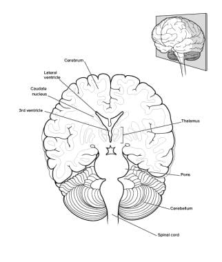
Cerebellum
Cerebellar Cortex
Cerebellar Diseases
Brain
Cerebellar Nuclei
Cerebellar Ataxia
Magnetic Resonance Imaging
Neurons
Cerebellar Neoplasms
Nerve Tissue Proteins
Brain Mapping
Brain Stem
Cerebral Cortex
Olivary Nucleus
Brain Chemistry
Ataxia
Pons
Tissue Distribution
Nerve Fibers
Blinking
Medulloblastoma
S100 Calcium Binding Protein G
Calbindins
In Situ Hybridization
Basal Ganglia
Image Processing, Computer-Assisted
Neuroglia
Tomography, Emission-Computed
Gene Expression Regulation, Developmental
Psychomotor Performance
Mesencephalon
Immunohistochemistry
Movement
Spinocerebellar Degenerations
Organ Specificity
Mice, Inbred C57BL
Synapses
Spinocerebellar Ataxias
Rats, Sprague-Dawley
Functional Laterality
Rats, Wistar
RNA, Messenger
Mice, Transgenic
Hippocampus
Dendrites
Central Nervous System
Autoradiography
Cerebrum
Thalamus
Nerve Net
Rhombencephalon
Mice, Knockout
Molecular Sequence Data
Reflex, Vestibulo-Ocular
Metencephalon
Diencephalon
Positron-Emission Tomography
Spinocerebellar Tracts
Glial Fibrillary Acidic Protein
Motor Cortex
Muscimol
Neuronal Plasticity
Learning
Gait Ataxia
Spinal Cord
Atrophy
Astrocytes
Aging
Frontal Lobe
Action Potentials
Receptors, Glutamate
Models, Neurological
Cats
Telencephalon
Carbon Radioisotopes
Rats, Inbred Strains
FGF8 induces formation of an ectopic isthmic organizer and isthmocerebellar development via a repressive effect on Otx2 expression. (1/6494)
Beads containing recombinant FGF8 (FGF8-beads) were implanted in the prospective caudal diencephalon or midbrain of chick embryos at stages 9-12. This induced the neuroepithelium rostral and caudal to the FGF8-bead to form two ectopic, mirror-image midbrains. Furthermore, cells in direct contact with the bead formed an outgrowth that protruded laterally from the neural tube. Tissue within such lateral outgrowths developed proximally into isthmic nuclei and distally into a cerebellum-like structure. These morphogenetic effects were apparently due to FGF8-mediated changes in gene expression in the vicinity of the bead, including a repressive effect on Otx2 and an inductive effect on En1, Fgf8 and Wnt1 expression. The ectopic Fgf8 and Wnt1 expression domains formed nearly complete concentric rings around the FGF8-bead, with the Wnt1 ring outermost. These observations suggest that FGF8 induces the formation of a ring-like ectopic signaling center (organizer) in the lateral wall of the brain, similar to the one that normally encircles the neural tube at the isthmic constriction, which is located at the boundary between the prospective midbrain and hindbrain. This ectopic isthmic organizer apparently sends long-range patterning signals both rostrally and caudally, resulting in the development of the two ectopic midbrains. Interestingly, our data suggest that these inductive signals spread readily in a caudal direction, but are inhibited from spreading rostrally across diencephalic neuromere boundaries. These results provide insights into the mechanism by which FGF8 induces an ectopic organizer and suggest that a negative feedback loop between Fgf8 and Otx2 plays a key role in patterning the midbrain and anterior hindbrain. (+info)Somatic recording of GABAergic autoreceptor current in cerebellar stellate and basket cells. (2/6494)
Patch-clamp recordings were performed from stellate and basket cells in rat cerebellar slices. Under somatic voltage clamp, short depolarizing pulses were applied to elicit action potentials in the axon. After the action potential, a bicuculline- and Cd2+-sensitive current transient was observed. A similar response was obtained when eliciting axonal firing by extracellular stimulation. With an isotonic internal Cl- solution, the peak amplitude of this current varied linearly with the holding potential, yielding an extrapolated reversal potential of -20 to 0 mV. Unlike synaptic or autaptic GABAergic currents obtained in the same preparation, the current transient had a slow rise-time and a low variability between trials. This current was blocked when 10 mM BAPTA was included in the recording solution. In some experiments, the current transient elicited axonal action potentials. The current transient was reliably observed in animals aged 12-15 d, with a mean amplitude of 82 pA at -70 mV, but was small and rare in the age group 29-49 d. Numerical simulations could account for all properties of the current transient by assuming that an action potential activates a distributed GABAergic conductance in the axon. The actual conductance is probably restricted to release sites, with an estimated mean presynaptic current response of 10 pA per site (-70 mV, age 12-15 d). We conclude that in developing rats, stellate and basket cell axons have a high density of GABAergic autoreceptors and that a sizable fraction of the corresponding current can be measured from the soma. (+info)Reproducibility studies with 11C-DTBZ, a monoamine vesicular transporter inhibitor in healthy human subjects. (3/6494)
The reproducibility of (+/-)-alpha-[11C] dihydrotetrabenazine (DTBZ) measures in PET was studied in 10 healthy human subjects, aged 22-76 y. METHODS: The scan-to-scan variation of several measures used in PET data analysis was determined, including the radioactivity ratio (target-to-reference), plasma-input Logan total distribution volume (DV), plasma-input Logan Bmax/Kd and tissue-input Logan Bmax/Kd values. RESULTS: The radioactivity ratios, plasma-input Bmax/Kd and tissue-input Bmax/Kd all have higher reliability than plasma-input total DV values. In addition, measures using the occipital cortex as the reference region have higher reliability than the same measures using the cerebellum as the reference region. CONCLUSION: Our results show that DTBZ is a reliable PET tracer that provides reproducible in vivo measurement of striatal vesicular monoamine transporter density. In the selection of reference regions for DTBZ PET data analysis, caution must be exercised in circumstances when DTBZ binding in the occipital cortex or the cerebellum may be altered. (+info)A genetic approach to visualization of multisynaptic neural pathways using plant lectin transgene. (4/6494)
The wiring patterns among various types of neurons via specific synaptic connections are the basis of functional logic employed by the brain for information processing. This study introduces a powerful method of analyzing the neuronal connectivity patterns by delivering a tracer selectively to specific types of neurons while simultaneously transsynaptically labeling their target neurons. We developed a novel genetic approach introducing cDNA for a plant lectin, wheat germ agglutinin (WGA), as a transgene under the control of specific promoter elements. Using this method, we demonstrate three examples of visualization of specific transsynaptic neural pathways: the mouse cerebellar efferent pathways, the mouse olfactory pathways, and the Drosophila visual pathways. This strategy should greatly facilitate studies on the anatomical and functional organization of the developing and mature nervous system. (+info)Control of neuronal precursor proliferation in the cerebellum by Sonic Hedgehog. (5/6494)
Cerebellar granule cells are the most abundant type of neuron in the brain, but the molecular mechanisms that control their generation are incompletely understood. We show that Sonic hedgehog (Shh), which is made by Purkinje cells, regulates the division of granule cell precursors (GCPs). Treatment of GCPs with Shh prevents differentiation and induces a potent, long-lasting proliferative response. This response can be inhibited by basic fibroblast growth factor or by activation of protein kinase A. Blocking Shh function in vivo dramatically reduces GCP proliferation. These findings provide insight into the mechanisms of normal growth and tumorigenesis in the cerebellum. (+info)Comparative effects of methylmercury on parallel-fiber and climbing-fiber responses of rat cerebellar slices. (6/6494)
The environmental neurotoxicant methylmercury (MeHg) causes profound disruption of cerebellar function. Previous studies have shown that acute exposure to MeHg impairs synaptic transmission in both the peripheral and central nervous systems. However, the effects of MeHg on cerebellar synaptic function have never been examined. In the present study, effects of acute exposure to MeHg on synaptic transmission between parallel fibers or climbing fibers and Purkinje cells were compared in 300- to 350-microm cerebellar slices by using extracellular and intracellular microelectrode-recording techniques. Field potentials of parallel-fiber volleys (PFVs) and the associated postsynaptic responses (PSRs) were recorded in the molecular layer by stimulating the parallel fibers in transverse cerebellar slices. The climbing-fiber responses were also recorded in the molecular layer by stimulating white matter in sagittal cerebellar slices. At 20, 100, and 500 microM, MeHg reduced the amplitude of both PFVs and the associated PSRs to complete block, however, it blocked PSRs more rapidly than PFVs. MeHg also decreased the amplitudes of climbing-fiber responses to complete block. For all responses, an initial increase in amplitude preceded MeHg-induced suppression. Intracellular recordings of excitatory postsynaptic potentials of Purkinje cells were compared before and after MeHg. At 100 microM and 20 microM, MeHg blocked the Na+-dependent, fast somatic spikes and Ca++-dependent, slow dendritic spike bursts. MeHg also hyperpolarized and then depolarized Purkinje cell membranes, suppressed current conduction from parallel fibers or climbing fibers to dendrites of Purkinje cells, and blocked synaptically activated local responses. MeHg switched the pattern of repetitive firing of Purkinje cells generated spontaneously or by depolarizing current injection at Purkinje cell soma from predominantly Na+-dependent, fast somatic spikes to predominantly Ca++-dependent, low amplitude, slow dendritic spike bursts. Thus, acute exposure to MeHg causes a complex pattern of effects on cerebellar synaptic transmission, with apparent actions on both neuronal excitability and chemical synaptic transmission. (+info)Long term lithium treatment suppresses p53 and Bax expression but increases Bcl-2 expression. A prominent role in neuroprotection against excitotoxicity. (7/6494)
This study was undertaken to investigate the molecular mechanisms underlying the neuroprotective actions of lithium against glutamate excitotoxicity with a focus on the role of proapoptotic and antiapoptotic genes. Long term, but not acute, treatment of cultured cerebellar granule cells with LiCl induces a concentration-dependent decrease in mRNA and protein levels of proapoptotic p53 and Bax; conversely, mRNA and protein levels of cytoprotective Bcl-2 are remarkably increased. The ratios of Bcl-2/Bax protein levels increase by approximately 5-fold after lithium treatment for 5-7 days. Exposure of cerebellar granule cells to glutamate induces a rapid increase in p53 and Bax mRNA and protein levels with no apparent effect on Bcl-2 expression. Pretreatment with LiCl for 7 days prevents glutamate-induced increase in p53 and Bax expression and maintains Bcl-2 in an elevated state. Glutamate exposure also triggers the release of cytochrome c from the mitochondria into the cytosol. Lithium pretreatment blocks glutamate-induced cytochrome c release and cleavage of lamin B1, a nuclear substrate for caspase-3. These results strongly suggest that lithium-induced Bcl-2 up-regulation and p53 and Bax down-regulation play a prominent role in neuroprotection against excitotoxicity. Our results further suggest that lithium, in addition to its use in the treatment of bipolar depressive illness, may have an expanded use in the intervention of neurodegeneration. (+info)The type and the localization of cAMP-dependent protein kinase regulate transmission of cAMP signals to the nucleus in cortical and cerebellar granule cells. (8/6494)
cAMP signals are received and transmitted by multiple isoforms of cAMP-dependent protein kinases, typically determined by their specific regulatory subunits. In the brain the major regulatory isoform RIIbeta and the RII-anchor protein, AKAP150 (rat) or 75 (bovine), are differentially expressed. Cortical neurons express RIIbeta and AKAP75; conversely, granule cerebellar cells express predominantly RIalpha and RIIalpha. Cortical neurons accumulate PKA catalytic subunit and phosphorylated cAMP responsive element binding protein very efficiently into nuclei upon cAMP induction, whereas granule cerebellar cells fail to do so. Down-regulation of RIIbeta synthesis by antisense oligonucleotides inhibited cAMP-induced nuclear signaling in cortical neurons. Expression in cerebellar granule cells of RIIbeta and AKAP75 genes by microinjection of specific expression vectors, markedly stimulated cAMP-induced transcription of the lacZ gene driven by a cAMP-responsive element promoter. These data indicate that the composition of PKA in cortical and granule cells underlies the differential ability of these cells to transmit cAMP signals to the nucleus. (+info)The cerebellum is a part of the brain that lies behind the brainstem and is involved in the regulation of motor movements, balance, and coordination. It contains two hemispheres and a central portion called the vermis. The cerebellum receives input from sensory systems and other areas of the brain and spinal cord and sends output to motor areas of the brain. Damage to the cerebellum can result in problems with movement, balance, and coordination.
Purkinje cells are a type of neuron located in the cerebellar cortex, which is the outer layer of the cerebellum, a part of the brain that plays a crucial role in motor control and coordination. These cells have large branching dendrites and receive input from many other neurons, particularly granule cells. The axons of Purkinje cells form the principal output pathway of the cerebellar cortex, synapsing with deep cerebellar nuclei. They are named after Johannes Evangelista Purkinje, a Czech physiologist who first described them in 1837.
The cerebellar cortex is the outer layer of the cerebellum, which is a part of the brain that plays a crucial role in motor control, balance, and coordination of muscle movements. The cerebellar cortex contains numerous small neurons called granule cells, as well as other types of neurons such as Purkinje cells, basket cells, and stellate cells. These neurons are organized into distinct layers and microcircuits that process information related to motor function and possibly other functions such as cognition and emotion. The cerebellar cortex receives input from various sources, including the spinal cord, vestibular system, and cerebral cortex, and sends output to brainstem nuclei and thalamus, which in turn project to the cerebral cortex. Damage to the cerebellar cortex can result in ataxia, dysmetria, dysdiadochokinesia, and other motor symptoms.
Cerebellar diseases refer to a group of medical conditions that affect the cerebellum, which is the part of the brain located at the back of the head, below the occipital lobe and above the brainstem. The cerebellum plays a crucial role in motor control, coordination, balance, and some cognitive functions.
Cerebellar diseases can be caused by various factors, including genetics, infections, tumors, stroke, trauma, or degenerative processes. These conditions can result in a wide range of symptoms, such as:
1. Ataxia: Loss of coordination and unsteady gait
2. Dysmetria: Inability to judge distance and force while performing movements
3. Intention tremors: Shaking or trembling that worsens during purposeful movements
4. Nystagmus: Rapid, involuntary eye movement
5. Dysarthria: Speech difficulty due to muscle weakness or incoordination
6. Hypotonia: Decreased muscle tone
7. Titubation: Rhythmic, involuntary oscillations of the head and neck
8. Cognitive impairment: Problems with memory, attention, and executive functions
Some examples of cerebellar diseases include:
1. Ataxia-telangiectasia
2. Friedrich's ataxia
3. Multiple system atrophy (MSA)
4. Spinocerebellar ataxias (SCAs)
5. Cerebellar tumors, such as medulloblastomas or astrocytomas
6. Infarctions or hemorrhages in the cerebellum due to stroke or trauma
7. Infections, such as viral encephalitis or bacterial meningitis
8. Autoimmune disorders, like multiple sclerosis (MS) or paraneoplastic syndromes
9. Metabolic disorders, such as Wilson's disease or phenylketonuria (PKU)
10. Chronic alcoholism and withdrawal
Treatment for cerebellar diseases depends on the underlying cause and may involve medications, physical therapy, surgery, or supportive care to manage symptoms and improve quality of life.
The brain is the central organ of the nervous system, responsible for receiving and processing sensory information, regulating vital functions, and controlling behavior, movement, and cognition. It is divided into several distinct regions, each with specific functions:
1. Cerebrum: The largest part of the brain, responsible for higher cognitive functions such as thinking, learning, memory, language, and perception. It is divided into two hemispheres, each controlling the opposite side of the body.
2. Cerebellum: Located at the back of the brain, it is responsible for coordinating muscle movements, maintaining balance, and fine-tuning motor skills.
3. Brainstem: Connects the cerebrum and cerebellum to the spinal cord, controlling vital functions such as breathing, heart rate, and blood pressure. It also serves as a relay center for sensory information and motor commands between the brain and the rest of the body.
4. Diencephalon: A region that includes the thalamus (a major sensory relay station) and hypothalamus (regulates hormones, temperature, hunger, thirst, and sleep).
5. Limbic system: A group of structures involved in emotional processing, memory formation, and motivation, including the hippocampus, amygdala, and cingulate gyrus.
The brain is composed of billions of interconnected neurons that communicate through electrical and chemical signals. It is protected by the skull and surrounded by three layers of membranes called meninges, as well as cerebrospinal fluid that provides cushioning and nutrients.
The cerebellar nuclei are clusters of neurons located within the white matter of the cerebellum, a region of the brain responsible for motor coordination, balance, and fine movement regulation. There are four main pairs of cerebellar nuclei: the fastigial, interpositus, dentate, and vestibular nuclei. These nuclei receive input from various parts of the cerebellar cortex and project to different areas of the brainstem and thalamus, contributing to the regulation of muscle tone, posture, and movement.
Neurologic mutant mice are genetically engineered or spontaneously mutated rodents that are used as models to study various neurological disorders and conditions. These mice have specific genetic modifications or mutations that affect their nervous system, leading to phenotypes that resemble human neurological diseases.
Some examples of neurologic mutant mice include:
1. Alzheimer's disease models: Mice that overexpress genes associated with Alzheimer's disease, such as the amyloid precursor protein (APP) or presenilin 1 (PS1), to study the pathogenesis and potential treatments of this disorder.
2. Parkinson's disease models: Mice that have genetic mutations in genes associated with Parkinson's disease, such as alpha-synuclein or parkin, to investigate the mechanisms underlying this condition and develop new therapies.
3. Huntington's disease models: Mice that carry an expanded CAG repeat in the huntingtin gene to replicate the genetic defect seen in humans with Huntington's disease and study disease progression and treatment strategies.
4. Epilepsy models: Mice with genetic mutations that cause spontaneous seizures or increased susceptibility to seizures, used to investigate the underlying mechanisms of epilepsy and develop new treatments.
5. Stroke models: Mice that have surgical induction of stroke or genetic modifications that increase the risk of stroke, used to study the pathophysiology of stroke and identify potential therapeutic targets.
Neurologic mutant mice are essential tools in biomedical research, allowing scientists to investigate the complex interactions between genes and the environment that contribute to neurological disorders. These models help researchers better understand disease mechanisms, develop new therapies, and test their safety and efficacy before moving on to clinical trials in humans.
Cerebellar ataxia is a type of ataxia, which refers to a group of disorders that cause difficulties with coordination and movement. Cerebellar ataxia specifically involves the cerebellum, which is the part of the brain responsible for maintaining balance, coordinating muscle movements, and regulating speech and eye movements.
The symptoms of cerebellar ataxia may include:
* Unsteady gait or difficulty walking
* Poor coordination of limb movements
* Tremors or shakiness, especially in the hands
* Slurred or irregular speech
* Abnormal eye movements, such as nystagmus (rapid, involuntary movement of the eyes)
* Difficulty with fine motor tasks, such as writing or buttoning a shirt
Cerebellar ataxia can be caused by a variety of underlying conditions, including:
* Genetic disorders, such as spinocerebellar ataxia or Friedreich's ataxia
* Brain injury or trauma
* Stroke or brain hemorrhage
* Infections, such as meningitis or encephalitis
* Exposure to toxins, such as alcohol or certain medications
* Tumors or other growths in the brain
Treatment for cerebellar ataxia depends on the underlying cause. In some cases, there may be no cure, and treatment is focused on managing symptoms and improving quality of life. Physical therapy, occupational therapy, and speech therapy can help improve coordination, balance, and communication skills. Medications may also be used to treat specific symptoms, such as tremors or muscle spasticity. In some cases, surgery may be recommended to remove tumors or repair damage to the brain.
Medical Definition:
Magnetic Resonance Imaging (MRI) is a non-invasive diagnostic imaging technique that uses a strong magnetic field and radio waves to create detailed cross-sectional or three-dimensional images of the internal structures of the body. The patient lies within a large, cylindrical magnet, and the scanner detects changes in the direction of the magnetic field caused by protons in the body. These changes are then converted into detailed images that help medical professionals to diagnose and monitor various medical conditions, such as tumors, injuries, or diseases affecting the brain, spinal cord, heart, blood vessels, joints, and other internal organs. MRI does not use radiation like computed tomography (CT) scans.
Neurons, also known as nerve cells or neurocytes, are specialized cells that constitute the basic unit of the nervous system. They are responsible for receiving, processing, and transmitting information and signals within the body. Neurons have three main parts: the dendrites, the cell body (soma), and the axon. The dendrites receive signals from other neurons or sensory receptors, while the axon transmits these signals to other neurons, muscles, or glands. The junction between two neurons is called a synapse, where neurotransmitters are released to transmit the signal across the gap (synaptic cleft) to the next neuron. Neurons vary in size, shape, and structure depending on their function and location within the nervous system.
Cerebellar neoplasms refer to abnormal growths or tumors that develop in the cerebellum, which is the part of the brain responsible for coordinating muscle movements and maintaining balance. These tumors can be benign (non-cancerous) or malignant (cancerous), and they can arise from various types of cells within the cerebellum.
The most common type of cerebellar neoplasm is a medulloblastoma, which arises from primitive nerve cells in the cerebellum. Other types of cerebellar neoplasms include astrocytomas, ependymomas, and brain stem gliomas. Symptoms of cerebellar neoplasms may include headaches, vomiting, unsteady gait, coordination problems, and visual disturbances. Treatment options depend on the type, size, and location of the tumor, as well as the patient's overall health and age. Treatment may involve surgery, radiation therapy, chemotherapy, or a combination of these approaches.
Eyelid conditioning, also known as eyelid classical conditioning or Ursinus' phenomenon, is a type of reflex conditioning that involves associating a neutral stimulus with the natural act of blinking. This concept was first described by Russian physiologist Ivan Pavlov and later studied in detail by German ophthalmologist Hermann Ludwig Ferdinand von Helmholtz and Austrian physician Sigmund Exner.
In this procedure, a conditioned stimulus (like a sound or light) is repeatedly presented just before the unconditioned stimulus (such as a puff of air directed at the eye), which naturally triggers the blink reflex. Over time, the subject begins to associate the conditioned stimulus with the blinking response and will start to blink even when only the conditioned stimulus is presented, without the presence of the unconditioned stimulus. This learning process is an example of classical conditioning and can be used in various research and clinical applications.
Nerve tissue proteins are specialized proteins found in the nervous system that provide structural and functional support to nerve cells, also known as neurons. These proteins include:
1. Neurofilaments: These are type IV intermediate filaments that provide structural support to neurons and help maintain their shape and size. They are composed of three subunits - NFL (light), NFM (medium), and NFH (heavy).
2. Neuronal Cytoskeletal Proteins: These include tubulins, actins, and spectrins that provide structural support to the neuronal cytoskeleton and help maintain its integrity.
3. Neurotransmitter Receptors: These are specialized proteins located on the postsynaptic membrane of neurons that bind neurotransmitters released by presynaptic neurons, triggering a response in the target cell.
4. Ion Channels: These are transmembrane proteins that regulate the flow of ions across the neuronal membrane and play a crucial role in generating and transmitting electrical signals in neurons.
5. Signaling Proteins: These include enzymes, receptors, and adaptor proteins that mediate intracellular signaling pathways involved in neuronal development, differentiation, survival, and death.
6. Adhesion Proteins: These are cell surface proteins that mediate cell-cell and cell-matrix interactions, playing a crucial role in the formation and maintenance of neural circuits.
7. Extracellular Matrix Proteins: These include proteoglycans, laminins, and collagens that provide structural support to nerve tissue and regulate neuronal migration, differentiation, and survival.
Brain mapping is a broad term that refers to the techniques used to understand the structure and function of the brain. It involves creating maps of the various cognitive, emotional, and behavioral processes in the brain by correlating these processes with physical locations or activities within the nervous system. Brain mapping can be accomplished through a variety of methods, including functional magnetic resonance imaging (fMRI), positron emission tomography (PET) scans, electroencephalography (EEG), and others. These techniques allow researchers to observe which areas of the brain are active during different tasks or thoughts, helping to shed light on how the brain processes information and contributes to our experiences and behaviors. Brain mapping is an important area of research in neuroscience, with potential applications in the diagnosis and treatment of neurological and psychiatric disorders.
The brainstem is the lower part of the brain that connects to the spinal cord. It consists of the midbrain, pons, and medulla oblongata. The brainstem controls many vital functions such as heart rate, breathing, and blood pressure. It also serves as a relay center for sensory and motor information between the cerebral cortex and the rest of the body. Additionally, several cranial nerves originate from the brainstem, including those that control eye movements, facial movements, and hearing.
The cerebral cortex is the outermost layer of the brain, characterized by its intricate folded structure and wrinkled appearance. It is a region of great importance as it plays a key role in higher cognitive functions such as perception, consciousness, thought, memory, language, and attention. The cerebral cortex is divided into two hemispheres, each containing four lobes: the frontal, parietal, temporal, and occipital lobes. These areas are responsible for different functions, with some regions specializing in sensory processing while others are involved in motor control or associative functions. The cerebral cortex is composed of gray matter, which contains neuronal cell bodies, and is covered by a layer of white matter that consists mainly of myelinated nerve fibers.
The olivary nucleus is a structure located in the medulla oblongata, which is a part of the brainstem. It consists of two main parts: the inferior olive and the accessory olive. The inferior olive is further divided into several subnuclei.
The olivary nucleus plays an important role in the coordination of movements, particularly in the regulation of fine motor control and rhythmic movements. It receives input from various sources, including the cerebellum, spinal cord, and other brainstem nuclei, and sends output to the cerebellum via the climbing fibers.
Damage to the olivary nucleus can result in a variety of neurological symptoms, including ataxia (loss of coordination), tremors, and dysarthria (speech difficulties). Certain neurodegenerative disorders, such as multiple system atrophy, may also affect the olivary nucleus and contribute to its degeneration.
Brain chemistry refers to the chemical processes that occur within the brain, particularly those involving neurotransmitters, neuromodulators, and neuropeptides. These chemicals are responsible for transmitting signals between neurons (nerve cells) in the brain, allowing for various cognitive, emotional, and physical functions.
Neurotransmitters are chemical messengers that transmit signals across the synapse (the tiny gap between two neurons). Examples of neurotransmitters include dopamine, serotonin, norepinephrine, GABA (gamma-aminobutyric acid), and glutamate. Each neurotransmitter has a specific role in brain function, such as regulating mood, motivation, attention, memory, and movement.
Neuromodulators are chemicals that modify the effects of neurotransmitters on neurons. They can enhance or inhibit the transmission of signals between neurons, thereby modulating brain activity. Examples of neuromodulators include acetylcholine, histamine, and substance P.
Neuropeptides are small protein-like molecules that act as neurotransmitters or neuromodulators. They play a role in various physiological functions, such as pain perception, stress response, and reward processing. Examples of neuropeptides include endorphins, enkephalins, and oxytocin.
Abnormalities in brain chemistry can lead to various neurological and psychiatric conditions, such as depression, anxiety disorders, schizophrenia, Parkinson's disease, and Alzheimer's disease. Understanding brain chemistry is crucial for developing effective treatments for these conditions.
Ataxia is a medical term that refers to a group of disorders affecting coordination, balance, and speech. It is characterized by a lack of muscle control during voluntary movements, causing unsteady or awkward movements, and often accompanied by tremors. Ataxia can affect various parts of the body, such as the limbs, trunk, eyes, and speech muscles. The condition can be congenital or acquired, and it can result from damage to the cerebellum, spinal cord, or sensory nerves. There are several types of ataxia, including hereditary ataxias, degenerative ataxias, cerebellar ataxias, and acquired ataxias, each with its own specific causes, symptoms, and prognosis. Treatment for ataxia typically focuses on managing symptoms and improving quality of life, as there is no cure for most forms of the disorder.
The pons is a part of the brainstem that lies between the medulla oblongata and the midbrain. Its name comes from the Latin word "ponte" which means "bridge," as it serves to connect these two regions of the brainstem. The pons contains several important structures, including nerve fibers that carry signals between the cerebellum (the part of the brain responsible for coordinating muscle movements) and the rest of the nervous system. It also contains nuclei (clusters of neurons) that help regulate various functions such as respiration, sleep, and facial movements.
"Newborn animals" refers to the very young offspring of animals that have recently been born. In medical terminology, newborns are often referred to as "neonates," and they are classified as such from birth until about 28 days of age. During this time period, newborn animals are particularly vulnerable and require close monitoring and care to ensure their survival and healthy development.
The specific needs of newborn animals can vary widely depending on the species, but generally, they require warmth, nutrition, hydration, and protection from harm. In many cases, newborns are unable to regulate their own body temperature or feed themselves, so they rely heavily on their mothers for care and support.
In medical settings, newborn animals may be examined and treated by veterinarians to ensure that they are healthy and receiving the care they need. This can include providing medical interventions such as feeding tubes, antibiotics, or other treatments as needed to address any health issues that arise. Overall, the care and support of newborn animals is an important aspect of animal medicine and conservation efforts.
Neural pathways, also known as nerve tracts or fasciculi, refer to the highly organized and specialized routes through which nerve impulses travel within the nervous system. These pathways are formed by groups of neurons (nerve cells) that are connected in a series, creating a continuous communication network for electrical signals to transmit information between different regions of the brain, spinal cord, and peripheral nerves.
Neural pathways can be classified into two main types: sensory (afferent) and motor (efferent). Sensory neural pathways carry sensory information from various receptors in the body (such as those for touch, temperature, pain, and vision) to the brain for processing. Motor neural pathways, on the other hand, transmit signals from the brain to the muscles and glands, controlling movements and other effector functions.
The formation of these neural pathways is crucial for normal nervous system function, as it enables efficient communication between different parts of the body and allows for complex behaviors, cognitive processes, and adaptive responses to internal and external stimuli.
Tissue distribution, in the context of pharmacology and toxicology, refers to the way that a drug or xenobiotic (a chemical substance found within an organism that is not naturally produced by or expected to be present within that organism) is distributed throughout the body's tissues after administration. It describes how much of the drug or xenobiotic can be found in various tissues and organs, and is influenced by factors such as blood flow, lipid solubility, protein binding, and the permeability of cell membranes. Understanding tissue distribution is important for predicting the potential effects of a drug or toxin on different parts of the body, and for designing drugs with improved safety and efficacy profiles.
Nerve fibers are specialized structures that constitute the long, slender processes (axons) of neurons (nerve cells). They are responsible for conducting electrical impulses, known as action potentials, away from the cell body and transmitting them to other neurons or effector organs such as muscles and glands. Nerve fibers are often surrounded by supportive cells called glial cells and are grouped together to form nerve bundles or nerves. These fibers can be myelinated (covered with a fatty insulating sheath called myelin) or unmyelinated, which influences the speed of impulse transmission.
Blinking is the rapid and repetitive closing and reopening of the eyelids. It is a normal physiological process that helps to keep the eyes moist, protected and comfortable by spreading tears over the surface of the eye and removing any foreign particles or irritants that may have accumulated on the eyelid or the conjunctiva (the mucous membrane that covers the front of the eye and lines the inside of the eyelids).
Blinking is controlled by the facial nerve (cranial nerve VII), which sends signals to the muscles that control the movement of the eyelids. On average, people blink about 15-20 times per minute, but this rate can vary depending on factors such as mood, level of attention, and visual tasks. For example, people tend to blink less frequently when they are concentrating on a visual task or looking at a screen, which can lead to dry eye symptoms.
Medulloblastoma is a type of malignant brain tumor that originates in the cerebellum, which is the part of the brain located at the back of the skull and controls coordination and balance. It is one of the most common types of pediatric brain tumors, although it can also occur in adults.
Medulloblastomas are typically made up of small, round cancer cells that grow quickly and can spread to other parts of the central nervous system, such as the spinal cord. They are usually treated with a combination of surgery, radiation therapy, and chemotherapy. The exact cause of medulloblastoma is not known, but it is thought to be related to genetic mutations or abnormalities that occur during development.
S100 calcium binding protein G, also known as calgranulin A or S100A8, is a member of the S100 family of proteins. These proteins are characterized by their ability to bind calcium ions and play a role in intracellular signaling and regulation of various cellular processes.
S100 calcium binding protein G forms a heterodimer with S100 calcium binding protein B (S100A9) and is involved in the inflammatory response, immune function, and tumor growth and progression. The S100A8/A9 heterocomplex has been shown to play a role in neutrophil activation and recruitment, as well as the regulation of cytokine production and cell proliferation.
Elevated levels of S100 calcium binding protein G have been found in various inflammatory conditions, such as rheumatoid arthritis, Crohn's disease, and psoriasis, as well as in several types of cancer, including breast, lung, and colon cancer. Therefore, it has been suggested that S100 calcium binding protein G may be a useful biomarker for the diagnosis and prognosis of these conditions.
Calbindins are a family of calcium-binding proteins that are widely distributed in various tissues, including the gastrointestinal tract, brain, and kidney. They play important roles in regulating intracellular calcium levels and modulating calcium-dependent signaling pathways. Calbindin D28k, one of the major isoforms, is particularly abundant in the central nervous system and has been implicated in neuroprotection, neuronal plasticity, and regulation of neurotransmitter release. Deficiencies or alterations in calbindins have been associated with various pathological conditions, including neurological disorders and cancer.
In situ hybridization (ISH) is a molecular biology technique used to detect and localize specific nucleic acid sequences, such as DNA or RNA, within cells or tissues. This technique involves the use of a labeled probe that is complementary to the target nucleic acid sequence. The probe can be labeled with various types of markers, including radioisotopes, fluorescent dyes, or enzymes.
During the ISH procedure, the labeled probe is hybridized to the target nucleic acid sequence in situ, meaning that the hybridization occurs within the intact cells or tissues. After washing away unbound probe, the location of the labeled probe can be visualized using various methods depending on the type of label used.
In situ hybridization has a wide range of applications in both research and diagnostic settings, including the detection of gene expression patterns, identification of viral infections, and diagnosis of genetic disorders.
The basal ganglia are a group of interconnected nuclei, or clusters of neurons, located in the base of the brain. They play a crucial role in regulating motor function, cognition, and emotion. The main components of the basal ganglia include the striatum (made up of the caudate nucleus, putamen, and ventral striatum), globus pallidus (divided into external and internal segments), subthalamic nucleus, and substantia nigra (with its pars compacta and pars reticulata).
The basal ganglia receive input from various regions of the cerebral cortex and other brain areas. They process this information and send output back to the thalamus and cortex, helping to modulate and coordinate movement. The basal ganglia also contribute to higher cognitive functions such as learning, decision-making, and habit formation. Dysfunction in the basal ganglia can lead to neurological disorders like Parkinson's disease, Huntington's disease, and dystonia.
Computer-assisted image processing is a medical term that refers to the use of computer systems and specialized software to improve, analyze, and interpret medical images obtained through various imaging techniques such as X-ray, CT (computed tomography), MRI (magnetic resonance imaging), ultrasound, and others.
The process typically involves several steps, including image acquisition, enhancement, segmentation, restoration, and analysis. Image processing algorithms can be used to enhance the quality of medical images by adjusting contrast, brightness, and sharpness, as well as removing noise and artifacts that may interfere with accurate diagnosis. Segmentation techniques can be used to isolate specific regions or structures of interest within an image, allowing for more detailed analysis.
Computer-assisted image processing has numerous applications in medical imaging, including detection and characterization of lesions, tumors, and other abnormalities; assessment of organ function and morphology; and guidance of interventional procedures such as biopsies and surgeries. By automating and standardizing image analysis tasks, computer-assisted image processing can help to improve diagnostic accuracy, efficiency, and consistency, while reducing the potential for human error.
Neuroglia, also known as glial cells or simply glia, are non-neuronal cells that provide support and protection for neurons in the nervous system. They maintain homeostasis, form myelin sheaths around nerve fibers, and provide structural support. They also play a role in the immune response of the central nervous system. Some types of neuroglia include astrocytes, oligodendrocytes, microglia, and ependymal cells.
Emission computed tomography (ECT) is a type of tomographic imaging technique in which an emission signal from within the body is detected to create cross-sectional images of that signal's distribution. In Emission-Computed Tomography (ECT), a radionuclide is introduced into the body, usually through injection, inhalation or ingestion. The radionuclide emits gamma rays that are then detected by external gamma cameras.
The data collected from these cameras is then used to create cross-sectional images of the distribution of the radiopharmaceutical within the body. This allows for the identification and quantification of functional information about specific organs or systems within the body, such as blood flow, metabolic activity, or receptor density.
One common type of Emission-Computed Tomography is Single Photon Emission Computed Tomography (SPECT), which uses a single gamma camera that rotates around the patient to collect data from multiple angles. Another type is Positron Emission Tomography (PET), which uses positron-emitting radionuclides and detects the coincident gamma rays emitted by the annihilation of positrons and electrons.
Overall, ECT is a valuable tool in medical imaging for diagnosing and monitoring various diseases, including cancer, heart disease, and neurological disorders.
Developmental gene expression regulation refers to the processes that control the activation or repression of specific genes during embryonic and fetal development. These regulatory mechanisms ensure that genes are expressed at the right time, in the right cells, and at appropriate levels to guide proper growth, differentiation, and morphogenesis of an organism.
Developmental gene expression regulation is a complex and dynamic process involving various molecular players, such as transcription factors, chromatin modifiers, non-coding RNAs, and signaling molecules. These regulators can interact with cis-regulatory elements, like enhancers and promoters, to fine-tune the spatiotemporal patterns of gene expression during development.
Dysregulation of developmental gene expression can lead to various congenital disorders and developmental abnormalities. Therefore, understanding the principles and mechanisms governing developmental gene expression regulation is crucial for uncovering the etiology of developmental diseases and devising potential therapeutic strategies.
Psychomotor performance refers to the integration and coordination of mental processes (cognitive functions) with physical movements. It involves the ability to perform complex tasks that require both cognitive skills, such as thinking, remembering, and perceiving, and motor skills, such as gross and fine motor movements. Examples of psychomotor performances include driving a car, playing a musical instrument, or performing surgical procedures.
In a medical context, psychomotor performance is often used to assess an individual's ability to perform activities of daily living (ADLs) and instrumental activities of daily living (IADLs), such as bathing, dressing, cooking, cleaning, and managing medications. Deficits in psychomotor performance can be a sign of neurological or psychiatric disorders, such as dementia, Parkinson's disease, or depression.
Assessment of psychomotor performance may involve tests that measure reaction time, coordination, speed, precision, and accuracy of movements, as well as cognitive functions such as attention, memory, and problem-solving skills. These assessments can help healthcare professionals develop appropriate treatment plans and monitor the progression of diseases or the effectiveness of interventions.
The mesencephalon, also known as the midbrain, is the middle portion of the brainstem that connects the hindbrain (rhombencephalon) and the forebrain (prosencephalon). It plays a crucial role in several important functions including motor control, vision, hearing, and the regulation of consciousness and sleep-wake cycles. The mesencephalon contains several important structures such as the cerebral aqueduct, tectum, tegmentum, cerebral peduncles, and several cranial nerve nuclei (III and IV).
Immunohistochemistry (IHC) is a technique used in pathology and laboratory medicine to identify specific proteins or antigens in tissue sections. It combines the principles of immunology and histology to detect the presence and location of these target molecules within cells and tissues. This technique utilizes antibodies that are specific to the protein or antigen of interest, which are then tagged with a detection system such as a chromogen or fluorophore. The stained tissue sections can be examined under a microscope, allowing for the visualization and analysis of the distribution and expression patterns of the target molecule in the context of the tissue architecture. Immunohistochemistry is widely used in diagnostic pathology to help identify various diseases, including cancer, infectious diseases, and immune-mediated disorders.
In the context of medicine and healthcare, "movement" refers to the act or process of changing physical location or position. It involves the contraction and relaxation of muscles, which allows for the joints to move and the body to be in motion. Movement can also refer to the ability of a patient to move a specific body part or limb, which is assessed during physical examinations. Additionally, "movement" can describe the progression or spread of a disease within the body.
Spinocerebellar degenerations (SCDs) are a group of genetic disorders that primarily affect the cerebellum, the part of the brain responsible for coordinating muscle movements, and the spinal cord. These conditions are characterized by progressive degeneration or loss of nerve cells in the cerebellum and/or spinal cord, leading to various neurological symptoms.
SCDs are often inherited in an autosomal dominant manner, meaning that only one copy of the altered gene from either parent is enough to cause the disorder. The most common type of SCD is spinocerebellar ataxia (SCA), which includes several subtypes (SCA1, SCA2, SCA3, etc.) differentiated by their genetic causes and specific clinical features.
Symptoms of spinocerebellar degenerations may include:
1. Progressive ataxia (loss of coordination and balance)
2. Dysarthria (speech difficulty)
3. Nystagmus (involuntary eye movements)
4. Oculomotor abnormalities (problems with eye movement control)
5. Tremors or other involuntary muscle movements
6. Muscle weakness and spasticity
7. Sensory disturbances, such as numbness or tingling sensations
8. Dysphagia (difficulty swallowing)
9. Cognitive impairment in some cases
The age of onset, severity, and progression of symptoms can vary significantly among different SCD subtypes and individuals. Currently, there is no cure for spinocerebellar degenerations, but various supportive treatments and therapies can help manage symptoms and improve quality of life.
Organ specificity, in the context of immunology and toxicology, refers to the phenomenon where a substance (such as a drug or toxin) or an immune response primarily affects certain organs or tissues in the body. This can occur due to various reasons such as:
1. The presence of specific targets (like antigens in the case of an immune response or receptors in the case of drugs) that are more abundant in these organs.
2. The unique properties of certain cells or tissues that make them more susceptible to damage.
3. The way a substance is metabolized or cleared from the body, which can concentrate it in specific organs.
For example, in autoimmune diseases, organ specificity describes immune responses that are directed against antigens found only in certain organs, such as the thyroid gland in Hashimoto's disease. Similarly, some toxins or drugs may have a particular affinity for liver cells, leading to liver damage or specific drug interactions.
C57BL/6 (C57 Black 6) is an inbred strain of laboratory mouse that is widely used in biomedical research. The term "inbred" refers to a strain of animals where matings have been carried out between siblings or other closely related individuals for many generations, resulting in a population that is highly homozygous at most genetic loci.
The C57BL/6 strain was established in 1920 by crossing a female mouse from the dilute brown (DBA) strain with a male mouse from the black strain. The resulting offspring were then interbred for many generations to create the inbred C57BL/6 strain.
C57BL/6 mice are known for their robust health, longevity, and ease of handling, making them a popular choice for researchers. They have been used in a wide range of biomedical research areas, including studies of cancer, immunology, neuroscience, cardiovascular disease, and metabolism.
One of the most notable features of the C57BL/6 strain is its sensitivity to certain genetic modifications, such as the introduction of mutations that lead to obesity or impaired glucose tolerance. This has made it a valuable tool for studying the genetic basis of complex diseases and traits.
Overall, the C57BL/6 inbred mouse strain is an important model organism in biomedical research, providing a valuable resource for understanding the genetic and molecular mechanisms underlying human health and disease.
A synapse is a structure in the nervous system that allows for the transmission of signals from one neuron (nerve cell) to another. It is the point where the axon terminal of one neuron meets the dendrite or cell body of another, and it is here that neurotransmitters are released and received. The synapse includes both the presynaptic and postsynaptic elements, as well as the cleft between them.
At the presynaptic side, an action potential travels down the axon and triggers the release of neurotransmitters into the synaptic cleft through exocytosis. These neurotransmitters then bind to receptors on the postsynaptic side, which can either excite or inhibit the receiving neuron. The strength of the signal between two neurons is determined by the number and efficiency of these synapses.
Synapses play a crucial role in the functioning of the nervous system, allowing for the integration and processing of information from various sources. They are also dynamic structures that can undergo changes in response to experience or injury, which has important implications for learning, memory, and recovery from neurological disorders.
Spinocerebellar ataxias (SCAs) are a group of genetic disorders that affect the cerebellum, which is the part of the brain responsible for coordinating muscle movements. SCAs are characterized by progressive problems with balance, speech, and coordination. They are caused by mutations in various genes that result in the production of abnormal proteins that accumulate in neurons, leading to their degeneration.
There are over 40 different types of SCAs, each caused by a different genetic mutation. Some of the more common types include SCA1, SCA2, SCA3, SCA6, and SCA7. The symptoms and age of onset can vary widely depending on the type of SCA.
In addition to problems with coordination and balance, people with SCAs may also experience muscle weakness, stiffness, tremors, spasticity, and difficulty swallowing or speaking. Some types of SCAs can also cause visual disturbances, hearing loss, and cognitive impairment. Currently, there is no cure for SCAs, but treatments such as physical therapy, speech therapy, and medications can help manage the symptoms.
Sprague-Dawley rats are a strain of albino laboratory rats that are widely used in scientific research. They were first developed by researchers H.H. Sprague and R.C. Dawley in the early 20th century, and have since become one of the most commonly used rat strains in biomedical research due to their relatively large size, ease of handling, and consistent genetic background.
Sprague-Dawley rats are outbred, which means that they are genetically diverse and do not suffer from the same limitations as inbred strains, which can have reduced fertility and increased susceptibility to certain diseases. They are also characterized by their docile nature and low levels of aggression, making them easier to handle and study than some other rat strains.
These rats are used in a wide variety of research areas, including toxicology, pharmacology, nutrition, cancer, and behavioral studies. Because they are genetically diverse, Sprague-Dawley rats can be used to model a range of human diseases and conditions, making them an important tool in the development of new drugs and therapies.
Functional laterality, in a medical context, refers to the preferential use or performance of one side of the body over the other for specific functions. This is often demonstrated in hand dominance, where an individual may be right-handed or left-handed, meaning they primarily use their right or left hand for tasks such as writing, eating, or throwing.
However, functional laterality can also apply to other bodily functions and structures, including the eyes (ocular dominance), ears (auditory dominance), or legs. It's important to note that functional laterality is not a strict binary concept; some individuals may exhibit mixed dominance or no strong preference for one side over the other.
In clinical settings, assessing functional laterality can be useful in diagnosing and treating various neurological conditions, such as stroke or traumatic brain injury, where understanding any resulting lateralized impairments can inform rehabilitation strategies.
"Wistar rats" are a strain of albino rats that are widely used in laboratory research. They were developed at the Wistar Institute in Philadelphia, USA, and were first introduced in 1906. Wistar rats are outbred, which means that they are genetically diverse and do not have a fixed set of genetic characteristics like inbred strains.
Wistar rats are commonly used as animal models in biomedical research because of their size, ease of handling, and relatively low cost. They are used in a wide range of research areas, including toxicology, pharmacology, nutrition, cancer, cardiovascular disease, and behavioral studies. Wistar rats are also used in safety testing of drugs, medical devices, and other products.
Wistar rats are typically larger than many other rat strains, with males weighing between 500-700 grams and females weighing between 250-350 grams. They have a lifespan of approximately 2-3 years. Wistar rats are also known for their docile and friendly nature, making them easy to handle and work with in the laboratory setting.
Messenger RNA (mRNA) is a type of RNA (ribonucleic acid) that carries genetic information copied from DNA in the form of a series of three-base code "words," each of which specifies a particular amino acid. This information is used by the cell's machinery to construct proteins, a process known as translation. After being transcribed from DNA, mRNA travels out of the nucleus to the ribosomes in the cytoplasm where protein synthesis occurs. Once the protein has been synthesized, the mRNA may be degraded and recycled. Post-transcriptional modifications can also occur to mRNA, such as alternative splicing and addition of a 5' cap and a poly(A) tail, which can affect its stability, localization, and translation efficiency.
Transgenic mice are genetically modified rodents that have incorporated foreign DNA (exogenous DNA) into their own genome. This is typically done through the use of recombinant DNA technology, where a specific gene or genetic sequence of interest is isolated and then introduced into the mouse embryo. The resulting transgenic mice can then express the protein encoded by the foreign gene, allowing researchers to study its function in a living organism.
The process of creating transgenic mice usually involves microinjecting the exogenous DNA into the pronucleus of a fertilized egg, which is then implanted into a surrogate mother. The offspring that result from this procedure are screened for the presence of the foreign DNA, and those that carry the desired genetic modification are used to establish a transgenic mouse line.
Transgenic mice have been widely used in biomedical research to model human diseases, study gene function, and test new therapies. They provide a valuable tool for understanding complex biological processes and developing new treatments for a variety of medical conditions.
The hippocampus is a complex, curved formation in the brain that resembles a seahorse (hence its name, from the Greek word "hippos" meaning horse and "kampos" meaning sea monster). It's part of the limbic system and plays crucial roles in the formation of memories, particularly long-term ones.
This region is involved in spatial navigation and cognitive maps, allowing us to recognize locations and remember how to get to them. Additionally, it's one of the first areas affected by Alzheimer's disease, which often results in memory loss as an early symptom.
Anatomically, it consists of two main parts: the Ammon's horn (or cornu ammonis) and the dentate gyrus. These structures are made up of distinct types of neurons that contribute to different aspects of learning and memory.
Dendrites are the branched projections of a neuron that receive and process signals from other neurons. They are typically short and highly branching, increasing the surface area for receiving incoming signals. Dendrites are covered in small protrusions called dendritic spines, which can form connections with the axon terminals of other neurons through chemical synapses. The structure and function of dendrites play a critical role in the integration and processing of information in the nervous system.
The Central Nervous System (CNS) is the part of the nervous system that consists of the brain and spinal cord. It is called the "central" system because it receives information from, and sends information to, the rest of the body through peripheral nerves, which make up the Peripheral Nervous System (PNS).
The CNS is responsible for processing sensory information, controlling motor functions, and regulating various autonomic processes like heart rate, respiration, and digestion. The brain, as the command center of the CNS, interprets sensory stimuli, formulates thoughts, and initiates actions. The spinal cord serves as a conduit for nerve impulses traveling to and from the brain and the rest of the body.
The CNS is protected by several structures, including the skull (which houses the brain) and the vertebral column (which surrounds and protects the spinal cord). Despite these protective measures, the CNS remains vulnerable to injury and disease, which can have severe consequences due to its crucial role in controlling essential bodily functions.
Autoradiography is a medical imaging technique used to visualize and localize the distribution of radioactively labeled compounds within tissues or organisms. In this process, the subject is first exposed to a radioactive tracer that binds to specific molecules or structures of interest. The tissue is then placed in close contact with a radiation-sensitive film or detector, such as X-ray film or an imaging plate.
As the radioactive atoms decay, they emit particles (such as beta particles) that interact with the film or detector, causing chemical changes and leaving behind a visible image of the distribution of the labeled compound. The resulting autoradiogram provides information about the location, quantity, and sometimes even the identity of the molecules or structures that have taken up the radioactive tracer.
Autoradiography has been widely used in various fields of biology and medical research, including pharmacology, neuroscience, genetics, and cell biology, to study processes such as protein-DNA interactions, gene expression, drug metabolism, and neuronal connectivity. However, due to the use of radioactive materials and potential hazards associated with them, this technique has been gradually replaced by non-radioactive alternatives like fluorescence in situ hybridization (FISH) or immunofluorescence techniques.
The cerebrum is the largest part of the brain, located in the frontal part of the skull. It is divided into two hemispheres, right and left, which are connected by a band of nerve fibers called the corpus callosum. The cerebrum is responsible for higher cognitive functions such as thinking, learning, memory, language, perception, and consciousness.
The outer layer of the cerebrum is called the cerebral cortex, which is made up of gray matter containing billions of neurons. This region is responsible for processing sensory information, generating motor commands, and performing higher-level cognitive functions. The cerebrum also contains several subcortical structures such as the thalamus, hypothalamus, hippocampus, and amygdala, which play important roles in various brain functions.
Damage to different parts of the cerebrum can result in a range of neurological symptoms, depending on the location and severity of the injury. For example, damage to the left hemisphere may affect language function, while damage to the right hemisphere may affect spatial perception and visual-spatial skills.
The thalamus is a large, paired structure in the brain that serves as a relay station for sensory and motor signals to the cerebral cortex. It is located in the dorsal part of the diencephalon and is made up of two symmetrical halves, each connected to the corresponding cerebral hemisphere.
The thalamus receives inputs from almost all senses, except for the olfactory system, and processes them before sending them to specific areas in the cortex. It also plays a role in regulating consciousness, sleep, and alertness. Additionally, the thalamus is involved in motor control by relaying information between the cerebellum and the motor cortex.
The thalamus is divided into several nuclei, each with distinct connections and functions. Some of these nuclei are involved in sensory processing, while others are involved in motor function or regulation of emotions and cognition. Overall, the thalamus plays a critical role in integrating information from various brain regions and modulating cognitive and emotional processes.
A nerve net, also known as a neural net or neuronal network, is not a medical term per se, but rather a concept in neuroscience and artificial intelligence (AI). It refers to a complex network of interconnected neurons that process and transmit information. In the context of the human body, the nervous system can be thought of as a type of nerve net, with the brain and spinal cord serving as the central processing unit and peripheral nerves carrying signals to and from various parts of the body.
In the field of AI, artificial neural networks are computational models inspired by the structure and function of biological nerve nets. These models consist of interconnected nodes or "neurons" that process information and learn patterns through a process of training and adaptation. They have been used in a variety of applications, including image recognition, natural language processing, and machine learning.
An axon is a long, slender extension of a neuron (a type of nerve cell) that conducts electrical impulses (nerve impulses) away from the cell body to target cells, such as other neurons or muscle cells. Axons can vary in length from a few micrometers to over a meter long and are typically surrounded by a myelin sheath, which helps to insulate and protect the axon and allows for faster transmission of nerve impulses.
Axons play a critical role in the functioning of the nervous system, as they provide the means by which neurons communicate with one another and with other cells in the body. Damage to axons can result in serious neurological problems, such as those seen in spinal cord injuries or neurodegenerative diseases like multiple sclerosis.
The rhombencephalon is a term used in the field of neuroanatomy, which refers to the most posterior region of the developing brain during embryonic development. It is also known as the hindbrain and it gives rise to several important structures in the adult brain.
More specifically, the rhombencephalon can be further divided into two main parts: the metencephalon and the myelencephalon. The metencephalon eventually develops into the pons and cerebellum, while the myelencephalon becomes the medulla oblongata.
The rhombencephalon plays a crucial role in several critical functions of the nervous system, including regulating heart rate and respiration, maintaining balance and posture, and coordinating motor movements. Defects or abnormalities in the development of the rhombencephalon can lead to various neurological disorders, such as cerebellar hypoplasia, Chiari malformation, and certain forms of brainstem tumors.
A "knockout" mouse is a genetically engineered mouse in which one or more genes have been deleted or "knocked out" using molecular biology techniques. This allows researchers to study the function of specific genes and their role in various biological processes, as well as potential associations with human diseases. The mice are generated by introducing targeted DNA modifications into embryonic stem cells, which are then used to create a live animal. Knockout mice have been widely used in biomedical research to investigate gene function, disease mechanisms, and potential therapeutic targets.
Molecular sequence data refers to the specific arrangement of molecules, most commonly nucleotides in DNA or RNA, or amino acids in proteins, that make up a biological macromolecule. This data is generated through laboratory techniques such as sequencing, and provides information about the exact order of the constituent molecules. This data is crucial in various fields of biology, including genetics, evolution, and molecular biology, allowing for comparisons between different organisms, identification of genetic variations, and studies of gene function and regulation.
In the field of medicine, "time factors" refer to the duration of symptoms or time elapsed since the onset of a medical condition, which can have significant implications for diagnosis and treatment. Understanding time factors is crucial in determining the progression of a disease, evaluating the effectiveness of treatments, and making critical decisions regarding patient care.
For example, in stroke management, "time is brain," meaning that rapid intervention within a specific time frame (usually within 4.5 hours) is essential to administering tissue plasminogen activator (tPA), a clot-busting drug that can minimize brain damage and improve patient outcomes. Similarly, in trauma care, the "golden hour" concept emphasizes the importance of providing definitive care within the first 60 minutes after injury to increase survival rates and reduce morbidity.
Time factors also play a role in monitoring the progression of chronic conditions like diabetes or heart disease, where regular follow-ups and assessments help determine appropriate treatment adjustments and prevent complications. In infectious diseases, time factors are crucial for initiating antibiotic therapy and identifying potential outbreaks to control their spread.
Overall, "time factors" encompass the significance of recognizing and acting promptly in various medical scenarios to optimize patient outcomes and provide effective care.
A vestibulo-ocular reflex (VOR) is a automatic motion of the eyes that helps to stabilize images on the retina during head movement. It is mediated by the vestibular system, which includes the semicircular canals and otolith organs in the inner ear.
When the head moves, the movement is detected by the vestibular system, which sends signals to the oculomotor nuclei in the brainstem. These nuclei then generate an eye movement that is equal and opposite to the head movement, allowing the eyes to remain fixed on a target while the head is moving. This reflex helps to maintain visual stability during head movements and is essential for activities such as reading, walking, and driving.
The VOR can be tested clinically by having the patient follow a target with their eyes while their head is moved passively. If the VOR is functioning properly, the eyes should remain fixed on the target despite the head movement. Abnormalities in the VOR can indicate problems with the vestibular system or the brainstem.
The metencephalon is a term used in the field of neuroanatomy, which refers to the portion of the brain that develops from the anterior rhombencephalic vesicle during embryonic development. It gives rise to two major structures in the adult brain: the pons and the cerebellum.
The pons is a region located in the brainstem that plays important roles in relaying sensory information, regulating respiration, and controlling facial movements. The cerebellum, on the other hand, is a structure located at the back of the brain that is responsible for coordinating muscle movements, maintaining balance, and contributing to cognitive functions such as attention and language processing.
Overall, the metencephalon is an essential part of the brain that plays critical roles in sensory perception, motor control, and various other physiological processes.
The diencephalon is a term used in anatomy to refer to the part of the brain that lies between the cerebrum and the midbrain. It includes several important structures, such as the thalamus, hypothalamus, epithalamus, and subthalamus.
The thalamus is a major relay station for sensory information, receiving input from all senses except smell and sending it to the appropriate areas of the cerebral cortex. The hypothalamus plays a crucial role in regulating various bodily functions, including hunger, thirst, body temperature, and sleep-wake cycles. It also produces hormones that regulate mood, growth, and development.
The epithalamus contains the pineal gland, which produces melatonin, a hormone that helps regulate sleep-wake cycles. The subthalamus is involved in motor control and coordination.
Overall, the diencephalon plays a critical role in integrating sensory information, regulating autonomic functions, and modulating behavior and emotion.
Positron-Emission Tomography (PET) is a type of nuclear medicine imaging that uses small amounts of radioactive material, called a radiotracer, to produce detailed, three-dimensional images. This technique measures metabolic activity within the body, such as sugar metabolism, to help distinguish between healthy and diseased tissue, identify cancerous cells, or examine the function of organs.
During a PET scan, the patient is injected with a radiotracer, typically a sugar-based compound labeled with a positron-emitting radioisotope, such as fluorine-18 (^18^F). The radiotracer accumulates in cells that are metabolically active, like cancer cells. As the radiotracer decays, it emits positrons, which then collide with electrons in nearby tissue, producing gamma rays. A special camera, called a PET scanner, detects these gamma rays and uses this information to create detailed images of the body's internal structures and processes.
PET is often used in conjunction with computed tomography (CT) or magnetic resonance imaging (MRI) to provide both functional and anatomical information, allowing for more accurate diagnosis and treatment planning. Common applications include detecting cancer recurrence, staging and monitoring cancer, evaluating heart function, and assessing brain function in conditions like dementia and epilepsy.
Spinocerebellar tracts are a type of white matter tract in the spinal cord that carry information related to proprioception, muscle tone, and movement coordination from the peripheral nervous system to the cerebellum. There are several different spinocerebellar tracts, including the dorsal (or posterior) spinocerebellar tract and the ventral (or anterior) spinocerebellar tract.
The dorsal spinocerebellar tract carries information about the position and movement of joints and muscles from receptors in the skin, muscles, and tendons to the cerebellum. This information is used by the cerebellum to help coordinate movements and maintain balance.
The ventral spinocerebellar tract carries information about muscle stretch and tension from receptors in the muscles to the cerebellum. This information is used by the cerebellum to regulate muscle tone and coordination.
Damage to the spinocerebellar tracts can result in a variety of neurological symptoms, including ataxia (loss of coordination), dysmetria (impaired ability to judge distance or speed of movement), and hypotonia (decreased muscle tone).
Glial Fibrillary Acidic Protein (GFAP) is a type of intermediate filament protein that is primarily found in astrocytes, which are a type of star-shaped glial cells in the central nervous system (CNS). These proteins play an essential role in maintaining the structural integrity and stability of astrocytes. They also participate in various cellular processes such as responding to injury, providing support to neurons, and regulating the extracellular environment.
GFAP is often used as a marker for astrocytic activation or reactivity, which can occur in response to CNS injuries, neuroinflammation, or neurodegenerative diseases. Elevated GFAP levels in cerebrospinal fluid (CSF) or blood can indicate astrocyte damage or dysfunction and are associated with several neurological conditions, including traumatic brain injury, stroke, multiple sclerosis, Alzheimer's disease, and Alexander's disease.
The motor cortex is a region in the frontal lobe of the brain that is responsible for controlling voluntary movements. It is involved in planning, initiating, and executing movements of the limbs, body, and face. The motor cortex contains neurons called Betz cells, which have large cell bodies and are responsible for transmitting signals to the spinal cord to activate muscles. Damage to the motor cortex can result in various movement disorders such as hemiplegia or paralysis on one side of the body.
Muscimol is defined as a cyclic psychoactive ingredient found in certain mushrooms, including Amanita muscaria and Amanita pantherina. It acts as a potent agonist at GABA-A receptors, which are involved in inhibitory neurotransmission in the central nervous system. Muscimol can cause symptoms such as altered consciousness, delirium, hallucinations, and seizures. It is used in research but has no medical applications.
Electric stimulation, also known as electrical nerve stimulation or neuromuscular electrical stimulation, is a therapeutic treatment that uses low-voltage electrical currents to stimulate nerves and muscles. It is often used to help manage pain, promote healing, and improve muscle strength and mobility. The electrical impulses can be delivered through electrodes placed on the skin or directly implanted into the body.
In a medical context, electric stimulation may be used for various purposes such as:
1. Pain management: Electric stimulation can help to block pain signals from reaching the brain and promote the release of endorphins, which are natural painkillers produced by the body.
2. Muscle rehabilitation: Electric stimulation can help to strengthen muscles that have become weak due to injury, illness, or surgery. It can also help to prevent muscle atrophy and improve range of motion.
3. Wound healing: Electric stimulation can promote tissue growth and help to speed up the healing process in wounds, ulcers, and other types of injuries.
4. Urinary incontinence: Electric stimulation can be used to strengthen the muscles that control urination and reduce symptoms of urinary incontinence.
5. Migraine prevention: Electric stimulation can be used as a preventive treatment for migraines by applying electrical impulses to specific nerves in the head and neck.
It is important to note that electric stimulation should only be administered under the guidance of a qualified healthcare professional, as improper use can cause harm or discomfort.
Neuronal plasticity, also known as neuroplasticity or neural plasticity, refers to the ability of the brain and nervous system to change and adapt as a result of experience, learning, injury, or disease. This can involve changes in the structure, organization, and function of neurons (nerve cells) and their connections (synapses) in the central and peripheral nervous systems.
Neuronal plasticity can take many forms, including:
* Synaptic plasticity: Changes in the strength or efficiency of synaptic connections between neurons. This can involve the formation, elimination, or modification of synapses.
* Neural circuit plasticity: Changes in the organization and connectivity of neural circuits, which are networks of interconnected neurons that process information.
* Structural plasticity: Changes in the physical structure of neurons, such as the growth or retraction of dendrites (branches that receive input from other neurons) or axons (projections that transmit signals to other neurons).
* Functional plasticity: Changes in the physiological properties of neurons, such as their excitability, responsiveness, or sensitivity to stimuli.
Neuronal plasticity is a fundamental property of the nervous system and plays a crucial role in many aspects of brain function, including learning, memory, perception, and cognition. It also contributes to the brain's ability to recover from injury or disease, such as stroke or traumatic brain injury.
In the context of medicine and healthcare, learning is often discussed in relation to learning abilities or disabilities that may impact an individual's capacity to acquire, process, retain, and apply new information or skills. Learning can be defined as the process of acquiring knowledge, understanding, behaviors, and skills through experience, instruction, or observation.
Learning disorders, also known as learning disabilities, are a type of neurodevelopmental disorder that affects an individual's ability to learn and process information in one or more areas, such as reading, writing, mathematics, or reasoning. These disorders are not related to intelligence or motivation but rather result from differences in the way the brain processes information.
It is important to note that learning can also be influenced by various factors, including age, cognitive abilities, physical and mental health status, cultural background, and educational experiences. Therefore, a comprehensive assessment of an individual's learning abilities and needs should take into account these various factors to provide appropriate support and interventions.
Cerebrovascular circulation refers to the network of blood vessels that supply oxygenated blood and nutrients to the brain tissue, and remove waste products. It includes the internal carotid arteries, vertebral arteries, circle of Willis, and the intracranial arteries that branch off from them.
The internal carotid arteries and vertebral arteries merge to form the circle of Willis, a polygonal network of vessels located at the base of the brain. The anterior cerebral artery, middle cerebral artery, posterior cerebral artery, and communicating arteries are the major vessels that branch off from the circle of Willis and supply blood to different regions of the brain.
Interruptions or abnormalities in the cerebrovascular circulation can lead to various neurological conditions such as stroke, transient ischemic attack (TIA), and vascular dementia.
Gait ataxia is a type of ataxia, which refers to a lack of coordination or stability, specifically involving walking or gait. It is characterized by an unsteady, uncoordinated, and typically wide-based gait pattern. This occurs due to dysfunction in the cerebellum or its connecting pathways, responsible for maintaining balance and coordinating muscle movements.
In gait ataxia, individuals often have difficulty with controlling the rhythm and pace of their steps, tend to veer or stagger off course, and may display a reeling or stumbling motion while walking. They might also have trouble performing rapid alternating movements like quickly tapping their foot or heel. These symptoms are usually worse when the person is tired or attempting to walk in the dark.
Gait ataxia can be caused by various underlying conditions, including degenerative neurological disorders (e.g., cerebellar atrophy, multiple sclerosis), stroke, brain injury, infection (e.g., alcoholism, HIV), or exposure to certain toxins. Proper diagnosis and identification of the underlying cause are essential for effective treatment and management of gait ataxia.
The spinal cord is a major part of the nervous system, extending from the brainstem and continuing down to the lower back. It is a slender, tubular bundle of nerve fibers (axons) and support cells (glial cells) that carries signals between the brain and the rest of the body. The spinal cord primarily serves as a conduit for motor information, which travels from the brain to the muscles, and sensory information, which travels from the body to the brain. It also contains neurons that can independently process and respond to information within the spinal cord without direct input from the brain.
The spinal cord is protected by the bony vertebral column (spine) and is divided into 31 segments: 8 cervical, 12 thoracic, 5 lumbar, 5 sacral, and 1 coccygeal. Each segment corresponds to a specific region of the body and gives rise to pairs of spinal nerves that exit through the intervertebral foramina at each level.
The spinal cord is responsible for several vital functions, including:
1. Reflexes: Simple reflex actions, such as the withdrawal reflex when touching a hot surface, are mediated by the spinal cord without involving the brain.
2. Muscle control: The spinal cord carries motor signals from the brain to the muscles, enabling voluntary movement and muscle tone regulation.
3. Sensory perception: The spinal cord transmits sensory information, such as touch, temperature, pain, and vibration, from the body to the brain for processing and awareness.
4. Autonomic functions: The sympathetic and parasympathetic divisions of the autonomic nervous system originate in the thoracolumbar and sacral regions of the spinal cord, respectively, controlling involuntary physiological responses like heart rate, blood pressure, digestion, and respiration.
Damage to the spinal cord can result in various degrees of paralysis or loss of sensation below the level of injury, depending on the severity and location of the damage.
Motor skills are defined as the abilities required to plan, control and execute physical movements. They involve a complex interplay between the brain, nerves, muscles, and the environment. Motor skills can be broadly categorized into two types: fine motor skills, which involve small, precise movements (such as writing or picking up small objects), and gross motor skills, which involve larger movements using the arms, legs, and torso (such as crawling, walking, or running).
Motor skills development is an essential aspect of child growth and development, and it continues to evolve throughout adulthood. Difficulties with motor skills can impact a person's ability to perform daily activities and can be associated with various neurological and musculoskeletal conditions.
Atrophy is a medical term that refers to the decrease in size and wasting of an organ or tissue due to the disappearance of cells, shrinkage of cells, or decreased number of cells. This process can be caused by various factors such as disuse, aging, degeneration, injury, or disease.
For example, if a muscle is immobilized for an extended period, it may undergo atrophy due to lack of use. Similarly, certain medical conditions like diabetes, cancer, and heart failure can lead to the wasting away of various tissues and organs in the body.
Atrophy can also occur as a result of natural aging processes, leading to decreased muscle mass and strength in older adults. In general, atrophy is characterized by a decrease in the volume or weight of an organ or tissue, which can have significant impacts on its function and overall health.
Astrocytes are a type of star-shaped glial cell found in the central nervous system (CNS), including the brain and spinal cord. They play crucial roles in supporting and maintaining the health and function of neurons, which are the primary cells responsible for transmitting information in the CNS.
Some of the essential functions of astrocytes include:
1. Supporting neuronal structure and function: Astrocytes provide structural support to neurons by ensheathing them and maintaining the integrity of the blood-brain barrier, which helps regulate the entry and exit of substances into the CNS.
2. Regulating neurotransmitter levels: Astrocytes help control the levels of neurotransmitters in the synaptic cleft (the space between two neurons) by taking up excess neurotransmitters and breaking them down, thus preventing excessive or prolonged activation of neuronal receptors.
3. Providing nutrients to neurons: Astrocytes help supply energy metabolites, such as lactate, to neurons, which are essential for their survival and function.
4. Modulating synaptic activity: Through the release of various signaling molecules, astrocytes can modulate synaptic strength and plasticity, contributing to learning and memory processes.
5. Participating in immune responses: Astrocytes can respond to CNS injuries or infections by releasing pro-inflammatory cytokines and chemokines, which help recruit immune cells to the site of injury or infection.
6. Promoting neuronal survival and repair: In response to injury or disease, astrocytes can become reactive and undergo morphological changes that aid in forming a glial scar, which helps contain damage and promote tissue repair. Additionally, they release growth factors and other molecules that support the survival and regeneration of injured neurons.
Dysfunction or damage to astrocytes has been implicated in several neurological disorders, including Alzheimer's disease, Parkinson's disease, amyotrophic lateral sclerosis (ALS), and multiple sclerosis (MS).
Gamma-Aminobutyric Acid (GABA) is a major inhibitory neurotransmitter in the mammalian central nervous system. It plays a crucial role in regulating neuronal excitability and preventing excessive neuronal firing, which helps to maintain neural homeostasis and reduce the risk of seizures. GABA functions by binding to specific receptors (GABA-A, GABA-B, and GABA-C) on the postsynaptic membrane, leading to hyperpolarization of the neuronal membrane and reduced neurotransmitter release from presynaptic terminals.
In addition to its role in the central nervous system, GABA has also been identified as a neurotransmitter in the peripheral nervous system, where it is involved in regulating various physiological processes such as muscle relaxation, hormone secretion, and immune function.
GABA can be synthesized in neurons from glutamate, an excitatory neurotransmitter, through the action of the enzyme glutamic acid decarboxylase (GAD). Once synthesized, GABA is stored in synaptic vesicles and released into the synapse upon neuronal activation. After release, GABA can be taken up by surrounding glial cells or degraded by the enzyme GABA transaminase (GABA-T) into succinic semialdehyde, which is further metabolized to form succinate and enter the Krebs cycle for energy production.
Dysregulation of GABAergic neurotransmission has been implicated in various neurological and psychiatric disorders, including epilepsy, anxiety, depression, and sleep disturbances. Therefore, modulating GABAergic signaling through pharmacological interventions or other therapeutic approaches may offer potential benefits for the treatment of these conditions.
Aging is a complex, progressive and inevitable process of bodily changes over time, characterized by the accumulation of cellular damage and degenerative changes that eventually lead to increased vulnerability to disease and death. It involves various biological, genetic, environmental, and lifestyle factors that contribute to the decline in physical and mental functions. The medical field studies aging through the discipline of gerontology, which aims to understand the underlying mechanisms of aging and develop interventions to promote healthy aging and extend the human healthspan.
The frontal lobe is the largest lobes of the human brain, located at the front part of each cerebral hemisphere and situated in front of the parietal and temporal lobes. It plays a crucial role in higher cognitive functions such as decision making, problem solving, planning, parts of social behavior, emotional expressions, physical reactions, and motor function. The frontal lobe is also responsible for what's known as "executive functions," which include the ability to focus attention, understand rules, switch focus, plan actions, and inhibit inappropriate behaviors. It is divided into five areas, each with its own specific functions: the primary motor cortex, premotor cortex, Broca's area, prefrontal cortex, and orbitofrontal cortex. Damage to the frontal lobe can result in a wide range of impairments, depending on the location and extent of the injury.
Neuroanatomy is the branch of anatomy that deals with the study of the structure, organization, and relationships of the nervous system, including the brain, spinal cord, and peripheral nerves. It involves understanding the complex arrangement of neurons, neural pathways, and support structures that make up the nervous system, as well as how these components work together to enable various functions such as sensation, movement, cognition, and emotion. Neuroanatomy is a fundamental area of study in neuroscience, medicine, and psychology, providing critical knowledge for understanding brain function and dysfunction, developing treatments for neurological disorders, and advancing our overall understanding of the human body.
An action potential is a brief electrical signal that travels along the membrane of a nerve cell (neuron) or muscle cell. It is initiated by a rapid, localized change in the permeability of the cell membrane to specific ions, such as sodium and potassium, resulting in a rapid influx of sodium ions and a subsequent efflux of potassium ions. This ion movement causes a brief reversal of the electrical potential across the membrane, which is known as depolarization. The action potential then propagates along the cell membrane as a wave, allowing the electrical signal to be transmitted over long distances within the body. Action potentials play a crucial role in the communication and functioning of the nervous system and muscle tissue.
Glutamate receptors are a type of neuroreceptor in the central nervous system that bind to the neurotransmitter glutamate. They play a crucial role in excitatory synaptic transmission, plasticity, and neuronal development. There are several types of glutamate receptors, including ionotropic and metabotropic receptors, which can be further divided into subclasses based on their pharmacological properties and molecular structure.
Ionotropic glutamate receptors, also known as iGluRs, are ligand-gated ion channels that directly mediate fast synaptic transmission. They include N-methyl-D-aspartate (NMDA) receptors, α-amino-3-hydroxy-5-methyl-4-isoxazolepropionic acid (AMPA) receptors, and kainite receptors.
Metabotropic glutamate receptors, also known as mGluRs, are G protein-coupled receptors that modulate synaptic transmission through second messenger systems. They include eight subtypes (mGluR1-8) that are classified into three groups based on their sequence homology, pharmacological properties, and signal transduction mechanisms.
Glutamate receptors have been implicated in various physiological processes, including learning and memory, motor control, sensory perception, and emotional regulation. Dysfunction of glutamate receptors has also been associated with several neurological disorders, such as epilepsy, Alzheimer's disease, Parkinson's disease, and psychiatric conditions like schizophrenia and depression.
Neurological models are simplified representations or simulations of various aspects of the nervous system, including its structure, function, and processes. These models can be theoretical, computational, or physical and are used to understand, explain, and predict neurological phenomena. They may focus on specific neurological diseases, disorders, or functions, such as memory, learning, or movement. The goal of these models is to provide insights into the complex workings of the nervous system that cannot be easily observed or understood through direct examination alone.
"Motor activity" is a general term used in the field of medicine and neuroscience to refer to any kind of physical movement or action that is generated by the body's motor system. The motor system includes the brain, spinal cord, nerves, and muscles that work together to produce movements such as walking, talking, reaching for an object, or even subtle actions like moving your eyes.
Motor activity can be voluntary, meaning it is initiated intentionally by the individual, or involuntary, meaning it is triggered automatically by the nervous system without conscious control. Examples of voluntary motor activity include deliberately lifting your arm or kicking a ball, while examples of involuntary motor activity include heartbeat, digestion, and reflex actions like jerking your hand away from a hot stove.
Abnormalities in motor activity can be a sign of neurological or muscular disorders, such as Parkinson's disease, cerebral palsy, or multiple sclerosis. Assessment of motor activity is often used in the diagnosis and treatment of these conditions.
"Cat" is a common name that refers to various species of small carnivorous mammals that belong to the family Felidae. The domestic cat, also known as Felis catus or Felis silvestris catus, is a popular pet and companion animal. It is a subspecies of the wildcat, which is found in Europe, Africa, and Asia.
Domestic cats are often kept as pets because of their companionship, playful behavior, and ability to hunt vermin. They are also valued for their ability to provide emotional support and therapy to people. Cats are obligate carnivores, which means that they require a diet that consists mainly of meat to meet their nutritional needs.
Cats are known for their agility, sharp senses, and predatory instincts. They have retractable claws, which they use for hunting and self-defense. Cats also have a keen sense of smell, hearing, and vision, which allow them to detect prey and navigate their environment.
In medical terms, cats can be hosts to various parasites and diseases that can affect humans and other animals. Some common feline diseases include rabies, feline leukemia virus (FeLV), feline immunodeficiency virus (FIV), and toxoplasmosis. It is important for cat owners to keep their pets healthy and up-to-date on vaccinations and preventative treatments to protect both the cats and their human companions.
The telencephalon is the most anterior (front) region of the embryonic brain, which eventually develops into the largest portion of the adult human brain, including the cerebral cortex, basal ganglia, and olfactory bulbs. It is derived from the prosencephalon (forebrain) during embryonic development and is responsible for higher cognitive functions such as thinking, perception, and language. The telencephalon can be further divided into two hemispheres, each containing regions associated with different functions.
Carbon radioisotopes are radioactive isotopes of carbon, which is an naturally occurring chemical element with the atomic number 6. The most common and stable isotope of carbon is carbon-12 (^12C), but there are also several radioactive isotopes, including carbon-11 (^11C), carbon-14 (^14C), and carbon-13 (^13C). These radioisotopes have different numbers of neutrons in their nuclei, which makes them unstable and causes them to emit radiation.
Carbon-11 has a half-life of about 20 minutes and is used in medical imaging techniques such as positron emission tomography (PET) scans. It is produced by bombarding nitrogen-14 with protons in a cyclotron.
Carbon-14, also known as radiocarbon, has a half-life of about 5730 years and is used in archaeology and geology to date organic materials. It is produced naturally in the atmosphere by cosmic rays.
Carbon-13 is stable and has a natural abundance of about 1.1% in carbon. It is not radioactive, but it can be used as a tracer in medical research and in the study of metabolic processes.
"Inbred strains of rats" are genetically identical rodents that have been produced through many generations of brother-sister mating. This results in a high degree of homozygosity, where the genes at any particular locus in the genome are identical in all members of the strain.
Inbred strains of rats are widely used in biomedical research because they provide a consistent and reproducible genetic background for studying various biological phenomena, including the effects of drugs, environmental factors, and genetic mutations on health and disease. Additionally, inbred strains can be used to create genetically modified models of human diseases by introducing specific mutations into their genomes.
Some commonly used inbred strains of rats include the Wistar Kyoto (WKY), Sprague-Dawley (SD), and Fischer 344 (F344) rat strains. Each strain has its own unique genetic characteristics, making them suitable for different types of research.
 Cerebellum
Bulbophyllum cerebellum
Glomerulus (cerebellum)
Culmen (cerebellum)
The Cerebellum
Lingula of cerebellum
Mossy fiber (cerebellum)
Vallecula of cerebellum
White cerebellum sign
Uvula of cerebellum
Posterior lobe of cerebellum
Horizontal fissure of cerebellum
Anterior lobe of cerebellum
Primary fissure of cerebellum
Anatomy of the cerebellum
Non-invasive cerebellar stimulation
Central lobule
Ataxia
Essential tremor
GRID2
Cerebellar hypoplasia (non-human)
Eric Courchesne
Cerebellar agenesis
Nodule of vermis
Transcranial direct-current stimulation
Rubicon (protein)
Granule-cell-Purkinje-cell synapse
Biventer lobule
Ageing
FMR1
Cerebellum - Wikipedia
cerebellum | Journal of Neuroscience
 The Cerebellum and Neurodevelopmental Disorders
 Locomotor activity modulates associative learning in mouse cerebellum | Nature Neuroscience
 Researcher finds altered cerebella in those w | EurekAlert!
Cerebellum activation associated with performance change but not motor learning
 Advances in the Pathogenesis of Auto-antibody-Induced Cerebellar Synaptopathies | The Cerebellum
Parameter tuning differentiates granule cell subtypes enriching transmission properties at the cerebellum input stage |...
 British Library EThOS: The cerebellum and eyeblink conditioning
 cerebellum - Washington University School of Medicine in St. Louis
 MBS151742 | Cerebellum, right Lysate | MyBiosource
 Researchers interested in Cerebellum | Yale School of Medicine
 talks.cam : Development and repair in the cerebellum
 Cerebellum: Sensing how to balance | eLife
 Cerebellum | Neurological Foundation
Total Protein - Progressive Supranuclear Palsy: Brain: Cerebellum | P1236039PSP | Biochain
Zic1 MGI Mouse Gene Detail - MGI:106683 - zinc finger protein of the cerebellum 1
 Contributions of the Cerebellum for Predictive and Instructional Control of Movement. | NCBS
fMRI Activities in the Emotional Cerebellum: A Preference for Negative Stimuli and Goal-Directed Behavior | The Cerebellum
Duvernoy's Atlas of the Human Brain Stem and Cerebellum | American Journal of Neuroradiology
 Tissue expression of AGK - Staining in cerebellum - The Human Protein Atlas
Tissue expression of CCL26 - Staining in cerebellum - The Human Protein Atlas
 Child With Missing Cerebellum Shows Power of Human Spirit | Wesley J. Smith | First Things
gDNA - Progressive Supranuclear Palsy: Brain: Cerebellum, from a single donor | D1236039PSP | Biochain
 Protein Expression Changes in Prefrontal Cortex, Corpus Callosum, and Anterior Dorsal Cerebellum After Ethanol Exposure in...
 Valentine's Day Giveaway: Win a Biology-Inspired Scarf, Bow Tie, or Necktie by Cerebella Design - Yanko Design
 Coordinating Movement, Language, and Thoughts? An Expanded Role for the Cerebellum | UC Psych
Cerebella<...
 Total GAD67 protein expression in the rat cerebellum under treatment conditions.
 Ultrastructural Change of Cerebellum in Exposed Rats to 3mT Electromagnetic Field
Cerebellar13
- This complex neural organization gives rise to a massive signal-processing capability, but almost all of the output from the cerebellar cortex passes through a set of small deep nuclei lying in the white matter interior of the cerebellum. (wikipedia.org)
- The unusual surface appearance of the cerebellum conceals the fact that most of its volume is made up of a very tightly folded layer of gray matter: the cerebellar cortex. (wikipedia.org)
- to make the point that the cerebellar architecture is stereotypical, the author ignores significant differences in anatomy and dynamics between different areas of the cerebellum (flocculus), and omits cell types. (scholarpedia.org)
- there is scarce information on the multifarious projections to and from the cerebellum and cerebellar system. (scholarpedia.org)
- The cerebellum is better understood as the olivo-cerebellar system, and thus the article could make good use of detailed sessions both on the cerebellar nuclei and the inferior olive. (scholarpedia.org)
- It feels to me like it would be more appropriate to have a parent page to cerebellum with the olivo-cerebellar system, including anatomy, dynamics and models of the Inferior Olive and of the Deep Cerebellar Nucleus. (scholarpedia.org)
- In the initial 9/2009 scan there was an area of concern in the cerebellum/cerebellar. (cancer.org)
- These instructional signals are well suited to guide cerebellar learning based on reward expectation and enable a cerebellar contribution to reward driven behaviors, suggesting a broad role for the lateral cerebellum in reward-based learning. (elifesciences.org)
- Structural, functional, and connectivity differences have implicated the cerebellum in this disorder, with neonatal cerebellar damage being the second highest predictor of ASD. (nih.gov)
- TGF-beta2 neutralization inhibits proliferation and activates apoptosis of cerebellar granule cell precurors in the developing cerebellum. (uni-bielefeld.de)
- Non-invasive cerebellar stimulation is the application of non-invasive neurostimulation techniques on the cerebellum to modify its electrical activity. (wikipedia.org)
- NICS targets the cerebellum, due to the high density of neurons at its superficial layer (the cerebellar cortex), the electrical properties, and network to neural circuits (involved in motor, cognitive, and emotional functions). (wikipedia.org)
- 1) NICS influences the excitability of cerebellar neurons and in the connectivity between cerebellar and other brain regions, which henceforth alters the cerebellums motor and cognitive functions. (wikipedia.org)
Basal4
- Hypomyelination with atrophy of basal ganglia and cerebellum (H-ABC) is a disease that affects certain parts of the brain. (nih.gov)
- During a mild-to-moderate reduction in perfusion, the brain's autoregulatory mechanisms preserve blood flow to the brain stem, cerebellum, and basal ganglia. (ajnr.org)
- The cerebellum communicates with brain areas critically involved in control of goal-directed behaviors including the prefrontal and orbitofrontal cortices and midbrain and basal ganglia structures. (eneuro.org)
- These findings suggest that in male mice, modulation of the posterior cerebellum may affect neuronal computation within basal ganglia, striatal, and cortical regions known to be relevant in alcohol use disorders. (eneuro.org)
Cerebral cortex1
- The cerebral cortex relates to the cerebellum (particularly its white matter) via corticothalamocerebellar pathways. (ajnr.org)
Cerebrum5
- Cerebellum literally means little brain - and it looks like a miniature version of the brain, or cerebrum . (osmosis.org)
- The cerebellum is a part of the brain that lies at the back of the head , beneath the cerebrum . (osmosis.org)
- The cerebellum is at the back of the brain, below the cerebrum. (kidshealth.org)
- The brain stem sits beneath the cerebrum and in front of the cerebellum. (kidshealth.org)
- The brain is composed of 3 main structural divisions: the cerebrum, the brainstem, and the cerebellum (see the images below). (medscape.com)
Mice10
- The mice preferred to spend time in the area where they received light-induced activation of the cerebellum. (nih.gov)
- But after researchers turned off the cerebellum-VTA connections, the mice no longer showed this preference. (nih.gov)
- Specifically, the Krook-Magnuson lab has found that when that type of "don't go" signal is missing from just a brain region called the cerebellum the mice have a tremor. (essentialtremor.org)
- To test how the cerebellum processes reward related signals in the same type of classical conditioning behavior typically studied to evaluate reward processing in the VTA and striatum, we have used calcium imaging to visualize instructional signals carried by climbing fibers across the lateral cerebellum in mice before and after learning. (elifesciences.org)
- Chemogenetic perturbation of the anterior cerebellum decreased locomotion in both male and female mice but had no effect on motor coordination or alcohol consumption. (eneuro.org)
- Before joining the NIH, Dr. Wagner studied bioengineering at Harvard University, researching human motor control with Maurice Smith during a combined B.A./M.S., then obtained a Ph.D. in Neuroscience from Stanford University under Mark Schnitzer, as well as postdoctoral training with Liqun Luo also at Stanford, developing novel strategies to study cortex-cerebellum circuitry in learning and behaving mice. (nih.gov)
- Jdp2 -KO mice exhibited impaired development of the cerebellum compared with wild-type (WT) mice. (biomedcentral.com)
- We generated the Jdp2 -promoter-Cre mice and Jdp2 -KO mice to examine the levels of Slc7a11, ROS levels and the expressions of antioxidation related genes were examined in the mouse cerebellum using the immunohistochemistry. (biomedcentral.com)
- Expression of Slc7a11, Nrf2, and p21 Cip1 was higher in the cerebellum of Jdp2 -KO mice than in WT mice. (biomedcentral.com)
- The researchers used this technique in mice, activating neurons in the cerebellum that connected to the VTA. (nih.gov)
Brainstem4
- First, they showed that the symptomatic SCA3 group has reduced brain volume in the cerebellum, brainstem, striatum, and spinal cord when compared to healthy controls. (ataxia.org)
- The cerebellum lies posterior to the brainstem and is attached to it by a stalk of tissue divided into three parts - the superior, middle, and inferior peduncles. (osmosis.org)
- These peduncles contain nerve axons going back and forth between the cerebellum and the brain, the internal ear , and the spinal cord via the brainstem . (osmosis.org)
- Posterior to the brainstem lies the cerebellum. (medscape.com)
Purkinje4
- The Purkinje cells and the granule cells are the most important targets in cerebellum for toxic substances. (nih.gov)
- Histology and preparations werw made of cells Purkinje after being observed in the cerebellum. (atlantis-press.com)
- The cerebellum contains many Purkinje cells, which are responsible for processing information related to movement and balance from other parts of the brain. (osmosis.org)
- The micrograph of the three layers of cerebellum (Light microscopy, ×100) The Purkinje cells counted were those having nucleoli clearly visible (as indicated by arrows). (jcimjournal.com)
Front of the cerebellum1
- The fourth ventricle, pons and medulla are in front of the cerebellum. (wikipedia.org)
Neurons in the cerebellum2
- The number of neurons in the cerebellum is related to the number of neurons in the neocortex. (wikipedia.org)
- There are about 3.6 times as many neurons in the cerebellum as in the neocortex, a ratio that is conserved across many different mammalian species. (wikipedia.org)
Function of the cerebellum2
- Active whisking is an important model sensorimotor behavior, but the function of the cerebellum in the rodent whisker system is unknown. (ntu.edu.sg)
- A description of the anatomy and function of the cerebellum. (frontalcortex.com)
Functions of the cerebellum2
- Yet, compared to studies aimed at understanding the contribution of the cerebellum to motor coordination, there is little research focused on elucidating the non-motor functions of the cerebellum, and our understanding of its cognitive functions is rudimentary at best. (grantome.com)
- Successful completion of the proposed aims would not only advance our understanding of the non- motor functions of the cerebellum, but has the potential to substantiate a number of mechanistic hypotheses on acquisition and extinction of addictive behaviors. (grantome.com)
Medulla1
- Cerebellum, Pons, and Medulla- Oh my! (ataxia.org)
Granule cell1
- MicrocircuitDB: Cerebellum granule cell FHF (Dover et al. (yale.edu)
Brain's cerebellum3
- STRESS or diet may suppress growth of the brain's cerebellum in the womb. (newscientist.com)
- Researchers found that the brain's cerebellum-known to play a role in motor coordination-also helps control the brain's reward circuitry. (nih.gov)
- These findings, published in Science , demonstrate for the first time that the brain's cerebellum plays a role in controlling reward and social preference behavior and sheds new light on the brain circuits critical to the affective and social dysfunction seen across multiple psychiatric disorders. (nih.gov)
Posterior cerebellum1
- In particular, the posterior cerebellum is important for cognitive flexibility and has been implicated in alcohol and drug-related memory. (eneuro.org)
Cortical3
- This study confirmed selective deep gray matter atrophy (mostly thalamic), revealed cerebellum WM atrophy from the earliest clinical stages, and showed that cortical thinning advances with disease progression. (nih.gov)
- signs and symptoms of Parenchymatous cortical degeneration of cerebellum may vary on an individual basis for each patient. (checkorphan.org)
- Only your doctor can provide adequate diagnosis of any signs or symptoms and whether they are indeed Parenchymatous cortical degeneration of cerebellum symptoms. (checkorphan.org)
Behaviors5
- For example, brain activity in the cerebellum has been linked to motivation, social and emotional behaviors, and reward learning. (nih.gov)
- messengers that sort out information for the spinal cord and cerebellum and are associated with various functions, including motor control, motor learning, executive functions and behaviors, and emotions. (parkinson.org)
- Currently, the concept of the cerebellum as a motor control device serves as a model for how the cerebellum also influences autonomic, vestibular, sensorimotor, emotional and cognitive behaviors ( Schmahmann, 2021 ). (eneuro.org)
- For example, abnormalities in the cerebellum have been linked to autism, schizophrenia and substance use disorders and brain activation in the cerebellum has been linked to motivation, social and emotional behaviors and reward learning, each of which can be disrupted in psychiatric disorders. (nih.gov)
- These earlier findings led Dr. Kamran Khodakhah of Albert Einstein College of Medicine and colleagues to wonder if there was a direct connection between the cerebellum and the VTA-a brain structure involved in controlling reward and motivational behaviors. (nih.gov)
Spinal3
- The human cerebellum does not initiate movement, but contributes to coordination, precision, and accurate timing: it receives input from sensory systems of the spinal cord and from other parts of the brain, and integrates these inputs to fine-tune motor activity. (wikipedia.org)
- The cerebellum processes input from other areas of the brain, spinal cord and sensory receptors to provide precise timing for coordinated, smooth movements of the skeletal muscular system. (medlineplus.gov)
- A subset of these brain regions including the spinal cord, brain stem, and anterior regions of the cerebellum are much smaller in the symptomatic SCA3 group compared to the pre-ataxia group. (ataxia.org)
Posture1
- The cerebellum coordinates movements, controls posture, balance and fine motor movement, and is involved in motor learning - like learning how to ride a bicycle. (osmosis.org)
Researchers5
- Researchers claim this could later contribute to schizophrenia, as people with the mental illness sometimes have abnormally small cerebellums. (newscientist.com)
- The researchers found that activating brain cells in the cerebellum led to increased activity in the VTA. (nih.gov)
- Researchers have long known that the cerebellum aids movement in much the same way a local command center supports military operations. (brainfacts.org)
- For example, in the mouse cerebellum, Slide-seq allowed the researchers to detect bands of variable gene activity across the tissues. (nih.gov)
- Researchers found a direct neural connection from the cerebellum to the ventral tegmental area (VTA) of the brain, which is an area long known to be involved in reward processing and encoding. (nih.gov)
Coordination4
- A stroke affecting the cerebellum may cause dizziness, nausea, balance and coordination problems. (medlineplus.gov)
- The cerebellum plays a well-recognized role in the coordination and regulation of motor activity. (nih.gov)
- The role of the cerebellum in regulating alcohol drinking behavior, independent from motor coordination, is largely unexplored. (eneuro.org)
- Compared to the role of the cerebellum in motor coordination, little is understood regarding its nonmotor, cognitive functions. (grantome.com)
TDCS2
- You would perform a series of motor and cognitive tasks after transcranial direct current stimulation (tDCS) is applied over the cerebellum. (jhu.edu)
- When directed on the cerebellum, tDCs can increase or suppress the excitability of neurons. (wikipedia.org)
Contributes3
- The literature provides compelling evidence consistent with the idea that the cerebellum contributes to addiction and drug-seeking behavior both in experimental animals and in humans. (grantome.com)
- There is good evidence to suggest that the cerebellum contributes to addictive behavior. (grantome.com)
- These findings provide evidence that the cerebellum is involved in non- motor tasks, but it remains entirely unclear how the cerebellum contributes to performance on these tasks. (nih.gov)
Amygdala1
- In the amygdala, the cerebellum, and elsewhere. (psychologytoday.com)
Anatomy2
- At the level of gross anatomy, the cerebellum consists of a tightly folded layer of cortex, with white matter underneath and a fluid-filled ventricle at the base. (wikipedia.org)
- The Cerebellum and Cognitive Function: 25 Years of Insight from Anatomy and Neuroimaging. (brainfacts.org)
Autism6
- Abnormalities in the cerebellum have been linked to autism, schizophrenia, and substance use disorders. (nih.gov)
- How is the cerebellum involved in autism spectrum disorder? (jhu.edu)
- The Developmental Neuroscience Lab is recruiting participants to take part in a study of autism and the cerebellum. (jhu.edu)
- There is broad agreement that the cerebellum does more than just coordinate movement, with clear indications that it participates in a number of cognitive functions, and that its dysfunction may contribute to mental health disorders such as schizophrenia, autism, and addiction. (grantome.com)
- Multiple psychiatric disorders, including autism spectrum disorders (ASD), have been linked to structural and functional differences in the cerebellum. (nih.gov)
- The K00 phase will investigate how the cerebellum performs in individuals with autism, who have deficits in social reward processing. (nih.gov)
Sensorimotor2
- In addition to its direct role in motor control, the cerebellum is necessary for several types of motor learning, most notably learning to adjust to changes in sensorimotor relationships. (wikipedia.org)
- Several theoretical models have been developed to explain sensorimotor calibration in terms of synaptic plasticity within the cerebellum. (wikipedia.org)
Vermis1
- The cerebellum consists of two hemispheres separated by a narrow, ridge in the middle called the vermis. (osmosis.org)
Neocortex1
- We are focused on the universally conserved circuits interconnecting the neocortex and cerebellum, which together account for ~99% of human neurons. (nih.gov)
Stand upright1
- Because of your cerebellum, you can stand upright, keep your balance, and move around. (kidshealth.org)
Abnormalities2
- The cerebellum is the sensitive region of the brain to developmental abnormalities related to the effects of oxidative stresses. (biomedcentral.com)
- The cerebellum is the most vulnerable region of the brain to developmental abnormalities related to the effects of oxidants. (biomedcentral.com)
Processes1
- Yet, there is a growing literature that points to a role of the cerebellum in processes across multiple domains. (nih.gov)
Pathways3
- Efferent pathways (cerebellothalamic) run from the deep gray matter nuclei of the cerebellum to the thalami via the superior peduncles. (ajnr.org)
- A primary aim of the current proposal is to delineate, using state-of-the-art anatomical and physiological approaches, the pathways by which the cerebellum can affect the activity of neurons in the VTA, and also those in the prefrontal cortex and nucleus accumbens (two additional brain regions which are target of the VTA projections and are intimately associated with addictive behavior). (grantome.com)
- The purpose of this proposal is to explore the brain pathways by which the cerebellum might contribute to processing of reward, and to addictive behavior. (grantome.com)
Neurological2
- Progressive deterioration of the superficial layer of the cerebellum in the brain resulting in various neurological symptoms. (checkorphan.org)
- The cerebellum is a high potential target for neuromodulation of neurological and psychiatric disorders due to the high density of neurons in its superficial layer, its electrical properties, and its participation in numerous closed-loop circuits involved in motor, cognitive, and emotional functions. (wikipedia.org)
Neuronal1
- The VZ progenitors produce all the GABAergic neurons and glial cells of the cerebellum, whereas the RL progenitors generate all the glutamatergic neuronal subtypes [ 13 ]. (biomedcentral.com)
Tremor2
- The cerebellum is a brain region that is known to play an important role in the control of movement, and has long been suspected to play a role in essential tremor. (essentialtremor.org)
- Part of what is exciting about these new findings is that it shows that a selective change that only happens in the cerebellum, (and even in just a particular cell type in the cerebellum), is enough to produce the tremor. (essentialtremor.org)
Mouse5
- This photo shows the cerebellum (a part of the brain) of a mouse embryo. (nih.gov)
- The cerebellum of a mouse shown in cross-section. (nih.gov)
- This approach allows genetically modified brain cells in the mouse cerebellum to be controlled using pulses of light. (nih.gov)
- This is a histology slide of a mouse cerebellum. (histology-world.com)
- Jdp2 -promoter-Cre transgenic mouse displayed a positive signal in the cerebellum, especially within granule cells. (biomedcentral.com)
Circuitry2
- We hypothesized that the cerebellum, through its multiple connections to reward-related brain circuitry, regulates alcohol consumption. (eneuro.org)
- Because the VTA is the seat of the mesolimbic dopamine projection that is critically important in addiction and reward, we propose the overarching hypothesis that the direct cerebellum to VTA projection (Cb?VTA) is a critical element of the neural circuitry underlying drug- seeking and natural reward-seeking behavior, as well as drug and natural reinforcement. (grantome.com)
Flocculus1
- It also ignores differences between flocculus and the rest of the cerebellum). (scholarpedia.org)
Cognitive functions1
- The development of transcranial magnetic stimulation (TMS) in the 1980s opened new possibilities for the application NICS, and brain imaging techniques developed in the latter half of the 20th century later revealed the effects of cerebellum stimulation on higher cognitive functions such as language, emotion and attention. (wikipedia.org)
Rats1
- The rats were finally sacrificed and the cerebellum of the rats was further processed for stereological analysis. (jcimjournal.com)
Role3
- In humans, the cerebellum plays an important role in motor control. (wikipedia.org)
- F99 Phase: Models have been successfully developed to explain the role of the cerebellum in motor processing. (nih.gov)
- It is unclear what computational role the cerebellum plays in this reward processing, as the cerebellum has been said to explicitly operate without reward information. (nih.gov)
Damage1
- Damage to the cerebellum can lead to problems with movement and balance. (osmosis.org)
Neural2
- Moreover, numerous functional magnetic resonance imaging (fMRI) studies have reported neural responses in the cerebellum related to a host of non-motor tasks. (nih.gov)
- 3) NICS induces selectivity of the activation and/or inhibition of specific neural circuits within the cerebellum. (wikipedia.org)
Computational2
- Because of its extensive connections with functionally diverse brain regions and the great computational power generated by its massive cell number, the cerebellum is a key contributor to CNS processing. (eneuro.org)
- The training plan in this phase of the proposal will focus on learning computational modeling of reinforcement learning and applying these models to cerebellum functioning with fMRI. (nih.gov)