Epiphyses, Slipped
Epiphyses
Slipped Capital Femoral Epiphyses
Femur Head
Legg-Calve-Perthes Disease
Femur Head Necrosis
Hip Joint
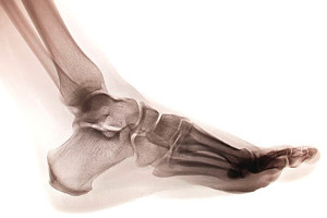
Tibia
Age Determination by Skeleton
Femoracetabular Impingement
Bone Development
Cartilage
Bone Nails
Bone Diseases, Developmental
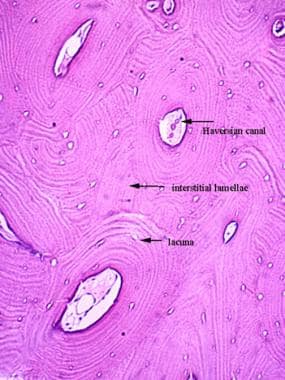
Bone and Bones
Fractures, Malunited
Exostoses
Casts, Surgical
Manipulation, Orthopedic
Hand Deformities, Congenital
Metacarpal Bones
Fibula
Orthopedic Procedures
Infusions, Intraosseous
Range of Motion, Articular
Bone Wires
Traction
Chondroblastoma
Acetabulum
Endocrine System Diseases
Craniopharyngioma
Osteomyelitis
Leg Length Inequality
Hyaline Cartilage
Hip Dislocation, Congenital
Shoulder Fractures
Fibrocartilage in tendons and ligaments--an adaptation to compressive load. (1/345)
Where tendons and ligaments are subject to compression, they are frequently fibrocartilaginous. This occurs at 2 principal sites: where tendons (and sometimes ligaments) wrap around bony or fibrous pulleys, and in the region where they attach to bone, i.e. at their entheses. Wrap-around tendons are most characteristic of the limbs and are commonly wider at their point of bony contact so that the pressure is reduced. The most fibrocartilaginous tendons are heavily loaded and permanently bent around their pulleys. There is often pronounced interweaving of collagen fibres that prevents the tendons from splaying apart under compression. The fibrocartilage can be located within fascicles, or in endo- or epitenon (where it may protect blood vessels from compression or allow fascicles to slide). Fibrocartilage cells are commonly packed with intermediate filaments which could be involved in transducing mechanical load. The ECM often contains aggrecan which allows the tendon to imbibe water and withstand compression. Type II collagen may also be present, particularly in tendons that are heavily loaded. Fibrocartilage is a dynamic tissue that disappears when the tendons are rerouted surgically and can be maintained in vitro when discs of tendon are compressed. Finite element analyses provide a good correlation between its distribution and levels of compressive stress, but at some locations fibrocartilage is a sign of pathology. Enthesis fibrocartilage is most typical of tendons or ligaments that attach to the epiphyses of long bones where it may also be accompanied by sesamoid and periosteal fibrocartilages. It is characteristic of sites where the angle of attachment changes throughout the range of joint movement and it reduces wear and tear by dissipating stress concentration at the bony interface. There is a good correlation between the distribution of fibrocartilage within an enthesis and the levels of compressive stress. The complex interlocking between calcified fibrocartilage and bone contributes to the mechanical strength of the enthesis and cartilage-like molecules (e.g. aggrecan and type II collagen) in the ECM contribute to its ability to withstand compression. Pathological changes are common and are known as enthesopathies. (+info)Retardation of bone growth in triamcinolone-treated mice. (2/345)
Immature mice were treated for up to 8 weeks with daily doses of triamcinolone diacetate. The epiphyseal cartilage plate and its surrounding bone from the humeral head were studied histologically at regular intervals. Concomitantly, roentgenographic measurements were performed on the humeri in toto. By the tenth injection significant morphological changes were noted in the cartilaginous plate, followed by complete cessation of bone growth. Severe triglyceride accumulation appeared in the experimental livers and humeral bone marrow. Osteoporosis also occurred and became severe from the fifth week of triamcinolone administration. Possible explanations for the above findings are discussed. (+info)The pathogenesis of Perthes' disease. (3/345)
It has been shown that in the puppy, two infarcts separated by an interval of four weeks produce a disorder of long duration which results in flattening and broadening of the femoral head and which reproduces the radiological changes seen in Perthes' disease in man. The histological appearances produced by two infarcts are characteristic. In this study the histological appearance of fifty-seven femoral head biopsy specimens in Perthes' disease in man have been studied. In 51 per cent of hips histopathological changes characteristic of double infarction were present, and there were grounds for postulating that double infarction might eventually occur in all cases. The findings support the concept that the deformation of the femoral head and the chronicity of Perthes' disease in man may be due at least as much or even more to repeated episodes of infarction and the ensuing abnormalities of growth as to mechanical factors related to weight-bearing. (+info)Spontaneous or traumatic premature closure of the tibial tubercle. (4/345)
A premature closure of the physis of the tibial tubercle in a young man has given rise to a shortening of the tibia, a patella alta and a reversed tibial slope of 20 degrees with clinical genu recurvatum. After a proximal open wedge tibial osteotomy all three postural deformities could be restored. The etiology of this complex deformity is discussed. (+info)Effect of strontium on the epiphyseal cartilage plate of rat tibiae-histological and radiographic studies. (5/345)
Following dietary administration of strontium carbonate, histological and radiographic changes in the epiphyseal cartilage plate of the rat tibiae were examined in the present study. The weight gain of the rat fed a strontium diet was less than that of the control rats. Longitudinal growth of tibiae and endochondral ossification were inhibited by strontium administration. The widths of both proximal and distal cartilage plates increased enormously as has also been shown by other investigators. Sizes of chondroblasts in columns of proximal cartilage plate in rats fed a strontium diet were smaller than those of the control rats and were not different between upper and lower parts. It is suggested that strontium inhibits bone growth through the inhibitory action on the maturation process of chondroblasts and the succeeding endochondral ossification. (+info)Evidence for the promotion of bone mineralization by 1alpha,25-dihydroxycholecalciferol in the rat unrelated to the correction of deficiencies in serum calcium and phosphorus. (6/345)
Concurrent administration of 1alpha,25-dihydroxycholecalciferol [1alpha,25-(OH)2-CC] to intact and thyroparathyroidectomized rats treated with ethane-1-hydroxy-1,1-diphosphonate (EHDP) prevented or reversed the EHDP-induced inhibition of bone mineralization as measured by changes in epiphyseal plate width and ash content of bone. An analog, 1alpha-droxycholecalciferol, was also effective. Recovery of bone after EHDP treatment was also significantly improved by administration of 1alpha,25-(OH)2-CC as evidenced by enhanced uptake of 45Ca by epiphyseal plates and decreased plate widths. Cholecalciferol (CC), ergocalciferol, dihydrotachysterol2, 5,6-trans-CC, 25-OH-CC, 5,6-Trans-25-OH-CC, and 1alpha24R,25-(OH)3-CC also blocked EHDP-induced epiphyseal plate widening, but required high, pharmacological dose levels. 24R,25- (OH)2-CC was inactive at doses up to 10 microgram/day. Since EHDP-treated rats are not deficient in calcium or phosphate, these data suggest that 1alpha,25-dihydroxycholecalciferol promoted bone mineralization independently of effects upon the intestinal absorption of calcium and phosphate. (+info)Sulfate incorporation from ascorbate 2-sulfate into chondroitin sulfate by embryonic chick cartilage epiphyses. (7/345)
Radioactivity was significantly incorporated from ascorbate 2-[35S]sulfate into chondroitin sulfate by embryonic chick cartilage epiphyses. The extent of incorporation was comparable with that from inorganic [35S]sulfate. The radioactive chondroitin sulfate formed from ascorbate 2-[35S]sulfate gave two radioactive disaccharides on chondroitinase-ABC [EC 4.2.2.4] digestion. The incorporation was markedly decreased by inorganic sulfate. The time course of incorporation from ascorbate 2-[35S]sulfate and inorganic [35S]sulfate into chondroitin sulfate and the constituent disaccharides suggest that the incorporation rates from the two radioactive substances are different. (+info)Identification of novel pro-alpha2(IX) collagen gene mutations in two families with distinctive oligo-epiphyseal forms of multiple epiphyseal dysplasia. (8/345)
Multiple epiphyseal dysplasia (MED) is a genetically heterogeneous disorder with marked clinical and radiographic variability. Traditionally, the mild "Ribbing" and severe "Fairbank" types have been used to define a broad phenotypic spectrum. Mutations in the gene encoding cartilage oligomeric-matrix protein have been shown to result in several types of MED, whereas mutations in the gene encoding the alpha2 chain of type IX collagen (COL9A2) have so far been found only in two families with the Fairbank type of MED. Type IX collagen is a heterotrimer of pro-alpha chains derived from three distinct genes-COL9A1, COL9A2, and COL9A3. In this article, we describe two families with distinctive oligo-epiphyseal forms of MED, which are heterozygous for different mutations in the COL9A2 exon 3/intron 3 splice-donor site. Both of these mutations result in the skipping of exon 3 from COL9A2 mRNA, but the position of the mutation in the splice-donor site determines the stability of the mRNA produced from the mutant COL9A2 allele. (+info)Slipped epiphyses refer to a medical condition where the growth plate (epiphysis) at the end of a bone slips away from the rest of the bone. This condition most commonly affects the hip joint in adolescents and is also known as slipped capital femoral epiphysis (SCFE).
The epiphysis is a layer of cartilage that is present at the ends of long bones in children and adolescents. It is responsible for the growth and development of the bone. In SCFE, the epiphysis on the upper end of the thighbone (femur) slips away from the shaft of the bone due to weakness or injury to the growth plate.
Slipped epiphyses can cause pain, stiffness, and limited mobility in the affected joint. If left untreated, it can lead to complications such as avascular necrosis (death of bone tissue due to lack of blood supply) and early arthritis. Treatment for slipped epiphyses typically involves surgery to realign and stabilize the growth plate with pins or screws.
The epiphyses are the rounded ends of long bones in the body, which articulate with other bones to form joints. They are separated from the main shaft of the bone (diaphysis) by a growth plate called the physis or epiphyseal plate. The epiphyses are made up of spongy bone and covered with articular cartilage, which allows for smooth movement between bones. During growth, the epiphyseal plates produce new bone cells that cause the bone to lengthen until they eventually fuse during adulthood, at which point growth stops.
Slipped Capital Femoral Epiphyses (SCFE) is a pediatric orthopedic condition that affects the growth plate (epiphysis) at the top of the thigh bone (femur). In SCFE, the epiphysis slips or shifts off the end of the femur, leading to abnormal hip function and potentially causing pain, stiffness, and limping. This condition typically occurs during periods of rapid growth, particularly in early adolescence, and is more common in overweight children. If left untreated, SCFE can result in significant long-term complications such as osteoarthritis or avascular necrosis (death of bone tissue due to lack of blood supply). Early diagnosis and appropriate medical intervention are crucial for optimal outcomes.
The femoral head is the rounded, ball-like top portion of the femur (thigh bone) that fits into the hip socket (acetabulum) to form the hip joint. It has a smooth, articular cartilage surface that allows for smooth and stable articulation with the pelvis. The femoral head is connected to the femoral neck, which is a narrower section of bone that angles downward and leads into the shaft of the femur. Together, the femoral head and neck provide stability and range of motion to the hip joint.
The femur is the medical term for the thigh bone, which is the longest and strongest bone in the human body. It connects the hip bone to the knee joint and plays a crucial role in supporting the weight of the body and allowing movement during activities such as walking, running, and jumping. The femur is composed of a rounded head, a long shaft, and two condyles at the lower end that articulate with the tibia and patella to form the knee joint.
Legg-Calve-Perthes disease is a childhood hip disorder that occurs when the blood supply to the ball part of the thigh bone (femoral head) is disrupted. This causes the bone tissue to die, leading to its collapse and deformity. The femoral head then regenerates itself, but often not as round and smooth as it should be, which can lead to hip problems in later life.
The disease is named after three doctors who independently described it: Arthur Legg, Jacques Calve, and Georg Perthes. It typically affects children between the ages of 4 and 10, more commonly boys than girls. Symptoms may include limping, pain in the hip or knee, reduced range of motion in the hip, and muscle wasting. Treatment often involves rest, physical therapy, and sometimes surgery to realign or reshape the femoral head.
Femoral head necrosis, also known as avascular necrosis of the femoral head, is a medical condition that results from the interruption of blood flow to the femoral head, which is the rounded end of the thigh bone that fits into the hip joint. This lack of blood supply can cause the bone tissue to die, leading to the collapse of the femoral head and eventually resulting in hip joint damage or arthritis.
The condition can be caused by a variety of factors, including trauma, alcohol abuse, corticosteroid use, radiation therapy, and certain medical conditions such as sickle cell disease and lupus. Symptoms may include pain in the hip or groin, limited range of motion, and difficulty walking. Treatment options depend on the severity and progression of the necrosis and may include medication, physical therapy, or surgical intervention.
A growth plate, also known as an epiphyseal plate or physis, is a layer of cartilaginous tissue found near the ends of long bones in children and adolescents. This region is responsible for the longitudinal growth of bones during development. The growth plate contains actively dividing cells that differentiate into chondrocytes, which produce and deposit new matrix, leading to bone elongation. Once growth is complete, usually in late adolescence or early adulthood, the growth plates ossify (harden) and are replaced by solid bone, transforming into the epiphyseal line.
The hip joint, also known as the coxal joint, is a ball-and-socket type synovial joint that connects the femur (thigh bone) to the pelvis. The "ball" is the head of the femur, while the "socket" is the acetabulum, a concave surface on the pelvic bone.
The hip joint is surrounded by a strong fibrous capsule and is reinforced by several ligaments, including the iliofemoral, ischiofemoral, and pubofemoral ligaments. The joint allows for flexion, extension, abduction, adduction, medial and lateral rotation, and circumduction movements, making it one of the most mobile joints in the body.
The hip joint is also supported by various muscles, including the gluteus maximus, gluteus medius, gluteus minimus, iliopsoas, and other hip flexors and extensors. These muscles provide stability and strength to the joint, allowing for weight-bearing activities such as walking, running, and jumping.
The tibia, also known as the shin bone, is the larger of the two bones in the lower leg and part of the knee joint. It supports most of the body's weight and is a major insertion point for muscles that flex the foot and bend the leg. The tibia articulates with the femur at the knee joint and with the fibula and talus bone at the ankle joint. Injuries to the tibia, such as fractures, are common in sports and other activities that put stress on the lower leg.
Age determination by skeleton, also known as skeletal aging or skeletal maturation, is the process of estimating a person's age based on the analysis of their skeletal remains. This technique is commonly used in forensic anthropology to help identify unknown individuals or determine the time since death.
The method involves examining various features of the skeleton, such as the degree of fusion of epiphyseal growth plates, the shape and size of certain bones, and the presence or absence of degenerative changes. These features change in a predictable way as a person grows and develops, allowing for an estimation of their age at death.
It is important to note that while skeletal aging can provide useful information, it is not always possible to determine an exact age. Instead, forensic anthropologists typically provide a range of ages that the individual may have fallen into based on the skeletal evidence. Additionally, factors such as genetics, nutrition, and health can affect the rate at which skeletal features develop, making it difficult to provide a precise estimate in some cases.
The humerus is the long bone in the upper arm that extends from the shoulder joint (glenohumeral joint) to the elbow joint. It articulates with the glenoid cavity of the scapula to form the shoulder joint and with the radius and ulna bones at the elbow joint. The proximal end of the humerus has a rounded head that provides for movement in multiple planes, making it one of the most mobile joints in the body. The greater and lesser tubercles are bony prominences on the humeral head that serve as attachment sites for muscles that move the shoulder and arm. The narrow shaft of the humerus provides stability and strength for weight-bearing activities, while the distal end forms two articulations: one with the ulna (trochlea) and one with the radius (capitulum). Together, these structures allow for a wide range of motion in the shoulder and elbow joints.
Osteotomy is a surgical procedure in which a bone is cut to shorten, lengthen, or change its alignment. It is often performed to correct deformities or to realign bones that have been damaged by trauma or disease. The bone may be cut straight across (transverse osteotomy) or at an angle (oblique osteotomy). After the bone is cut, it can be realigned and held in place with pins, plates, or screws until it heals. This procedure is commonly performed on bones in the leg, such as the femur or tibia, but can also be done on other bones in the body.
The diaphysis refers to the shaft or middle portion of a long bone in the body. It is the part that is typically cylindrical in shape and contains the medullary cavity, which is filled with yellow marrow. The diaphysis is primarily composed of compact bone tissue, which provides strength and support for weight-bearing and movement.
In contrast to the diaphysis, the ends of long bones are called epiphyses, and they are covered with articular cartilage and contain spongy bone tissue filled with red marrow, which is responsible for producing blood cells. The area where the diaphysis meets the epiphysis is known as the metaphysis, and it contains growth plates that are responsible for the longitudinal growth of bones during development.
Femoroacetabular impingement (FAI) is a medical condition that affects the hip joint. It occurs when there is abnormal contact between the femoral head (the ball at the top of the thigh bone) and the acetabulum (the socket in the pelvis) during normal movement of the hip. This abnormal contact can cause damage to the cartilage and labrum (a ring of cartilage that helps to stabilize the hip joint) leading to pain, stiffness and decreased range of motion.
FAI is classified into two types: cam impingement and pincer impingement. Cam impingement occurs when there is an abnormal shape of the femoral head or neck, which leads to abnormal contact with the acetabulum during hip flexion and internal rotation. Pincer impingement occurs when there is overcoverage of the acetabulum, leading to abnormal contact with the femoral head or neck.
In some cases, both cam and pincer impingement can be present, which is referred to as mixed impingement. Symptoms of FAI may include hip pain, stiffness, limping, and reduced range of motion. Treatment options for FAI may include physical therapy, activity modification, medications, and in some cases, surgery.
The "femur neck" is the narrow, upper part of the femur (thigh bone) where it connects to the pelvis. It is the region through which the femoral head articulates with the acetabulum to form the hip joint. The femur neck is a common site for fractures, especially in older adults with osteoporosis.
Osteochondrodysplasias are a group of genetic disorders that affect the development of bones and cartilage. These conditions can result in dwarfism or short stature, as well as other skeletal abnormalities. Osteochondrodysplasias can be caused by mutations in genes that regulate bone and cartilage growth, and they are often characterized by abnormalities in the shape, size, and/or structure of the bones and cartilage.
There are many different types of osteochondrodysplasias, each with its own specific symptoms and patterns of inheritance. Some common examples include achondroplasia, thanatophoric dysplasia, and spondyloepiphyseal dysplasia. These conditions can vary in severity, and some may be associated with other health problems, such as respiratory difficulties or neurological issues.
Treatment for osteochondrodysplasias typically focuses on managing the symptoms and addressing any related health concerns. This may involve physical therapy, bracing or surgery to correct skeletal abnormalities, and treatment for any associated medical conditions. In some cases, genetic counseling may also be recommended for individuals with osteochondrodysplasias and their families.
Bone development, also known as ossification, is the process by which bone tissue is formed and grows. This complex process involves several different types of cells, including osteoblasts, which produce new bone matrix, and osteoclasts, which break down and resorb existing bone tissue.
There are two main types of bone development: intramembranous and endochondral ossification. Intramembranous ossification occurs when bone tissue forms directly from connective tissue, while endochondral ossification involves the formation of a cartilage model that is later replaced by bone.
During fetal development, most bones develop through endochondral ossification, starting as a cartilage template that is gradually replaced by bone tissue. However, some bones, such as those in the skull and clavicles, develop through intramembranous ossification.
Bone development continues after birth, with new bone tissue being laid down and existing tissue being remodeled throughout life. This ongoing process helps to maintain the strength and integrity of the skeleton, allowing it to adapt to changing mechanical forces and repair any damage that may occur.
Cartilage is a type of connective tissue that is found throughout the body in various forms. It is made up of specialized cells called chondrocytes, which are embedded in a firm, flexible matrix composed of collagen fibers and proteoglycans. This unique structure gives cartilage its characteristic properties of being both strong and flexible.
There are three main types of cartilage in the human body: hyaline cartilage, elastic cartilage, and fibrocartilage.
1. Hyaline cartilage is the most common type and is found in areas such as the articular surfaces of bones (where they meet to form joints), the nose, trachea, and larynx. It has a smooth, glassy appearance and provides a smooth, lubricated surface for joint movement.
2. Elastic cartilage contains more elastin fibers than hyaline cartilage, which gives it greater flexibility and resilience. It is found in structures such as the external ear and parts of the larynx and epiglottis.
3. Fibrocartilage has a higher proportion of collagen fibers and fewer chondrocytes than hyaline or elastic cartilage. It is found in areas that require high tensile strength, such as the intervertebral discs, menisci (found in joints like the knee), and the pubic symphysis.
Cartilage plays a crucial role in supporting and protecting various structures within the body, allowing for smooth movement and providing a cushion between bones to absorb shock and prevent wear and tear. However, cartilage has limited capacity for self-repair and regeneration, making damage or degeneration of cartilage tissue a significant concern in conditions such as osteoarthritis.
I believe you are referring to "bone pins" or "bone nails" rather than "bone nails." These terms are used in the medical field to describe surgical implants made of metal or biocompatible materials that are used to stabilize and hold together fractured bones during the healing process. They can also be used in spinal fusion surgery to provide stability and promote bone growth between vertebrae.
Bone pins or nails typically have a threaded or smooth shaft, with a small diameter that allows them to be inserted into the medullary canal of long bones such as the femur or tibia. They may also have a head or eyelet on one end that allows for attachment to external fixation devices or other surgical instruments.
The use of bone pins and nails has revolutionized orthopedic surgery, allowing for faster healing times, improved stability, and better functional outcomes for patients with fractures or spinal deformities.
Osteochondritis is a joint condition where a piece of cartilage or bone in the joint separates from its attachment due to a lack of blood supply. This can cause pain, stiffness, and potentially restricted movement in the affected joint. It often occurs in weight-bearing joints like the knee or ankle, and is more common in children and adolescents. The separated piece may sometimes float around in the joint space, causing further damage to the cartilage and bone. If left untreated, it can lead to long-term joint problems. Also known as osteochondrosis or osteochondritis dissecans.
Developmental bone diseases are a group of medical conditions that affect the growth and development of bones. These diseases are present at birth or develop during childhood and adolescence, when bones are growing rapidly. They can result from genetic mutations, hormonal imbalances, or environmental factors such as poor nutrition.
Some examples of developmental bone diseases include:
1. Osteogenesis imperfecta (OI): Also known as brittle bone disease, OI is a genetic disorder that affects the body's production of collagen, a protein necessary for healthy bones. People with OI have fragile bones that break easily and may also experience other symptoms such as blue sclerae (whites of the eyes), hearing loss, and joint laxity.
2. Achondroplasia: This is the most common form of dwarfism, caused by a genetic mutation that affects bone growth. People with achondroplasia have short limbs and a large head relative to their body size.
3. Rickets: A condition caused by vitamin D deficiency or an inability to absorb or use vitamin D properly. This leads to weak, soft bones that can bow or bend easily, particularly in children.
4. Fibrous dysplasia: A rare bone disorder where normal bone is replaced with fibrous tissue, leading to weakened bones and deformities.
5. Scoliosis: An abnormal curvature of the spine that can develop during childhood or adolescence. While not strictly a developmental bone disease, scoliosis can be caused by various underlying conditions such as cerebral palsy, muscular dystrophy, or spina bifida.
Treatment for developmental bone diseases varies depending on the specific condition and its severity. Treatment may include medication, physical therapy, bracing, or surgery to correct deformities and improve function. Regular follow-up with a healthcare provider is essential to monitor growth, manage symptoms, and prevent complications.
Bone screws are medical devices used in orthopedic and trauma surgery to affix bone fracture fragments or to attach bones to other bones or to metal implants such as plates, rods, or artificial joints. They are typically made of stainless steel or titanium alloys and have a threaded shaft that allows for purchase in the bone when tightened. The head of the screw may have a hexagonal or star-shaped design to allow for precise tightening with a screwdriver. Bone screws come in various shapes, sizes, and designs, including fully threaded, partially threaded, cannulated (hollow), and headless types, depending on their intended use and location in the body.
"Bone" is the hard, dense connective tissue that makes up the skeleton of vertebrate animals. It provides support and protection for the body's internal organs, and serves as a attachment site for muscles, tendons, and ligaments. Bone is composed of cells called osteoblasts and osteoclasts, which are responsible for bone formation and resorption, respectively, and an extracellular matrix made up of collagen fibers and mineral crystals.
Bones can be classified into two main types: compact bone and spongy bone. Compact bone is dense and hard, and makes up the outer layer of all bones and the shafts of long bones. Spongy bone is less dense and contains large spaces, and makes up the ends of long bones and the interior of flat and irregular bones.
The human body has 206 bones in total. They can be further classified into five categories based on their shape: long bones, short bones, flat bones, irregular bones, and sesamoid bones.
A hip dislocation is a medical emergency that occurs when the head of the femur (thighbone) slips out of its socket in the pelvis. This can happen due to high-energy trauma, such as a car accident or a severe fall. Hip dislocations can also occur in people with certain health conditions that make their hips more prone to displacement, such as developmental dysplasia of the hip.
There are two main types of hip dislocations: posterior and anterior. In a posterior dislocation, the femur head moves out of the back of the socket, which is the most common type. In an anterior dislocation, the femur head moves out of the front of the socket. Both types of hip dislocations can cause severe pain, swelling, and difficulty moving the affected leg.
Immediate medical attention is necessary for a hip dislocation to realign the bones and prevent further damage. Treatment typically involves sedation or anesthesia to relax the muscles around the joint, followed by a closed reduction procedure to gently guide the femur head back into the socket. In some cases, surgery may be required to repair any associated injuries, such as fractures or damaged ligaments. After treatment, physical therapy and rehabilitation are usually necessary to restore strength, mobility, and function to the affected hip joint.
Dysostosis is a term used to describe a group of genetic disorders that are characterized by abnormal development and formation of one or more bones in the body. The condition is typically present at birth (congenital) and can affect any bone, but it most commonly involves the bones of the skull, face, hands, and feet.
The term "dysostosis" comes from the Greek words "dys," meaning difficult or abnormal, and "osteon," meaning bone. Dysostoses are usually caused by mutations in specific genes that regulate bone development. These genetic changes can be inherited from one or both parents or can occur spontaneously during fetal development.
There are many different types of dysostoses, each with its own set of symptoms and characteristics. Some common examples include:
1. Cleidocranial Dysplasia: This is a rare genetic disorder that affects the development of the skull and collarbones (cleido). People with cleidocranial dysplasia may have a larger than normal head, wide-set eyes, a prominent forehead, and underdeveloped or missing collarbones.
2. Acrocephalopolysyndactyly Type II: Also known as ACPS II or Greig cephalopolysyndactyly syndrome, this disorder is characterized by a pointed skull (acrocephaly), extra fingers and toes (polydactyly), and wide-set eyes.
3. Osteogenesis Imperfecta: This is a group of genetic disorders that affect the body's production of collagen, a protein that helps to strengthen bones. People with osteogenesis imperfecta have fragile bones that break easily, often as a result of minor trauma.
4. Diastrophic Dysplasia: This is a rare genetic disorder that affects the development of the bones and cartilage in the body. People with diastrophic dysplasia may have short limbs, a deformed spine, and a characteristic "hitchhiker's thumb" appearance.
5. Thanatophoric Dysplasia: This is a severe genetic disorder that affects the development of the bones in the body. People with thanatophoric dysplasia have very short limbs, a small chest, and a deformed skull. The condition is often fatal in infancy or early childhood.
These are just a few examples of the many different types of skeletal dysplasias that exist. While some forms of these disorders can be managed with medical treatment and therapy, others may require surgery or other interventions to help improve quality of life. In some cases, genetic counseling and testing may be recommended for individuals who are considering starting a family and have a history of skeletal dysplasia in their family.
Malunited fractures refer to a type of fracture where the bones do not heal in their proper alignment or position. This can occur due to various reasons such as inadequate reduction of the fracture fragments during initial treatment, improper casting or immobilization, or failure of the patient to follow proper immobilization instructions. Malunited fractures can result in deformity, limited range of motion, and decreased functionality of the affected limb. Additional treatments such as surgery may be required to correct the malunion and restore normal function.
Exostoses are benign (noncancerous) bone growths that develop on the surface of a bone, usually in response to repeated stress or friction. They are often small and smooth, but can become larger and more irregular over time. In some cases, they may cause pain or discomfort, especially if they continue to grow and put pressure on nearby nerves, muscles, or other bones.
Exostoses can occur in various parts of the body, but they are most commonly found in the long bones of the arms and legs, as well as in the small bones of the feet. They may also develop in response to chronic irritation or injury, such as from jogging or playing sports that involve a lot of running or jumping.
In some cases, exostoses may be surgically removed if they cause persistent pain or other symptoms. However, in many cases, they do not require treatment and can be left alone. If you are concerned about any bone growths or other unusual symptoms, it is always best to consult with a healthcare professional for an accurate diagnosis and treatment plan.
Surgical casts are medical devices used to immobilize and protect injured body parts, typically fractured or broken bones, during the healing process. They are usually made of plaster or fiberglass materials that harden when wet and conform to the shape of the affected area once applied. The purpose of a surgical cast is to restrict movement and provide stability to the injured site, allowing for proper alignment and healing of the bones.
The casting process involves first aligning the broken bone fragments into their correct positions, often through manual manipulation or surgical intervention. Once aligned, the cast material is applied in layers, with each layer being allowed to dry before adding the next. This creates a rigid structure that encases and supports the injured area. The cast must be kept dry during the healing process to prevent it from becoming weakened or damaged.
Surgical casts come in various shapes and sizes depending on the location and severity of the injury. They may also include additional components such as padding, Velcro straps, or window openings to allow for regular monitoring of the skin and underlying tissue. In some cases, removable splints or functional braces may be used instead of traditional casts, providing similar support while allowing for limited movement and easier adjustments.
It is essential to follow proper care instructions when wearing a surgical cast, including elevating the injured limb, avoiding excessive weight-bearing, and monitoring for signs of complications such as swelling, numbness, or infection. Regular check-ups with a healthcare provider are necessary to ensure proper healing and adjust the cast if needed.
Orthopedic manipulation is a hands-on technique that is used by healthcare professionals, such as orthopedic doctors, chiropractors, and physical therapists, to diagnose and treat muscle and joint disorders. This manual procedure involves moving the joints or soft tissues in a specific direction and amplitude with the aim of improving joint mobility, reducing pain, relieving muscle tension, and enhancing overall function.
Orthopedic manipulation can be performed on various parts of the body, including the spine, extremities, and cranial structures. It is often used as a complementary treatment alongside other therapeutic interventions, such as exercise, medication, or surgery, to manage a wide range of musculoskeletal conditions, including but not limited to:
* Back pain and stiffness
* Neck pain and stiffness
* Joint pain and inflammation
* Muscle spasms and tension
* Headaches and migraines
* Disc disorders
* Sprains and strains
* Postural dysfunctions
It is important to note that orthopedic manipulation should only be performed by trained and licensed healthcare professionals, as improper techniques can lead to injury or further damage. Patients should consult with their healthcare provider to determine if orthopedic manipulation is an appropriate treatment option for their specific condition.
Birth injuries refer to damages or injuries that a baby suffers during the birthing process. These injuries can result from various factors, such as mechanical forces during delivery, medical negligence, or complications during pregnancy or labor. Some common examples of birth injuries include:
1. Brachial plexus injuries: Damage to the nerves that control movement and feeling in the arms and hands, often caused by excessive pulling or stretching during delivery.
2. Cephalohematoma: A collection of blood between the skull and the periosteum (the membrane covering the bone), usually caused by trauma during delivery.
3. Caput succedaneum: Swelling of the soft tissues of the baby's scalp, often resulting from pressure on the head during labor and delivery.
4. Fractures: Broken bones, such as a clavicle or skull fracture, can occur due to mechanical forces during delivery.
5. Intracranial hemorrhage: Bleeding in or around the brain, which can result from trauma during delivery or complications like high blood pressure in the mother.
6. Perinatal asphyxia: A lack of oxygen supply to the baby before, during, or immediately after birth, which can lead to brain damage and other health issues.
7. Subconjunctival hemorrhage: Bleeding under the conjunctiva (the clear membrane covering the eye), often caused by pressure on the head during delivery.
8. Spinal cord injuries: Damage to the spinal cord, which can result in paralysis or other neurological issues, may occur due to excessive force during delivery or medical negligence.
It's important to note that some birth injuries are unavoidable and may not be a result of medical malpractice. However, if a healthcare provider fails to provide the standard of care expected during pregnancy, labor, or delivery, they may be held liable for any resulting injuries.
Congenital hand deformities refer to physical abnormalities or malformations of the hand, wrist, and/or digits (fingers) that are present at birth. These deformities can result from genetic factors, environmental influences during pregnancy, or a combination of both. They may affect the bones, muscles, tendons, joints, and other structures in the hand, leading to varying degrees of impairment in function and appearance.
There are numerous types of congenital hand deformities, some of which include:
1. Polydactyly: The presence of extra digits on the hand, which can be fully formed or rudimentary.
2. Syndactyly: Webbing or fusion of two or more fingers, which may involve soft tissue only or bone as well.
3. Clinodactyly: A curved finger due to a sideways deviation of the fingertip, often affecting the little finger.
4. Camptodactyly: Permanent flexion or bending of one or more fingers, typically involving the proximal interphalangeal joint.
5. Trigger Finger/Thumb: A condition where a finger or thumb becomes locked in a bent position due to thickening and narrowing of the tendon sheath.
6. Radial Club Hand (Radial Ray Deficiency): Underdevelopment or absence of the radius bone, resulting in a short, curved forearm and hand deformity.
7. Ulnar Club Hand (Ulnar Ray Deficiency): Underdevelopment or absence of the ulna bone, leading to a short, curved forearm and hand deformity.
8. Cleidocranial Dysplasia: A genetic disorder affecting bone growth, resulting in underdeveloped or absent collarbones, dental abnormalities, and occasionally hand deformities.
9. Apert Syndrome: A rare genetic disorder characterized by the fusion of fingers and toes (syndactyly) and other skeletal abnormalities.
10. Holt-Oram Syndrome: A genetic disorder involving heart defects and upper limb deformities, such as radial ray deficiency or thumb anomalies.
Treatment for hand deformities varies depending on the specific condition and severity. Options may include physical therapy, bracing, splinting, medications, or surgical intervention.
The metacarpal bones are the long slender bones that make up the middle part of the hand, located between the carpals (wrist bones) and the phalanges (finger bones). There are five metacarpal bones in total, with one for each finger and thumb. Each bone has a base attached to the carpals, a shaft, and a head that connects to the phalanges. The metacarpal bones play a crucial role in hand function, providing stability and support during gripping and manipulation movements.
The fibula is a slender bone located in the lower leg of humans and other vertebrates. It runs parallel to the larger and more robust tibia, and together they are known as the bones of the leg or the anterior tibial segment. The fibula is the lateral bone in the leg, positioned on the outside of the tibia.
In humans, the fibula extends from the knee joint proximally to the ankle joint distally. Its proximal end, called the head of the fibula, articulates with the lateral condyle of the tibia and forms part of the inferior aspect of the knee joint. The narrowed portion below the head is known as the neck of the fibula.
The shaft of the fibula, also called the body of the fibula, is a long, thin structure that descends from the neck and serves primarily for muscle attachment rather than weight-bearing functions. The distal end of the fibula widens to form the lateral malleolus, which is an important bony landmark in the ankle region. The lateral malleolus articulates with the talus bone of the foot and forms part of the ankle joint.
The primary functions of the fibula include providing attachment sites for muscles that act on the lower leg, ankle, and foot, as well as contributing to the stability of the ankle joint through its articulation with the talus bone. Fractures of the fibula can occur due to various injuries, such as twisting or rotational forces applied to the ankle or direct trauma to the lateral aspect of the lower leg.
Orthopedic procedures are surgical or nonsurgical methods used to treat musculoskeletal conditions, including injuries, deformities, or diseases of the bones, joints, muscles, ligaments, and tendons. These procedures can range from simple splinting or casting to complex surgeries such as joint replacements, spinal fusions, or osteotomies (cutting and repositioning bones). The primary goal of orthopedic procedures is to restore function, reduce pain, and improve the quality of life for patients.
Intraosseous infusion is a medical procedure that involves the injection of fluid or medication directly into the bone marrow, specifically through the tibia or humerus bones. This route is used when intravenous access is difficult or impossible to obtain in emergency situations, such as cardiac arrest, severe trauma, or shock. The goal is to deliver essential fluids and medications rapidly into the systemic circulation, bypassing the need for traditional venous access. Intraosseous infusions are considered a temporary measure until intravenous access can be established.
Articular Range of Motion (AROM) is a term used in physiotherapy and orthopedics to describe the amount of movement available in a joint, measured in degrees of a circle. It refers to the range through which synovial joints can actively move without causing pain or injury. AROM is assessed by measuring the degree of motion achieved by active muscle contraction, as opposed to passive range of motion (PROM), where the movement is generated by an external force.
Assessment of AROM is important in evaluating a patient's functional ability and progress, planning treatment interventions, and determining return to normal activities or sports participation. It is also used to identify any restrictions in joint mobility that may be due to injury, disease, or surgery, and to monitor the effectiveness of rehabilitation programs.
The ulna is one of the two long bones in the forearm, the other being the radius. It runs from the elbow to the wrist and is located on the medial side of the forearm, next to the bone called the humerus in the upper arm. The ulna plays a crucial role in the movement of the forearm and also serves as an attachment site for various muscles.
I'm not aware of a medical term called "bone wires." The term "wiring" is used in orthopedic surgery to describe the use of metal wire to hold bones or fractures in place during healing. However, I couldn't find any specific medical definition or term related to "bone wires." It may be a colloquialism, a term used in a specific context, or a term from science fiction. If you could provide more context about where you encountered this term, I might be able to give a more accurate answer.
Traction, in medical terms, refers to the application of a pulling force to distract or align parts of the body, particularly bones, joints, or muscles, with the aim of immobilizing, reducing displacement, or realigning them. This is often achieved through the use of various devices such as tongs, pulleys, weights, or specialized traction tables. Traction may be applied manually or mechanically and can be continuous or intermittent, depending on the specific medical condition being treated. Common indications for traction include fractures, dislocations, spinal cord injuries, and certain neurological conditions.
Chondroblastoma is a rare, benign (non-cancerous) bone tumor that typically develops in the epiphysis, which is the rounded end of a long bone near a joint. It primarily affects children and adolescents, with around 90% of cases occurring before the age of 20.
The tumor arises from chondroblasts, cells responsible for producing cartilage during bone growth. Chondroblastoma is usually slow-growing and typically causes localized pain, swelling, or tenderness in the affected area. In some cases, it may weaken the bone and lead to fractures.
Treatment generally involves surgical removal of the tumor, followed by curettage (scraping) of the surrounding bone tissue and replacement with bone grafts or substitutes. Recurrence is possible but rare, and long-term prognosis is usually favorable.
The acetabulum is the cup-shaped cavity in the pelvic bone (specifically, the os coxa) where the head of the femur bone articulates to form the hip joint. It provides a stable and flexible connection between the lower limb and the trunk, allowing for a wide range of movements such as flexion, extension, abduction, adduction, rotation, and circumduction. The acetabulum is lined with articular cartilage, which facilitates smooth and frictionless movement of the hip joint. Its stability is further enhanced by various ligaments, muscles, and the labrum, a fibrocartilaginous rim that deepens the socket and increases its contact area with the femoral head.
The endocrine system is a complex network of glands and organs that produce, store, and secrete hormones. It plays a crucial role in regulating various functions in the body, including metabolism, growth and development, tissue function, sexual function, reproduction, sleep, and mood.
Endocrine system diseases or disorders occur when there is a problem with the production or regulation of hormones. This can result from:
1. Overproduction or underproduction of hormones by the endocrine glands.
2. Impaired response of target cells to hormones.
3. Disruption in the feedback mechanisms that regulate hormone production.
Examples of endocrine system diseases include:
1. Diabetes Mellitus - a group of metabolic disorders characterized by high blood sugar levels due to insulin deficiency or resistance.
2. Hypothyroidism - underactive thyroid gland leading to slow metabolism, weight gain, fatigue, and depression.
3. Hyperthyroidism - overactive thyroid gland causing rapid heartbeat, anxiety, weight loss, and heat intolerance.
4. Cushing's Syndrome - excess cortisol production resulting in obesity, high blood pressure, and weak muscles.
5. Addison's Disease - insufficient adrenal hormone production leading to weakness, fatigue, and low blood pressure.
6. Acromegaly - overproduction of growth hormone after puberty causing enlargement of bones, organs, and soft tissues.
7. Gigantism - similar to acromegaly but occurs before puberty resulting in excessive height and body size.
8. Hypopituitarism - underactive pituitary gland leading to deficiencies in various hormones.
9. Hyperparathyroidism - overactivity of the parathyroid glands causing calcium imbalances and kidney stones.
10. Precocious Puberty - early onset of puberty due to premature activation of the pituitary gland.
Treatment for endocrine system diseases varies depending on the specific disorder and may involve medication, surgery, lifestyle changes, or a combination of these approaches.
Osteogenesis is the process of bone formation or development. It involves the differentiation and maturation of osteoblasts, which are bone-forming cells that synthesize and deposit the organic matrix of bone tissue, composed mainly of type I collagen. This organic matrix later mineralizes to form the inorganic crystalline component of bone, primarily hydroxyapatite.
There are two primary types of osteogenesis: intramembranous and endochondral. Intramembranous osteogenesis occurs directly within connective tissue, where mesenchymal stem cells differentiate into osteoblasts and form bone tissue without an intervening cartilage template. This process is responsible for the formation of flat bones like the skull and clavicles.
Endochondral osteogenesis, on the other hand, involves the initial development of a cartilaginous model or template, which is later replaced by bone tissue. This process forms long bones, such as those in the limbs, and occurs through several stages involving chondrocyte proliferation, hypertrophy, and calcification, followed by invasion of blood vessels and osteoblasts to replace the cartilage with bone tissue.
Abnormalities in osteogenesis can lead to various skeletal disorders and diseases, such as osteogenesis imperfecta (brittle bone disease), achondroplasia (a form of dwarfism), and cleidocranial dysplasia (a disorder affecting skull and collarbone development).
A craniopharyngioma is a type of brain tumor that develops near the pituitary gland, which is a small gland located at the base of the brain. These tumors arise from remnants of Rathke's pouch, an embryonic structure involved in the development of the pituitary gland.
Craniopharyngiomas are typically slow-growing and benign (non-cancerous), but they can still cause significant health problems due to their location. They can compress nearby structures such as the optic nerves, hypothalamus, and pituitary gland, leading to symptoms like vision loss, hormonal imbalances, and cognitive impairment.
Treatment for craniopharyngiomas usually involves surgical removal of the tumor, followed by radiation therapy in some cases. Regular follow-up with a healthcare team is essential to monitor for recurrence and manage any long-term effects of treatment.
Osteomyelitis is a medical condition characterized by an infection that involves the bone or the bone marrow. It can occur as a result of a variety of factors, including bacterial or fungal infections that spread to the bone from another part of the body, or direct infection of the bone through trauma or surgery.
The symptoms of osteomyelitis may include pain and tenderness in the affected area, fever, chills, fatigue, and difficulty moving the affected limb. In some cases, there may also be redness, swelling, and drainage from the infected area. The diagnosis of osteomyelitis typically involves imaging tests such as X-rays, CT scans, or MRI scans, as well as blood tests and cultures to identify the underlying cause of the infection.
Treatment for osteomyelitis usually involves a combination of antibiotics or antifungal medications to eliminate the infection, as well as pain management and possibly surgical debridement to remove infected tissue. In severe cases, hospitalization may be necessary to monitor and manage the condition.
"Bone retroversion" is not a widely recognized medical term or concept with a specific definition. The term "retroversion" is used in anatomy to describe the position of an organ or structure when it is turned backward or inward. In relation to bones, retroversion typically describes the orientation of a bone or joint when it is angled or positioned in such a way that its posterior (back) aspect faces more anteriorly (toward the front).
However, I was unable to find a widely accepted medical definition for "bone retroversion" as a specific pathological or anatomical condition. It's possible that the term may be used in a more specialized context within certain medical subspecialties. If you have more context or information about where this term is being used, I may be able to provide a more precise answer.
'Leg length inequality' (LLIS) is a condition where there is a discrepancy in the lengths of an individual's lower extremities, specifically the bones of the thigh (femur) and/or the leg (tibia/fibula). This discrepancy can be congenital or acquired due to various causes such as fractures, infections, or surgical procedures. The inequality can lead to functional scoliosis, lower back pain, and other musculoskeletal issues. It is typically diagnosed through physical examination and imaging studies like X-rays, and may be treated with various methods including orthotics, shoe lifts, or in some cases, surgical intervention.
Hyaline cartilage is a type of cartilaginous tissue that is primarily found in the articulating surfaces of bones, ribcage, nose, ears, and trachea. It has a smooth, glassy appearance (hence the name "hyaline," derived from the Greek word "hyalos" meaning glass) due to the presence of type II collagen fibers that are arranged in a precise pattern and embedded in a proteoglycan-rich matrix.
The high concentration of proteoglycans, which are complex molecules made up of a protein core and glycosaminoglycan side chains, gives hyaline cartilage its firm yet flexible properties. This type of cartilage is avascular, meaning it does not contain blood vessels, and receives nutrients through diffusion from the surrounding synovial fluid in joints or adjacent tissues.
Hyaline cartilage plays a crucial role in providing structural support, reducing friction between articulating bones, and facilitating smooth movement in joints. It also serves as a template for endochondral ossification, a process by which long bones grow in length during development.
Congenital hip dislocation, also known as developmental dysplasia of the hip (DDH), is a condition where the hip joint fails to develop normally in utero or during early infancy. In a healthy hip, the head of the femur (thigh bone) fits snugly into the acetabulum (hip socket). However, in congenital hip dislocation, the femoral head is not held firmly in place within the acetabulum due to abnormal development or laxity of the ligaments that support the joint.
There are two types of congenital hip dislocations:
1. Teratologic dislocation: This type is present at birth and occurs due to abnormalities in the development of the hip joint during fetal growth. The femoral head may be completely outside the acetabulum or partially dislocated.
2. Developmental dysplasia: This type develops after birth, often within the first few months of life, as a result of ligamentous laxity and shallow acetabulum. In some cases, it can progress to a complete hip dislocation if left untreated.
Risk factors for congenital hip dislocation include family history, breech presentation during delivery, and female gender. Early diagnosis and treatment are crucial to prevent long-term complications such as pain, limited mobility, and osteoarthritis. Treatment options may include bracing, closed reduction, or surgical intervention, depending on the severity and age of the child at diagnosis.
A shoulder fracture refers to a break in one or more bones that make up the shoulder joint, which includes the humerus (upper arm bone), scapula (shoulder blade), and clavicle (collarbone). These types of fractures can occur due to various reasons such as high-energy trauma, falls, or degenerative conditions. Symptoms may include severe pain, swelling, bruising, limited range of motion, deformity, and in some cases, numbness or tingling sensations. Treatment options depend on the severity and location of the fracture but can include immobilization with a sling or brace, surgery, or physical therapy.
Cartilage diseases refer to conditions that affect the cartilaginous tissues in the body. Cartilage is a firm, flexible connective tissue found in many areas of the body, including the joints, ribcage, ears, and nose. It provides structure and support, allows for smooth movement between bones, and protects the ends of bones from friction.
There are several types of cartilage diseases, including:
1. Osteoarthritis (OA): This is a degenerative joint disease that occurs when the protective cartilage that cushions the ends of your bones wears down over time. It can cause pain, stiffness, and loss of mobility in the affected joints.
2. Rheumatoid arthritis (RA): This is an autoimmune disorder that causes inflammation in the lining of the joints, leading to cartilage damage and bone erosion.
3. Traumatic arthritis: This occurs when a joint is injured, causing damage to the cartilage and resulting in pain, stiffness, and loss of mobility.
4. Infectious arthritis: This occurs when a joint becomes infected, leading to inflammation and potential damage to the cartilage.
5. Chondromalacia patellae: This is a condition that affects the cartilage on the back of the kneecap, causing pain and stiffness in the knee.
6. Costochondritis: This is an inflammation of the cartilage in the ribcage, causing chest pain and discomfort.
7. Nasal septal deviation: This is a condition where the cartilage that separates the nostrils is crooked or off-center, causing difficulty breathing through the nose.
8. Osteochondritis dissecans (OCD): This is a joint condition that occurs when a piece of cartilage and bone in a joint becomes detached, causing pain and stiffness.
9. Synovial chondromatosis: This is a rare condition where nodules made up of cartilage form in the lining of a joint, causing pain, swelling, and limited mobility.
Treatment for cartilage diseases varies depending on the specific condition and severity, but may include medication, physical therapy, surgery, or a combination of these.
Bone lengthening is a surgical procedure that involves cutting and then gradually stretching the bone apart, allowing new bone to grow in its place. This process is also known as distraction osteogenesis. The goal of bone lengthening is to increase the length of a bone, either to improve function or to correct a deformity.
The procedure typically involves making an incision in the skin over the bone and using specialized tools to cut through the bone. Once the bone is cut, a device called an external fixator is attached to the bone on either side of the cut. The external fixator is then gradually adjusted over time to slowly stretch the bone apart, creating a gap between the two ends of the bone. As the bone is stretched, new bone tissue begins to grow in the space between the two ends, eventually filling in the gap and lengthening the bone.
Bone lengthening can be used to treat a variety of conditions, including limb length discrepancies, congenital deformities, and injuries that result in bone loss. It is typically performed by an orthopedic surgeon and may require several months of follow-up care to ensure proper healing and growth of the new bone tissue.
 Stippled epiphyses
Epiphysis
Slipped capital femoral epiphysis
Acrodysostosis
Humerus
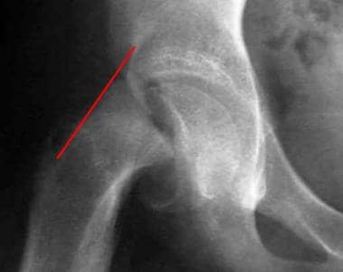
Southwick angle
Metatarsal bones
First metatarsal bone
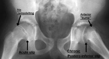
Klein's line
Fetal warfarin syndrome
Maxilla
Fitzsimmons-Guilbert syndrome
Greater trochanter
Ammitocyon
Bone age
Meharia
Ossification center
Pseudoachondroplasia
Long bone
Tibia
Enhydriodon
Regional Acceleratory Phenomenon
Diaphysis
Scapula
Erlikosaurus
Metachondromatosis
Fibula
Salter-Harris fracture
Epiphyseal plate
Multiple epiphyseal dysplasia
SCFE15
- But in some kids, the hipbone doesn't connect to the thighbone as well as it should due to slipped capital femoral epiphysis (SCFE). (kidshealth.org)
- Slipped capital femoral epiphysis (SCFE) is a condition of the hip joint that affects children. (uhhospitals.org)
- Slipped capital femoral epiphysis (SCFE) is a disorder of the adolescent hip in which the upper part of the femur (head) slips through the growth plate (physis) and results in displacement of the overlying head on the neck of the femur. (luriechildrens.org)
- Slipped capital femoral epiphysis (SCFE) usually occurs in early adolescence and preferentially affects boys. (msdmanuals.com)
- Slipped Capital Femoral Epiphysis SCFE is seen most often in obese adolescent African American boys during their rapid growth spurt (10-16 years of age). (orthofixar.com)
- Ultrasonography and MRI have been used for diagnosis and evaluation of Slipped Capital Femoral Epiphysis SCFE. (orthofixar.com)
- Separation of the hip joint and femur, a slipped capital femoral epiphysis (SCFE), occurs at the bone's growth plate, making walking difficult and causing pain. (stanfordhealthcare.org)
- Slipped capital femoral epiphysis (SCFE), or slip, is a disorder of the proximal femoral growth plate leading to slippage of the epiphysis relative to the metaphysis. (envisionmi.com.au)
- Slipped capital femoral epiphysis (SCFE) is one of the most important pediatric and adolescent hip disorders encountered in medical practice. (medscape.com)
- Slipped capital femoral epiphysis (SCFE) is displacement of the femoral head (epiphysis) relative to the femoral neck (metaphysis). (unboundmedicine.com)
- Slipped capital femoral epiphysis (SCFE) is an unusual disorder of the hip where the ball at the upper end of the thighbone (femur) slips in a backward direction. (houstonscoliosis.com)
- Slipped capital femoral epiphysis (SCFE) is defined as the disruption of the relationship between the proximal femoral epiphysis and the femoral neck because of softening of the proximal femoral physis and increased shear forces due to rapid growth and increased body weight during puberty. (totbid.org.tr)
- Total hip arthroplasty (THA) in patients with a history of Slipped Capital Femoral Epiphysis (SCFE), is typically indicated to address the consequent deformity of the proximal femur and/or acetabulum. (openorthopaedicsjournal.com)
- One theory suggests that subtle slipped capital femoral epiphysis (SCFE) leads to proximal femoral changes resulting in cam morphology. (elsevierpure.com)
- Dysplasia can already develop in newborns and cam deformity is a common consequence of slipped capital femoral epiphysis (SCFE), the most common hip disease in adolescents. (lu.se)
Metaphysis5
- thus, the metaphysis actually moves proximally and anteriorly while the epiphysis remains in the acetabulum. (medscape.com)
- The epiphyses (singular: epiphysis) are the rounded portions at the ends of a bone separated from the metaphysis by the physis . (radiopaedia.org)
- Once the growth plate has fused, the epiphysis and metaphysis are joined. (radiopaedia.org)
- The physis is the region that separates the epiphysis from the metaphysis. (medscape.com)
- The epiphysis and the metaphysis are connected by mammillary processes internally and by the tough fibrous periosteum externally, both of which resist displacement forces. (medscape.com)
Relative to the femoral neck1
- Slipped Capital Femoral Epiphysis is a disorder of proximal femoral epiphysis where there is a slippage of the epiphysis relative to the femoral neck. (orthofixar.com)
Remains in the acetabulum1
- The femoral epiphysis remains in the acetabulum, and the neck is displaced anteriorly and rotates externally. (orthofixar.com)
Distal10
- An abnormality of the fifth finger in which the epiphysis surrounds a phalangeal bone, having a bracket-like form and reaching from the proximal side of a phalanx to the distal side. (mcw.edu)
- Although an epiphysis is present at each end of the long limb bones, it is found at only one end of the metacarpals (proximal first and distal second through the fifth metacarpals), metatarsals (proximal first and distal second through fifth metatarsals), phalanges (proximal ends), clavicles, and ribs. (medscape.com)
- The purpose of this case report is to report a atypical case of a patient who had the lateral aspect of the distal ulnar epiphysis involved which has not been reported before, review the literature and discuss the management. (ispub.com)
- Radiographs of the wrist showed an enlarged lateral half of the distal ulnar epiphysis. (ispub.com)
- The height of the distal ulnar epiphysis was more than the styloid process of the ulna. (ispub.com)
- This enlarged portion of the epiphysis abutted against the medial part of the distal end of the radius. (ispub.com)
- With none involving the lateral half of the distal ulnar epiphysis. (ispub.com)
- Summary The objective of this study was to examine the associations of neuromuscular and cardiovascular impairments with the bone strength index of the hemiparetic distal radius epiphysis in chronic stroke survivors. (edu.hk)
- Multiple regression analyses revealed that the cBSI of the hemiparetic distal radius epiphysis had a stronger association with neuromuscular factors than cardiovascular factors. (edu.hk)
- Conclusions Muscle weakness is the most predominant determinant of cBSI in the hemiparetic distal radius epiphysis among chronic stroke patients. (edu.hk)
Capital femoral epiphyses2
- Narrow window of bone age in children with slipped capital femoral epiphyses. (ijpoonline.com)
- We retrospectively reviewed 29 hips in which intertrochanteric osteotomies were performed for severe slipped capital femoral epiphyses. (elsevierpure.com)
Femur4
- A slipped capital femoral epiphysis is a separation of the ball of the hip joint from the thigh bone (femur) at the upper growing end (growth plate) of the bone. (medlineplus.gov)
- A slipped capital femoral epiphysis occurs when the upper end of the thigh bone (femur) slips at the area where the bone is growing (growth plate or physis) and does not fit in the hip socket correctly. (healthlinkbc.ca)
- The head of the femur (capital femoral epiphysis) should sit squarely on the femoral neck and forms most of the "ball" on the ball-and-socket hip joint. (luriechildrens.org)
- Klein's line is abnormal when the line passes lateral to the epiphysis of the proximal femur. (journalfeed.org)
Epidemiology1
- 14. Loder RT, Aronson DD, Greenfield ML. The epidemiology of bilateral slipped capital femoral epiphysis. (ijpoonline.com)
Bone10
- Stippled epiphyses is a pattern of focal bone calcification. (wikipedia.org)
- An epiphysis is an area at the end of a long bone . (medlineplus.gov)
- In the long bones, the epiphysis is the region between the growth plate or growth plate scar and the expanded end of bone, covered by articular cartilage. (medscape.com)
- An epiphysis in a skeletally mature person consists of abundant trabecular bone and a thin shell of cortical bone (see the image below). (medscape.com)
- Knowledge of the location of the epiphysis and its equivalents in various bones aids clinicians in the recognition of the origin of bone lesions and further facilitates the diagnostic considerations, in that some bone tumors (eg, chondroblastoma) have a strong predilection for the epiphysis or epiphysioid bones. (medscape.com)
- Slipped capital femoral epiphysis Epiphysis The head of a long bone that is separated from the shaft by the epiphyseal plate until bone growth stops. (lecturio.com)
- Slippage of the epiphysis (ball at the upper and of the thigh bone) is a gradual and slow process, however it may occur suddenly in cases of trauma or falls. (corralesadvancedjoints.com)
- Dysplasia epiphysialis hemimelica (DEH) is a rare developmental disorder 1 affecting one or more epiphyses of the long bone and/or short bones of the carpus or tarsus 2.It usually affects the lower limb but upper limb involvements have been reported. (ispub.com)
- The epiphysis is the growing end of the long bone and is responsible for an increase in the length of the long bones (see the image below). (medscape.com)
- Structure of epiphysis demonstrates how bone achieves increase in length. (medscape.com)
Slippage3
- Onset is usually insidious, and symptoms of slipped capital femoral epiphysis are associated with stage of slippage. (msdmanuals.com)
- Up to 15% of patients with slipped capital femoral epiphysis present with knee or thigh pain, and the true problem (hip) may be missed until slippage worsens. (msdmanuals.com)
- Because treatment of advanced slippage is difficult, early diagnosis of slipped capital femoral epiphysis is vital. (msdmanuals.com)
Vertebral epiphysis1
- A 7/8" solid sterling silver casting of a deer vertebral epiphysis with a 5mm grey moonstone. (littlegypsybones.com)
Lateral3
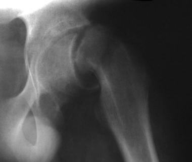
- A cross-table, or true, lateral view can help determine the extent of posterior displacement of the epiphysis, and a "frog-leg" lateral view best shows subtle slipping. (orthofixar.com)
- A modification of this measurement considered a slip to have occurred if the maximal width of the epiphysis lateral to the Klein line differed 2 mm or more from the contralateral hip. (orthofixar.com)
- The lesion usually affects one half of the epiphysis medial or the lateral side hence the term hemimelica in its description. (ispub.com)
Epiphyseal plate1
- 1. Enderle A. Changes in the epiphysis and epiphyseal plate in systemic and genetically-induced diseases. (radiopaedia.org)
Physeal2
- Acute Slipped capital femoral epiphysis: the importance of physeal stability. (ijpoonline.com)
- Rang states that the epiphysis is periarticular and that forces typically causing dislocation in the adult are likely to cause epiphyseal or physeal injury in the child. (medscape.com)
Avascular necrosis3
- If blood supply to the area is compromised, avascular necrosis and collapse of the epiphysis may occur. (msdmanuals.com)
- Chondrolysis and avascular necrosis: complications of slipped capital femoral epiphysis. (ijpoonline.com)
- Radiography revealed epiphysiodesis in coxa vara with resorption of the femoral neck and evidence of avascular necrosis of the capital femoral epiphysis ( Fig. 1 ). (canjsurg.ca)
Physis1
- the epiphysis is a cartilaginous structure that sits atop the physis. (medscape.com)
Skeletally1
- Slipped capital femoral epiphysis in skeletally immature patients. (musc.edu)
Dislocation1
- These techniques include surgical dislocation of the hip, the Ganz Periacetabular Osteotomy, and surgical head realignment after slipped capital femoral epiphysis. (hopkinsmedicine.org)
Slips2
- In slipped capital femoral epiphysis (ih-PIF-eh-siss), the ball slips off the back through the growth plate, almost the way a scoop of ice cream might slip off a cone. (kidshealth.org)
- In early slips, the epiphysis is flush with or below this line. (orthofixar.com)
Valgus1
- [ 2 ] Occasionally, the slip appears to be in a valgus position, with the epiphysis displaced superiorly in relation to the neck. (medscape.com)
Occurs1
- Slipped capital femoral epiphysis occurs in about 2 out of every 100,000 children. (medlineplus.gov)
Hips2
- A slipped capital femoral epiphysis may affect both hips. (medlineplus.gov)
- Radiograph of the hips 1 year after open reduction for slipped capital femoral epiphysis in the left hip. (canjsurg.ca)
Osteotomy2
- Osteotomy through the lesser trochanter for slipped capital femoral epiphysis. (ijpoonline.com)
- Complications after modi ed Dunn osteotomy for the treatment of adolescent slipped capital femoral epiphysis. (ijpoonline.com)
Diagnosis2
- 21. Cowell HR. The significance of early diagnosis and management of slipping capital femoral epiphysis. (ijpoonline.com)
- This is an all too common error in slipping of the capital femoral epiphysis, and may delay the diagnosis for months or years. (canjsurg.ca)
Bones2
- The structure of the epiphysis is more complex in bones that are fused from more than one part during development. (medscape.com)
- Carpal bones, tarsal bones, and the patella are also called epiphysioid bones and are developmentally equivalent to the epiphyses of the long bones. (medscape.com)
Bilateral1
- A reduction in body mass index lowers risk for bilateral clipped capital femoral epiphysis. (ijpoonline.com)
Pediatrics1
- Pediatrics Central , peds.unboundmedicine.com/pedscentral/view/5-Minute-Pediatric-Consult/617760/all/Slipped_Capital_Femoral_Epiphysis. (unboundmedicine.com)
Separation2
- Radiography revealed a complete separation of the capital femoral epiphysis. (canjsurg.ca)
- Our case differs because in our patient there was no history of trauma, complete separation of the epiphysis, delayed treatment, open reduction and a satisfactory functional outcome. (canjsurg.ca)
Neck3
- Slipped capital femoral epiphysis is movement of the femoral neck upward and forward on the femoral epiphysis. (msdmanuals.com)
- The Klein line is a line along the anterior or superior aspect of the femoral neck that normally is intersected by the epiphysis. (orthofixar.com)
- The capital femoral epiphysis appeared completely separated, and the femoral neck revealed cloacae. (canjsurg.ca)
Treatment3
- Nonoperative treatment by traction and spica cast immobilization is rarely used today in Slipped Capital Femoral Epiphysis treatment because of its complications. (orthofixar.com)
- Knee pain as the initial symptom of slipped capital femoral epiphysis: an analysis of initial presentation and treatment. (ijpoonline.com)
- Despite a higher failure rate, compared to total hip arthroplasty in the treatment of primary osteoarthritis, total hip arthroplasty can be considered a feasible option for patients with secondary osteoarthritis of the hip due to slipped capital femoral epiphysis. (openorthopaedicsjournal.com)
Obesity1
- Slipped capital femoral epiphysis: Rising rates with obesity and aboriginality in Southern Australia. (ijpoonline.com)
Growth1
- The cause of slipped capital femoral epiphysis is mostly unknown (idiopathic), although it may be associated with endocrine disorders (e.g., hypothyroidism or administration of growth hormone), renal osteodystrophy, malnutrition and radiation therapy. (canjsurg.ca)
Long1
- 2. Carney BT, Weinstein SL, Noble J. Long-term follow-up of slipped capital femoral epiphysis. (ijpoonline.com)
Children1
- Complete slipping of the capital femoral epiphysis secondary to hematogenous osteomyelitis is hitherto unreported in adolescent children. (canjsurg.ca)
Diseases1
- Slipped Capital Femoral Epiphysis is one of the unique diseases where clinical as well as radiological features are of paramount importance both in planning and prognosis of the disease. (ijpoonline.com)
Stability1
- Slipped Capital Femoral Epiphysis traditionally has been classified according to the stability, the duration of symptoms and the severity of the slip. (orthofixar.com)
Early1
- A slipped capital femoral epiphysis may lead to early degenerative arthritis of the hip if it is not detected early and treated properly. (healthlinkbc.ca)
Patients2
- Imaging modalities in patients with slipped capital femoral epiphysis. (ijpoonline.com)
- All patients with secondary osteoarthritis due to slipped capital femoral epiphysis, treated with total hip arthroplasty between 1987 and 2005, were included in this retrospective study (n=30). (openorthopaedicsjournal.com)
Joint1
- The epiphysis contributes to a joint, compared with an apophysis which is a site of tendon or ligament attachment. (radiopaedia.org)
Concepts1
- Slipped capital femoral epiphysis: Current concepts. (ijpoonline.com)
Report2
- Acute slipped capital femoral epiphysis: review of the literature and report of ten cases. (ijpoonline.com)
- In April 2021, we searched the FDA's "Drug Data and Adverse Event Report System" utilizing the terms "isotretinoin" and "epiphysis premature fusion. (degruyter.com)