Pancreatic Neoplasms
Carcinoma, Acinar Cell
Creosote
Killifishes
Pancreatic Cyst
Carcinoma, Pancreatic Ductal
Cystadenoma, Serous
Endoscopic Ultrasound-Guided Fine Needle Aspiration
Adenocarcinoma, Mucinous
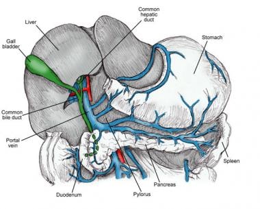
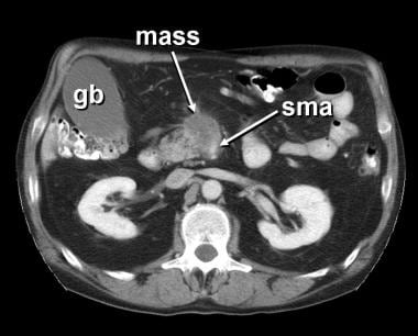
Pancreas
Endosonography
Adenoma, Islet Cell
Pancreatic Ducts
Adenocarcinoma, Papillary
Carcinoma, Papillary
Pancreatitis
Tomography, X-Ray Computed
Neoplasms
Neoplasms, Cystic, Mucinous, and Serous
Neoplasms, Multiple Primary
Neoplasms, Second Primary
Myeloproliferative Disorders
Cystadenoma
Neoplasms, Connective and Soft Tissue
Neoplasms, Plasma Cell
Cystadenoma, Mucinous
Ovarian Neoplasms
Gastrointestinal Neoplasms
Neoplasms, Experimental
Neoplasms, Vascular Tissue
Immunohistochemistry
Neoplasms, Radiation-Induced
Testicular Neoplasms
Neoplasms, Muscle Tissue
Neoplasms, Glandular and Epithelial
Cystadenocarcinoma, Mucinous
Soft Tissue Neoplasms
Hematologic Neoplasms
Neoplasm Proteins
Neoplasms, Adnexal and Skin Appendage
Neoplasm Staging
Vascular Neoplasms
Sweat Gland Neoplasms
Palatal Neoplasms
Antigens, Neoplasm
Cystadenocarcinoma
Rational sequence of tests for pancreatic function. (1/9206)
Of 144 patients with suspected pancreatic disease in whom a 75Se-selenomethionine scan was performed, endoscopic retrograde pancreatography (ERP) was successful in 108 (75%). The final diagnosis is known in 100 patients and has been compared with scan and ERP findings. A normal scan reliably indicated a normal pancreas, but the scan was falsely abnormal in 30%. ERP distinguished between carcinoma and chronic pancreatitis in 84% of cases but was falsely normal in five patients with pancreatic disease. In extrahepatic biliary disease both tests tended to give falsely abnormal results. A sequence of tests to provide a rapid and reliable assessment of pancreatic function should be a radio-isotope scan, followed by ERP if the results of the scan are abnormal, and a Lundh test if the scan is abnormal but the findings on ERP are normal. (+info)Diphtheria toxin effects on human cells in tissue culture. (2/9206)
HeLa cells exposed to a single sublethal concentration of diphtheria toxin were found to have diminished sensitivity when subsequently reexposed to the toxin. Three cells strains exhibiting toxin resistance were developed. In the cells that had previously been exposed to toxin at 0.015 mug/ml, 50% inhibition of protein synthesis required a toxin concentration of 0.3 mug/ml, which is more than 10 times that required in normal HeLa cells. There appears to be a threshold level of diphtheria toxin action. Concentrations of toxin greater than that required for 50% inhibition of protein synthesis (0.01 mug/ml) are associated with cytotoxicity, whereas those below this concentration may not be lethal. Several established human cell lines of both normal and neoplastic origin were tested for their sensitivity to the effects of the toxin. No special sensitivity was observed with the cells of tumor origin. Fifty % inhibition of protein synthesis of HeLa cells was achieved with diphtheria toxin (0.01 mug/ml) as compared to the normal human cell lines tested (0.03 and 0.5 mug/ml) and a cell line derived from a human pancreatic adenocarcinoma (0.2 mug/ml). A human breast carcinoma cell line showed a maximum of 45% inhibition of protein synthesis. This required a diphtheria toxin concentration of 5 mug/ml. These results suggest that different human cell lines show wide variation in their sensitivity to the toxin. (+info)Expression and differential regulation of connective tissue growth factor in pancreatic cancer cells. (3/9206)
CTGF is an immediate early growth responsive gene that has been shown to be a downstream mediator of TGFbeta actions in fibroblasts and vascular endothelial cells. In the present study hCTGF was isolated as immediate early target gene of EGF/TGFalpha in human pancreatic cancer cells by suppression hybridization. CTGF transcripts were found in 13/15 pancreatic cancer cell lines incubated with 10% serum. In 3/7 pancreatic cancer cell lines EGF/TGFalpha induced a significant rise of CTGF transcript levels peaking 1-2 h after the start of treatment. TGFbeta increased CTGF transcript levels in 2/7 pancreatic cancer cell lines after 4 h of treatment and this elevation was sustained after 24 h. Only treatment with TGFbeta was accompanied by a parallel induction of collagen type I transcription. 15/19 human pancreatic cancer tissues were shown to overexpress high levels of CTGF transcripts. CTGF transcript levels in pancreatic cancer tissues and nude mouse xenograft tumors showed a good correlation to the degree of fibrosis. In situ hybridization and the nude mouse experiments revealed that in pancreatic cancer tissues, fibroblasts are the predominant site of CTGF transcription, whereas the tumor cells appear to contribute to a lesser extent. We conclude that CTGF may be of paramount importance for the development of the characteristic desmoplastic reaction in pancreatic cancer tissues. (+info)Detection of liver metastases from pancreatic cancer using FDG PET. (4/9206)
We evaluated the potential of the glucose analog [18F]fluorodeoxyglucose (FDG) as a PET tracer for the hepatic staging in 168 patients designated for resective pancreatic surgery. METHODS: Metastatic liver disease was confirmed or excluded during surgery or with CT follow-up for at least 6 mo. Proven metastases were then retrospectively identified on preoperative CT (gold standard). Hepatic PET scans of all patients were interpreted blindly. Any focal FDG uptake was considered malignant. Both proven hepatic metastases and suspicious hepatic PET lesions were then compared, lesion by lesion, with CT. Standardized uptake values (SUV) and tumor-to-liver ratios (T/L) were determined for the most intense lesion of each patient. RESULTS: Sensitivity of FDG PET was 68% (15 of 22 patients). The lesion detection rate was 97% (28 of 29 metastases) for lesions >1 cm and 43% (16 of 37 metastases) for lesions < or = 1 cm. Specificity was 95% (138 of 146 patients). Six of eight patients with false-positive results had marked intrahepatic cholestasis (versus 3 of 15 patients with true-positive lesions), one had an infrahepatic abscess and one had a right basal pulmonary metastasis. The SUV and T/L were 4.6+/-1.4 and 2.3+/-1.1, respectively, for malignant lesions and 4.1+/-1.5 and 1.9+/-0.3, respectively, for false-positive lesions and therefore are of limited value. CONCLUSION: FDG PET provides reliable hepatic staging for lesions >1 cm. False-positive results are associated with the presence of marked intrahepatic cholestasis. For lesions < or = 1 cm, FDG PET can define malignancy in 43% of suspicious CT lesions in the absence of dilated bile ducts. (+info)Intensive weekly chemotherapy is not effective in advanced pancreatic cancer patients: a report from the Italian Group for the Study of Digestive Tract Cancer (GISCAD). (5/9206)
Twenty-two patients, with locally advanced unresectable and/or metastatic pancreatic carcinoma, received weekly administration of cisplatin 40 mg m(-2), 5-fluorouracil 500 mg m(-2), epidoxorubicin 35 mg m(-2), 6S stereoisomer of leucovorin 250 mg m(-2) and glutathione 1.5 mg m(-2), supported by a daily administration of lenograstim at a dose of 5 microg kg(-1). Nineteen patients were men and three were women. Median age was 63 years (range 47-70). At study entry, pain was present in 15 out of 22 patients (68%) with a mean value of Scott-Huskisson scale of 27.6+/-23.8, whereas a weight loss >10% was present in 15 patients. After eight weekly treatments, three partial responses were achieved for a response rate of 13% (95% CI 0-26%), five patients had stable disease and 14 progressed on therapy. Pain was present in 9 out of 22 patients (40%) with a mean value of Scott-Huskisson scale of 12.3+/-18.4. Eight patients (36%) (three partial response and five stable disease) had a positive weight change. Toxicity was mild: WHO grade III or IV toxicity was recorded in terms of anaemia in 7 out of 188 cycles (3.7%), of neutropenia in 9 out of 188 cycles (4.7%) and of thrombocytopenia in 3 out of 188 cycles (1.5%). Median survival of all patients was 6 months. The outcome of this intensive chemotherapy regimen does not support its use in pancreatic cancer. (+info)Treatment of advanced pancreatic cancer with the long-acting somatostatin analogue lanreotide: in vitro and in vivo results. (6/9206)
Fourteen patients with metastatic pancreatic adenocarcinoma were treated with the long-acting somatostatin (SST) analogue lanreotide. No objective response was obtained, and the median survival was 4 months (range 1.8-7 months). Pancreatic cancer could not be visualized by means of SST-receptor (R) scintigraphy in our patients. In vitro data also demonstrated absence of SSTR2 expression, suggesting pancreatic cancer not to be a potential target for treatment with SST analogues. (+info)Gallstones, cholecystectomy and risk of cancers of the liver, biliary tract and pancreas. (7/9206)
To examine the association between gallstones and cholecystectomy, we conducted a nationwide population-based cohort study in Denmark. Patients with a discharge diagnosis of gallstones from 1977 to 1989 were identified from the Danish National Registry of Patients and followed up for cancer occurrence until death or the end of 1993 by record linkage to the Danish Cancer Registry. Included in the cohort were 60 176 patients, with 471 450 person-years of follow-up. Cancer risks were estimated by standardized incidence ratios (SIRs) and 95% confidence intervals (CIs) stratified by years of follow-up and by cholecystectomy status. Among patients without cholecystectomy, the risks at 5 or more years of follow-up were significantly elevated for cancers of liver (SIR = 2.0, CI = 1.2-3.1) and gallbladder (SIR = 2.7, CI = 1.5-4.4) and near unity for cancers of extrahepatic bile duct (SIR = 1.1), ampulla of Vater (SIR = 1.0) and pancreas (SIR = 1.1). The excess risk of liver cancer was seen only among patients with a history of hepatic disease. Among cholecystectomy patients, the risks at 5 or more years of follow-up declined for cancers of liver (SIR = 1.1) and extrahepatic bile duct (SIR = 0.7), but were elevated for cancers of ampulla of Vater (SIR = 2.0, CI = 1.0-3.7) and pancreas (SIR = 1.3, CI = 1.1-1.6). These findings confirm that gallstone disease increases the risk of gallbladder cancer, whereas cholecystectomy appears to increase the risk of cancers of ampulla of Vater and pancreas. Further research is needed to clarify the carcinogenic risks associated with gallstones and cholecystectomy and to define the mechanisms involved. (+info)Mutations and allelic deletions of the MEN1 gene are associated with a subset of sporadic endocrine pancreatic and neuroendocrine tumors and not restricted to foregut neoplasms. (8/9206)
Endocrine pancreatic tumors (EPT) and neuroendocrine tumors (NET) occur sporadically and rarely in association with multiple endocrine neoplasia type 1 (MEN1). We analyzed the frequency of allelic deletions and mutations of the recently identified MEN1 gene in 53 sporadic tumors including 30 EPT and 23 NET (carcinoids) of different locations and types. Allelic deletion of the MEN1 locus was identified in 18/49 (36.7%) tumors (13/30, 43.3% in EPT and 5/19, 26.3% in NET) and mutations of the MEN1 gene were present in 8/52 (15.3%) tumors (4/30 (13.3%) EPT and 4/22 (18.1%) NET). The somatic mutations were clustered in the 5' region of the coding sequence and most frequently encompassed missense mutations. All tumors with mutations exhibited a loss of the other allele and a wild-type sequence of the MEN1 gene in nontumorous DNA. In one additional patient with a NET of the lung and no clinical signs or history of MEN1, a 5178-9G-->A splice donor site mutation in intron 4 was identified in both the tumor and blood DNA, indicating the presence of a thus far unknown MEN1 syndrome. In most tumor groups the frequency of allelic deletions at 11q13 was 2 to 3 times higher than the frequency of identified MEN1 gene mutations. Some tumor types, including rare forms of EPT and NET of the duodenum and small intestine, exhibited mutations more frequently than other types. Furthermore, somatic mutations were not restricted to foregut tumors but were also detectable in a midgut tumor (15.2% versus 16.6%). Our data indicate that somatic MEN1 gene mutations contribute to a subset of sporadic EPT and NET, including midgut tumors. Because the frequency of mutations varies significantly among the investigated tumor subgroups and allelic deletions are 2 to 3 times more frequently observed, factors other than MEN1 gene inactivation, including other tumor-suppressor genes on 11q13, may also be involved in the tumorigenesis of these neoplasms. (+info)Pancreatic neoplasms refer to abnormal growths in the pancreas that can be benign or malignant. The pancreas is a gland located behind the stomach that produces hormones and digestive enzymes. Pancreatic neoplasms can interfere with the normal functioning of the pancreas, leading to various health complications.
Benign pancreatic neoplasms are non-cancerous growths that do not spread to other parts of the body. They are usually removed through surgery to prevent any potential complications, such as blocking the bile duct or causing pain.
Malignant pancreatic neoplasms, also known as pancreatic cancer, are cancerous growths that can invade and destroy surrounding tissues and organs. They can also spread (metastasize) to other parts of the body, such as the liver, lungs, or bones. Pancreatic cancer is often aggressive and difficult to treat, with a poor prognosis.
There are several types of pancreatic neoplasms, including adenocarcinomas, neuroendocrine tumors, solid pseudopapillary neoplasms, and cystic neoplasms. The specific type of neoplasm is determined through various diagnostic tests, such as imaging studies, biopsies, and blood tests. Treatment options depend on the type, stage, and location of the neoplasm, as well as the patient's overall health and preferences.
Carcinoma, acinar cell is a type of pancreatic cancer that originates in the acinar cells of the pancreas. The acinar cells are responsible for producing digestive enzymes. This type of cancer is relatively rare and accounts for less than 5% of all pancreatic cancers. It typically presents with symptoms such as abdominal pain, weight loss, and jaundice. Treatment options may include surgery, chemotherapy, and radiation therapy.
Creosote is a thick, dark brown or black liquid that has a strong, tarry odor and is produced when wood, coal, or other organic materials are burned or distilled. It is a complex mixture of chemicals, including polycyclic aromatic hydrocarbons (PAHs), which have been linked to an increased risk of cancer.
In the medical context, creosote is not typically used as a treatment for any condition. However, it has been used historically as a topical antiseptic and wound dressing, due to its antibacterial properties. However, its use in this way has largely been replaced by more modern and effective treatments.
It's important to note that creosote is considered a hazardous substance and can be harmful if swallowed, inhaled, or comes into contact with the skin. It can cause irritation to the eyes, skin, and respiratory tract, and prolonged exposure has been linked to an increased risk of cancer. Therefore, it should be handled with care and used only under the supervision of a medical professional.
I'm sorry for any confusion, but "killifishes" is not a term used in medical definitions. Killifishes are a type of small, freshwater or saltwater fish that belong to the family Cyprinodontidae. They are known for their bright colors and patterns, and are often kept as aquarium pets. If you have any questions about biology or animal species, I'd be happy to try to help answer them!
A pancreatectomy is a surgical procedure in which all or part of the pancreas is removed. There are several types of pancreatectomies, including:
* **Total pancreatectomy:** Removal of the entire pancreas, as well as the spleen and nearby lymph nodes. This type of pancreatectomy is usually done for patients with cancer that has spread throughout the pancreas or for those who have had multiple surgeries to remove pancreatic tumors.
* **Distal pancreatectomy:** Removal of the body and tail of the pancreas, as well as nearby lymph nodes. This type of pancreatectomy is often done for patients with tumors in the body or tail of the pancreas.
* **Partial (or segmental) pancreatectomy:** Removal of a portion of the head or body of the pancreas, as well as nearby lymph nodes. This type of pancreatectomy is often done for patients with tumors in the head or body of the pancreas that can be removed without removing the entire organ.
* **Pylorus-preserving pancreaticoduodenectomy (PPPD):** A type of surgery used to treat tumors in the head of the pancreas, as well as other conditions such as chronic pancreatitis. In this procedure, the head of the pancreas, duodenum, gallbladder, and bile duct are removed, but the stomach and lower portion of the esophagus (pylorus) are left in place.
After a pancreatectomy, patients may experience problems with digestion and blood sugar regulation, as the pancreas plays an important role in these functions. Patients may need to take enzyme supplements to help with digestion and may require insulin therapy to manage their blood sugar levels.
A pancreatic cyst is a fluid-filled sac that forms in the pancreas, a gland located behind the stomach that produces enzymes to help with digestion and hormones to regulate blood sugar levels. Pancreatic cysts can be classified into several types, including congenital (present at birth), retention (formed due to blockage of pancreatic ducts), and pseudocysts (formed as a result of injury or inflammation).
While some pancreatic cysts may not cause any symptoms, others can lead to abdominal pain, bloating, nausea, vomiting, or jaundice. Some cysts may also have the potential to become cancerous over time. Therefore, it is essential to monitor and evaluate pancreatic cysts through imaging tests such as ultrasound, CT scan, or MRI, and in some cases, endoscopic ultrasound (EUS) with fine-needle aspiration (FNA) may be necessary for further evaluation.
Treatment options for pancreatic cysts depend on the type, size, location, and symptoms of the cyst, as well as the patient's overall health condition. Some cysts may require surgical removal, while others can be managed with regular monitoring and follow-up care. It is essential to consult a healthcare provider for proper evaluation and management of pancreatic cysts.
Pancreatic ductal carcinoma (PDC) is a specific type of cancer that forms in the ducts that carry digestive enzymes out of the pancreas. It's the most common form of exocrine pancreatic cancer, making up about 90% of all cases.
The symptoms of PDC are often vague and can include abdominal pain, jaundice (yellowing of the skin and eyes), unexplained weight loss, and changes in bowel movements. These symptoms can be similar to those caused by other less serious conditions, which can make diagnosis difficult.
Pancreatic ductal carcinoma is often aggressive and difficult to treat. The prognosis for PDC is generally poor, with a five-year survival rate of only about 9%. Treatment options may include surgery, chemotherapy, radiation therapy, or a combination of these approaches. However, because PDC is often not detected until it has advanced, treatment is frequently focused on palliative care to relieve symptoms and improve quality of life.
A serous cystadenoma is a type of benign tumor that arises from the epithelial cells lining the serous glands, which are glands that produce a watery, lubricating fluid. This type of tumor typically develops in the ovary or the pancreas.
Serous cystadenomas of the ovary are usually filled with a clear, watery fluid and have multiple loculations (compartments). They can vary in size from a few millimeters to several centimeters in diameter. Although these tumors are benign, they can cause symptoms if they become large enough to press on surrounding organs or if they rupture and release their contents into the abdominal cavity.
Serous cystadenomas of the pancreas are less common than ovarian serous cystadenomas. They typically occur in the tail of the pancreas and can range in size from a few millimeters to several centimeters in diameter. These tumors are usually asymptomatic, but they can cause symptoms such as abdominal pain or discomfort if they become large enough to press on surrounding organs.
It is important to note that while serous cystadenomas are generally benign, there is a small risk that they may undergo malignant transformation and develop into a type of cancer known as a serous cystadenocarcinoma. For this reason, it is important for patients with these tumors to be followed closely by a healthcare provider and to have regular imaging studies and/or surgical excision to monitor for any changes in the tumor.
Endoscopic Ultrasound-Guided Fine Needle Aspiration (EUS-FNA) is a medical procedure that combines the use of endoscopy and ultrasound to guide the fine needle aspiration biopsy of internal organs or lesions. This technique allows for the sampling of tissue from inside the gastrointestinal tract and adjacent organs such as the pancreas, lymph nodes, and liver.
During the procedure, an endoscope equipped with an ultrasound probe is inserted through the patient's mouth and advanced to the area of interest. The ultrasound probe provides real-time images of the internal organs and lesions, allowing the physician to guide the fine needle into the target tissue. Once the needle is in position, suction is applied to collect a sample of cells or fluid for further examination under a microscope.
EUS-FNA is commonly used to diagnose and stage various types of cancer, as well as to evaluate other conditions such as pancreatitis, chronic liver disease, and gastrointestinal submucosal tumors. The procedure is generally safe and well-tolerated, with minimal risks and complications. However, as with any medical procedure, there are potential risks and benefits that should be discussed with a healthcare provider before undergoing EUS-FNA.
Adenocarcinoma, mucinous is a type of cancer that begins in the glandular cells that line certain organs and produce mucin, a substance that lubricates and protects tissues. This type of cancer is characterized by the presence of abundant pools of mucin within the tumor. It typically develops in organs such as the colon, rectum, lungs, pancreas, and ovaries.
Mucinous adenocarcinomas tend to have a distinct appearance under the microscope, with large pools of mucin pushing aside the cancer cells. They may also have a different clinical behavior compared to other types of adenocarcinomas, such as being more aggressive or having a worse prognosis in some cases.
It is important to note that while a diagnosis of adenocarcinoma, mucinous can be serious, the prognosis and treatment options may vary depending on several factors, including the location of the cancer, the stage at which it was diagnosed, and the individual's overall health.
The pancreas is a glandular organ located in the abdomen, posterior to the stomach. It has both exocrine and endocrine functions. The exocrine portion of the pancreas consists of acinar cells that produce and secrete digestive enzymes into the duodenum via the pancreatic duct. These enzymes help in the breakdown of proteins, carbohydrates, and fats in food.
The endocrine portion of the pancreas consists of clusters of cells called islets of Langerhans, which include alpha, beta, delta, and F cells. These cells produce and secrete hormones directly into the bloodstream, including insulin, glucagon, somatostatin, and pancreatic polypeptide. Insulin and glucagon are critical regulators of blood sugar levels, with insulin promoting glucose uptake and storage in tissues and glucagon stimulating glycogenolysis and gluconeogenesis to raise blood glucose when it is low.
Endosonography, also known as endoscopic ultrasound (EUS), is a medical procedure that combines endoscopy and ultrasound to obtain detailed images and information about the digestive tract and surrounding organs. An endoscope, which is a flexible tube with a light and camera at its tip, is inserted through the mouth or rectum to reach the area of interest. A high-frequency ultrasound transducer at the tip of the endoscope generates sound waves that bounce off body tissues and create echoes, which are then translated into detailed images by a computer.
Endosonography allows doctors to visualize structures such as the esophageal, stomach, and intestinal walls, lymph nodes, blood vessels, and organs like the pancreas, liver, and gallbladder. It can help diagnose conditions such as tumors, inflammation, and infections, and it can also be used to guide biopsies or fine-needle aspirations of suspicious lesions.
Overall, endosonography is a valuable tool for the diagnosis and management of various gastrointestinal and related disorders.
An islet cell adenoma is a rare, typically benign tumor that develops in the islets of Langerhans, which are clusters of hormone-producing cells in the pancreas. The islets of Langerhans contain several types of cells, including beta cells that produce insulin, alpha cells that produce glucagon, and delta cells that produce somatostatin.
Islet cell adenomas can cause various endocrine disorders depending on the type of hormone-producing cells involved. For example, if the tumor consists mainly of beta cells, it may secrete excessive amounts of insulin, leading to hypoglycemia (low blood sugar). Conversely, if the tumor is composed primarily of alpha cells, it may produce too much glucagon, resulting in hyperglycemia (high blood sugar) and a condition known as glucagonoma.
Islet cell adenomas are usually slow-growing and small but can become quite large in some cases. They are typically diagnosed through imaging tests such as CT scans or MRI, and hormone levels may be measured to determine the type of cells involved. Treatment options include surgical removal of the tumor, medication to manage hormonal imbalances, and, in rare cases, radiofrequency ablation or embolization.
The pancreatic ducts are a set of tubular structures within the pancreas that play a crucial role in the digestive system. The main pancreatic duct, also known as the duct of Wirsung, is responsible for transporting pancreatic enzymes and bicarbonate-rich fluid from the pancreas to the duodenum, which is the first part of the small intestine.
The exocrine portion of the pancreas contains numerous smaller ducts called interlobular ducts and intralobular ducts that merge and ultimately join the main pancreatic duct. This system ensures that the digestive enzymes and fluids produced by the pancreas are effectively delivered to the small intestine, where they aid in the breakdown and absorption of nutrients from food.
In addition to the main pancreatic duct, there is an accessory pancreatic duct, also known as Santorini's duct, which can sometimes join the common bile duct before emptying into the duodenum through a shared opening called the ampulla of Vater. However, in most individuals, the accessory pancreatic duct usually drains into the main pancreatic duct before entering the duodenum.
Adenocarcinoma, papillary is a type of cancer that begins in the glandular cells and grows in a finger-like projection (called a papilla). This type of cancer can occur in various organs, including the lungs, pancreas, thyroid, and female reproductive system. The prognosis and treatment options for papillary adenocarcinoma depend on several factors, such as the location and stage of the tumor, as well as the patient's overall health. It is important to consult with a healthcare professional for an accurate diagnosis and personalized treatment plan.
Carcinoma, papillary is a type of cancer that begins in the cells that line the glandular structures or the lining of organs. In a papillary carcinoma, the cancerous cells grow and form small finger-like projections, called papillae, within the tumor. This type of cancer most commonly occurs in the thyroid gland, but can also be found in other organs such as the lung, breast, and kidney. Papillary carcinoma of the thyroid gland is usually slow-growing and has a good prognosis, especially when it is diagnosed at an early stage.
Adenocarcinoma is a type of cancer that arises from glandular epithelial cells. These cells line the inside of many internal organs, including the breasts, prostate, colon, and lungs. Adenocarcinomas can occur in any of these organs, as well as in other locations where glands are present.
The term "adenocarcinoma" is used to describe a cancer that has features of glandular tissue, such as mucus-secreting cells or cells that produce hormones. These cancers often form glandular structures within the tumor mass and may produce mucus or other substances.
Adenocarcinomas are typically slow-growing and tend to spread (metastasize) to other parts of the body through the lymphatic system or bloodstream. They can be treated with surgery, radiation therapy, chemotherapy, targeted therapy, or a combination of these treatments. The prognosis for adenocarcinoma depends on several factors, including the location and stage of the cancer, as well as the patient's overall health and age.
Pancreatitis is a medical condition characterized by inflammation of the pancreas, a gland located in the abdomen that plays a crucial role in digestion and regulating blood sugar levels. The inflammation can be acute (sudden and severe) or chronic (persistent and recurring), and it can lead to various complications if left untreated.
Acute pancreatitis often results from gallstones or excessive alcohol consumption, while chronic pancreatitis may be caused by long-term alcohol abuse, genetic factors, autoimmune conditions, or metabolic disorders like high triglyceride levels. Symptoms of acute pancreatitis include severe abdominal pain, nausea, vomiting, fever, and increased heart rate, while chronic pancreatitis may present with ongoing abdominal pain, weight loss, diarrhea, and malabsorption issues due to impaired digestive enzyme production. Treatment typically involves supportive care, such as intravenous fluids, pain management, and addressing the underlying cause. In severe cases, hospitalization and surgery may be necessary.
X-ray computed tomography (CT or CAT scan) is a medical imaging method that uses computer-processed combinations of many X-ray images taken from different angles to produce cross-sectional (tomographic) images (virtual "slices") of the body. These cross-sectional images can then be used to display detailed internal views of organs, bones, and soft tissues in the body.
The term "computed tomography" is used instead of "CT scan" or "CAT scan" because the machines take a series of X-ray measurements from different angles around the body and then use a computer to process these data to create detailed images of internal structures within the body.
CT scanning is a noninvasive, painless medical test that helps physicians diagnose and treat medical conditions. CT imaging provides detailed information about many types of tissue including lung, bone, soft tissue and blood vessels. CT examinations can be performed on every part of the body for a variety of reasons including diagnosis, surgical planning, and monitoring of therapeutic responses.
In computed tomography (CT), an X-ray source and detector rotate around the patient, measuring the X-ray attenuation at many different angles. A computer uses this data to construct a cross-sectional image by the process of reconstruction. This technique is called "tomography". The term "computed" refers to the use of a computer to reconstruct the images.
CT has become an important tool in medical imaging and diagnosis, allowing radiologists and other physicians to view detailed internal images of the body. It can help identify many different medical conditions including cancer, heart disease, lung nodules, liver tumors, and internal injuries from trauma. CT is also commonly used for guiding biopsies and other minimally invasive procedures.
In summary, X-ray computed tomography (CT or CAT scan) is a medical imaging technique that uses computer-processed combinations of many X-ray images taken from different angles to produce cross-sectional images of the body. It provides detailed internal views of organs, bones, and soft tissues in the body, allowing physicians to diagnose and treat medical conditions.
Neoplasms are abnormal growths of cells or tissues in the body that serve no physiological function. They can be benign (non-cancerous) or malignant (cancerous). Benign neoplasms are typically slow growing and do not spread to other parts of the body, while malignant neoplasms are aggressive, invasive, and can metastasize to distant sites.
Neoplasms occur when there is a dysregulation in the normal process of cell division and differentiation, leading to uncontrolled growth and accumulation of cells. This can result from genetic mutations or other factors such as viral infections, environmental exposures, or hormonal imbalances.
Neoplasms can develop in any organ or tissue of the body and can cause various symptoms depending on their size, location, and type. Treatment options for neoplasms include surgery, radiation therapy, chemotherapy, immunotherapy, and targeted therapy, among others.
Neoplasms: Neoplasms refer to abnormal growths of tissue that can be benign (non-cancerous) or malignant (cancerous). They occur when the normal control mechanisms that regulate cell growth and division are disrupted, leading to uncontrolled cell proliferation.
Cystic Neoplasms: Cystic neoplasms are tumors that contain fluid-filled sacs or cysts. These tumors can be benign or malignant and can occur in various organs of the body, including the pancreas, ovary, and liver.
Mucinous Neoplasms: Mucinous neoplasms are a type of cystic neoplasm that is characterized by the production of mucin, a gel-like substance produced by certain types of cells. These tumors can occur in various organs, including the ovary, pancreas, and colon. Mucinous neoplasms can be benign or malignant, and malignant forms are often aggressive and have a poor prognosis.
Serous Neoplasms: Serous neoplasms are another type of cystic neoplasm that is characterized by the production of serous fluid, which is a thin, watery fluid. These tumors commonly occur in the ovary and can be benign or malignant. Malignant serous neoplasms are often aggressive and have a poor prognosis.
In summary, neoplasms refer to abnormal tissue growths that can be benign or malignant. Cystic neoplasms contain fluid-filled sacs and can occur in various organs of the body. Mucinous neoplasms produce a gel-like substance called mucin and can also occur in various organs, while serous neoplasms produce thin, watery fluid and commonly occur in the ovary. Both mucinous and serous neoplasms can be benign or malignant, with malignant forms often being aggressive and having a poor prognosis.
Skin neoplasms refer to abnormal growths or tumors in the skin that can be benign (non-cancerous) or malignant (cancerous). They result from uncontrolled multiplication of skin cells, which can form various types of lesions. These growths may appear as lumps, bumps, sores, patches, or discolored areas on the skin.
Benign skin neoplasms include conditions such as moles, warts, and seborrheic keratoses, while malignant skin neoplasms are primarily classified into melanoma, squamous cell carcinoma, and basal cell carcinoma. These three types of cancerous skin growths are collectively known as non-melanoma skin cancers (NMSCs). Melanoma is the most aggressive and dangerous form of skin cancer, while NMSCs tend to be less invasive but more common.
It's essential to monitor any changes in existing skin lesions or the appearance of new growths and consult a healthcare professional for proper evaluation and treatment if needed.
Multiple primary neoplasms refer to the occurrence of more than one primary malignant tumor in an individual, where each tumor is unrelated to the other and originates from separate cells or organs. This differs from metastatic cancer, where a single malignancy spreads to multiple sites in the body. Multiple primary neoplasms can be synchronous (occurring at the same time) or metachronous (occurring at different times). The risk of developing multiple primary neoplasms increases with age and is associated with certain genetic predispositions, environmental factors, and lifestyle choices such as smoking and alcohol consumption.
Kidney neoplasms refer to abnormal growths or tumors in the kidney tissues that can be benign (non-cancerous) or malignant (cancerous). These growths can originate from various types of kidney cells, including the renal tubules, glomeruli, and the renal pelvis.
Malignant kidney neoplasms are also known as kidney cancers, with renal cell carcinoma being the most common type. Benign kidney neoplasms include renal adenomas, oncocytomas, and angiomyolipomas. While benign neoplasms are generally not life-threatening, they can still cause problems if they grow large enough to compromise kidney function or if they undergo malignant transformation.
Early detection and appropriate management of kidney neoplasms are crucial for improving patient outcomes and overall prognosis. Regular medical check-ups, imaging studies, and urinalysis can help in the early identification of these growths, allowing for timely intervention and treatment.
A "second primary neoplasm" is a distinct, new cancer or malignancy that develops in a person who has already had a previous cancer. It is not a recurrence or metastasis of the original tumor, but rather an independent cancer that arises in a different location or organ system. The development of second primary neoplasms can be influenced by various factors such as genetic predisposition, environmental exposures, and previous treatments like chemotherapy or radiation therapy.
It is important to note that the definition of "second primary neoplasm" may vary slightly depending on the specific source or context. In general medical usage, it refers to a new, separate cancer; however, in some research or clinical settings, there might be more precise criteria for defining and diagnosing second primary neoplasms.
Thyroid neoplasms refer to abnormal growths or tumors in the thyroid gland, which can be benign (non-cancerous) or malignant (cancerous). These growths can vary in size and may cause a noticeable lump or nodule in the neck. Thyroid neoplasms can also affect the function of the thyroid gland, leading to hormonal imbalances and related symptoms. The exact causes of thyroid neoplasms are not fully understood, but risk factors include radiation exposure, family history, and certain genetic conditions. It is important to note that most thyroid nodules are benign, but a proper medical evaluation is necessary to determine the nature of the growth and develop an appropriate treatment plan.
Myeloproliferative disorders (MPDs) are a group of rare, chronic blood cancers that originate from the abnormal proliferation or growth of one or more types of blood-forming cells in the bone marrow. These disorders result in an overproduction of mature but dysfunctional blood cells, which can lead to serious complications such as blood clots, bleeding, and organ damage.
There are several subtypes of MPDs, including:
1. Chronic Myeloid Leukemia (CML): A disorder characterized by the overproduction of mature granulocytes (a type of white blood cell) in the bone marrow, leading to an increased number of these cells in the blood. CML is caused by a genetic mutation that results in the formation of the BCR-ABL fusion protein, which drives uncontrolled cell growth and division.
2. Polycythemia Vera (PV): A disorder characterized by the overproduction of all three types of blood cells - red blood cells, white blood cells, and platelets - in the bone marrow. This can lead to an increased risk of blood clots, bleeding, and enlargement of the spleen.
3. Essential Thrombocythemia (ET): A disorder characterized by the overproduction of platelets in the bone marrow, leading to an increased risk of blood clots and bleeding.
4. Primary Myelofibrosis (PMF): A disorder characterized by the replacement of normal bone marrow tissue with scar tissue, leading to impaired blood cell production and anemia, enlargement of the spleen, and increased risk of infections and bleeding.
5. Chronic Neutrophilic Leukemia (CNL): A rare disorder characterized by the overproduction of neutrophils (a type of white blood cell) in the bone marrow, leading to an increased number of these cells in the blood. CNL can lead to an increased risk of infections and organ damage.
MPDs are typically treated with a combination of therapies, including chemotherapy, targeted therapy, immunotherapy, and stem cell transplantation. The choice of treatment depends on several factors, including the subtype of MPD, the patient's age and overall health, and the presence of any comorbidities.
The term "DNA, neoplasm" is not a standard medical term or concept. DNA refers to deoxyribonucleic acid, which is the genetic material present in the cells of living organisms. A neoplasm, on the other hand, is a tumor or growth of abnormal tissue that can be benign (non-cancerous) or malignant (cancerous).
In some contexts, "DNA, neoplasm" may refer to genetic alterations found in cancer cells. These genetic changes can include mutations, amplifications, deletions, or rearrangements of DNA sequences that contribute to the development and progression of cancer. Identifying these genetic abnormalities can help doctors diagnose and treat certain types of cancer more effectively.
However, it's important to note that "DNA, neoplasm" is not a term that would typically be used in medical reports or research papers without further clarification. If you have any specific questions about DNA changes in cancer cells or neoplasms, I would recommend consulting with a healthcare professional or conducting further research on the topic.
Lung neoplasms refer to abnormal growths or tumors in the lung tissue. These tumors can be benign (non-cancerous) or malignant (cancerous). Malignant lung neoplasms are further classified into two main types: small cell lung carcinoma and non-small cell lung carcinoma. Lung neoplasms can cause symptoms such as cough, chest pain, shortness of breath, and weight loss. They are often caused by smoking or exposure to secondhand smoke, but can also occur due to genetic factors, radiation exposure, and other environmental carcinogens. Early detection and treatment of lung neoplasms is crucial for improving outcomes and survival rates.
Parotid neoplasms refer to abnormal growths or tumors in the parotid gland, which is the largest of the salivary glands and is located in front of the ear and extends down the neck. These neoplasms can be benign (non-cancerous) or malignant (cancerous).
Benign parotid neoplasms are typically slow-growing, painless masses that may cause facial asymmetry or difficulty in chewing or swallowing if they become large enough to compress surrounding structures. The most common type of benign parotid tumor is a pleomorphic adenoma.
Malignant parotid neoplasms, on the other hand, are more aggressive and can invade nearby tissues and spread to other parts of the body. They may present as rapidly growing masses that are firm or fixed to surrounding structures. Common types of malignant parotid tumors include mucoepidermoid carcinoma, adenoid cystic carcinoma, and squamous cell carcinoma.
The diagnosis of parotid neoplasms typically involves a thorough clinical evaluation, imaging studies such as CT or MRI scans, and fine-needle aspiration biopsy (FNAB) to determine the nature of the tumor. Treatment options depend on the type, size, and location of the neoplasm but may include surgical excision, radiation therapy, and chemotherapy.
Cystadenoma is a type of benign tumor (not cancerous), which arises from glandular epithelial cells and is covered by a thin layer of connective tissue. These tumors can develop in various locations within the body, including the ovaries, pancreas, and other organs that contain glands.
There are two main types of cystadenomas: serous and mucinous. Serous cystadenomas are filled with a clear or watery fluid, while mucinous cystadenomas contain a thick, gelatinous material. Although they are generally not harmful, these tumors can grow quite large and cause discomfort or other symptoms due to their size or location. In some cases, cystadenomas may undergo malignant transformation and develop into cancerous tumors, known as cystadenocarcinomas. Regular medical follow-up and monitoring are essential for individuals diagnosed with cystadenomas to ensure early detection and treatment of any potential complications.
Neoplasms of connective and soft tissue are abnormal growths or tumors that develop in the body's supportive tissues, such as cartilage, tendons, ligaments, fascia, and fat. These neoplasms can be benign (non-cancerous) or malignant (cancerous).
Benign connective and soft tissue neoplasms include:
- Lipomas: slow-growing, fatty tumors that develop under the skin.
- Fibromas: firm, benign tumors that develop in connective tissue such as tendons or ligaments.
- Nevi (plural of nevus): benign growths made up of cells called melanocytes, which produce pigment.
Malignant connective and soft tissue neoplasms include:
- Sarcomas: a type of cancer that develops in the body's supportive tissues such as muscle, bone, fat, cartilage, or blood vessels. There are many different types of sarcomas, including liposarcoma (fatty tissue), rhabdomyosarcoma (muscle), and osteosarcoma (bone).
- Desmoid tumors: a rare type of benign tumor that can become aggressive and invade surrounding tissues. While not considered cancerous, desmoid tumors can cause significant morbidity due to their tendency to grow and infiltrate nearby structures.
Connective and soft tissue neoplasms can present with various symptoms depending on their location and size. Treatment options include surgery, radiation therapy, chemotherapy, or a combination of these modalities. Regular follow-up care is essential to monitor for recurrence or metastasis (spread) of the tumor.
Plasma cell neoplasms are a type of cancer that originates from plasma cells, which are a type of white blood cell found in the bone marrow. These cells are responsible for producing antibodies to help fight off infections. When plasma cells become cancerous and multiply out of control, they can form a tumor called a plasmacytoma.
There are two main types of plasma cell neoplasms: solitary plasmacytoma and multiple myeloma. Solitary plasmacytoma is a localized tumor that typically forms in the bone, while multiple myeloma is a systemic disease that affects multiple bones and can cause a variety of symptoms such as bone pain, fatigue, and anemia.
Plasma cell neoplasms are diagnosed through a combination of tests, including blood tests, imaging studies, and bone marrow biopsy. Treatment options depend on the stage and extent of the disease, but may include radiation therapy, chemotherapy, and stem cell transplantation.
Appendiceal neoplasms refer to various types of tumors that can develop in the appendix, a small tube-like structure attached to the large intestine. These neoplasms can be benign or malignant and can include:
1. Adenomas: These are benign tumors that arise from the glandular cells lining the appendix. They are usually slow-growing and may not cause any symptoms.
2. Carcinoids: These are neuroendocrine tumors that arise from the hormone-producing cells in the appendix. They are typically small and slow-growing, but some can be aggressive and spread to other parts of the body.
3. Mucinous neoplasms: These are tumors that produce mucin, a slippery substance that can cause the appendix to become distended and filled with mucus. They can be low-grade (less aggressive) or high-grade (more aggressive) and may spread to other parts of the abdomen.
4. Adenocarcinomas: These are malignant tumors that arise from the glandular cells lining the appendix. They are relatively rare but can be aggressive and spread to other parts of the body.
5. Pseudomyxoma peritonei: This is a condition in which mucin produced by an appendiceal neoplasm leaks into the abdominal cavity, causing a jelly-like accumulation of fluid and tissue. It can be caused by both benign and malignant tumors.
Treatment for appendiceal neoplasms depends on the type and stage of the tumor, as well as the patient's overall health. Treatment options may include surgery, chemotherapy, or radiation therapy.
Liver neoplasms refer to abnormal growths in the liver that can be benign or malignant. Benign liver neoplasms are non-cancerous tumors that do not spread to other parts of the body, while malignant liver neoplasms are cancerous tumors that can invade and destroy surrounding tissue and spread to other organs.
Liver neoplasms can be primary, meaning they originate in the liver, or secondary, meaning they have metastasized (spread) to the liver from another part of the body. Primary liver neoplasms can be further classified into different types based on their cell of origin and behavior, including hepatocellular carcinoma, cholangiocarcinoma, and hepatic hemangioma.
The diagnosis of liver neoplasms typically involves a combination of imaging studies, such as ultrasound, CT scan, or MRI, and biopsy to confirm the type and stage of the tumor. Treatment options depend on the type and extent of the neoplasm and may include surgery, radiation therapy, chemotherapy, or liver transplantation.
Mucinous cystadenoma is a type of benign tumor that arises from the epithelial cells lining the mucous membranes of the body. It is most commonly found in the ovary, but can also occur in other locations such as the pancreas or appendix.
Mucinous cystadenomas are characterized by the production of large amounts of mucin, a slippery, gel-like substance that accumulates inside the tumor and causes it to grow into a cystic mass. These tumors can vary in size, ranging from a few centimeters to over 20 centimeters in diameter.
While mucinous cystadenomas are generally benign, they have the potential to become cancerous (mucinous cystadenocarcinoma) if left untreated. Symptoms of mucinous cystadenoma may include abdominal pain or swelling, bloating, and changes in bowel movements or urinary habits. Treatment typically involves surgical removal of the tumor.
Ovarian neoplasms refer to abnormal growths or tumors in the ovary, which can be benign (non-cancerous) or malignant (cancerous). These growths can originate from various cell types within the ovary, including epithelial cells, germ cells, and stromal cells. Ovarian neoplasms are often classified based on their cell type of origin, histological features, and potential for invasive or metastatic behavior.
Epithelial ovarian neoplasms are the most common type and can be further categorized into several subtypes, such as serous, mucinous, endometrioid, clear cell, and Brenner tumors. Some of these epithelial tumors have a higher risk of becoming malignant and spreading to other parts of the body.
Germ cell ovarian neoplasms arise from the cells that give rise to eggs (oocytes) and can include teratomas, dysgerminomas, yolk sac tumors, and embryonal carcinomas. Stromal ovarian neoplasms develop from the connective tissue cells supporting the ovary and can include granulosa cell tumors, thecomas, and fibromas.
It is essential to diagnose and treat ovarian neoplasms promptly, as some malignant forms can be aggressive and potentially life-threatening if not managed appropriately. Regular gynecological exams, imaging studies, and tumor marker tests are often used for early detection and monitoring of ovarian neoplasms. Treatment options may include surgery, chemotherapy, or radiation therapy, depending on the type, stage, and patient's overall health condition.
Endocrine gland neoplasms refer to abnormal growths (tumors) that develop in the endocrine glands. These glands are responsible for producing hormones, which are chemical messengers that regulate various functions and processes in the body. Neoplasms can be benign or malignant (cancerous). Benign neoplasms tend to grow slowly and do not spread to other parts of the body. Malignant neoplasms, on the other hand, can invade nearby tissues and organs and may also metastasize (spread) to distant sites.
Endocrine gland neoplasms can occur in any of the endocrine glands, including:
1. Pituitary gland: located at the base of the brain, it produces several hormones that regulate growth and development, as well as other bodily functions.
2. Thyroid gland: located in the neck, it produces thyroid hormones that regulate metabolism and calcium balance.
3. Parathyroid glands: located near the thyroid gland, they produce parathyroid hormone that regulates calcium levels in the blood.
4. Adrenal glands: located on top of each kidney, they produce hormones such as adrenaline, cortisol, and aldosterone that regulate stress response, metabolism, and blood pressure.
5. Pancreas: located behind the stomach, it produces insulin and glucagon, which regulate blood sugar levels, and digestive enzymes that help break down food.
6. Pineal gland: located in the brain, it produces melatonin, a hormone that regulates sleep-wake cycles.
7. Gonads (ovaries and testicles): located in the pelvis (ovaries) and scrotum (testicles), they produce sex hormones such as estrogen, progesterone, and testosterone that regulate reproductive function and secondary sexual characteristics.
Endocrine gland neoplasms can cause various symptoms depending on the type and location of the tumor. For example, a pituitary gland neoplasm may cause headaches, vision problems, or hormonal imbalances, while an adrenal gland neoplasm may cause high blood pressure, weight gain, or mood changes.
Diagnosis of endocrine gland neoplasms typically involves a combination of medical history, physical examination, imaging studies such as CT or MRI scans, and laboratory tests to measure hormone levels. Treatment options may include surgery, radiation therapy, chemotherapy, or hormonal therapy, depending on the type and stage of the tumor.
Gastrointestinal (GI) neoplasms refer to abnormal growths in the gastrointestinal tract, which can be benign or malignant. The gastrointestinal tract includes the mouth, esophagus, stomach, small intestine, large intestine, rectum, and anus.
Benign neoplasms are non-cancerous growths that do not invade nearby tissues or spread to other parts of the body. They can sometimes be removed completely and may not cause any further health problems.
Malignant neoplasms, on the other hand, are cancerous growths that can invade nearby tissues and organs and spread to other parts of the body through the bloodstream or lymphatic system. These types of neoplasms can be life-threatening if not diagnosed and treated promptly.
GI neoplasms can cause various symptoms, including abdominal pain, bloating, changes in bowel habits, nausea, vomiting, weight loss, and anemia. The specific symptoms may depend on the location and size of the neoplasm.
There are many types of GI neoplasms, including adenocarcinomas, gastrointestinal stromal tumors (GISTs), lymphomas, and neuroendocrine tumors. The diagnosis of GI neoplasms typically involves a combination of medical history, physical examination, imaging studies, and biopsy. Treatment options may include surgery, radiation therapy, chemotherapy, targeted therapy, or immunotherapy.
Experimental neoplasms refer to abnormal growths or tumors that are induced and studied in a controlled laboratory setting, typically in animals or cell cultures. These studies are conducted to understand the fundamental mechanisms of cancer development, progression, and potential treatment strategies. By manipulating various factors such as genetic mutations, environmental exposures, and pharmacological interventions, researchers can gain valuable insights into the complex processes underlying neoplasm formation and identify novel targets for cancer therapy. It is important to note that experimental neoplasms may not always accurately represent human cancers, and further research is needed to translate these findings into clinically relevant applications.
A neoplasm of vascular tissue is an abnormal growth or mass of cells in the blood vessels or lymphatic vessels. These growths can be benign (non-cancerous) or malignant (cancerous). Benign neoplasms, such as hemangiomas and lymphangiomas, are typically not harmful and may not require treatment. However, they can cause symptoms if they grow large enough to press on nearby organs or tissues. Malignant neoplasms, such as angiosarcomas, are cancerous and can invade and destroy surrounding tissue, as well as spread (metastasize) to other parts of the body. Treatment for vascular tissue neoplasms depends on the type, size, location, and stage of the growth, and may include surgery, radiation therapy, chemotherapy, or a combination of these.
Eye neoplasms, also known as ocular tumors or eye cancer, refer to abnormal growths of tissue in the eye. These growths can be benign (non-cancerous) or malignant (cancerous). Eye neoplasms can develop in various parts of the eye, including the eyelid, conjunctiva, cornea, iris, ciliary body, choroid, retina, and optic nerve.
Benign eye neoplasms are typically slow-growing and do not spread to other parts of the body. They may cause symptoms such as vision changes, eye pain, or a noticeable mass in the eye. Treatment options for benign eye neoplasms include monitoring, surgical removal, or radiation therapy.
Malignant eye neoplasms, on the other hand, can grow and spread rapidly to other parts of the body. They may cause symptoms such as vision changes, eye pain, floaters, or flashes of light. Treatment options for malignant eye neoplasms depend on the type and stage of cancer but may include surgery, radiation therapy, chemotherapy, or a combination of these treatments.
It is important to note that early detection and treatment of eye neoplasms can improve outcomes and prevent complications. Regular eye exams with an ophthalmologist are recommended for early detection and prevention of eye diseases, including eye neoplasms.
Immunohistochemistry (IHC) is a technique used in pathology and laboratory medicine to identify specific proteins or antigens in tissue sections. It combines the principles of immunology and histology to detect the presence and location of these target molecules within cells and tissues. This technique utilizes antibodies that are specific to the protein or antigen of interest, which are then tagged with a detection system such as a chromogen or fluorophore. The stained tissue sections can be examined under a microscope, allowing for the visualization and analysis of the distribution and expression patterns of the target molecule in the context of the tissue architecture. Immunohistochemistry is widely used in diagnostic pathology to help identify various diseases, including cancer, infectious diseases, and immune-mediated disorders.
Nose neoplasms refer to abnormal growths or tumors in the nasal cavity or paranasal sinuses. These growths can be benign (non-cancerous) or malignant (cancerous). Benign neoplasms are typically slow-growing and do not spread to other parts of the body, while malignant neoplasms can invade surrounding tissues and have the potential to metastasize.
Nose neoplasms can cause various symptoms such as nasal congestion, nosebleeds, difficulty breathing through the nose, loss of smell, facial pain or numbness, and visual changes if they affect the eye. The diagnosis of nose neoplasms usually involves a combination of physical examination, imaging studies (such as CT or MRI scans), and biopsy to determine the type and extent of the growth. Treatment options depend on the type, size, location, and stage of the neoplasm and may include surgery, radiation therapy, chemotherapy, or a combination of these approaches.
Salivary gland neoplasms refer to abnormal growths or tumors that develop in the salivary glands. These glands are responsible for producing saliva, which helps in digestion, lubrication of food and maintaining oral health. Salivary gland neoplasms can be benign (non-cancerous) or malignant (cancerous).
Benign neoplasms are slow-growing and typically do not spread to other parts of the body. They may cause symptoms such as swelling, painless lumps, or difficulty swallowing if they grow large enough to put pressure on surrounding tissues.
Malignant neoplasms, on the other hand, can be aggressive and have the potential to invade nearby structures and metastasize (spread) to distant organs. Symptoms of malignant salivary gland neoplasms may include rapid growth, pain, numbness, or paralysis of facial nerves.
Salivary gland neoplasms can occur in any of the major salivary glands (parotid, submandibular, and sublingual glands) or in the minor salivary glands located throughout the mouth and throat. The exact cause of these neoplasms is not fully understood, but risk factors may include exposure to radiation, certain viral infections, and genetic predisposition.
Radiation-induced neoplasms are a type of cancer or tumor that develops as a result of exposure to ionizing radiation. Ionizing radiation is radiation with enough energy to remove tightly bound electrons from atoms or molecules, leading to the formation of ions. This type of radiation can damage DNA and other cellular structures, which can lead to mutations and uncontrolled cell growth, resulting in the development of a neoplasm.
Radiation-induced neoplasms can occur after exposure to high levels of ionizing radiation, such as that received during radiation therapy for cancer treatment or from nuclear accidents. The risk of developing a radiation-induced neoplasm depends on several factors, including the dose and duration of radiation exposure, the type of radiation, and the individual's genetic susceptibility to radiation-induced damage.
Radiation-induced neoplasms can take many years to develop after initial exposure to ionizing radiation, and they often occur at the site of previous radiation therapy. Common types of radiation-induced neoplasms include sarcomas, carcinomas, and thyroid cancer. It is important to note that while ionizing radiation can increase the risk of developing cancer, the overall risk is still relatively low, especially when compared to other well-established cancer risk factors such as smoking and exposure to certain chemicals.
Testicular neoplasms are abnormal growths or tumors in the testicle that can be benign (non-cancerous) or malignant (cancerous). They are a type of genitourinary cancer, which affects the reproductive and urinary systems. Testicular neoplasms can occur in men of any age but are most commonly found in young adults between the ages of 15 and 40.
Testicular neoplasms can be classified into two main categories: germ cell tumors and non-germ cell tumors. Germ cell tumors, which arise from the cells that give rise to sperm, are further divided into seminomas and non-seminomas. Seminomas are typically slow-growing and have a good prognosis, while non-seminomas tend to grow more quickly and can spread to other parts of the body.
Non-germ cell tumors are less common than germ cell tumors and include Leydig cell tumors, Sertoli cell tumors, and lymphomas. These tumors can have a variety of clinical behaviors, ranging from benign to malignant.
Testicular neoplasms often present as a painless mass or swelling in the testicle. Other symptoms may include a feeling of heaviness or discomfort in the scrotum, a dull ache in the lower abdomen or groin, and breast enlargement (gynecomastia).
Diagnosis typically involves a physical examination, imaging studies such as ultrasound or CT scan, and blood tests to detect tumor markers. Treatment options depend on the type and stage of the neoplasm but may include surgery, radiation therapy, chemotherapy, or a combination of these modalities. Regular self-examinations of the testicles are recommended for early detection and improved outcomes.
Neoplasms in muscle tissue refer to abnormal and excessive growths of muscle cells that can be benign or malignant. These growths can arise from any of the three types of muscle tissue: skeletal, cardiac, or smooth muscle. Neoplasms in muscle tissue are classified based on their origin, behavior, and histological features.
Benign neoplasms in muscle tissue include leiomyomas (smooth muscle), rhabdomyomas (skeletal muscle), and myxomas (cardiac muscle). These tumors are usually slow-growing and do not invade surrounding tissues or spread to other parts of the body.
Malignant neoplasms in muscle tissue, also known as sarcomas, include leiomyosarcoma (smooth muscle), rhabdomyosarcoma (skeletal muscle), and angiosarcoma (cardiac muscle). These tumors are aggressive, invasive, and have the potential to metastasize to other parts of the body.
Symptoms of neoplasms in muscle tissue depend on their location, size, and type. They may include a painless or painful mass, weakness, fatigue, weight loss, and difficulty swallowing or breathing. Treatment options for neoplasms in muscle tissue include surgery, radiation therapy, chemotherapy, and targeted therapy. The choice of treatment depends on the type, stage, location, and patient's overall health condition.
Neoplasms are abnormal growths of cells or tissues that serve no purpose and can be benign (non-cancerous) or malignant (cancerous). Glandular and epithelial neoplasms refer to specific types of tumors that originate from the glandular and epithelial tissues, respectively.
Glandular neoplasms arise from the glandular tissue, which is responsible for producing and secreting substances such as hormones, enzymes, or other fluids. These neoplasms can be further classified into adenomas (benign) and adenocarcinomas (malignant).
Epithelial neoplasms, on the other hand, develop from the epithelial tissue that lines the outer surfaces of organs and the inner surfaces of cavities. These neoplasms can also be benign or malignant and are classified as papillomas (benign) and carcinomas (malignant).
It is important to note that while both glandular and epithelial neoplasms can become cancerous, not all of them do. However, if they do, the malignant versions can invade surrounding tissues and spread to other parts of the body, making them potentially life-threatening.
Mucinous cystadenocarcinoma is a type of cancer that arises from the mucin-producing cells in the lining of a cyst. It is a subtype of cystadenocarcinoma, which is a malignant tumor that develops within a cyst. Mucinous cystadenocarcinomas are typically found in the ovary or pancreas but can also occur in other organs such as the appendix and the respiratory tract.
These tumors are characterized by the production of large amounts of mucin, a gel-like substance that can accumulate within the cyst and cause it to grow. Mucinous cystadenocarcinomas tend to grow slowly but can become quite large and may eventually spread (metastasize) to other parts of the body if left untreated.
Symptoms of mucinous cystadenocarcinoma depend on the location and size of the tumor, but they may include abdominal pain or discomfort, bloating, changes in bowel movements, or vaginal bleeding. Treatment typically involves surgical removal of the tumor, followed by chemotherapy or radiation therapy to kill any remaining cancer cells. The prognosis for mucinous cystadenocarcinoma depends on several factors, including the stage of the disease at diagnosis and the patient's overall health.
An adenoma is a benign (noncancerous) tumor that develops from glandular epithelial cells. These types of cells are responsible for producing and releasing fluids, such as hormones or digestive enzymes, into the surrounding tissues. Adenomas can occur in various organs and glands throughout the body, including the thyroid, pituitary, adrenal, and digestive systems.
Depending on their location, adenomas may cause different symptoms or remain asymptomatic. Some common examples of adenomas include:
1. Colorectal adenoma (also known as a polyp): These growths occur in the lining of the colon or rectum and can develop into colorectal cancer if left untreated. Regular screenings, such as colonoscopies, are essential for early detection and removal of these polyps.
2. Thyroid adenoma: This type of adenoma affects the thyroid gland and may result in an overproduction or underproduction of hormones, leading to conditions like hyperthyroidism (overactive thyroid) or hypothyroidism (underactive thyroid).
3. Pituitary adenoma: These growths occur in the pituitary gland, which is located at the base of the brain and controls various hormonal functions. Depending on their size and location, pituitary adenomas can cause vision problems, headaches, or hormonal imbalances that affect growth, reproduction, and metabolism.
4. Liver adenoma: These rare benign tumors develop in the liver and may not cause any symptoms unless they become large enough to press on surrounding organs or structures. In some cases, liver adenomas can rupture and cause internal bleeding.
5. Adrenal adenoma: These growths occur in the adrenal glands, which are located above the kidneys and produce hormones that regulate stress responses, metabolism, and blood pressure. Most adrenal adenomas are nonfunctioning, meaning they do not secrete excess hormones. However, functioning adrenal adenomas can lead to conditions like Cushing's syndrome or Conn's syndrome, depending on the type of hormone being overproduced.
It is essential to monitor and manage benign tumors like adenomas to prevent potential complications, such as rupture, bleeding, or hormonal imbalances. Treatment options may include surveillance with imaging studies, medication to manage hormonal issues, or surgical removal of the tumor in certain cases.
Soft tissue neoplasms refer to abnormal growths or tumors that develop in the soft tissues of the body. Soft tissues include muscles, tendons, ligaments, fascia, nerves, blood vessels, fat, and synovial membranes (the thin layer of cells that line joints and tendons). Neoplasms can be benign (non-cancerous) or malignant (cancerous), and their behavior and potential for spread depend on the specific type of neoplasm.
Benign soft tissue neoplasms are typically slow-growing, well-circumscribed, and rarely spread to other parts of the body. They can often be removed surgically with a low risk of recurrence. Examples of benign soft tissue neoplasms include lipomas (fat tumors), schwannomas (nerve sheath tumors), and hemangiomas (blood vessel tumors).
Malignant soft tissue neoplasms, on the other hand, can grow rapidly, invade surrounding tissues, and may metastasize (spread) to distant parts of the body. They are often more difficult to treat than benign neoplasms and require a multidisciplinary approach, including surgery, radiation therapy, and chemotherapy. Examples of malignant soft tissue neoplasms include sarcomas, such as rhabdomyosarcoma (arising from skeletal muscle), leiomyosarcoma (arising from smooth muscle), and angiosarcoma (arising from blood vessels).
It is important to note that soft tissue neoplasms can occur in any part of the body, and their diagnosis and treatment require a thorough evaluation by a healthcare professional with expertise in this area.
Hematologic neoplasms, also known as hematological malignancies, are a group of diseases characterized by the uncontrolled growth and accumulation of abnormal blood cells or bone marrow cells. These disorders can originate from the myeloid or lymphoid cell lines, which give rise to various types of blood cells, including red blood cells, white blood cells, and platelets.
Hematologic neoplasms can be broadly classified into three categories:
1. Leukemias: These are cancers that primarily affect the bone marrow and blood-forming tissues. They result in an overproduction of abnormal white blood cells, which interfere with the normal functioning of the blood and immune system. There are several types of leukemia, including acute lymphoblastic leukemia (ALL), chronic lymphocytic leukemia (CLL), acute myeloid leukemia (AML), and chronic myeloid leukemia (CML).
2. Lymphomas: These are cancers that develop from the lymphatic system, which is a part of the immune system responsible for fighting infections. Lymphomas can affect lymph nodes, spleen, bone marrow, and other organs. The two main types of lymphoma are Hodgkin lymphoma (HL) and non-Hodgkin lymphoma (NHL).
3. Myelomas: These are cancers that arise from the plasma cells, a type of white blood cell responsible for producing antibodies. Multiple myeloma is the most common type of myeloma, characterized by an excessive proliferation of malignant plasma cells in the bone marrow, leading to the production of abnormal amounts of monoclonal immunoglobulins (M proteins) and bone destruction.
Hematologic neoplasms can have various symptoms, such as fatigue, weakness, frequent infections, easy bruising or bleeding, weight loss, swollen lymph nodes, and bone pain. The diagnosis typically involves a combination of medical history, physical examination, laboratory tests, imaging studies, and sometimes bone marrow biopsy. Treatment options depend on the type and stage of the disease and may include chemotherapy, radiation therapy, targeted therapy, immunotherapy, stem cell transplantation, or a combination of these approaches.
A neoplasm is a tumor or growth that is formed by an abnormal and excessive proliferation of cells, which can be benign or malignant. Neoplasm proteins are therefore any proteins that are expressed or produced in these neoplastic cells. These proteins can play various roles in the development, progression, and maintenance of neoplasms.
Some neoplasm proteins may contribute to the uncontrolled cell growth and division seen in cancer, such as oncogenic proteins that promote cell cycle progression or inhibit apoptosis (programmed cell death). Others may help the neoplastic cells evade the immune system, allowing them to proliferate undetected. Still others may be involved in angiogenesis, the formation of new blood vessels that supply the tumor with nutrients and oxygen.
Neoplasm proteins can also serve as biomarkers for cancer diagnosis, prognosis, or treatment response. For example, the presence or level of certain neoplasm proteins in biological samples such as blood or tissue may indicate the presence of a specific type of cancer, help predict the likelihood of cancer recurrence, or suggest whether a particular therapy will be effective.
Overall, understanding the roles and behaviors of neoplasm proteins can provide valuable insights into the biology of cancer and inform the development of new diagnostic and therapeutic strategies.
Uterine neoplasms refer to abnormal growths in the uterus, which can be benign (non-cancerous) or malignant (cancerous). These growths can originate from different types of cells within the uterus, leading to various types of uterine neoplasms. The two main categories of uterine neoplasms are endometrial neoplasms and uterine sarcomas.
Endometrial neoplasms develop from the endometrium, which is the inner lining of the uterus. Most endometrial neoplasms are classified as endometrioid adenocarcinomas, arising from glandular cells in the endometrium. Other types include serous carcinoma, clear cell carcinoma, and mucinous carcinoma.
Uterine sarcomas, on the other hand, are less common and originate from the connective tissue (stroma) or muscle (myometrium) of the uterus. Uterine sarcomas can be further divided into several subtypes, such as leiomyosarcoma, endometrial stromal sarcoma, and undifferentiated uterine sarcoma.
Uterine neoplasms can cause various symptoms, including abnormal vaginal bleeding or discharge, pelvic pain, and difficulty urinating or having bowel movements. The diagnosis typically involves a combination of imaging tests (such as ultrasound, CT, or MRI scans) and tissue biopsies to determine the type and extent of the neoplasm. Treatment options depend on the type, stage, and patient's overall health but may include surgery, radiation therapy, chemotherapy, or hormone therapy.
Intestinal neoplasms refer to abnormal growths in the tissues of the intestines, which can be benign or malignant. These growths are called neoplasms and they result from uncontrolled cell division. In the case of intestinal neoplasms, these growths occur in the small intestine, large intestine (colon), rectum, or appendix.
Benign intestinal neoplasms are not cancerous and often do not invade surrounding tissues or spread to other parts of the body. However, they can still cause problems if they grow large enough to obstruct the intestines or cause bleeding. Common types of benign intestinal neoplasms include polyps, leiomyomas, and lipomas.
Malignant intestinal neoplasms, on the other hand, are cancerous and can invade surrounding tissues and spread to other parts of the body. The most common type of malignant intestinal neoplasm is adenocarcinoma, which arises from the glandular cells lining the inside of the intestines. Other types of malignant intestinal neoplasms include lymphomas, sarcomas, and carcinoid tumors.
Symptoms of intestinal neoplasms can vary depending on their size, location, and type. Common symptoms include abdominal pain, bloating, changes in bowel habits, rectal bleeding, weight loss, and fatigue. If you experience any of these symptoms, it is important to seek medical attention promptly.
Neoplasms, adnexal and skin appendage refer to abnormal growths or tumors that develop in the sweat glands, hair follicles, or other structures associated with the skin. These growths can be benign (non-cancerous) or malignant (cancerous), and they can occur anywhere on the body.
Adnexal neoplasms are tumors that arise from the sweat glands or hair follicles, including the sebaceous glands, eccrine glands, and apocrine glands. These tumors can range in size and severity, and they may cause symptoms such as pain, itching, or changes in the appearance of the skin.
Skin appendage neoplasms are similar to adnexal neoplasms, but they specifically refer to tumors that arise from structures such as hair follicles, nails, and sweat glands. Examples of skin appendage neoplasms include pilomatricomas (tumors of the hair follicle), trichilemmomas (tumors of the outer root sheath of the hair follicle), and sebaceous adenomas (tumors of the sebaceous glands).
It is important to note that while many adnexal and skin appendage neoplasms are benign, some can be malignant and may require aggressive treatment. If you notice any unusual growths or changes in your skin, it is important to consult with a healthcare professional for further evaluation and care.
Neoplasm staging is a systematic process used in medicine to describe the extent of spread of a cancer, including the size and location of the original (primary) tumor and whether it has metastasized (spread) to other parts of the body. The most widely accepted system for this purpose is the TNM classification system developed by the American Joint Committee on Cancer (AJCC) and the Union for International Cancer Control (UICC).
In this system, T stands for tumor, and it describes the size and extent of the primary tumor. N stands for nodes, and it indicates whether the cancer has spread to nearby lymph nodes. M stands for metastasis, and it shows whether the cancer has spread to distant parts of the body.
Each letter is followed by a number that provides more details about the extent of the disease. For example, a T1N0M0 cancer means that the primary tumor is small and has not spread to nearby lymph nodes or distant sites. The higher the numbers, the more advanced the cancer.
Staging helps doctors determine the most appropriate treatment for each patient and estimate the patient's prognosis. It is an essential tool for communication among members of the healthcare team and for comparing outcomes of treatments in clinical trials.
Vascular neoplasms are a type of tumor that develops from cells that line the blood vessels or lymphatic vessels. These tumors can be benign (non-cancerous) or malignant (cancerous). Benign vascular neoplasms, such as hemangiomas and lymphangiomas, are usually harmless and may not require treatment unless they cause symptoms or complications. Malignant vascular neoplasms, on the other hand, are known as angiosarcomas and can be aggressive, spreading to other parts of the body and potentially causing serious health problems.
Angiosarcomas can develop in any part of the body but are most commonly found in the skin, particularly in areas exposed to radiation or chronic lymph edema. They can also occur in the breast, liver, spleen, and heart. Treatment for vascular neoplasms depends on the type, location, size, and stage of the tumor, as well as the patient's overall health. Treatment options may include surgery, radiation therapy, chemotherapy, or a combination of these approaches.
Sweat gland neoplasms are abnormal growths that develop in the sweat glands. These growths can be benign (noncancerous) or malignant (cancerous). Benign sweat gland neoplasms include hidradenomas and syringomas, which are usually slow-growing and cause little to no symptoms. Malignant sweat gland neoplasms, also known as sweat gland carcinomas, are rare but aggressive cancers that can spread to other parts of the body. They may cause symptoms such as a lump or mass under the skin, pain, swelling, and redness. Treatment typically involves surgical removal of the growth.
Lymphoma is a type of cancer that originates from the white blood cells called lymphocytes, which are part of the immune system. These cells are found in various parts of the body such as the lymph nodes, spleen, bone marrow, and other organs. Lymphoma can be classified into two main types: Hodgkin lymphoma (HL) and non-Hodgkin lymphoma (NHL).
HL is characterized by the presence of a specific type of abnormal lymphocyte called Reed-Sternberg cells, while NHL includes a diverse group of lymphomas that lack these cells. The symptoms of lymphoma may include swollen lymph nodes, fever, night sweats, weight loss, and fatigue.
The exact cause of lymphoma is not known, but it is believed to result from genetic mutations in the lymphocytes that lead to uncontrolled cell growth and division. Exposure to certain viruses, chemicals, and radiation may increase the risk of developing lymphoma. Treatment options for lymphoma depend on various factors such as the type and stage of the disease, age, and overall health of the patient. Common treatments include chemotherapy, radiation therapy, immunotherapy, and stem cell transplantation.
Bone neoplasms are abnormal growths or tumors that develop in the bone. They can be benign (non-cancerous) or malignant (cancerous). Benign bone neoplasms do not spread to other parts of the body and are rarely a threat to life, although they may cause problems if they grow large enough to press on surrounding tissues or cause fractures. Malignant bone neoplasms, on the other hand, can invade and destroy nearby tissue and may spread (metastasize) to other parts of the body.
There are many different types of bone neoplasms, including:
1. Osteochondroma - a benign tumor that develops from cartilage and bone
2. Enchondroma - a benign tumor that forms in the cartilage that lines the inside of the bones
3. Chondrosarcoma - a malignant tumor that develops from cartilage
4. Osteosarcoma - a malignant tumor that develops from bone cells
5. Ewing sarcoma - a malignant tumor that develops in the bones or soft tissues around the bones
6. Giant cell tumor of bone - a benign or occasionally malignant tumor that develops from bone tissue
7. Fibrosarcoma - a malignant tumor that develops from fibrous tissue in the bone
The symptoms of bone neoplasms vary depending on the type, size, and location of the tumor. They may include pain, swelling, stiffness, fractures, or limited mobility. Treatment options depend on the type and stage of the tumor but may include surgery, radiation therapy, chemotherapy, or a combination of these treatments.
Palatal neoplasms refer to abnormal growths or tumors that occur on the palate, which is the roof of the mouth. These growths can be benign (non-cancerous) or malignant (cancerous). Benign neoplasms are typically slower growing and less likely to spread, while malignant neoplasms are more aggressive and can invade nearby tissues and organs.
Palatal neoplasms can have various causes, including genetic factors, environmental exposures, and viral infections. They may present with symptoms such as mouth pain, difficulty swallowing, swelling or lumps in the mouth, bleeding, or numbness in the mouth or face.
The diagnosis of palatal neoplasms typically involves a thorough clinical examination, imaging studies, and sometimes biopsy to determine the type and extent of the growth. Treatment options depend on the type, size, location, and stage of the neoplasm but may include surgery, radiation therapy, chemotherapy, or a combination of these approaches. Regular follow-up care is essential to monitor for recurrence or spread of the neoplasm.
Neoplasms are abnormal growths of cells or tissues in the body that can be benign (non-cancerous) or malignant (cancerous). When referring to "Complex and Mixed Neoplasms," it is typically used in the context of histopathology, where it describes tumors with a mixture of different types of cells or growth patterns.
A complex neoplasm usually contains areas with various architectural patterns, cell types, or both, making its classification challenging. It may require extensive sampling and careful examination to determine its nature and behavior. These neoplasms can be either benign or malignant, depending on the specific characteristics of the tumor cells and their growth pattern.
A mixed neoplasm, on the other hand, is a tumor that contains more than one type of cell or tissue component, often arising from different germ layers (the three primary layers of embryonic development: ectoderm, mesoderm, and endoderm). A common example of a mixed neoplasm is a teratoma, which can contain tissues derived from all three germ layers, such as skin, hair, teeth, bone, and muscle. Mixed neoplasms can also be benign or malignant, depending on the specific components of the tumor.
It's important to note that the classification and behavior of complex and mixed neoplasms can vary significantly based on their location in the body, cellular composition, and other factors. Accurate diagnosis typically requires a thorough examination by an experienced pathologist and may involve additional tests, such as immunohistochemistry or molecular analysis, to determine the appropriate treatment and management strategies.
Neoplasm antigens, also known as tumor antigens, are substances that are produced by cancer cells (neoplasms) and can stimulate an immune response. These antigens can be proteins, carbohydrates, or other molecules that are either unique to the cancer cells or are overexpressed or mutated versions of normal cellular proteins.
Neoplasm antigens can be classified into two main categories: tumor-specific antigens (TSAs) and tumor-associated antigens (TAAs). TSAs are unique to cancer cells and are not expressed by normal cells, while TAAs are present at low levels in normal cells but are overexpressed or altered in cancer cells.
TSAs can be further divided into viral antigens and mutated antigens. Viral antigens are produced when cancer is caused by a virus, such as human papillomavirus (HPV) in cervical cancer. Mutated antigens are the result of genetic mutations that occur during cancer development and are unique to each patient's tumor.
Neoplasm antigens play an important role in the immune response against cancer. They can be recognized by the immune system, leading to the activation of immune cells such as T cells and natural killer (NK) cells, which can then attack and destroy cancer cells. However, cancer cells often develop mechanisms to evade the immune response, allowing them to continue growing and spreading.
Understanding neoplasm antigens is important for the development of cancer immunotherapies, which aim to enhance the body's natural immune response against cancer. These therapies include checkpoint inhibitors, which block proteins that inhibit T cell activation, and therapeutic vaccines, which stimulate an immune response against specific tumor antigens.
Mandibular neoplasms refer to abnormal growths or tumors that develop in the mandible, which is the lower jawbone. These growths can be benign (non-cancerous) or malignant (cancerous). Benign neoplasms are typically slow-growing and rarely spread to other parts of the body, while malignant neoplasms can invade surrounding tissues and may metastasize (spread) to distant sites.
Mandibular neoplasms can have various causes, including genetic mutations, exposure to certain chemicals or radiation, and infection with certain viruses. The symptoms of mandibular neoplasms may include swelling or pain in the jaw, difficulty chewing or speaking, numbness in the lower lip or chin, loose teeth, and/or a lump or mass in the mouth or neck.
The diagnosis of mandibular neoplasms typically involves a thorough clinical examination, imaging studies such as X-rays, CT scans, or MRI scans, and sometimes a biopsy to confirm the type and extent of the tumor. Treatment options depend on the type, stage, and location of the neoplasm, and may include surgery, radiation therapy, chemotherapy, or a combination of these approaches. Regular follow-up care is essential to monitor for recurrence or metastasis.
Cystadenocarcinoma is a type of tumor that arises from the epithelial lining of a cyst, and it has the potential to invade surrounding tissues and spread (metastasize) to other parts of the body. It typically affects glandular organs such as the ovaries, pancreas, and salivary glands.
Cystadenocarcinomas can be classified into two types: serous and mucinous. Serous cystadenocarcinomas produce a watery fluid, while mucinous cystadenocarcinomas produce a thick, mucus-like fluid. Both types of tumors can be benign or malignant, but malignant cystadenocarcinomas are more aggressive and have a higher risk of metastasis.
Symptoms of cystadenocarcinoma depend on the location and size of the tumor. In some cases, there may be no symptoms until the tumor has grown large enough to cause pain or other problems. Treatment typically involves surgical removal of the tumor, along with any affected surrounding tissue. Chemotherapy and radiation therapy may also be used in some cases to help prevent recurrence or spread of the cancer.
Bile duct neoplasms, also known as cholangiocarcinomas, refer to a group of malignancies that arise from the bile ducts. These are the tubes that carry bile from the liver to the gallbladder and small intestine. Bile duct neoplasms can be further classified based on their location as intrahepatic (within the liver), perihilar (at the junction of the left and right hepatic ducts), or distal (in the common bile duct).
These tumors are relatively rare, but their incidence has been increasing in recent years. They can cause a variety of symptoms, including jaundice, abdominal pain, weight loss, and fever. The diagnosis of bile duct neoplasms typically involves imaging studies such as CT or MRI scans, as well as blood tests to assess liver function. In some cases, a biopsy may be necessary to confirm the diagnosis.
Treatment options for bile duct neoplasms depend on several factors, including the location and stage of the tumor, as well as the patient's overall health. Surgical resection is the preferred treatment for early-stage tumors, while chemotherapy and radiation therapy may be used in more advanced cases. For patients who are not candidates for surgery, palliative treatments such as stenting or bypass procedures may be recommended to relieve symptoms and improve quality of life.
Neoplasm invasiveness is a term used in pathology and oncology to describe the aggressive behavior of cancer cells as they invade surrounding tissues and organs. This process involves the loss of cell-to-cell adhesion, increased motility and migration, and the ability of cancer cells to degrade the extracellular matrix (ECM) through the production of enzymes such as matrix metalloproteinases (MMPs).
Invasive neoplasms are cancers that have spread beyond the original site where they first developed and have infiltrated adjacent tissues or structures. This is in contrast to non-invasive or in situ neoplasms, which are confined to the epithelial layer where they originated and have not yet invaded the underlying basement membrane.
The invasiveness of a neoplasm is an important prognostic factor in cancer diagnosis and treatment, as it can indicate the likelihood of metastasis and the potential effectiveness of various therapies. In general, more invasive cancers are associated with worse outcomes and require more aggressive treatment approaches.
Thymus neoplasms are abnormal growths in the thymus gland that result from uncontrolled cell division. The term "neoplasm" refers to any new and abnormal growth of tissue, also known as a tumor. Thymus neoplasms can be benign or malignant (cancerous).
Malignant thymus neoplasms are called thymomas or thymic carcinomas. Thymomas are the most common type and tend to grow slowly, invading nearby tissues and organs. They can also spread (metastasize) to other parts of the body. Thymic carcinomas are rarer and more aggressive, growing and spreading more quickly than thymomas.
Symptoms of thymus neoplasms may include coughing, chest pain, difficulty breathing, or swelling in the neck or upper chest. Treatment options for thymus neoplasms depend on the type, size, location, and stage of the tumor, as well as the patient's overall health. Treatment may include surgery, radiation therapy, chemotherapy, or a combination of these approaches.
Splenic neoplasms refer to abnormal growths or tumors in the spleen, which can be benign (non-cancerous) or malignant (cancerous). These growths can arise from various cell types present within the spleen, including hematopoietic cells (red and white blood cells, platelets), stromal cells (supporting tissue), or lymphoid cells (part of the immune system).
There are several types of splenic neoplasms:
1. Hematologic malignancies: These are cancers that affect the blood and bone marrow, such as leukemias, lymphomas, and multiple myeloma. They often involve the spleen, causing enlargement (splenomegaly) and neoplastic infiltration of splenic tissue.
2. Primary splenic tumors: These are rare and include benign lesions like hemangiomas, lymphangiomas, and hamartomas, as well as malignant tumors such as angiosarcoma, littoral cell angiosarcoma, and primary splenic lymphoma.
3. Metastatic splenic tumors: These occur when cancer cells from other primary sites spread (metastasize) to the spleen. Common sources of metastasis include lung, breast, colon, and ovarian cancers, as well as melanomas and sarcomas.
Symptoms of splenic neoplasms may vary depending on the type and extent of the disease but often include abdominal pain or discomfort, fatigue, weight loss, and anemia. Diagnosis typically involves imaging studies (such as ultrasound, CT, or MRI scans) and sometimes requires a biopsy for confirmation. Treatment options depend on the type of neoplasm and may include surgery, chemotherapy, radiation therapy, targeted therapy, or immunotherapy.
 Pancreatic mucinous cystic neoplasm
Ralph H. Hruban
Markus Büchler
TUM School of Medicine
University of Tokyo
Intraepithelial neoplasia
Mucinous cystic neoplasm
David B. Adams
Pancreatic neuroendocrine tumor
MIRN21
Hepatoid tumor
Pancreatic mucinous cystadenoma
Intraductal papillary mucinous neoplasm
HHIP
Insulinoma
Pancreatic cancer
Ludwig Maximilian University of Munich
Solid pseudopapillary tumour
Phakomatosis
Pancreatic tumor
Malassezia
Blastoma
Pancreatic pseudocyst
Pancreatic serous cystadenoma
Hamartoma
Saliva testing
Lomustine
Ovarian serous cystadenoma
Neuroendocrine tumor
Endocrine gland neoplasm
Carney complex
Pancreatic cyst
DUSP6
MCN
Pancreatic disease
Pancreatic mucinous cystic neoplasm - Wikipedia
 Pancreatic Neuroendocrine Neoplasms (pNENs) - Symptoms, Causes, Treatment | NORD
 Candidate protein biomarkers in pancreatic neuroendocrine neoplasms grade 3
Treatment Strategy for Pancreatic Neuroendocrine Neoplasms (pNEN)
 RePub, Erasmus University Repository:
Unmet Needs in Functional and Nonfunctional Pancreatic Neuroendocrine Neoplasms
RePub, Erasmus University Repository:
Unmet Needs in Functional and Nonfunctional Pancreatic Neuroendocrine Neoplasms
 Adrenal uptake in PET/CT in a patient with pancreatic neoplasm: Not always metastasis | Gastroenterología y Hepatología ...
 Combining radiomic features with a miRNA classifier may improve prediction of malignant pathology for pancreatic intraductal...
 Pancreatic Neoplasms: Lund University - Expertscape.com
 Pathology and Genetics of Pancreatic Neoplasms - PDF Free Download
Glypican-3 expression in gastrointestinal and pancreatic epithelial neoplasms<...
 Pancreatic resections for cystic neoplasms: from the surgeon's presumption to the pathologist's reality
 Pancreatic neoplasm: a unique size and presentation - Pourmorteza - Translational Gastrointestinal Cancer
 Normal pancreatic epithelium progresses through various stages of pancreatic intraepithelial neoplasms - Gene editing with...
 Pancreatic Cancer: Practice Essentials, Pathophysiology, Etiology
 Review Articles
| British Journal of Cancer
Review Articles
| British Journal of Cancer
 Clinicopathological features of pancreatic mucinous cystic neoplasm and influencing factors for its malignancy
 Page 1 | Search Results | Onkologie | Karger Publishers
![New staging classification for pancreatic neuroendocrine neoplasms combining TNM stage and WHO grade classification []. |...](data:image/png;base64,iVBORw0KGgoAAAANSUhEUgAAABAAAAAQCAYAAAAf8/9hAAACpklEQVQ4jaXTe2iNcRzH8ffz7Jzncp4ZZyZjZ6bmEvPPxrDIJSnJZZJIbn8Qk1CI2pQ7JX8h5fKHW4o/3EuaWnJvrUNqiFZssTWcs83z7DnPc87XH88Ziv98//z1+7369v38vvCfpQBY0w6LeHb2RCHl+fg9vaiGxuyq0ZQMzef0tSeoOSqGEQaR4Go4QghAPBvxHRzbBddjUGGUhXMmULN0KhVlxQDMLC+iZt9VEt+6MPNMCIwAAEj7aSrGxFg5r5KVCyoZGM2lx3Y5dbGB6jnlLJs7nvIxMZbvOE9TvAUjagXtAygVu2Tk3P3ieb6IiPjpjBw9Vy+F02uFYWulaEadnLn2WEREJCOyetdFYdRGMSfXSQ4ABVV7Jo4rYtWCiVyvf8nizWe4cuMZdkYwBlh863a4fa+JR/EWyscWs37pFAxD4/7T96hB/2lKhuYDcORsPW+aWzHz+6FrIRDB1MMY0Vwe3I9Td+IuALMmjyIjZAGBkiFRACKmBqb+77hyDQw9DED8TRukvCygKpQWFwCQyUb0zxIwtQD40NoZPAVQdY3S2EAAkj9cfo/3b0DJ+h8/fwdVCYA8S6d0WNBBbHB/6LJJeWmUPkiB3pQPPQ7RPDMAviQglBMAtuNSs/cqyW6HO8fXcaB2CZYWwk78IOWlcb72UDjA4tihFRzcOg+Ato4EaigIkUjVbmHEBimbf1Diza0iIvKupV2qN52W/MrtsuPoDUl22dJXl2+9EHP8NtEn1YnSB0i6F6fbwYronKxdwupFkwCwbZdIRKe9s5sLN59z6W4jr962oZs6OVrk91dGhEg/A8f1WbPzPI3Nnzi8ZT7NLe2cuPyQWw2vSXQmwdAwLIM/xvP3NmYyGXptl+GxAlo7kvi2S9gyCIfVX0sEwTb+BBBpI5QgousUAAAAAElFTkSuQmCC) New staging classification for pancreatic neuroendocrine neoplasms combining TNM stage and WHO grade classification []. |...
New staging classification for pancreatic neuroendocrine neoplasms combining TNM stage and WHO grade classification []. |...
Pancreatitis | Medscape
Quality assessment of the guidelines on cystic neoplasms of the pancreas
Pancreatic intraductal tubulopapillary neoplasm is genetically distinct from intraductal papillary mucinous neoplasm and ductal...
A pilot study of in vivo identification of pancreatic cystic neoplasms with needle-based confocal laser endomicroscopy under...
Pancreatic Cancer Medication: Antineoplastic agents
Pathways of Progression From Intraductal Papillary Mucinous Neoplasm to Pancreatic Ductal Adenocarcinoma Based on Molecular...
 Pregnancy following Radical Resection of Solid Pseudopapillary Tumor of the Pancreas
Contrast-enhanced harmonic endoscopic ultrasound using time-intensity curve analysis predicts pathological grade of pancreatic...
 Loop | Publication Impact | CT-Based Radiomics Analysis for Preoperative Diagnosis of Pancreatic Mucinous Cystic Neoplasm and...
NIOSHTIC-2 Search Results - Full View
 Surgery for pancreatic cancer - discharge: MedlinePlus Medical Encyclopedia
Adenocarcinoma21
- Pancreatic cancer as a general term usually refers to pancreatic adenocarcinoma, an aggressive malignant cancer with a poor prognosis. (rarediseases.org)
- [ 4 ] IPMN can progress from lower to higher grades of dysplasia and, ultimately, pancreatic ductal adenocarcinoma (PDAC). (medscape.com)
- PANCREATIC DUCTAL ADENOCARCINOMA CLINICAL FEATURES Ductal adenocarcinoma of the pancreas is the most common malignant neoplasm of the pancreas. (coek.info)
- Surprisingly, 23 cysts (5%) were found to be ductal adenocarcinoma, whereas 45 patients (9%) underwent a pancreatic resection for a non-neoplastic condition. (univr.it)
- Normal pancreatic epithelium progresses through various stages of pancreatic intraepithelial neoplasms (PanINs) in the development of pancreatic ductal adenocarcinoma (PDAC). (crispr-reagents.com)
- The negative correlation between HNF6 expression and pancreatic cancer progression suggests that HNF6 maintains pancreatic epithelial homeostasis in humans and that its loss contributes to the progression from PanIN to ductal adenocarcinoma. (crispr-reagents.com)
- The more common member of this family, intraductal papillary mucinous neoplasm, often carries genetic alterations typical of pancreatic infiltrating ductal adenocarcinoma (KRAS, TP53, and CDKN2A) but additionally has mutations in GNAS and RNF43 genes. (nygenome.org)
- Stage 2 assessed whether the specific criteria defined in Stage 1 could identify pancreatic cystic neoplasms (PCN) including intraductal papillary mucinous neoplasms, mucinous cystic adenoma, or adenocarcinoma in an off-line blinded consensus review. (elsevierpure.com)
- Gross section of an adenocarcinoma of the pancreas measuring 5 × 6 cm resected from the pancreatic body and tail. (medscape.com)
- See also Pancreatic Adenocarcinoma Imaging: What You Need to Know , a Critical Images slideshow, to help identify which imaging studies to use to identify and evaluate this disease. (medscape.com)
- Olaparib, a poly (ADP-ribose) polymerase (PARP) inhibitor, is used for maintenance therapy in pancreatic adenocarcinoma. (medscape.com)
- Olaparib has US Food and Drug Administration (FDA) approval for adults with germline BRCA -mutated metastatic pancreatic adenocarcinoma whose disease has not progressed on at least 16 weeks of a first-line platinum-based chemotherapy regimen. (medscape.com)
- It is indicated for maintenance treatment of adults with deleterious or suspected deleterious gBRCAm metastatic pancreatic adenocarcinoma whose disease has not progressed on at least 16 weeks of a first-line platinum-based chemotherapy regimen. (medscape.com)
- IPMN is thought to represent a precursor lesion to pancreatic adenocarcinoma due to its association with invasive carcinoma [ 5 ]. (gastrores.org)
- In an attempt to identify pancreatic cystic lesions with an increased risk of associated invasive adenocarcinoma, carcinoembryonic antigen (CEA) level has emerged over the last few decades as a tool to assess pancreatic cystic fluid pre-operatively. (gastrores.org)
- Purpose This study aims to compare tubular pancreatic ductal adenocarcinoma (tPDAC) and colloid subtype pancreatic ductal adenocarcinoma (cPDAC) associated with intraductal papillary mucinous neoplasms (IPMNs) on computed tomography. (elsevierpure.com)
- Pancreatic ductal adenocarcinoma (PDAC), which constitutes 90% of pancreatic cancers, is the fourth leading cause of cancer-related deaths in the world. (lu.se)
- PURPOSE: Pancreatic adenocarcinoma is currently the fourth leading cause of cancer death in the United States, and most pancreatic cancers develop locally advanced disease or metastasis at the time of diagnosis. (duke.edu)
- Gross section of an adenocarcinoma of the pancreas measuring 5 X 6 cm resected from the pancreatic body and tail. (medscape.com)
- Is lung involvement a favorable prognostic factor for pancreatic ductal adenocarcinoma with synchronous liver metastases? (lu.se)
- Purpose Pancreatic ductal adenocarcinoma (PDAC) has a poor prognosis. (medscape.com)
Pancreas19
- Pancreatic mucinous cystic neoplasm (MCN) is a type of cystic lesion that occurs in the pancreas. (wikipedia.org)
- Pancreatic neuroendocrine neoplasms (pNENs) are an increasingly common group of malignancies that arise within the endocrine tissue of the pancreas. (rarediseases.org)
- Yilmaz & Deshpande occasionally, they are cystic and thus can mimic primary cystic neoplasms of the pancreas. (coek.info)
- In addition, non-neoplastic abnormalities that affect the pancreas can simulate the appearance of a pancreatic neoplasm. (elsevierpure.com)
- Serum CA19-9 is an important complementary tool within the context of preoperative investigation of cystic neoplasms of the pancreas. (univr.it)
- Symptomatic pancreatic VIPomas are usually solitary, more than 3 cm in diameter, and occur in the tail of pancreas in 75 percent of patients. (amegroups.com)
- Steroidogenesis in hepatic mucinous cystic neoplasm: Aim Mucinous cystic neoplasms (MCNs) occur in the ovary, pancreas, and retroperitoneum but very rarely in the liver. (elsevierpure.com)
- To this end we examined expression of HNF6 mRNA and protein expression in samples of normal human pancreas and of pancreatic cancer. (crispr-reagents.com)
- Moreover, in contrast to most ductal neoplasms of the pancreas, MAP-kinase pathway was not involved. (nygenome.org)
- Intraductal tubulopapillary neoplasm is a distinct clinicopathologic entity in the pancreas. (nygenome.org)
- Pancreatic surgery is done to treat cancer and other diseases of the pancreas. (medlineplus.gov)
- View of Multifocal Pancreatic Ductal Adenocarcinomas Concomitant with Intraductal Papillary Mucinous Neoplasms of the Pancreas Detected by Intraoperative Pancreatic Juice Cytology. (unina.it)
- Pancreatic neuroendocrine neoplasms (PNEN) are rare cancers originating from the endocrine tissue within the pancreas. (bookpi.org)
- These features are typical for a mucinous cystic neoplasm of the pancreas and demonstrate a potential pitfall associated with the diagnosis of pancreatic cysts by chemical analyses. (johnshopkins.edu)
- Pancreatic cancer medical term = malignant neoplasm of pancreas. (statista.com)
- The pancreatic neck is the arbitrary junction between the head and body of the pancreas. (medscape.com)
- Adenocarcinomas of the distal pancreas account for 15-25% of pancreatic adenocarcinomas . (medscape.com)
- Approximately 75% of all pancreatic carcinomas occur within the head or neck of the pancreas, 15-20% occur in the body of the pancreas, and 5-10% occur in the tail. (medscape.com)
- SPPT) is a rare exocrine pancreatic neoplasm, tumor' of the pancreas.4 not conflict of interests. (bvsalud.org)
Papillary8
- The most frequently encountered neoplastic pancreatic cysts include intraductal papillary mucinous neoplasm (IPMN), serous cystadenoma (SCA), mucinous cystic neoplasm with ovarian stroma (MCN), and solid pseudopapillary epithelial neoplasm. (medscape.com)
- Intraductal papillary mucinous neoplasms (IPMNs) are pancreatic cancer precursors incidentally discovered by cross-sectional imaging. (oncotarget.com)
- However, the majority of the previously reported intraductal papillary mucinous neoplasm-related alterations were absent. (nygenome.org)
- Although its intraductal nature and some clinicopathologic features resemble those of intraductal papillary mucinous neoplasm, our results suggest that intraductal tubulopapillary neoplasm has distinguishing genetic characteristics. (nygenome.org)
- Background & Aims: Intraductal papillary mucinous neoplasms (IPMNs) are regarded as precursors of pancreatic ductal adenocarcinomas (PDAs), but little is known about the mechanism of progression. (elsevierpure.com)
- To study the relationship between carcinoembryonic antigen (CEA) level, intraductal papillary mucinous neoplasm (IPMN) subtype, and the presence of invasive carcinoma. (gastrores.org)
- Intraductal papillary mucinous neoplasm (IPMN) is a substantial subclass of pancreatic pathology, representing 20-50% of cystic pancreatic neoplasms [ 1 - 3 ]. (gastrores.org)
- Thirteen individuals with FPC (6.1%) underwent surgical resection for a suspected PRL, but only four (1.9%) had high-risk lesions (ie, high-grade intraductal papillary mucinous neoplasms or grade 3 pancreatic intraepithelial neoplasms). (medscape.com)
Metastasis3
- Grants classified within the biology field of interest focus on understanding the development, progression, and metastasis (spread) of pancreatic cancer. (pancan.org)
- EXPERIMENTAL DESIGN: To identify the genes involved in pancreatic cancer metastasis, we analyzed the gene expression profiles between highly metastatic Colo357L3.6pl and parental Colo357FG pancreatic cancer cell lines using cDNA microarrays and confirmed differential gene expression by reverse transcription-PCR, Western blotting, and immunologic analysis of 54 samples from pancreatic cancer patients. (duke.edu)
- CONCLUSION: These results suggest that overexpression of TrkB and activation of mitogen-activated protein kinase and AP-1, which may in turn induce the expression of vascular endothelial growth factor and interleukin 8, may mediate the cardinal clinical features of locally aggressive growth and metastasis of pancreatic cancer. (duke.edu)
Tumors11
- Triphasic pancreatic-protocol computed tomography is the best initial diagnostic test for pancreatic cancer, and on this modality these tumors appear as a hypodense solid mass. (coek.info)
- VIPomas are rare pancreatic endocrine tumors (PETs), detected in 1 in 10 million people per year and usually present as a constellation of well-defined clinical features characterized by watery diarrhea, hypokalemia, and achlorhydria (WDHA). (amegroups.com)
- Background: To better characterize short-term and long-term outcomes in children with pancreatic tumors treated with pancreaticoduodenectomy (PD). (elsevierpure.com)
- Pancreatic cysts include inflammatory pseudocysts, cystic tumors (serous and mucinous), and various rare cystic lesions. (johnshopkins.edu)
- Pancreatic neuroendocrine tumors may be functional or nonfunctional. (medscape.com)
- Pancreatic islet cell tumors may secrete 2 or more polypeptide hormones. (medscape.com)
- Spleen-preserving distal pancreatectomy is reserved for chronic pancreatitis , proven benign pancreatic lesions and cysts, neuroendocrine tumors, and trauma . (medscape.com)
- Neuroendocrine tumors represent 2-4% of pancreatic tumors. (medscape.com)
- These tumors represent 0.9-2.5% of solid pancreatic tumors, with 82-93% of cases diagnosed in young women. (medscape.com)
- Serous cystadenomas are typically benign and represent 20-40% of cystic pancreatic tumors. (medscape.com)
- Because of their similarities in appearance on diagnostic imaging, pancreatic pseudocysts and cystic tumors may be distinguished by endoscopic ultrasonography with fine-needle aspiration. (medscape.com)
Cancer42
- However, cyst fluid analysis cannot rule out the presence of pancreatic cancer or high grade dysplasia. (wikipedia.org)
- Pathology and Genetics o f P a n c re a t i c N e o p l a s m s Omer H. Yilmaz, MD, PhD, Vikram Deshpande, MD* KEYWORDS Pancreatic cancer Molecula. (coek.info)
- INTRODUCTION Pancreatic cancer is the fourth leading cause of cancer death in both men and women in the United States.1 There have been significant advances in our understanding of the genetics of pancreatic neoplasms in the past 2 decades, as well as an explosion of information over the past 3 years, largely because of the availability of whole genome and exome sequencing technologies. (coek.info)
- Nonetheless, there are several pathways that could be targeted in pancreatic cancer, and consequently there are numerous clinical trials currently under way. (coek.info)
- This chapter reviews the clinical and imaging features of rare pancreatic neoplasms and the non-neoplastic processes that can mimic pancreatic cancer. (elsevierpure.com)
- In Pancreatic Cancer (pp. 175-192). (elsevierpure.com)
- Takahashi, N 2008, Rare pancreatic neoplasms and mimics of pancreatic cancer . (elsevierpure.com)
- In addition gene expression analyses of human pancreatic cancer reveal decreased expression of and its direct and indirect target genes compared to normal tissue and up-regulation of genes that act in opposition to HNF6 and its targets. (crispr-reagents.com)
- Insight on the role of HNF6 in pancreatic cancer development could lead to its use as a biomarker for early detection and prognosis. (crispr-reagents.com)
- cilia formation in a mouse pancreatic cancer cell line [22]. (crispr-reagents.com)
- Thus we predicted that loss of HNF6 in exocrine cells would be observed early in the course of pancreatic cancer progression and would correlate with development of PanINs and PDAC in humans. (crispr-reagents.com)
- HNF6 protein expression was analyzed at different stages throughout human pancreatic cancer progression. (crispr-reagents.com)
- 8 Department of Surgery and The Sol Goldman Pancreatic Cancer Research Center, Johns Hopkins University, Baltimore, MD, USA. (nih.gov)
- The following types of surgeries are used in the surgical treatment of pancreatic cancer. (medlineplus.gov)
- Whipple procedure -- This is the most common surgery for pancreatic cancer. (medlineplus.gov)
- Both the incidence and the death rates for pancreatic cancer are increasing. (medscape.com)
- The initial manifestations of pancreatic cancer are often nonspecific, and consequently are often misinterpreted. (medscape.com)
- Pancreatic cancer is notoriously difficult to diagnose in its early stages. (medscape.com)
- Laboratory findings in patients with pancreatic cancer are usually nonspecific. (medscape.com)
- Occasionally, the presence of pancreatic cancer is suggested by elevated liver transaminase, bilirubin, and alkaline phosphatase levels on testing performed for other purposes. (medscape.com)
- Patients with advanced pancreatic cancer and weight loss have laboratory evidence of malnutrition (eg, low serum albumin or cholesterol level). (medscape.com)
- Surgery is the only potentially curative treatment modality for early-stage pancreatic cancer. (medscape.com)
- Methods: We obtained 30 pancreatic tissues with concurrent PDAs and IPMNs, and 168 lesions, including incipient foci, were mapped, microdissected, and analyzed for mutations in 18 pancreatic cancer-associated genes and expression of tumor suppressors. (elsevierpure.com)
- The most active agents for pancreatic cancer have been 5-fluorouracil (5-FU) and gemcitabine. (medscape.com)
- Response rates are typically less than 20% in pancreatic cancer. (medscape.com)
- Erlotinib has been approved by the FDA for use, in combination with gemcitabine, as a first-line treatment for locally advanced, unresectable, or metastatic pancreatic cancer. (medscape.com)
- In the fight against pancreatic cancer, clinical trials often provide the best treatment options. (pancan.org)
- Endoscopic retrograde cholangiopancreatography guided interventions in the management of pancreatic cancer. (wjgnet.com)
- This statistic shows the rate of registrations of newly diagnosed cases of pancreatic cancer per 100,000 population in England in 2020, by region and gender. (statista.com)
- In this year, the rate of newly diagnosed cases of pancreatic cancer among women was highest in South East region of England at 16.1 cases per 100,000 population, whereas the highest rate among men was in North East region at 21 per 100,000 population. (statista.com)
- Scholars@Duke publication: Overexpression of tropomysin-related kinase B in metastatic human pancreatic cancer cells. (duke.edu)
- Our results also imply that TrkB receptor may be a novel therapeutic target for pancreatic cancer. (duke.edu)
- Pancreatic cancer is an aggressive disease with poor prognosis, due, in part, to the lack of disease-specific biomarkers that could afford early and accurate diagnosis. (lu.se)
- With a recombinant antibody microarray platform, targeting mainly immunoregulatory proteins, we screened sera from 148 patients with pancreatic cancer, chronic pancreatitis, autoimmune pancreatitis (AIP), and healthy controls (N). Serum biomarker signatures were derived from training cohorts and the predictive power was evaluated using independent test cohorts. (lu.se)
- The results identified serum portraits distinguishing pancreatic cancer from N [receiver operating characteristics area under the curve (AUC) of 0.95], chronic pancreatitis (0.86), and AIP (0.99). (lu.se)
- Importantly, a 25-serum biomarker signature discriminating pancreatic cancer from the combined group of N, chronic pancreatitis, and AIP was determined. (lu.se)
- In summary, we present the first prevalidated, multiplexed serum biomarker signature for diagnosis of pancreatic cancer that may improve diagnosis and prevention in premalignant diseases and in screening of high-risk individuals. (lu.se)
- Pancreatic cancer / edited by Isidore Cohn and Paul R. Hastings. (who.int)
- Pancreatic cancer is the fourth leading cause of cancer deaths, being responsible for 7% of all cancer-related deaths in both men and women. (medscape.com)
- The initial symptoms of pancreatic cancer are often quite nonspecific and subtle in onset. (medscape.com)
- Surgery is the primary mode of treatment for pancreatic cancer. (medscape.com)
- Patients and Methods Screening outcomes were collected from three European centers that conduct prospective screening in high-risk groups including families with clustering of PDAC (familial pancreatic cancer [FPC]) or families with a gene defect that predisposes to PDAC. (medscape.com)
Cancers2
- The 5-year survival rate for patients with localized disease after surgical resection is 20% and for those with metastatic disease, the survival rate is only 2%.1 Only about 20% of pancreatic cancers are detected early enough to be surgically resectable. (coek.info)
- Other less common pancreatic cancers include acinar cell carcinoma, anaplastic carcinoma, osteoclast-like giant cell tumor, mucinous non-cystic carcinoma, adenosquamous carcinoma, signet-ring cell carcinoma and small cell carcinoma [1]. (elsevierpure.com)
Carcinoma5
- The availability of tools to interrogate the transcriptome and proteome, such as expression profiling, have identified a substantial library of products that could aid in distinguishing a well-differentiated pancreatic carcinoma from a benign pancreatic lesion.2 Many of these proteins can be detected immunohistochemically and are thus available to the practicing pathologist (Table 2).3 Some of these markers have been validated on biopsy specimens as well. (coek.info)
- Acinar cell carcinoma is a rare pancreatic tumor comprising 1% of all exocrine neoplasms, despite the fact that acinar cells make up most of the pancreatic parenchyma [2]. (elsevierpure.com)
- Anaplastic carcinoma comprises 2-7% of all pancreatic carcinomas. (elsevierpure.com)
- Small cell carcinoma comprises 1% of all pancreatic carcinomas. (elsevierpure.com)
- In addition to CEA being used to stratify mucinous vs. non-mucinous lesions, several studies have attempted to correlate pancreatic cyst fluid CEA level and the presence of invasive carcinoma, with discordant results [ 17 , 18 ]. (gastrores.org)
Lesions6
- [ 5 ] Other, rarer cystic lesions, such as solid pseudopapillary epithelial neoplasm and cystic pancreatic neuroendocrine tumor (cPNET), tend to harbor features that suggest a specific diagnosis, usually leading to surgical removal. (medscape.com)
- Current guidelines for the management of pancreatic cystic neoplasms are based on the assumption that these lesions can be classified correctly on the basis of features of cross-sectional imaging. (univr.it)
- Pancreatic cystic lesions. (nih.gov)
- Background and study aims: Endoscopic ultrasound (EUS) with fine-needle aspiration (FNA) of pancreatic cystic lesions (PCL) is flawed by inadequate diagnostic yield. (elsevierpure.com)
- During the diagnostic workup for pancreatic cysts, CEA is often utilized to distinguish those pancreatic cystic lesions that are likely to be mucinous [ 13 - 16 ]. (gastrores.org)
- However, with the increasing use of imaging studies, more incidental pancreatic lesions are discovered and diagnosed. (medscape.com)
Islet cells2
- Malonaldehyde produced adenomas and carcinomas of the thyroid gland and adenomas of the pancreatic islet cells during a long term gavage treatment of rats. (cdc.gov)
- Pancreatic islet cells are part of the diffuse neuroendocrine system of the gut and pancreatic endocrine system. (medscape.com)
Neuroendocrine tumor2
- Recently, the European Neuroendocrine Tumor Society (ENETS) held working sessions composed of members of the advisory board and other neuroendocrine neoplasm (NEN) experts to attempt to identify unmet needs in NENs in different locations or with advanced/poorly differentiated NENs. (eur.nl)
- Two individuals (0.9%) in the FPC cohort had a pancreatic tumor, including one advanced PDAC and one early grade 2 neuroendocrine tumor. (medscape.com)
Adenocarcinomas1
- Approximately 95 percent of pancreatic malignancies are adenocarcinomas, for which the prognoses are in general worse than the prognosis of G1- and G2 pNENs. (rarediseases.org)
Mucinous cystic neoplasms3
- Amongst individuals undergoing surgical resection of a pancreatic cyst, about 23 percent were mucinous cystic neoplasms. (wikipedia.org)
- Where possible, surgical resection of mucinous cystic neoplasms is preferable. (wikipedia.org)
- Mucinous cystic neoplasms are known to harbor ovarian-like mesenchymal stroma (OLS) expressing progesterone and estrogen receptors. (elsevierpure.com)
PNEN3
- We present two pancreatic neuroendocrine neoplasm (PNEN) cases featuring disparate presentations and treatment modalities. (bookpi.org)
- Pancreatic neuroendocrine neoplasms (PNEN) are a diverse group of disorders that present in various ways. (bookpi.org)
- Purpose: Histological grading is important for the treatment algorithm in pancreatic neuroendocrine neoplasms (PNEN). (elsevierpure.com)
Types of pancreatic1
- Certain histologic types of pancreatic neoplasms are encountered uncommonly. (elsevierpure.com)
Pancreaticoduodenectomy1
- Morbidity and mortality of pancreaticoduodenectomy for benign and premalignant pancreatic neoplasms. (umassmed.edu)
IPMNs1
- On the basis of involvement of the pancreatic duct, IPMNs are classified as either main duct IPMN, side-branch IPMN, or mixed variant IPMN involving both the main pancreatic duct and the side branches. (medscape.com)
Pancreatectomy2
- Pancreatosplenectomy (pancreaticosplenectomy) or spleen-preserving pancreatectomy is performed to surgically treat pancreatic disease of the tail and body. (medscape.com)
- Symptomatic or enlarging pancreatic pseudocysts can be treated with distal pancreatectomy after the acute pancreatitis episode has resolved. (medscape.com)
Main panc1
- Enhancing mural nodules (MN), solid component (SC), main pancreatic duct (MPD) diameter, and abrupt change in MPD caliber were evaluated. (elsevierpure.com)
Genes2
- Twenty-two intraductal tubulopapillary neoplasms were analyzed by either targeted next-generation sequencing, which enabled the identification of sequence mutations, copy number alterations, and selected structural rearrangements involving all targeted (≥300) genes, or whole-exome sequencing. (nygenome.org)
- In fact, 2/22 (9%) of intraductal tubulopapillary neoplasms did not reveal any mutations in the tested genes. (nygenome.org)
Epithelial1
- Conclusions These results indicated that locally produced androgens in OLS could be pivotal for tumorigenesis of both hepatic and pancreatic MCN and influence epithelial cells, possibly in a paracrine fashion, which could represent biological significance of OLS in these neoplasms. (elsevierpure.com)
Patients4
- FASLG concentration in blood was significantly lower in patients with pancreatic NENs G3 compared to controls, and the expression in tumour tissue was variable. (nih.gov)
- This report briefly summarizes the main proposed areas of unmet needs in patients with functional and nonfunctional pancreatic NENs. (eur.nl)
- The increased detection of pancreatic cysts in asymptomatic patients has also led to an increase in the number of pancreatic biopsies and surgical interventions. (coek.info)
- To address this issue, preoperative and final pathologic diagnoses of patients resected for a presumed pancreatic cystic neoplasm were compared. (univr.it)
Clinical1
- AJCC TNM stage and WHO grade (G) are two widely used staging systems to guide clinical management for pancreatic neuroendocrine neoplasms (panNENs), based on clinical staging and pathological grading information, respectively. (bvsalud.org)
Diagnostic1
- Though cystic pancreatic neoplasms (CPNs) are being increasingly detected, their evaluation and management are still debated and have lead to publication of multiple guidelines for diagnostic work-up, indications for resection, and non-operative management with follow-up strategies of CPNs. (nih.gov)
PanNENs1
- Pancreatic neuroendocrine neoplasms (PanNENs) are rare tumours that compose 1-2% of all pancreatic tumours. (nih.gov)