Radiotherapy, Conformal
Radiotherapy, Adjuvant
Radiotherapy, Intensity-Modulated
Radiotherapy Planning, Computer-Assisted
Radiotherapy, High-Energy
Combined Modality Therapy
Dose Fractionation
Radiation Injuries
Radiotherapy, Computer-Assisted
Radiotherapy, Image-Guided
Radiation Oncology
Treatment Outcome
Radiosurgery
Dose-Response Relationship, Radiation
Head and Neck Neoplasms
Brachytherapy
Neoplasm Recurrence, Local
Gene Dosage
Neoplasm Staging
Dosage Compensation, Genetic
Antineoplastic Combined Chemotherapy Protocols
Brain Neoplasms
Disease-Free Survival
Carcinoma, Squamous Cell
Radiation Tolerance
Retrospective Studies
Survival Rate
Organs at Risk
Survival Analysis
Dosage Forms
Radiation-Sensitizing Agents
Follow-Up Studies
Chemotherapy, Adjuvant
Cisplatin
Particle Accelerators
Prognosis
Cranial Irradiation
Radiation Dosage
Radiometry
Radiation Pneumonitis
Laryngeal Neoplasms
Mastectomy, Segmental
Hodgkin Disease
Neoplasms, Radiation-Induced
Radiodermatitis
Heavy Ion Radiotherapy
Dacarbazine
Tomography, X-Ray Computed
Neoplasms, Second Primary
Neoplasms
Organ Sparing Treatments
Procarbazine
Fluorouracil
Neoadjuvant Therapy
Radioisotope Teletherapy
Drug Administration Schedule
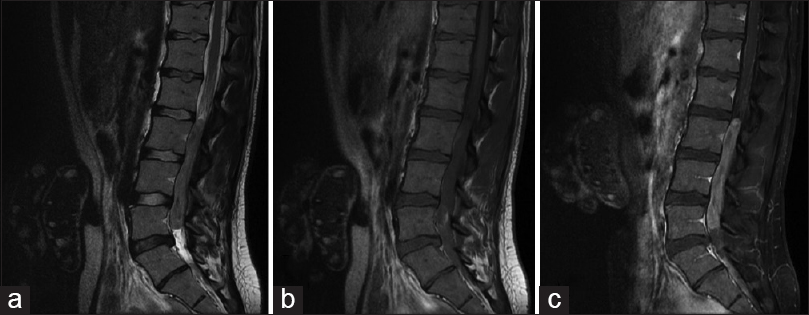
Spinal Neoplasms
Cobalt Radioisotopes
Carcinoma, Non-Small-Cell Lung
Lymphatic Irradiation
Mechlorethamine
Carcinoma
Radiation-Protective Agents
Glioblastoma
Prospective Studies
Salvage Therapy
Kaplan-Meier Estimate
Bleomycin
Doxorubicin
Antineoplastic Agents, Alkylating
Iridium Radioisotopes
Glioma
Pelvis
Lymphatic Metastasis
Cyclophosphamide
Thoracic Neoplasms
Tumor Burden
Patient Positioning
Feasibility Studies
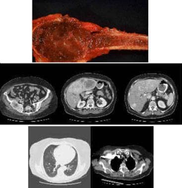
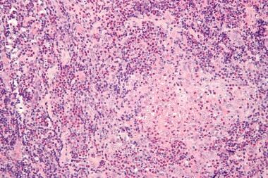
Sarcoma
Radiotherapy Setup Errors
Prednisone
Remission Induction
Heavy Ions
Dose-Response Relationship, Drug
Supratentorial Neoplasms
Nimustine
Positron-Emission Tomography
Neoplasm Metastasis
Amifostine
Radiopharmaceuticals
Etoposide
Mucositis
Central Nervous System Neoplasms
Skull Base Neoplasms
Radiation, Ionizing
Chemoradiotherapy, Adjuvant
Clinical Trials as Topic
Quality of Life
Postoperative Care
Treatment Failure
Esthesioneuroblastoma, Olfactory
Vindesine
Methotrexate
Meningeal Neoplasms
Proportional Hazards Models
Preoperative Care
Magnetic Resonance Imaging
Astrocytoma
Prostate-Specific Antigen
Health Physics
Actuarial Analysis
Four-Dimensional Computed Tomography
Prostatectomy
Radiobiology
Testicular Neoplasms
Mastectomy, Modified Radical
Soft Tissue Neoplasms
Carcinoma, Small Cell
Multivariate Analysis
Meningioma
Disease Progression
Deoxycytidine
Risk Factors
Spinal Cord Neoplasms
Lymphoma, Non-Hodgkin
Abdominal Neoplasms
Cone-Beam Computed Tomography
Oligodendroglioma
Eye Enucleation
Fluorodeoxyglucose F18
Spinal Cord Compression
Gamma Rays
Karnofsky Performance Status
Medulloblastoma
Seminoma
Brain Stem Neoplasms
Cerebellar Neoplasms
Lymph Node Excision
Tumor Markers, Biological
Leukopenia
Rhabdomyosarcoma
Randomized Controlled Trials as Topic
Ependymoma
Antineoplastic Agents, Hormonal
Photons
Risk Assessment
Relative Biological Effectiveness
Choroid Neoplasms
Hyperthermia, Induced
Radiation Protection
Cohort Studies
Age Factors
Radiation Injuries, Experimental
Neck Dissection
Ifosfamide
Phantoms, Imaging
Pancreatic Neoplasms
Phase II trial of primary chemotherapy followed by reduced-dose radiation for CNS germ cell tumors. (1/2836)
PURPOSE: A prospective phase II study was initiated to assess the response rate, survival, and late effects of treatment in patients with newly diagnosed CNS germ cell tumors (GCT), using etoposide plus cisplatin followed by radiation therapy prescribed by extent of disease, histology, and response to chemotherapy. PATIENTS AND METHODS: Seventeen patients aged 8 to 24 years with histologically proven CNS GCT received etoposide (100 mg/m2/d) plus cisplatin (20 mg/m2/d) daily for 5 days every 3 weeks for four cycles, followed by radiation therapy. Nine patients had germinomas; eight had mixed GCT. Four patients (three with germinomas and one with mixed GCT) presented with leptomeningeal dissemination. RESULTS: Radiographically, 14 of 17 patients were assessable for response; 11 patients experienced complete regression, and three had major partial regression before radiation. Six of seven assessable patients with elevated CSF levels of alpha-fetoprotein or betahuman chorionic gonadotropin had normalization with chemotherapy alone; all normalized with combined chemotherapy and radiation therapy. All 17 patients are alive without evidence of disease (median follow-up, 51 months). One patient developed a relapse in the spinal leptomeninges and was rendered free of disease with spinal radiation more than 5 years ago. One patient developed carotid stenosis requiring surgery. Thus far, only minimal long-term deterioration in neurocognitive function has been detected as a consequence of protocol treatment. CONCLUSION: Conventional-dose intravenous chemotherapy with etoposide and cisplatin can effect tumor regression in a high proportion of patients with CNS GCT, including those with leptomeningeal metastases. Acute and long-term toxicities are acceptable. Progression-free survival and overall survival are excellent. (+info)Dose-related effects of single focal irradiation in the medial temporal lobe structures in rats--magnetic resonance imaging and histological study. (2/2836)
The dose-related effects of single focal irradiation on the medial temporal lobe in rats were investigated by sequential magnetic resonance imaging and histological examination. Irradiation of 200 Gy as a maximum dose using 4 mm collimators with a gamma unit created an area of necrosis consistently at the target site within 2 weeks after irradiation. Irradiation of 100 Gy caused necrosis within 10 weeks, and 75 Gy caused necrosis within one year. Irradiation of less than 50 Gy did not induce necrosis consistently, although a restricted area of necrosis was created in the medial temporal structures including the intraparenchymal portion of the optic tract. 75 Gy may be the optimum dose for creating necrosis consistently in the medial temporal lobe structures. However, careful dose planning considering both dose-time and dose-volume relationships in necrosis development is necessary to avoid injury to vulnerable neural structures such as the optic tract when applying radiosurgical techniques to treat functional brain disorders in medial temporal lobe structures such as temporal lobe epilepsy. (+info)Dosimetry of 131I-labeled 81C6 monoclonal antibody administered into surgically created resection cavities in patients with malignant brain tumors. (3/2836)
The objective of this study was to perform the dosimetry of 131I-labeled 81C6 monoclonal antibody (MAb) in patients with recurrent malignant brain tumors, treated by direct injections of MAb into surgically created resection cavities (SCRCs). METHODS: Absorbed dose estimates were performed for nine patients. Dosimetry was performed retrospectively using probe counts (during patient isolation) and whole-body and SPECT images thereafter. Absorbed doses were calculated for the SCRC interface and for regions of interest (ROIs) 1 and 2 cm thick, measured from the margins of cavity interface. Also, mean absorbed doses were calculated for normal brain, liver, spleen, thyroid gland, stomach, bone marrow and whole body. The average residence time for the SCRC was 111 h (65-200h). RESULTS: The average absorbed dose per unit injected activity (range) to the SCRC interface and ROIs 1 and 2 cm thick from the cavity interface were 31.9 (7.8-84.2), 1.9 (0.7-3.6) and 1.0 (0.4-1.8) cGy/MBq, respectively. Average absorbed doses per unit administered activity to brain, liver, spleen, thyroid, stomach, bone marrow and whole body were 0.18, 0.03, 0.08, 0.05, 0.02, 0.02 and 0.01 cGy/MBq, respectively. The high absorbed dose delivered to the SCRC interface may have produced an increase in cavity volume independent of tumor progression. CONCLUSION: At the maximum tolerated dose of 3700 MBq 131I-labeled 81C6 MAb, the absorbed doses to the SCRC interface and ROIs of 1 and 2 cm thickness were estimated to be 1180, 71 and 39 Gy, respectively. The estimated average absorbed dose to the brain was 6.5 Gy. There was no neurological toxicity and minimal hematologic toxicity at this maximum tolerated administration level. (+info)Phase I study of 90Y-labeled B72.3 intraperitoneal administration in patients with ovarian cancer: effect of dose and EDTA coadministration on pharmacokinetics and toxicity. (4/2836)
The tumor-associated glycoprotein 72 (TAG-72) antigen is present on a high percentage of tumor types including ovarian carcinomas. Antibody B72.3 is a murine monoclonal recognizing the surface domain of the TAG-72 antigen and has been widely used in human clinical trials. After our initial encouraging studies (M. G. Rosenblum et al., J. Natl. Cancer Inst., 83: 1629-1636, 1991) of tissue disposition, metabolism, and pharmacokinetics in 9 patients with ovarian cancer, we designed an escalating dose, multi-arm Phase I study of 90Y-labeled B72.3 i.p. administration. In the first arm of the study, patients (3 pts/dose level) received an i.p. infusion of either 2 or 10 mg of B72.3 labeled with either 1, 10, 15, or 25 mCi of 90Y. Pharmacokinetic studies demonstrated that concentrations of 90Y-labeled B72.3 persist in peritoneal fluid with half-lives >24 h after i.p. administration. In addition, 90Y-labeled B72.3 was absorbed rapidly into the plasma with peak levels achieved within 48 h, and levels declined slowly thereafter. Cumulative urinary excretion of the 90Y label was 10-20% of the administered dose which suggests significant whole-body retention of the radiolabel. Biopsy specimens of bone and marrow obtained at 72 h after administration demonstrated significant content of the label in bone (0.015% of the dose/g) with relatively little in marrow (0.005% of the dose/g). The maximal tolerated dose was determined to be 10 mCi because of hematological toxicity and platelet suppression. This typically occurred on the 29th day after administration and was thought to be a consequence of the irradiation of the marrow from the bony deposition of the radiolabel. In an effort to suppress the bone uptake of 90Y, patients were treated with a continuous i.v. infusion of EDTA (25 mg/kg/12 h x 6) infused immediately before i.p. administration of the radiolabeled antibody. Patients (3 pts/dose level) were treated with doses of 10, 15, 20, 25, 30, 35, 40, or 45 mCi of 90Y-labeled B72.3 for a total of 38 patients. EDTA administration resulted in significant myeloprotection, which allowed escalation to the maximal tolerated dose of 40 mCi. Dose-limiting toxicity was thrombocytopenia and neutropenia. Studies of plasma and peritoneal fluid pharmacokinetics demonstrate no changes compared with patients without EDTA pretreatment. Cumulative urinary excretion of the radiolabel was not increased in patients pretreated with EDTA compared with the untreated group. However, analysis of biopsy specimens of bone and marrow demonstrated that bone and marrow content of the 90Y label was 15-fold lower (<0.001% injected dose/g) than a companion group without EDTA. Four responses were noted in patients who received 15-30 mCi of 90Y-labeled B72.3 with response durations of 1-12 months. These results demonstrate the myeloprotective ability of EDTA, which allows safe i.p. administration of higher doses of 90Y-labeled B72.3 and, therefore, clearly warrant an expanded Phase II trial in patients with minimal residual disease after standard chemotherapy or for the palliation of refractory ascites. (+info)Locoregional regulatory peptide receptor targeting with the diffusible somatostatin analogue 90Y-labeled DOTA0-D-Phe1-Tyr3-octreotide (DOTATOC): a pilot study in human gliomas. (5/2836)
Human gliomas, especially of low-grade type, have been shown to express high-affinity somatostatin receptor type 2 (J-C. Reubi et al., Am. J. Pathol, 134: 337-344, 1989). We enrolled seven low-grade and four anaplastic glioma patients in a pilot study using the diffusible peptidic vector 90Y-labeled DOTA0-D-Phe1-Tyr3-octreotide (DOTATOC) for receptor targeting. The radiopharmakon was locoregionally injected into a stereotactically inserted Port-a-cath. DOTATOC competes specifically with somatostatin binding to somatostatin receptor type 2 in the low nanomolar range as shown by a displacement curve of 125I-[Tyr3]-octreotide in tumor tissue sections. Diagnostic (111)In-labeled DOTATOC-scintigraphy following local injection displayed homogeneous to nodular intratumoral vector distribution. The cumulative activity of regionally injected peptide-bound 90Y amounted to 370-3300 MBq, which is equivalent to an effective dose range between 60 +/- 15 and 550 +/- 110 Gy. Activity was injected in one to four fractions according to tumor volumes; 1110 MBq of 90Y-labeled DOTATOC was the maximum activity per single injection. We obtained six disease stabilizations and shrinking of a cystic low-grade astrocytoma component. The only toxicity observed was secondary perifocal edema. The activity:dose ratio (MBq:Gy) represents a measure for the stability of peptide retention in receptor-positive tissue and might predict the clinical course. We conclude that SR-positive human gliomas, especially of low-grade type, can be successfully targeted by intratumoral injection of the metabolically stable small regulatory peptide DOTATOC. (+info)A radionuclide therapy treatment planning and dose estimation system. (6/2836)
An object-oriented software system is described for estimating internal emitter absorbed doses using a set of computer modules operating within a personal computer environment. The system is called the Radionuclide Treatment Planning and Absorbed Dose Estimation System (RTDS). It is intended for radioimmunotherapy applications, although other forms of internal emitter therapy may also be considered. METHODS: Four software modules interact through a database backend. Clinical, demographic and image data are directly entered into the database. Modules include those devoted to clinical imaging (nuclear, CT and MR), activity determination, organ compartmental modeling and absorbed dose estimation. RESULTS: Both standard phantom (Medical Internal Radiation Dose [MIRD]) and patient-specific absorbed doses are estimated. All modules interact with the database backend so that changes in one process do not influence other operations. Results of the modular operations are written to the database as computations are completed. Dose-volume histograms are an intrinsic part of the output for patient-specific absorbed dose estimates. A sample dose estimate for a potential 90Y monoclonal antibody is described. CONCLUSION: A four-module software system has been implemented to estimate MIRD phantom and patient-specific absorbed doses. Computations of the doses and their statistical distribution for a pure beta emitter such as 90Y take approximately 1 min on a 300 MHz personal computer. (+info)The increment of micronucleus frequency in cervical carcinoma during irradiation in vivo and its prognostic value for tumour radiocurability. (7/2836)
A potential usefulness of micronucleus assay for prediction of tumour radiosensitivity has been tested in 64 patients with advanced stage (II B-IV B) cervical carcinoma treated by radiotherapy. The study of cellular radiosensitivity in vitro was conducted in parallel with the study of cellular damage after tumour irradiation in vivo. Radiosensitivity of in vitro cultured primary cells isolated from tumour biopsies taken before radiotherapy was evaluated using cytokinesis-block micronucleus assay. Frequency of micronuclei per binucleated cell (MN/BNC) at 2 Gy was used as a measure of radiosensitivity. Radiation sensitivity in vivo was expressed as per cent increment of micronucleus frequency in cells isolated from biopsy taken after 20 Gy (external irradiation, 10 x 2 Gy) over the pre-treatment spontaneous micronucleus level and was called MN20. Very low correlation (r = 0.324) was observed between micronucleus frequency in vitro and in vivo. Although micronucleus frequency at 2 Gy differed widely between tumours evaluated (mean MN/BNC was 0.224; range 0.08-0.416), no significant correlation was observed between this parameter and clinical outcome. The average increment of micronucleus frequency after 20 Gy amounted to 193% of spontaneous level (range 60-610%) and was independent of spontaneous micronucleation before radiotherapy. In contrast to in vitro results, these from in vivo assay seem to have a predictive value for radiotherapy of cervix cancer. The micronucleus increment in vivo that reached at least 117.5% of pretreatment value (first quartile for MN20 data set) correlated significantly with better tumour local control (P < 0.008) and overall survival (P < 0.045). Our results suggest that evaluation of increment of micronucleus frequency during radiotherapy (after fixed tested dose of 20 Gy) offers a potentially valuable approach to predicting individual radioresponsiveness and may be helpful for individualization of treatment strategy in advanced stage cervical cancer. (+info)Prognostic values of proliferating cell nuclear antigen (PCNA) and Ki-67 for radiotherapy of oesophageal squamous cell carcinomas. (8/2836)
The relationship of immunohistochemical indices of proliferating cell nuclear antigen (PCNA) and Ki-67 to local control and survival rates for patients with oesophageal squamous cell carcinomas treated by definitive radiotherapy (RT) was investigated. Biopsy materials before RT were obtained from 65 patients with oesophageal cancer. The median PCNA labelling index (LI) and the median Ki-67 LI were 52% and 45% respectively. The PCNA LI was independent of known prognostic factors on local control for oesophageal cancer, although Ki-67 LI correlated with several prognostic factors. In the univariate analysis, patients with the PCNA LI of < 52% or the Ki-67 LI of < 45% showed significantly higher local recurrence rates than those with higher LIs (both P < 0.05). This difference in local control rate according to LIs was prominent for the patients treated with conventional fractionation. In the multivariate analysis, T-stage (P = 0.0056) and PCNA LI (P = 0.0332) were significant factors for local control in the final model using a stepwise regression procedure. In conclusion, PCNA LI and Ki-67 LI were significantly correlated with local control probabilities in oesophageal squamous cell carcinomas treated by definitive RT. (+info)Radiotherapy dosage refers to the total amount of radiation energy that is absorbed by tissues or organs, typically measured in units of Gray (Gy), during a course of radiotherapy treatment. It is the product of the dose rate (the amount of radiation delivered per unit time) and the duration of treatment. The prescribed dosage for cancer treatments can range from a few Gray to more than 70 Gy, depending on the type and location of the tumor, the patient's overall health, and other factors. The goal of radiotherapy is to deliver a sufficient dosage to destroy the cancer cells while minimizing damage to surrounding healthy tissues.
Radiotherapy, also known as radiation therapy, is a medical treatment that uses ionizing radiation to kill cancer cells, shrink tumors, and prevent the growth and spread of cancer. The radiation can be delivered externally using machines or internally via radioactive substances placed in or near the tumor. Radiotherapy works by damaging the DNA of cancer cells, which prevents them from dividing and growing. Normal cells are also affected by radiation, but they have a greater ability to repair themselves compared to cancer cells. The goal of radiotherapy is to destroy as many cancer cells as possible while minimizing damage to healthy tissue.
Conformal radiotherapy is a type of external beam radiation therapy that uses advanced technology to conform the radiation beam to the shape of the tumor, allowing for more precise and targeted treatment while minimizing exposure to healthy surrounding tissue. This can help reduce the risk of side effects and improve the therapeutic ratio. Conformal radiotherapy techniques include 3D conformal radiation therapy (3D-CRT), intensity-modulated radiation therapy (IMRT), and volumetric modulated arc therapy (VMAT). These techniques use sophisticated imaging and treatment planning systems to create a personalized treatment plan for each patient, based on the size, shape, and location of their tumor.
Adjuvant radiotherapy is a type of cancer treatment that uses radiation therapy as an adjunct to a primary surgical procedure. The goal of adjuvant radiotherapy is to eliminate any remaining microscopic cancer cells that may be present in the surrounding tissues after surgery, thereby reducing the risk of local recurrence and improving the chances of cure.
Radiotherapy involves the use of high-energy radiation to destroy cancer cells and shrink tumors. In adjuvant radiotherapy, the radiation is usually delivered to the tumor bed and regional lymph nodes in order to target any potential sites of residual disease. The timing and dosing of adjuvant radiotherapy may vary depending on the type and stage of cancer being treated, as well as other factors such as patient age and overall health status.
Adjuvant radiotherapy is commonly used in the treatment of various types of cancer, including breast, colorectal, lung, head and neck, and gynecologic cancers. Its use has been shown to improve survival rates and reduce the risk of recurrence in many cases, making it an important component of comprehensive cancer care.
Intensity-modulated radiotherapy (IMRT) is a type of external beam radiation therapy that uses advanced technology to precisely target tumors while minimizing exposure to healthy tissues. In IMRT, the intensity of the radiation beam is modulated or varied during treatment, allowing for more conformal dose distributions and better sparing of normal structures. This is achieved through the use of computer-controlled linear accelerators that shape the radiation beam to match the three-dimensional shape of the tumor. The result is improved treatment accuracy, reduced side effects, and potentially higher cure rates compared to conventional radiotherapy techniques.
Computer-assisted radiotherapy planning (CARP) is the use of computer systems and software to assist in the process of creating a treatment plan for radiotherapy. The goal of radiotherapy is to deliver a precise and effective dose of radiation to a tumor while minimizing exposure to healthy tissue. CARP involves using imaging data, such as CT or MRI scans, to create a 3D model of the patient's anatomy. This model is then used to simulate the delivery of radiation from different angles and determine the optimal treatment plan. The use of computers in this process allows for more accurate and efficient planning, as well as the ability to easily adjust the plan as needed.
High-energy radiotherapy, also known as external beam radiation therapy (EBRT), is a type of cancer treatment that uses high-energy radiation beams to destroy cancer cells and shrink tumors. The radiation beams are produced by a machine called a linear accelerator (LINAC) and are directed at the tumor site from outside the body. High-energy radiotherapy can be used to treat many different types of cancer, either alone or in combination with other treatments such as surgery or chemotherapy.
The high-energy radiation beams used in this type of radiotherapy are able to penetrate deep into the body and target large areas, making it an effective treatment for cancers that have spread or are too large to be removed surgically. The dose and duration of treatment will depend on the type and stage of cancer being treated, as well as the patient's overall health.
High-energy radiotherapy works by damaging the DNA of cancer cells, which prevents them from dividing and growing. This ultimately leads to the death of the cancer cells. While radiation therapy can also damage normal cells, they are generally better able to repair themselves compared to cancer cells. Therefore, the goal of high-energy radiotherapy is to deliver a high enough dose to destroy the cancer cells while minimizing harm to surrounding healthy tissue.
It's important to note that high-energy radiotherapy requires careful planning and delivery to ensure that the radiation beams are focused on the tumor site and avoid healthy tissues as much as possible. This is typically done using imaging techniques such as CT, MRI, or PET scans to create a treatment plan that maps out the exact location and shape of the tumor. The patient will then undergo a series of treatments, usually scheduled daily over several weeks.
Combined modality therapy (CMT) is a medical treatment approach that utilizes more than one method or type of therapy simultaneously or in close succession, with the goal of enhancing the overall effectiveness of the treatment. In the context of cancer care, CMT often refers to the combination of two or more primary treatment modalities, such as surgery, radiation therapy, and systemic therapies (chemotherapy, immunotherapy, targeted therapy, etc.).
The rationale behind using combined modality therapy is that each treatment method can target cancer cells in different ways, potentially increasing the likelihood of eliminating all cancer cells and reducing the risk of recurrence. The specific combination and sequence of treatments will depend on various factors, including the type and stage of cancer, patient's overall health, and individual preferences.
For example, a common CMT approach for locally advanced rectal cancer may involve preoperative (neoadjuvant) chemoradiation therapy, followed by surgery to remove the tumor, and then postoperative (adjuvant) chemotherapy. This combined approach allows for the reduction of the tumor size before surgery, increases the likelihood of complete tumor removal, and targets any remaining microscopic cancer cells with systemic chemotherapy.
It is essential to consult with a multidisciplinary team of healthcare professionals to determine the most appropriate CMT plan for each individual patient, considering both the potential benefits and risks associated with each treatment method.
Dose fractionation is a medical term that refers to the practice of dividing the total dose of radiation therapy or chemotherapy into smaller doses, which are given over a longer period. This approach allows for the delivery of a higher total dose of treatment while minimizing damage to healthy tissues and reducing side effects.
In radiation therapy, fractionation is used to target cancer cells while sparing surrounding normal tissues. By delivering smaller doses of radiation over several treatments, healthy tissue has time to recover between treatments, reducing the risk of complications. The number and size of fractions can vary depending on the type and location of the tumor, as well as other factors such as the patient's overall health.
Similarly, in chemotherapy, dose fractionation is used to maximize the effectiveness of the treatment while minimizing toxicity. By administering smaller doses of chemotherapy over time, the body has a chance to recover between treatments, reducing side effects and allowing for higher total doses to be given. The schedule and duration of chemotherapy fractionation may vary depending on the type of drug used, the type and stage of cancer, and other factors.
Overall, dose fractionation is an important technique in both radiation therapy and chemotherapy that allows for more effective treatment while minimizing harm to healthy tissues.
Radiation injuries refer to the damages that occur to living tissues as a result of exposure to ionizing radiation. These injuries can be acute, occurring soon after exposure to high levels of radiation, or chronic, developing over a longer period after exposure to lower levels of radiation. The severity and type of injury depend on the dose and duration of exposure, as well as the specific tissues affected.
Acute radiation syndrome (ARS), also known as radiation sickness, is the most severe form of acute radiation injury. It can cause symptoms such as nausea, vomiting, diarrhea, fatigue, fever, and skin burns. In more severe cases, it can lead to neurological damage, hemorrhage, infection, and death.
Chronic radiation injuries, on the other hand, may not appear until months or even years after exposure. They can cause a range of symptoms, including fatigue, weakness, skin changes, cataracts, reduced fertility, and an increased risk of cancer.
Radiation injuries can be treated with supportive care, such as fluids and electrolytes replacement, antibiotics, wound care, and blood transfusions. In some cases, surgery may be necessary to remove damaged tissue or control bleeding. Prevention is the best approach to radiation injuries, which includes limiting exposure through proper protective measures and monitoring radiation levels in the environment.
Computer-assisted radiotherapy, also known as computerized radiation therapy planning or treatment planning system, is a medical procedure that utilizes advanced computer software to design and implement a radiotherapy treatment plan for patients with cancer. This process involves using imaging technologies such as CT, MRI, or PET scans to create a 3D model of the tumor and surrounding healthy tissues. The software then calculates the optimal radiation dose and beam orientation to deliver the maximum radiation to the tumor while minimizing exposure to healthy tissue.
The computer-assisted radiotherapy system allows for more precise and accurate treatment planning, which can lead to improved outcomes and reduced side effects for patients undergoing radiation therapy. It also enables clinicians to simulate and compare different treatment plans, allowing them to choose the most effective and safe option for each individual patient. Additionally, the use of computer-assisted radiotherapy can increase efficiency and streamline the treatment planning process, reducing wait times for patients and improving workflow in radiotherapy departments.
Image-guided radiotherapy (IGRT) is a type of radiation therapy that uses medical imaging techniques to improve the precision and accuracy of radiation delivery. It allows for real-time or periodic imaging during the course of radiation treatment, which can be used to confirm the position of the targeted tumor and make any necessary adjustments to the patient's position or the radiation beam. This helps ensure that the radiation is focused on the intended target, while minimizing exposure to surrounding healthy tissue. IGRT may be used to treat a variety of cancer types and can be delivered using various radiation therapy techniques such as 3D-conformal radiotherapy, intensity-modulated radiotherapy (IMRT), or stereotactic body radiotherapy (SBRT).
Radiation oncology is a branch of medicine that uses ionizing radiation in the treatment and management of cancer. The goal of radiation therapy, which is the primary treatment modality in radiation oncology, is to destroy cancer cells or inhibit their growth while minimizing damage to normal tissues. This is achieved through the use of high-energy radiation beams, such as X-rays, gamma rays, and charged particles, that are directed at the tumor site with precision. Radiation oncologists work in interdisciplinary teams with other healthcare professionals, including medical physicists, dosimetrists, and radiation therapists, to plan and deliver effective radiation treatments for cancer patients.
Treatment outcome is a term used to describe the result or effect of medical treatment on a patient's health status. It can be measured in various ways, such as through symptoms improvement, disease remission, reduced disability, improved quality of life, or survival rates. The treatment outcome helps healthcare providers evaluate the effectiveness of a particular treatment plan and make informed decisions about future care. It is also used in clinical research to compare the efficacy of different treatments and improve patient care.
Radiosurgery is a non-invasive surgical procedure that uses precisely focused beams of radiation to treat various medical conditions, primarily in the field of neurosurgery and oncology. It allows for the destruction of targeted tissue while minimizing damage to surrounding healthy structures. Unlike traditional surgery, radiosurgery does not require any incisions, as it delivers radiation through the skin to reach the intended target.
The term "stereotactic" is often associated with radiosurgery, which refers to the use of a three-dimensional coordinate system to precisely locate and target the affected area. This technique enables high doses of radiation to be delivered accurately and efficiently, maximizing therapeutic effectiveness while minimizing side effects.
Radiosurgery can be used to treat various conditions such as brain tumors (both malignant and benign), arteriovenous malformations (AVMs), trigeminal neuralgia, acoustic neuromas, pituitary adenomas, and spinal cord tumors. Common radiosurgery platforms include the Gamma Knife, CyberKnife, and linear accelerator-based systems like Novalis Tx or TrueBeam.
It is essential to note that although it is called "surgery," radiosurgery does not involve any physical incisions or removal of tissue. Instead, it relies on the destructive effects of high-dose radiation to ablate or damage targeted cells over time, leading to their eventual death and resolution of symptoms or tumor control.
A dose-response relationship in radiation refers to the correlation between the amount of radiation exposure (dose) and the biological response or adverse health effects observed in exposed individuals. As the level of radiation dose increases, the severity and frequency of the adverse health effects also tend to increase. This relationship is crucial in understanding the risks associated with various levels of radiation exposure and helps inform radiation protection standards and guidelines.
The effects of ionizing radiation can be categorized into two types: deterministic and stochastic. Deterministic effects have a threshold dose below which no effect is observed, and above this threshold, the severity of the effect increases with higher doses. Examples include radiation-induced cataracts or radiation dermatitis. Stochastic effects, on the other hand, do not have a clear threshold and are based on probability; as the dose increases, so does the likelihood of the adverse health effect occurring, such as an increased risk of cancer.
Understanding the dose-response relationship in radiation exposure is essential for setting limits on occupational and public exposure to ionizing radiation, optimizing radiation protection practices, and developing effective medical countermeasures in case of radiation emergencies.
Head and neck neoplasms refer to abnormal growths or tumors in the head and neck region, which can be benign (non-cancerous) or malignant (cancerous). These tumors can develop in various sites, including the oral cavity, nasopharynx, oropharynx, larynx, hypopharynx, paranasal sinuses, salivary glands, and thyroid gland.
Benign neoplasms are slow-growing and generally do not spread to other parts of the body. However, they can still cause problems if they grow large enough to press on surrounding tissues or structures. Malignant neoplasms, on the other hand, can invade nearby tissues and organs and may also metastasize (spread) to other parts of the body.
Head and neck neoplasms can have various symptoms depending on their location and size. Common symptoms include difficulty swallowing, speaking, or breathing; pain in the mouth, throat, or ears; persistent coughing or hoarseness; and swelling or lumps in the neck or face. Early detection and treatment of head and neck neoplasms are crucial for improving outcomes and reducing the risk of complications.
Brachytherapy is a type of cancer treatment that involves placing radioactive material directly into or near the tumor site. The term "brachy" comes from the Greek word for "short," which refers to the short distance that the radiation travels. This allows for a high dose of radiation to be delivered directly to the tumor while minimizing exposure to healthy surrounding tissue.
There are two main types of brachytherapy:
1. Intracavitary brachytherapy: The radioactive material is placed inside a body cavity, such as the uterus or windpipe.
2. Interstitial brachytherapy: The radioactive material is placed directly into the tumor or surrounding tissue using needles, seeds, or catheters.
Brachytherapy can be used alone or in combination with other cancer treatments such as surgery, external beam radiation therapy, and chemotherapy. It may be recommended for a variety of cancers, including prostate, cervical, vaginal, vulvar, head and neck, and skin cancers. The specific type of brachytherapy used will depend on the size, location, and stage of the tumor.
The advantages of brachytherapy include its ability to deliver a high dose of radiation directly to the tumor while minimizing exposure to healthy tissue, which can result in fewer side effects compared to other forms of radiation therapy. Additionally, brachytherapy is often a shorter treatment course than external beam radiation therapy, with some treatments lasting only a few minutes or hours.
However, there are also potential risks and side effects associated with brachytherapy, including damage to nearby organs and tissues, bleeding, infection, and pain. Patients should discuss the benefits and risks of brachytherapy with their healthcare provider to determine if it is an appropriate treatment option for them.
Local neoplasm recurrence is the return or regrowth of a tumor in the same location where it was originally removed or treated. This means that cancer cells have survived the initial treatment and started to grow again in the same area. It's essential to monitor and detect any local recurrence as early as possible, as it can affect the prognosis and may require additional treatment.
Gene dosage, in genetic terms, refers to the number of copies of a particular gene present in an organism's genome. Each gene usually has two copies (alleles) in diploid organisms, one inherited from each parent. An increase or decrease in the number of copies of a specific gene can lead to changes in the amount of protein it encodes, which can subsequently affect various biological processes and phenotypic traits.
For example, gene dosage imbalances have been associated with several genetic disorders, such as Down syndrome (trisomy 21), where an individual has three copies of chromosome 21 instead of the typical two copies, leading to developmental delays and intellectual disabilities. Similarly, in certain cases of cancer, gene amplification (an increase in the number of copies of a particular gene) can result in overexpression of oncogenes, contributing to tumor growth and progression.
Neoplasm staging is a systematic process used in medicine to describe the extent of spread of a cancer, including the size and location of the original (primary) tumor and whether it has metastasized (spread) to other parts of the body. The most widely accepted system for this purpose is the TNM classification system developed by the American Joint Committee on Cancer (AJCC) and the Union for International Cancer Control (UICC).
In this system, T stands for tumor, and it describes the size and extent of the primary tumor. N stands for nodes, and it indicates whether the cancer has spread to nearby lymph nodes. M stands for metastasis, and it shows whether the cancer has spread to distant parts of the body.
Each letter is followed by a number that provides more details about the extent of the disease. For example, a T1N0M0 cancer means that the primary tumor is small and has not spread to nearby lymph nodes or distant sites. The higher the numbers, the more advanced the cancer.
Staging helps doctors determine the most appropriate treatment for each patient and estimate the patient's prognosis. It is an essential tool for communication among members of the healthcare team and for comparing outcomes of treatments in clinical trials.
Genetic dosage compensation is a process that evens out the effects of genes on an organism's phenotype (observable traits), even when there are differences in the number of copies of those genes present. This is especially important in cases where sex chromosomes are involved, as males and females often have different numbers of sex chromosomes.
In many species, including humans, females have two X chromosomes, while males have one X and one Y chromosome. To compensate for the difference in dosage, one of the female's X chromosomes is randomly inactivated during early embryonic development, resulting in each cell having only one active X chromosome, regardless of sex. This process ensures that both males and females have similar levels of gene expression from their X chromosomes and helps to prevent an imbalance in gene dosage between the sexes.
Defects in dosage compensation can lead to various genetic disorders, such as Turner syndrome (where a female has only one X chromosome) or Klinefelter syndrome (where a male has two or more X chromosomes). These conditions can result in developmental abnormalities and health issues due to the imbalance in gene dosage.
Chemoradiotherapy is a medical treatment that combines chemotherapy and radiotherapy. Chemotherapy involves the use of drugs to kill or damage cancer cells, while radiotherapy uses ionizing radiation to achieve the same goal. In chemoradiotherapy, these two modalities are used simultaneously or sequentially to treat a malignancy.
The aim of chemoradiotherapy is to increase the effectiveness of treatment by targeting cancer cells with both chemotherapy and radiation therapy. This approach can be particularly effective in treating certain types of cancer, such as head and neck cancer, lung cancer, esophageal cancer, cervical cancer, anal cancer, and rectal cancer.
The specific drugs used in chemoradiotherapy and the doses and schedules of both chemotherapy and radiotherapy vary depending on the type and stage of cancer being treated. The side effects of chemoradiotherapy can be significant and may include fatigue, skin reactions, mucositis, nausea, vomiting, diarrhea, and myelosuppression. However, these side effects are usually manageable with appropriate supportive care.
Antineoplastic combined chemotherapy protocols refer to a treatment plan for cancer that involves the use of more than one antineoplastic (chemotherapy) drug given in a specific sequence and schedule. The combination of drugs is used because they may work better together to destroy cancer cells compared to using a single agent alone. This approach can also help to reduce the likelihood of cancer cells becoming resistant to the treatment.
The choice of drugs, dose, duration, and frequency are determined by various factors such as the type and stage of cancer, patient's overall health, and potential side effects. Combination chemotherapy protocols can be used in various settings, including as a primary treatment, adjuvant therapy (given after surgery or radiation to kill any remaining cancer cells), neoadjuvant therapy (given before surgery or radiation to shrink the tumor), or palliative care (to alleviate symptoms and prolong survival).
It is important to note that while combined chemotherapy protocols can be effective in treating certain types of cancer, they can also cause significant side effects, including nausea, vomiting, hair loss, fatigue, and an increased risk of infection. Therefore, patients undergoing such treatment should be closely monitored and managed by a healthcare team experienced in administering chemotherapy.
Nasopharyngeal neoplasms refer to abnormal growths or tumors in the nasopharynx, which is the upper part of the pharynx (throat) behind the nose. These growths can be benign (non-cancerous) or malignant (cancerous).
Malignant nasopharyngeal neoplasms are often referred to as nasopharyngeal carcinoma or cancer. There are different types of nasopharyngeal carcinomas, including keratinizing squamous cell carcinoma, non-keratinizing carcinoma, and basaloid squamous cell carcinoma.
The risk factors for developing nasopharyngeal neoplasms include exposure to the Epstein-Barr virus (EBV), consumption of certain foods, smoking, and genetic factors. Symptoms may include a lump in the neck, nosebleeds, hearing loss, ringing in the ears, and difficulty swallowing or speaking. Treatment options depend on the type, size, and stage of the neoplasm and may include surgery, radiation therapy, chemotherapy, or a combination of these treatments.
Brain neoplasms, also known as brain tumors, are abnormal growths of cells within the brain. These growths can be benign (non-cancerous) or malignant (cancerous). Benign brain tumors typically grow slowly and do not spread to other parts of the body. However, they can still cause serious problems if they press on sensitive areas of the brain. Malignant brain tumors, on the other hand, are cancerous and can grow quickly, invading surrounding brain tissue and spreading to other parts of the brain or spinal cord.
Brain neoplasms can arise from various types of cells within the brain, including glial cells (which provide support and insulation for nerve cells), neurons (nerve cells that transmit signals in the brain), and meninges (the membranes that cover the brain and spinal cord). They can also result from the spread of cancer cells from other parts of the body, known as metastatic brain tumors.
Symptoms of brain neoplasms may vary depending on their size, location, and growth rate. Common symptoms include headaches, seizures, weakness or paralysis in the limbs, difficulty with balance and coordination, changes in speech or vision, confusion, memory loss, and changes in behavior or personality.
Treatment for brain neoplasms depends on several factors, including the type, size, location, and grade of the tumor, as well as the patient's age and overall health. Treatment options may include surgery, radiation therapy, chemotherapy, targeted therapy, or a combination of these approaches. Regular follow-up care is essential to monitor for recurrence and manage any long-term effects of treatment.
Disease-free survival (DFS) is a term used in medical research and clinical practice, particularly in the field of oncology. It refers to the length of time after primary treatment for a cancer during which no evidence of the disease can be found. This means that the patient shows no signs or symptoms of the cancer, and any imaging studies or other tests do not reveal any tumors or other indications of the disease.
DFS is often used as an important endpoint in clinical trials to evaluate the effectiveness of different treatments for cancer. By measuring the length of time until the cancer recurs or a new cancer develops, researchers can get a better sense of how well a particular treatment is working and whether it is improving patient outcomes.
It's important to note that DFS is not the same as overall survival (OS), which refers to the length of time from primary treatment until death from any cause. While DFS can provide valuable information about the effectiveness of cancer treatments, it does not necessarily reflect the impact of those treatments on patients' overall survival.
Squamous cell carcinoma is a type of skin cancer that begins in the squamous cells, which are flat, thin cells that form the outer layer of the skin (epidermis). It commonly occurs on sun-exposed areas such as the face, ears, lips, and backs of the hands. Squamous cell carcinoma can also develop in other areas of the body including the mouth, lungs, and cervix.
This type of cancer usually develops slowly and may appear as a rough or scaly patch of skin, a red, firm nodule, or a sore or ulcer that doesn't heal. While squamous cell carcinoma is not as aggressive as some other types of cancer, it can metastasize (spread) to other parts of the body if left untreated, making early detection and treatment important.
Risk factors for developing squamous cell carcinoma include prolonged exposure to ultraviolet (UV) radiation from the sun or tanning beds, fair skin, a history of sunburns, a weakened immune system, and older age. Prevention measures include protecting your skin from the sun by wearing protective clothing, using a broad-spectrum sunscreen with an SPF of at least 30, avoiding tanning beds, and getting regular skin examinations.
Radiation tolerance, in the context of medicine and particularly radiation oncology, refers to the ability of tissues or organs to withstand and recover from exposure to ionizing radiation without experiencing significant damage or loss of function. It is often used to describe the maximum dose of radiation that can be safely delivered to a specific area of the body during radiotherapy treatments.
Radiation tolerance varies depending on the type and location of the tissue or organ. For example, some tissues such as the brain, spinal cord, and lungs have lower radiation tolerance than others like the skin or bone. Factors that can affect radiation tolerance include the total dose of radiation, the fractionation schedule (the number and size of radiation doses), the volume of tissue treated, and the individual patient's overall health and genetic factors.
Assessing radiation tolerance is critical in designing safe and effective radiotherapy plans for cancer patients, as excessive radiation exposure can lead to serious side effects such as radiation-induced injury, fibrosis, or even secondary malignancies.
Retrospective studies, also known as retrospective research or looking back studies, are a type of observational study that examines data from the past to draw conclusions about possible causal relationships between risk factors and outcomes. In these studies, researchers analyze existing records, medical charts, or previously collected data to test a hypothesis or answer a specific research question.
Retrospective studies can be useful for generating hypotheses and identifying trends, but they have limitations compared to prospective studies, which follow participants forward in time from exposure to outcome. Retrospective studies are subject to biases such as recall bias, selection bias, and information bias, which can affect the validity of the results. Therefore, retrospective studies should be interpreted with caution and used primarily to generate hypotheses for further testing in prospective studies.
Medical survival rate is a statistical measure used to determine the percentage of patients who are still alive for a specific period of time after their diagnosis or treatment for a certain condition or disease. It is often expressed as a five-year survival rate, which refers to the proportion of people who are alive five years after their diagnosis. Survival rates can be affected by many factors, including the stage of the disease at diagnosis, the patient's age and overall health, the effectiveness of treatment, and other health conditions that the patient may have. It is important to note that survival rates are statistical estimates and do not necessarily predict an individual patient's prognosis.
"Organs at Risk" (OARs) is a term commonly used in the field of radiation oncology. It refers to normal, vital organs and tissues that are located near a tumor or within the path of a radiation beam during cancer treatment. These structures are at risk of being damaged or injured by the radiation therapy, which can lead to side effects and complications. Examples of OARs include the heart, lungs, spinal cord, brain, kidneys, liver, and intestines. The goal of radiation therapy planning is to maximize the dose delivered to the tumor while minimizing the dose to the surrounding OARs.
Survival analysis is a branch of statistics that deals with the analysis of time to event data. It is used to estimate the time it takes for a certain event of interest to occur, such as death, disease recurrence, or treatment failure. The event of interest is called the "failure" event, and survival analysis estimates the probability of not experiencing the failure event until a certain point in time, also known as the "survival" probability.
Survival analysis can provide important information about the effectiveness of treatments, the prognosis of patients, and the identification of risk factors associated with the event of interest. It can handle censored data, which is common in medical research where some participants may drop out or be lost to follow-up before the event of interest occurs.
Survival analysis typically involves estimating the survival function, which describes the probability of surviving beyond a certain time point, as well as hazard functions, which describe the instantaneous rate of failure at a given time point. Other important concepts in survival analysis include median survival times, restricted mean survival times, and various statistical tests to compare survival curves between groups.
A dosage form refers to the physical or pharmaceutical preparation of a drug that determines how it is administered and taken by the patient. The dosage form influences the rate and extent of drug absorption, distribution, metabolism, and excretion in the body, which ultimately affects the drug's therapeutic effectiveness and safety profile.
There are various types of dosage forms available, including:
1. Solid dosage forms: These include tablets, capsules, caplets, and powders that are intended to be swallowed or chewed. They may contain a single active ingredient or multiple ingredients in a fixed-dose combination.
2. Liquid dosage forms: These include solutions, suspensions, emulsions, and syrups that are intended to be taken orally or administered parenterally (e.g., intravenously, intramuscularly, subcutaneously).
3. Semi-solid dosage forms: These include creams, ointments, gels, pastes, and suppositories that are intended to be applied topically or administered rectally.
4. Inhalation dosage forms: These include metered-dose inhalers (MDIs), dry powder inhalers (DPIs), and nebulizers that are used to deliver drugs directly to the lungs.
5. Transdermal dosage forms: These include patches, films, and sprays that are applied to the skin to deliver drugs through the skin into the systemic circulation.
6. Implantable dosage forms: These include surgically implanted devices or pellets that release drugs slowly over an extended period.
The choice of dosage form depends on various factors, such as the drug's physicochemical properties, pharmacokinetics, therapeutic indication, patient population, and route of administration. The goal is to optimize the drug's efficacy and safety while ensuring patient compliance and convenience.
Lung neoplasms refer to abnormal growths or tumors in the lung tissue. These tumors can be benign (non-cancerous) or malignant (cancerous). Malignant lung neoplasms are further classified into two main types: small cell lung carcinoma and non-small cell lung carcinoma. Lung neoplasms can cause symptoms such as cough, chest pain, shortness of breath, and weight loss. They are often caused by smoking or exposure to secondhand smoke, but can also occur due to genetic factors, radiation exposure, and other environmental carcinogens. Early detection and treatment of lung neoplasms is crucial for improving outcomes and survival rates.
Radiation-sensitizing agents are drugs that make cancer cells more sensitive to radiation therapy. These agents work by increasing the ability of radiation to damage the DNA of cancer cells, which can lead to more effective tumor cell death. This means that lower doses of radiation may be required to achieve the same therapeutic effect, reducing the potential for damage to normal tissues surrounding the tumor.
Radiation-sensitizing agents are often used in conjunction with radiation therapy to improve treatment outcomes for patients with various types of cancer. They can be given either systemically (through the bloodstream) or locally (directly to the tumor site). The choice of agent and the timing of administration depend on several factors, including the type and stage of cancer, the patient's overall health, and the specific radiation therapy protocol being used.
It is important to note that while radiation-sensitizing agents can enhance the effectiveness of radiation therapy, they may also increase the risk of side effects. Therefore, careful monitoring and management of potential toxicities are essential during treatment.
Follow-up studies are a type of longitudinal research that involve repeated observations or measurements of the same variables over a period of time, in order to understand their long-term effects or outcomes. In medical context, follow-up studies are often used to evaluate the safety and efficacy of medical treatments, interventions, or procedures.
In a typical follow-up study, a group of individuals (called a cohort) who have received a particular treatment or intervention are identified and then followed over time through periodic assessments or data collection. The data collected may include information on clinical outcomes, adverse events, changes in symptoms or functional status, and other relevant measures.
The results of follow-up studies can provide important insights into the long-term benefits and risks of medical interventions, as well as help to identify factors that may influence treatment effectiveness or patient outcomes. However, it is important to note that follow-up studies can be subject to various biases and limitations, such as loss to follow-up, recall bias, and changes in clinical practice over time, which must be carefully considered when interpreting the results.
Rectal neoplasms refer to abnormal growths in the tissues of the rectum, which can be benign or malignant. They are characterized by uncontrolled cell division and can invade nearby tissues or spread to other parts of the body (metastasis). The most common type of rectal neoplasm is rectal cancer, which often begins as a small polyp or growth in the lining of the rectum. Other types of rectal neoplasms include adenomas, carcinoids, and gastrointestinal stromal tumors (GISTs). Regular screenings are recommended for early detection and treatment of rectal neoplasms.
Adjuvant chemotherapy is a medical treatment that is given in addition to the primary therapy, such as surgery or radiation, to increase the chances of a cure or to reduce the risk of recurrence in patients with cancer. It involves the use of chemicals (chemotherapeutic agents) to destroy any remaining cancer cells that may not have been removed by the primary treatment. This type of chemotherapy is typically given after the main treatment has been completed, and its goal is to kill any residual cancer cells that may be present in the body and reduce the risk of the cancer coming back. The specific drugs used and the duration of treatment will depend on the type and stage of cancer being treated.
Cisplatin is a chemotherapeutic agent used to treat various types of cancers, including testicular, ovarian, bladder, head and neck, lung, and cervical cancers. It is an inorganic platinum compound that contains a central platinum atom surrounded by two chloride atoms and two ammonia molecules in a cis configuration.
Cisplatin works by forming crosslinks between DNA strands, which disrupts the structure of DNA and prevents cancer cells from replicating. This ultimately leads to cell death and slows down or stops the growth of tumors. However, cisplatin can also cause damage to normal cells, leading to side effects such as nausea, vomiting, hearing loss, and kidney damage. Therefore, it is essential to monitor patients closely during treatment and manage any adverse effects promptly.
I'm sorry for any confusion, but "Particle Accelerators" are not a medical term. Particle accelerators are large pieces of scientific equipment that use electromagnetic fields to propel charged particles to high speeds and contain them in well-defined beams. They are used for a variety of purposes, including research in particle physics, nuclear physics, and synchrotron light sources in a wide range of disciplines, such as condensed matter physics, chemistry, biology, and materials science. If you have any questions about medical terminology or concepts, I would be happy to help with those!
Prognosis is a medical term that refers to the prediction of the likely outcome or course of a disease, including the chances of recovery or recurrence, based on the patient's symptoms, medical history, physical examination, and diagnostic tests. It is an important aspect of clinical decision-making and patient communication, as it helps doctors and patients make informed decisions about treatment options, set realistic expectations, and plan for future care.
Prognosis can be expressed in various ways, such as percentages, categories (e.g., good, fair, poor), or survival rates, depending on the nature of the disease and the available evidence. However, it is important to note that prognosis is not an exact science and may vary depending on individual factors, such as age, overall health status, and response to treatment. Therefore, it should be used as a guide rather than a definitive forecast.
Cranial irradiation is a medical treatment that involves the use of radiation therapy to target the brain. It is often used to treat various conditions affecting the brain, such as brain tumors, leukemia, and certain neurological disorders. The radiation is directed at the skull and can be focused on specific areas of the brain or delivered more broadly, depending on the nature and location of the condition being treated.
The goal of cranial irradiation may be to destroy cancer cells, reduce the size of tumors, prevent the spread of cancer, or provide symptomatic relief for patients with advanced disease. However, it is important to note that cranial irradiation can have side effects, including hair loss, fatigue, memory problems, and cognitive changes, among others. These side effects can vary in severity and duration depending on the individual patient and the specific treatment regimen.
Radiation dosage, in the context of medical physics, refers to the amount of radiation energy that is absorbed by a material or tissue, usually measured in units of Gray (Gy), where 1 Gy equals an absorption of 1 Joule of radiation energy per kilogram of matter. In the clinical setting, radiation dosage is used to plan and assess the amount of radiation delivered to a patient during treatments such as radiotherapy. It's important to note that the biological impact of radiation also depends on other factors, including the type and energy level of the radiation, as well as the sensitivity of the irradiated tissues or organs.
Radiometry is the measurement of electromagnetic radiation, including visible light. It quantifies the amount and characteristics of radiant energy in terms of power or intensity, wavelength, direction, and polarization. In medical physics, radiometry is often used to measure therapeutic and diagnostic radiation beams used in various imaging techniques and cancer treatments such as X-rays, gamma rays, and ultraviolet or infrared light. Radiometric measurements are essential for ensuring the safe and effective use of these medical technologies.
Prostatic neoplasms refer to abnormal growths in the prostate gland, which can be benign or malignant. The term "neoplasm" simply means new or abnormal tissue growth. When it comes to the prostate, neoplasms are often referred to as tumors.
Benign prostatic neoplasms, such as prostate adenomas, are non-cancerous overgrowths of prostate tissue. They usually grow slowly and do not spread to other parts of the body. While they can cause uncomfortable symptoms like difficulty urinating, they are generally not life-threatening.
Malignant prostatic neoplasms, on the other hand, are cancerous growths. The most common type of prostate cancer is adenocarcinoma, which arises from the glandular cells in the prostate. Prostate cancer often grows slowly and may not cause any symptoms for many years. However, some types of prostate cancer can be aggressive and spread quickly to other parts of the body, such as the bones or lymph nodes.
It's important to note that while prostate neoplasms can be concerning, early detection and treatment can significantly improve outcomes for many men. Regular check-ups with a healthcare provider are key to monitoring prostate health and catching any potential issues early on.
Radiation pneumonitis is a inflammatory reaction in the lung tissue that occurs as a complication of thoracic radiation therapy. It usually develops 1-3 months following the completion of radiation treatment. The symptoms can range from mild to severe and may include cough, shortness of breath, fever, and chest discomfort. In severe cases, it can lead to fibrosis (scarring) of the lung tissue, which can cause permanent lung damage. Radiation pneumonitis is diagnosed through a combination of clinical symptoms, imaging studies such as chest X-ray or CT scan, and sometimes through bronchoscopy with lavage. Treatment typically involves corticosteroids to reduce inflammation and supportive care to manage symptoms.
Pelvic neoplasms refer to abnormal growths or tumors located in the pelvic region. These growths can be benign (non-cancerous) or malignant (cancerous). They can originate from various tissues within the pelvis, including the reproductive organs (such as ovaries, uterus, cervix, vagina, and vulva in women; and prostate, testicles, and penis in men), the urinary system (kidneys, ureters, bladder, and urethra), the gastrointestinal tract (colon, rectum, and anus), as well as the muscles, nerves, blood vessels, and other connective tissues.
Malignant pelvic neoplasms can invade surrounding tissues and spread to distant parts of the body (metastasize). The symptoms of pelvic neoplasms may vary depending on their location, size, and type but often include abdominal or pelvic pain, bloating, changes in bowel or bladder habits, unusual vaginal bleeding or discharge, and unintentional weight loss. Early detection and prompt treatment are crucial for improving the prognosis of malignant pelvic neoplasms.
Breast neoplasms refer to abnormal growths in the breast tissue that can be benign or malignant. Benign breast neoplasms are non-cancerous tumors or growths, while malignant breast neoplasms are cancerous tumors that can invade surrounding tissues and spread to other parts of the body.
Breast neoplasms can arise from different types of cells in the breast, including milk ducts, milk sacs (lobules), or connective tissue. The most common type of breast cancer is ductal carcinoma, which starts in the milk ducts and can spread to other parts of the breast and nearby structures.
Breast neoplasms are usually detected through screening methods such as mammography, ultrasound, or MRI, or through self-examination or clinical examination. Treatment options for breast neoplasms depend on several factors, including the type and stage of the tumor, the patient's age and overall health, and personal preferences. Treatment may include surgery, radiation therapy, chemotherapy, hormone therapy, or targeted therapy.
Laryngeal neoplasms refer to abnormal growths or tumors in the larynx, also known as the voice box. These growths can be benign (non-cancerous) or malignant (cancerous). Laryngeal neoplasms can affect any part of the larynx, including the vocal cords, epiglottis, and the area around the vocal cords called the ventricle.
Benign laryngeal neoplasms may include papillomas, hemangiomas, or polyps. Malignant laryngeal neoplasms are typically squamous cell carcinomas, which account for more than 95% of all malignant laryngeal tumors. Other types of malignant laryngeal neoplasms include adenocarcinoma, sarcoma, and lymphoma.
Risk factors for developing laryngeal neoplasms include smoking, alcohol consumption, exposure to industrial chemicals, and a history of acid reflux. Symptoms may include hoarseness, difficulty swallowing, sore throat, ear pain, or a lump in the neck. Treatment options depend on the type, size, location, and stage of the neoplasm but may include surgery, radiation therapy, chemotherapy, or a combination of these treatments.
A segmental mastectomy, also known as a partial mastectomy, is a surgical procedure that involves the removal of a portion of the breast tissue. This type of mastectomy is typically used to treat breast cancer that is limited to a specific area of the breast. During the procedure, the surgeon removes the cancerous tumor along with some surrounding healthy tissue, as well as the lining of the chest wall below the tumor and the lymph nodes in the underarm area.
In a segmental mastectomy, the goal is to remove the cancer while preserving as much of the breast tissue as possible. This approach can help to achieve a more cosmetic outcome compared to a total or simple mastectomy, which involves removing the entire breast. However, the extent of the surgery will depend on the size and location of the tumor, as well as other factors such as the patient's overall health and personal preferences.
It is important to note that while a segmental mastectomy can be an effective treatment option for breast cancer, it may not be appropriate for all patients or tumors. The decision to undergo this procedure should be made in consultation with a healthcare provider, taking into account the individual patient's medical history, diagnosis, and treatment goals.
Hodgkin disease, also known as Hodgkin lymphoma, is a type of cancer that originates in the white blood cells called lymphocytes. It typically affects the lymphatic system, which is a network of vessels and glands spread throughout the body. The disease is characterized by the presence of a specific type of abnormal cell, known as a Reed-Sternberg cell, within the affected lymph nodes.
The symptoms of Hodgkin disease may include painless swelling of the lymph nodes in the neck, armpits, or groin; fever; night sweats; weight loss; and fatigue. The exact cause of Hodgkin disease is unknown, but it is thought to involve a combination of genetic, environmental, and infectious factors.
Hodgkin disease is typically treated with a combination of chemotherapy, radiation therapy, and/or immunotherapy, depending on the stage and extent of the disease. With appropriate treatment, the prognosis for Hodgkin disease is generally very good, with a high cure rate. However, long-term side effects of treatment may include an increased risk of secondary cancers and other health problems.
In the field of medicine, "time factors" refer to the duration of symptoms or time elapsed since the onset of a medical condition, which can have significant implications for diagnosis and treatment. Understanding time factors is crucial in determining the progression of a disease, evaluating the effectiveness of treatments, and making critical decisions regarding patient care.
For example, in stroke management, "time is brain," meaning that rapid intervention within a specific time frame (usually within 4.5 hours) is essential to administering tissue plasminogen activator (tPA), a clot-busting drug that can minimize brain damage and improve patient outcomes. Similarly, in trauma care, the "golden hour" concept emphasizes the importance of providing definitive care within the first 60 minutes after injury to increase survival rates and reduce morbidity.
Time factors also play a role in monitoring the progression of chronic conditions like diabetes or heart disease, where regular follow-ups and assessments help determine appropriate treatment adjustments and prevent complications. In infectious diseases, time factors are crucial for initiating antibiotic therapy and identifying potential outbreaks to control their spread.
Overall, "time factors" encompass the significance of recognizing and acting promptly in various medical scenarios to optimize patient outcomes and provide effective care.
Radiation-induced neoplasms are a type of cancer or tumor that develops as a result of exposure to ionizing radiation. Ionizing radiation is radiation with enough energy to remove tightly bound electrons from atoms or molecules, leading to the formation of ions. This type of radiation can damage DNA and other cellular structures, which can lead to mutations and uncontrolled cell growth, resulting in the development of a neoplasm.
Radiation-induced neoplasms can occur after exposure to high levels of ionizing radiation, such as that received during radiation therapy for cancer treatment or from nuclear accidents. The risk of developing a radiation-induced neoplasm depends on several factors, including the dose and duration of radiation exposure, the type of radiation, and the individual's genetic susceptibility to radiation-induced damage.
Radiation-induced neoplasms can take many years to develop after initial exposure to ionizing radiation, and they often occur at the site of previous radiation therapy. Common types of radiation-induced neoplasms include sarcomas, carcinomas, and thyroid cancer. It is important to note that while ionizing radiation can increase the risk of developing cancer, the overall risk is still relatively low, especially when compared to other well-established cancer risk factors such as smoking and exposure to certain chemicals.
Radiodermatitis is a cutaneous adverse reaction that occurs as a result of exposure to ionizing radiation. It is characterized by inflammation, erythema, dryness, and desquamation of the skin, which can progress to moist desquamation, ulceration, and necrosis in severe cases. Radiodermatitis typically affects areas of the skin that have received high doses of radiation therapy during cancer treatment. The severity and duration of radiodermatitis depend on factors such as the total dose, fraction size, dose rate, and volume of radiation administered, as well as individual patient characteristics.
Heavy Ion Radiotherapy is a type of external beam radiation therapy used in the treatment of cancer. It uses beams of heavy, charged particles such as carbon or lead ions to deliver high doses of radiation directly to tumor cells while minimizing exposure and damage to surrounding healthy tissues. This is achieved by taking advantage of the unique physical properties of these particles, which can deposit their energy more densely in tissue and stop closer to the tumor site compared to conventional photon or electron beams.
The process involves accelerating the heavy ions to near-light speeds using a particle accelerator, then directing them at the tumor with precision. Upon interaction with the tumor cells, these high-energy particles cause ionization and DNA damage, leading to cell death and shrinkage or eradication of the tumor. Heavy Ion Radiotherapy has been shown to be effective in treating certain types of cancer, including some radioresistant tumors, due to its increased biological effectiveness compared to conventional radiotherapy techniques.
Vincristine is an antineoplastic agent, specifically a vinca alkaloid. It is derived from the Madagascar periwinkle plant (Catharanthus roseus). Vincristine binds to tubulin, a protein found in microtubules, and inhibits their polymerization, which results in disruption of mitotic spindles leading to cell cycle arrest and apoptosis (programmed cell death). It is used in the treatment of various types of cancer including leukemias, lymphomas, and solid tumors. Common side effects include peripheral neuropathy, constipation, and alopecia.
Dacarbazine is a medical term that refers to a chemotherapeutic agent used in the treatment of various types of cancer. It is an alkylating agent, which means it works by modifying the DNA of cancer cells, preventing them from dividing and growing. Dacarbazine is often used to treat malignant melanoma, Hodgkin's lymphoma, and soft tissue sarcomas.
The drug is typically administered intravenously in a hospital or clinic setting, and the dosage and schedule may vary depending on the type and stage of cancer being treated, as well as the patient's overall health and response to treatment. Common side effects of dacarbazine include nausea, vomiting, loss of appetite, and weakness or fatigue. More serious side effects, such as low white blood cell counts, anemia, and liver damage, may also occur.
It is important for patients receiving dacarbazine to follow their doctor's instructions carefully and report any unusual symptoms or side effects promptly. Regular monitoring of blood counts and other laboratory tests may be necessary to ensure safe and effective treatment.
X-ray computed tomography (CT or CAT scan) is a medical imaging method that uses computer-processed combinations of many X-ray images taken from different angles to produce cross-sectional (tomographic) images (virtual "slices") of the body. These cross-sectional images can then be used to display detailed internal views of organs, bones, and soft tissues in the body.
The term "computed tomography" is used instead of "CT scan" or "CAT scan" because the machines take a series of X-ray measurements from different angles around the body and then use a computer to process these data to create detailed images of internal structures within the body.
CT scanning is a noninvasive, painless medical test that helps physicians diagnose and treat medical conditions. CT imaging provides detailed information about many types of tissue including lung, bone, soft tissue and blood vessels. CT examinations can be performed on every part of the body for a variety of reasons including diagnosis, surgical planning, and monitoring of therapeutic responses.
In computed tomography (CT), an X-ray source and detector rotate around the patient, measuring the X-ray attenuation at many different angles. A computer uses this data to construct a cross-sectional image by the process of reconstruction. This technique is called "tomography". The term "computed" refers to the use of a computer to reconstruct the images.
CT has become an important tool in medical imaging and diagnosis, allowing radiologists and other physicians to view detailed internal images of the body. It can help identify many different medical conditions including cancer, heart disease, lung nodules, liver tumors, and internal injuries from trauma. CT is also commonly used for guiding biopsies and other minimally invasive procedures.
In summary, X-ray computed tomography (CT or CAT scan) is a medical imaging technique that uses computer-processed combinations of many X-ray images taken from different angles to produce cross-sectional images of the body. It provides detailed internal views of organs, bones, and soft tissues in the body, allowing physicians to diagnose and treat medical conditions.
A "second primary neoplasm" is a distinct, new cancer or malignancy that develops in a person who has already had a previous cancer. It is not a recurrence or metastasis of the original tumor, but rather an independent cancer that arises in a different location or organ system. The development of second primary neoplasms can be influenced by various factors such as genetic predisposition, environmental exposures, and previous treatments like chemotherapy or radiation therapy.
It is important to note that the definition of "second primary neoplasm" may vary slightly depending on the specific source or context. In general medical usage, it refers to a new, separate cancer; however, in some research or clinical settings, there might be more precise criteria for defining and diagnosing second primary neoplasms.
Neoplasms are abnormal growths of cells or tissues in the body that serve no physiological function. They can be benign (non-cancerous) or malignant (cancerous). Benign neoplasms are typically slow growing and do not spread to other parts of the body, while malignant neoplasms are aggressive, invasive, and can metastasize to distant sites.
Neoplasms occur when there is a dysregulation in the normal process of cell division and differentiation, leading to uncontrolled growth and accumulation of cells. This can result from genetic mutations or other factors such as viral infections, environmental exposures, or hormonal imbalances.
Neoplasms can develop in any organ or tissue of the body and can cause various symptoms depending on their size, location, and type. Treatment options for neoplasms include surgery, radiation therapy, chemotherapy, immunotherapy, and targeted therapy, among others.
Organ sparing treatments refer to medical interventions that are designed to preserve the structure and function of an organ, while still effectively treating the underlying disease or condition. These treatments can include surgical techniques, radiation therapy, or medications that aim to target specific cells or processes involved in the disease, while minimizing damage to healthy tissues.
Organ sparing treatments may be used in a variety of medical contexts, such as cancer treatment, where the goal is to eliminate malignant cells while preserving as much normal tissue as possible. For example, radiation therapy may be delivered with precise techniques that limit exposure to surrounding organs, or medications may be used to target specific receptors on cancer cells, reducing the need for more extensive surgical interventions.
Similarly, in the context of kidney disease, organ sparing treatments may include medications that help control blood pressure and reduce proteinuria (protein in the urine), which can help slow the progression of kidney damage and potentially delay or prevent the need for dialysis or transplantation.
Overall, organ sparing treatments represent an important area of medical research and practice, as they offer the potential to improve patient outcomes, reduce treatment-related morbidity, and maintain quality of life.
Antineoplastic agents are a class of drugs used to treat malignant neoplasms or cancer. These agents work by inhibiting the growth and proliferation of cancer cells, either by killing them or preventing their division and replication. Antineoplastic agents can be classified based on their mechanism of action, such as alkylating agents, antimetabolites, topoisomerase inhibitors, mitotic inhibitors, and targeted therapy agents.
Alkylating agents work by adding alkyl groups to DNA, which can cause cross-linking of DNA strands and ultimately lead to cell death. Antimetabolites interfere with the metabolic processes necessary for DNA synthesis and replication, while topoisomerase inhibitors prevent the relaxation of supercoiled DNA during replication. Mitotic inhibitors disrupt the normal functioning of the mitotic spindle, which is essential for cell division. Targeted therapy agents are designed to target specific molecular abnormalities in cancer cells, such as mutated oncogenes or dysregulated signaling pathways.
It's important to note that antineoplastic agents can also affect normal cells and tissues, leading to various side effects such as nausea, vomiting, hair loss, and myelosuppression (suppression of bone marrow function). Therefore, the use of these drugs requires careful monitoring and management of their potential adverse effects.
Procarbazine is an antineoplastic agent, specifically an alkylating agent, used in the treatment of certain types of cancer such as Hodgkin's lymphoma and brain tumors. It works by interfering with the DNA of cancer cells, preventing them from dividing and growing. Procarbazine is often used in combination with other chemotherapy drugs to increase its effectiveness.
It is important to note that procarbazine can have significant side effects, including nausea, vomiting, loss of appetite, and weakness. It can also suppress the immune system, increasing the risk of infection. Additionally, it can cause damage to cells outside of the cancerous tissue, which can result in side effects such as hair loss and mouth sores.
Procarbazine is a prescription medication that should only be used under the supervision of a healthcare professional. It is important for patients to follow their doctor's instructions carefully when taking this medication and to report any side effects or concerns promptly.
Fluorouracil is a antineoplastic medication, which means it is used to treat cancer. It is a type of chemotherapy drug known as an antimetabolite. Fluorouracil works by interfering with the growth of cancer cells and ultimately killing them. It is often used to treat colon, esophageal, stomach, and breast cancers, as well as skin conditions such as actinic keratosis and superficial basal cell carcinoma. Fluorouracil may be given by injection or applied directly to the skin in the form of a cream.
It is important to note that fluorouracil can have serious side effects, including suppression of bone marrow function, mouth sores, stomach and intestinal ulcers, and nerve damage. It should only be used under the close supervision of a healthcare professional.
Xerostomia is a medical term that describes the subjective feeling of dryness in the mouth due to decreased or absent saliva flow. It's also commonly referred to as "dry mouth." This condition can result from various factors, including medications, dehydration, radiation therapy, Sjögren's syndrome, and other medical disorders. Prolonged xerostomia may lead to oral health issues such as dental caries, oral candidiasis, and difficulty with speaking, chewing, and swallowing.
Neoadjuvant therapy is a treatment regimen that is administered to patients before they undergo definitive or curative surgery for their cancer. The main goal of neoadjuvant therapy is to reduce the size and extent of the tumor, making it easier to remove surgically and increasing the likelihood of complete resection. This type of therapy often involves the use of chemotherapy, radiation therapy, or targeted therapy, and it can help improve treatment outcomes by reducing the risk of recurrence and improving overall survival rates. Neoadjuvant therapy is commonly used in the treatment of various types of cancer, including breast, lung, esophageal, rectal, and bladder cancer.
Radioisotope teletherapy is a type of cancer treatment that uses high-energy radiation from a radioisotope to destroy cancer cells. In this procedure, the radioisotope is placed outside the body and aimed at the tumor site, rather than being inserted into the body like in brachytherapy. The radiation travels through space and penetrates the tissue to reach the tumor, where it damages the DNA of cancer cells and inhibits their ability to divide and grow. This type of radiotherapy is often used for larger or more difficult-to-reach tumors, as well as for palliative care in advanced stages of cancer. Examples of radioisotopes commonly used in teletherapy include cobalt-60 and cesium-137.
A "Drug Administration Schedule" refers to the plan for when and how a medication should be given to a patient. It includes details such as the dose, frequency (how often it should be taken), route (how it should be administered, such as orally, intravenously, etc.), and duration (how long it should be taken) of the medication. This schedule is often created and prescribed by healthcare professionals, such as doctors or pharmacists, to ensure that the medication is taken safely and effectively. It may also include instructions for missed doses or changes in the dosage.
Spinal neoplasms refer to abnormal growths or tumors found within the spinal column, which can be benign (non-cancerous) or malignant (cancerous). These tumors can originate in the spine itself, called primary spinal neoplasms, or they can spread to the spine from other parts of the body, known as secondary or metastatic spinal neoplasms. Spinal neoplasms can cause various symptoms, such as back pain, neurological deficits, and even paralysis, depending on their location and size. Early diagnosis and treatment are crucial to prevent or minimize long-term complications and improve the patient's prognosis.
Palliative care is a type of medical care that focuses on relieving the pain, symptoms, and stress of serious illnesses. The goal is to improve quality of life for both the patient and their family. It is provided by a team of doctors, nurses, and other specialists who work together to address the physical, emotional, social, and spiritual needs of the patient. Palliative care can be provided at any stage of an illness, alongside curative treatments, and is not dependent on prognosis.
The World Health Organization (WHO) defines palliative care as: "an approach that improves the quality of life of patients and their families facing the problems associated with life-threatening illness, through the prevention and relief of suffering by means of early identification and impeccable assessment and treatment of pain and other problems, physical, psychological and spiritual."
Adenocarcinoma is a type of cancer that arises from glandular epithelial cells. These cells line the inside of many internal organs, including the breasts, prostate, colon, and lungs. Adenocarcinomas can occur in any of these organs, as well as in other locations where glands are present.
The term "adenocarcinoma" is used to describe a cancer that has features of glandular tissue, such as mucus-secreting cells or cells that produce hormones. These cancers often form glandular structures within the tumor mass and may produce mucus or other substances.
Adenocarcinomas are typically slow-growing and tend to spread (metastasize) to other parts of the body through the lymphatic system or bloodstream. They can be treated with surgery, radiation therapy, chemotherapy, targeted therapy, or a combination of these treatments. The prognosis for adenocarcinoma depends on several factors, including the location and stage of the cancer, as well as the patient's overall health and age.
Cobalt radioisotopes are radioactive forms of the element cobalt, which are used in various medical applications. The most commonly used cobalt radioisotope is Cobalt-60 (Co-60), which has a half-life of 5.27 years.
Co-60 emits gamma rays and beta particles, making it useful for radiation therapy to treat cancer, as well as for sterilizing medical equipment and food irradiation. In radiation therapy, Co-60 is used in teletherapy machines to deliver a focused beam of radiation to tumors, helping to destroy cancer cells while minimizing damage to surrounding healthy tissue.
It's important to note that handling and disposal of cobalt radioisotopes require strict safety measures due to their radioactive nature, as they can pose risks to human health and the environment if not managed properly.
Carcinoma, non-small-cell lung (NSCLC) is a type of lung cancer that includes several subtypes of malignant tumors arising from the epithelial cells of the lung. These subtypes are classified based on the appearance of the cancer cells under a microscope and include adenocarcinoma, squamous cell carcinoma, and large cell carcinoma. NSCLC accounts for about 85% of all lung cancers and tends to grow and spread more slowly than small-cell lung cancer (SCLC).
NSCLC is often asymptomatic in its early stages, but as the tumor grows, symptoms such as coughing, chest pain, shortness of breath, hoarseness, and weight loss may develop. Treatment options for NSCLC depend on the stage and location of the cancer, as well as the patient's overall health and lung function. Common treatments include surgery, radiation therapy, chemotherapy, targeted therapy, or a combination of these approaches.
Lymphatic irradiation is a medical procedure that involves the use of radiation therapy to target and treat the lymphatic system. This type of treatment is often used in cancer care, specifically in cases where cancer has spread to the lymph nodes. The goal of lymphatic irradiation is to destroy any remaining cancer cells in the lymphatic system and reduce the risk of cancer recurrence.
The procedure typically involves the use of a linear accelerator, which directs high-energy X-rays or electrons at the affected area. The radiation oncologist will determine the appropriate dose and duration of treatment based on the location and extent of the cancer, as well as the patient's overall health and medical history.
It is important to note that lymphatic irradiation can have side effects, including fatigue, skin changes, and swelling in the affected area. Patients may also experience longer-term side effects, such as lymphedema, which is a chronic swelling of the limbs due to damage to the lymphatic system.
Overall, lymphatic irradiation is an important tool in cancer care and can help improve outcomes for patients with cancer that has spread to the lymphatic system. However, it should be administered by trained medical professionals and accompanied by appropriate supportive care to manage side effects and optimize patient outcomes.
Mechlorethamine is an antineoplastic agent, which means it is used to treat cancer. It is a type of alkylating agent, which is a class of drugs that work by interfering with the DNA of cancer cells, preventing them from dividing and growing. Mechlorethamine is used in the treatment of Hodgkin's lymphoma and non-Hodgkin's lymphoma, as well as some other types of cancer. It can be administered intravenously or topically (as a cream) to treat skin lesions caused by certain types of cancer.
Mechlorethamine is a potent drug that can have significant side effects, including nausea, vomiting, hair loss, and an increased risk of infection due to suppression of the immune system. It can also cause damage to the heart, lungs, and reproductive system with long-term use. As with all chemotherapy drugs, mechlorethamine should be administered under the close supervision of a healthcare professional.
Carcinoma is a type of cancer that develops from epithelial cells, which are the cells that line the inner and outer surfaces of the body. These cells cover organs, glands, and other structures within the body. Carcinomas can occur in various parts of the body, including the skin, lungs, breasts, prostate, colon, and pancreas. They are often characterized by the uncontrolled growth and division of abnormal cells that can invade surrounding tissues and spread to other parts of the body through a process called metastasis. Carcinomas can be further classified based on their appearance under a microscope, such as adenocarcinoma, squamous cell carcinoma, and basal cell carcinoma.
Uterine cervical neoplasms, also known as cervical cancer or cervical dysplasia, refer to abnormal growths or lesions on the lining of the cervix that have the potential to become cancerous. These growths are usually caused by human papillomavirus (HPV) infection and can be detected through routine Pap smears.
Cervical neoplasms are classified into different grades based on their level of severity, ranging from mild dysplasia (CIN I) to severe dysplasia or carcinoma in situ (CIN III). In some cases, cervical neoplasms may progress to invasive cancer if left untreated.
Risk factors for developing cervical neoplasms include early sexual activity, multiple sexual partners, smoking, and a weakened immune system. Regular Pap smears and HPV testing are recommended for early detection and prevention of cervical cancer.
Radiation-protective agents, also known as radioprotectors, are substances that help in providing protection against the harmful effects of ionizing radiation. They can be used to prevent or reduce damage to biological tissues, including DNA, caused by exposure to radiation. These agents work through various mechanisms such as scavenging free radicals, modulating cellular responses to radiation-induced damage, and enhancing DNA repair processes.
Radiation-protective agents can be categorized into two main groups:
1. Radiosensitizers: These are substances that make cancer cells more sensitive to the effects of radiation therapy, increasing their susceptibility to damage and potentially improving treatment outcomes. However, radiosensitizers do not provide protection to normal tissues against radiation exposure.
2. Radioprotectors: These agents protect both normal and cancerous cells from radiation-induced damage. They can be further divided into two categories: direct and indirect radioprotectors. Direct radioprotectors interact directly with radiation, absorbing or scattering it away from sensitive tissues. Indirect radioprotectors work by neutralizing free radicals and reactive oxygen species generated during radiation exposure, which would otherwise cause damage to cellular structures and DNA.
Examples of radiation-protective agents include antioxidants like vitamins C and E, chemical compounds such as amifostine and cysteamine, and various natural substances found in plants and foods. It is important to note that while some radiation-protective agents have shown promise in preclinical studies, their efficacy and safety in humans require further investigation before they can be widely used in clinical settings.
Glioblastoma, also known as Glioblastoma multiforme (GBM), is a highly aggressive and malignant type of brain tumor that arises from the glial cells in the brain. These tumors are characterized by their rapid growth, invasion into surrounding brain tissue, and resistance to treatment.
Glioblastomas are composed of various cell types, including astrocytes and other glial cells, which make them highly heterogeneous and difficult to treat. They typically have a poor prognosis, with a median survival rate of 14-15 months from the time of diagnosis, even with aggressive treatment.
Symptoms of glioblastoma can vary depending on the location and size of the tumor but may include headaches, seizures, nausea, vomiting, memory loss, difficulty speaking or understanding speech, changes in personality or behavior, and weakness or paralysis on one side of the body.
Standard treatment for glioblastoma typically involves surgical resection of the tumor, followed by radiation therapy and chemotherapy with temozolomide. However, despite these treatments, glioblastomas often recur, leading to a poor overall prognosis.
Prospective studies, also known as longitudinal studies, are a type of cohort study in which data is collected forward in time, following a group of individuals who share a common characteristic or exposure over a period of time. The researchers clearly define the study population and exposure of interest at the beginning of the study and follow up with the participants to determine the outcomes that develop over time. This type of study design allows for the investigation of causal relationships between exposures and outcomes, as well as the identification of risk factors and the estimation of disease incidence rates. Prospective studies are particularly useful in epidemiology and medical research when studying diseases with long latency periods or rare outcomes.
Salvage therapy, in the context of medical oncology, refers to the use of treatments that are typically considered less desirable or more aggressive, often due to greater side effects or lower efficacy, when standard treatment options have failed. These therapies are used to attempt to salvage a response or delay disease progression in patients with refractory or relapsed cancers.
In other words, salvage therapy is a last-resort treatment approach for patients who have not responded to first-line or subsequent lines of therapy. It may involve the use of different drug combinations, higher doses of chemotherapy, immunotherapy, targeted therapy, or radiation therapy. The goal of salvage therapy is to extend survival, improve quality of life, or achieve disease stabilization in patients with limited treatment options.
The Kaplan-Meier estimate is a statistical method used to calculate the survival probability over time in a population. It is commonly used in medical research to analyze time-to-event data, such as the time until a patient experiences a specific event like disease progression or death. The Kaplan-Meier estimate takes into account censored data, which occurs when some individuals are lost to follow-up before experiencing the event of interest.
The method involves constructing a survival curve that shows the proportion of subjects still surviving at different time points. At each time point, the survival probability is calculated as the product of the conditional probabilities of surviving from one time point to the next. The Kaplan-Meier estimate provides an unbiased and consistent estimator of the survival function, even when censoring is present.
In summary, the Kaplan-Meier estimate is a crucial tool in medical research for analyzing time-to-event data and estimating survival probabilities over time while accounting for censored observations.
Bleomycin is a type of chemotherapeutic agent used to treat various types of cancer, including squamous cell carcinoma, testicular cancer, and lymphomas. It works by causing DNA damage in rapidly dividing cells, which can inhibit the growth and proliferation of cancer cells.
Bleomycin is an antibiotic derived from Streptomyces verticillus and is often administered intravenously or intramuscularly. While it can be effective in treating certain types of cancer, it can also have serious side effects, including lung toxicity, which can lead to pulmonary fibrosis and respiratory failure. Therefore, bleomycin should only be used under the close supervision of a healthcare professional who is experienced in administering chemotherapy drugs.
Doxorubicin is a type of chemotherapy medication known as an anthracycline. It works by interfering with the DNA in cancer cells, which prevents them from growing and multiplying. Doxorubicin is used to treat a wide variety of cancers, including leukemia, lymphoma, breast cancer, lung cancer, ovarian cancer, and many others. It may be given alone or in combination with other chemotherapy drugs.
Doxorubicin is usually administered through a vein (intravenously) and can cause side effects such as nausea, vomiting, hair loss, mouth sores, and increased risk of infection. It can also cause damage to the heart muscle, which can lead to heart failure in some cases. For this reason, doctors may monitor patients' heart function closely while they are receiving doxorubicin treatment.
It is important for patients to discuss the potential risks and benefits of doxorubicin therapy with their healthcare provider before starting treatment.
Antineoplastic agents, alkylating, are a class of chemotherapeutic drugs that work by alkylating (adding alkyl groups) to DNA, which can lead to the death or dysfunction of cancer cells. These agents can form cross-links between strands of DNA, preventing DNA replication and transcription, ultimately leading to cell cycle arrest and apoptosis (programmed cell death). Examples of alkylating agents include cyclophosphamide, melphalan, and cisplatin. While these drugs are designed to target rapidly dividing cancer cells, they can also affect normal cells that divide quickly, such as those in the bone marrow and digestive tract, leading to side effects like anemia, neutropenia, thrombocytopenia, and nausea/vomiting.
A mastectomy is a surgical procedure where the entire breast tissue along with the nipple and areola is removed. This is usually performed to treat or prevent breast cancer. There are different types of mastectomies, such as simple (total) mastectomy, skin-sparing mastectomy, and nipple-sparing mastectomy. The choice of procedure depends on various factors including the type and stage of cancer, patient's preference, and the recommendation of the surgical team.
Iridium radioisotopes are unstable isotopes or variants of the element iridium that emit radiation as they decay into more stable forms. These isotopes can be used in various medical applications, such as brachytherapy, a type of cancer treatment where a small amount of radioactive material is placed inside the body near the tumor site to deliver targeted radiation therapy.
Iridium-192 is one commonly used iridium radioisotope for this purpose. It has a half-life of 74.2 days and emits gamma rays, making it useful for treating various types of cancer, including breast, gynecological, prostate, and head and neck cancers.
It's important to note that handling and using radioisotopes requires specialized training and equipment due to the potential radiation hazards associated with them.
A glioma is a type of tumor that originates from the glial cells in the brain. Glial cells are non-neuronal cells that provide support and protection for nerve cells (neurons) within the central nervous system, including providing nutrients, maintaining homeostasis, and insulating neurons.
Gliomas can be classified into several types based on the specific type of glial cell from which they originate. The most common types include:
1. Astrocytoma: Arises from astrocytes, a type of star-shaped glial cells that provide structural support to neurons.
2. Oligodendroglioma: Develops from oligodendrocytes, which produce the myelin sheath that insulates nerve fibers.
3. Ependymoma: Originate from ependymal cells, which line the ventricles (fluid-filled spaces) in the brain and spinal cord.
4. Glioblastoma multiforme (GBM): A highly aggressive and malignant type of astrocytoma that tends to spread quickly within the brain.
Gliomas can be further classified based on their grade, which indicates how aggressive and fast-growing they are. Lower-grade gliomas tend to grow more slowly and may be less aggressive, while higher-grade gliomas are more likely to be aggressive and rapidly growing.
Symptoms of gliomas depend on the location and size of the tumor but can include headaches, seizures, cognitive changes, and neurological deficits such as weakness or paralysis in certain parts of the body. Treatment options for gliomas may include surgery, radiation therapy, chemotherapy, or a combination of these approaches.
Esophageal neoplasms refer to abnormal growths in the tissue of the esophagus, which is the muscular tube that connects the throat to the stomach. These growths can be benign (non-cancerous) or malignant (cancerous). Malignant esophageal neoplasms are typically classified as either squamous cell carcinomas or adenocarcinomas, depending on the type of cell from which they originate.
Esophageal cancer is a serious and often life-threatening condition that can cause symptoms such as difficulty swallowing, chest pain, weight loss, and coughing. Risk factors for esophageal neoplasms include smoking, heavy alcohol consumption, gastroesophageal reflux disease (GERD), and Barrett's esophagus. Treatment options may include surgery, radiation therapy, chemotherapy, or a combination of these approaches.
Osteoradionecrosis (ORN) is a serious and potentially disabling complication of radiation therapy, particularly in the head and neck region. It is defined as an area of exposed necrotic bone that fails to heal over a period of 3-6 months in a patient who has received radiation therapy. The pathophysiology of ORN involves damage to blood vessels, connective tissue, and bone, leading to hypoxia, hypocellularity, and hypovascularity.
The clinical presentation of ORN includes pain, swelling, trismus (difficulty opening the mouth), foul odor, and purulent drainage. The diagnosis is typically made based on clinical examination and imaging studies such as CT or MRI scans. Treatment options for ORN include hyperbaric oxygen therapy, surgical debridement, and antibiotic therapy. Preventive measures include good oral hygiene, dental evaluation before radiation therapy, and avoidance of tobacco and alcohol use.
The pelvis is the lower part of the trunk, located between the abdomen and the lower limbs. It is formed by the fusion of several bones: the ilium, ischium, and pubis (which together form the hip bone on each side), and the sacrum and coccyx in the back. The pelvis has several functions including supporting the weight of the upper body when sitting, protecting the lower abdominal organs, and providing attachment for muscles that enable movement of the lower limbs. In addition, it serves as a bony canal through which the reproductive and digestive tracts pass. The pelvic cavity contains several vital organs such as the bladder, parts of the large intestine, and in females, the uterus, ovaries, and fallopian tubes.
Lymphatic metastasis is the spread of cancer cells from a primary tumor to distant lymph nodes through the lymphatic system. It occurs when malignant cells break away from the original tumor, enter the lymphatic vessels, and travel to nearby or remote lymph nodes. Once there, these cancer cells can multiply and form new tumors, leading to further progression of the disease. Lymphatic metastasis is a common way for many types of cancer to spread and can have significant implications for prognosis and treatment strategies.
Cyclophosphamide is an alkylating agent, which is a type of chemotherapy medication. It works by interfering with the DNA of cancer cells, preventing them from dividing and growing. This helps to stop the spread of cancer in the body. Cyclophosphamide is used to treat various types of cancer, including lymphoma, leukemia, multiple myeloma, and breast cancer. It can be given orally as a tablet or intravenously as an injection.
Cyclophosphamide can also have immunosuppressive effects, which means it can suppress the activity of the immune system. This makes it useful in treating certain autoimmune diseases, such as rheumatoid arthritis and lupus. However, this immunosuppression can also increase the risk of infections and other side effects.
Like all chemotherapy medications, cyclophosphamide can cause a range of side effects, including nausea, vomiting, hair loss, fatigue, and increased susceptibility to infections. It is important for patients receiving cyclophosphamide to be closely monitored by their healthcare team to manage these side effects and ensure the medication is working effectively.
Thoracic neoplasms refer to abnormal growths or tumors that develop in the thorax, which is the area of the body that includes the chest and lungs. These neoplasms can be benign (non-cancerous) or malignant (cancerous). Malignant thoracic neoplasms are often referred to as lung cancer, but they can also include other types of cancer such as mesothelioma, thymoma, and esophageal cancer.
Thoracic neoplasms can cause various symptoms depending on their location and size. Common symptoms include coughing, chest pain, shortness of breath, hoarseness, and difficulty swallowing. Treatment options for thoracic neoplasms depend on the type, stage, and location of the tumor, as well as the patient's overall health. Treatment may include surgery, radiation therapy, chemotherapy, targeted therapy, or a combination of these approaches.
Tumor burden is a term used to describe the total amount of cancer in the body. It can refer to the number of tumors, the size of the tumors, or the amount of cancer cells in the body. In research and clinical trials, tumor burden is often measured to assess the effectiveness of treatments or to monitor disease progression. High tumor burden can cause various symptoms and complications, depending on the type and location of the cancer. It can also affect a person's prognosis and treatment options.
Vinblastine is an alkaloid derived from the Madagascar periwinkle plant (Catharanthus roseus) and is primarily used in cancer chemotherapy. It is classified as a vinca alkaloid, along with vincristine, vinorelbine, and others.
Medically, vinblastine is an antimicrotubule agent that binds to tubulin, a protein involved in the formation of microtubules during cell division. By binding to tubulin, vinblastine prevents the assembly of microtubules, which are essential for mitosis (cell division). This leads to the inhibition of cell division and ultimately results in the death of rapidly dividing cells, such as cancer cells.
Vinblastine is used to treat various types of cancers, including Hodgkin's lymphoma, non-Hodgkin's lymphoma, testicular cancer, breast cancer, and others. It is often administered intravenously in a healthcare setting and may be given as part of a combination chemotherapy regimen with other anticancer drugs.
As with any medication, vinblastine can have side effects, including bone marrow suppression (leading to an increased risk of infection, anemia, and bleeding), neurotoxicity (resulting in peripheral neuropathy, constipation, and jaw pain), nausea, vomiting, hair loss, and mouth sores. Regular monitoring by a healthcare professional is necessary during vinblastine treatment to manage side effects and ensure the safe and effective use of this medication.
Patient positioning in a medical context refers to the arrangement and placement of a patient's body in a specific posture or alignment on a hospital bed, examination table, or other medical device during medical procedures, surgeries, or diagnostic imaging examinations. The purpose of patient positioning is to optimize the patient's comfort, ensure their safety, facilitate access to the surgical site or area being examined, enhance the effectiveness of medical interventions, and improve the quality of medical images in diagnostic tests.
Proper patient positioning can help prevent complications such as pressure ulcers, nerve injuries, and respiratory difficulties. It may involve adjusting the height and angle of the bed, using pillows, blankets, or straps to support various parts of the body, and communicating with the patient to ensure they are comfortable and aware of what to expect during the procedure.
In surgical settings, patient positioning is carefully planned and executed by a team of healthcare professionals, including surgeons, anesthesiologists, nurses, and surgical technicians, to optimize surgical outcomes and minimize risks. In diagnostic imaging examinations, such as X-rays, CT scans, or MRIs, patient positioning is critical for obtaining high-quality images that can aid in accurate diagnosis and treatment planning.
A feasibility study is a preliminary investigation or analysis conducted to determine the viability of a proposed project, program, or product. In the medical field, feasibility studies are often conducted before implementing new treatments, procedures, equipment, or facilities. These studies help to assess the practicality and effectiveness of the proposed intervention, as well as its potential benefits and risks.
Feasibility studies in healthcare typically involve several steps:
1. Problem identification: Clearly define the problem that the proposed project, program, or product aims to address.
2. Objectives setting: Establish specific, measurable, achievable, relevant, and time-bound (SMART) objectives for the study.
3. Literature review: Conduct a thorough review of existing research and best practices related to the proposed intervention.
4. Methodology development: Design a methodology for data collection and analysis that will help answer the research questions and achieve the study's objectives.
5. Resource assessment: Evaluate the availability and adequacy of resources, including personnel, time, and finances, required to carry out the proposed intervention.
6. Risk assessment: Identify potential risks and challenges associated with the implementation of the proposed intervention and develop strategies to mitigate them.
7. Cost-benefit analysis: Estimate the costs and benefits of the proposed intervention, including direct and indirect costs, as well as short-term and long-term benefits.
8. Stakeholder engagement: Engage relevant stakeholders, such as patients, healthcare providers, administrators, and policymakers, to gather their input and support for the proposed intervention.
9. Decision-making: Based on the findings of the feasibility study, make an informed decision about whether or not to proceed with the proposed project, program, or product.
Feasibility studies are essential in healthcare as they help ensure that resources are allocated efficiently and effectively, and that interventions are evidence-based, safe, and beneficial for patients.
Sarcoma is a type of cancer that develops from certain types of connective tissue (such as muscle, fat, fibrous tissue, blood vessels, or nerves) found throughout the body. It can occur in any part of the body, but it most commonly occurs in the arms, legs, chest, and abdomen.
Sarcomas are classified into two main groups: bone sarcomas and soft tissue sarcomas. Bone sarcomas develop in the bones, while soft tissue sarcomas develop in the soft tissues of the body, such as muscles, tendons, ligaments, fat, blood vessels, and nerves.
Sarcomas can be further classified into many subtypes based on their specific characteristics, such as the type of tissue they originate from, their genetic makeup, and their appearance under a microscope. The different subtypes of sarcoma have varying symptoms, prognoses, and treatment options.
Overall, sarcomas are relatively rare cancers, accounting for less than 1% of all cancer diagnoses in the United States each year. However, they can be aggressive and may require intensive treatment, such as surgery, radiation therapy, and chemotherapy.
Radiotherapy setup errors refer to inaccuracies or discrepancies in the positioning and alignment of patients, target volumes (tumors), and surrounding healthy tissues during radiotherapy treatments. These errors can occur due to various factors, including improper patient immobilization, incorrect identification of the treatment area, miscommunication between healthcare professionals, and mechanical malfunctions of the radiation equipment.
Setup errors can lead to unintended irradiation of normal tissues or inadequate dosing of the tumor, potentially resulting in reduced treatment efficacy and increased side effects for patients. Therefore, rigorous quality assurance programs are essential to minimize setup errors and ensure precise and accurate delivery of radiotherapy treatments.
Fiducial markers, also known as fiducials, are small markers that are often used in medical imaging to help identify and target specific locations within the body. These markers can be made of various materials, such as metal or plastic, and are typically placed at or near the site of interest through a minimally invasive procedure.
In radiation therapy, fiducial markers are often used to help ensure that the treatment is accurately targeted to the correct location. The markers can be seen on imaging scans, such as X-rays or CT scans, and can be used to align the treatment beam with the target area. This helps to improve the precision of the radiation therapy and reduce the risk of harm to surrounding healthy tissue.
Fiducial markers may also be used in other medical procedures, such as image-guided surgery or interventional radiology, to help guide the placement of instruments or devices within the body.
A laryngectomy is a surgical procedure that involves the removal of the larynx, also known as the voice box. This is typically performed in cases of advanced laryngeal cancer or other severe diseases of the larynx. After the surgery, the patient will have a permanent stoma (opening) in the neck to allow for breathing. The ability to speak after a total laryngectomy can be restored through various methods such as esophageal speech, tracheoesophageal puncture with a voice prosthesis, or electronic devices.
Prednisone is a synthetic glucocorticoid, which is a type of corticosteroid hormone. It is primarily used to reduce inflammation in various conditions such as asthma, allergies, arthritis, and autoimmune disorders. Prednisone works by mimicking the effects of natural hormones produced by the adrenal glands, suppressing the immune system's response and reducing the release of substances that cause inflammation.
It is available in oral tablet form and is typically prescribed to be taken at specific times during the day, depending on the condition being treated. Common side effects of prednisone include increased appetite, weight gain, mood changes, insomnia, and easy bruising. Long-term use or high doses can lead to more serious side effects such as osteoporosis, diabetes, cataracts, and increased susceptibility to infections.
Healthcare providers closely monitor patients taking prednisone for extended periods to minimize the risk of adverse effects. It is essential to follow the prescribed dosage regimen and not discontinue the medication abruptly without medical supervision, as this can lead to withdrawal symptoms or a rebound of the underlying condition.
Androgen antagonists are a class of drugs that block the action of androgens, which are hormones that contribute to male sexual development and characteristics. They work by binding to androgen receptors in cells, preventing the natural androgens from attaching and exerting their effects. This can be useful in treating conditions that are caused or worsened by androgens, such as prostate cancer, hirsutism (excessive hair growth in women), and acne. Examples of androgen antagonists include flutamide, bicalutamide, and spironolactone.
Oropharyngeal neoplasms refer to abnormal growths or tumors in the oropharynx, which is the middle part of the pharynx (throat) that includes the back one-third of the tongue, the soft palate, the side and back walls of the throat, and the tonsils. These neoplasms can be benign (non-cancerous) or malignant (cancerous). Oropharyngeal cancer is a significant global health concern, with squamous cell carcinoma being the most common type of malignant neoplasm in this region. The primary risk factors for oropharyngeal cancers include tobacco use, alcohol consumption, and human papillomavirus (HPV) infection. Early detection and treatment are crucial for improving outcomes and survival rates.
Remission induction is a treatment approach in medicine, particularly in the field of oncology and hematology. It refers to the initial phase of therapy aimed at reducing or eliminating the signs and symptoms of active disease, such as cancer or autoimmune disorders. The primary goal of remission induction is to achieve a complete response (disappearance of all detectable signs of the disease) or a partial response (a decrease in the measurable extent of the disease). This phase of treatment is often intensive and may involve the use of multiple drugs or therapies, including chemotherapy, immunotherapy, or targeted therapy. After remission induction, patients may receive additional treatments to maintain the remission and prevent relapse, known as consolidation or maintenance therapy.
Heavy ions, in the context of medicine, typically refer to charged particles that are used in the field of radiation therapy for cancer treatment. These particles are much heavier than electrons and carry a positive charge, unlike the negatively charged electrons or neutral photons used in conventional radiotherapy.
The term "heavy ions" is often associated with carbon ions or other ions like oxygen or neon. The high mass and charge of these particles result in unique physical properties that allow for more targeted and precise cancer treatment compared to traditional radiation therapy methods.
When heavy ions pass through tissue, they deposit most of their energy at the end of their range, creating a narrow, highly-damaging track known as the Bragg peak. This property enables clinicians to concentrate the dose of radiation within the tumor while minimizing exposure to surrounding healthy tissues. The result is a potentially more effective and less toxic treatment option for certain types of cancer, particularly those that are radioresistant or located near critical organs.
It's important to note that heavy ion therapy requires specialized equipment, such as particle accelerators and gantry systems, which limits its availability to a smaller number of medical facilities worldwide.
A dose-response relationship in the context of drugs refers to the changes in the effects or symptoms that occur as the dose of a drug is increased or decreased. Generally, as the dose of a drug is increased, the severity or intensity of its effects also increases. Conversely, as the dose is decreased, the effects of the drug become less severe or may disappear altogether.
The dose-response relationship is an important concept in pharmacology and toxicology because it helps to establish the safe and effective dosage range for a drug. By understanding how changes in the dose of a drug affect its therapeutic and adverse effects, healthcare providers can optimize treatment plans for their patients while minimizing the risk of harm.
The dose-response relationship is typically depicted as a curve that shows the relationship between the dose of a drug and its effect. The shape of the curve may vary depending on the drug and the specific effect being measured. Some drugs may have a steep dose-response curve, meaning that small changes in the dose can result in large differences in the effect. Other drugs may have a more gradual dose-response curve, where larger changes in the dose are needed to produce significant effects.
In addition to helping establish safe and effective dosages, the dose-response relationship is also used to evaluate the potential therapeutic benefits and risks of new drugs during clinical trials. By systematically testing different doses of a drug in controlled studies, researchers can identify the optimal dosage range for the drug and assess its safety and efficacy.
Supratentorial neoplasms refer to tumors that originate in the region of the brain located above the tentorium cerebelli, which is a dual layer of dura mater (the protective outer covering of the brain) that separates the cerebrum from the cerebellum. This area includes the cerebral hemispheres, basal ganglia, thalamus, hypothalamus, and pineal gland. Supratentorial neoplasms can be benign or malignant and may arise from various cell types such as neurons, glial cells, meninges, or blood vessels. They can cause a variety of neurological symptoms depending on their size, location, and rate of growth.
Lomustine is a medical term for a specific antineoplastic agent, which is a type of medication used to treat cancer. It's a nitrosourea compound that is classified as an alkylating agent, meaning it works by preventing the reproduction of cancer cells. Lomustine is used in the treatment of various types of cancer, including brain tumors, Hodgkin's lymphoma, and non-Hodgkin's lymphoma. It's usually administered orally in the form of a capsule. As with any medication, it can have side effects, which can include nausea, vomiting, and lowered blood cell counts.
Proton therapy, also known as proton beam therapy, is a type of radiation therapy used in the treatment of various types of cancer. It uses a focused beam of high-energy protons instead of X-rays (photons) to deliver radiation directly to the tumor site, minimizing exposure to healthy tissues surrounding the tumor.
The main advantage of proton therapy is its ability to precisely target the tumor while sparing nearby organs and critical structures, potentially reducing side effects and complications associated with conventional radiation therapy. Proton therapy is particularly beneficial for treating tumors located close to sensitive tissues, such as those found in the brain, base of the skull, spine, eye, or prostate gland.
During proton therapy, a cyclotron or synchrotron accelerates protons to nearly the speed of light, creating a high-energy proton beam. The proton beam is then carefully aimed and directed at the tumor using advanced imaging techniques, such as computed tomography (CT), magnetic resonance imaging (MRI), or positron emission tomography (PET) scans.
The depth of penetration and energy deposition of protons within tissue are controlled by adjusting the beam's intensity and energy. This allows for a highly conformal dose distribution, where most of the radiation is deposited directly within the tumor while minimizing exposure to healthy tissues beyond it. The Bragg peak, a characteristic feature of proton therapy, describes this distinct energy deposition pattern, where the majority of the radiation energy is released at a specific depth, just prior to stopping inside the tumor.
Proton therapy has been shown to be effective in treating various types of cancer, including brain tumors, head and neck cancers, base-of-skull tumors, spinal cord tumors, prostate cancer, lung cancer, liver cancer, and pediatric cancers. While it offers several advantages over conventional radiation therapy, proton therapy is generally more expensive and less widely available. However, its unique properties make it an increasingly popular treatment option for patients with specific types of cancer who may benefit from reduced side effects and improved quality of life during and after treatment.
Bone neoplasms are abnormal growths or tumors that develop in the bone. They can be benign (non-cancerous) or malignant (cancerous). Benign bone neoplasms do not spread to other parts of the body and are rarely a threat to life, although they may cause problems if they grow large enough to press on surrounding tissues or cause fractures. Malignant bone neoplasms, on the other hand, can invade and destroy nearby tissue and may spread (metastasize) to other parts of the body.
There are many different types of bone neoplasms, including:
1. Osteochondroma - a benign tumor that develops from cartilage and bone
2. Enchondroma - a benign tumor that forms in the cartilage that lines the inside of the bones
3. Chondrosarcoma - a malignant tumor that develops from cartilage
4. Osteosarcoma - a malignant tumor that develops from bone cells
5. Ewing sarcoma - a malignant tumor that develops in the bones or soft tissues around the bones
6. Giant cell tumor of bone - a benign or occasionally malignant tumor that develops from bone tissue
7. Fibrosarcoma - a malignant tumor that develops from fibrous tissue in the bone
The symptoms of bone neoplasms vary depending on the type, size, and location of the tumor. They may include pain, swelling, stiffness, fractures, or limited mobility. Treatment options depend on the type and stage of the tumor but may include surgery, radiation therapy, chemotherapy, or a combination of these treatments.
Nimustine is a medical term for a specific anti-cancer drug, also known as a cytotoxic chemotherapeutic agent. Its chemical name is nimustine hydrochloride and it belongs to the class of alkylating agents. It works by interfering with the DNA of cancer cells, preventing them from dividing and growing. Nimustine is used in the treatment of various types of cancers, including brain tumors and Hodgkin's lymphoma.
The drug is administered intravenously under the supervision of a healthcare professional, as it can have serious side effects, such as bone marrow suppression, nausea, vomiting, and hair loss. It is important for patients to be closely monitored during treatment with nimustine and to receive appropriate supportive care to manage these side effects.
It's worth noting that the use of nimustine should be based on a thorough evaluation of the patient's medical condition, the type and stage of cancer, and other factors. The decision to use this drug should be made by a qualified healthcare professional in consultation with the patient.
Hypopharyngeal neoplasms refer to abnormal growths or tumors in the hypopharynx, which is the lower part of the pharynx or throat. These growths can be benign (non-cancerous) or malignant (cancerous). Malignant hypopharyngeal neoplasms are often squamous cell carcinomas and are aggressive with a poor prognosis due to their location and tendency to spread early. They can cause symptoms such as difficulty swallowing, pain when swallowing, sore throat, ear pain, and neck masses. Risk factors for hypopharyngeal cancer include smoking, heavy alcohol consumption, and poor nutrition.
Positron-Emission Tomography (PET) is a type of nuclear medicine imaging that uses small amounts of radioactive material, called a radiotracer, to produce detailed, three-dimensional images. This technique measures metabolic activity within the body, such as sugar metabolism, to help distinguish between healthy and diseased tissue, identify cancerous cells, or examine the function of organs.
During a PET scan, the patient is injected with a radiotracer, typically a sugar-based compound labeled with a positron-emitting radioisotope, such as fluorine-18 (^18^F). The radiotracer accumulates in cells that are metabolically active, like cancer cells. As the radiotracer decays, it emits positrons, which then collide with electrons in nearby tissue, producing gamma rays. A special camera, called a PET scanner, detects these gamma rays and uses this information to create detailed images of the body's internal structures and processes.
PET is often used in conjunction with computed tomography (CT) or magnetic resonance imaging (MRI) to provide both functional and anatomical information, allowing for more accurate diagnosis and treatment planning. Common applications include detecting cancer recurrence, staging and monitoring cancer, evaluating heart function, and assessing brain function in conditions like dementia and epilepsy.
Neoplasm metastasis is the spread of cancer cells from the primary site (where the original or primary tumor formed) to other places in the body. This happens when cancer cells break away from the original (primary) tumor and enter the bloodstream or lymphatic system. The cancer cells can then travel to other parts of the body and form new tumors, called secondary tumors or metastases.
Metastasis is a key feature of malignant neoplasms (cancers), and it is one of the main ways that cancer can cause harm in the body. The metastatic tumors may continue to grow and may cause damage to the organs and tissues where they are located. They can also release additional cancer cells into the bloodstream or lymphatic system, leading to further spread of the cancer.
The metastatic tumors are named based on the location where they are found, as well as the type of primary cancer. For example, if a patient has a primary lung cancer that has metastasized to the liver, the metastatic tumor would be called a liver metastasis from lung cancer.
It is important to note that the presence of metastases can significantly affect a person's prognosis and treatment options. In general, metastatic cancer is more difficult to treat than cancer that has not spread beyond its original site. However, there are many factors that can influence a person's prognosis and response to treatment, so it is important for each individual to discuss their specific situation with their healthcare team.
Amifostine is a medication that is used to protect tissues from the harmful effects of radiation therapy and certain chemotherapy drugs. It is an organic thiophosphate compound, chemically known as (3-Aminopropyl)amidophosphoric acid, and is administered intravenously.
Amifostine works by scavenging free radicals and converting them into non-reactive substances, which helps to prevent damage to normal cells during cancer treatment. It is particularly useful in protecting the kidneys from cisplatin-induced nephrotoxicity and reducing xerostomia (dry mouth) caused by radiation therapy in head and neck cancers.
The medication is typically given as a slow intravenous infusion over 15 minutes before cancer treatment, and its use should be monitored carefully due to potential side effects such as nausea, vomiting, hypotension, and allergic reactions. Healthcare professionals must consider the benefits and risks of amifostine therapy on a case-by-case basis, taking into account the patient's overall health status, cancer type, and treatment plan.
Radiopharmaceuticals are defined as pharmaceutical preparations that contain radioactive isotopes and are used for diagnosis or therapy in nuclear medicine. These compounds are designed to interact specifically with certain biological targets, such as cells, tissues, or organs, and emit radiation that can be detected and measured to provide diagnostic information or used to destroy abnormal cells or tissue in therapeutic applications.
The radioactive isotopes used in radiopharmaceuticals have carefully controlled half-lives, which determine how long they remain radioactive and how long the pharmaceutical preparation remains effective. The choice of radioisotope depends on the intended use of the radiopharmaceutical, as well as factors such as its energy, range of emission, and chemical properties.
Radiopharmaceuticals are used in a wide range of medical applications, including imaging, cancer therapy, and treatment of other diseases and conditions. Examples of radiopharmaceuticals include technetium-99m for imaging the heart, lungs, and bones; iodine-131 for treating thyroid cancer; and samarium-153 for palliative treatment of bone metastases.
The use of radiopharmaceuticals requires specialized training and expertise in nuclear medicine, as well as strict adherence to safety protocols to minimize radiation exposure to patients and healthcare workers.
Etoposide is a chemotherapy medication used to treat various types of cancer, including lung cancer, testicular cancer, and certain types of leukemia. It works by inhibiting the activity of an enzyme called topoisomerase II, which is involved in DNA replication and transcription. By doing so, etoposide can interfere with the growth and multiplication of cancer cells.
Etoposide is often administered intravenously in a hospital or clinic setting, although it may also be given orally in some cases. The medication can cause a range of side effects, including nausea, vomiting, hair loss, and an increased risk of infection. It can also have more serious side effects, such as bone marrow suppression, which can lead to anemia, bleeding, and a weakened immune system.
Like all chemotherapy drugs, etoposide is not without risks and should only be used under the close supervision of a qualified healthcare provider. It is important for patients to discuss the potential benefits and risks of this medication with their doctor before starting treatment.
Mucositis is a common side effect of cancer treatment, particularly chemotherapy and radiation therapy. It's defined as the inflammation and damage to the mucous membranes that line the digestive tract, from the mouth to the anus. This condition can cause symptoms such as pain, redness, swelling, and ulcers in the mouth, throat, esophagus, stomach, and intestines.
Mucositis can make it difficult for patients to eat, drink, and swallow, which can lead to dehydration, malnutrition, and weight loss. It can also increase the risk of infection, as the damaged mucous membranes provide an entry point for bacteria and other microorganisms.
The severity of mucositis can vary depending on the type and dose of chemotherapy or radiation therapy, as well as individual patient factors such as age, overall health status, and genetic makeup. Mucositis typically occurs within a few days to a week after starting cancer treatment and may persist for several weeks or even months after treatment has ended.
Management of mucositis typically involves a combination of strategies, including pain relief, oral hygiene measures, nutritional support, and infection prevention. In severe cases, hospitalization and intravenous fluids may be necessary to prevent dehydration and manage infection.
Central nervous system (CNS) neoplasms refer to a group of abnormal growths or tumors that develop within the brain or spinal cord. These tumors can be benign or malignant, and their growth can compress or disrupt the normal functioning of surrounding brain or spinal cord tissue.
Benign CNS neoplasms are slow-growing and rarely spread to other parts of the body. However, they can still cause significant problems if they grow large enough to put pressure on vital structures within the brain or spinal cord. Malignant CNS neoplasms, on the other hand, are aggressive tumors that can invade and destroy surrounding tissue. They may also spread to other parts of the CNS or, rarely, to other organs in the body.
CNS neoplasms can arise from various types of cells within the brain or spinal cord, including nerve cells, glial cells (which provide support and insulation for nerve cells), and supportive tissues such as blood vessels. The specific type of CNS neoplasm is often used to help guide treatment decisions and determine prognosis.
Symptoms of CNS neoplasms can vary widely depending on the location and size of the tumor, but may include headaches, seizures, weakness or paralysis, vision or hearing changes, balance problems, memory loss, and changes in behavior or personality. Treatment options for CNS neoplasms may include surgery, radiation therapy, chemotherapy, or a combination of these approaches.
Skull base neoplasms refer to abnormal growths or tumors located in the skull base, which is the region where the skull meets the spine and where the brain connects with the blood vessels and nerves that supply the head and neck. These neoplasms can be benign (non-cancerous) or malignant (cancerous), and they can arise from various types of cells in this area, including bone, nerve, glandular, and vascular tissue.
Skull base neoplasms can cause a range of symptoms depending on their size, location, and growth rate. Some common symptoms include headaches, vision changes, hearing loss, facial numbness or weakness, difficulty swallowing, and balance problems. Treatment options for skull base neoplasms may include surgery, radiation therapy, chemotherapy, or a combination of these approaches. The specific treatment plan will depend on the type, size, location, and stage of the tumor, as well as the patient's overall health and medical history.
Nose neoplasms refer to abnormal growths or tumors in the nasal cavity or paranasal sinuses. These growths can be benign (non-cancerous) or malignant (cancerous). Benign neoplasms are typically slow-growing and do not spread to other parts of the body, while malignant neoplasms can invade surrounding tissues and have the potential to metastasize.
Nose neoplasms can cause various symptoms such as nasal congestion, nosebleeds, difficulty breathing through the nose, loss of smell, facial pain or numbness, and visual changes if they affect the eye. The diagnosis of nose neoplasms usually involves a combination of physical examination, imaging studies (such as CT or MRI scans), and biopsy to determine the type and extent of the growth. Treatment options depend on the type, size, location, and stage of the neoplasm and may include surgery, radiation therapy, chemotherapy, or a combination of these approaches.
Ionizing radiation is a type of radiation that carries enough energy to ionize atoms or molecules, which means it can knock electrons out of their orbits and create ions. These charged particles can cause damage to living tissue and DNA, making ionizing radiation dangerous to human health. Examples of ionizing radiation include X-rays, gamma rays, and some forms of subatomic particles such as alpha and beta particles. The amount and duration of exposure to ionizing radiation are important factors in determining the potential health effects, which can range from mild skin irritation to an increased risk of cancer and other diseases.
Chemoradiotherapy, adjuvant is a medical treatment approach that involves the use of both chemotherapy and radiotherapy in combination to kill any remaining cancer cells after surgery. The goal of this therapy is to reduce the risk of recurrence or spread of the cancer. Adjuvant chemoradiotherapy may be recommended for certain types of cancers, such as colon, rectal, breast, head and neck, and lung cancer, among others.
Adjuvant chemotherapy involves the use of drugs that kill cancer cells throughout the body, while adjuvant radiotherapy uses high-energy radiation to target specific areas where the cancer was removed during surgery. The combination of these two treatments can be more effective than either treatment alone in preventing cancer recurrence and improving survival rates.
The timing and duration of chemoradiotherapy, as well as the specific drugs and doses used, may vary depending on the type and stage of cancer being treated, as well as the individual patient's overall health and medical history. It is important for patients to discuss their treatment options with their healthcare team to determine the best approach for their particular situation.
Clinical trials are research studies that involve human participants and are designed to evaluate the safety and efficacy of new medical treatments, drugs, devices, or behavioral interventions. The purpose of clinical trials is to determine whether a new intervention is safe, effective, and beneficial for patients, as well as to compare it with currently available treatments. Clinical trials follow a series of phases, each with specific goals and criteria, before a new intervention can be approved by regulatory authorities for widespread use.
Clinical trials are conducted according to a protocol, which is a detailed plan that outlines the study's objectives, design, methodology, statistical analysis, and ethical considerations. The protocol is developed and reviewed by a team of medical experts, statisticians, and ethicists, and it must be approved by an institutional review board (IRB) before the trial can begin.
Participation in clinical trials is voluntary, and participants must provide informed consent before enrolling in the study. Informed consent involves providing potential participants with detailed information about the study's purpose, procedures, risks, benefits, and alternatives, as well as their rights as research subjects. Participants can withdraw from the study at any time without penalty or loss of benefits to which they are entitled.
Clinical trials are essential for advancing medical knowledge and improving patient care. They help researchers identify new treatments, diagnostic tools, and prevention strategies that can benefit patients and improve public health. However, clinical trials also pose potential risks to participants, including adverse effects from experimental interventions, time commitment, and inconvenience. Therefore, it is important for researchers to carefully design and conduct clinical trials to minimize risks and ensure that the benefits outweigh the risks.
Quality of Life (QOL) is a broad, multidimensional concept that usually includes an individual's physical health, psychological state, level of independence, social relationships, personal beliefs, and their relationship to salient features of their environment. It reflects the impact of disease and treatment on a patient's overall well-being and ability to function in daily life.
The World Health Organization (WHO) defines QOL as "an individual's perception of their position in life in the context of the culture and value systems in which they live and in relation to their goals, expectations, standards and concerns." It is a subjective concept, meaning it can vary greatly from person to person.
In healthcare, QOL is often used as an outcome measure in clinical trials and other research studies to assess the impact of interventions or treatments on overall patient well-being.
Postoperative care refers to the comprehensive medical treatment and nursing attention provided to a patient following a surgical procedure. The goal of postoperative care is to facilitate the patient's recovery, prevent complications, manage pain, ensure proper healing of the incision site, and maintain overall health and well-being until the patient can resume their normal activities.
This type of care includes monitoring vital signs, managing pain through medication or other techniques, ensuring adequate hydration and nutrition, helping the patient with breathing exercises to prevent lung complications, encouraging mobility to prevent blood clots, monitoring for signs of infection or other complications, administering prescribed medications, providing wound care, and educating the patient about postoperative care instructions.
The duration of postoperative care can vary depending on the type and complexity of the surgical procedure, as well as the individual patient's needs and overall health status. It may be provided in a hospital setting, an outpatient surgery center, or in the patient's home, depending on the level of care required.
Stomatitis is a medical term that refers to inflammation of the mucous membrane of any of the soft tissues in the mouth, including the lips, gums, tongue, palate, and cheek lining. It can cause discomfort, pain, and sores or lesions in the mouth. Stomatitis may result from a variety of causes, such as infection, injury, allergic reaction, or systemic diseases. Treatment depends on the underlying cause and may include medications, mouth rinses, or changes in oral hygiene practices.
Treatment failure is a term used in medicine to describe the situation when a prescribed treatment or intervention is not achieving the desired therapeutic goals or objectives. This may occur due to various reasons, such as:
1. Development of drug resistance by the pathogen or disease being treated.
2. Inadequate dosage or frequency of the medication.
3. Poor adherence or compliance to the treatment regimen by the patient.
4. The presence of underlying conditions or comorbidities that may affect the efficacy of the treatment.
5. The severity or progression of the disease despite appropriate treatment.
When treatment failure occurs, healthcare providers may need to reassess the patient's condition and modify the treatment plan accordingly, which may include adjusting the dosage, changing the medication, adding new medications, or considering alternative treatments.
Carboplatin is a chemotherapeutic agent used to treat various types of cancers, including ovarian, lung, and head and neck cancer. It is a platinum-containing compound that works by forming crosslinks in DNA, which leads to the death of rapidly dividing cells, such as cancer cells. Carboplatin is often used in combination with other chemotherapy drugs and is administered intravenously.
The medical definition of Carboplatin is:
"A platinum-containing antineoplastic agent that forms crosslinks with DNA, inducing cell cycle arrest and apoptosis. It is used to treat a variety of cancers, including ovarian, lung, and head and neck cancer."
Esthesioneuroblastoma, also known as olfactory neuroblastoma, is a rare type of malignant tumor that develops in the upper part of the nasal cavity, near the area responsible for the sense of smell (olfaction). It arises from the olfactory nerve cells and typically affects adults between 20 to 50 years old, although it can occur at any age.
Esthesioneuroblastomas are characterized by their aggressive growth and potential to spread to other parts of the head and neck, as well as distant organs such as the lungs, bones, and bone marrow. Symptoms may include nasal congestion, nosebleeds, loss of smell, facial pain or numbness, bulging eyes, and visual disturbances.
Diagnosis is usually made through a combination of clinical examination, imaging studies (such as MRI or CT scans), and biopsy. Treatment typically involves surgical resection of the tumor, followed by radiation therapy and/or chemotherapy to reduce the risk of recurrence. Regular follow-up care is essential due to the possibility of late relapse.
Overall, prognosis varies depending on factors such as the stage of the disease at diagnosis, the patient's age, and the effectiveness of treatment. While some individuals may experience long-term survival or even cure, others may face more aggressive tumor behavior and a higher risk of recurrence.
Vindesine is a type of chemotherapy medication known as a vinca alkaloid. It is derived from the Madagascar periwinkle plant and works by interfering with the formation of microtubules, which are necessary for cell division. This causes the cancer cells to stop growing and dividing, ultimately leading to their death.
Vindesine is used to treat several types of cancer, including lung cancer, Kaposi's sarcoma, and certain types of leukemia. It may be given alone or in combination with other chemotherapy drugs. The medication is typically administered intravenously (through an IV) in a healthcare setting.
Like all chemotherapy drugs, vindesine can cause side effects, including nausea, vomiting, hair loss, and increased risk of infection. It may also cause peripheral neuropathy, which is damage to the nerves that can result in numbness, tingling, or pain in the hands and feet. Vindesine can also affect blood cell production, leading to anemia, bleeding, or bruising.
It's important for patients receiving vindesine to be closely monitored by their healthcare team to manage any side effects and adjust the dosage as needed.
Methotrexate is a medication used in the treatment of certain types of cancer and autoimmune diseases. It is an antimetabolite that inhibits the enzyme dihydrofolate reductase, which is necessary for the synthesis of purines and pyrimidines, essential components of DNA and RNA. By blocking this enzyme, methotrexate interferes with cell division and growth, making it effective in treating rapidly dividing cells such as cancer cells.
In addition to its use in cancer treatment, methotrexate is also used to manage autoimmune diseases such as rheumatoid arthritis, psoriasis, and inflammatory bowel disease. In these conditions, methotrexate modulates the immune system and reduces inflammation.
It's important to note that methotrexate can have significant side effects and should be used under the close supervision of a healthcare provider. Regular monitoring of blood counts, liver function, and kidney function is necessary during treatment with methotrexate.
Meningeal neoplasms, also known as malignant meningitis or leptomeningeal carcinomatosis, refer to cancerous tumors that originate in the meninges, which are the membranes covering the brain and spinal cord. These tumors can arise primarily from the meningeal cells themselves, although they more commonly result from the spread (metastasis) of cancer cells from other parts of the body, such as breast, lung, or melanoma.
Meningeal neoplasms can cause a variety of symptoms, including headaches, nausea and vomiting, mental status changes, seizures, and focal neurological deficits. Diagnosis typically involves imaging studies (such as MRI) and analysis of cerebrospinal fluid obtained through a spinal tap. Treatment options may include radiation therapy, chemotherapy, or surgery, depending on the type and extent of the tumor. The prognosis for patients with meningeal neoplasms is generally poor, with a median survival time of several months to a year.
Proportional hazards models are a type of statistical analysis used in medical research to investigate the relationship between covariates (predictor variables) and survival times. The most common application of proportional hazards models is in the Cox regression model, which is named after its developer, Sir David Cox.
In a proportional hazards model, the hazard rate or risk of an event occurring at a given time is assumed to be proportional to the hazard rate of a reference group, after adjusting for the covariates. This means that the ratio of the hazard rates between any two individuals remains constant over time, regardless of their survival times.
Mathematically, the hazard function h(t) at time t for an individual with a set of covariates X can be expressed as:
h(t|X) = h0(t) \* exp(β1X1 + β2X2 + ... + βpXp)
where h0(t) is the baseline hazard function, X1, X2, ..., Xp are the covariates, and β1, β2, ..., βp are the regression coefficients that represent the effect of each covariate on the hazard rate.
The assumption of proportionality is crucial in the interpretation of the results from a Cox regression model. If the assumption is violated, then the estimated regression coefficients may be biased and misleading. Therefore, it is important to test for the proportional hazards assumption before interpreting the results of a Cox regression analysis.
Preoperative care refers to the series of procedures, interventions, and preparations that are conducted before a surgical operation. The primary goal of preoperative care is to ensure the patient's well-being, optimize their physical condition, reduce potential risks, and prepare them mentally and emotionally for the upcoming surgery.
Preoperative care typically includes:
1. Preoperative assessment: A thorough evaluation of the patient's overall health status, including medical history, physical examination, laboratory tests, and diagnostic imaging, to identify any potential risk factors or comorbidities that may impact the surgical procedure and postoperative recovery.
2. Informed consent: The process of ensuring the patient understands the nature of the surgery, its purpose, associated risks, benefits, and alternative treatment options. The patient signs a consent form indicating they have been informed and voluntarily agree to undergo the surgery.
3. Preoperative instructions: Guidelines provided to the patient regarding their diet, medication use, and other activities in the days leading up to the surgery. These instructions may include fasting guidelines, discontinuing certain medications, or arranging for transportation after the procedure.
4. Anesthesia consultation: A meeting with the anesthesiologist to discuss the type of anesthesia that will be used during the surgery and address any concerns related to anesthesia risks, side effects, or postoperative pain management.
5. Preparation of the surgical site: Cleaning and shaving the area where the incision will be made, as well as administering appropriate antimicrobial agents to minimize the risk of infection.
6. Medical optimization: Addressing any underlying medical conditions or correcting abnormalities that may negatively impact the surgical outcome. This may involve adjusting medications, treating infections, or managing chronic diseases such as diabetes.
7. Emotional and psychological support: Providing counseling, reassurance, and education to help alleviate anxiety, fear, or emotional distress related to the surgery.
8. Preoperative holding area: The patient is transferred to a designated area near the operating room where they are prepared for surgery by changing into a gown, having intravenous (IV) lines inserted, and receiving monitoring equipment.
By following these preoperative care guidelines, healthcare professionals aim to ensure that patients undergo safe and successful surgical procedures with optimal outcomes.
Medical Definition:
Magnetic Resonance Imaging (MRI) is a non-invasive diagnostic imaging technique that uses a strong magnetic field and radio waves to create detailed cross-sectional or three-dimensional images of the internal structures of the body. The patient lies within a large, cylindrical magnet, and the scanner detects changes in the direction of the magnetic field caused by protons in the body. These changes are then converted into detailed images that help medical professionals to diagnose and monitor various medical conditions, such as tumors, injuries, or diseases affecting the brain, spinal cord, heart, blood vessels, joints, and other internal organs. MRI does not use radiation like computed tomography (CT) scans.
Astrocytoma is a type of brain tumor that arises from astrocytes, which are star-shaped glial cells in the brain. These tumors can occur in various parts of the brain and can have different grades of malignancy, ranging from low-grade (I or II) to high-grade (III or IV). Low-grade astrocytomas tend to grow slowly and may not cause any symptoms for a long time, while high-grade astrocytomas are more aggressive and can grow quickly, causing neurological problems.
Symptoms of astrocytoma depend on the location and size of the tumor but may include headaches, seizures, weakness or numbness in the limbs, difficulty speaking or swallowing, changes in vision or behavior, and memory loss. Treatment options for astrocytomas include surgery, radiation therapy, chemotherapy, or a combination of these approaches. The prognosis for astrocytoma varies widely depending on the grade and location of the tumor, as well as the age and overall health of the patient.
Prostate-Specific Antigen (PSA) is a glycoprotein enzyme produced by the epithelial cells of the prostate gland. It is primarily involved in liquefying semen after ejaculation, allowing sperm mobility.
In clinical medicine, PSA is used as a tumor marker, mainly for monitoring the treatment and recurrence of prostate cancer. Elevated levels of PSA can indicate inflammation, infection, benign prostatic hyperplasia (BPH), or prostate cancer. However, it's important to note that an elevated PSA level does not necessarily confirm cancer; further diagnostic tests like digital rectal examination, transrectal ultrasound, and prostate biopsy are often required for definitive diagnosis.
Doctors may also use PSA isoforms or derivatives, such as free PSA, total PSA, and PSA density, to help improve the specificity of cancer detection and differentiate between malignant and benign conditions.
Health physics is a branch of physics that deals with the applications of ionizing and non-ionizing radiation in medicine, industry, and research, with the primary focus on protecting people and the environment from potential radiation hazards. It involves the assessment, measurement, and control of radiation doses to ensure that exposures are kept below established limits, as well as the development and implementation of safety procedures and regulations. Health physicists may also be involved in radiation therapy, diagnostic imaging, nuclear medicine, and other fields where radiation is used for beneficial purposes.
Actuarial analysis is a process used in the field of actuarial science to evaluate and manage risk, typically for financial or insurance purposes. It involves the use of statistical modeling, mathematical calculations, and data analysis to estimate the probability and potential financial impact of various events or outcomes.
In a medical context, actuarial analysis may be used to assess the risks and costs associated with different health conditions, treatments, or patient populations. For example, an actuary might use data on morbidity rates, mortality rates, and healthcare utilization patterns to estimate the expected costs of providing coverage to a group of patients with a particular medical condition.
Actuarial analysis can help healthcare organizations, insurers, and policymakers make informed decisions about resource allocation, pricing, and risk management. It can also be used to develop predictive models that identify high-risk populations or forecast future trends in healthcare utilization and costs.
Four-dimensional computed tomography (4D CT) is not a separate type of imaging technology, but rather an advanced application of standard computed tomography (CT). In 4D CT, the traditional three dimensions of CT images (x, y, and z axes representing width, height, and depth respectively) are combined with a fourth dimension - time. This technique allows for the visualization and analysis of changes in structures or processes over time.
In other words, 4D CT is a series of CT scans taken at multiple time points, creating a dynamic volumetric dataset that can be used to assess temporal changes within anatomy or physiology. This approach has been increasingly applied in various clinical settings such as:
1. Monitoring respiratory motion during radiation therapy planning and treatment delivery.
2. Assessing the function of organs like the heart, lungs, or gastrointestinal tract.
3. Studying the dynamics of blood flow and vascular structures.
4. Evaluating the response to treatments, such as tumor shrinkage or changes in organ size and shape.
Overall, 4D CT provides valuable information for better understanding and managing various medical conditions by capturing the spatial and temporal complexities of biological systems.
A prostatectomy is a surgical procedure where all or part of the prostate gland is removed. This surgery can be performed through various approaches such as open surgery, laparoscopic surgery, or robotic-assisted surgery. The type of prostatectomy performed depends on the reason for the surgery and the patient's individual circumstances.
There are two main types of prostatectomies: radical and simple. A radical prostatectomy is a surgical procedure to remove the entire prostate gland, seminal vesicles, and surrounding lymph nodes. This type of prostatectomy is typically performed as a treatment for prostate cancer.
A simple prostatectomy, on the other hand, involves removing only the inner part of the prostate gland that is causing symptoms such as difficulty urinating or bladder obstruction. Simple prostatectomies are usually performed to alleviate benign prostatic hyperplasia (BPH), which is a non-cancerous enlargement of the prostate gland.
Regardless of the type of prostatectomy, potential risks and complications include bleeding, infection, urinary incontinence, erectile dysfunction, and changes in sexual function. It is important for patients to discuss these risks with their healthcare provider before undergoing surgery.
Radiobiology is the scientific study of the effects of ionizing radiation on living organisms, including both normal tissue and tumors. It encompasses the investigation of the biological responses to various types and doses of radiation, as well as the mechanisms behind these reactions at molecular, cellular, tissue, and systemic levels. The knowledge gained from radiobiology is crucial for optimizing radiation therapy in cancer treatment, setting radiation safety standards, and understanding the biological consequences of radiation exposure in diagnostic and occupational settings.
Testicular neoplasms are abnormal growths or tumors in the testicle that can be benign (non-cancerous) or malignant (cancerous). They are a type of genitourinary cancer, which affects the reproductive and urinary systems. Testicular neoplasms can occur in men of any age but are most commonly found in young adults between the ages of 15 and 40.
Testicular neoplasms can be classified into two main categories: germ cell tumors and non-germ cell tumors. Germ cell tumors, which arise from the cells that give rise to sperm, are further divided into seminomas and non-seminomas. Seminomas are typically slow-growing and have a good prognosis, while non-seminomas tend to grow more quickly and can spread to other parts of the body.
Non-germ cell tumors are less common than germ cell tumors and include Leydig cell tumors, Sertoli cell tumors, and lymphomas. These tumors can have a variety of clinical behaviors, ranging from benign to malignant.
Testicular neoplasms often present as a painless mass or swelling in the testicle. Other symptoms may include a feeling of heaviness or discomfort in the scrotum, a dull ache in the lower abdomen or groin, and breast enlargement (gynecomastia).
Diagnosis typically involves a physical examination, imaging studies such as ultrasound or CT scan, and blood tests to detect tumor markers. Treatment options depend on the type and stage of the neoplasm but may include surgery, radiation therapy, chemotherapy, or a combination of these modalities. Regular self-examinations of the testicles are recommended for early detection and improved outcomes.
The rectum is the lower end of the digestive tract, located between the sigmoid colon and the anus. It serves as a storage area for feces before they are eliminated from the body. The rectum is about 12 cm long in adults and is surrounded by layers of muscle that help control defecation. The mucous membrane lining the rectum allows for the detection of stool, which triggers the reflex to have a bowel movement.
Recurrence, in a medical context, refers to the return of symptoms or signs of a disease after a period of improvement or remission. It indicates that the condition has not been fully eradicated and may require further treatment. Recurrence is often used to describe situations where a disease such as cancer comes back after initial treatment, but it can also apply to other medical conditions. The likelihood of recurrence varies depending on the type of disease and individual patient factors.
A modified radical mastectomy is a surgical procedure that involves the removal of the whole breast tissue (including the nipple and areola), some of the axillary lymph nodes, and the lining over the chest muscles. However, unlike a radical mastectomy, the underlying major chest muscle (the pectoralis major) is left intact unless it is directly involved by cancer. This type of mastectomy is often performed for breast cancer staging, particularly in cases where there's confirmation or suspicion of cancer in the lymph nodes, but the tumor is too large to be treated with breast-conserving surgery (lumpectomy).
Soft tissue neoplasms refer to abnormal growths or tumors that develop in the soft tissues of the body. Soft tissues include muscles, tendons, ligaments, fascia, nerves, blood vessels, fat, and synovial membranes (the thin layer of cells that line joints and tendons). Neoplasms can be benign (non-cancerous) or malignant (cancerous), and their behavior and potential for spread depend on the specific type of neoplasm.
Benign soft tissue neoplasms are typically slow-growing, well-circumscribed, and rarely spread to other parts of the body. They can often be removed surgically with a low risk of recurrence. Examples of benign soft tissue neoplasms include lipomas (fat tumors), schwannomas (nerve sheath tumors), and hemangiomas (blood vessel tumors).
Malignant soft tissue neoplasms, on the other hand, can grow rapidly, invade surrounding tissues, and may metastasize (spread) to distant parts of the body. They are often more difficult to treat than benign neoplasms and require a multidisciplinary approach, including surgery, radiation therapy, and chemotherapy. Examples of malignant soft tissue neoplasms include sarcomas, such as rhabdomyosarcoma (arising from skeletal muscle), leiomyosarcoma (arising from smooth muscle), and angiosarcoma (arising from blood vessels).
It is important to note that soft tissue neoplasms can occur in any part of the body, and their diagnosis and treatment require a thorough evaluation by a healthcare professional with expertise in this area.
Antimetabolites are a class of antineoplastic (chemotherapy) drugs that interfere with the metabolism of cancer cells and inhibit their growth and proliferation. These agents are structurally similar to naturally occurring metabolites, such as amino acids, nucleotides, and folic acid, which are essential for cellular replication and growth. Antimetabolites act as false analogs and get incorporated into the growing cells' DNA or RNA, causing disruption of the normal synthesis process, leading to cell cycle arrest and apoptosis (programmed cell death).
Examples of antimetabolite drugs include:
1. Folate antagonists: Methotrexate, Pemetrexed
2. Purine analogs: Mercaptopurine, Thioguanine, Fludarabine, Cladribine
3. Pyrimidine analogs: 5-Fluorouracil (5-FU), Capecitabine, Cytarabine, Gemcitabine
These drugs are used to treat various types of cancers, such as leukemias, lymphomas, breast, ovarian, and gastrointestinal cancers. Due to their mechanism of action, antimetabolites can also affect normal, rapidly dividing cells in the body, leading to side effects like myelosuppression (decreased production of blood cells), mucositis (inflammation and ulceration of the gastrointestinal tract), and alopecia (hair loss).
Carcinoma, small cell is a type of lung cancer that typically starts in the bronchi (the airways that lead to the lungs). It is called "small cell" because the cancer cells are small and appear round or oval in shape. This type of lung cancer is also sometimes referred to as "oat cell carcinoma" due to the distinctive appearance of the cells, which can resemble oats when viewed under a microscope.
Small cell carcinoma is a particularly aggressive form of lung cancer that tends to spread quickly to other parts of the body. It is strongly associated with smoking and is less common than non-small cell lung cancer (NSCLC), which accounts for about 85% of all lung cancers.
Like other types of lung cancer, small cell carcinoma may not cause any symptoms in its early stages. However, as the tumor grows and spreads, it can cause a variety of symptoms, including coughing, chest pain, shortness of breath, hoarseness, and weight loss. Treatment for small cell carcinoma typically involves a combination of chemotherapy, radiation therapy, and sometimes surgery.
Multivariate analysis is a statistical method used to examine the relationship between multiple independent variables and a dependent variable. It allows for the simultaneous examination of the effects of two or more independent variables on an outcome, while controlling for the effects of other variables in the model. This technique can be used to identify patterns, associations, and interactions among multiple variables, and is commonly used in medical research to understand complex health outcomes and disease processes. Examples of multivariate analysis methods include multiple regression, factor analysis, cluster analysis, and discriminant analysis.
A meningioma is a type of slow-growing tumor that forms on the membranes (meninges) surrounding the brain and spinal cord. It's usually benign, meaning it doesn't spread to other parts of the body, but it can still cause serious problems if it grows and presses on nearby tissues.
Meningiomas most commonly occur in adults, and are more common in women than men. They can cause various symptoms depending on their location and size, including headaches, seizures, vision or hearing problems, memory loss, and changes in personality or behavior. In some cases, they may not cause any symptoms at all and are discovered only during imaging tests for other conditions.
Treatment options for meningiomas include monitoring with regular imaging scans, surgery to remove the tumor, and radiation therapy to shrink or kill the tumor cells. The best treatment approach depends on factors such as the size and location of the tumor, the patient's age and overall health, and their personal preferences.
Cancer care facilities are healthcare institutions that provide medical and supportive services to patients diagnosed with cancer. These facilities offer a range of treatments, including surgery, radiation therapy, chemotherapy, immunotherapy, and hormone therapy. They also provide diagnostic services, pain management, rehabilitation, palliative care, and psychosocial support to help patients cope with the physical and emotional challenges of cancer and its treatment.
Cancer care facilities can vary in size and scope, from large academic medical centers that offer cutting-edge clinical trials and specialized treatments, to community hospitals and outpatient clinics that provide more routine cancer care. Some cancer care facilities specialize in specific types of cancer or treatments, while others offer a comprehensive range of services for all types of cancer.
In addition to medical treatment, cancer care facilities may also provide complementary therapies such as acupuncture, massage, and yoga to help patients manage symptoms and improve their quality of life during and after treatment. They may also offer support groups, counseling, and other resources to help patients and their families cope with the challenges of cancer.
Overall, cancer care facilities play a critical role in diagnosing, treating, and supporting patients with cancer, helping them to achieve the best possible outcomes and quality of life.
A cell line that is derived from tumor cells and has been adapted to grow in culture. These cell lines are often used in research to study the characteristics of cancer cells, including their growth patterns, genetic changes, and responses to various treatments. They can be established from many different types of tumors, such as carcinomas, sarcomas, and leukemias. Once established, these cell lines can be grown and maintained indefinitely in the laboratory, allowing researchers to conduct experiments and studies that would not be feasible using primary tumor cells. It is important to note that tumor cell lines may not always accurately represent the behavior of the original tumor, as they can undergo genetic changes during their time in culture.
Disease progression is the worsening or advancement of a medical condition over time. It refers to the natural course of a disease, including its development, the severity of symptoms and complications, and the impact on the patient's overall health and quality of life. Understanding disease progression is important for developing appropriate treatment plans, monitoring response to therapy, and predicting outcomes.
The rate of disease progression can vary widely depending on the type of medical condition, individual patient factors, and the effectiveness of treatment. Some diseases may progress rapidly over a short period of time, while others may progress more slowly over many years. In some cases, disease progression may be slowed or even halted with appropriate medical interventions, while in other cases, the progression may be inevitable and irreversible.
In clinical practice, healthcare providers closely monitor disease progression through regular assessments, imaging studies, and laboratory tests. This information is used to guide treatment decisions and adjust care plans as needed to optimize patient outcomes and improve quality of life.
Deoxycytidine is a chemical compound that is a component of DNA, one of the nucleic acids in living organisms. It is a nucleoside, consisting of the sugar deoxyribose and the base cytosine. Deoxycytidine pairs with guanine via hydrogen bonds to form base pairs in the double helix structure of DNA.
In biochemistry, deoxycytidine can also exist as a free nucleoside, not bound to other molecules. It is involved in various cellular processes related to DNA metabolism and replication. Deoxycytidine can be phosphorylated to form deoxycytidine monophosphate (dCMP), which is an important intermediate in the synthesis of DNA.
It's worth noting that while deoxycytidine is a component of DNA, its counterpart in RNA is cytidine, which contains ribose instead of deoxyribose as the sugar component.
Medical Definition:
"Risk factors" are any attribute, characteristic or exposure of an individual that increases the likelihood of developing a disease or injury. They can be divided into modifiable and non-modifiable risk factors. Modifiable risk factors are those that can be changed through lifestyle choices or medical treatment, while non-modifiable risk factors are inherent traits such as age, gender, or genetic predisposition. Examples of modifiable risk factors include smoking, alcohol consumption, physical inactivity, and unhealthy diet, while non-modifiable risk factors include age, sex, and family history. It is important to note that having a risk factor does not guarantee that a person will develop the disease, but rather indicates an increased susceptibility.
Spinal cord neoplasms refer to abnormal growths or tumors within the spinal cord. These can be benign (non-cancerous) or malignant (cancerous). They originate from the cells within the spinal cord itself (primary tumors), or they may spread to the spinal cord from other parts of the body (metastatic tumors). Spinal cord neoplasms can cause various symptoms depending on their location and size, including back pain, neurological deficits, and even paralysis. Treatment options include surgery, radiation therapy, and chemotherapy.
Non-Hodgkin lymphoma (NHL) is a type of cancer that originates in the lymphatic system, which is part of the immune system. It involves the abnormal growth and proliferation of malignant lymphocytes (a type of white blood cell), leading to the formation of tumors in lymph nodes, spleen, bone marrow, or other organs. NHL can be further classified into various subtypes based on the specific type of lymphocyte involved and its characteristics.
The symptoms of Non-Hodgkin lymphoma may include:
* Painless swelling of lymph nodes in the neck, armpits, or groin
* Persistent fatigue
* Unexplained weight loss
* Fever
* Night sweats
* Itchy skin
The exact cause of Non-Hodgkin lymphoma is not well understood, but it has been associated with certain risk factors such as age (most common in people over 60), exposure to certain chemicals, immune system deficiencies, and infection with viruses like Epstein-Barr virus or HIV.
Treatment for Non-Hodgkin lymphoma depends on the stage and subtype of the disease, as well as the patient's overall health. Treatment options may include chemotherapy, radiation therapy, immunotherapy, targeted therapy, stem cell transplantation, or a combination of these approaches. Regular follow-up care is essential to monitor the progression of the disease and manage any potential long-term side effects of treatment.
Abdominal neoplasms refer to abnormal growths or tumors in the abdomen that can be benign (non-cancerous) or malignant (cancerous). These growths can occur in any of the organs within the abdominal cavity, including the stomach, small intestine, large intestine, liver, pancreas, spleen, and kidneys.
Abdominal neoplasms can cause various symptoms depending on their size, location, and type. Some common symptoms include abdominal pain or discomfort, bloating, changes in bowel habits, unexplained weight loss, fatigue, and fever. In some cases, abdominal neoplasms may not cause any symptoms until they have grown quite large or spread to other parts of the body.
The diagnosis of abdominal neoplasms typically involves a combination of physical exam, medical history, imaging studies such as CT scans or MRIs, and sometimes biopsy to confirm the type of tumor. Treatment options depend on the type, stage, and location of the neoplasm but may include surgery, radiation therapy, chemotherapy, targeted therapy, or a combination of these approaches.
Cone-beam computed tomography (CBCT) is a medical imaging technique that uses a cone-shaped X-ray beam to create detailed, cross-sectional images of the body. In dental and maxillofacial radiology, CBCT is used to produce three-dimensional images of the teeth, jaws, and surrounding bones.
CBCT differs from traditional computed tomography (CT) in that it uses a cone-shaped X-ray beam instead of a fan-shaped beam, which allows for a faster scan time and lower radiation dose. The X-ray beam is rotated around the patient's head, capturing data from multiple angles, which is then reconstructed into a three-dimensional image using specialized software.
CBCT is commonly used in dental implant planning, orthodontic treatment planning, airway analysis, and the diagnosis and management of jaw pathologies such as tumors and fractures. It provides detailed information about the anatomy of the teeth, jaws, and surrounding structures, which can help clinicians make more informed decisions about patient care.
However, it is important to note that CBCT should only be used when necessary, as it still involves exposure to ionizing radiation. The benefits of using CBCT must be weighed against the potential risks associated with radiation exposure.
Oligodendroglioma is a type of brain tumor that originates from the glial cells, specifically the oligodendrocytes, which normally provide support and protection for the nerve cells (neurons) within the brain. This type of tumor is typically slow-growing and located in the cerebrum, particularly in the frontal or temporal lobes.
Oligodendrogliomas are characterized by their distinct appearance under a microscope, where the tumor cells have a round nucleus with a clear halo around it, resembling a "fried egg." They often contain calcifications and have a tendency to infiltrate the brain tissue, making them difficult to completely remove through surgery.
Oligodendrogliomas are classified based on their genetic profile, which includes the presence or absence of certain chromosomal abnormalities like 1p/19q co-deletion. This genetic information can help predict the tumor's behavior and response to specific treatments. Overall, oligodendrogliomas tend to have a better prognosis compared to other types of brain tumors, but their treatment and management depend on various factors, including the patient's age, overall health, and the extent of the tumor.
Eye enucleation is a surgical procedure that involves the removal of the entire eyeball, leaving the eye muscles, eyelids, and orbital structures intact. This procedure is typically performed to treat severe eye conditions or injuries, such as uncontrollable pain, blindness, cancer, or trauma. After the eyeball is removed, an implant may be placed in the socket to help maintain its shape and appearance. The optic nerve and other surrounding tissues are cut during the enucleation procedure, which means that vision cannot be restored in the affected eye. However, the remaining eye structures can still function normally, allowing for regular blinking, tear production, and eyelid movement.
Fluorodeoxyglucose F18 (FDG-18) is not a medical condition, but a radiopharmaceutical used in medical imaging. It is a type of glucose (a simple sugar) that has been chemically combined with a small amount of a radioactive isotope called fluorine-18.
FDG-18 is used in positron emission tomography (PET) scans to help identify areas of the body where cells are using more energy than normal, such as cancerous tumors. The FDG-18 is injected into the patient's vein and travels throughout the body. Because cancer cells often use more glucose than normal cells, they tend to absorb more FDG-18.
Once inside the body, the FDG-18 emits positrons, which interact with electrons in nearby tissue, producing gamma rays that can be detected by a PET scanner. The resulting images can help doctors locate and assess the size and activity of cancerous tumors, as well as monitor the effectiveness of treatment.
The postoperative period is the time following a surgical procedure during which the patient's response to the surgery and anesthesia is monitored, and any complications or adverse effects are managed. This period can vary in length depending on the type of surgery and the individual patient's needs, but it typically includes the immediate recovery phase in the post-anesthesia care unit (PACU) or recovery room, as well as any additional time spent in the hospital for monitoring and management of pain, wound healing, and other aspects of postoperative care.
The goals of postoperative care are to ensure the patient's safety and comfort, promote optimal healing and rehabilitation, and minimize the risk of complications such as infection, bleeding, or other postoperative issues. The specific interventions and treatments provided during this period will depend on a variety of factors, including the type and extent of surgery performed, the patient's overall health and medical history, and any individualized care plans developed in consultation with the patient and their healthcare team.
Spinal cord compression is a medical condition that refers to the narrowing of the spinal canal, which puts pressure on the spinal cord and the nerves that branch out from it. This can occur due to various reasons such as degenerative changes in the spine, herniated discs, bone spurs, tumors, or fractures. The compression can lead to a range of symptoms including pain, numbness, tingling, weakness, or loss of bladder and bowel control. In severe cases, it can cause paralysis. Treatment options depend on the underlying cause and may include physical therapy, medication, surgery, or radiation therapy.
Anus neoplasms refer to abnormal growths or tumors in the anus, which is the opening at the end of the digestive tract where solid waste leaves the body. These growths can be benign (non-cancerous) or malignant (cancerous). Common types of anus neoplasms include squamous cell carcinoma, adenocarcinoma, and melanoma.
Squamous cell carcinoma is the most common type of anus cancer, accounting for about 80% of all cases. It begins in the squamous cells that line the anal canal and can spread to other parts of the body if left untreated.
Adenocarcinoma is a less common type of anus cancer that arises from glandular cells in the anus. This type of cancer is often associated with long-standing inflammatory conditions, such as anal fistulas or ulcerative colitis.
Melanoma is a rare form of skin cancer that can also occur in the anus. It develops from pigment-producing cells called melanocytes and tends to be aggressive with a high risk of spreading to other parts of the body.
Other less common types of anus neoplasms include basal cell carcinoma, sarcoma, and lymphoma. Treatment options for anus neoplasms depend on the type, stage, and location of the tumor, as well as the patient's overall health.
Gamma rays are a type of ionizing radiation that is released from the nucleus of an atom during radioactive decay. They are high-energy photons, with wavelengths shorter than 0.01 nanometers and frequencies greater than 3 x 10^19 Hz. Gamma rays are electromagnetic radiation, similar to X-rays, but with higher energy levels and the ability to penetrate matter more deeply. They can cause damage to living tissue and are used in medical imaging and cancer treatment.
Genital neoplasms in females refer to abnormal growths or tumors that occur in the female reproductive organs. These can be benign (non-cancerous) or malignant (cancerous). The most common types of female genital neoplasms are:
1. Cervical cancer: This is a malignancy that arises from the cells lining the cervix, usually caused by human papillomavirus (HPV) infection.
2. Uterine cancer: Also known as endometrial cancer, this type of female genital neoplasm originates in the lining of the uterus (endometrium).
3. Ovarian cancer: This is a malignancy that develops from the cells in the ovaries, which can be difficult to detect at an early stage due to its location and lack of symptoms.
4. Vulvar cancer: A rare type of female genital neoplasm that affects the external female genital area (vulva).
5. Vaginal cancer: This is a malignancy that occurs in the vagina, often caused by HPV infection.
6. Gestational trophoblastic neoplasia: A rare group of tumors that develop from placental tissue and can occur during or after pregnancy.
Regular screening and early detection are crucial for successful treatment and management of female genital neoplasms.
The Karnofsky Performance Status (KPS) is a clinical tool used by healthcare professionals to assess the functional impairment and overall health of a patient with a chronic illness or malignancy. It was originally developed in 1948 by Dr. David A. Karnofsky and Dr. Joseph H. Burchenal to evaluate the ability of cancer patients to undergo specific treatments.
The KPS scale ranges from 0 to 100, with increments of 10, and it is based on the patient's ability to perform daily activities independently and their need for assistance or medical intervention. The following is a brief overview of the KPS scale:
* 100: Normal; no complaints; no evidence of disease
* 90: Able to carry on normal activity; minor symptoms of disease
* 80: Normal activity with effort; some symptoms of disease
* 70: Cares for self; unable to carry on normal activity or do active work
* 60: Requires occasional assistance but can take care of most needs
* 50: Requires considerable assistance and frequent medical care
* 40: Disabled; requires special care and assistance
* 30: Severely disabled; hospitalization is indicated although death not imminent
* 20: Very sick; hospitalization necessary; active supportive treatment required
* 10: Moribund; fatal processes progressing rapidly
* 0: Dead
The KPS score helps healthcare professionals determine the appropriate treatment plan, prognosis, and potential for recovery in patients with various medical conditions. It is widely used in oncology, palliative care, and clinical trials to assess the overall health status of patients and their ability to tolerate specific therapies.
Pharyngeal neoplasms refer to abnormal growths or tumors in the pharynx, which is the part of the throat that lies behind the nasal cavity and mouth, and above the esophagus and larynx. These growths can be benign (non-cancerous) or malignant (cancerous).
Pharyngeal neoplasms can occur in any part of the pharynx, which is divided into three regions: the nasopharynx, oropharynx, and hypopharynx. The most common type of pharyngeal cancer is squamous cell carcinoma, which arises from the flat cells that line the mucosal surface of the pharynx.
Risk factors for developing pharyngeal neoplasms include tobacco use, heavy alcohol consumption, and infection with human papillomavirus (HPV). Symptoms may include sore throat, difficulty swallowing, ear pain, neck masses, and changes in voice or speech. Treatment options depend on the type, size, location, and stage of the neoplasm, and may include surgery, radiation therapy, chemotherapy, or a combination of these approaches.
Medulloblastoma is a type of malignant brain tumor that originates in the cerebellum, which is the part of the brain located at the back of the skull and controls coordination and balance. It is one of the most common types of pediatric brain tumors, although it can also occur in adults.
Medulloblastomas are typically made up of small, round cancer cells that grow quickly and can spread to other parts of the central nervous system, such as the spinal cord. They are usually treated with a combination of surgery, radiation therapy, and chemotherapy. The exact cause of medulloblastoma is not known, but it is thought to be related to genetic mutations or abnormalities that occur during development.
Seminoma is a type of germ cell tumor that develops in the testicle. It is a malignant tumor, meaning it can spread to other parts of the body if left untreated. Seminomas are typically slow-growing and tend to remain localized to the testicle for a longer period compared to other types of testicular cancer. They usually occur in men between the ages of 25 and 45 but can develop at any age.
Seminomas can be classified into two main subtypes: classical seminoma and spermatocytic seminoma. Classical seminoma is more common and typically responds well to treatment, while spermatocytic seminoma is rarer and tends to have a better prognosis with a lower risk of spreading.
Seminomas are usually treated with surgery to remove the affected testicle (orchiectomy), followed by radiation therapy or chemotherapy to kill any remaining cancer cells. The prognosis for seminoma is generally good, especially when caught and treated early. Regular self-examinations of the testicles can help detect any lumps or abnormalities that may indicate the presence of a seminoma or other type of testicular cancer.
Brain stem neoplasms refer to tumors that originate in the brainstem, which is the lower part of the brain that connects to the spinal cord. These tumors can be benign or malignant and can arise from various types of cells within the brainstem, such as nerve cells, glial cells (which support and protect nerve cells), or cells that make up blood vessels.
Brain stem neoplasms are relatively rare, accounting for about 2% of all primary brain tumors. They can cause a variety of symptoms depending on their size and location, including headache, vomiting, double vision, difficulty swallowing, facial weakness, and problems with balance and coordination. Treatment options may include surgery, radiation therapy, and chemotherapy, depending on the type, location, and extent of the tumor.
Cerebellar neoplasms refer to abnormal growths or tumors that develop in the cerebellum, which is the part of the brain responsible for coordinating muscle movements and maintaining balance. These tumors can be benign (non-cancerous) or malignant (cancerous), and they can arise from various types of cells within the cerebellum.
The most common type of cerebellar neoplasm is a medulloblastoma, which arises from primitive nerve cells in the cerebellum. Other types of cerebellar neoplasms include astrocytomas, ependymomas, and brain stem gliomas. Symptoms of cerebellar neoplasms may include headaches, vomiting, unsteady gait, coordination problems, and visual disturbances. Treatment options depend on the type, size, and location of the tumor, as well as the patient's overall health and age. Treatment may involve surgery, radiation therapy, chemotherapy, or a combination of these approaches.
Lymph node excision is a surgical procedure in which one or more lymph nodes are removed from the body for the purpose of examination. This procedure is often conducted to help diagnose or stage various types of cancer, as malignant cells may spread to the lymphatic system and eventually accumulate within nearby lymph nodes.
During a lymph node excision, an incision is made in the skin overlying the affected lymph node(s). The surgeon carefully dissects the tissue surrounding the lymph node(s) to isolate them from adjacent structures before removing them. In some cases, a sentinel lymph node biopsy may be performed instead, where only the sentinel lymph node (the first lymph node to which cancer cells are likely to spread) is removed and examined.
The excised lymph nodes are then sent to a laboratory for histopathological examination, which involves staining and microscopic evaluation of the tissue to determine whether it contains any malignant cells. The results of this examination can help guide further treatment decisions and provide valuable prognostic information.
Oral administration is a route of giving medications or other substances by mouth. This can be in the form of tablets, capsules, liquids, pastes, or other forms that can be swallowed. Once ingested, the substance is absorbed through the gastrointestinal tract and enters the bloodstream to reach its intended target site in the body. Oral administration is a common and convenient route of medication delivery, but it may not be appropriate for all substances or in certain situations, such as when rapid onset of action is required or when the patient has difficulty swallowing.
Orbital neoplasms refer to abnormal growths or tumors that develop in the orbit, which is the bony cavity that contains the eyeball, muscles, nerves, fat, and blood vessels. These neoplasms can be benign (non-cancerous) or malignant (cancerous), and they can arise from various types of cells within the orbit.
Orbital neoplasms can cause a variety of symptoms depending on their size, location, and rate of growth. Common symptoms include protrusion or displacement of the eyeball, double vision, limited eye movement, pain, swelling, and numbness in the face. In some cases, orbital neoplasms may not cause any noticeable symptoms, especially if they are small and slow-growing.
There are many different types of orbital neoplasms, including:
1. Optic nerve glioma: a rare tumor that arises from the optic nerve's supportive tissue.
2. Orbital meningioma: a tumor that originates from the membranes covering the brain and extends into the orbit.
3. Lacrimal gland tumors: benign or malignant growths that develop in the lacrimal gland, which produces tears.
4. Orbital lymphangioma: a non-cancerous tumor that arises from the lymphatic vessels in the orbit.
5. Rhabdomyosarcoma: a malignant tumor that develops from the skeletal muscle cells in the orbit.
6. Metastatic tumors: cancerous growths that spread to the orbit from other parts of the body, such as the breast, lung, or prostate.
The diagnosis and treatment of orbital neoplasms depend on several factors, including the type, size, location, and extent of the tumor. Imaging tests, such as CT scans and MRI, are often used to visualize the tumor and determine its extent. A biopsy may also be performed to confirm the diagnosis and determine the tumor's type and grade. Treatment options include surgery, radiation therapy, chemotherapy, or a combination of these approaches.
Tumor markers are substances that can be found in the body and their presence can indicate the presence of certain types of cancer or other conditions. Biological tumor markers refer to those substances that are produced by cancer cells or by other cells in response to cancer or certain benign (non-cancerous) conditions. These markers can be found in various bodily fluids such as blood, urine, or tissue samples.
Examples of biological tumor markers include:
1. Proteins: Some tumor markers are proteins that are produced by cancer cells or by other cells in response to the presence of cancer. For example, prostate-specific antigen (PSA) is a protein produced by normal prostate cells and in higher amounts by prostate cancer cells.
2. Genetic material: Tumor markers can also include genetic material such as DNA, RNA, or microRNA that are shed by cancer cells into bodily fluids. For example, circulating tumor DNA (ctDNA) is genetic material from cancer cells that can be found in the bloodstream.
3. Metabolites: Tumor markers can also include metabolic products produced by cancer cells or by other cells in response to cancer. For example, lactate dehydrogenase (LDH) is an enzyme that is released into the bloodstream when cancer cells break down glucose for energy.
It's important to note that tumor markers are not specific to cancer and can be elevated in non-cancerous conditions as well. Therefore, they should not be used alone to diagnose cancer but rather as a tool in conjunction with other diagnostic tests and clinical evaluations.
Leukopenia is a medical term used to describe an abnormally low white blood cell (WBC) count in the blood. White blood cells are crucial components of the body's immune system, helping to fight infections and diseases. A normal WBC count ranges from 4,500 to 11,000 cells per microliter (μL) of blood in most laboratories. Leukopenia is typically diagnosed when the WBC count falls below 4,500 cells/μL.
There are several types of white blood cells, including neutrophils, lymphocytes, monocytes, eosinophils, and basophils. Neutropenia, a specific type of leukopenia, refers to an abnormally low neutrophil count (less than 1,500 cells/μL). Neutropenia increases the risk of bacterial and fungal infections since neutrophils play a significant role in combating these types of pathogens.
Leukopenia can result from various factors, such as viral infections, certain medications (like chemotherapy or radiation therapy), bone marrow disorders, autoimmune diseases, or congenital conditions affecting white blood cell production. It is essential to identify the underlying cause of leukopenia to provide appropriate treatment and prevent complications.
Rhabdomyosarcoma is a type of cancer that develops in the body's soft tissues, specifically in the muscle cells. It is a rare and aggressive form of sarcoma, which is a broader category of cancers that affect the connective tissues such as muscles, tendons, cartilages, bones, blood vessels, and fatty tissues.
Rhabdomyosarcomas can occur in various parts of the body, including the head, neck, arms, legs, trunk, and genitourinary system. They are more common in children than adults, with most cases diagnosed before the age of 18. The exact cause of rhabdomyosarcoma is not known, but genetic factors and exposure to radiation or certain chemicals may increase the risk.
There are several subtypes of rhabdomyosarcoma, including embryonal, alveolar, pleomorphic, and spindle cell/sclerosing. The type and stage of the cancer determine the treatment options, which may include surgery, radiation therapy, chemotherapy, or a combination of these approaches. Early diagnosis and prompt treatment are crucial for improving the prognosis and long-term survival rates.
A randomized controlled trial (RCT) is a type of clinical study in which participants are randomly assigned to receive either the experimental intervention or the control condition, which may be a standard of care, placebo, or no treatment. The goal of an RCT is to minimize bias and ensure that the results are due to the intervention being tested rather than other factors. This design allows for a comparison between the two groups to determine if there is a significant difference in outcomes. RCTs are often considered the gold standard for evaluating the safety and efficacy of medical interventions, as they provide a high level of evidence for causal relationships between the intervention and health outcomes.
Ependymoma is a type of brain or spinal cord tumor that develops from the ependymal cells that line the ventricles (fluid-filled spaces) in the brain, or the central canal of the spinal cord. These tumors can be benign or malignant, and they can cause various symptoms depending on their location and size.
Ependymomas are relatively rare, accounting for about 2-3% of all primary brain and central nervous system tumors. They most commonly occur in children and young adults, but they can also affect older individuals. Treatment typically involves surgical removal of the tumor, followed by radiation therapy or chemotherapy, depending on the grade and location of the tumor. The prognosis for ependymomas varies widely, with some patients experiencing long-term survival and others having more aggressive tumors that are difficult to treat.
Antineoplastic agents, hormonal, are a class of drugs used to treat cancers that are sensitive to hormones. These agents work by interfering with the production or action of hormones in the body. They can be used to slow down or stop the growth of cancer cells and may also help to relieve symptoms caused by the spread of cancer.
Hormonal therapies can work in one of two ways: they can either block the production of hormones or prevent their action on cancer cells. For example, some hormonal therapies work by blocking the action of estrogen or testosterone, which are hormones that can stimulate the growth of certain types of cancer cells.
Examples of hormonal agents used to treat cancer include:
* Aromatase inhibitors (such as letrozole, anastrozole, and exemestane), which block the production of estrogen in postmenopausal women
* Selective estrogen receptor modulators (such as tamoxifen and raloxifene), which block the action of estrogen on cancer cells
* Luteinizing hormone-releasing hormone agonists (such as leuprolide, goserelin, and triptorelin), which block the production of testosterone in men
* Antiandrogens (such as bicalutamide, flutamide, and enzalutamide), which block the action of testosterone on cancer cells
Hormonal therapies are often used in combination with other treatments, such as surgery or radiation therapy. They may be used to shrink tumors before surgery, to kill any remaining cancer cells after surgery, or to help control the spread of cancer that cannot be removed by surgery. Hormonal therapies can also be used to relieve symptoms and improve quality of life in people with advanced cancer.
It's important to note that hormonal therapies are not effective for all types of cancer. They are most commonly used to treat breast, prostate, and endometrial cancers, which are known to be sensitive to hormones. Hormonal therapies may also be used to treat other types of cancer in certain situations.
Like all medications, hormonal therapies can have side effects. These can vary depending on the specific drug and the individual person. Common side effects of hormonal therapies include hot flashes, fatigue, mood changes, and sexual dysfunction. Some hormonal therapies can also cause more serious side effects, such as an increased risk of osteoporosis or blood clots. It's important to discuss the potential risks and benefits of hormonal therapy with a healthcare provider before starting treatment.
A photon is not a term that has a specific medical definition, as it is a fundamental concept in physics. Photons are elementary particles that carry electromagnetic energy, such as light. They have no mass or electric charge and exhibit both particle-like and wave-like properties. In the context of medicine, photons are often discussed in relation to various medical imaging techniques (e.g., X-ray imaging, CT scans, and PET scans) and therapeutic interventions like laser therapy and radiation therapy, where photons are used to diagnose or treat medical conditions.
Risk assessment in the medical context refers to the process of identifying, evaluating, and prioritizing risks to patients, healthcare workers, or the community related to healthcare delivery. It involves determining the likelihood and potential impact of adverse events or hazards, such as infectious diseases, medication errors, or medical devices failures, and implementing measures to mitigate or manage those risks. The goal of risk assessment is to promote safe and high-quality care by identifying areas for improvement and taking action to minimize harm.
Relative Biological Effectiveness (RBE) is a term used in radiation biology and medicine to describe the relative effectiveness of different types or energies of ionizing radiation in causing biological damage, compared to a reference radiation such as high-energy photons (X-rays or gamma rays). RBE takes into account the differences in biological impact between various types of radiation, which can be due to differences in linear energy transfer (LET), quality factor, and other factors. It is used to estimate the biological effects of mixed radiation fields, such as those encountered in radiotherapy treatments that combine different types or energies of radiation. The RBE value for a specific type of radiation is determined through experimental studies that compare its biological impact to that of the reference radiation.
Choroid neoplasms are abnormal growths that develop in the choroid, a layer of blood vessels that lies between the retina and the sclera (the white of the eye). These growths can be benign or malignant (cancerous). Benign choroid neoplasms include choroidal hemangiomas and choroidal osteomas. Malignant choroid neoplasms are typically choroidal melanomas, which are the most common primary eye tumors in adults. Other types of malignant choroid neoplasms include metastatic tumors that have spread to the eye from other parts of the body. Symptoms of choroid neoplasms can vary depending on the size and location of the growth, but may include blurred vision, floaters, or a dark spot in the visual field. Treatment options depend on the type, size, and location of the tumor, as well as the patient's overall health and personal preferences.
Hyperthermia, induced, is a medically controlled increase in core body temperature beyond the normal range (36.5-37.5°C or 97.7-99.5°F) to a target temperature typically between 38-42°C (100.4-107.6°F). This therapeutic intervention is used in various medical fields, including oncology and critical care medicine. Induced hyperthermia can be achieved through different methods such as whole-body heating or localized heat application, often combined with chemotherapy or radiation therapy to enhance treatment efficacy.
In the context of oncology, hyperthermia is used as a sensitizer for cancer treatments by increasing blood flow to tumors, enhancing drug delivery, and directly damaging cancer cells through protein denaturation and apoptosis at higher temperatures. In critical care settings, induced hyperthermia may be applied in therapeutic hypothermia protocols to protect the brain after cardiac arrest or other neurological injuries by decreasing metabolic demand and reducing oxidative stress.
It is essential to closely monitor patients undergoing induced hyperthermia for potential adverse effects, including cardiovascular instability, electrolyte imbalances, and infections, and manage these complications promptly to ensure patient safety during the procedure.
Eye neoplasms, also known as ocular tumors or eye cancer, refer to abnormal growths of tissue in the eye. These growths can be benign (non-cancerous) or malignant (cancerous). Eye neoplasms can develop in various parts of the eye, including the eyelid, conjunctiva, cornea, iris, ciliary body, choroid, retina, and optic nerve.
Benign eye neoplasms are typically slow-growing and do not spread to other parts of the body. They may cause symptoms such as vision changes, eye pain, or a noticeable mass in the eye. Treatment options for benign eye neoplasms include monitoring, surgical removal, or radiation therapy.
Malignant eye neoplasms, on the other hand, can grow and spread rapidly to other parts of the body. They may cause symptoms such as vision changes, eye pain, floaters, or flashes of light. Treatment options for malignant eye neoplasms depend on the type and stage of cancer but may include surgery, radiation therapy, chemotherapy, or a combination of these treatments.
It is important to note that early detection and treatment of eye neoplasms can improve outcomes and prevent complications. Regular eye exams with an ophthalmologist are recommended for early detection and prevention of eye diseases, including eye neoplasms.
Radiation protection, also known as radiation safety, is a field of study and practice that aims to protect people and the environment from harmful effects of ionizing radiation. It involves various measures and techniques used to minimize or eliminate exposure to ionizing radiation, such as:
1. Time: Reducing the amount of time spent near a radiation source.
2. Distance: Increasing the distance between oneself and a radiation source.
3. Shielding: Using materials that can absorb or block radiation to reduce exposure.
4. Containment: Preventing the release of radiation into the environment.
5. Training and education: Providing information and training to individuals who work with radiation sources.
6. Dosimetry and monitoring: Measuring and monitoring radiation doses received by individuals and populations.
7. Emergency planning and response: Developing plans and procedures for responding to radiation emergencies or accidents.
Radiation protection is an important consideration in various fields, including medicine, nuclear energy, research, and manufacturing, where ionizing radiation sources are used or produced.
A cohort study is a type of observational study in which a group of individuals who share a common characteristic or exposure are followed up over time to determine the incidence of a specific outcome or outcomes. The cohort, or group, is defined based on the exposure status (e.g., exposed vs. unexposed) and then monitored prospectively to assess for the development of new health events or conditions.
Cohort studies can be either prospective or retrospective in design. In a prospective cohort study, participants are enrolled and followed forward in time from the beginning of the study. In contrast, in a retrospective cohort study, researchers identify a cohort that has already been assembled through medical records, insurance claims, or other sources and then look back in time to assess exposure status and health outcomes.
Cohort studies are useful for establishing causality between an exposure and an outcome because they allow researchers to observe the temporal relationship between the two. They can also provide information on the incidence of a disease or condition in different populations, which can be used to inform public health policy and interventions. However, cohort studies can be expensive and time-consuming to conduct, and they may be subject to bias if participants are not representative of the population or if there is loss to follow-up.
"Age factors" refer to the effects, changes, or differences that age can have on various aspects of health, disease, and medical care. These factors can encompass a wide range of issues, including:
1. Physiological changes: As people age, their bodies undergo numerous physical changes that can affect how they respond to medications, illnesses, and medical procedures. For example, older adults may be more sensitive to certain drugs or have weaker immune systems, making them more susceptible to infections.
2. Chronic conditions: Age is a significant risk factor for many chronic diseases, such as heart disease, diabetes, cancer, and arthritis. As a result, age-related medical issues are common and can impact treatment decisions and outcomes.
3. Cognitive decline: Aging can also lead to cognitive changes, including memory loss and decreased decision-making abilities. These changes can affect a person's ability to understand and comply with medical instructions, leading to potential complications in their care.
4. Functional limitations: Older adults may experience physical limitations that impact their mobility, strength, and balance, increasing the risk of falls and other injuries. These limitations can also make it more challenging for them to perform daily activities, such as bathing, dressing, or cooking.
5. Social determinants: Age-related factors, such as social isolation, poverty, and lack of access to transportation, can impact a person's ability to obtain necessary medical care and affect their overall health outcomes.
Understanding age factors is critical for healthcare providers to deliver high-quality, patient-centered care that addresses the unique needs and challenges of older adults. By taking these factors into account, healthcare providers can develop personalized treatment plans that consider a person's age, physical condition, cognitive abilities, and social circumstances.
'Radiation injuries, experimental' is not a widely recognized medical term. However, in the field of radiation biology and medicine, it may refer to the study and understanding of radiation-induced damage using various experimental models (e.g., cell cultures, animal models) before applying this knowledge to human health situations. These experiments aim to investigate the effects of ionizing radiation on living organisms' biological processes, tissue responses, and potential therapeutic interventions. The findings from these studies contribute to the development of medical countermeasures, diagnostic tools, and treatment strategies for accidental or intentional radiation exposures in humans.
Neoplasm invasiveness is a term used in pathology and oncology to describe the aggressive behavior of cancer cells as they invade surrounding tissues and organs. This process involves the loss of cell-to-cell adhesion, increased motility and migration, and the ability of cancer cells to degrade the extracellular matrix (ECM) through the production of enzymes such as matrix metalloproteinases (MMPs).
Invasive neoplasms are cancers that have spread beyond the original site where they first developed and have infiltrated adjacent tissues or structures. This is in contrast to non-invasive or in situ neoplasms, which are confined to the epithelial layer where they originated and have not yet invaded the underlying basement membrane.
The invasiveness of a neoplasm is an important prognostic factor in cancer diagnosis and treatment, as it can indicate the likelihood of metastasis and the potential effectiveness of various therapies. In general, more invasive cancers are associated with worse outcomes and require more aggressive treatment approaches.
Neck dissection is a surgical procedure that involves the removal of lymph nodes and other tissues from the neck. It is typically performed as part of cancer treatment, particularly in cases of head and neck cancer, to help determine the stage of the cancer, prevent the spread of cancer, or treat existing metastases. There are several types of neck dissections, including radical, modified radical, and selective neck dissection, which vary based on the extent of tissue removal. The specific type of neck dissection performed depends on the location and extent of the cancer.
Ifosfamide is an alkylating agent, which is a type of chemotherapy medication. It works by interfering with the DNA of cancer cells, preventing them from dividing and growing. Ifosfamide is used to treat various types of cancers, such as testicular cancer, small cell lung cancer, ovarian cancer, cervical cancer, and certain types of sarcomas.
The medical definition of Ifosfamide is:
Ifosfamide is a synthetic antineoplastic agent, an oxazaphosphorine derivative, with the chemical formula C6H15Cl2N2O2P. It is used in the treatment of various malignancies, including germ cell tumors, sarcomas, lymphomas, and testicular cancer. The drug is administered intravenously and exerts its cytotoxic effects through the alkylation and cross-linking of DNA, leading to the inhibition of DNA replication and transcription. Ifosfamide can cause significant myelosuppression and has been associated with urotoxicity, neurotoxicity, and secondary malignancies. Therefore, it is essential to monitor patients closely during treatment and manage any adverse effects promptly.
In the field of medical imaging, "phantoms" refer to physical objects that are specially designed and used for calibration, quality control, and evaluation of imaging systems. These phantoms contain materials with known properties, such as attenuation coefficients or spatial resolution, which allow for standardized measurement and comparison of imaging parameters across different machines and settings.
Imaging phantoms can take various forms depending on the modality of imaging. For example, in computed tomography (CT), a common type of phantom is the "water-equivalent phantom," which contains materials with similar X-ray attenuation properties as water. This allows for consistent measurement of CT dose and image quality. In magnetic resonance imaging (MRI), phantoms may contain materials with specific relaxation times or magnetic susceptibilities, enabling assessment of signal-to-noise ratio, spatial resolution, and other imaging parameters.
By using these standardized objects, healthcare professionals can ensure the accuracy, consistency, and reliability of medical images, ultimately contributing to improved patient care and safety.
Pancreatic neoplasms refer to abnormal growths in the pancreas that can be benign or malignant. The pancreas is a gland located behind the stomach that produces hormones and digestive enzymes. Pancreatic neoplasms can interfere with the normal functioning of the pancreas, leading to various health complications.
Benign pancreatic neoplasms are non-cancerous growths that do not spread to other parts of the body. They are usually removed through surgery to prevent any potential complications, such as blocking the bile duct or causing pain.
Malignant pancreatic neoplasms, also known as pancreatic cancer, are cancerous growths that can invade and destroy surrounding tissues and organs. They can also spread (metastasize) to other parts of the body, such as the liver, lungs, or bones. Pancreatic cancer is often aggressive and difficult to treat, with a poor prognosis.
There are several types of pancreatic neoplasms, including adenocarcinomas, neuroendocrine tumors, solid pseudopapillary neoplasms, and cystic neoplasms. The specific type of neoplasm is determined through various diagnostic tests, such as imaging studies, biopsies, and blood tests. Treatment options depend on the type, stage, and location of the neoplasm, as well as the patient's overall health and preferences.
In a medical context, "survivors" typically refers to individuals who have lived through or recovered from a serious illness, injury, or life-threatening event. This may include people who have survived cancer, heart disease, trauma, or other conditions that posed a significant risk to their health and well-being. The term is often used to describe the resilience and strength of these individuals, as well as to highlight the importance of ongoing support and care for those who have faced serious medical challenges. It's important to note that the definition may vary depending on the context in which it's used.
"Male urogenital diseases" refer to a range of medical conditions that affect the urinary and reproductive systems in males. This can include:
1. Benign Prostatic Hyperplasia (BPH): An enlarged prostate gland that can cause difficulties with urination.
2. Prostatitis: Inflammation of the prostate gland, which can cause pain, urinary frequency and difficulty, and sexual dysfunction.
3. Erectile Dysfunction (ED): The inability to achieve or maintain an erection sufficient for sexual activity.
4. Peyronie's Disease: A condition where scar tissue causes the penis to bend or curve during an erection.
5. Testicular Cancer: A malignant tumor that develops in the testicle.
6. Epididymitis: Inflammation of the epididymis, a coiled tube at the back of the testicle where sperm matures.
7. Orchitis: Inflammation of the testicle, often caused by an infection.
8. Urinary Tract Infections (UTIs): Bacterial infections that can occur anywhere along the urinary tract.
9. Kidney Stones: Small, hard mineral deposits that form in the kidneys and can cause severe pain when passed.
10. Bladder Cancer: A malignant tumor that develops in the bladder.
These conditions can vary greatly in severity and treatment, so it's important for individuals to seek medical advice if they suspect they may have a urogenital disease.
Pituitary neoplasms refer to abnormal growths or tumors in the pituitary gland, a small endocrine gland located at the base of the brain. These neoplasms can be benign (non-cancerous) or malignant (cancerous), with most being benign. They can vary in size and may cause various symptoms depending on their location, size, and hormonal activity.
Pituitary neoplasms can produce and secrete excess hormones, leading to a variety of endocrine disorders such as Cushing's disease (caused by excessive ACTH production), acromegaly (caused by excessive GH production), or prolactinoma (caused by excessive PRL production). They can also cause local compression symptoms due to their size, leading to headaches, vision problems, and cranial nerve palsies.
The exact causes of pituitary neoplasms are not fully understood, but genetic factors, radiation exposure, and certain inherited conditions may increase the risk of developing these tumors. Treatment options for pituitary neoplasms include surgical removal, radiation therapy, and medical management with drugs that can help control hormonal imbalances.
Hemibody irradiation is a medical procedure that involves the delivery of a large dose of radiation to one half (hemi) of the body. This technique is used in palliative care for patients with advanced cancer, particularly hematologic malignancies such as lymphoma and leukemia, who have widespread disease involvement in a particular hemibody.
The procedure can help alleviate symptoms like pain, bleeding, and discomfort caused by the cancer. It is typically administered as a single treatment or in a few sessions, depending on the individual case and response to therapy. Potential side effects include nausea, vomiting, diarrhea, and decreased blood cell counts.
Reproducibility of results in a medical context refers to the ability to obtain consistent and comparable findings when a particular experiment or study is repeated, either by the same researcher or by different researchers, following the same experimental protocol. It is an essential principle in scientific research that helps to ensure the validity and reliability of research findings.
In medical research, reproducibility of results is crucial for establishing the effectiveness and safety of new treatments, interventions, or diagnostic tools. It involves conducting well-designed studies with adequate sample sizes, appropriate statistical analyses, and transparent reporting of methods and findings to allow other researchers to replicate the study and confirm or refute the results.
The lack of reproducibility in medical research has become a significant concern in recent years, as several high-profile studies have failed to produce consistent findings when replicated by other researchers. This has led to increased scrutiny of research practices and a call for greater transparency, rigor, and standardization in the conduct and reporting of medical research.
A residual neoplasm is a term used in pathology and oncology to describe the remaining abnormal tissue or cancer cells after a surgical procedure or treatment aimed at completely removing a tumor. This means that some cancer cells have been left behind and continue to persist in the body. The presence of residual neoplasm can increase the risk of recurrence or progression of the disease, as these remaining cells may continue to grow and divide.
Residual neoplasm is often assessed during follow-up appointments and monitoring, using imaging techniques like CT scans, MRIs, or PET scans, and sometimes through biopsies. The extent of residual neoplasm can influence the choice of further treatment options, such as additional surgery, radiation therapy, chemotherapy, or targeted therapies, to eliminate the remaining cancer cells and reduce the risk of recurrence.
I'm sorry for any confusion, but "pilot projects" is not a medical term per se. Instead, it is a general term used in various fields, including healthcare and medicine, to describe a small-scale initiative that is implemented on a temporary basis to evaluate its feasibility, effectiveness, or impact before deciding whether to expand or continue it.
In the context of healthcare, pilot projects might involve testing new treatment protocols, implementing innovative care models, or introducing technology solutions in a limited setting to assess their potential benefits and drawbacks. The results of these projects can help inform decisions about broader implementation and provide valuable insights for improving the quality and efficiency of healthcare services.
Deglutition disorders, also known as swallowing disorders, are conditions that affect the ability to move food or liquids from the mouth to the stomach safely and efficiently. These disorders can occur at any stage of the swallowing process, which includes oral preparation (chewing and manipulating food in the mouth), pharyngeal phase (activating muscles and structures in the throat to move food toward the esophagus), and esophageal phase (relaxing and contracting the esophagus to propel food into the stomach).
Symptoms of deglutition disorders may include coughing or choking during or after eating, difficulty initiating a swallow, food sticking in the throat or chest, regurgitation, unexplained weight loss, and aspiration (inhaling food or liquids into the lungs), which can lead to pneumonia.
Deglutition disorders can be caused by various factors, such as neurological conditions (e.g., stroke, Parkinson's disease, multiple sclerosis), structural abnormalities (e.g., narrowing or blockage of the esophagus), muscle weakness or dysfunction, and cognitive or behavioral issues. Treatment for deglutition disorders may involve dietary modifications, swallowing exercises, medications, or surgical interventions, depending on the underlying cause and severity of the condition.
A mouth neoplasm refers to an abnormal growth or tumor in the oral cavity, which can be benign (non-cancerous) or malignant (cancerous). Malignant mouth neoplasms are also known as oral cancer. They can develop on the lips, gums, tongue, roof and floor of the mouth, inside the cheeks, and in the oropharynx (the middle part of the throat at the back of the mouth).
Mouth neoplasms can have various causes, including genetic factors, tobacco use, alcohol consumption, and infection with human papillomavirus (HPV). Symptoms may include a lump or thickening in the oral soft tissues, white or red patches, persistent mouth sores, difficulty swallowing or speaking, and numbness in the mouth. Early detection and treatment of mouth neoplasms are crucial for improving outcomes and preventing complications.
Eyelid neoplasms refer to abnormal growths or tumors that develop in the tissues of the eyelids. These growths can be benign (non-cancerous) or malignant (cancerous). Common types of benign eyelid neoplasms include papillomas, hemangiomas, and nevi. Malignant eyelid neoplasms are typically classified as basal cell carcinomas, squamous cell carcinomas, or melanomas. These malignant tumors can be aggressive and may spread to other parts of the body if left untreated. Treatment options for eyelid neoplasms depend on the type, size, and location of the growth, as well as the patient's overall health. Surgical excision is often the preferred treatment approach, although radiation therapy and chemotherapy may also be used in some cases. Regular follow-up care is important to monitor for recurrence or new growths.
Lymphoma is a type of cancer that originates from the white blood cells called lymphocytes, which are part of the immune system. These cells are found in various parts of the body such as the lymph nodes, spleen, bone marrow, and other organs. Lymphoma can be classified into two main types: Hodgkin lymphoma (HL) and non-Hodgkin lymphoma (NHL).
HL is characterized by the presence of a specific type of abnormal lymphocyte called Reed-Sternberg cells, while NHL includes a diverse group of lymphomas that lack these cells. The symptoms of lymphoma may include swollen lymph nodes, fever, night sweats, weight loss, and fatigue.
The exact cause of lymphoma is not known, but it is believed to result from genetic mutations in the lymphocytes that lead to uncontrolled cell growth and division. Exposure to certain viruses, chemicals, and radiation may increase the risk of developing lymphoma. Treatment options for lymphoma depend on various factors such as the type and stage of the disease, age, and overall health of the patient. Common treatments include chemotherapy, radiation therapy, immunotherapy, and stem cell transplantation.
Three-dimensional (3D) imaging in medicine refers to the use of technologies and techniques that generate a 3D representation of internal body structures, organs, or tissues. This is achieved by acquiring and processing data from various imaging modalities such as X-ray computed tomography (CT), magnetic resonance imaging (MRI), ultrasound, or confocal microscopy. The resulting 3D images offer a more detailed visualization of the anatomy and pathology compared to traditional 2D imaging techniques, allowing for improved diagnostic accuracy, surgical planning, and minimally invasive interventions.
In 3D imaging, specialized software is used to reconstruct the acquired data into a volumetric model, which can be manipulated and viewed from different angles and perspectives. This enables healthcare professionals to better understand complex anatomical relationships, detect abnormalities, assess disease progression, and monitor treatment response. Common applications of 3D imaging include neuroimaging, orthopedic surgery planning, cancer staging, dental and maxillofacial reconstruction, and interventional radiology procedures.
Multimodal imaging is a medical term that refers to the combination of two or more imaging techniques to obtain complementary information about the structure, function, and/or physiology of tissues, organs, or organ systems. This approach allows for a more comprehensive assessment of normal and abnormal processes in the body than can be achieved with any single imaging modality alone.
Commonly used imaging modalities in multimodal imaging include computed tomography (CT), magnetic resonance imaging (MRI), positron emission tomography (PET), single-photon emission computed tomography (SPECT), ultrasound, and optical imaging techniques. Each modality provides unique information that can be integrated to improve diagnostic accuracy, guide treatment planning, and monitor response to therapy.
For example, a patient with a suspected brain tumor may undergo both MRI and PET scans. The MRI provides detailed anatomical information about the size, shape, and location of the tumor, while the PET scan shows metabolic activity within the tumor, which can help distinguish between benign and malignant lesions.
Multimodal imaging is also used in research settings to study various physiological processes, such as blood flow, oxygenation, and neurotransmission, in both health and disease.
Immunohistochemistry (IHC) is a technique used in pathology and laboratory medicine to identify specific proteins or antigens in tissue sections. It combines the principles of immunology and histology to detect the presence and location of these target molecules within cells and tissues. This technique utilizes antibodies that are specific to the protein or antigen of interest, which are then tagged with a detection system such as a chromogen or fluorophore. The stained tissue sections can be examined under a microscope, allowing for the visualization and analysis of the distribution and expression patterns of the target molecule in the context of the tissue architecture. Immunohistochemistry is widely used in diagnostic pathology to help identify various diseases, including cancer, infectious diseases, and immune-mediated disorders.
Adenoid cystic carcinoma (AdCC) is a rare type of cancer that can occur in various glands and tissues of the body, most commonly in the salivary glands. AdCC is characterized by its slow growth and tendency to spread along nerves. It typically forms solid, cystic, or mixed tumors with distinct histological features, including epithelial cells arranged in tubular, cribriform, or solid patterns.
The term "carcinoma" refers to a malignant tumor originating from the epithelial cells lining various organs and glands. In this case, adenoid cystic carcinoma is a specific type of carcinoma that arises in the salivary glands or other glandular tissues.
The primary treatment options for AdCC include surgical resection, radiation therapy, and sometimes chemotherapy. Despite its slow growth, adenoid cystic carcinoma has a propensity to recur locally and metastasize to distant sites such as the lungs, bones, and liver. Long-term follow-up is essential due to the risk of late recurrences.
Liver neoplasms refer to abnormal growths in the liver that can be benign or malignant. Benign liver neoplasms are non-cancerous tumors that do not spread to other parts of the body, while malignant liver neoplasms are cancerous tumors that can invade and destroy surrounding tissue and spread to other organs.
Liver neoplasms can be primary, meaning they originate in the liver, or secondary, meaning they have metastasized (spread) to the liver from another part of the body. Primary liver neoplasms can be further classified into different types based on their cell of origin and behavior, including hepatocellular carcinoma, cholangiocarcinoma, and hepatic hemangioma.
The diagnosis of liver neoplasms typically involves a combination of imaging studies, such as ultrasound, CT scan, or MRI, and biopsy to confirm the type and stage of the tumor. Treatment options depend on the type and extent of the neoplasm and may include surgery, radiation therapy, chemotherapy, or liver transplantation.
The X chromosome is one of the two types of sex-determining chromosomes in humans (the other being the Y chromosome). It's one of the 23 pairs of chromosomes that make up a person's genetic material. Females typically have two copies of the X chromosome (XX), while males usually have one X and one Y chromosome (XY).
The X chromosome contains hundreds of genes that are responsible for the production of various proteins, many of which are essential for normal bodily functions. Some of the critical roles of the X chromosome include:
1. Sex Determination: The presence or absence of the Y chromosome determines whether an individual is male or female. If there is no Y chromosome, the individual will typically develop as a female.
2. Genetic Disorders: Since females have two copies of the X chromosome, they are less likely to be affected by X-linked genetic disorders than males. Males, having only one X chromosome, will express any recessive X-linked traits they inherit.
3. Dosage Compensation: To compensate for the difference in gene dosage between males and females, a process called X-inactivation occurs during female embryonic development. One of the two X chromosomes is randomly inactivated in each cell, resulting in a single functional copy per cell.
The X chromosome plays a crucial role in human genetics and development, contributing to various traits and characteristics, including sex determination and dosage compensation.
Induction chemotherapy is a type of cancer treatment that involves the use of cytotoxic drugs to reduce the size of tumors prior to administering other forms of therapy, such as radiation therapy or surgery. The goal of induction chemotherapy is to eliminate as many cancer cells as possible and shrink the tumor to improve the chances of a successful outcome with subsequent treatments.
This approach is often used in the treatment of certain types of cancer, including lymphoma, leukemia, and testicular cancer, among others. The specific drugs used and the duration of treatment may vary depending on the type and stage of cancer being treated.
It's important to note that induction chemotherapy is a complex medical procedure that should be administered under the close supervision of an experienced oncologist. Patients undergoing this treatment may experience side effects, such as nausea, vomiting, fatigue, and hair loss, among others. However, these side effects can often be managed with supportive care and medications.
Hemangiopericytoma is a rare type of soft tissue sarcoma, which is a cancer that develops from the cells that surround blood vessels. It specifically arises from the pericytes, which are cells that help regulate blood flow in capillaries. Hemangiopericytomas typically form in the membranes surrounding the brain and spinal cord (meninges), but they can also occur in other parts of the body such as the lungs, abdomen, or extremities.
These tumors usually grow slowly, but they can become aggressive and spread to other parts of the body (metastasize). Symptoms depend on the location of the tumor, but may include headaches, seizures, weakness, or numbness in the arms or legs. Diagnosis typically involves imaging tests like MRI or CT scans, followed by a biopsy to confirm the presence of cancer cells. Treatment usually consists of surgical removal of the tumor, often accompanied by radiation therapy and/or chemotherapy to help prevent recurrence or spread of the disease.
The Maximum Tolerated Dose (MTD) is a term used in medical research, particularly in clinical trials of new drugs or treatments. It refers to the highest dose of a medication or treatment that can be given without causing unacceptable or severe side effects or toxicity to the patient.
Determining the MTD is an important step in developing new medications, as it helps researchers establish a safe and effective dosage range for future use. This process typically involves gradually increasing the dose in a group of subjects (often healthy volunteers in early phase trials) until intolerable side effects occur, at which point the previous dose is considered the MTD.
It's important to note that the MTD may vary between individuals and populations, depending on factors such as age, sex, genetic makeup, and overall health status. Therefore, individualized dosing strategies may be necessary to ensure safe and effective treatment with new medications.
Fatigue is a state of feeling very tired, weary, or exhausted, which can be physical, mental, or both. It is a common symptom that can be caused by various factors, including lack of sleep, poor nutrition, stress, medical conditions (such as anemia, diabetes, heart disease, or cancer), medications, and substance abuse. Fatigue can also be a symptom of depression or other mental health disorders. In medical terms, fatigue is often described as a subjective feeling of tiredness that is not proportional to recent activity levels and interferes with usual functioning. It is important to consult a healthcare professional if experiencing persistent or severe fatigue to determine the underlying cause and develop an appropriate treatment plan.
Esophagitis is a medical condition characterized by inflammation and irritation of the esophageal lining, which is the muscular tube that connects the throat to the stomach. This inflammation can cause symptoms such as difficulty swallowing, chest pain, heartburn, and acid reflux.
Esophagitis can be caused by various factors, including gastroesophageal reflux disease (GERD), infection, allergies, medications, and chronic vomiting. Prolonged exposure to stomach acid can also cause esophagitis, leading to a condition called reflux esophagitis.
If left untreated, esophagitis can lead to complications such as strictures, ulcers, and Barrett's esophagus, which is a precancerous condition that increases the risk of developing esophageal cancer. Treatment for esophagitis typically involves addressing the underlying cause, managing symptoms, and protecting the esophageal lining to promote healing.
Cell hypoxia, also known as cellular hypoxia or tissue hypoxia, refers to a condition in which the cells or tissues in the body do not receive an adequate supply of oxygen. Oxygen is essential for the production of energy in the form of ATP (adenosine triphosphate) through a process called oxidative phosphorylation. When the cells are deprived of oxygen, they switch to anaerobic metabolism, which produces lactic acid as a byproduct and can lead to acidosis.
Cell hypoxia can result from various conditions, including:
1. Low oxygen levels in the blood (hypoxemia) due to lung diseases such as chronic obstructive pulmonary disease (COPD), pneumonia, or high altitude.
2. Reduced blood flow to tissues due to cardiovascular diseases such as heart failure, peripheral artery disease, or shock.
3. Anemia, which reduces the oxygen-carrying capacity of the blood.
4. Carbon monoxide poisoning, which binds to hemoglobin and prevents it from carrying oxygen.
5. Inadequate ventilation due to trauma, drug overdose, or other causes that can lead to respiratory failure.
Cell hypoxia can cause cell damage, tissue injury, and organ dysfunction, leading to various clinical manifestations depending on the severity and duration of hypoxia. Treatment aims to correct the underlying cause and improve oxygen delivery to the tissues.
Intraoperative care refers to the medical care and interventions provided to a patient during a surgical procedure. This care is typically administered by a team of healthcare professionals, including anesthesiologists, surgeons, nurses, and other specialists as needed. The goal of intraoperative care is to maintain the patient's physiological stability throughout the surgery, minimize complications, and ensure the best possible outcome.
Intraoperative care may include:
1. Anesthesia management: Administering and monitoring anesthetic drugs to keep the patient unconscious and free from pain during the surgery.
2. Monitoring vital signs: Continuously tracking the patient's heart rate, blood pressure, oxygen saturation, body temperature, and other key physiological parameters to ensure they remain within normal ranges.
3. Fluid and blood product administration: Maintaining adequate intravascular volume and oxygen-carrying capacity through the infusion of fluids and blood products as needed.
4. Intraoperative imaging: Utilizing real-time imaging techniques, such as X-ray, ultrasound, or CT scans, to guide the surgical procedure and ensure accurate placement of implants or other devices.
5. Neuromonitoring: Using electrophysiological methods to monitor the functional integrity of nerves and neural structures during surgery, particularly in procedures involving the brain, spine, or peripheral nerves.
6. Intraoperative medication management: Administering various medications as needed for pain control, infection prophylaxis, or the treatment of medical conditions that may arise during the surgery.
7. Temperature management: Regulating the patient's body temperature to prevent hypothermia or hyperthermia, which can have adverse effects on surgical outcomes and overall patient health.
8. Communication and coordination: Ensuring effective communication among the members of the surgical team to optimize patient care and safety.
"Nude mice" is a term used in the field of laboratory research to describe a strain of mice that have been genetically engineered to lack a functional immune system. Specifically, nude mice lack a thymus gland and have a mutation in the FOXN1 gene, which results in a failure to develop a mature T-cell population. This means that they are unable to mount an effective immune response against foreign substances or organisms.
The name "nude" refers to the fact that these mice also have a lack of functional hair follicles, resulting in a hairless or partially hairless phenotype. This feature is actually a secondary consequence of the same genetic mutation that causes their immune deficiency.
Nude mice are commonly used in research because their weakened immune system makes them an ideal host for transplanted tumors, tissues, and cells from other species, including humans. This allows researchers to study the behavior of these foreign substances in a living organism without the complication of an immune response. However, it's important to note that because nude mice lack a functional immune system, they must be kept in sterile conditions and are more susceptible to infection than normal mice.
A chordoma is a rare, slow-growing tumor that typically develops in the bones of the spine or skull. These tumors originate from remnants of the notochord, a structure that forms during embryonic development and eventually becomes part of the spinal cord. Chordomas are usually low-grade malignancies but can be aggressive and locally invasive, potentially causing pain, neurological symptoms, or structural damage to the spine or skull. Treatment typically involves surgical resection, often combined with radiation therapy.
Iodine radioisotopes are radioactive isotopes of the element iodine, which decays and emits radiation in the form of gamma rays. Some commonly used iodine radioisotopes include I-123, I-125, I-131. These radioisotopes have various medical applications such as in diagnostic imaging, therapy for thyroid disorders, and cancer treatment.
For example, I-131 is commonly used to treat hyperthyroidism and differentiated thyroid cancer due to its ability to destroy thyroid tissue. On the other hand, I-123 is often used in nuclear medicine scans of the thyroid gland because it emits gamma rays that can be detected by a gamma camera, allowing for detailed images of the gland's structure and function.
It is important to note that handling and administering radioisotopes require specialized training and safety precautions due to their radiation-emitting properties.
Paranasal sinus neoplasms refer to abnormal growths or tumors that develop within the paranasal sinuses, which are air-filled cavities located inside the skull near the nasal cavity. These tumors can be benign (noncancerous) or malignant (cancerous), and they can arise from various types of tissue within the sinuses, such as the lining of the sinuses (mucosa), bone, or other soft tissues.
Paranasal sinus neoplasms can cause a variety of symptoms, including nasal congestion, nosebleeds, facial pain or numbness, and visual disturbances. The diagnosis of these tumors typically involves a combination of imaging studies (such as CT or MRI scans) and biopsy to determine the type and extent of the tumor. Treatment options may include surgery, radiation therapy, chemotherapy, or a combination of these approaches, depending on the specific type and stage of the neoplasm.
Mitomycin is an antineoplastic antibiotic derived from Streptomyces caespitosus. It is primarily used in cancer chemotherapy, particularly in the treatment of various carcinomas including gastrointestinal tract malignancies and breast cancer. Mitomycin works by forming cross-links in DNA, thereby inhibiting its replication and transcription, which ultimately leads to cell death.
In addition to its systemic use, mitomycin is also used topically in ophthalmology for the treatment of certain eye conditions such as glaucoma and various ocular surface disorders. The topical application of mitomycin can help reduce scarring and fibrosis by inhibiting the proliferation of fibroblasts.
It's important to note that mitomycin has a narrow therapeutic index, meaning there is only a small range between an effective dose and a toxic one. Therefore, its use should be closely monitored to minimize side effects, which can include myelosuppression, mucositis, alopecia, and potential secondary malignancies.
Drug dosage calculations refer to the process of determining the appropriate amount of a medication that should be administered to a patient, based on various factors such as the patient's weight, age, kidney and liver function, and the route of administration. The calculation is crucial to ensure that the patient receives a safe and effective dose, neither too much nor too little.
The formula used to calculate drug dosages may vary depending on the medication and the route of administration. For instance, the dosage for intravenous (IV) medications may be calculated based on the patient's body surface area, while oral medications may be dosed based on weight or age.
Accurate drug dosage calculations require a solid understanding of mathematical principles, as well as knowledge of the medication being administered and the patient's individual health status. Healthcare professionals, such as nurses, pharmacists, and physicians, are trained to perform these calculations and must adhere to strict protocols to minimize errors and ensure patient safety.
Thymus neoplasms are abnormal growths in the thymus gland that result from uncontrolled cell division. The term "neoplasm" refers to any new and abnormal growth of tissue, also known as a tumor. Thymus neoplasms can be benign or malignant (cancerous).
Malignant thymus neoplasms are called thymomas or thymic carcinomas. Thymomas are the most common type and tend to grow slowly, invading nearby tissues and organs. They can also spread (metastasize) to other parts of the body. Thymic carcinomas are rarer and more aggressive, growing and spreading more quickly than thymomas.
Symptoms of thymus neoplasms may include coughing, chest pain, difficulty breathing, or swelling in the neck or upper chest. Treatment options for thymus neoplasms depend on the type, size, location, and stage of the tumor, as well as the patient's overall health. Treatment may include surgery, radiation therapy, chemotherapy, or a combination of these approaches.
Tonsillar neoplasms refer to abnormal growths or tumors that develop in the tonsils, which are two masses of lymphoid tissue located on either side of the back of the throat (oropharynx). These growths can be benign or malignant (cancerous), and their symptoms may include difficulty swallowing, sore throat, ear pain, and swollen lymph nodes in the neck.
Tonsillar neoplasms are relatively rare, but they can occur at any age. The most common type of malignant tonsillar neoplasm is squamous cell carcinoma, which accounts for about 90% of all cases. Other types of malignant tonsillar neoplasms include lymphomas and sarcomas.
The diagnosis of tonsillar neoplasms typically involves a physical examination, imaging studies such as CT or MRI scans, and sometimes a biopsy to confirm the type of tumor. Treatment options depend on the stage and location of the tumor, as well as the patient's overall health. Treatment may include surgery, radiation therapy, chemotherapy, or a combination of these approaches. Regular follow-up care is essential to monitor for recurrence and manage any long-term effects of treatment.
Paclitaxel is a chemotherapeutic agent derived from the bark of the Pacific yew tree (Taxus brevifolia). It is an antimicrotubule agent that promotes the assembly and stabilization of microtubules, thereby interfering with the normal dynamic reorganization of the microtubule network that is essential for cell division.
Paclitaxel is used in the treatment of various types of cancer including ovarian, breast, lung, and pancreatic cancers. It works by inhibiting the disassembly of microtubules, which prevents the separation of chromosomes during mitosis, leading to cell cycle arrest and apoptosis (programmed cell death).
Common side effects of paclitaxel include neutropenia (low white blood cell count), anemia (low red blood cell count), alopecia (hair loss), peripheral neuropathy (nerve damage causing numbness or tingling in the hands and feet), myalgias (muscle pain), arthralgias (joint pain), and hypersensitivity reactions.
Maxillary sinus neoplasms refer to abnormal growths or tumors that develop in the maxillary sinuses, which are located in the upper part of your cheekbones, below your eyes. These growths can be benign (non-cancerous) or malignant (cancerous).
Benign neoplasms may include conditions such as an osteoma (a benign bone tumor), a papilloma (a benign growth of the lining of the sinus), or a fibrous dysplasia (a condition where bone is replaced by fibrous tissue).
Malignant neoplasms, on the other hand, can be primary (originating in the maxillary sinuses) or secondary (spreading to the maxillary sinuses from another site in the body). Common types of malignant tumors that arise in the maxillary sinus include squamous cell carcinoma, adenocarcinoma, and mucoepidermoid carcinoma.
Symptoms of maxillary sinus neoplasms may include nasal congestion, nosebleeds, facial pain or numbness, vision changes, and difficulty swallowing or speaking. Treatment options depend on the type, size, and location of the tumor but may include surgery, radiation therapy, chemotherapy, or a combination of these approaches.
Stereotaxic techniques are minimally invasive surgical procedures used in neuroscience and neurology that allow for precise targeting and manipulation of structures within the brain. These methods use a stereotactic frame, which is attached to the skull and provides a three-dimensional coordinate system to guide the placement of instruments such as electrodes, cannulas, or radiation sources. The main goal is to reach specific brain areas with high precision and accuracy, minimizing damage to surrounding tissues. Stereotaxic techniques are widely used in research, diagnosis, and treatment of various neurological disorders, including movement disorders, pain management, epilepsy, and psychiatric conditions.
Adenosquamous carcinoma is a rare type of cancer that contains two types of cells: glandular (adeno) and squamous. This mixed composition leads to a unique microscopic appearance and more aggressive behavior compared to other types of carcinomas. Adenosquamous carcinoma can occur in various organs, such as the lung, pancreas, cervix, and skin.
The glandular (adeno) component is made up of columnar epithelial cells that form glands or tubular structures. These cells produce mucus or other secretions. The squamous component consists of flat, scale-like cells that resemble the cells found in the outer layer of the skin.
The presence of both adeno and squamous components in a single tumor can lead to more rapid growth, increased likelihood of metastasis (spreading to other parts of the body), and poorer prognosis compared to carcinomas with only one cell type. Treatment typically involves surgical resection, radiation therapy, chemotherapy, or a combination of these approaches, depending on the location and stage of the cancer.
A biopsy is a medical procedure in which a small sample of tissue is taken from the body to be examined under a microscope for the presence of disease. This can help doctors diagnose and monitor various medical conditions, such as cancer, infections, or autoimmune disorders. The type of biopsy performed will depend on the location and nature of the suspected condition. Some common types of biopsies include:
1. Incisional biopsy: In this procedure, a surgeon removes a piece of tissue from an abnormal area using a scalpel or other surgical instrument. This type of biopsy is often used when the lesion is too large to be removed entirely during the initial biopsy.
2. Excisional biopsy: An excisional biopsy involves removing the entire abnormal area, along with a margin of healthy tissue surrounding it. This technique is typically employed for smaller lesions or when cancer is suspected.
3. Needle biopsy: A needle biopsy uses a thin, hollow needle to extract cells or fluid from the body. There are two main types of needle biopsies: fine-needle aspiration (FNA) and core needle biopsy. FNA extracts loose cells, while a core needle biopsy removes a small piece of tissue.
4. Punch biopsy: In a punch biopsy, a round, sharp tool is used to remove a small cylindrical sample of skin tissue. This type of biopsy is often used for evaluating rashes or other skin abnormalities.
5. Shave biopsy: During a shave biopsy, a thin slice of tissue is removed from the surface of the skin using a sharp razor-like instrument. This technique is typically used for superficial lesions or growths on the skin.
After the biopsy sample has been collected, it is sent to a laboratory where a pathologist will examine the tissue under a microscope and provide a diagnosis based on their findings. The results of the biopsy can help guide further treatment decisions and determine the best course of action for managing the patient's condition.
Skin neoplasms refer to abnormal growths or tumors in the skin that can be benign (non-cancerous) or malignant (cancerous). They result from uncontrolled multiplication of skin cells, which can form various types of lesions. These growths may appear as lumps, bumps, sores, patches, or discolored areas on the skin.
Benign skin neoplasms include conditions such as moles, warts, and seborrheic keratoses, while malignant skin neoplasms are primarily classified into melanoma, squamous cell carcinoma, and basal cell carcinoma. These three types of cancerous skin growths are collectively known as non-melanoma skin cancers (NMSCs). Melanoma is the most aggressive and dangerous form of skin cancer, while NMSCs tend to be less invasive but more common.
It's essential to monitor any changes in existing skin lesions or the appearance of new growths and consult a healthcare professional for proper evaluation and treatment if needed.
Medical oncology is a branch of medicine that deals with the prevention, diagnosis, and treatment of cancer using systemic medications, including chemotherapy, hormonal therapy, targeted therapy, and immunotherapy. Medical oncologists are specialized physicians who manage cancer patients throughout their illness, from diagnosis to survivorship or end-of-life care. They work closely with other healthcare professionals, such as surgeons, radiation oncologists, radiologists, pathologists, and nurses, to provide comprehensive cancer care for their patients. The primary goal of medical oncology is to improve the quality of life and overall survival of cancer patients while minimizing side effects and toxicities associated with cancer treatments.
Carcinoma, ductal, breast is a type of breast cancer that begins in the milk ducts (the tubes that carry milk from the lobules of the breast to the nipple). It is called "ductal" because it starts in the cells that line the milk ducts. Ductal carcinoma can be further classified as either non-invasive or invasive, based on whether the cancer cells are confined to the ducts or have spread beyond them into the surrounding breast tissue.
Non-invasive ductal carcinoma (also known as intraductal carcinoma or ductal carcinoma in situ) is a condition where abnormal cells have been found in the lining of the milk ducts, but they have not spread outside of the ducts. These cells have the potential to become invasive and spread to other parts of the breast or body if left untreated.
Invasive ductal carcinoma (IDC) is a type of breast cancer that starts in a milk duct and then grows into the surrounding breast tissue. From there, it can spread to other parts of the body through the bloodstream and lymphatic system. IDC is the most common form of breast cancer, accounting for about 80% of all cases.
Symptoms of ductal carcinoma may include a lump or thickening in the breast, changes in the size or shape of the breast, dimpling or puckering of the skin on the breast, nipple discharge (especially if it is clear or bloody), and/or redness or scaling of the nipple or breast skin. However, many cases of ductal carcinoma are detected through mammography before any symptoms develop.
Treatment for ductal carcinoma depends on several factors, including the stage and grade of the cancer, as well as the patient's overall health and personal preferences. Treatment options may include surgery (such as a lumpectomy or mastectomy), radiation therapy, chemotherapy, hormone therapy, and/or targeted therapies.
In the context of medicine and healthcare, 'probability' does not have a specific medical definition. However, in general terms, probability is a branch of mathematics that deals with the study of numerical quantities called probabilities, which are assigned to events or sets of events. Probability is a measure of the likelihood that an event will occur. It is usually expressed as a number between 0 and 1, where 0 indicates that the event is impossible and 1 indicates that the event is certain to occur.
In medical research and statistics, probability is often used to quantify the uncertainty associated with statistical estimates or hypotheses. For example, a p-value is a probability that measures the strength of evidence against a hypothesis. A small p-value (typically less than 0.05) suggests that the observed data are unlikely under the assumption of the null hypothesis, and therefore provides evidence in favor of an alternative hypothesis.
Probability theory is also used to model complex systems and processes in medicine, such as disease transmission dynamics or the effectiveness of medical interventions. By quantifying the uncertainty associated with these models, researchers can make more informed decisions about healthcare policies and practices.
Retinoblastoma is a rare type of eye cancer that primarily affects young children, typically developing in the retina (the light-sensitive tissue at the back of the eye) before the age of 5. This malignancy originates from immature retinal cells called retinoblasts and can occur in one or both eyes (bilateral or unilateral).
There are two main types of Retinoblastoma: heritable and non-heritable. The heritable form is caused by a genetic mutation that can be inherited from a parent or may occur spontaneously during embryonic development. This type often affects both eyes and has an increased risk of developing other cancers. Non-heritable Retinoblastoma, on the other hand, occurs due to somatic mutations (acquired during life) that affect only the retinal cells in one eye.
Symptoms of Retinoblastoma may include a white pupil or glow in photographs, crossed eyes, strabismus (misalignment of the eyes), poor vision, redness, or swelling in the eye. Treatment options depend on various factors such as the stage and location of the tumor(s), patient's age, and overall health. These treatments may include chemotherapy, radiation therapy, laser therapy, cryotherapy (freezing), thermotherapy (heating), or enucleation (removal of the affected eye) in advanced cases.
Early detection and prompt treatment are crucial for improving the prognosis and preserving vision in children with Retinoblastoma. Regular eye examinations by a pediatric ophthalmologist or oncologist are recommended to monitor any changes and ensure timely intervention if necessary.
Multiple primary neoplasms refer to the occurrence of more than one primary malignant tumor in an individual, where each tumor is unrelated to the other and originates from separate cells or organs. This differs from metastatic cancer, where a single malignancy spreads to multiple sites in the body. Multiple primary neoplasms can be synchronous (occurring at the same time) or metachronous (occurring at different times). The risk of developing multiple primary neoplasms increases with age and is associated with certain genetic predispositions, environmental factors, and lifestyle choices such as smoking and alcohol consumption.
Intravenous (IV) infusion is a medical procedure in which liquids, such as medications, nutrients, or fluids, are delivered directly into a patient's vein through a needle or a catheter. This route of administration allows for rapid absorption and distribution of the infused substance throughout the body. IV infusions can be used for various purposes, including resuscitation, hydration, nutrition support, medication delivery, and blood product transfusion. The rate and volume of the infusion are carefully controlled to ensure patient safety and efficacy of treatment.
The parotid gland is the largest of the major salivary glands. It is a bilobed, accessory digestive organ that secretes serous saliva into the mouth via the parotid duct (Stensen's duct), located near the upper second molar tooth. The parotid gland is primarily responsible for moistening and lubricating food to aid in swallowing and digestion.
Anatomically, the parotid gland is located in the preauricular region, extending from the zygomatic arch superiorly to the angle of the mandible inferiorly, and from the masseter muscle anteriorly to the sternocleidomastoid muscle posteriorly. It is enclosed within a fascial capsule and has a rich blood supply from the external carotid artery and a complex innervation pattern involving both parasympathetic and sympathetic fibers.
Parotid gland disorders can include salivary gland stones (sialolithiasis), infections, inflammatory conditions, benign or malignant tumors, and autoimmune diseases such as Sjögren's syndrome.
The Surveillance, Epidemiology, and End Results (SEER) Program is not a medical condition or diagnosis, but rather a research program run by the National Cancer Institute (NCI), which is part of the National Institutes of Health (NIH). The SEER Program collects and publishes cancer incidence and survival data from population-based cancer registries covering approximately 34.6% of the U.S. population.
The primary goal of the SEER Program is to provide reliable, up-to-date, and accessible information about cancer incidence and survival in the United States. This information is used by researchers, clinicians, policymakers, and the public to monitor cancer trends, identify factors that influence cancer risk, inform cancer prevention and control efforts, and improve cancer care.
The SEER Program collects data on patient demographics, primary tumor site, morphology, stage at diagnosis, first course of treatment, and survival. The program also supports research on the causes and effects of cancer, as well as the development of new methods for cancer surveillance and data analysis.
Mediastinal neoplasms refer to abnormal growths or tumors located in the mediastinum, which is the central compartment of the thoracic cavity that lies between the lungs and contains various vital structures such as the heart, esophagus, trachea, blood vessels, lymph nodes, and nerves. Mediastinal neoplasms can be benign (non-cancerous) or malignant (cancerous), and they can arise from any of the tissues or organs within the mediastinum.
Benign mediastinal neoplasms may include thymomas, lipomas, neurofibromas, or teratomas, among others. These tumors are typically slow-growing and rarely spread to other parts of the body. However, they can still cause symptoms or complications by compressing adjacent structures within the mediastinum, such as the airways, blood vessels, or nerves.
Malignant mediastinal neoplasms are cancerous tumors that can invade and destroy surrounding tissues and may spread (metastasize) to other parts of the body. Common types of malignant mediastinal neoplasms include thymic carcinomas, lymphomas, germ cell tumors, and neuroendocrine tumors. These tumors often require aggressive treatment, such as surgery, radiation therapy, and chemotherapy, to control their growth and spread.
It is important to note that mediastinal neoplasms can present with various symptoms depending on their location, size, and type. Some patients may be asymptomatic, while others may experience cough, chest pain, difficulty breathing, hoarseness, or swallowing difficulties. A thorough diagnostic workup, including imaging studies and biopsies, is necessary to confirm the diagnosis and determine the best course of treatment for mediastinal neoplasms.
A pinealoma is a rare type of brain tumor that originates in the pineal gland, a small endocrine gland located in the center of the brain. The pineal gland is responsible for producing melatonin, a hormone that helps regulate sleep-wake cycles. Pinealomas can be benign or malignant, with malignant pinealomas being more aggressive and likely to spread to other parts of the body.
Pinealomas are typically classified as either pineocytomas or pineoblastomas, depending on their appearance under a microscope. Pineocytomas are slow-growing and less aggressive, while pineoblastomas are fast-growing and more likely to spread. Symptoms of pinealomas can include headaches, nausea, vomiting, vision problems, and hormonal imbalances.
Treatment for pinealomas typically involves surgery to remove as much of the tumor as possible, followed by radiation therapy and/or chemotherapy to kill any remaining cancer cells. The prognosis for pinealomas varies depending on the type and stage of the tumor, as well as the patient's age and overall health.
Hemangiosarcoma is a type of cancer that arises from the cells that line the blood vessels (endothelial cells). It most commonly affects middle-aged to older dogs, but it can also occur in cats and other animals, as well as rarely in humans.
This cancer can develop in various parts of the body, including the skin, heart, spleen, liver, and lungs. Hemangiosarcomas of the skin tend to be more benign and have a better prognosis than those that arise internally.
Hemangiosarcomas are highly invasive and often metastasize (spread) to other organs, making them difficult to treat. The exact cause of hemangiosarcoma is not known, but exposure to certain chemicals, radiation, and viruses may increase the risk of developing this cancer. Treatment options typically include surgery, chemotherapy, and/or radiation therapy, depending on the location and stage of the tumor.
Proctitis is a medical condition that refers to inflammation of the lining of the rectum, which is the lower end of the colon. The symptoms of proctitis may include rectal pain, discomfort, or a feeling of fullness; rectal bleeding, often in the form of mucus or blood; diarrhea; and urgency to have a bowel movement.
Proctitis can be caused by a variety of factors, including infections (such as sexually transmitted infections, foodborne illnesses, or inflammatory bowel diseases like Crohn's disease or ulcerative colitis), radiation therapy, trauma, or autoimmune disorders. The diagnosis of proctitis typically involves a physical examination, medical history, and sometimes endoscopic procedures to visualize the rectum and take tissue samples for further testing. Treatment depends on the underlying cause but may include antibiotics, anti-inflammatory medications, or other therapies.
In the context of medicine, risk is the probability or likelihood of an adverse health effect or the occurrence of a negative event related to treatment or exposure to certain hazards. It is usually expressed as a ratio or percentage and can be influenced by various factors such as age, gender, lifestyle, genetics, and environmental conditions. Risk assessment involves identifying, quantifying, and prioritizing risks to make informed decisions about prevention, mitigation, or treatment strategies.
Neoplasm grading is a system used by pathologists to classify the degree of abnormality in cells that make up a tumor (neoplasm). It provides an assessment of how quickly the tumor is likely to grow and spread. The grade helps doctors predict the prognosis and determine the best treatment options.
Neoplasm grading typically involves evaluating certain cellular features under a microscope, such as:
1. Differentiation or degree of maturity: This refers to how closely the tumor cells resemble their normal counterparts in terms of size, shape, and organization. Well-differentiated tumors have cells that look more like normal cells and are usually slower growing. Poorly differentiated tumors have cells that appear very abnormal and tend to grow and spread more aggressively.
2. Mitotic count: This is the number of times the tumor cells divide (mitosis) within a given area. A higher mitotic count indicates a faster-growing tumor.
3. Necrosis: This refers to areas of dead tissue within the tumor. A significant amount of necrosis may suggest a more aggressive tumor.
Based on these and other factors, pathologists assign a grade to the tumor using a standardized system, such as the Bloom-Richardson or Scarff-Bloom-Richardson grading systems for breast cancer or the Fuhrman grading system for kidney cancer. The grade usually consists of a number or a range (e.g., G1, G2, G3, or G4) or a combination of grades (e.g., low grade, intermediate grade, and high grade).
In general, higher-grade tumors have a worse prognosis than lower-grade tumors because they are more likely to grow quickly, invade surrounding tissues, and metastasize (spread) to other parts of the body. However, neoplasm grading is just one aspect of cancer diagnosis and treatment planning. Other factors, such as the stage of the disease, location of the tumor, patient's overall health, and specific molecular markers, are also considered when making treatment decisions.
Antineoplastic agents, phytogenic, also known as plant-derived anticancer drugs, are medications that are derived from plants and used to treat cancer. These agents have natural origins and work by interfering with the growth and multiplication of cancer cells, helping to slow or stop the spread of the disease. Some examples of antineoplastic agents, phytogenic include paclitaxel (Taxol), vincristine, vinblastine, and etoposide. These drugs are often used in combination with other treatments such as surgery, radiation therapy, and other medications to provide a comprehensive approach to cancer care.
Small Cell Lung Carcinoma (SCLC) is a type of lung cancer that typically originates in the central part of the lungs. It is called "small cell" because the tumor cells appear small and round under a microscope. SCLC is an aggressive form of lung cancer that tends to spread rapidly to other parts of the body, such as the lymph nodes, liver, bones, and brain.
SCLC is strongly associated with smoking and is relatively uncommon in people who have never smoked. It accounts for about 10-15% of all lung cancer cases. SCLC is often diagnosed at a later stage because it can grow quickly and cause symptoms such as coughing, chest pain, shortness of breath, and weight loss.
Treatment for SCLC typically involves a combination of chemotherapy and radiation therapy. Surgery is not usually an option due to the advanced stage of the disease at diagnosis. The prognosis for SCLC is generally poor, with a five-year survival rate of less than 7%. However, early detection and treatment can improve outcomes in some cases.
Misonidazole is defined as a radiosensitizer drug, which is primarily used in the field of radiation oncology. It works by making cancer cells more sensitive to radiation therapy, thereby increasing the effectiveness of the treatment. Misonidazole is an nitroimidazole compound that gets reduced under hypoxic conditions (when there is a lack of oxygen) and forms free radicals, which can damage DNA and kill the cells.
It's important to note that misonidazole is not commonly used in current clinical practice due to its narrow therapeutic index and significant side effects, such as neurotoxicity. Other nitroimidazole radiosensitizers, such as nimorazole, have been developed and are more widely used because they have a lower risk of neurotoxicity.
Pleural neoplasms refer to abnormal growths or tumors that develop in the pleura, which is the thin, double layered membrane that surrounds the lungs and lines the inside of the chest wall. These neoplasms can be benign (non-cancerous) or malignant (cancerous).
Malignant pleural neoplasms are often associated with lung cancer, mesothelioma, or metastasis from other types of cancer. They can cause symptoms such as chest pain, cough, shortness of breath, and weight loss. Diagnosis typically involves imaging tests like X-rays or CT scans, followed by biopsy to confirm the type of tumor. Treatment options may include surgery, radiation therapy, chemotherapy, or a combination of these approaches.
Vaginal neoplasms refer to abnormal growths or tumors in the vagina. These growths can be benign (non-cancerous) or malignant (cancerous). The two main types of vaginal neoplasms are:
1. Vaginal intraepithelial neoplasia (VAIN): This is a condition where the cells on the inner lining of the vagina become abnormal but have not invaded deeper tissues. VAIN can be low-grade or high-grade, depending on the severity of the cell changes.
2. Vaginal cancer: This is a malignant tumor that arises from the cells in the vagina. The two main types of vaginal cancer are squamous cell carcinoma and adenocarcinoma. Squamous cell carcinoma is the most common type, accounting for about 85% of all cases.
Risk factors for vaginal neoplasms include human papillomavirus (HPV) infection, smoking, older age, history of cervical cancer or precancerous changes, and exposure to diethylstilbestrol (DES) in utero. Treatment options depend on the type, stage, and location of the neoplasm but may include surgery, radiation therapy, chemotherapy, or a combination of these approaches.
Radiation-induced leukemia is a type of cancer that affects the blood-forming tissues of the body, such as the bone marrow. It is caused by exposure to high levels of radiation, which can damage the DNA of cells and lead to their uncontrolled growth and division.
There are several types of radiation-induced leukemia, depending on the specific type of blood cell that becomes cancerous. The most common types are acute myeloid leukemia (AML) and acute lymphoblastic leukemia (ALL). These forms of leukemia tend to progress quickly and require prompt treatment.
Radiation-induced leukemia is a rare complication of radiation therapy, which is used to treat many types of cancer. The risk of developing this type of leukemia increases with the dose and duration of radiation exposure. It is important to note that the benefits of radiation therapy in treating cancer generally outweigh the small increased risk of developing radiation-induced leukemia.
Symptoms of radiation-induced leukemia may include fatigue, fever, frequent infections, easy bruising or bleeding, and weight loss. If you have been exposed to high levels of radiation and are experiencing these symptoms, it is important to seek medical attention promptly. A diagnosis of radiation-induced leukemia is typically made through a combination of physical exam, medical history, and laboratory tests, such as blood counts and bone marrow biopsy. Treatment may include chemotherapy, radiation therapy, and/or stem cell transplantation.
A fatal outcome is a term used in medical context to describe a situation where a disease, injury, or illness results in the death of an individual. It is the most severe and unfortunate possible outcome of any medical condition, and is often used as a measure of the severity and prognosis of various diseases and injuries. In clinical trials and research, fatal outcome may be used as an endpoint to evaluate the effectiveness and safety of different treatments or interventions.
Urinary Bladder Neoplasms are abnormal growths or tumors in the urinary bladder, which can be benign (non-cancerous) or malignant (cancerous). Malignant neoplasms can be further classified into various types of bladder cancer, such as urothelial carcinoma, squamous cell carcinoma, and adenocarcinoma. These malignant tumors often invade surrounding tissues and organs, potentially spreading to other parts of the body (metastasis), which can lead to serious health consequences if not detected and treated promptly and effectively.
"Random allocation," also known as "random assignment" or "randomization," is a process used in clinical trials and other research studies to distribute participants into different intervention groups (such as experimental group vs. control group) in a way that minimizes selection bias and ensures the groups are comparable at the start of the study.
In random allocation, each participant has an equal chance of being assigned to any group, and the assignment is typically made using a computer-generated randomization schedule or other objective methods. This process helps to ensure that any differences between the groups are due to the intervention being tested rather than pre-existing differences in the participants' characteristics.
Optic nerve neoplasms refer to abnormal growths or tumors that develop within or near the optic nerve. These tumors can be benign (non-cancerous) or malignant (cancerous).
Benign optic nerve neoplasms include optic nerve meningiomas and schwannomas, which originate from the sheaths surrounding the optic nerve. They usually grow slowly and may not cause significant vision loss, but they can lead to compression of the optic nerve, resulting in visual field defects or optic disc swelling (papilledema).
Malignant optic nerve neoplasms are rare but more aggressive. The most common type is optic nerve glioma, which arises from the glial cells within the optic nerve. These tumors can quickly damage the optic nerve and cause severe vision loss.
It's important to note that any optic nerve neoplasm requires prompt medical evaluation and treatment, as they can potentially lead to significant visual impairment or even blindness if left untreated.
Anatomic landmarks are specific, identifiable structures or features on the body that are used as references in medicine and surgery. These landmarks can include bones, muscles, joints, or other visible or palpable features that help healthcare professionals identify specific locations, orient themselves during procedures, or measure changes in the body.
Examples of anatomic landmarks include:
* The anterior iliac spine, a bony prominence on the front of the pelvis that can be used to locate the hip joint.
* The cubital fossa, a depression at the elbow where the median nerve and brachial artery can be palpated.
* The navel (umbilicus), which serves as a reference point for measuring distances in the abdomen.
* The xiphoid process, a small piece of cartilage at the bottom of the breastbone that can be used to locate the heart and other structures in the chest.
Anatomic landmarks are important for accurate diagnosis, treatment planning, and surgical procedures, as they provide reliable and consistent reference points that can help ensure safe and effective care.
An algorithm is not a medical term, but rather a concept from computer science and mathematics. In the context of medicine, algorithms are often used to describe step-by-step procedures for diagnosing or managing medical conditions. These procedures typically involve a series of rules or decision points that help healthcare professionals make informed decisions about patient care.
For example, an algorithm for diagnosing a particular type of heart disease might involve taking a patient's medical history, performing a physical exam, ordering certain diagnostic tests, and interpreting the results in a specific way. By following this algorithm, healthcare professionals can ensure that they are using a consistent and evidence-based approach to making a diagnosis.
Algorithms can also be used to guide treatment decisions. For instance, an algorithm for managing diabetes might involve setting target blood sugar levels, recommending certain medications or lifestyle changes based on the patient's individual needs, and monitoring the patient's response to treatment over time.
Overall, algorithms are valuable tools in medicine because they help standardize clinical decision-making and ensure that patients receive high-quality care based on the latest scientific evidence.
Melanoma is defined as a type of cancer that develops from the pigment-containing cells known as melanocytes. It typically occurs in the skin but can rarely occur in other parts of the body, including the eyes and internal organs. Melanoma is characterized by the uncontrolled growth and multiplication of melanocytes, which can form malignant tumors that invade and destroy surrounding tissue.
Melanoma is often caused by exposure to ultraviolet (UV) radiation from the sun or tanning beds, but it can also occur in areas of the body not exposed to the sun. It is more likely to develop in people with fair skin, light hair, and blue or green eyes, but it can affect anyone, regardless of their skin type.
Melanoma can be treated effectively if detected early, but if left untreated, it can spread to other parts of the body and become life-threatening. Treatment options for melanoma include surgery, radiation therapy, chemotherapy, immunotherapy, and targeted therapy, depending on the stage and location of the cancer. Regular skin examinations and self-checks are recommended to detect any changes or abnormalities in moles or other pigmented lesions that may indicate melanoma.
The Predictive Value of Tests, specifically the Positive Predictive Value (PPV) and Negative Predictive Value (NPV), are measures used in diagnostic tests to determine the probability that a positive or negative test result is correct.
Positive Predictive Value (PPV) is the proportion of patients with a positive test result who actually have the disease. It is calculated as the number of true positives divided by the total number of positive results (true positives + false positives). A higher PPV indicates that a positive test result is more likely to be a true positive, and therefore the disease is more likely to be present.
Negative Predictive Value (NPV) is the proportion of patients with a negative test result who do not have the disease. It is calculated as the number of true negatives divided by the total number of negative results (true negatives + false negatives). A higher NPV indicates that a negative test result is more likely to be a true negative, and therefore the disease is less likely to be present.
The predictive value of tests depends on the prevalence of the disease in the population being tested, as well as the sensitivity and specificity of the test. A test with high sensitivity and specificity will generally have higher predictive values than a test with low sensitivity and specificity. However, even a highly sensitive and specific test can have low predictive values if the prevalence of the disease is low in the population being tested.
Spiral Computed Tomography (CT), also known as Helical CT, is a type of computed tomography scan in which the X-ray tube and detector rotate around the patient in a spiral path, capturing data as the table moves the patient through the scanner. This continuous spiral motion allows for faster and more detailed volumetric imaging of internal organs and structures, reducing the need for multiple slices and providing improved image reconstruction. It is commonly used to diagnose and monitor various medical conditions, including cancer, heart disease, and trauma injuries.
Urogenital neoplasms refer to abnormal growths or tumors that occur in the urinary and genital organs. These can include various types of cancer, such as bladder cancer, kidney cancer, prostate cancer, testicular cancer, cervical cancer, ovarian cancer, and others. Some urogenital neoplasms may be benign (non-cancerous), while others are malignant (cancerous) and can spread to other parts of the body.
The term "urogenital" refers to the combined urinary and genital systems in the human body. The urinary system includes the kidneys, ureters, bladder, and urethra, which are responsible for filtering waste from the blood and eliminating it as urine. The genital system includes the reproductive organs such as the ovaries, fallopian tubes, uterus, vagina, prostate gland, testicles, and penis.
Urogenital neoplasms can cause various symptoms depending on their location and size. Common symptoms include blood in urine, pain during urination, difficulty urinating, abnormal discharge, lumps or swelling in the genital area, and unexplained weight loss. If you experience any of these symptoms, it is important to consult a healthcare professional for further evaluation and treatment.
A xenograft model antitumor assay is a type of preclinical cancer research study that involves transplanting human tumor cells or tissues into an immunodeficient mouse. This model allows researchers to study the effects of various treatments, such as drugs or immune therapies, on human tumors in a living organism.
In this assay, human tumor cells or tissues are implanted into the mouse, typically under the skin or in another organ, where they grow and form a tumor. Once the tumor has established, the mouse is treated with the experimental therapy, and the tumor's growth is monitored over time. The response of the tumor to the treatment is then assessed by measuring changes in tumor size or weight, as well as other parameters such as survival rate and metastasis.
Xenograft model antitumor assays are useful for evaluating the efficacy and safety of new cancer therapies before they are tested in human clinical trials. They provide valuable information on how the tumors respond to treatment, drug pharmacokinetics, and toxicity, which can help researchers optimize dosing regimens and identify potential side effects. However, it is important to note that xenograft models have limitations, such as differences in tumor biology between mice and humans, and may not always predict how well a therapy will work in human patients.
Otorhinolaryngologic surgical procedures are surgeries that are performed on the head and neck region, specifically involving the ear, nose, and throat (ENT) regions. This field is also known as otolaryngology-head and neck surgery. The procedures can range from relatively minor ones, such as removing a small nasal polyp or inserting ear tubes, to more complex surgeries like cochlear implantation, endoscopic sinus surgery, or removal of tumors in the head and neck region. These surgical procedures are typically performed by specialized physicians called otorhinolaryngologists (also known as ENT surgeons) who have completed extensive training in this area.
Monoclonal antibodies are laboratory-produced proteins that mimic the immune system's ability to fight off harmful antigens such as viruses and cancer cells. They are created by fusing a single B cell (the type of white blood cell responsible for producing antibodies) with a tumor cell, resulting in a hybrid cell called a hybridoma. This hybridoma can then be cloned to produce a large number of identical cells, all producing the same antibody, hence "monoclonal."
Humanized monoclonal antibodies are a type of monoclonal antibody that have been genetically engineered to include human components. This is done to reduce the risk of an adverse immune response in patients receiving the treatment. In this process, the variable region of the mouse monoclonal antibody, which contains the antigen-binding site, is grafted onto a human constant region. The resulting humanized monoclonal antibody retains the ability to bind to the target antigen while minimizing the immunogenicity associated with murine (mouse) antibodies.
In summary, "antibodies, monoclonal, humanized" refers to a type of laboratory-produced protein that mimics the immune system's ability to fight off harmful antigens, but with reduced immunogenicity due to the inclusion of human components in their structure.
I'm sorry for any confusion, but the "Monte Carlo method" is actually a term from the field of mathematics and computer science, not medicine. It refers to a statistical technique that allows for the modeling of complex systems by running multiple simulations with random inputs. This method is widely used in various fields such as physics, engineering, and finance, but it is not a medical concept or term.
Analysis of Variance (ANOVA) is a statistical technique used to compare the means of two or more groups and determine whether there are any significant differences between them. It is a way to analyze the variance in a dataset to determine whether the variability between groups is greater than the variability within groups, which can indicate that the groups are significantly different from one another.
ANOVA is based on the concept of partitioning the total variance in a dataset into two components: variance due to differences between group means (also known as "between-group variance") and variance due to differences within each group (also known as "within-group variance"). By comparing these two sources of variance, ANOVA can help researchers determine whether any observed differences between groups are statistically significant, or whether they could have occurred by chance.
ANOVA is a widely used technique in many areas of research, including biology, psychology, engineering, and business. It is often used to compare the means of two or more experimental groups, such as a treatment group and a control group, to determine whether the treatment had a significant effect. ANOVA can also be used to compare the means of different populations or subgroups within a population, to identify any differences that may exist between them.
A craniopharyngioma is a type of brain tumor that develops near the pituitary gland, which is a small gland located at the base of the brain. These tumors arise from remnants of Rathke's pouch, an embryonic structure involved in the development of the pituitary gland.
Craniopharyngiomas are typically slow-growing and benign (non-cancerous), but they can still cause significant health problems due to their location. They can compress nearby structures such as the optic nerves, hypothalamus, and pituitary gland, leading to symptoms like vision loss, hormonal imbalances, and cognitive impairment.
Treatment for craniopharyngiomas usually involves surgical removal of the tumor, followed by radiation therapy in some cases. Regular follow-up with a healthcare team is essential to monitor for recurrence and manage any long-term effects of treatment.
In the context of medical terminology, tablets refer to pharmaceutical dosage forms that contain various active ingredients. They are often manufactured in a solid, compressed form and can be administered orally. Tablets may come in different shapes, sizes, colors, and flavors, depending on their intended use and the manufacturer's specifications.
Some tablets are designed to disintegrate or dissolve quickly in the mouth, making them easier to swallow, while others are formulated to release their active ingredients slowly over time, allowing for extended drug delivery. These types of tablets are known as sustained-release or controlled-release tablets.
Tablets may contain a single active ingredient or a combination of several ingredients, depending on the intended therapeutic effect. They are typically manufactured using a variety of excipients, such as binders, fillers, and disintegrants, which help to hold the tablet together and ensure that it breaks down properly when ingested.
Overall, tablets are a convenient and widely used dosage form for administering medications, offering patients an easy-to-use and often palatable option for receiving their prescribed treatments.
Leucovorin is the pharmaceutical name for a form of folic acid, also known as folinic acid. It is used in medicine as a medication to reduce the toxic effects of certain chemotherapy drugs, such as methotrexate, that work by blocking the action of folic acid in the body. Leucovorin is able to bypass this blockage and restore some of the necessary functions of folic acid, helping to prevent or reduce the severity of side effects like nausea, vomiting, and damage to the mucous membranes.
Leucovorin may also be used in combination with fluorouracil chemotherapy to enhance its effectiveness in treating certain types of cancer. It is important to note that leucovorin should only be used under the supervision of a healthcare professional, as it can interact with other medications and have potentially serious side effects if not used properly.
X-rays, also known as radiographs, are a type of electromagnetic radiation with higher energy and shorter wavelength than visible light. In medical imaging, X-rays are used to produce images of the body's internal structures, such as bones and organs, by passing the X-rays through the body and capturing the resulting shadows or patterns on a specialized film or digital detector.
The amount of X-ray radiation used is carefully controlled to minimize exposure and ensure patient safety. Different parts of the body absorb X-rays at different rates, allowing for contrast between soft tissues and denser structures like bone. This property makes X-rays an essential tool in diagnosing and monitoring a wide range of medical conditions, including fractures, tumors, infections, and foreign objects within the body.
Neurosurgical procedures are operations that are performed on the brain, spinal cord, and peripheral nerves. These procedures are typically carried out by neurosurgeons, who are medical doctors with specialized training in the diagnosis and treatment of disorders of the nervous system. Neurosurgical procedures can be used to treat a wide range of conditions, including traumatic injuries, tumors, aneurysms, vascular malformations, infections, degenerative diseases, and congenital abnormalities.
Some common types of neurosurgical procedures include:
* Craniotomy: A procedure in which a bone flap is temporarily removed from the skull to gain access to the brain. This type of procedure may be performed to remove a tumor, repair a blood vessel, or relieve pressure on the brain.
* Spinal fusion: A procedure in which two or more vertebrae in the spine are fused together using bone grafts and metal hardware. This is often done to stabilize the spine and alleviate pain caused by degenerative conditions or spinal deformities.
* Microvascular decompression: A procedure in which a blood vessel that is causing pressure on a nerve is repositioned or removed. This type of procedure is often used to treat trigeminal neuralgia, a condition that causes severe facial pain.
* Deep brain stimulation: A procedure in which electrodes are implanted in specific areas of the brain and connected to a battery-operated device called a neurostimulator. The neurostimulator sends electrical impulses to the brain to help alleviate symptoms of movement disorders such as Parkinson's disease or dystonia.
* Stereotactic radiosurgery: A non-invasive procedure that uses focused beams of radiation to treat tumors, vascular malformations, and other abnormalities in the brain or spine. This type of procedure is often used for patients who are not good candidates for traditional surgery due to age, health status, or location of the lesion.
Neurosurgical procedures can be complex and require a high degree of skill and expertise. Patients considering neurosurgical treatment should consult with a qualified neurosurgeon to discuss their options and determine the best course of action for their individual situation.
Organoplatinum compounds are a group of chemical substances that contain at least one carbon-platinum bond. These compounds have been widely studied and used in the field of medicine, particularly in cancer chemotherapy. The most well-known organoplatinum compound is cisplatin, which is a platinum-based drug used to treat various types of cancers such as testicular, ovarian, bladder, and lung cancers. Cisplatin works by forming crosslinks with the DNA of cancer cells, disrupting their ability to replicate and ultimately leading to cell death. Other examples of organoplatinum compounds used in cancer treatment include carboplatin and oxaliplatin.
Tissue expansion devices are medical implants used in plastic and reconstructive surgery to enable the body to grow new tissue. These devices consist of a silicone balloon that is inserted under the skin near the area where additional tissue is needed. Over time, the balloon is gradually filled with a sterile saline solution through an integrated valve system, causing the overlying skin to stretch and thicken.
The expansion process can take several weeks or months, depending on the desired amount of tissue growth. Once enough new tissue has been generated, the expander is removed, and the expanded skin is used to reconstruct the defect or deficiency in the adjacent area. Tissue expansion devices are commonly used for breast reconstruction after mastectomy, as well as for repairing burns, wounds, and other soft-tissue defects.
I'm sorry for any confusion, but "Japan" is not a medical term. Japan is the name of a country, officially known as Nippon-koku or Nihon-koku in Japanese, and is located in East Asia. It is an island nation in the Pacific Ocean with a population of about 126 million people.
If you have any medical questions or terms that you would like me to define, please let me know!
Thymoma is a type of tumor that originates from the thymus gland, which is a part of the immune system located in the chest behind the breastbone. Thymomas are typically slow-growing and often do not cause any symptoms until they have grown quite large or spread to other parts of the body.
Thymomas can be classified into different types based on their appearance under a microscope, such as type A, AB, B1, B2, and B3. These classifications are important because they can help predict how aggressive the tumor is likely to be and how it should be treated.
Symptoms of thymoma may include cough, chest pain, difficulty breathing, or swelling in the face or neck. Thymomas can also be associated with autoimmune disorders such as myasthenia gravis, which affects muscle strength and mobility. Treatment for thymoma typically involves surgical removal of the tumor, often followed by radiation therapy or chemotherapy to help prevent recurrence.
Trismus is a term used in medicine to describe the inability to open the mouth fully due to spasm or prolonged stiffness of the muscles involved in jaw movement, specifically the masseter and temporalis muscles. This condition can result from various causes such as dental procedures, infections, tetanus, radiation therapy to the head and neck region, or trauma. In some cases, trismus can lead to complications like difficulty eating, speaking, and maintaining oral hygiene, which can negatively impact a person's quality of life. Treatment typically involves physical therapy, stretching exercises, medication, or in severe cases, surgery.
In epidemiology, the incidence of a disease is defined as the number of new cases of that disease within a specific population over a certain period of time. It is typically expressed as a rate, with the number of new cases in the numerator and the size of the population at risk in the denominator. Incidence provides information about the risk of developing a disease during a given time period and can be used to compare disease rates between different populations or to monitor trends in disease occurrence over time.
Taxoids are a class of naturally occurring compounds that are derived from the bark of the Pacific yew tree (Taxus brevifolia) and other species of the genus Taxus. They are known for their antineoplastic (cancer-fighting) properties and have been used in chemotherapy to treat various types of cancer, including ovarian, breast, and lung cancer.
The most well-known taxoid is paclitaxel (also known by the brand name Taxol), which was first discovered in the 1960s and has since become a widely used cancer drug. Paclitaxel works by stabilizing microtubules, which are important components of the cell's skeleton, and preventing them from disassembling. This disrupts the normal function of the cell's mitotic spindle, leading to cell cycle arrest and ultimately apoptosis (programmed cell death).
Other taxoids that have been developed for clinical use include docetaxel (Taxotere), which is a semi-synthetic analogue of paclitaxel, and cabazitaxel (Jevtana), which is a second-generation taxoid. These drugs have similar mechanisms of action to paclitaxel but may have different pharmacokinetic properties or be effective against cancer cells that have developed resistance to other taxoids.
While taxoids have been successful in treating certain types of cancer, they can also cause significant side effects, including neutropenia (low white blood cell count), anemia (low red blood cell count), and peripheral neuropathy (nerve damage). As with all chemotherapy drugs, the use of taxoids must be carefully balanced against their potential benefits and risks.
Monoclonal antibodies are a type of antibody that are identical because they are produced by a single clone of cells. They are laboratory-produced molecules that act like human antibodies in the immune system. They can be designed to attach to specific proteins found on the surface of cancer cells, making them useful for targeting and treating cancer. Monoclonal antibodies can also be used as a therapy for other diseases, such as autoimmune disorders and inflammatory conditions.
Monoclonal antibodies are produced by fusing a single type of immune cell, called a B cell, with a tumor cell to create a hybrid cell, or hybridoma. This hybrid cell is then able to replicate indefinitely, producing a large number of identical copies of the original antibody. These antibodies can be further modified and engineered to enhance their ability to bind to specific targets, increase their stability, and improve their effectiveness as therapeutic agents.
Monoclonal antibodies have several mechanisms of action in cancer therapy. They can directly kill cancer cells by binding to them and triggering an immune response. They can also block the signals that promote cancer growth and survival. Additionally, monoclonal antibodies can be used to deliver drugs or radiation directly to cancer cells, increasing the effectiveness of these treatments while minimizing their side effects on healthy tissues.
Monoclonal antibodies have become an important tool in modern medicine, with several approved for use in cancer therapy and other diseases. They are continuing to be studied and developed as a promising approach to treating a wide range of medical conditions.
Patient selection, in the context of medical treatment or clinical research, refers to the process of identifying and choosing appropriate individuals who are most likely to benefit from a particular medical intervention or who meet specific criteria to participate in a study. This decision is based on various factors such as the patient's diagnosis, stage of disease, overall health status, potential risks, and expected benefits. The goal of patient selection is to ensure that the selected individuals will receive the most effective and safe care possible while also contributing to meaningful research outcomes.
Retroperitoneal neoplasms refer to abnormal growths or tumors that develop in the retroperitoneal space. This is the area located behind the peritoneum, which is the membrane that lines the abdominal cavity and covers the abdominal organs. The retroperitoneal space contains several vital structures such as the kidneys, adrenal glands, pancreas, aorta, and lymphatic vessels.
Retroperitoneal neoplasms can be benign or malignant (cancerous). Malignant retroperitoneal neoplasms are often aggressive and can invade surrounding tissues and organs, leading to various complications. Common types of retroperitoneal neoplasms include lymphomas, sarcomas, and metastatic tumors from other primary sites. Symptoms may vary depending on the size and location of the tumor but can include abdominal or back pain, weight loss, and swelling in the legs. Diagnosis typically involves imaging studies such as CT scans or MRI, followed by a biopsy to determine the type and grade of the tumor. Treatment options may include surgery, radiation therapy, chemotherapy, or a combination of these approaches.
Lymph nodes are small, bean-shaped organs that are part of the immune system. They are found throughout the body, especially in the neck, armpits, groin, and abdomen. Lymph nodes filter lymph fluid, which carries waste and unwanted substances such as bacteria, viruses, and cancer cells. They contain white blood cells called lymphocytes that help fight infections and diseases by attacking and destroying the harmful substances found in the lymph fluid. When an infection or disease is present, lymph nodes may swell due to the increased number of immune cells and fluid accumulation as they work to fight off the invaders.
Apoptosis is a programmed and controlled cell death process that occurs in multicellular organisms. It is a natural process that helps maintain tissue homeostasis by eliminating damaged, infected, or unwanted cells. During apoptosis, the cell undergoes a series of morphological changes, including cell shrinkage, chromatin condensation, and fragmentation into membrane-bound vesicles called apoptotic bodies. These bodies are then recognized and engulfed by neighboring cells or phagocytic cells, preventing an inflammatory response. Apoptosis is regulated by a complex network of intracellular signaling pathways that involve proteins such as caspases, Bcl-2 family members, and inhibitors of apoptosis (IAPs).
In the context of medicine, particularly in relation to cancer treatment, protons refer to positively charged subatomic particles found in the nucleus of an atom. Proton therapy, a type of radiation therapy, uses a beam of protons to target and destroy cancer cells with high precision, minimizing damage to surrounding healthy tissue. The concentrated dose of radiation is delivered directly to the tumor site, reducing side effects and improving quality of life during treatment.
Prednisolone is a synthetic glucocorticoid drug, which is a class of steroid hormones. It is commonly used in the treatment of various inflammatory and autoimmune conditions due to its potent anti-inflammatory and immunosuppressive effects. Prednisolone works by binding to specific receptors in cells, leading to changes in gene expression that reduce the production of substances involved in inflammation, such as cytokines and prostaglandins.
Prednisolone is available in various forms, including tablets, syrups, and injectable solutions. It can be used to treat a wide range of medical conditions, including asthma, rheumatoid arthritis, inflammatory bowel disease, allergies, skin conditions, and certain types of cancer.
Like other steroid medications, prednisolone can have significant side effects if used in high doses or for long periods of time. These may include weight gain, mood changes, increased risk of infections, osteoporosis, diabetes, and adrenal suppression. As a result, the use of prednisolone should be closely monitored by a healthcare professional to ensure that its benefits outweigh its risks.
Salivation is the process of producing and secreting saliva by the salivary glands in the mouth. It is primarily a reflex response to various stimuli such as thinking about or tasting food, chewing, and speaking. Saliva plays a crucial role in digestion by moistening food and helping to create a food bolus that can be swallowed easily. Additionally, saliva contains enzymes like amylase which begin the process of digesting carbohydrates even before food enters the stomach. Excessive salivation is known as hypersalivation or ptyalism, while reduced salivation is called xerostomia.
Craniospinal irradiation is a type of radiation therapy that is used to treat certain types of cancer, such as medulloblastoma and other primary brain tumors, that have spread or have the potential to spread along the neuraxis (the continuous series of nerve cells that make up the central nervous system). This treatment involves delivering targeted beams of radiation to the entire craniospinal axis, including the brain and spinal cord. The goal of craniospinal irradiation is to eliminate any cancer cells that may have spread beyond the primary tumor site and reduce the risk of recurrence.
The procedure typically involves using a linear accelerator or other radiation therapy machine to deliver high-energy X-rays or other types of radiation to the brain and spinal cord. The treatment is usually given in fractions, with daily sessions over several weeks. The exact dose and duration of treatment may vary depending on the type and stage of cancer being treated.
Craniospinal irradiation can have significant side effects, including fatigue, hair loss, skin changes, and potential long-term effects on cognitive function and fertility. As with any medical procedure, the benefits and risks of craniospinal irradiation should be carefully weighed and discussed with a healthcare provider before treatment is initiated.
Dysgerminoma is a type of germ cell tumor that develops in the ovaries. It is a malignant (cancerous) tumor that primarily affects girls and women of reproductive age, although it can occur at any age. Dysgerminomas are composed of large, round, or polygonal cells with clear cytoplasm and distinct cell borders, arranged in nests or sheets. They may also contain lymphoid aggregates and may produce hormones such as estrogen or testosterone.
Dysgerminomas are usually unilateral (affecting one ovary), but they can be bilateral (affecting both ovaries) in about 10-15% of cases. They tend to grow and spread rapidly, so early detection and treatment are crucial for a favorable prognosis.
The standard treatment for dysgerminoma is surgical removal of the affected ovary or ovaries, followed by chemotherapy with agents such as bleomycin, etoposide, and cisplatin (BEP). With appropriate treatment, the five-year survival rate for patients with dysgerminoma is high, ranging from 80% to 95%.
Ewing sarcoma is a type of cancer that originates in bones or the soft tissues surrounding them, such as muscles and tendons. It primarily affects children and adolescents, although it can occur in adults as well. The disease is characterized by small, round tumor cells that typically grow quickly and are prone to metastasize (spread) to other parts of the body, most commonly the lungs, bones, and bone marrow.
Ewing sarcoma is caused by a genetic abnormality, specifically a chromosomal translocation that results in the fusion of two genes, EWSR1 and FLI1. This gene fusion leads to the formation of an abnormal protein that disrupts normal cell growth and division processes, ultimately resulting in cancer.
Symptoms of Ewing sarcoma can vary depending on the location and size of the tumor but may include pain or swelling in the affected area, fever, fatigue, and weight loss. Diagnosis typically involves imaging studies such as X-rays, CT scans, or MRI scans to locate the tumor, followed by a biopsy to confirm the presence of cancer cells. Treatment may involve surgery, radiation therapy, chemotherapy, or a combination of these approaches, depending on the stage and location of the disease.
Tongue neoplasms refer to abnormal growths or tumors that develop in the tongue tissue. These growths can be benign (non-cancerous) or malignant (cancerous).
Benign tongue neoplasms may include entities such as papillomas, fibromas, or granular cell tumors. They are typically slow growing and less likely to spread to other parts of the body.
Malignant tongue neoplasms, on the other hand, are cancers that can invade surrounding tissues and spread to other parts of the body. The most common type of malignant tongue neoplasm is squamous cell carcinoma, which arises from the thin, flat cells (squamous cells) that line the surface of the tongue.
Tongue neoplasms can cause various symptoms such as a lump or thickening on the tongue, pain or burning sensation in the mouth, difficulty swallowing or speaking, and unexplained bleeding from the mouth. Early detection and treatment are crucial for improving outcomes and preventing complications.
Parotid neoplasms refer to abnormal growths or tumors in the parotid gland, which is the largest of the salivary glands and is located in front of the ear and extends down the neck. These neoplasms can be benign (non-cancerous) or malignant (cancerous).
Benign parotid neoplasms are typically slow-growing, painless masses that may cause facial asymmetry or difficulty in chewing or swallowing if they become large enough to compress surrounding structures. The most common type of benign parotid tumor is a pleomorphic adenoma.
Malignant parotid neoplasms, on the other hand, are more aggressive and can invade nearby tissues and spread to other parts of the body. They may present as rapidly growing masses that are firm or fixed to surrounding structures. Common types of malignant parotid tumors include mucoepidermoid carcinoma, adenoid cystic carcinoma, and squamous cell carcinoma.
The diagnosis of parotid neoplasms typically involves a thorough clinical evaluation, imaging studies such as CT or MRI scans, and fine-needle aspiration biopsy (FNAB) to determine the nature of the tumor. Treatment options depend on the type, size, and location of the neoplasm but may include surgical excision, radiation therapy, and chemotherapy.
Cell survival refers to the ability of a cell to continue living and functioning normally, despite being exposed to potentially harmful conditions or treatments. This can include exposure to toxins, radiation, chemotherapeutic drugs, or other stressors that can damage cells or interfere with their normal processes.
In scientific research, measures of cell survival are often used to evaluate the effectiveness of various therapies or treatments. For example, researchers may expose cells to a particular drug or treatment and then measure the percentage of cells that survive to assess its potential therapeutic value. Similarly, in toxicology studies, measures of cell survival can help to determine the safety of various chemicals or substances.
It's important to note that cell survival is not the same as cell proliferation, which refers to the ability of cells to divide and multiply. While some treatments may promote cell survival, they may also inhibit cell proliferation, making them useful for treating diseases such as cancer. Conversely, other treatments may be designed to specifically target and kill cancer cells, even if it means sacrificing some healthy cells in the process.
Squamous cell neoplasms are abnormal growths or tumors that originate from squamous cells, which are flat, scale-like cells that make up the outer layer of the skin and the lining of mucous membranes. These neoplasms can be benign (noncancerous) or malignant (cancerous). When malignant, they are called squamous cell carcinomas.
Squamous cell carcinomas often develop in areas exposed to excessive sunlight or ultraviolet radiation, such as the skin, lips, and mouth. They can also occur in other areas of the body, including the cervix, anus, and lungs. Risk factors for developing squamous cell carcinoma include fair skin, a history of sunburns, exposure to certain chemicals or radiation, and a weakened immune system.
Symptoms of squamous cell carcinomas may include rough or scaly patches on the skin, a sore that doesn't heal, a wart-like growth, or a raised bump with a central depression. Treatment for squamous cell carcinomas typically involves surgical removal of the tumor, along with radiation therapy or chemotherapy in some cases. Early detection and treatment can help prevent the spread of the cancer to other parts of the body.
Epidural neoplasms refer to abnormal growths or tumors that develop in the epidural space, which is the area between the dura mater (the outermost protective covering of the spinal cord) and the vertebral column. These tumors can be either primary, originating directly from the cells in the epidural space, or secondary, resulting from the spread (metastasis) of cancerous cells from other parts of the body.
Epidural neoplasms can cause various symptoms due to the compression of the spinal cord and nerve roots. These symptoms may include localized back pain, radiating pain, sensory changes, motor weakness, and autonomic dysfunction. The diagnosis typically involves imaging studies such as MRI or CT scans, followed by a biopsy for histopathological examination to confirm the type and grade of the tumor. Treatment options depend on several factors, including the patient's overall health, the location and size of the tumor, and the type and extent of neurological deficits. Treatment may involve surgical resection, radiation therapy, chemotherapy, or a combination of these approaches.
Retinal neoplasms are abnormal growths or tumors that develop in the retina, which is the light-sensitive tissue located at the back of the eye. These neoplasms can be benign or malignant and can have varying effects on vision depending on their size, location, and type.
Retinal neoplasms can be classified into two main categories: primary and secondary. Primary retinal neoplasms originate from the retina or its surrounding tissues, while secondary retinal neoplasms spread to the retina from other parts of the body.
The most common type of primary retinal neoplasm is a retinoblastoma, which is a malignant tumor that typically affects children under the age of five. Other types of primary retinal neoplasms include capillary hemangioma, cavernous hemangioma, and combined hamartoma of the retina and RPE (retinal pigment epithelium).
Secondary retinal neoplasms are usually metastatic tumors that spread to the eye from other parts of the body, such as the lung, breast, or skin. These tumors can cause vision loss, eye pain, or floaters, and may require treatment with radiation therapy, chemotherapy, or surgery.
It is important to note that retinal neoplasms are relatively rare, and any symptoms or changes in vision should be evaluated by an ophthalmologist as soon as possible to rule out other potential causes and develop an appropriate treatment plan.
Erectile dysfunction (ED) is the inability to achieve or maintain an erection sufficient for satisfactory sexual performance. It can have physical and psychological causes, such as underlying health conditions like diabetes, heart disease, obesity, and mental health issues like stress, anxiety, and depression. ED can also be a side effect of certain medications. Treatment options include lifestyle changes, medication, counseling, and in some cases, surgery.
X-ray therapy, also known as radiation therapy, is a medical treatment that uses high-energy radiation to destroy cancer cells and shrink or control the growth of tumors. The radiation used in x-ray therapy can come from a machine outside the body (external beam radiation) or from radioactive material placed in or near the tumor (internal radiation or brachytherapy).
The goal of x-ray therapy is to kill cancer cells while minimizing harm to normal cells. The treatment is carefully planned and tailored to the size, shape, and location of the tumor, as well as the patient's overall health. X-ray therapy can be used alone or in combination with other cancer treatments, such as surgery and chemotherapy.
It is important to note that x-ray therapy itself does not cause cancer, but it can increase the risk of developing secondary cancers in the future. This risk is generally low and will be weighed against the potential benefits of treatment. Patients should discuss any concerns about this risk with their healthcare provider.
Skull neoplasms refer to abnormal growths or tumors that develop within the skull. These growths can be benign (non-cancerous) or malignant (cancerous). They can originate from various types of cells, such as bone cells, nerve cells, or soft tissues. Skull neoplasms can cause various symptoms depending on their size and location, including headaches, seizures, vision problems, hearing loss, and neurological deficits. Treatment options include surgery, radiation therapy, and chemotherapy. It is important to note that a neoplasm in the skull can also refer to metastatic cancer, which has spread from another part of the body to the skull.
Sensitivity and specificity are statistical measures used to describe the performance of a diagnostic test or screening tool in identifying true positive and true negative results.
* Sensitivity refers to the proportion of people who have a particular condition (true positives) who are correctly identified by the test. It is also known as the "true positive rate" or "recall." A highly sensitive test will identify most or all of the people with the condition, but may also produce more false positives.
* Specificity refers to the proportion of people who do not have a particular condition (true negatives) who are correctly identified by the test. It is also known as the "true negative rate." A highly specific test will identify most or all of the people without the condition, but may also produce more false negatives.
In medical testing, both sensitivity and specificity are important considerations when evaluating a diagnostic test. High sensitivity is desirable for screening tests that aim to identify as many cases of a condition as possible, while high specificity is desirable for confirmatory tests that aim to rule out the condition in people who do not have it.
It's worth noting that sensitivity and specificity are often influenced by factors such as the prevalence of the condition in the population being tested, the threshold used to define a positive result, and the reliability and validity of the test itself. Therefore, it's important to consider these factors when interpreting the results of a diagnostic test.
A multicenter study is a type of clinical research study that involves multiple centers or institutions. These studies are often conducted to increase the sample size and diversity of the study population, which can improve the generalizability of the study results. In a multicenter study, data is collected from participants at multiple sites and then analyzed together to identify patterns, trends, and relationships in the data. This type of study design can be particularly useful for researching rare diseases or conditions, or for testing new treatments or interventions that require a large number of participants.
Multicenter studies can be either interventional (where participants are randomly assigned to receive different treatments or interventions) or observational (where researchers collect data on participants' characteristics and outcomes without intervening). In both cases, it is important to ensure standardization of data collection and analysis procedures across all study sites to minimize bias and ensure the validity and reliability of the results.
Multicenter studies can provide valuable insights into the effectiveness and safety of new treatments or interventions, as well as contribute to our understanding of disease mechanisms and risk factors. However, they can also be complex and expensive to conduct, requiring careful planning, coordination, and management to ensure their success.
Combination drug therapy is a treatment approach that involves the use of multiple medications with different mechanisms of action to achieve better therapeutic outcomes. This approach is often used in the management of complex medical conditions such as cancer, HIV/AIDS, and cardiovascular diseases. The goal of combination drug therapy is to improve efficacy, reduce the risk of drug resistance, decrease the likelihood of adverse effects, and enhance the overall quality of life for patients.
In combining drugs, healthcare providers aim to target various pathways involved in the disease process, which may help to:
1. Increase the effectiveness of treatment by attacking the disease from multiple angles.
2. Decrease the dosage of individual medications, reducing the risk and severity of side effects.
3. Slow down or prevent the development of drug resistance, a common problem in chronic diseases like HIV/AIDS and cancer.
4. Improve patient compliance by simplifying dosing schedules and reducing pill burden.
Examples of combination drug therapy include:
1. Antiretroviral therapy (ART) for HIV treatment, which typically involves three or more drugs from different classes to suppress viral replication and prevent the development of drug resistance.
2. Chemotherapy regimens for cancer treatment, where multiple cytotoxic agents are used to target various stages of the cell cycle and reduce the likelihood of tumor cells developing resistance.
3. Cardiovascular disease management, which may involve combining medications such as angiotensin-converting enzyme (ACE) inhibitors, beta-blockers, diuretics, and statins to control blood pressure, heart rate, fluid balance, and cholesterol levels.
4. Treatment of tuberculosis, which often involves a combination of several antibiotics to target different aspects of the bacterial life cycle and prevent the development of drug-resistant strains.
When prescribing combination drug therapy, healthcare providers must carefully consider factors such as potential drug interactions, dosing schedules, adverse effects, and contraindications to ensure safe and effective treatment. Regular monitoring of patients is essential to assess treatment response, manage side effects, and adjust the treatment plan as needed.
Neoplasms of connective tissue are abnormal growths or tumors that develop from the cells that form the body's supportive framework, including bones, cartilage, tendons, ligaments, and other connective tissues. These neoplasms can be benign (non-cancerous) or malignant (cancerous), and they can cause various symptoms depending on their location and size.
There are several types of connective tissue neoplasms, including:
1. Fibroma: A benign tumor that arises from fibrous connective tissue.
2. Fibrosarcoma: A malignant tumor that develops from fibrous connective tissue.
3. Lipoma: A benign tumor that arises from fat cells.
4. Liposarcoma: A malignant tumor that develops from fat cells.
5. Chondroma: A benign tumor that arises from cartilage.
6. Chondrosarcoma: A malignant tumor that develops from cartilage.
7. Osteoma: A benign tumor that arises from bone.
8. Osteosarcoma: A malignant tumor that develops from bone.
9. Giant cell tumors: Benign or malignant tumors that contain many giant cells, which are large, multinucleated cells.
10. Synovial sarcoma: A malignant tumor that arises from the synovial tissue that lines joints and tendons.
Connective tissue neoplasms can cause various symptoms depending on their location and size. For example, a benign lipoma may cause a painless lump under the skin, while a malignant osteosarcoma may cause bone pain, swelling, and fractures. Treatment options for connective tissue neoplasms include surgery, radiation therapy, chemotherapy, or a combination of these approaches.
The thoracic wall refers to the anatomical structure that surrounds and protects the chest cavity or thorax, which contains the lungs, heart, and other vital organs. It is composed of several components:
1. Skeletal framework: This includes the 12 pairs of ribs, the sternum (breastbone) in the front, and the thoracic vertebrae in the back. The upper seven pairs of ribs are directly attached to the sternum in the front through costal cartilages. The lower five pairs of ribs are not directly connected to the sternum but are joined to the ribs above them.
2. Muscles: The thoracic wall contains several muscles, including the intercostal muscles (located between the ribs), the scalene muscles (at the side and back of the neck), and the serratus anterior muscle (on the sides of the chest). These muscles help in breathing by expanding and contracting the ribcage.
3. Soft tissues: The thoracic wall also contains various soft tissues, such as fascia, nerves, blood vessels, and fat. These structures support the functioning of the thoracic organs and contribute to the overall stability and protection of the chest cavity.
The primary function of the thoracic wall is to protect the vital organs within the chest cavity while allowing for adequate movement during respiration. Additionally, it provides a stable base for the attachment of various muscles involved in upper limb movement and posture.
Carmustine is a chemotherapy drug used to treat various types of cancer, including brain tumors, multiple myeloma, and Hodgkin's lymphoma. It belongs to a class of drugs called alkylating agents, which work by damaging the DNA in cancer cells, preventing them from dividing and growing.
Carmustine is available as an injectable solution that is administered intravenously (into a vein) or as implantable wafers that are placed directly into the brain during surgery. The drug can cause side effects such as nausea, vomiting, hair loss, and low blood cell counts, among others. It may also increase the risk of certain infections and bleeding complications.
As with all chemotherapy drugs, carmustine can have serious and potentially life-threatening side effects, and it should only be administered under the close supervision of a qualified healthcare professional. Patients receiving carmustine treatment should be closely monitored for signs of toxicity and other adverse reactions.
Computer-assisted radiographic image interpretation is the use of computer algorithms and software to assist and enhance the interpretation and analysis of medical images produced by radiography, such as X-rays, CT scans, and MRI scans. The computer-assisted system can help identify and highlight certain features or anomalies in the image, such as tumors, fractures, or other abnormalities, which may be difficult for the human eye to detect. This technology can improve the accuracy and speed of diagnosis, and may also reduce the risk of human error. It's important to note that the final interpretation and diagnosis is always made by a qualified healthcare professional, such as a radiologist, who takes into account the computer-assisted analysis in conjunction with their clinical expertise and knowledge.
Radioisotopes, also known as radioactive isotopes or radionuclides, are variants of chemical elements that have unstable nuclei and emit radiation in the form of alpha particles, beta particles, gamma rays, or conversion electrons. These isotopes are formed when an element's nucleus undergoes natural or artificial radioactive decay.
Radioisotopes can be produced through various processes, including nuclear fission, nuclear fusion, and particle bombardment in a cyclotron or other types of particle accelerators. They have a wide range of applications in medicine, industry, agriculture, research, and energy production. In the medical field, radioisotopes are used for diagnostic imaging, radiation therapy, and in the labeling of molecules for research purposes.
It is important to note that handling and using radioisotopes requires proper training, safety measures, and regulatory compliance due to their ionizing radiation properties, which can pose potential health risks if not handled correctly.
The mediastinum is the medical term for the area in the middle of the chest that separates the two lungs. It contains various vital organs and structures, including:
* The heart and its blood vessels
* The trachea (windpipe) and esophagus (tube connecting the throat to the stomach)
* The thymus gland
* Lymph nodes
* Nerves, including the vagus nerve and phrenic nerves
* Connective tissue and fat
The mediastinum is enclosed by the breastbone in front, the spine in back, and the lungs on either side. Abnormalities in the structures contained within the mediastinum can lead to various medical conditions, such as tumors or infections.
Intraductal carcinoma, noninfiltrating is a medical term used to describe a type of breast cancer that is confined to the milk ducts of the breast. It is also sometimes referred to as ductal carcinoma in situ (DCIS). Noninfiltrating means that the cancer cells have not spread beyond the ducts into the surrounding breast tissue or elsewhere in the body.
In this type of cancer, abnormal cells line the milk ducts and fill the inside of the ducts. These abnormal cells may look like cancer cells under a microscope, but they have not grown through the walls of the ducts into the surrounding breast tissue. However, if left untreated, noninfiltrating intraductal carcinoma can progress to an invasive form of breast cancer where the cancer cells spread beyond the milk ducts and invade the surrounding breast tissue.
It is important to note that while noninfiltrating intraductal carcinoma is considered a precancerous condition, it still requires medical treatment to prevent the development of invasive breast cancer. Treatment options may include surgery, radiation therapy, or hormone therapy, depending on the size and location of the tumor and other individual factors.
A hysterectomy is a surgical procedure that involves the removal of the uterus (womb). Depending on the specific medical condition and necessity, a hysterectomy may also include the removal of the ovaries, fallopian tubes, and surrounding tissues. There are different types of hysterectomies, including:
1. Total hysterectomy: The uterus and cervix are removed.
2. Supracervical (or subtotal) hysterectomy: Only the upper part of the uterus is removed, leaving the cervix intact.
3. Radical hysterectomy: This procedure involves removing the uterus, cervix, surrounding tissues, and the upper part of the vagina. It is typically performed in cases of cervical cancer.
4. Oophorectomy: The removal of one or both ovaries can be performed along with a hysterectomy depending on the patient's medical condition and age.
5. Salpingectomy: The removal of one or both fallopian tubes can also be performed along with a hysterectomy if needed.
The reasons for performing a hysterectomy may include but are not limited to: uterine fibroids, heavy menstrual bleeding, endometriosis, adenomyosis, pelvic prolapse, cervical or uterine cancer, and chronic pelvic pain. The choice of the type of hysterectomy depends on the patient's medical condition, age, and personal preferences.
Otorhinolaryngologic neoplasms refer to abnormal growths or tumors that occur in the structures related to the head and neck, which are studied and managed by the medical specialty of otorhinolaryngology (also known as ENT - ear, nose, and throat). These neoplasms can be benign or malignant and can develop in various areas such as:
1. The external auditory canal (the ear canal)
2. The middle ear and inner ear
3. The nasal cavity and paranasal sinuses
4. The pharynx (throat), including the nasopharynx, oropharynx, and hypopharynx
5. The larynx (voice box)
The symptoms and treatment options for otorhinolaryngologic neoplasms depend on their location, size, and type (benign or malignant). Common symptoms include:
* A mass or growth in the ear, nose, or throat
* Difficulty swallowing or speaking
* Hearing loss or tinnitus (ringing in the ears)
* Nosebleeds or nasal congestion
* Facial pain or numbness
* Swelling in the neck or face
It is essential to consult an otorhinolaryngologist if any concerning symptoms are present, as early detection and treatment can significantly improve outcomes.
 Radiation burn
Side effects of radiotherapy on fertility
Beam spoiler
Mucositis
Jean Bernard (physician)
Diffuse intrinsic pontine glioma
Hypofractionated high-dose intensity-modulated radiotherapy
Central neurocytoma
Limited-stage small cell lung carcinoma
Primary testicular diffuse large B-cell lymphoma
James Ralston Kennedy Paterson
Samantha Poling
Theodore Puck
Preclinical imaging
T-cell lymphoma
POEMS syndrome
Claudius Regaud
Library of Congress Classification:Class R -- Medicine
Intraperitoneal injection
Carboplatin
Carl Simon Fried
History of radiation therapy
Proton therapy
Radiography
Diffuse large B-cell lymphoma
Kimura's disease
Particle therapy
Plasmablastic lymphoma
Dose-volume histogram
Chemotherapy
Radiotherapy Dosage | Profiles RNS
 Randomized controlled trial of resection versus radiotherapy after induction chemotherapy in stage IIIA-N2 non-small-cell lung...
 Arimidex (Anastrozole): Uses, Dosage, Side Effects, Interactions, Warning
Whole brain radiation therapy with or without stereotactic radiosurgery boost for patients with one to three brain metastases:...
 "Radiation Treatment for Cervical Esophagus: Patterns of Practice Study in Canada, 1996" by Patricia...
 Kimura Disease: Practice Essentials, Epidemiology
 Taxotere Dosage Guide - Drugs.com
Kytril: Uses, Dosage & Side Effects - Drugs.com
Radiation burn - Wikipedia
 Inflammatory Myofibroblastic Tumors in Paranasal Sinus and Nasopharynx: A Clinical Retrospective Study of 13 Cases
 Mantle Cell Lymphoma - Symptoms, Causes, Treatment | NORD
 Radiation Dose to Dysphagia Aspiration Related Structures and its Effect on Swallowing: Comparison of Three Dimensional...
Seth Kaufman's research topics | Profiles RNS
 Cancer Treatment and Your Fertility | Canteen Australia
NIH Clinical Center Search the Studies: Study Number, Study Title
 DailyMed - TEMOZOLOMIDE capsule
20 ways to improve your Radiotherapy experience | CANCERactive
 Ondansetron Dosage & Rx Info | Uses, Side Effects
 Scientists hope radiotherapy breakthroughs will transform cancer treatment
 What is Colloidal Gold
 Our research impact: prostate cancer radiotherapy - The Institute of Cancer Research, London
 Portal LIS - Localizador de Informação em Saúde
Three-drug intra-arterial chemotherapy using simultaneous carboplatin, topotecan and melphalan for intraocular retinoblastoma:...
Information about Pre-Cancerous Skin Lesions
 Cancer Patients to Receive Faster, Precise Radiotherapy at Good Sam | The Rockland County Times
 Toxicity associated with adjuvant postoperative therapy for adenocarcinoma of the rectum. | Emmes
Phase I/II trials of 186Re-HEDP in metastatic castration-resistant prostate cancer: post-hoc analysis of the impact of...
Angiosarcoma: Practice Essentials, Pathophysiology, Etiology
 Type and dose of radiotherapy used for initial treatment of non-metastatic prostate cancer
 Radiation Oncology Physics | IAEA
Adjuvant1
- Contrary to this finding, other researchers suggest that adjuvant radiotherapy increases the risk of breast angiosarcoma 9-fold. (medscape.com)
Patients25
- Responding patients were subsequently randomly assigned to surgical resection or radiotherapy. (nih.gov)
- A total of 167 patients were allocated to resection and 165 to radiotherapy. (nih.gov)
- Postoperative radiotherapy was administered to 62 (40%) of patients in the surgery arm. (nih.gov)
- Median and 5-year overall survival for patients randomly assigned to resection versus radiotherapy were 16.4 versus 17.5 months and 15.7% versus 14%, respectively (hazard ratio = 1.06, 95% CI = 0.84 to 1.35). (nih.gov)
- In selected patients with pathologically proven stage IIIA-N2 NSCLC and a response to induction chemotherapy, surgical resection did not improve overall or progression-free survival compared with radiotherapy. (nih.gov)
- In view of its low morbidity and mortality, radiotherapy should be considered the preferred locoregional treatment for these patients. (nih.gov)
- No dosage adjustment is necessary for patients with renal impairment or for elderly patients [see Use In Specific Populations ]. (rxlist.com)
- Temozolomide capsules are indicated for the treatment of adult patients with newly diagnosed glioblastoma concomitantly with radiotherapy and then as maintenance treatment. (nih.gov)
- There is a group called Radiotherapy Action Group Exposure (RAGE) in the UK, who once calculated that with old-fashioned radiotherapy 25% of patients suffered side-effects and even permanent damage. (canceractive.com)
- Whatever your oncologist says, or you may read on the Internet, patients undertaking radiotherapy owe it to their long term-health to fully understand this treatment. (canceractive.com)
- and radiotherapy in patients receiving total body irradiation, single high-dose fraction to the abdomen, or daily fractions to the abdomen. (empr.com)
- There were also major advantages to both patients and healthcare systems in finding ways to reduce the number of hospital visits required for a course of radiotherapy. (icr.ac.uk)
- Seventeen patients (68%) had recurrence after prior intravenous chemotherapy with or without radiotherapy. (bmj.com)
- Good Samaritan Hospital will soon begin treating cancer patients using the new TrueBeam Linear Accelerator system, an innovative medical technology that enables a radically different approach to treating cancer with image-guided radiotherapy. (rocklandtimes.com)
- We sought to describe patterns of initial radiotherapy among non-metastatic prostate cancer (PC) patients by recurrence risk groups. (cdc.gov)
- Scholars@Duke publication: Dosimetric effect of translational and rotational errors for patients undergoing image-guided stereotactic body radiotherapy for spinal metastases. (duke.edu)
- METHODS AND MATERIALS: The data from 20 patients treated for metastatic spinal cancer using image-guided stereotactic body radiotherapy were investigated in this study. (duke.edu)
- Mycelex ® Troches are also indicated prophylactically to reduce the incidence of oropharyngeal candidiasis in patients immunocompromised by conditions that include chemotherapy, radiotherapy, or steroid therapy utilized in the treatment of leukemia, solid tumors, or renal transplantation. (nih.gov)
- Maintain records, reports and files as required, including such information as radiation dosages, equipment settings and patients' reactions. (collegerecruiter.com)
- For the study group, patients were treated with radiotherapy combined Irisquinones 140 mg(M 2 d). (100md.com)
- Patients in the control group received radiotherapy alone. (100md.com)
- For patients with NPC and HNC in study group, average PR and CR radiation dosage CR rate of primary disease were superior than that in control group. (100md.com)
- For patients with NSCLC and esophagus carcinoma, the response of half dosage (67.52% v 45.58% and 52.32% v 25.31% ) and CR rate (36.04% v 10.23% and 50.77% v 23.13% ) of primary disease were also superior in study group compared with control group. (100md.com)
- MATERIALS AND METHODS: Patients with T2-4N0-3M0 MIBC were recruited to a phase II study assessing the feasibility of intensity-modulated radiotherapy to the bladder and pelvic lymph nodes. (regionh.dk)
- The dosage was blinded for the patients. (bvsalud.org)
Intensity-modulated radiotherapy2
- Intensity modulated radiotherapy (IMRT) - pioneered at the ICR - is one such approach which has improved outcomes and quality of life for many men with prostate cancer. (icr.ac.uk)
- TrueBeam can be used for many forms of advanced treatments such as image-guided radiotherapy (IGRT), including intensity-modulated radiotherapy (IMRT), RapidArc® radiotherapy technology and Gated RapidArc. (rocklandtimes.com)
External beam radioth1
- Twenty-three of 26 eyes avoided both enucleation and external beam radiotherapy and retained electroretinogram function. (bmj.com)
Conformal1
- IMRT is a type of external-beam 'conformal' radiotherapy which shapes the radiation beam to match the contours of the tumour, delivering higher doses to target areas of the prostate while avoiding healthy organs and tissues. (icr.ac.uk)
Dose12
- 75 mg/m 2 once daily for 42 days concomitant with focal radiotherapy followed by initial maintenance dose of 150 mg/m 2 once daily for Days 1 to 5 of each 28-day cycle for 6 cycles. (nih.gov)
- Single high-dose fraction radiotherapy to abdomen: 8mg 1-2hrs before radiotherapy, then 8mg every 8hrs after 1st dose for 1-2 days after radiation completed. (empr.com)
- However, in order to make the most of the treatment, the accuracy of the radiation dose from proton beam treatment must be similar to that achieved using existing radiotherapy treatments. (yahoo.com)
- New ways of targeting tumours more precisely have helped increase the effectiveness of radiotherapy - increasing the dose that can safely be given while minimising damage to surrounding tissues. (icr.ac.uk)
- Since the calcium folinate rescue dosage regimen depends heavily on the posology and method of the intermediate- or high-dose methotrexate administration, the methotrexate protocol will dictate the dosage regimen of calcium folinate rescue. (janusinfo.se)
- AIMS: To determine the relationship between local relapse following radical radiotherapy for muscle-invasive bladder cancer (MIBC) and radiation dose. (regionh.dk)
- CONCLUSION: Relapse following radiotherapy occurred within close proximity to the original bladder tumour volume and within the planned high-dose region, suggesting possible biological causes for failure. (regionh.dk)
- We advise caution when considering margin reduction for future reduced high-dose radiation volume or partial bladder radiotherapy protocols. (regionh.dk)
- Our extensive experience and well-equipped radiotherapy service enables us to put into practice the current available techniques to deliver a maximum radiation dose to the tumour. (ivo.es)
- In this case customized radiotherapy is used working with previous treatments and new objectives, taking into account dose-tolerance limits for organs at risk. (ivo.es)
- BACKGROUND: Low-dose radiotherapy (LDRT) for pain reduction in osteoarthritis (OA) is a frequently used treatment in Germany and Eastern European countries. (maastrichtuniversity.nl)
- OBJECTIVE: Updated report about the randomized comparison of the effect of radiotherapy on painful osteoarthritis (OA) applying a standard dose vs. a very low dose regime after a follow-up of 1 year. (bvsalud.org)
Prostate Cancer7
- Gege Li spoke to several of our researchers here at The Institute of Cancer Research, London, who have pioneered improved use of radiotherapy in the treatment of prostate cancer. (icr.ac.uk)
- In general, radiotherapy and surgery are considered the most effective treatment options for cancer, with around 30 per cent of men with prostate cancer receiving radiotherapy as their main treatment. (icr.ac.uk)
- Tony Herbert, who was diagnosed with prostate cancer in 2018, said: "My radiotherapy treatment was quite intense. (icr.ac.uk)
- Before our practice-changing research in prostate cancer radiotherapy, ICR researchers including Professor Judith Bliss and Professor John Yarnold had already showed that hypofractionated radiotherapy had advantages for women with breast cancer without increasing side effects. (icr.ac.uk)
- PURPOSE: To investigate the role of patient-specific dosimetry as a predictive marker of survival and as a potential tool for individualised molecular radiotherapy treatment planning of bone metastases from castration-resistant prostate cancer, and to assess whether higher administered levels of activity are associated with a survival benefit. (mcmaster.ca)
- A pioneer in the treatment of prostate cancer using brachytherapy (internal radiotherapy), IMOR also uses this technique in breast brachytherapy, gynaecologic brachytherapy, ophthalmic brachytherapy, etc. (treatmentabroad.com)
- MRI-only radiotherapy from an economic perspective: Can new techniques in prostate cancer treatment be cost saving? (lu.se)
Treatments3
- Flash treatments can be delivered in fewer or even single deliveries in comparison to conventional radiotherapy which is often delivered in fractions over a period of about six weeks with the patient needing to attend hospital every day. (yahoo.com)
- Johnson et al also defined CM as "unproven cancer treatments administered by nonmedical personnel in addition to at least 1 conventional cancer treatment such as surgery, radiotherapy, chemotherapy and/or hormone therapy. (naturalmedicinejournal.com)
- The properties of the proton beams allow a high dosage of the particles against the target and a limited release of radiation on the surrounding healthy tissue, and consequentially a limited probability of collateral effects typical of radiotherapy treatments. (hexagon.com)
Stereotactic3
- Stereotactic radiotherapy and standard radiotherapy take a line from the machine to the tumour, while gamma knife and Cyberknife use multiple lines. (canceractive.com)
- PURPOSE: To investigate the dosimetric effects of translational and rotational patient positioning errors on the treatment of spinal and paraspinal metastases using computed tomography image-guided stereotactic body radiotherapy. (duke.edu)
- Modulation of the Human Pancreatic Ductal Adenocarcinoma Immune Microenvironment by Stereotactic Body Radiotherapy. (rochester.edu)
Carcinoma1
- Radiation-induced angiosarcomas occur in the absence of chronic lymphedema after radiotherapy for carcinoma of the cervix , ovary , endometrium , or breast and Hodgkin lymphoma . (medscape.com)
MeSH1
- Radiotherapy Dosage" is a descriptor in the National Library of Medicine's controlled vocabulary thesaurus, MeSH (Medical Subject Headings) . (sdsu.edu)
Tumour3
- Unlike traditional radiotherapy, the radiation from proton beam radiotherapy can be confined largely to the tumour, minimising the damage to surrounding healthy tissue. (yahoo.com)
- Currently, any form of radiotherapy results in unwanted but unavoidable deposition of radiation to healthy tissue around the targeted tumour. (yahoo.com)
- Using a sophisticated computer program, technicians calculate the time each radioactive source must remain in place in order to give the tumour the dosage the doctor considers necessary. (treatmentabroad.com)
Irradiation1
- Total body irradiation: 8mg 1-2hrs before each fraction of radiotherapy administered each day. (empr.com)
Induction1
- We hypothesized that, following a response to induction chemotherapy, surgical resection would be superior to thoracic radiotherapy as locoregional therapy. (nih.gov)
Oncology3
- It will be particularly useful to graduate students and residents in medical physics programmes, to residents in radiation oncology, as well as to students in dosimetry and radiotherapy technology programmes. (iaea.org)
- It will assist those preparing for their professional certification examinations in radiation oncology, medical physics, dosimetry or radiotherapy technology. (iaea.org)
- Newswise - Women undergoing radiotherapy for many cancers are more likely than men to be cured, but the side effects are more brutal, according to one of Australia's most experienced radiation oncology medical physicists. (newswise.com)
Tissues3
- The total amount of radiation absorbed by tissues as a result of radiotherapy. (sdsu.edu)
- Radiotherapy only affects the cells and tissues within a specific area (unlike chemo, which affects the whole body). (canteen.org.au)
- Because they are more radio-resistant than women, their healthy tissues are better protected when receiving radiotherapy with fewer side effects, but their long-term survival rates are shorter. (newswise.com)
Newly Diagnosed Glioblastoma3
- Newly diagnosed glioblastoma concomitantly with radiotherapy and then as maintenance treatment. (nih.gov)
- One approved indication for temozolomide remains the same: treatment of adults with newly diagnosed glioblastoma , alongside radiotherapy, and then as maintenance treatment. (medscape.com)
- The FDA also updated temozolomide's dosage regimen for newly diagnosed glioblastoma and refractory anaplastic astrocytoma, added information on risks from exposure to opened capsules, and updated patient counseling information and the Patient Information document. (medscape.com)
Therapy8
- For unresectable nasal IMT, combined therapy with glucocorticoids, chemotherapy, and radiotherapy is sometimes a better choice. (hindawi.com)
- Many people with cancer will be offered radiotherapy, (radiation or radiation therapy), which is often claimed to be orthodox medicine's number 1 weapon against cancer. (canceractive.com)
- Radiotherapy and oxygen therapy combine to damage the Hypoxic pocket and multiple research studies clearly show that the combination makes Radiation Therapy more effective and reduces side-effects. (canceractive.com)
- This therapy can more precisely target tumours without damaging surrounding healthy tissue, minimising the side effects of radiotherapy - particularly in children - such as heart failure, pulmonary fibrosis, and secondary cancer. (yahoo.com)
- The organic performance of the therapy appears to be because of the local deposition of radiation dosage near the nanoparticles. (kxcad.net)
- The authors conclude that combined radiotherapy and chemotherapy, although significantly more effective in reducing recurrence than no therapy, is significantly more toxic than single-modality therapy in many parameters, although most of the toxicity is transient and therefore not limiting. (emmes.com)
- Dosage and duration of calcium folinate rescue primarily depend on the type and dosage of methotrexate therapy, the occurrence of toxicity symptoms, and the individual excretion capacity for methotrexate. (janusinfo.se)
- Treatment refusal was defined as any NCDB-documented refusal of chemotherapy, radiotherapy, surgery, and/or hormonal therapy in the patient record. (naturalmedicinejournal.com)
Diagnostic1
- High exposure to X-rays during diagnostic medical imaging or radiotherapy can also result in radiation burns. (wikipedia.org)
Side Effects1
- This means they're always giving the correct dosage, which helps keep side effects to a minimum. (elekta.com)
Beams1
- A laser tracker and its ability to be the reference for positioning tasks makes the targeting of proton beams used in radiotherapy more precise. (hexagon.com)
Survival1
- However, the role of combine radiotherapy and Iq7611 on long term survival remains to be answered. (100md.com)
Acute2
- There are three specific types of radiodermatitis: acute radiodermatitis, chronic radiodermatitis, and eosinophilic, polymorphic, and pruritic eruption associated with radiotherapy. (wikipedia.org)
- CONTEXT: Radiotherapy techniques have improved over years but have also raised concerns over subsequent acute and late effects. (amrita.edu)
Cancers1
- For vulvar cancers, radiotherapy is targeting cancer cells, but also affects the host immune system. (bvsalud.org)
Proton3
- The experts have developed a range of projects that significantly improve the accuracy of a type of radiotherapy treatment called proton beam radiotherapy. (yahoo.com)
- They have been able to produce their own highly accurate tool for measurement and assuring radiation dosage amounts called the Primary Standard Proton Calorimeter (PSPC). (yahoo.com)
- He added: "I think with proton provision in the UK now we're probably treating slightly less than 2% of the radiotherapy population that would benefit - 5% to 20% is the estimate. (yahoo.com)
Clinical1
- CONCLUSION: LDRT showed a clinical relevant pain relief at 6 weeks after radiotherapy. (maastrichtuniversity.nl)
Metastatic1
- In study group, average PR and CR radiation dosage CR rate of NPC metastatic disease were better than in control group. (100md.com)
Tumor3
- Radiotherapy exacerbates tumor-promoting innervation and nerve signaling in rectal cancer. (rochester.edu)
- Co-administration of a clinically relevant dexamethasone dosage with ablative radiotherapy reduces peripheral lymphocytes but does not alter in vivo intratumoral lymphocyte phenotype or inhibit efficacy of radiotherapy in a murine colorectal tumor model. (rochester.edu)
- Because clinicians can monitor changes in the tumor as treatment progresses, they can adapt and plan the dosage accordingly. (elekta.com)
Electrons1
- Radiotherapy uses high energy x-rays, gamma rays or electrons to kill cancer cells in a specific part of the body. (canteen.org.au)
Cancer6
- 130 Eosinophilic, polymorphic, and pruritic eruption associated with radiotherapy is a skin condition that occurs most often in women receiving cobalt radiotherapy for internal cancer. (wikipedia.org)
- Using oxygen under a little pressure overcomes the areas of hypoxia and pre-sensitises (weakens) cancer cells so that more are killed by the action of radiotherapy. (canceractive.com)
- Evidence from ICR research and wider studies pointed to the idea of delivering smaller, higher doses of radiotherapy - an approach called hypofractionation, which might be able to reduce treatment toxicity without compromising cancer control. (icr.ac.uk)
- [ 9 ] and gynecologic cancer, the excess does not appear to be strongly related to radiotherapy. (medscape.com)
- New insights into the responder/nonresponder divide in rectal cancer: Damage-induced Type I IFNs dictate treatment efficacy and can be targeted to enhance radiotherapy. (rochester.edu)
- It is also important to collect data retrospectively so we can compare the radiotherapy outcomes for men and women who were prescribed radiotherapy for the same cancer. (newswise.com)
Orally1
- 2 mg orally given within 1 hour of radiotherapy. (drugs.com)
Surgery2
- The treatment methods used in this study were different combinations of surgery, prednisone, radiotherapy, and chemotherapy or observation alone. (hindawi.com)
- It is concluded that surgery and radiotherapy give comparable results in Group I tumours, but radiotherapy gives superior results in Group II tumours. (mcmaster.ca)
Toxicity1
- Analysis of all parameters of radiotherapy quality assurance data was not significantly associated with toxicity. (emmes.com)