Motion
Range of Motion, Articular
Motion Sickness
Movement
Photic Stimulation
Psychophysics
Rotation
Figural Aftereffect
Biomechanical Phenomena
Artifacts
Pattern Recognition, Visual
Contrast Sensitivity
Pursuit, Smooth
Motion Pictures as Topic
Visual Pathways
Vision Disparity
Motion Therapy, Continuous Passive
Visual Cortex
Head Movements
Lighting
Computer Simulation
Cues
Algorithms
Visual Perception
Models, Biological
Fixation, Ocular
Color Perception
Adaptation, Ocular
Respiratory-Gated Imaging Techniques
Image Processing, Computer-Assisted
Imaging, Three-Dimensional
Reproducibility of Results
Magnetic Resonance Imaging
Visual Fields
Models, Molecular
Nystagmus, Optokinetic
Models, Neurological
Phantoms, Imaging
Perceptual Disorders
Image Interpretation, Computer-Assisted
Attention
Perceptual Distortion
Arthrometry, Articular
Diptera
Time and Motion Studies
Image Enhancement
Vision, Ocular
Video Recording
Protein Conformation
Signal Detection, Psychological
Shoulder Joint
Psychomotor Performance
Macaca mulatta
Vestibule, Labyrinth
Adaptation, Physiological
Sensitivity and Specificity
Evoked Potentials, Visual
Molecular Dynamics Simulation
Models, Psychological
Perceptual Masking
Diffusion
Temporal Lobe
Mathematics
Differential Threshold
Ankle Joint
Brain Mapping
Distance Perception
Radiotherapy Planning, Computer-Assisted
Respiration
Reflex, Vestibulo-Ocular
Cervical Vertebrae
Analysis of Variance
Models, Theoretical
Weight-Bearing
Wrist Joint
Vibration
Fourier Analysis
Echocardiography
Saccades
Otolithic Membrane
Models, Chemical
Shoulder
Robotics
Viscosity
Space Motion Sickness
Joint Instability
Total Disc Replacement
Neurons
Magnetic Resonance Spectroscopy
Lumbar Vertebrae
Cardiac-Gated Imaging Techniques
Dobutamine
Thermodynamics
Torque
Walking
Retina
Kinesthesis
Hip Joint
Stress, Mechanical
Magnetic Resonance Imaging, Cine
Nuclear Magnetic Resonance, Biomolecular
Spin Labels
Gravitation
Anisotropy
Head
Thorax
Locomotion
Ventricular Function, Left
Normal Distribution
Mass Behavior
Recovery of Function
Molecular Motor Proteins
Torsion, Mechanical
Respiratory Mechanics
Equipment Failure Analysis
Proprioception
Fluorescence Polarization
Rheology
Macaca
Holography
Heart Ventricles
Tomography, Emission-Computed, Single-Photon
Gated Blood-Pool Imaging
Hydrodynamics
Tympanic Membrane
Scapula
Water
Physical Therapy Modalities
Postural Balance
Feasibility Studies
Intervertebral Disc
Observer Variation
Hand
Carpal Joints
Radiographic Image Interpretation, Computer-Assisted
Protein Structure, Secondary
Pliability
Functional Laterality
Echocardiography, Stress
Nystagmus, Physiologic
Flicker Fusion
Judgment
Sound
Tomography, X-Ray Computed
Electromyography
Cone-Beam Computed Tomography
Gravity Sensing
Task Performance and Analysis
Friction
Pressure
Ventricular Dysfunction, Left
Temperature
Breath Holding
Color
Protein Structure, Tertiary
Signal Processing, Computer-Assisted
Brain
Fovea Centralis
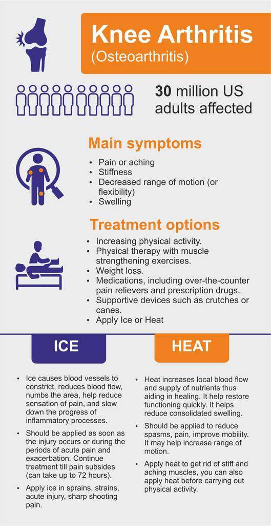
Reconstruction of the anterior cruciate ligament: comparison of outside-in and all-inside techniques. (1/4075)
The aim of this prospective study was to compare two arthroscopic techniques for reconstructing the anterior cruciate ligament, the "outside-in" (two incisions) and the "all-inside" (one incision) techniques. The results obtained for 30 patients operated on using the "outside-in" technique (group I) were compared with those for 29 patients operated on using the "all-inside" technique (group II). Before surgery, there were no significant differences between the groups in terms of Lysholm score, Tegner activity level, patellofemoral pain score, or knee laxity. Both groups displayed significant improvements in Lysholm score after 24 months, from 69 (16) to 91 (9) in group I and from 70 (17) to 90 (15) in group II (means (SD)). There were also significant improvements in patellofemoral pain scores in both groups, from 13 (6) to 18 (5) in group I and from 14 (6) to 18 (4) in group II after 24 months. No difference was found between the groups in knee stability at the 24 month follow up. The IKDC score was identical in both groups at follow up. The operation took significantly longer for patients in group I (mean 94 (15)) than for those in group II (mean 86 (20)) (p = 0.03). The mean sick leave was 7.7 (6.2) weeks in group I and 12.3 (9.7) weeks in group II (p = 0.026), indicating that there may be a higher morbidity associated with the "all-inside" technique. It can be concluded that there were no significant differences between the two different techniques in terms of functional results, knee laxity, or postoperative complications. The results were satisfactory and the outcome was similar in both treatment groups. (+info)Phase reversal of biomechanical functions and muscle activity in backward pedaling. (2/4075)
Computer simulations of pedaling have shown that a wide range of pedaling tasks can be performed if each limb has the capability of executing six biomechanical functions, which are arranged into three pairs of alternating antagonistic functions. An Ext/Flex pair accelerates the limb into extension or flexion, a Plant/Dorsi pair accelerates the foot into plantarflexion or dorsiflexion, and an Ant/Post pair accelerates the foot anteriorly or posteriorly relative to the pelvis. Because each biomechanical function (i.e., Ext, Flex, Plant, Dorsi, Ant, or Post) contributes to crank propulsion during a specific region in the cycle, phasing of a muscle is hypothesized to be a consequence of its ability to contribute to one or more of the biomechanical functions. Analysis of electromyogram (EMG) patterns has shown that this biomechanical framework assists in the interpretation of muscle activity in healthy and hemiparetic subjects during forward pedaling. Simulations show that backward pedaling can be produced with a phase shift of 180 degrees in the Ant/Post pair. No phase shifts in the Ext/Flex and Plant/Dorsi pairs are then necessary. To further test whether this simple yet biomechanically viable strategy may be used by the nervous system, EMGs from 7 muscles in 16 subjects were measured during backward as well as forward pedaling. As predicted, phasing in vastus medialis (VM), tibialis anterior (TA), medial gastrocnemius (MG), and soleus (SL) were unaffected by pedaling direction, with VM and SL contributing to Ext, MG to Plant, and TA to Dorsi. In contrast, phasing in biceps femoris (BF) and semimembranosus (SM) were affected by pedaling direction, as predicted, compatible with their contribution to the directionally sensitive Post function. Phasing of rectus femoris (RF) was also affected by pedaling direction; however, its ability to contribute to the directionally sensitive Ant function may only be expressed in forward pedaling. RF also contributed significantly to the directionally insensitive Ext function in both forward and backward pedaling. Other muscles also appear to have contributed to more than one function, which was especially evident in backward pedaling (i.e. , BF, SM, MG, and TA to Flex). We conclude that the phasing of only the Ant and Post biomechanical functions are directionally sensitive. Further, we suggest that task-dependent modulation of the expression of the functions in the motor output provides this biomechanics-based neural control scheme with the capability to execute a variety of lower limb tasks, including walking. (+info)Manipulation of total knee replacements. Is the flexion gained retained? (3/4075)
As part of a prospective study of 476 total knee replacements (TKR), we evaluated the use of manipulation under anaesthesia in 47 knees. Manipulation was considered when intensive physiotherapy failed to increase flexion to more than 80 degrees. The mean time from arthroplasty to manipulation was 11.3 weeks (median 9, range 2 to 41). The mean active flexion before manipulation was 62 degrees (35 to 80). One year later the mean gain was 33 degrees (Wilcoxon signed-rank test, range -5 to 70, 95% CI 28.5 to 38.5). Definite sustained gains in flexion were achieved even when manipulation was performed four or more months after arthroplasty (paired t-test, p < 0.01, CI 8.4 to 31.4). A further 21 patients who met our criteria for manipulation declined the procedure. Despite continued physiotherapy, there was no significant increase in flexion in their knees. Six weeks to one year after TKR, the mean change was 3.1 degrees (paired t-test, p = 0.23, CI -8.1 to +2). (+info)The tourniquet in total knee arthroplasty. A prospective, randomised study. (4/4075)
We assessed the influence of the use of a tourniquet in total knee arthroplasty in a prospective, randomised study. After satisfying exclusion criteria, we divided 77 patients into two groups, one to undergo surgery with a tourniquet and one without. Both groups were well matched. The mean change in knee flexion in the group that had surgery without a tourniquet was significantly better at one week (p = 0.03) than in the other group, but movement was similar at six weeks and at four months. There was no significant difference in the surgical time, postoperative pain, need for analgesia, the volume collected in the drains, postoperative swelling, and the incidence of wound complications or of deep-venous thrombosis. We conclude that the use of a tourniquet is safe and that current practice can be continued. (+info)The relationship between submaximal activity of the lumbar extensor muscles and lumbar posteroanterior stiffness. (5/4075)
BACKGROUND AND PURPOSE: Some patients with low back pain are thought to have increased lumbar posteroanterior (PA) stiffness. Increased activity of the lumbar extensors could contribute to this stiffness. This activity may be seen when a PA force is applied and is thought to represent much less force than occurs with a maximal voluntary contraction (MVC). Although MVCs of the lumbar extensors are known to increase lumbar PA stiffness, the effect of small amounts of voluntary contraction is not known. In this study, the effect of varying amounts of voluntary isometric muscle activity of the lumbar extensors on lumbar PA stiffness was examined. SUBJECTS: Twenty subjects without low back pain, aged 26 to 45 years (X=34, SD=5.6), participated in the study. METHODS: Subjects were asked to perform an isometric MVC of their lumbar extensor muscles with their pelvis fixed by exerting a force against a steel plate located over their T4 spinous process. They were then asked to perform contractions generating force equivalent to 0%, 10%, 30%, 50%, and 100% of that obtained with an MVC. Posteroanterior stiffness at L4 was measured during these contractions. RESULTS: A Friedman one-way analysis of variance for repeated measures demonstrated a difference in PA stiffness among all levels of muscle activity. CONCLUSION AND DISCUSSION: Voluntary contraction of the lumbar extensor muscles will result in an increase in lumbar PA stiffness even at low levels of activity. (+info)Effects of aggressive early rehabilitation on the outcome of anterior cruciate ligament reconstruction with multi-strand semitendinosus tendon. (6/4075)
To evaluate the effects of aggressive early rehabilitation on the clinical outcome of anterior cruciate ligament reconstruction using semitendinosus (and gracilis) tendon, 103 of 110 consecutive patients who underwent ACL reconstruction using multistrand semitendinosus tendon (ST) or the central one-third of patellar tendon with bony attachments (BTB) were analyzed prospectively. Subjectively, the Lysholm score was not different among the groups. The Lachman test indicated a trend of less negative grade in the ST men's group than that in the BTB men's group. On the patellofemoral grinding test, only women patients of both groups showed pain, with less positive crepitation in the ST group than in the BTB group. KT measurements at manual maximum showed more patients with more than 5 mm differences in the ST group than in the BTB group. The results of this study suggest that aggressive early rehabilitation after the ACL reconstruction using the semitendinosus (and gracilis) tendon has more risk of residual laxity than with the BTB. (+info)The role of fibular length and the width of the ankle mortise in post-traumatic osteoarthrosis after malleolar fracture. (7/4075)
We assessed the role of fibular length and the width of the ankle mortise as risk factors in the occurrence of post-traumatic osteoarthritis of the ankle joint by comparison of radiographs of the affected and unaffected sides. A shortened fibular malleolus (P < 0.01), a wide ankle mortise (P < 0.01) and Weber type B fracture (P < 0.01) were significantly associated with the development of osteoarthrosis but an elongated fibular (P > 0.05) and a narrowing of the ankle mortise (P > 0.07) were not. (+info)Modified Bankart procedure for recurrent anterior dislocation and subluxation of the shoulder in athletes. (8/4075)
Thirty-four athletes (34 shoulders) with recurrent anterior glenohumeral instability were treated with a modified Bankart procedure, using a T-shaped capsular incision in the anterior capsule. The inferior flap was advanced medially and/or superiorly and rigidly fixed at the point of the Bankart lesion by a small cancellous screw and a spike-washer. The superior flap was advanced inferiority and sutured over the inferior flap. Twenty-five athletes (median age: 22) were evaluated over a mean period of follow-up of 65 months. The clinical results were graded, according to Rowe, as 22 (88%) excellent, 3 (12%) good, and none as fair or poor. The mean postoperative range of movement was 92 degrees of external rotation in 90 degrees of abduction. Elevation and internal rotation was symmetrical with the opposite side. Twenty-four patients returned to active sport, 22 at their previous level. This modified Bankart procedure is an effective treatment for athletes with recurrent anterior glenohumeral instability. (+info)In the context of medical terminology, "motion" generally refers to the act or process of moving or changing position. It can also refer to the range of movement of a body part or joint. However, there is no single specific medical definition for the term "motion." The meaning may vary depending on the context in which it is used.
Motion perception is the ability to interpret and understand the movement of objects in our environment. It is a complex process that involves multiple areas of the brain and the visual system. In medical terms, motion perception refers to the specific function of the visual system to detect and analyze the movement of visual stimuli. This allows us to perceive and respond to moving objects in our environment, which is crucial for activities such as driving, sports, and even maintaining balance. Disorders in motion perception can lead to conditions like motion sickness or difficulty with depth perception.
Articular Range of Motion (AROM) is a term used in physiotherapy and orthopedics to describe the amount of movement available in a joint, measured in degrees of a circle. It refers to the range through which synovial joints can actively move without causing pain or injury. AROM is assessed by measuring the degree of motion achieved by active muscle contraction, as opposed to passive range of motion (PROM), where the movement is generated by an external force.
Assessment of AROM is important in evaluating a patient's functional ability and progress, planning treatment interventions, and determining return to normal activities or sports participation. It is also used to identify any restrictions in joint mobility that may be due to injury, disease, or surgery, and to monitor the effectiveness of rehabilitation programs.
Motion sickness is a condition characterized by a disturbance in the balance and orientation senses, often triggered by conflicting information received from the eyes, inner ears, and other bodily sensory systems. It's typically brought on by motion such as that experienced during travel in cars, trains, boats, or airplanes, or even while using virtual reality devices. Symptoms can include dizziness, nausea, vomiting, and cold sweats.
The inner ear's vestibular system plays a key role in this condition. When the body is in motion but the inner ear remains still, or vice versa, it can cause the brain to receive conflicting signals about the body's state of motion, leading to feelings of disorientation and sickness.
Preventative measures for motion sickness include fixating on a stationary point outside the vehicle, avoiding reading or looking at electronic screens during travel, taking over-the-counter medications like dimenhydrinate (Dramamine) or scopolamine (Transderm Scop), and engaging in relaxation techniques such as deep breathing.
In the context of medicine and healthcare, "movement" refers to the act or process of changing physical location or position. It involves the contraction and relaxation of muscles, which allows for the joints to move and the body to be in motion. Movement can also refer to the ability of a patient to move a specific body part or limb, which is assessed during physical examinations. Additionally, "movement" can describe the progression or spread of a disease within the body.
Photic stimulation is a medical term that refers to the exposure of the eyes to light, specifically repetitive pulses of light, which is used as a method in various research and clinical settings. In neuroscience, it's often used in studies related to vision, circadian rhythms, and brain function.
In a clinical context, photic stimulation is sometimes used in the diagnosis of certain medical conditions such as seizure disorders (like epilepsy). By observing the response of the brain to this light stimulus, doctors can gain valuable insights into the functioning of the brain and the presence of any neurological disorders.
However, it's important to note that photic stimulation should be conducted under the supervision of a trained healthcare professional, as improper use can potentially trigger seizures in individuals who are susceptible to them.
Psychophysics is not a medical term per se, but rather a subfield of psychology and neuroscience that studies the relationship between physical stimuli and the sensations and perceptions they produce. It involves the quantitative investigation of psychological functions, such as how brightness or loudness is perceived relative to the physical intensity of light or sound.
In medical contexts, psychophysical methods may be used in research or clinical settings to understand how patients with neurological conditions or sensory impairments perceive and respond to different stimuli. This information can inform diagnostic assessments, treatment planning, and rehabilitation strategies.
Optical illusions are visual phenomena that occur when the brain perceives an image or scene differently from the actual physical properties of that image or scene. They often result from the brain's attempt to interpret and make sense of ambiguous, contradictory, or incomplete information provided by the eyes. This can lead to visually perceived images that are different from the objective reality. Optical illusions can be categorized into different types such as literal illusions, physiological illusions, and cognitive illusions, based on the nature of the illusion and the underlying cause.
In the context of medicine, particularly in anatomy and physiology, "rotation" refers to the movement of a body part around its own axis or the long axis of another structure. This type of motion is three-dimensional and can occur in various planes. A common example of rotation is the movement of the forearm bones (radius and ulna) around each other during pronation and supination, which allows the hand to be turned palm up or down. Another example is the rotation of the head during mastication (chewing), where the mandible moves in a circular motion around the temporomandibular joint.
"Figural aftereffect" is not a widely recognized or established term in medical or clinical neuroscience literature. However, it seems to be related to the concept of "perceptual aftereffects," which are well-documented phenomena in visual and other sensory perception. Here's a definition that may help you understand figural aftereffects within this context:
Perceptual aftereffect is a phenomenon where exposure to a specific stimulus for a certain period can temporarily alter the perception of subsequent stimuli, making them appear different from what they would have been without the initial exposure. This effect arises due to neural adaptation in response to the prolonged exposure.
In the case of "figural aftereffect," it likely refers to a specific type of perceptual aftereffect where the perception of figures or shapes is affected by prior exposure. For example, if someone stares at a curved line for a while and then looks at a straight line, they might initially perceive the straight line as being more curved than it actually is due to the lingering influence of the initial stimulus.
However, since "figural aftereffect" isn't a standard term in medical or neuroscience literature, I would recommend consulting original research articles or experts in visual perception for a more precise definition and context.
Biomechanics is the application of mechanical laws to living structures and systems, particularly in the field of medicine and healthcare. A biomechanical phenomenon refers to a observable event or occurrence that involves the interaction of biological tissues or systems with mechanical forces. These phenomena can be studied at various levels, from the molecular and cellular level to the tissue, organ, and whole-body level.
Examples of biomechanical phenomena include:
1. The way that bones and muscles work together to produce movement (known as joint kinematics).
2. The mechanical behavior of biological tissues such as bone, cartilage, tendons, and ligaments under various loads and stresses.
3. The response of cells and tissues to mechanical stimuli, such as the way that bone tissue adapts to changes in loading conditions (known as Wolff's law).
4. The biomechanics of injury and disease processes, such as the mechanisms of joint injury or the development of osteoarthritis.
5. The use of mechanical devices and interventions to treat medical conditions, such as orthopedic implants or assistive devices for mobility impairments.
Understanding biomechanical phenomena is essential for developing effective treatments and prevention strategies for a wide range of medical conditions, from musculoskeletal injuries to neurological disorders.
Sensory thresholds are the minimum levels of stimulation that are required to produce a sensation in an individual, as determined through psychophysical testing. These tests measure the point at which a person can just barely detect the presence of a stimulus, such as a sound, light, touch, or smell.
There are two types of sensory thresholds: absolute and difference. Absolute threshold is the minimum level of intensity required to detect a stimulus 50% of the time. Difference threshold, also known as just noticeable difference (JND), is the smallest change in intensity that can be detected between two stimuli.
Sensory thresholds can vary between individuals and are influenced by factors such as age, attention, motivation, and expectations. They are often used in clinical settings to assess sensory function and diagnose conditions such as hearing or vision loss.
An artifact, in the context of medical terminology, refers to something that is created or introduced during a scientific procedure or examination that does not naturally occur in the patient or specimen being studied. Artifacts can take many forms and can be caused by various factors, including contamination, damage, degradation, or interference from equipment or external sources.
In medical imaging, for example, an artifact might appear as a distortion or anomaly on an X-ray, MRI, or CT scan that is not actually present in the patient's body. This can be caused by factors such as patient movement during the scan, metal implants or other foreign objects in the body, or issues with the imaging equipment itself.
Similarly, in laboratory testing, an artifact might refer to a substance or characteristic that is introduced into a sample during collection, storage, or analysis that can interfere with accurate results. This could include things like contamination from other samples, degradation of the sample over time, or interference from chemicals used in the testing process.
In general, artifacts are considered to be sources of error or uncertainty in medical research and diagnosis, and it is important to identify and account for them in order to ensure accurate and reliable results.
Depth perception is the ability to accurately judge the distance or separation of an object in three-dimensional space. It is a complex visual process that allows us to perceive the world in three dimensions and to understand the spatial relationships between objects.
Depth perception is achieved through a combination of monocular cues, which are visual cues that can be perceived with one eye, and binocular cues, which require input from both eyes. Monocular cues include perspective (the relative size of objects), texture gradients (finer details become smaller as distance increases), and atmospheric perspective (colors become less saturated and lighter in value as distance increases). Binocular cues include convergence (the degree to which the eyes must turn inward to focus on an object) and retinal disparity (the slight difference in the images projected onto the two retinas due to the slightly different positions of the eyes).
Deficits in depth perception can occur due to a variety of factors, including eye disorders, brain injuries, or developmental delays. These deficits can result in difficulties with tasks such as driving, sports, or navigating complex environments. Treatment for depth perception deficits may include vision therapy, corrective lenses, or surgery.
Visual pattern recognition is the ability to identify and interpret patterns in visual information. In a medical context, it often refers to the process by which healthcare professionals recognize and diagnose medical conditions based on visible signs or symptoms. This can involve recognizing the characteristic appearance of a rash, wound, or other physical feature associated with a particular disease or condition. It may also involve recognizing patterns in medical images such as X-rays, CT scans, or MRIs.
In the field of radiology, for example, visual pattern recognition is a critical skill. Radiologists are trained to recognize the typical appearances of various diseases and conditions in medical images. This allows them to make accurate diagnoses based on the patterns they see. Similarly, dermatologists use visual pattern recognition to identify skin abnormalities and diseases based on the appearance of rashes, lesions, or other skin changes.
Overall, visual pattern recognition is an essential skill in many areas of medicine, allowing healthcare professionals to quickly and accurately diagnose medical conditions based on visible signs and symptoms.
Eye movements, also known as ocular motility, refer to the voluntary or involuntary motion of the eyes that allows for visual exploration of our environment. There are several types of eye movements, including:
1. Saccades: rapid, ballistic movements that quickly shift the gaze from one point to another.
2. Pursuits: smooth, slow movements that allow the eyes to follow a moving object.
3. Vergences: coordinated movements of both eyes in opposite directions, usually in response to a three-dimensional stimulus.
4. Vestibulo-ocular reflex (VOR): automatic eye movements that help stabilize the gaze during head movement.
5. Optokinetic nystagmus (OKN): rhythmic eye movements that occur in response to large moving visual patterns, such as when looking out of a moving vehicle.
Abnormalities in eye movements can indicate neurological or ophthalmological disorders and are often assessed during clinical examinations.
Contrast sensitivity is a measure of the ability to distinguish between an object and its background based on differences in contrast, rather than differences in luminance. Contrast refers to the difference in light intensity between an object and its immediate surroundings. Contrast sensitivity is typically measured using specially designed charts that have patterns of parallel lines with varying widths and contrast levels.
In clinical settings, contrast sensitivity is often assessed as part of a comprehensive visual examination. Poor contrast sensitivity can affect a person's ability to perform tasks such as reading, driving, or distinguishing objects from their background, especially in low-light conditions. Reduced contrast sensitivity is a common symptom of various eye conditions, including cataracts, glaucoma, and age-related macular degeneration.
In the context of medical terminology, I believe you may be referring to "pursuit" as it relates to neurological tests. A smooth pursuit is a type of eye movement in which the eyes smoothly and slowly follow a moving object. It requires coordination between the extraocular muscles, vestibular system, and visual system. If there are issues with any of these systems, smooth pursuit can be affected, leading to abnormalities such as jerky or saccadic movements.
Therefore, "smooth pursuit" is a medical term used to describe the normal, coordinated movement of the eyes that allows for the tracking of moving objects in a smooth and continuous manner.
I'm sorry for any confusion, but "Motion Pictures as Topic" is not a medical term or concept. It is actually a subject heading used in library and information sciences to categorize materials related to the study or analysis of motion pictures as a medium or art form. This could include books, articles, and other resources about film theory, film history, film criticism, and so on.
If you have any questions about medical terminology or concepts, I would be happy to help!
Visual pathways, also known as the visual system or the optic pathway, refer to the series of specialized neurons in the nervous system that transmit visual information from the eyes to the brain. This complex network includes the retina, optic nerve, optic chiasma, optic tract, lateral geniculate nucleus, pulvinar, and the primary and secondary visual cortices located in the occipital lobe of the brain.
The process begins when light enters the eye and strikes the photoreceptor cells (rods and cones) in the retina, converting the light energy into electrical signals. These signals are then transmitted to bipolar cells and subsequently to ganglion cells, whose axons form the optic nerve. The fibers from each eye's nasal hemiretina cross at the optic chiasma, while those from the temporal hemiretina continue without crossing. This results in the formation of the optic tract, which carries visual information from both eyes to the opposite side of the brain.
The majority of fibers in the optic tract synapse with neurons in the lateral geniculate nucleus (LGN), a part of the thalamus. The LGN sends this information to the primary visual cortex, also known as V1 or Brodmann area 17, located in the occipital lobe. Here, simple features like lines and edges are initially processed. Further processing occurs in secondary (V2) and tertiary (V3-V5) visual cortices, where more complex features such as shape, motion, and depth are analyzed. Ultimately, this information is integrated to form our perception of the visual world.
Form perception, also known as shape perception, is not a term that has a specific medical definition. However, in the field of neuropsychology and sensory perception, form perception refers to the ability to recognize and interpret different shapes and forms of objects through visual processing. This ability is largely dependent on the integrity of the visual cortex and its ability to process and interpret information received from the retina.
Damage to certain areas of the brain, particularly in the occipital and parietal lobes, can result in deficits in form perception, leading to difficulties in recognizing and identifying objects based on their shape or form. This condition is known as visual agnosia and can be a symptom of various neurological disorders such as stroke, brain injury, or degenerative diseases like Alzheimer's disease.
In a medical context, "orientation" typically refers to an individual's awareness and understanding of their personal identity, place, time, and situation. It is a critical component of cognitive functioning and mental status. Healthcare professionals often assess a person's orientation during clinical evaluations, using tests that inquire about their name, location, the current date, and the circumstances of their hospitalization or visit.
There are different levels of orientation:
1. Person (or self): The individual knows their own identity, including their name, age, and other personal details.
2. Place: The individual is aware of where they are, such as the name of the city, hospital, or healthcare facility.
3. Time: The individual can accurately state the current date, day of the week, month, and year.
4. Situation or event: The individual understands why they are in the healthcare setting, what happened leading to their hospitalization or visit, and the nature of any treatments or procedures they are undergoing.
Impairments in orientation can be indicative of various neurological or psychiatric conditions, such as delirium, dementia, or substance intoxication or withdrawal. It is essential for healthcare providers to monitor and address orientation issues to ensure appropriate diagnosis, treatment, and patient safety.
An illusion is a perception in the brain that does not match the actual stimulus in the environment. It is often described as a false or misinterpreted sensory experience, where the senses perceive something that is different from the reality. Illusions can occur in any of the senses, including vision, hearing, touch, taste, and smell.
In medical terms, illusions are sometimes associated with certain neurological conditions, such as migraines, brain injuries, or mental health disorders like schizophrenia. They can also be a side effect of certain medications or substances. In these cases, the illusions may be a symptom of an underlying medical condition and should be evaluated by a healthcare professional.
It's important to note that while illusions are often used in the context of entertainment and art, they can also have serious implications for individuals who experience them frequently or as part of a medical condition.
Vision disparity, also known as binocular vision disparity, refers to the difference in the image that is perceived by each eye. This can occur due to a variety of reasons such as misalignment of the eyes (strabismus), unequal refractive power in each eye (anisometropia), or abnormalities in the shape of the eye (astigmatism).
When there is a significant difference in the image that is perceived by each eye, the brain may have difficulty combining the two images into a single, three-dimensional perception. This can result in visual symptoms such as double vision (diplopia), eyestrain, headaches, and difficulty with depth perception.
Vision disparity can be detected through a comprehensive eye examination and may be treated with corrective lenses, prism lenses, vision therapy, or surgery, depending on the underlying cause and severity of the condition.
Continuous Passive Motion (CPM) therapy is a type of motion therapy that is often used in physical rehabilitation following surgery or injury. In CPM therapy, the affected body part is moved continuously through a range of motion without any active participation from the patient. This is typically accomplished with the use of a motorized device that gently and slowly moves the limb.
The goal of CPM therapy is to help prevent stiffness, reduce pain, improve circulation, and promote healing in the affected area. It is often used following joint replacement surgery, such as knee or hip replacements, as well as after injuries that limit mobility and range of motion. By providing continuous, passive movement to the affected limb, CPM therapy can help prevent the formation of scar tissue and adhesions, which can restrict movement and cause pain.
CPM therapy is usually prescribed by a healthcare provider and administered under the supervision of a physical therapist or other rehabilitation specialist. The range of motion and speed of the movement are carefully controlled to ensure safety and effectiveness. While CPM therapy can be an important part of the recovery process, it is typically used in conjunction with other rehabilitation techniques, such as exercises and manual therapy, to achieve optimal outcomes.
Binocular vision refers to the ability to use both eyes together to create a single, three-dimensional image of our surroundings. This is achieved through a process called binocular fusion, where the images from each eye are aligned and combined in the brain to form a unified perception.
The term "binocular vision" specifically refers to the way that our visual system integrates information from both eyes to create depth perception and enhance visual clarity. When we view an object with both eyes, they focus on the same point in space and send slightly different images to the brain due to their slightly different positions. The brain then combines these images to create a single, three-dimensional image that allows us to perceive depth and distance.
Binocular vision is important for many everyday activities, such as driving, reading, and playing sports. Disorders of binocular vision can lead to symptoms such as double vision, eye strain, and difficulty with depth perception.
The visual cortex is the part of the brain that processes visual information. It is located in the occipital lobe, which is at the back of the brain. The visual cortex is responsible for receiving and interpreting signals from the retina, which are then transmitted through the optic nerve and optic tract.
The visual cortex contains several areas that are involved in different aspects of visual processing, such as identifying shapes, colors, and movements. These areas work together to help us recognize and understand what we see. Damage to the visual cortex can result in various visual impairments, such as blindness or difficulty with visual perception.
Head movements refer to the voluntary or involuntary motion of the head in various directions. These movements can occur in different planes, including flexion (moving the head forward), extension (moving the head backward), rotation (turning the head to the side), and lateral bending (leaning the head to one side).
Head movements can be a result of normal physiological processes, such as when nodding in agreement or shaking the head to indicate disagreement. They can also be caused by neurological conditions, such as abnormal head movements in patients with Parkinson's disease or cerebellar disorders. Additionally, head movements may occur in response to sensory stimuli, such as turning the head toward a sound.
In a medical context, an examination of head movements can provide important clues about a person's neurological function and help diagnose various conditions affecting the brain and nervous system.
I'm sorry for any confusion, but "lighting" does not have a specific medical definition. In a general context, lighting refers to the arrangement or use of light sources to achieve a particular effect or atmosphere. However, if you are referring to a term in medicine that may be similar to "lighting," you might be thinking of "lumination" or "illumination," which refer to the act of providing or admitting light, especially for medical examination or surgical procedures. I hope this helps! If you have any other questions, please don't hesitate to ask.
Optic flow is not a medical term per se, but rather a term used in the field of visual perception and neuroscience. It refers to the pattern of motion of objects in the visual field that occurs as an observer moves through the environment. This pattern of motion is important for the perception of self-motion and the estimation of egocentric distance (the distance of objects in the environment relative to the observer). Optic flow has been studied in relation to various clinical populations, such as individuals with vestibular disorders or visual impairments, who may have difficulty processing optic flow information.
A computer simulation is a process that involves creating a model of a real-world system or phenomenon on a computer and then using that model to run experiments and make predictions about how the system will behave under different conditions. In the medical field, computer simulations are used for a variety of purposes, including:
1. Training and education: Computer simulations can be used to create realistic virtual environments where medical students and professionals can practice their skills and learn new procedures without risk to actual patients. For example, surgeons may use simulation software to practice complex surgical techniques before performing them on real patients.
2. Research and development: Computer simulations can help medical researchers study the behavior of biological systems at a level of detail that would be difficult or impossible to achieve through experimental methods alone. By creating detailed models of cells, tissues, organs, or even entire organisms, researchers can use simulation software to explore how these systems function and how they respond to different stimuli.
3. Drug discovery and development: Computer simulations are an essential tool in modern drug discovery and development. By modeling the behavior of drugs at a molecular level, researchers can predict how they will interact with their targets in the body and identify potential side effects or toxicities. This information can help guide the design of new drugs and reduce the need for expensive and time-consuming clinical trials.
4. Personalized medicine: Computer simulations can be used to create personalized models of individual patients based on their unique genetic, physiological, and environmental characteristics. These models can then be used to predict how a patient will respond to different treatments and identify the most effective therapy for their specific condition.
Overall, computer simulations are a powerful tool in modern medicine, enabling researchers and clinicians to study complex systems and make predictions about how they will behave under a wide range of conditions. By providing insights into the behavior of biological systems at a level of detail that would be difficult or impossible to achieve through experimental methods alone, computer simulations are helping to advance our understanding of human health and disease.
An afterimage is a visual phenomenon that occurs when the eye's retina continues to send signals to the brain even after exposure to a stimulus has ended. This can result in the perception of a lingering image, often in complementary colors to the original stimulus. Afterimages can be either positive or negative, with a positive afterimage appearing as the same color as the original stimulus and a negative afterimage appearing as its complementary color.
Afterimages are typically caused by exposure to bright or intense light sources, such as a camera flash or the sun. They can also occur after prolonged exposure to a particular color or pattern. The phenomenon is thought to be related to the adaptation of photoreceptor cells in the retina, which become less responsive to stimuli after prolonged exposure.
Afterimages are generally harmless and temporary, lasting only a few seconds to several minutes. However, they can sometimes be used as a tool for visual perception experiments or to study the mechanisms of visual processing in the brain.
In the field of medicine, "time factors" refer to the duration of symptoms or time elapsed since the onset of a medical condition, which can have significant implications for diagnosis and treatment. Understanding time factors is crucial in determining the progression of a disease, evaluating the effectiveness of treatments, and making critical decisions regarding patient care.
For example, in stroke management, "time is brain," meaning that rapid intervention within a specific time frame (usually within 4.5 hours) is essential to administering tissue plasminogen activator (tPA), a clot-busting drug that can minimize brain damage and improve patient outcomes. Similarly, in trauma care, the "golden hour" concept emphasizes the importance of providing definitive care within the first 60 minutes after injury to increase survival rates and reduce morbidity.
Time factors also play a role in monitoring the progression of chronic conditions like diabetes or heart disease, where regular follow-ups and assessments help determine appropriate treatment adjustments and prevent complications. In infectious diseases, time factors are crucial for initiating antibiotic therapy and identifying potential outbreaks to control their spread.
Overall, "time factors" encompass the significance of recognizing and acting promptly in various medical scenarios to optimize patient outcomes and provide effective care.
In the context of medicine and physiology, acceleration refers to the process of increasing or quickening a function or process. For example, heart rate acceleration is an increase in the speed at which the heart beats. It can also refer to the rate at which something increases, such as the acceleration of muscle strength during rehabilitation. In physics terms, acceleration refers to the rate at which an object changes its velocity, but this definition is not typically used in a medical context.
In the context of medicine, "cues" generally refer to specific pieces of information or signals that can help healthcare professionals recognize and respond to a particular situation or condition. These cues can come in various forms, such as:
1. Physical examination findings: For example, a patient's abnormal heart rate or blood pressure reading during a physical exam may serve as a cue for the healthcare professional to investigate further.
2. Patient symptoms: A patient reporting chest pain, shortness of breath, or other concerning symptoms can act as a cue for a healthcare provider to consider potential diagnoses and develop an appropriate treatment plan.
3. Laboratory test results: Abnormal findings on laboratory tests, such as elevated blood glucose levels or abnormal liver function tests, may serve as cues for further evaluation and diagnosis.
4. Medical history information: A patient's medical history can provide valuable cues for healthcare professionals when assessing their current health status. For example, a history of smoking may increase the suspicion for chronic obstructive pulmonary disease (COPD) in a patient presenting with respiratory symptoms.
5. Behavioral or environmental cues: In some cases, behavioral or environmental factors can serve as cues for healthcare professionals to consider potential health risks. For instance, exposure to secondhand smoke or living in an area with high air pollution levels may increase the risk of developing respiratory conditions.
Overall, "cues" in a medical context are essential pieces of information that help healthcare professionals make informed decisions about patient care and treatment.
An algorithm is not a medical term, but rather a concept from computer science and mathematics. In the context of medicine, algorithms are often used to describe step-by-step procedures for diagnosing or managing medical conditions. These procedures typically involve a series of rules or decision points that help healthcare professionals make informed decisions about patient care.
For example, an algorithm for diagnosing a particular type of heart disease might involve taking a patient's medical history, performing a physical exam, ordering certain diagnostic tests, and interpreting the results in a specific way. By following this algorithm, healthcare professionals can ensure that they are using a consistent and evidence-based approach to making a diagnosis.
Algorithms can also be used to guide treatment decisions. For instance, an algorithm for managing diabetes might involve setting target blood sugar levels, recommending certain medications or lifestyle changes based on the patient's individual needs, and monitoring the patient's response to treatment over time.
Overall, algorithms are valuable tools in medicine because they help standardize clinical decision-making and ensure that patients receive high-quality care based on the latest scientific evidence.
Space perception, in the context of neuroscience and psychology, refers to the ability to perceive and understand the spatial arrangement of objects and their relationship to oneself. It involves integrating various sensory inputs such as visual, auditory, tactile, and proprioceptive information to create a coherent three-dimensional representation of our environment.
This cognitive process enables us to judge distances, sizes, shapes, and movements of objects around us. It also helps us navigate through space, reach for objects, avoid obstacles, and maintain balance. Disorders in space perception can lead to difficulties in performing everyday activities and may be associated with neurological conditions such as stroke, brain injury, or neurodevelopmental disorders like autism.
Visual perception refers to the ability to interpret and organize information that comes from our eyes to recognize and understand what we are seeing. It involves several cognitive processes such as pattern recognition, size estimation, movement detection, and depth perception. Visual perception allows us to identify objects, navigate through space, and interact with our environment. Deficits in visual perception can lead to learning difficulties and disabilities.
Biological models, also known as physiological models or organismal models, are simplified representations of biological systems, processes, or mechanisms that are used to understand and explain the underlying principles and relationships. These models can be theoretical (conceptual or mathematical) or physical (such as anatomical models, cell cultures, or animal models). They are widely used in biomedical research to study various phenomena, including disease pathophysiology, drug action, and therapeutic interventions.
Examples of biological models include:
1. Mathematical models: These use mathematical equations and formulas to describe complex biological systems or processes, such as population dynamics, metabolic pathways, or gene regulation networks. They can help predict the behavior of these systems under different conditions and test hypotheses about their underlying mechanisms.
2. Cell cultures: These are collections of cells grown in a controlled environment, typically in a laboratory dish or flask. They can be used to study cellular processes, such as signal transduction, gene expression, or metabolism, and to test the effects of drugs or other treatments on these processes.
3. Animal models: These are living organisms, usually vertebrates like mice, rats, or non-human primates, that are used to study various aspects of human biology and disease. They can provide valuable insights into the pathophysiology of diseases, the mechanisms of drug action, and the safety and efficacy of new therapies.
4. Anatomical models: These are physical representations of biological structures or systems, such as plastic models of organs or tissues, that can be used for educational purposes or to plan surgical procedures. They can also serve as a basis for developing more sophisticated models, such as computer simulations or 3D-printed replicas.
Overall, biological models play a crucial role in advancing our understanding of biology and medicine, helping to identify new targets for therapeutic intervention, develop novel drugs and treatments, and improve human health.
Ocular fixation is a term used in ophthalmology and optometry to refer to the ability of the eyes to maintain steady gaze or visual focus on an object. It involves the coordinated movement of the extraocular muscles that control eye movements, allowing for clear and stable vision.
In medical terminology, fixation specifically refers to the state in which the eyes are aligned and focused on a single point in space. This is important for maintaining visual perception and preventing blurring or double vision. Ocular fixation can be affected by various factors such as muscle weakness, nerve damage, or visual processing disorders.
Assessment of ocular fixation is often used in eye examinations to evaluate visual acuity, eye alignment, and muscle function. Abnormalities in fixation may indicate the presence of underlying eye conditions or developmental delays that require further investigation and treatment.
Color perception refers to the ability to detect, recognize, and differentiate various colors and color patterns in the visual field. This complex process involves the functioning of both the eyes and the brain.
The eye's retina contains two types of photoreceptor cells called rods and cones. Rods are more sensitive to light and dark changes and help us see in low-light conditions, but they do not contribute much to color vision. Cones, on the other hand, are responsible for color perception and function best in well-lit conditions.
There are three types of cone cells, each sensitive to a particular range of wavelengths corresponding to blue, green, and red colors. The combination of signals from these three types of cones allows us to perceive a wide spectrum of colors.
The brain then interprets these signals and translates them into the perception of different colors and hues. It is important to note that color perception can be influenced by various factors, including cultural background, personal experiences, and even language. Some individuals may also have deficiencies in color perception due to genetic or acquired conditions, such as color blindness or cataracts.
Ocular adaptation is the ability of the eye to adjust and accommodate to changes in visual input and lighting conditions. This process allows the eye to maintain a clear and focused image over a range of different environments and light levels. There are several types of ocular adaptation, including:
1. Light Adaptation: This refers to the eye's ability to adjust to different levels of illumination. When moving from a dark environment to a bright one, the pupils constrict to let in less light, and the sensitivity of the retina decreases. Conversely, when moving from a bright environment to a dark one, the pupils dilate to let in more light, and the sensitivity of the retina increases.
2. Dark Adaptation: This is the process by which the eye adjusts to low light conditions. It involves the dilation of the pupils and an increase in the sensitivity of the rods (specialised cells in the retina that are responsible for vision in low light conditions). Dark adaptation can take several minutes to occur fully.
3. Color Adaptation: This refers to the eye's ability to adjust to changes in the color temperature of light sources. For example, when moving from a room lit by incandescent light to one lit by fluorescent light, the eye may need to adjust its perception of colors to maintain accurate color vision.
4. Accommodation: This is the process by which the eye changes focus from distant to near objects. The lens of the eye changes shape to bend the light rays entering the eye and bring them into sharp focus on the retina.
Overall, ocular adaptation is an essential function that allows us to see clearly and accurately in a wide range of environments and lighting conditions.
Respiratory-gated imaging techniques are medical imaging procedures that synchronize the data acquisition with the patient's respiratory cycle, in order to reduce motion artifacts and improve image quality. These techniques are often used in CT (computed tomography) and MR (magnetic resonance) imaging for thoracic and abdominal examinations, where respiratory motion can degrade the images and compromise diagnostic accuracy.
In a respiratory-gated imaging technique, the patient's breathing pattern is monitored using sensors such as pressure belts or navigators, which detect the movement of the diaphragm or chest wall. The imaging data are then acquired only during specific phases of the respiratory cycle, typically during the end-expiration phase when motion is minimal. This allows for the creation of sharp and detailed images that accurately represent the anatomy and pathology of interest.
Respiratory gating can be particularly useful in imaging patients with lung cancer, liver tumors, or other conditions that involve moving structures in the chest and abdomen. By reducing motion artifacts, these techniques can help ensure more accurate diagnosis, staging, and treatment planning.
Computer-assisted image processing is a medical term that refers to the use of computer systems and specialized software to improve, analyze, and interpret medical images obtained through various imaging techniques such as X-ray, CT (computed tomography), MRI (magnetic resonance imaging), ultrasound, and others.
The process typically involves several steps, including image acquisition, enhancement, segmentation, restoration, and analysis. Image processing algorithms can be used to enhance the quality of medical images by adjusting contrast, brightness, and sharpness, as well as removing noise and artifacts that may interfere with accurate diagnosis. Segmentation techniques can be used to isolate specific regions or structures of interest within an image, allowing for more detailed analysis.
Computer-assisted image processing has numerous applications in medical imaging, including detection and characterization of lesions, tumors, and other abnormalities; assessment of organ function and morphology; and guidance of interventional procedures such as biopsies and surgeries. By automating and standardizing image analysis tasks, computer-assisted image processing can help to improve diagnostic accuracy, efficiency, and consistency, while reducing the potential for human error.
Three-dimensional (3D) imaging in medicine refers to the use of technologies and techniques that generate a 3D representation of internal body structures, organs, or tissues. This is achieved by acquiring and processing data from various imaging modalities such as X-ray computed tomography (CT), magnetic resonance imaging (MRI), ultrasound, or confocal microscopy. The resulting 3D images offer a more detailed visualization of the anatomy and pathology compared to traditional 2D imaging techniques, allowing for improved diagnostic accuracy, surgical planning, and minimally invasive interventions.
In 3D imaging, specialized software is used to reconstruct the acquired data into a volumetric model, which can be manipulated and viewed from different angles and perspectives. This enables healthcare professionals to better understand complex anatomical relationships, detect abnormalities, assess disease progression, and monitor treatment response. Common applications of 3D imaging include neuroimaging, orthopedic surgery planning, cancer staging, dental and maxillofacial reconstruction, and interventional radiology procedures.
Fiducial markers, also known as fiducials, are small markers that are often used in medical imaging to help identify and target specific locations within the body. These markers can be made of various materials, such as metal or plastic, and are typically placed at or near the site of interest through a minimally invasive procedure.
In radiation therapy, fiducial markers are often used to help ensure that the treatment is accurately targeted to the correct location. The markers can be seen on imaging scans, such as X-rays or CT scans, and can be used to align the treatment beam with the target area. This helps to improve the precision of the radiation therapy and reduce the risk of harm to surrounding healthy tissue.
Fiducial markers may also be used in other medical procedures, such as image-guided surgery or interventional radiology, to help guide the placement of instruments or devices within the body.
Reproducibility of results in a medical context refers to the ability to obtain consistent and comparable findings when a particular experiment or study is repeated, either by the same researcher or by different researchers, following the same experimental protocol. It is an essential principle in scientific research that helps to ensure the validity and reliability of research findings.
In medical research, reproducibility of results is crucial for establishing the effectiveness and safety of new treatments, interventions, or diagnostic tools. It involves conducting well-designed studies with adequate sample sizes, appropriate statistical analyses, and transparent reporting of methods and findings to allow other researchers to replicate the study and confirm or refute the results.
The lack of reproducibility in medical research has become a significant concern in recent years, as several high-profile studies have failed to produce consistent findings when replicated by other researchers. This has led to increased scrutiny of research practices and a call for greater transparency, rigor, and standardization in the conduct and reporting of medical research.
Medical Definition:
Magnetic Resonance Imaging (MRI) is a non-invasive diagnostic imaging technique that uses a strong magnetic field and radio waves to create detailed cross-sectional or three-dimensional images of the internal structures of the body. The patient lies within a large, cylindrical magnet, and the scanner detects changes in the direction of the magnetic field caused by protons in the body. These changes are then converted into detailed images that help medical professionals to diagnose and monitor various medical conditions, such as tumors, injuries, or diseases affecting the brain, spinal cord, heart, blood vessels, joints, and other internal organs. MRI does not use radiation like computed tomography (CT) scans.
Monocular vision refers to the ability to see and process visual information using only one eye. It is the type of vision that an individual has when they are using only one eye to look at something, while the other eye may be covered or not functioning. This can be contrasted with binocular vision, which involves the use of both eyes working together to provide depth perception and a single, combined visual field.
Monocular vision is important for tasks that only require the use of one eye, such as when looking through a microscope or using a telescope. However, it does not provide the same level of depth perception and spatial awareness as binocular vision. In some cases, individuals may have reduced visual acuity or other visual impairments in one eye, leading to limited monocular vision in that eye. It is important for individuals with monocular vision to have regular eye exams to monitor their eye health and ensure that any visual impairments are detected and treated promptly.
Visual fields refer to the total area in which objects can be seen while keeping the eyes focused on a central point. It is the entire area that can be observed using peripheral (side) vision while the eye gazes at a fixed point. A visual field test is used to detect blind spots or gaps (scotomas) in a person's vision, which could indicate various medical conditions such as glaucoma, retinal damage, optic nerve disease, brain tumors, or strokes. The test measures both the central and peripheral vision and maps the entire area that can be seen when focusing on a single point.
Molecular models are three-dimensional representations of molecular structures that are used in the field of molecular biology and chemistry to visualize and understand the spatial arrangement of atoms and bonds within a molecule. These models can be physical or computer-generated and allow researchers to study the shape, size, and behavior of molecules, which is crucial for understanding their function and interactions with other molecules.
Physical molecular models are often made up of balls (representing atoms) connected by rods or sticks (representing bonds). These models can be constructed manually using materials such as plastic or wooden balls and rods, or they can be created using 3D printing technology.
Computer-generated molecular models, on the other hand, are created using specialized software that allows researchers to visualize and manipulate molecular structures in three dimensions. These models can be used to simulate molecular interactions, predict molecular behavior, and design new drugs or chemicals with specific properties. Overall, molecular models play a critical role in advancing our understanding of molecular structures and their functions.
Optokinetic nystagmus (OKN) is a type of involuntary eye movement that occurs in response to large moving visual patterns. It serves as a mechanism for stabilizing the image on the retina during head movement and helps in maintaining visual fixation.
In OKN, there are two phases of eye movement: a slow phase where the eyes follow or track the moving pattern, and a fast phase where the eyes quickly reset to the starting position. This results in a back-and-forth or "to-and-fro" motion of the eyes.
Optokinetic nystagmus can be elicited by observing a large moving object or a series of alternating visual stimuli, such as stripes on a rotating drum. It is often used in clinical settings to assess various aspects of the visual system, including oculomotor function and visual acuity.
Abnormalities in OKN can indicate problems with the vestibular system, brainstem, or cerebellum, and may be associated with conditions such as brain injury, multiple sclerosis, or cerebral palsy.
Neurological models are simplified representations or simulations of various aspects of the nervous system, including its structure, function, and processes. These models can be theoretical, computational, or physical and are used to understand, explain, and predict neurological phenomena. They may focus on specific neurological diseases, disorders, or functions, such as memory, learning, or movement. The goal of these models is to provide insights into the complex workings of the nervous system that cannot be easily observed or understood through direct examination alone.
In the field of medical imaging, "phantoms" refer to physical objects that are specially designed and used for calibration, quality control, and evaluation of imaging systems. These phantoms contain materials with known properties, such as attenuation coefficients or spatial resolution, which allow for standardized measurement and comparison of imaging parameters across different machines and settings.
Imaging phantoms can take various forms depending on the modality of imaging. For example, in computed tomography (CT), a common type of phantom is the "water-equivalent phantom," which contains materials with similar X-ray attenuation properties as water. This allows for consistent measurement of CT dose and image quality. In magnetic resonance imaging (MRI), phantoms may contain materials with specific relaxation times or magnetic susceptibilities, enabling assessment of signal-to-noise ratio, spatial resolution, and other imaging parameters.
By using these standardized objects, healthcare professionals can ensure the accuracy, consistency, and reliability of medical images, ultimately contributing to improved patient care and safety.
Perceptual disorders are conditions that affect the way a person perceives or interprets sensory information from their environment. These disorders can involve any of the senses, including sight, sound, touch, taste, and smell. They can cause a person to have difficulty recognizing, interpreting, or responding appropriately to sensory stimuli.
Perceptual disorders can result from damage to the brain or nervous system, such as from a head injury, stroke, or degenerative neurological condition. They can also be caused by certain mental health conditions, such as schizophrenia or severe depression.
Symptoms of perceptual disorders may include:
* Misinterpretations of sensory information, such as seeing things that are not there or hearing voices that are not present
* Difficulty recognizing familiar objects or people
* Problems with depth perception or spatial awareness
* Difficulty judging the size, shape, or distance of objects
* Trouble distinguishing between similar sounds or colors
* Impaired sense of smell or taste
Perceptual disorders can have a significant impact on a person's daily life and functioning. Treatment may involve medication, therapy, or rehabilitation to help the person better cope with their symptoms and improve their ability to interact with their environment.
Computer-assisted image interpretation is the use of computer algorithms and software to assist healthcare professionals in analyzing and interpreting medical images. These systems use various techniques such as pattern recognition, machine learning, and artificial intelligence to help identify and highlight abnormalities or patterns within imaging data, such as X-rays, CT scans, MRI, and ultrasound images. The goal is to increase the accuracy, consistency, and efficiency of image interpretation, while also reducing the potential for human error. It's important to note that these systems are intended to assist healthcare professionals in their decision making process and not to replace them.
Reaction time, in the context of medicine and physiology, refers to the time period between the presentation of a stimulus and the subsequent initiation of a response. This complex process involves the central nervous system, particularly the brain, which perceives the stimulus, processes it, and then sends signals to the appropriate muscles or glands to react.
There are different types of reaction times, including simple reaction time (responding to a single, expected stimulus) and choice reaction time (choosing an appropriate response from multiple possibilities). These measures can be used in clinical settings to assess various aspects of neurological function, such as cognitive processing speed, motor control, and alertness.
However, it is important to note that reaction times can be influenced by several factors, including age, fatigue, attention, and the use of certain medications or substances.
In a medical or psychological context, attention is the cognitive process of selectively concentrating on certain aspects of the environment while ignoring other things. It involves focusing mental resources on specific stimuli, sensory inputs, or internal thoughts while blocking out irrelevant distractions. Attention can be divided into different types, including:
1. Sustained attention: The ability to maintain focus on a task or stimulus over time.
2. Selective attention: The ability to concentrate on relevant stimuli while ignoring irrelevant ones.
3. Divided attention: The capacity to pay attention to multiple tasks or stimuli simultaneously.
4. Alternating attention: The skill of shifting focus between different tasks or stimuli as needed.
Deficits in attention are common symptoms of various neurological and psychiatric conditions, such as ADHD, dementia, depression, and anxiety disorders. Assessment of attention is an essential part of neuropsychological evaluations and can be measured using various tests and tasks.
Perceptual distortion is not explicitly defined within the realm of medicine, but it does fall under the broader category of cognitive impairments and abnormalities. It generally refers to the incorrect interpretation or misrepresentation of sensory information by the brain. This can result in various experiences such as hallucinations, illusions, or distorted perceptions of reality. Perceptual distortions are often associated with certain medical conditions like mental disorders (e.g., schizophrenia, bipolar disorder), neurological disorders (e.g., migraines, epilepsy), and substance use disorders.
Arthrometry is a measurement technique used in the field of orthopedics and rheumatology to assess the integrity and mobility of joints. When qualified with the term "articular," it specifically refers to the measurement of articular motion or range of motion (ROM) within a synovial joint.
Articular arthrometry involves using specialized instruments, such as goniometers, inclinometers, or digital devices like smartphone applications and wearable sensors, to quantify the degree of flexion, extension, abduction, adduction, rotation, or other movements in a joint. This information can help medical professionals evaluate joint function, diagnose injuries or conditions affecting joint mobility, monitor disease progression, and assess treatment outcomes.
In summary, articular arthrometry is the measurement of articular motion within synovial joints to evaluate joint health and function.
Diptera is an order of insects that includes flies, mosquitoes, and gnats. The name "Diptera" comes from the Greek words "di," meaning two, and "pteron," meaning wing. This refers to the fact that all members of this order have a single pair of functional wings for flying, while the other pair is reduced to small knob-like structures called halteres, which help with balance and maneuverability during flight.
Some common examples of Diptera include houseflies, fruit flies, horseflies, tsetse flies, and midges. Many species in this order are important pollinators, while others can be significant pests or disease vectors. The study of Diptera is called dipterology.
"Time and motion studies" is not a term that has a specific medical definition. However, it is a term commonly used in the field of industrial engineering and ergonomics to describe a systematic analytical approach to improve the efficiency and effectiveness of a particular task or process. This method involves carefully observing and measuring the time and motion required to complete a task, with the goal of identifying unnecessary steps, reducing wasted motion, and optimizing the workflow. While not a medical term per se, time and motion studies can be applied in healthcare settings to improve patient care, staff efficiency, and overall operational performance.
Image enhancement in the medical context refers to the process of improving the quality and clarity of medical images, such as X-rays, CT scans, MRI scans, or ultrasound images, to aid in the diagnosis and treatment of medical conditions. Image enhancement techniques may include adjusting contrast, brightness, or sharpness; removing noise or artifacts; or applying specialized algorithms to highlight specific features or structures within the image.
The goal of image enhancement is to provide clinicians with more accurate and detailed information about a patient's anatomy or physiology, which can help inform medical decision-making and improve patient outcomes.
Ocular vision refers to the ability to process and interpret visual information that is received by the eyes. This includes the ability to see clearly and make sense of the shapes, colors, and movements of objects in the environment. The ocular system, which includes the eye and related structures such as the optic nerve and visual cortex of the brain, works together to enable vision.
There are several components of ocular vision, including:
* Visual acuity: the clarity or sharpness of vision
* Field of vision: the extent of the visual world that is visible at any given moment
* Color vision: the ability to distinguish different colors
* Depth perception: the ability to judge the distance of objects in three-dimensional space
* Contrast sensitivity: the ability to distinguish an object from its background based on differences in contrast
Disorders of ocular vision can include refractive errors such as nearsightedness or farsightedness, as well as more serious conditions such as cataracts, glaucoma, and macular degeneration. These conditions can affect one or more aspects of ocular vision and may require medical treatment to prevent further vision loss.
I apologize, but "video recording" is not a term that has a specific medical definition. It generally refers to the process of capturing and storing moving visual images on electronic media, which is used in various medical fields for different purposes such as surgical training, telemedicine consultations, or monitoring patient conditions. However, there is no unique medical meaning associated with this term.
Protein conformation refers to the specific three-dimensional shape that a protein molecule assumes due to the spatial arrangement of its constituent amino acid residues and their associated chemical groups. This complex structure is determined by several factors, including covalent bonds (disulfide bridges), hydrogen bonds, van der Waals forces, and ionic bonds, which help stabilize the protein's unique conformation.
Protein conformations can be broadly classified into two categories: primary, secondary, tertiary, and quaternary structures. The primary structure represents the linear sequence of amino acids in a polypeptide chain. The secondary structure arises from local interactions between adjacent amino acid residues, leading to the formation of recurring motifs such as α-helices and β-sheets. Tertiary structure refers to the overall three-dimensional folding pattern of a single polypeptide chain, while quaternary structure describes the spatial arrangement of multiple folded polypeptide chains (subunits) that interact to form a functional protein complex.
Understanding protein conformation is crucial for elucidating protein function, as the specific three-dimensional shape of a protein directly influences its ability to interact with other molecules, such as ligands, nucleic acids, or other proteins. Any alterations in protein conformation due to genetic mutations, environmental factors, or chemical modifications can lead to loss of function, misfolding, aggregation, and disease states like neurodegenerative disorders and cancer.
In psychology, Signal Detection Theory (SDT) is a framework used to understand the ability to detect the presence or absence of a signal (such as a stimulus or event) in the presence of noise or uncertainty. It is often applied in sensory perception research, such as hearing and vision, where it helps to separate an observer's sensitivity to the signal from their response bias.
SDT involves measuring both hits (correct detections of the signal) and false alarms (incorrect detections when no signal is present). These measures are then used to calculate measures such as d', which reflects the observer's ability to discriminate between the signal and noise, and criterion (C), which reflects the observer's response bias.
SDT has been applied in various fields of psychology, including cognitive psychology, clinical psychology, and neuroscience, to study decision-making, memory, attention, and perception. It is a valuable tool for understanding how people make decisions under uncertainty and how they trade off accuracy and caution in their responses.
The shoulder joint, also known as the glenohumeral joint, is the most mobile joint in the human body. It is a ball and socket synovial joint that connects the head of the humerus (upper arm bone) to the glenoid cavity of the scapula (shoulder blade). The shoulder joint allows for a wide range of movements including flexion, extension, abduction, adduction, internal rotation, and external rotation. It is surrounded by a group of muscles and tendons known as the rotator cuff that provide stability and enable smooth movement of the joint.
Psychomotor performance refers to the integration and coordination of mental processes (cognitive functions) with physical movements. It involves the ability to perform complex tasks that require both cognitive skills, such as thinking, remembering, and perceiving, and motor skills, such as gross and fine motor movements. Examples of psychomotor performances include driving a car, playing a musical instrument, or performing surgical procedures.
In a medical context, psychomotor performance is often used to assess an individual's ability to perform activities of daily living (ADLs) and instrumental activities of daily living (IADLs), such as bathing, dressing, cooking, cleaning, and managing medications. Deficits in psychomotor performance can be a sign of neurological or psychiatric disorders, such as dementia, Parkinson's disease, or depression.
Assessment of psychomotor performance may involve tests that measure reaction time, coordination, speed, precision, and accuracy of movements, as well as cognitive functions such as attention, memory, and problem-solving skills. These assessments can help healthcare professionals develop appropriate treatment plans and monitor the progression of diseases or the effectiveness of interventions.
Gait is a medical term used to describe the pattern of movement of the limbs during walking or running. It includes the manner or style of walking, including factors such as rhythm, speed, and step length. A person's gait can provide important clues about their physical health and neurological function, and abnormalities in gait may indicate the presence of underlying medical conditions, such as neuromuscular disorders, orthopedic problems, or injuries.
A typical human gait cycle involves two main phases: the stance phase, during which the foot is in contact with the ground, and the swing phase, during which the foot is lifted and moved forward in preparation for the next step. The gait cycle can be further broken down into several sub-phases, including heel strike, foot flat, midstance, heel off, and toe off.
Gait analysis is a specialized field of study that involves observing and measuring a person's gait pattern using various techniques, such as video recordings, force plates, and motion capture systems. This information can be used to diagnose and treat gait abnormalities, improve mobility and function, and prevent injuries.
"Macaca mulatta" is the scientific name for the Rhesus macaque, a species of monkey that is native to South, Central, and Southeast Asia. They are often used in biomedical research due to their genetic similarity to humans.
The vestibular system is a part of the inner ear that contributes to our sense of balance and spatial orientation. It is made up of two main components: the vestibule and the labyrinth.
The vestibule is a bony chamber in the inner ear that contains two important structures called the utricle and saccule. These structures contain hair cells and fluid-filled sacs that help detect changes in head position and movement, allowing us to maintain our balance and orientation in space.
The labyrinth, on the other hand, is a more complex structure that includes the vestibule as well as three semicircular canals. These canals are also filled with fluid and contain hair cells that detect rotational movements of the head. Together, the vestibule and labyrinth work together to provide us with information about our body's position and movement in space.
Overall, the vestibular system plays a crucial role in maintaining our balance, coordinating our movements, and helping us navigate through our environment.
Equipment design, in the medical context, refers to the process of creating and developing medical equipment and devices, such as surgical instruments, diagnostic machines, or assistive technologies. This process involves several stages, including:
1. Identifying user needs and requirements
2. Concept development and brainstorming
3. Prototyping and testing
4. Design for manufacturing and assembly
5. Safety and regulatory compliance
6. Verification and validation
7. Training and support
The goal of equipment design is to create safe, effective, and efficient medical devices that meet the needs of healthcare providers and patients while complying with relevant regulations and standards. The design process typically involves a multidisciplinary team of engineers, clinicians, designers, and researchers who work together to develop innovative solutions that improve patient care and outcomes.
Physiological adaptation refers to the changes or modifications that occur in an organism's biological functions or structures as a result of environmental pressures or changes. These adaptations enable the organism to survive and reproduce more successfully in its environment. They can be short-term, such as the constriction of blood vessels in response to cold temperatures, or long-term, such as the evolution of longer limbs in animals that live in open environments.
In the context of human physiology, examples of physiological adaptation include:
1. Acclimatization: The process by which the body adjusts to changes in environmental conditions, such as altitude or temperature. For example, when a person moves to a high-altitude location, their body may produce more red blood cells to compensate for the lower oxygen levels, leading to improved oxygen delivery to tissues.
2. Exercise adaptation: Regular physical activity can lead to various physiological adaptations, such as increased muscle strength and endurance, enhanced cardiovascular function, and improved insulin sensitivity.
3. Hormonal adaptation: The body can adjust hormone levels in response to changes in the environment or internal conditions. For instance, during prolonged fasting, the body releases stress hormones like cortisol and adrenaline to help maintain energy levels and prevent muscle wasting.
4. Sensory adaptation: Our senses can adapt to different stimuli over time. For example, when we enter a dark room after being in bright sunlight, it takes some time for our eyes to adjust to the new light level. This process is known as dark adaptation.
5. Aging-related adaptations: As we age, various physiological changes occur that help us adapt to the changing environment and maintain homeostasis. These include changes in body composition, immune function, and cognitive abilities.
Sensitivity and specificity are statistical measures used to describe the performance of a diagnostic test or screening tool in identifying true positive and true negative results.
* Sensitivity refers to the proportion of people who have a particular condition (true positives) who are correctly identified by the test. It is also known as the "true positive rate" or "recall." A highly sensitive test will identify most or all of the people with the condition, but may also produce more false positives.
* Specificity refers to the proportion of people who do not have a particular condition (true negatives) who are correctly identified by the test. It is also known as the "true negative rate." A highly specific test will identify most or all of the people without the condition, but may also produce more false negatives.
In medical testing, both sensitivity and specificity are important considerations when evaluating a diagnostic test. High sensitivity is desirable for screening tests that aim to identify as many cases of a condition as possible, while high specificity is desirable for confirmatory tests that aim to rule out the condition in people who do not have it.
It's worth noting that sensitivity and specificity are often influenced by factors such as the prevalence of the condition in the population being tested, the threshold used to define a positive result, and the reliability and validity of the test itself. Therefore, it's important to consider these factors when interpreting the results of a diagnostic test.
Evoked potentials, visual, also known as visually evoked potentials (VEPs), are electrical responses recorded from the brain following the presentation of a visual stimulus. These responses are typically measured using electroencephalography (EEG) and can provide information about the functioning of the visual pathways in the brain.
There are several types of VEPs, including pattern-reversal VEPs and flash VEPs. Pattern-reversal VEPs are elicited by presenting alternating checkerboard patterns, while flash VEPs are elicited by flashing a light. The responses are typically analyzed in terms of their latency (the time it takes for the response to occur) and amplitude (the size of the response).
VEPs are often used in clinical settings to help diagnose and monitor conditions that affect the visual system, such as multiple sclerosis, optic neuritis, and brainstem tumors. They can also be used in research to study the neural mechanisms underlying visual perception.
Molecular Dynamics (MD) simulation is a computational method used in the field of molecular modeling and molecular physics. It involves simulating the motions and interactions of atoms and molecules over time, based on classical mechanics or quantum mechanics. In MD simulations, the equations of motion for each atom are repeatedly solved, allowing researchers to study the dynamic behavior of molecular systems, such as protein folding, ligand-protein binding, and chemical reactions. These simulations provide valuable insights into the structural and functional properties of biological macromolecules at the atomic level, and have become an essential tool in modern drug discovery and development.
Posture is the position or alignment of body parts supported by the muscles, especially the spine and head in relation to the vertebral column. It can be described as static (related to a stationary position) or dynamic (related to movement). Good posture involves training your body to stand, walk, sit, and lie in positions where the least strain is placed on supporting muscles and ligaments during movement or weight-bearing activities. Poor posture can lead to various health issues such as back pain, neck pain, headaches, and respiratory problems.
Psychological models are theoretical frameworks used in psychology to explain and predict mental processes and behaviors. They are simplified representations of complex phenomena, consisting of interrelated concepts, assumptions, and hypotheses that describe how various factors interact to produce specific outcomes. These models can be quantitative (e.g., mathematical equations) or qualitative (e.g., conceptual diagrams) in nature and may draw upon empirical data, theoretical insights, or both.
Psychological models serve several purposes:
1. They provide a systematic and organized way to understand and describe psychological phenomena.
2. They generate hypotheses and predictions that can be tested through empirical research.
3. They integrate findings from different studies and help synthesize knowledge across various domains of psychology.
4. They inform the development of interventions and treatments for mental health disorders.
Examples of psychological models include:
1. The Five Factor Model (FFM) of personality, which posits that individual differences in personality can be described along five broad dimensions: Openness, Conscientiousness, Extraversion, Agreeableness, and Neuroticism.
2. The Cognitive-Behavioral Therapy (CBT) model, which suggests that maladaptive thoughts, feelings, and behaviors are interconnected and can be changed through targeted interventions.
3. The Dual Process Theory of Attitudes, which proposes that attitudes are formed and influenced by two distinct processes: a rapid, intuitive process (heuristic) and a slower, deliberative process (systematic).
4. The Social Cognitive Theory, which emphasizes the role of observational learning, self-efficacy, and outcome expectations in shaping behavior.
5. The Attachment Theory, which describes the dynamics of long-term relationships between humans, particularly the parent-child relationship.
It is important to note that psychological models are provisional and subject to revision or replacement as new evidence emerges. They should be considered as useful tools for understanding and explaining psychological phenomena rather than definitive truths.
Perceptual masking, also known as sensory masking or just masking, is a concept in sensory perception that refers to the interference in the ability to detect or recognize a stimulus (the target) due to the presence of another stimulus (the mask). This phenomenon can occur across different senses, including audition and vision.
In the context of hearing, perceptual masking occurs when one sound (the masker) makes it difficult to hear another sound (the target) because the two sounds are presented simultaneously or in close proximity to each other. The masker can make the target sound less detectable, harder to identify, or even completely inaudible.
There are different types of perceptual masking, including:
1. Simultaneous Masking: When the masker and target sounds occur at the same time.
2. Temporal Masking: When the masker sound precedes or follows the target sound by a short period. This type of masking can be further divided into forward masking (when the masker comes before the target) and backward masking (when the masker comes after the target).
3. Informational Masking: A more complex form of masking that occurs when the listener's cognitive processes, such as attention or memory, are affected by the presence of the masker sound. This type of masking can make it difficult to understand speech in noisy environments, even if the signal-to-noise ratio is favorable.
Perceptual masking has important implications for understanding and addressing hearing difficulties, particularly in situations with background noise or multiple sounds occurring simultaneously.
Diffusion, in the context of medicine and physiology, refers to the process by which molecules move from an area of high concentration to an area of low concentration until they are evenly distributed throughout a space or solution. This passive transport mechanism does not require energy and relies solely on the random motion of particles. Diffusion is a vital process in many biological systems, including the exchange of gases in the lungs, the movement of nutrients and waste products across cell membranes, and the spread of drugs and other substances throughout tissues.
The temporal lobe is one of the four main lobes of the cerebral cortex in the brain, located on each side of the head roughly level with the ears. It plays a major role in auditory processing, memory, and emotion. The temporal lobe contains several key structures including the primary auditory cortex, which is responsible for analyzing sounds, and the hippocampus, which is crucial for forming new memories. Damage to the temporal lobe can result in various neurological symptoms such as hearing loss, memory impairment, and changes in emotional behavior.
I'm sorry for any confusion, but there seems to be a misunderstanding. Mathematics is not a medical term; it is a branch of science dedicated to the study of numbers, shapes, and structures. However, mathematics does have many applications in medicine, such as in modeling disease spread, analyzing medical images, or designing clinical trials. If you have any questions related to mathematics in a medical context, I'd be happy to help clarify those for you!
The Differential Threshold, also known as the Just Noticeable Difference (JND), is the minimum change in a stimulus that can be detected or perceived as different from another stimulus by an average human observer. It is a fundamental concept in psychophysics, which deals with the relationship between physical stimuli and the sensations and perceptions they produce.
The differential threshold is typically measured using methods such as the method of limits or the method of constant stimuli, in which the intensity of a stimulus is gradually increased or decreased until the observer can reliably detect a difference. The difference between the original stimulus and the barely detectable difference is then taken as the differential threshold.
The differential threshold can vary depending on a number of factors, including the type of stimulus (e.g., visual, auditory, tactile), the intensity of the original stimulus, the observer's attention and expectations, and individual differences in sensory sensitivity. Understanding the differential threshold is important for many applications, such as designing sensory aids for people with hearing or vision impairments, optimizing the design of multimedia systems, and developing more effective methods for detecting subtle changes in physiological signals.
Biophysics is a interdisciplinary field that combines the principles and methods of physics with those of biology to study biological systems and phenomena. It involves the use of physical theories, models, and techniques to understand and explain the properties, functions, and behaviors of living organisms and their constituents, such as cells, proteins, and DNA.
Biophysics can be applied to various areas of biology, including molecular biology, cell biology, neuroscience, and physiology. It can help elucidate the mechanisms of biological processes at the molecular and cellular levels, such as protein folding, ion transport, enzyme kinetics, gene expression, and signal transduction. Biophysical methods can also be used to develop diagnostic and therapeutic tools for medical applications, such as medical imaging, drug delivery, and gene therapy.
Examples of biophysical techniques include X-ray crystallography, nuclear magnetic resonance (NMR) spectroscopy, electron microscopy, fluorescence microscopy, atomic force microscopy, and computational modeling. These methods allow researchers to probe the structure, dynamics, and interactions of biological molecules and systems with high precision and resolution, providing insights into their functions and behaviors.
The ankle joint, also known as the talocrural joint, is the articulation between the bones of the lower leg (tibia and fibula) and the talus bone in the foot. It is a synovial hinge joint that allows for dorsiflexion and plantarflexion movements, which are essential for walking, running, and jumping. The ankle joint is reinforced by strong ligaments on both sides to provide stability during these movements.
Brain mapping is a broad term that refers to the techniques used to understand the structure and function of the brain. It involves creating maps of the various cognitive, emotional, and behavioral processes in the brain by correlating these processes with physical locations or activities within the nervous system. Brain mapping can be accomplished through a variety of methods, including functional magnetic resonance imaging (fMRI), positron emission tomography (PET) scans, electroencephalography (EEG), and others. These techniques allow researchers to observe which areas of the brain are active during different tasks or thoughts, helping to shed light on how the brain processes information and contributes to our experiences and behaviors. Brain mapping is an important area of research in neuroscience, with potential applications in the diagnosis and treatment of neurological and psychiatric disorders.
Myocardial contraction refers to the rhythmic and forceful shortening of heart muscle cells (myocytes) in the myocardium, which is the muscular wall of the heart. This process is initiated by electrical signals generated by the sinoatrial node, causing a wave of depolarization that spreads throughout the heart.
During myocardial contraction, calcium ions flow into the myocytes, triggering the interaction between actin and myosin filaments, which are the contractile proteins in the muscle cells. This interaction causes the myofilaments to slide past each other, resulting in the shortening of the sarcomeres (the functional units of muscle contraction) and ultimately leading to the contraction of the heart muscle.
Myocardial contraction is essential for pumping blood throughout the body and maintaining adequate circulation to vital organs. Any impairment in myocardial contractility can lead to various cardiac disorders, such as heart failure, cardiomyopathy, and arrhythmias.
Distance perception refers to the ability to accurately judge the distance or depth of an object in relation to oneself or other objects. It is a complex process that involves both visual and non-visual cues, such as perspective, size, texture, motion parallax, binocular disparity, and familiarity with the object or scene.
In the visual system, distance perception is primarily mediated by the convergence of the two eyes on an object, which provides information about its depth and location in three-dimensional space. The brain then integrates this information with other sensory inputs and prior knowledge to create a coherent perception of the environment.
Disorders of distance perception can result from various conditions that affect the visual system, such as amblyopia, strabismus, or traumatic brain injury. These disorders can cause difficulties in tasks that require accurate depth perception, such as driving, sports, or manual work.
Computer-assisted radiotherapy planning (CARP) is the use of computer systems and software to assist in the process of creating a treatment plan for radiotherapy. The goal of radiotherapy is to deliver a precise and effective dose of radiation to a tumor while minimizing exposure to healthy tissue. CARP involves using imaging data, such as CT or MRI scans, to create a 3D model of the patient's anatomy. This model is then used to simulate the delivery of radiation from different angles and determine the optimal treatment plan. The use of computers in this process allows for more accurate and efficient planning, as well as the ability to easily adjust the plan as needed.
Medical Definition of Respiration:
Respiration, in physiology, is the process by which an organism takes in oxygen and gives out carbon dioxide. It's also known as breathing. This process is essential for most forms of life because it provides the necessary oxygen for cellular respiration, where the cells convert biochemical energy from nutrients into adenosine triphosphate (ATP), and releases waste products, primarily carbon dioxide.
In humans and other mammals, respiration is a two-stage process:
1. Breathing (or external respiration): This involves the exchange of gases with the environment. Air enters the lungs through the mouth or nose, then passes through the pharynx, larynx, trachea, and bronchi, finally reaching the alveoli where the actual gas exchange occurs. Oxygen from the inhaled air diffuses into the blood, while carbon dioxide, a waste product of metabolism, diffuses from the blood into the alveoli to be exhaled.
2. Cellular respiration (or internal respiration): This is the process by which cells convert glucose and other nutrients into ATP, water, and carbon dioxide in the presence of oxygen. The carbon dioxide produced during this process then diffuses out of the cells and into the bloodstream to be exhaled during breathing.
In summary, respiration is a vital physiological function that enables organisms to obtain the necessary oxygen for cellular metabolism while eliminating waste products like carbon dioxide.
A vestibulo-ocular reflex (VOR) is a automatic motion of the eyes that helps to stabilize images on the retina during head movement. It is mediated by the vestibular system, which includes the semicircular canals and otolith organs in the inner ear.
When the head moves, the movement is detected by the vestibular system, which sends signals to the oculomotor nuclei in the brainstem. These nuclei then generate an eye movement that is equal and opposite to the head movement, allowing the eyes to remain fixed on a target while the head is moving. This reflex helps to maintain visual stability during head movements and is essential for activities such as reading, walking, and driving.
The VOR can be tested clinically by having the patient follow a target with their eyes while their head is moved passively. If the VOR is functioning properly, the eyes should remain fixed on the target despite the head movement. Abnormalities in the VOR can indicate problems with the vestibular system or the brainstem.
The cervical vertebrae are the seven vertebrae that make up the upper part of the spine, also known as the neck region. They are labeled C1 to C7, with C1 being closest to the skull and C7 connecting to the thoracic vertebrae in the chest region. The cervical vertebrae have unique structures to allow for a wide range of motion in the neck while also protecting the spinal cord and providing attachment points for muscles and ligaments.
Analysis of Variance (ANOVA) is a statistical technique used to compare the means of two or more groups and determine whether there are any significant differences between them. It is a way to analyze the variance in a dataset to determine whether the variability between groups is greater than the variability within groups, which can indicate that the groups are significantly different from one another.
ANOVA is based on the concept of partitioning the total variance in a dataset into two components: variance due to differences between group means (also known as "between-group variance") and variance due to differences within each group (also known as "within-group variance"). By comparing these two sources of variance, ANOVA can help researchers determine whether any observed differences between groups are statistically significant, or whether they could have occurred by chance.
ANOVA is a widely used technique in many areas of research, including biology, psychology, engineering, and business. It is often used to compare the means of two or more experimental groups, such as a treatment group and a control group, to determine whether the treatment had a significant effect. ANOVA can also be used to compare the means of different populations or subgroups within a population, to identify any differences that may exist between them.
The term "Theoretical Models" is used in various scientific fields, including medicine, to describe a representation of a complex system or phenomenon. It is a simplified framework that explains how different components of the system interact with each other and how they contribute to the overall behavior of the system. Theoretical models are often used in medical research to understand and predict the outcomes of diseases, treatments, or public health interventions.
A theoretical model can take many forms, such as mathematical equations, computer simulations, or conceptual diagrams. It is based on a set of assumptions and hypotheses about the underlying mechanisms that drive the system. By manipulating these variables and observing the effects on the model's output, researchers can test their assumptions and generate new insights into the system's behavior.
Theoretical models are useful for medical research because they allow scientists to explore complex systems in a controlled and systematic way. They can help identify key drivers of disease or treatment outcomes, inform the design of clinical trials, and guide the development of new interventions. However, it is important to recognize that theoretical models are simplifications of reality and may not capture all the nuances and complexities of real-world systems. Therefore, they should be used in conjunction with other forms of evidence, such as experimental data and observational studies, to inform medical decision-making.
"Animal Flight" is not a medical term per se, but it is a concept that is studied in the field of comparative physiology and biomechanics, which are disciplines related to medicine. Animal flight refers to the ability of certain animal species to move through the air by flapping their wings or other appendages. This mode of locomotion is most commonly associated with birds, bats, and insects, but some mammals such as flying squirrels and sugar gliders are also capable of gliding through the air.
The study of animal flight involves understanding the biomechanics of how animals generate lift and propulsion, as well as the physiological adaptations that allow them to sustain flight. For example, birds have lightweight skeletons and powerful chest muscles that enable them to flap their wings rapidly and generate lift. Bats, on the other hand, use a more complex system of membranes and joints to manipulate their wings and achieve maneuverability in flight.
Understanding animal flight has important implications for the design of aircraft and other engineering systems, as well as for our broader understanding of how animals have evolved to adapt to their environments.
The knee joint, also known as the tibiofemoral joint, is the largest and one of the most complex joints in the human body. It is a synovial joint that connects the thighbone (femur) to the shinbone (tibia). The patella (kneecap), which is a sesamoid bone, is located in front of the knee joint and helps in the extension of the leg.
The knee joint is made up of three articulations: the femorotibial joint between the femur and tibia, the femoropatellar joint between the femur and patella, and the tibiofibular joint between the tibia and fibula. These articulations are surrounded by a fibrous capsule that encloses the synovial membrane, which secretes synovial fluid to lubricate the joint.
The knee joint is stabilized by several ligaments, including the medial and lateral collateral ligaments, which provide stability to the sides of the joint, and the anterior and posterior cruciate ligaments, which prevent excessive forward and backward movement of the tibia relative to the femur. The menisci, which are C-shaped fibrocartilaginous structures located between the femoral condyles and tibial plateaus, also help to stabilize the joint by absorbing shock and distributing weight evenly across the articular surfaces.
The knee joint allows for flexion, extension, and a small amount of rotation, making it essential for activities such as walking, running, jumping, and sitting.
"Weight-bearing" is a term used in the medical field to describe the ability of a body part or limb to support the weight or pressure exerted upon it, typically while standing, walking, or performing other physical activities. In a clinical setting, healthcare professionals often use the term "weight-bearing exercise" to refer to physical activities that involve supporting one's own body weight, such as walking, jogging, or climbing stairs. These exercises can help improve bone density, muscle strength, and overall physical function, particularly in individuals with conditions affecting the bones, joints, or muscles.
In addition, "weight-bearing" is also used to describe the positioning of a body part during medical imaging studies, such as X-rays or MRIs. For example, a weight-bearing X-ray of the foot or ankle involves taking an image while the patient stands on the affected limb, allowing healthcare providers to assess any alignment or stability issues that may not be apparent in a non-weight-bearing position.
The wrist joint, also known as the radiocarpal joint, is a condyloid joint that connects the distal end of the radius bone in the forearm to the proximal row of carpal bones in the hand (scaphoid, lunate, and triquetral bones). It allows for flexion, extension, radial deviation, and ulnar deviation movements of the hand. The wrist joint is surrounded by a capsule and reinforced by several ligaments that provide stability and strength to the joint.
Biophysical phenomena refer to the observable events and processes that occur in living organisms, which can be explained and studied using the principles and methods of physics. These phenomena can include a wide range of biological processes at various levels of organization, from molecular interactions to whole-organism behaviors. Examples of biophysical phenomena include the mechanics of muscle contraction, the electrical activity of neurons, the transport of molecules across cell membranes, and the optical properties of biological tissues. By applying physical theories and techniques to the study of living systems, biophysicists seek to better understand the fundamental principles that govern life and to develop new approaches for diagnosing and treating diseases.
A cadaver is a deceased body that is used for medical research or education. In the field of medicine, cadavers are often used in anatomy lessons, surgical training, and other forms of medical research. The use of cadavers allows medical professionals to gain a deeper understanding of the human body and its various systems without causing harm to living subjects. Cadavers may be donated to medical schools or obtained through other means, such as through consent of the deceased or their next of kin. It is important to handle and treat cadavers with respect and dignity, as they were once living individuals who deserve to be treated with care even in death.
In the context of medicine and physiology, vibration refers to the mechanical oscillation of a physical body or substance with a periodic back-and-forth motion around an equilibrium point. This motion can be produced by external forces or internal processes within the body.
Vibration is often measured in terms of frequency (the number of cycles per second) and amplitude (the maximum displacement from the equilibrium position). In clinical settings, vibration perception tests are used to assess peripheral nerve function and diagnose conditions such as neuropathy.
Prolonged exposure to whole-body vibration or hand-transmitted vibration in certain occupational settings can also have adverse health effects, including hearing loss, musculoskeletal disorders, and vascular damage.
I'm sorry for any confusion, but Fourier Analysis is not a medical term. It is a mathematical concept used in various scientific fields, including physics, engineering, and signal processing.
Fourier Analysis is a method to decompose functions into sinusoidal components (sines and cosines) of different frequencies. This allows for the representation of a function or a signal as a sum of these frequency components. It's particularly useful in analyzing periodic functions, understanding signals, and solving partial differential equations.
If you have any medical terms you would like me to define, please let me know!
Echocardiography is a medical procedure that uses sound waves to produce detailed images of the heart's structure, function, and motion. It is a non-invasive test that can help diagnose various heart conditions, such as valve problems, heart muscle damage, blood clots, and congenital heart defects.
During an echocardiogram, a transducer (a device that sends and receives sound waves) is placed on the chest or passed through the esophagus to obtain images of the heart. The sound waves produced by the transducer bounce off the heart structures and return to the transducer, which then converts them into electrical signals that are processed to create images of the heart.
There are several types of echocardiograms, including:
* Transthoracic echocardiography (TTE): This is the most common type of echocardiogram and involves placing the transducer on the chest.
* Transesophageal echocardiography (TEE): This type of echocardiogram involves passing a specialized transducer through the esophagus to obtain images of the heart from a closer proximity.
* Stress echocardiography: This type of echocardiogram is performed during exercise or medication-induced stress to assess how the heart functions under stress.
* Doppler echocardiography: This type of echocardiogram uses sound waves to measure blood flow and velocity in the heart and blood vessels.
Echocardiography is a valuable tool for diagnosing and managing various heart conditions, as it provides detailed information about the structure and function of the heart. It is generally safe, non-invasive, and painless, making it a popular choice for doctors and patients alike.
A saccade is a quick, rapid, and ballistic conjugate eye movement that shifts the point of fixation from one target to another. It helps in rapidly repositioning the fovea (the central part of the retina with the highest visual acuity) to focus on different targets of interest in the visual scene. Saccades are essential for efficient scanning and exploration of our environment, allowing us to direct our high-resolution vision towards various points of interest. They typically take only about 20-200 milliseconds to complete and can reach peak velocities of up to 500 degrees per second or more, depending on the amplitude of the movement. Saccades are a critical component of normal visual function and are often studied in fields such as ophthalmology, neurology, and neuroscience.
I apologize for any confusion, but "baseball" is not a medical term. It is a sport that involves two teams of nine players each trying to score runs by hitting a ball with a bat and running around four bases on a field. If you have any questions about medical terminology or concepts, I would be happy to help clarify those for you.
The otolithic membrane is a part of the inner ear's vestibular system, which contributes to our sense of balance and spatial orientation. It is composed of a gelatinous material containing tiny calcium carbonate crystals called otoconia or otoliths. These crystals provide weight to the membrane, allowing it to detect linear acceleration and gravity-induced head movements.
There are two otolithic membranes in each inner ear, located within the utricle and saccule, two of the three main vestibular organs. The utricle is primarily responsible for detecting horizontal movement and head tilts, while the saccule senses vertical motion and linear acceleration.
Damage to the otolithic membrane can result in balance disorders, vertigo, or dizziness.
A chemical model is a simplified representation or description of a chemical system, based on the laws of chemistry and physics. It is used to explain and predict the behavior of chemicals and chemical reactions. Chemical models can take many forms, including mathematical equations, diagrams, and computer simulations. They are often used in research, education, and industry to understand complex chemical processes and develop new products and technologies.
For example, a chemical model might be used to describe the way that atoms and molecules interact in a particular reaction, or to predict the properties of a new material. Chemical models can also be used to study the behavior of chemicals at the molecular level, such as how they bind to each other or how they are affected by changes in temperature or pressure.
It is important to note that chemical models are simplifications of reality and may not always accurately represent every aspect of a chemical system. They should be used with caution and validated against experimental data whenever possible.
In anatomical terms, the shoulder refers to the complex joint of the human body that connects the upper limb to the trunk. It is formed by the union of three bones: the clavicle (collarbone), scapula (shoulder blade), and humerus (upper arm bone). The shoulder joint is a ball-and-socket type of synovial joint, allowing for a wide range of movements such as flexion, extension, abduction, adduction, internal rotation, and external rotation.
The shoulder complex includes not only the glenohumeral joint but also other structures that contribute to its movement and stability, including:
1. The acromioclavicular (AC) joint: where the clavicle meets the acromion process of the scapula.
2. The coracoclavicular (CC) ligament: connects the coracoid process of the scapula to the clavicle, providing additional stability to the AC joint.
3. The rotator cuff: a group of four muscles (supraspinatus, infraspinatus, teres minor, and subscapularis) that surround and reinforce the shoulder joint, contributing to its stability and range of motion.
4. The biceps tendon: originates from the supraglenoid tubercle of the scapula and passes through the shoulder joint, helping with flexion, supination, and stability.
5. Various ligaments and capsular structures that provide additional support and limit excessive movement in the shoulder joint.
The shoulder is a remarkable joint due to its wide range of motion, but this also makes it susceptible to injuries and disorders such as dislocations, subluxations, sprains, strains, tendinitis, bursitis, and degenerative conditions like osteoarthritis. Proper care, exercise, and maintenance are essential for maintaining shoulder health and function throughout one's life.
In medicine, elasticity refers to the ability of a tissue or organ to return to its original shape after being stretched or deformed. This property is due to the presence of elastic fibers in the extracellular matrix of the tissue, which can stretch and recoil like rubber bands.
Elasticity is an important characteristic of many tissues, particularly those that are subjected to repeated stretching or compression, such as blood vessels, lungs, and skin. For example, the elasticity of the lungs allows them to expand and contract during breathing, while the elasticity of blood vessels helps maintain normal blood pressure by allowing them to expand and constrict in response to changes in blood flow.
In addition to its role in normal physiology, elasticity is also an important factor in the diagnosis and treatment of various medical conditions. For example, decreased elasticity in the lungs can be a sign of lung disease, while increased elasticity in the skin can be a sign of aging or certain genetic disorders. Medical professionals may use techniques such as pulmonary function tests or skin biopsies to assess elasticity and help diagnose these conditions.
The elbow joint, also known as the cubitus joint, is a hinge joint that connects the humerus bone of the upper arm to the radius and ulna bones of the forearm. It allows for flexion and extension movements of the forearm, as well as some degree of rotation. The main articulation occurs between the trochlea of the humerus and the trochlear notch of the ulna, while the radial head of the radius also contributes to the joint's stability and motion. Ligaments, muscles, and tendons surround and support the elbow joint, providing strength and protection during movement.
Robotics, in the medical context, refers to the branch of technology that deals with the design, construction, operation, and application of robots in medical fields. These machines are capable of performing a variety of tasks that can aid or replicate human actions, often with high precision and accuracy. They can be used for various medical applications such as surgery, rehabilitation, prosthetics, patient care, and diagnostics. Surgical robotics, for example, allows surgeons to perform complex procedures with increased dexterity, control, and reduced fatigue, while minimizing invasiveness and improving patient outcomes.
Viscosity is a physical property of a fluid that describes its resistance to flow. In medical terms, viscosity is often discussed in relation to bodily fluids such as blood or synovial fluid (found in joints). The unit of measurement for viscosity is the poise, although it is more commonly expressed in millipascals-second (mPa.s) in SI units. Highly viscous fluids flow more slowly than less viscous fluids. Changes in the viscosity of bodily fluids can have significant implications for health and disease; for example, increased blood viscosity has been associated with cardiovascular diseases, while decreased synovial fluid viscosity can contribute to joint pain and inflammation in conditions like osteoarthritis.
Space motion sickness (SMS) is a condition that affects individuals exposed to weightless or microgravity environments, such as those experienced during space travel. It's similar to motion sickness that occurs on Earth and is characterized by symptoms like nausea, vomiting, dizziness, headache, and disorientation.
The exact cause of SMS isn't fully understood, but it's believed to result from conflicting signals sent to the brain from the eyes, inner ears (which help with balance), and the body's sense of movement. In space, the lack of gravity can disrupt these normal sensory inputs, leading to feelings of disorientation and sickness.
Preventive measures for SMS include gradual adaptation to microgravity through pre-flight training, medication, and dietary changes. Treatment typically involves supportive care, such as rehydration and anti-nausea medications.
In medical terms, the heart is a muscular organ located in the thoracic cavity that functions as a pump to circulate blood throughout the body. It's responsible for delivering oxygen and nutrients to the tissues and removing carbon dioxide and other wastes. The human heart is divided into four chambers: two atria on the top and two ventricles on the bottom. The right side of the heart receives deoxygenated blood from the body and pumps it to the lungs, while the left side receives oxygenated blood from the lungs and pumps it out to the rest of the body. The heart's rhythmic contractions and relaxations are regulated by a complex electrical conduction system.
Time perception, in the context of medicine and neuroscience, refers to the subjective experience and cognitive representation of time intervals. It is a complex process that involves the integration of various sensory, attentional, and emotional factors.
Disorders or injuries to certain brain regions, such as the basal ganglia, thalamus, or cerebellum, can affect time perception, leading to symptoms such as time distortion, where time may seem to pass more slowly or quickly than usual. Additionally, some neurological and psychiatric conditions, such as Parkinson's disease, attention deficit hyperactivity disorder (ADHD), and depression, have been associated with altered time perception.
Assessment of time perception is often used in neuropsychological evaluations to help diagnose and monitor the progression of certain neurological disorders. Various tests exist to measure time perception, such as the temporal order judgment task, where individuals are asked to judge which of two stimuli occurred first, or the duration estimation task, where individuals are asked to estimate the duration of a given stimulus.
Joint instability is a condition characterized by the loss of normal joint function and increased risk of joint injury due to impaired integrity of the supporting structures, such as ligaments, muscles, or cartilage. This can result in excessive movement or laxity within the joint, leading to decreased stability and increased susceptibility to dislocations or subluxations. Joint instability may cause pain, swelling, and limited range of motion, and it can significantly impact a person's mobility and quality of life. It is often caused by trauma, degenerative conditions, or congenital abnormalities and may require medical intervention, such as physical therapy, bracing, or surgery, to restore joint stability.
Total disc replacement (TDR), also known as total disc arthroplasty, is a surgical procedure in which the damaged or degenerated intervertebral disc in the spine is removed and replaced with an artificial device. The primary goal of this procedure is to maintain motion within the spinal segment while alleviating pain and other symptoms caused by the damaged disc.
The artificial disc, typically made from materials such as metal or polymer, is designed to replicate the natural movement and function of a healthy intervertebral disc. The surgery can be performed at various levels of the spine, including cervical (neck) and lumbar (lower back), depending on the location of the damaged disc.
TDR is generally considered for patients with degenerative disc disease who have not responded to non-surgical treatments such as physical therapy or pain management. The potential benefits of TDR over traditional spinal fusion surgery include preserving motion, reducing the risk of adjacent segment degeneration, and potentially faster recovery times. However, as with any surgical procedure, there are risks involved, including infection, implant wear, dislocation, or subsidence (sinking of the implant into the bone). It is essential to discuss these potential risks and benefits with a qualified medical professional before making a decision about undergoing TDR surgery.
Stroboscopy is a medical examination technique used primarily for the evaluation of voice and swallowing disorders. It involves the use of a strobe light that flickers at a rate equal to or close to the vibration rate of the vocal folds (vocal cords). This allows the examiner to visualize the movement of the vocal folds in slow motion, which can help identify any abnormalities in their movement or structure.
During the procedure, a thin, flexible tube called a stroboscope is inserted through the nose and into the throat. The strobe light is then activated, and the examiner observes the vibration of the vocal folds using an attached camera and video monitor. This technique can help diagnose conditions such as vocal fold nodules, polyps, paralysis, and other disorders that affect voice production.
It's important to note that stroboscopy should be performed by a trained healthcare professional, such as an otolaryngologist (ear, nose, and throat specialist) or speech-language pathologist, who has experience in evaluating voice and swallowing disorders.
Neurons, also known as nerve cells or neurocytes, are specialized cells that constitute the basic unit of the nervous system. They are responsible for receiving, processing, and transmitting information and signals within the body. Neurons have three main parts: the dendrites, the cell body (soma), and the axon. The dendrites receive signals from other neurons or sensory receptors, while the axon transmits these signals to other neurons, muscles, or glands. The junction between two neurons is called a synapse, where neurotransmitters are released to transmit the signal across the gap (synaptic cleft) to the next neuron. Neurons vary in size, shape, and structure depending on their function and location within the nervous system.
Magnetic Resonance Spectroscopy (MRS) is a non-invasive diagnostic technique that provides information about the biochemical composition of tissues, including their metabolic state. It is often used in conjunction with Magnetic Resonance Imaging (MRI) to analyze various metabolites within body tissues, such as the brain, heart, liver, and muscles.
During MRS, a strong magnetic field, radio waves, and a computer are used to produce detailed images and data about the concentration of specific metabolites in the targeted tissue or organ. This technique can help detect abnormalities related to energy metabolism, neurotransmitter levels, pH balance, and other biochemical processes, which can be useful for diagnosing and monitoring various medical conditions, including cancer, neurological disorders, and metabolic diseases.
There are different types of MRS, such as Proton (^1^H) MRS, Phosphorus-31 (^31^P) MRS, and Carbon-13 (^13^C) MRS, each focusing on specific elements or metabolites within the body. The choice of MRS technique depends on the clinical question being addressed and the type of information needed for diagnosis or monitoring purposes.
The lumbar vertebrae are the five largest and strongest vertebrae in the human spine, located in the lower back region. They are responsible for bearing most of the body's weight and providing stability during movement. The lumbar vertebrae have a characteristic shape, with a large body in the front, which serves as the main weight-bearing structure, and a bony ring in the back, formed by the pedicles, laminae, and processes. This ring encloses and protects the spinal cord and nerves. The lumbar vertebrae are numbered L1 to L5, starting from the uppermost one. They allow for flexion, extension, lateral bending, and rotation movements of the trunk.
Cardiac-gated imaging techniques are medical diagnostic procedures that involve synchronizing the acquisition of data with the electrical activity of the heart, typically the R-wave of the electrocardiogram (ECG). This allows for the capture of images during specific phases of the cardiac cycle, reducing motion artifacts and improving image quality. These techniques are commonly used in various imaging modalities such as echocardiography, cardiac magnetic resonance imaging (MRI), and nuclear medicine studies like myocardial perfusion imaging. By obtaining images at specific points in the cardiac cycle, these techniques help assess heart function, wall motion abnormalities, valve function, and myocardial perfusion, ultimately aiding in the diagnosis and management of various cardiovascular diseases.
Dobutamine is a synthetic catecholamine used in medical treatment, specifically as a positive inotrope and vasodilator. It works by stimulating the beta-1 adrenergic receptors of the heart, thereby increasing its contractility and stroke volume. This results in an improved cardiac output, making dobutamine beneficial in treating heart failure, cardiogenic shock, and other conditions where heart function is compromised.
It's important to note that dobutamine should be administered under strict medical supervision due to its potential to cause adverse effects such as arrhythmias, hypotension, or hypertension. The dosage, frequency, and duration of administration are determined by the patient's specific condition and response to treatment.
I'm sorry for any confusion, but "thermodynamics" is not a term that has a specific medical definition. It is a branch of physics that deals with the relationships between heat and other forms of energy. However, the principles of thermodynamics can be applied to biological systems, including those in the human body, such as in the study of metabolism or muscle function. But in a medical context, "thermodynamics" would not be a term used independently as a diagnosis, treatment, or any medical condition.
"Torque" is not a term that has a specific medical definition. It is a physical concept used in the fields of physics and engineering, referring to a twisting force that causes rotation around an axis. However, in certain medical contexts, such as in discussions of spinal or joint biomechanics, the term "torque" may be used to describe a rotational force applied to a body part. But generally speaking, "torque" is not a term commonly used in medical terminology.
Medical science often defines and describes "walking" as a form of locomotion or mobility where an individual repeatedly lifts and sets down each foot to move forward, usually bearing weight on both legs. It is a complex motor activity that requires the integration and coordination of various systems in the human body, including the musculoskeletal, neurological, and cardiovascular systems.
Walking involves several components such as balance, coordination, strength, and endurance. The ability to walk independently is often used as a measure of functional mobility and overall health status. However, it's important to note that the specific definition of walking may vary depending on the context and the medical or scientific field in question.
The retina is the innermost, light-sensitive layer of tissue in the eye of many vertebrates and some cephalopods. It receives light that has been focused by the cornea and lens, converts it into neural signals, and sends these to the brain via the optic nerve. The retina contains several types of photoreceptor cells including rods (which handle vision in low light) and cones (which are active in bright light and are capable of color vision).
In medical terms, any pathological changes or diseases affecting the retinal structure and function can lead to visual impairment or blindness. Examples include age-related macular degeneration, diabetic retinopathy, retinal detachment, and retinitis pigmentosa among others.
Kinesthesia, also known as proprioception, refers to the perception or awareness of the position and movement of the body parts in space. It is a type of sensory information that comes from receptors located in muscles, tendons, ligaments, and joints, which detect changes in tension, length, and pressure of these tissues during movement. This information is then sent to the brain, where it is integrated with visual and vestibular (inner ear) inputs to create a sense of body position and movement.
Kinesthesia allows us to perform complex movements and maintain balance without having to consciously think about each movement. It helps us to coordinate our movements, adjust our posture, and navigate through our environment with ease. Deficits in kinesthetic perception can lead to difficulties with motor coordination, balance, and mobility.
The hip joint, also known as the coxal joint, is a ball-and-socket type synovial joint that connects the femur (thigh bone) to the pelvis. The "ball" is the head of the femur, while the "socket" is the acetabulum, a concave surface on the pelvic bone.
The hip joint is surrounded by a strong fibrous capsule and is reinforced by several ligaments, including the iliofemoral, ischiofemoral, and pubofemoral ligaments. The joint allows for flexion, extension, abduction, adduction, medial and lateral rotation, and circumduction movements, making it one of the most mobile joints in the body.
The hip joint is also supported by various muscles, including the gluteus maximus, gluteus medius, gluteus minimus, iliopsoas, and other hip flexors and extensors. These muscles provide stability and strength to the joint, allowing for weight-bearing activities such as walking, running, and jumping.
Mechanical stress, in the context of physiology and medicine, refers to any type of force that is applied to body tissues or organs, which can cause deformation or displacement of those structures. Mechanical stress can be either external, such as forces exerted on the body during physical activity or trauma, or internal, such as the pressure changes that occur within blood vessels or other hollow organs.
Mechanical stress can have a variety of effects on the body, depending on the type, duration, and magnitude of the force applied. For example, prolonged exposure to mechanical stress can lead to tissue damage, inflammation, and chronic pain. Additionally, abnormal or excessive mechanical stress can contribute to the development of various musculoskeletal disorders, such as tendinitis, osteoarthritis, and herniated discs.
In order to mitigate the negative effects of mechanical stress, the body has a number of adaptive responses that help to distribute forces more evenly across tissues and maintain structural integrity. These responses include changes in muscle tone, joint positioning, and connective tissue stiffness, as well as the remodeling of bone and other tissues over time. However, when these adaptive mechanisms are overwhelmed or impaired, mechanical stress can become a significant factor in the development of various pathological conditions.
Magnetic Resonance Imaging (MRI) is a non-invasive diagnostic technique that uses a strong magnetic field and radio waves to create detailed cross-sectional images of the body's internal structures. In MRI, Cine is a specific mode of imaging that allows for the evaluation of moving structures, such as the heart, by acquiring and displaying a series of images in rapid succession. This technique is particularly useful in cardiac imaging, where it can help assess heart function, valve function, and blood flow. The term "Cine" refers to the continuous playback of these images, similar to watching a movie, allowing doctors to evaluate motion and timing within the heart.
Nuclear Magnetic Resonance (NMR) Biomolecular is a research technique that uses magnetic fields and radio waves to study the structure and dynamics of biological molecules, such as proteins and nucleic acids. This technique measures the magnetic properties of atomic nuclei within these molecules, specifically their spin, which can be influenced by the application of an external magnetic field.
When a sample is placed in a strong magnetic field, the nuclei absorb and emit electromagnetic radiation at specific frequencies, known as resonance frequencies, which are determined by the molecular structure and environment of the nuclei. By analyzing these resonance frequencies and their interactions, researchers can obtain detailed information about the three-dimensional structure, dynamics, and interactions of biomolecules.
NMR spectroscopy is a non-destructive technique that allows for the study of biological molecules in solution, which makes it an important tool for understanding the function and behavior of these molecules in their natural environment. Additionally, NMR can be used to study the effects of drugs, ligands, and other small molecules on biomolecular structure and dynamics, making it a valuable tool in drug discovery and development.
"Spin labels" are a term used in the field of magnetic resonance, including nuclear magnetic resonance (NMR) and electron paramagnetic resonance (EPR). They refer to molecules or atoms that have been chemically attached to a system of interest and possess a stable, unpaired electron. This unpaired electron behaves like a tiny magnet and can be manipulated using magnetic fields and radiofrequency pulses in EPR experiments. The resulting changes in the electron's spin state can provide information about the local environment, dynamics, and structure of the system to which it is attached. Spin labels are often used in biochemistry and materials science to study complex biological systems or materials at the molecular level.
I'm sorry for any confusion, but "gravitation" is not a term that is typically used in the context of medical definitions. Gravitation is a fundamental force that attracts two objects with mass towards each other. It is the force that causes objects to fall towards the earth and keeps the planets in orbit around the sun.
In the field of medicine, the concepts of gravity or gravitational forces are not directly relevant to the diagnosis or treatment of medical conditions. However, there may be some indirect applications related to physiology and human health, such as the effects of microgravity on the human body during space travel.
Anisotropy is a medical term that refers to the property of being directionally dependent, meaning that its properties or characteristics vary depending on the direction in which they are measured. In the context of medicine and biology, anisotropy can refer to various biological structures, tissues, or materials that exhibit different physical or chemical properties along different axes.
For example, certain types of collagen fibers in tendons and ligaments exhibit anisotropic behavior because they are stronger and stiffer when loaded along their long axis compared to being loaded perpendicular to it. Similarly, some brain tissues may show anisotropy due to the presence of nerve fibers that are organized in specific directions, leading to differences in electrical conductivity or diffusion properties depending on the orientation of the measurement.
Anisotropy is an important concept in various medical fields, including radiology, neurology, and materials science, as it can provide valuable information about the structure and function of biological tissues and help guide diagnostic and therapeutic interventions.
In medical terms, the "head" is the uppermost part of the human body that contains the brain, skull, face, eyes, nose, mouth, and ears. It is connected to the rest of the body by the neck and is responsible for many vital functions such as sight, hearing, smell, taste, touch, and thought processing. The head also plays a crucial role in maintaining balance, speech, and eating.
The thorax is the central part of the human body, located between the neck and the abdomen. In medical terms, it refers to the portion of the body that contains the heart, lungs, and associated structures within a protective cage made up of the sternum (breastbone), ribs, and thoracic vertebrae. The thorax is enclosed by muscles and protected by the ribcage, which helps to maintain its structural integrity and protect the vital organs contained within it.
The thorax plays a crucial role in respiration, as it allows for the expansion and contraction of the lungs during breathing. This movement is facilitated by the flexible nature of the ribcage, which expands and contracts with each breath, allowing air to enter and exit the lungs. Additionally, the thorax serves as a conduit for major blood vessels, such as the aorta and vena cava, which carry blood to and from the heart and the rest of the body.
Understanding the anatomy and function of the thorax is essential for medical professionals, as many conditions and diseases can affect this region of the body. These may include respiratory disorders such as pneumonia or chronic obstructive pulmonary disease (COPD), cardiovascular conditions like heart attacks or aortic aneurysms, and musculoskeletal issues involving the ribs, spine, or surrounding muscles.
Locomotion, in a medical context, refers to the ability to move independently and change location. It involves the coordinated movement of the muscles, bones, and nervous system that enables an individual to move from one place to another. This can include walking, running, jumping, or using assistive devices such as wheelchairs or crutches. Locomotion is a fundamental aspect of human mobility and is often assessed in medical evaluations to determine overall health and functioning.
Left ventricular function refers to the ability of the left ventricle (the heart's lower-left chamber) to contract and relax, thereby filling with and ejecting blood. The left ventricle is responsible for pumping oxygenated blood to the rest of the body. Its function is evaluated by measuring several parameters, including:
1. Ejection fraction (EF): This is the percentage of blood that is pumped out of the left ventricle with each heartbeat. A normal ejection fraction ranges from 55% to 70%.
2. Stroke volume (SV): The amount of blood pumped by the left ventricle in one contraction. A typical SV is about 70 mL/beat.
3. Cardiac output (CO): The total volume of blood that the left ventricle pumps per minute, calculated as the product of stroke volume and heart rate. Normal CO ranges from 4 to 8 L/minute.
Assessment of left ventricular function is crucial in diagnosing and monitoring various cardiovascular conditions such as heart failure, coronary artery disease, valvular heart diseases, and cardiomyopathies.
To the best of my knowledge, "Normal Distribution" is not a term that has a specific medical definition. It is a statistical concept that describes a distribution of data points in which the majority of the data falls around a central value, with fewer and fewer data points appearing as you move further away from the center in either direction. This type of distribution is also known as a "bell curve" because of its characteristic shape.
In medical research, normal distribution may be used to describe the distribution of various types of data, such as the results of laboratory tests or patient outcomes. For example, if a large number of people are given a particular laboratory test, their test results might form a normal distribution, with most people having results close to the average and fewer people having results that are much higher or lower than the average.
It's worth noting that in some cases, data may not follow a normal distribution, and other types of statistical analyses may be needed to accurately describe and analyze the data.
Fluoroscopy is a type of medical imaging that uses X-rays to obtain real-time moving images of the internal structures of the body. A continuous X-ray beam is passed through the body part being examined, and the resulting fluoroscopic images are transmitted to a monitor, allowing the medical professional to view the structure and movement of the internal organs and bones in real time.
Fluoroscopy is often used to guide minimally invasive procedures such as catheterization, stent placement, or joint injections. It can also be used to diagnose and monitor a variety of medical conditions, including gastrointestinal disorders, musculoskeletal injuries, and cardiovascular diseases.
It is important to note that fluoroscopy involves exposure to ionizing radiation, and the risks associated with this exposure should be carefully weighed against the benefits of the procedure. Medical professionals are trained to use the lowest possible dose of radiation necessary to obtain the desired diagnostic information.
I'm sorry for any confusion, but "mass behavior" is not a term that has a specific medical definition. It is a social science concept that refers to the behavior of large groups of people, often in response to certain stimuli or situations. If you have any questions about a related concept or term within the field of medicine, I would be happy to help further!
"Recovery of function" is a term used in medical rehabilitation to describe the process in which an individual regains the ability to perform activities or tasks that were previously difficult or impossible due to injury, illness, or disability. This can involve both physical and cognitive functions. The goal of recovery of function is to help the person return to their prior level of independence and participation in daily activities, work, and social roles as much as possible.
Recovery of function may be achieved through various interventions such as physical therapy, occupational therapy, speech-language therapy, and other rehabilitation strategies. The specific approach used will depend on the individual's needs and the nature of their impairment. Recovery of function can occur spontaneously as the body heals, or it may require targeted interventions to help facilitate the process.
It is important to note that recovery of function does not always mean a full return to pre-injury or pre-illness levels of ability. Instead, it often refers to the person's ability to adapt and compensate for any remaining impairments, allowing them to achieve their maximum level of functional independence and quality of life.
Size perception in a medical context typically refers to the way an individual's brain interprets and perceives the size or volume of various stimuli. This can include visual stimuli, such as objects or distances, as well as tactile stimuli, like the size of an object being held or touched.
Disorders in size perception can occur due to neurological conditions, brain injuries, or certain developmental disorders. For example, individuals with visual agnosia may have difficulty recognizing or perceiving the size of objects they see, even though their eyes are functioning normally. Similarly, those with somatoparaphrenia may not recognize the size of their own limbs due to damage in specific areas of the brain.
It's important to note that while 'size perception' is not a medical term per se, it can still be used in a medical or clinical context to describe these types of symptoms and conditions.
Molecular conformation, also known as spatial arrangement or configuration, refers to the specific three-dimensional shape and orientation of atoms that make up a molecule. It describes the precise manner in which bonds between atoms are arranged around a molecular framework, taking into account factors such as bond lengths, bond angles, and torsional angles.
Conformational isomers, or conformers, are different spatial arrangements of the same molecule that can interconvert without breaking chemical bonds. These isomers may have varying energies, stability, and reactivity, which can significantly impact a molecule's biological activity and function. Understanding molecular conformation is crucial in fields such as drug design, where small changes in conformation can lead to substantial differences in how a drug interacts with its target.
Molecular motor proteins are a type of protein that convert chemical energy into mechanical work at the molecular level. They play a crucial role in various cellular processes, such as cell division, muscle contraction, and intracellular transport. There are several types of molecular motor proteins, including myosin, kinesin, and dynein.
Myosin is responsible for muscle contraction and movement along actin filaments in the cytoplasm. Kinesin and dynein are involved in intracellular transport along microtubules, moving cargo such as vesicles, organelles, and mRNA to various destinations within the cell.
These motor proteins move in a stepwise fashion, with each step driven by the hydrolysis of adenosine triphosphate (ATP) into adenosine diphosphate (ADP) and inorganic phosphate (Pi). The directionality and speed of movement are determined by the structure and regulation of the motor proteins, as well as the properties of the tracks along which they move.
Eye movement measurements, also known as oculometry, refer to the measurement and analysis of eye movements. This can include assessing the direction, speed, range, and patterns of eye movement. These measurements are often used in research and clinical settings to understand various aspects of vision, perception, and cognition. They can be used to diagnose and monitor conditions that affect eye movement, such as strabismus (crossed eyes), amblyopia (lazy eye), or neurological disorders. Additionally, eye movement measurements are also used in areas such as human-computer interaction, marketing research, and virtual reality to understand how individuals interact with their environment.
In medical terms, the arm refers to the upper limb of the human body, extending from the shoulder to the wrist. It is composed of three major bones: the humerus in the upper arm, and the radius and ulna in the lower arm. The arm contains several joints, including the shoulder joint, elbow joint, and wrist joint, which allow for a wide range of motion. The arm also contains muscles, blood vessels, nerves, and other soft tissues that are essential for normal function.
Mechanical torsion in a medical context refers to the twisting or rotational deformation of a body or structure due to an applied torque or force. This can occur in various biological structures, such as blood vessels, intestines, or muscles, leading to impaired function, pain, or even tissue necrosis if severe or prolonged.
For example, in the case of the gastrointestinal tract, torsion can cause a segment of the bowel to twist around its own axis, cutting off blood flow and causing ischemia or necrosis. This is a surgical emergency that requires prompt intervention to prevent further complications. Similarly, in the eye, torsion can refer to the rotation of the eyeball within the orbit, which can cause double vision or other visual disturbances.
Respiratory mechanics refers to the biomechanical properties and processes that involve the movement of air through the respiratory system during breathing. It encompasses the mechanical behavior of the lungs, chest wall, and the muscles of respiration, including the diaphragm and intercostal muscles.
Respiratory mechanics includes several key components:
1. **Compliance**: The ability of the lungs and chest wall to expand and recoil during breathing. High compliance means that the structures can easily expand and recoil, while low compliance indicates greater resistance to expansion and recoil.
2. **Resistance**: The opposition to airflow within the respiratory system, primarily due to the friction between the air and the airway walls. Airway resistance is influenced by factors such as airway diameter, length, and the viscosity of the air.
3. **Lung volumes and capacities**: These are the amounts of air present in the lungs during different phases of the breathing cycle. They include tidal volume (the amount of air inspired or expired during normal breathing), inspiratory reserve volume (additional air that can be inspired beyond the tidal volume), expiratory reserve volume (additional air that can be exhaled beyond the tidal volume), and residual volume (the air remaining in the lungs after a forced maximum exhalation).
4. **Work of breathing**: The energy required to overcome the resistance and elastic forces during breathing. This work is primarily performed by the respiratory muscles, which contract to generate negative intrathoracic pressure and expand the chest wall, allowing air to flow into the lungs.
5. **Pressure-volume relationships**: These describe how changes in lung volume are associated with changes in pressure within the respiratory system. Important pressure components include alveolar pressure (the pressure inside the alveoli), pleural pressure (the pressure between the lungs and the chest wall), and transpulmonary pressure (the difference between alveolar and pleural pressures).
Understanding respiratory mechanics is crucial for diagnosing and managing various respiratory disorders, such as chronic obstructive pulmonary disease (COPD), asthma, and restrictive lung diseases.
Equipment Failure Analysis is a process of identifying the cause of failure in medical equipment or devices. This involves a systematic examination and evaluation of the equipment, its components, and operational history to determine why it failed. The analysis may include physical inspection, chemical testing, and review of maintenance records, as well as assessment of design, manufacturing, and usage factors that may have contributed to the failure.
The goal of Equipment Failure Analysis is to identify the root cause of the failure, so that corrective actions can be taken to prevent similar failures in the future. This is important in medical settings to ensure patient safety and maintain the reliability and effectiveness of medical equipment.
Proprioception is the unconscious perception of movement and spatial orientation arising from stimuli within the body itself. It is sometimes described as the "sixth sense" and it's all about knowing where your body parts are, how they are moving, and the effort being used to move them. This information is crucial for motor control, balance, and coordination.
The proprioceptive system includes sensory receptors called proprioreceptors located in muscles, tendons, and joints that send messages to the brain through nerves regarding body position and movement. These messages are then integrated with information from other senses, such as vision and vestibular sense (related to balance), to create a complete understanding of the body's position and motion in space.
Deficits in proprioception can lead to problems with coordination, balance, and fine motor skills.
Fluorescence Polarization (FP) is not a medical term per se, but a technique used in medical research and diagnostics. Here's a general definition:
Fluorescence Polarization is a biophysical technique used to measure the rotational movement of molecules in solution after they have been excited by polarized light. When a fluorophore (a fluorescent molecule) absorbs light, its electrons become excited and then return to their ground state, releasing energy in the form of light. This emitted light often has different properties than the incident light, one of which can be its polarization. If the fluorophore is large or bound to a large structure, it may not rotate significantly during the time between absorption and emission, resulting in emitted light that maintains the same polarization as the excitation light. Conversely, if the fluorophore is small or unbound, it will rotate rapidly during this period, and the emitted light will be depolarized. By measuring the degree of polarization of the emitted light, researchers can gain information about the size, shape, and mobility of the fluorophore and the molecules to which it is attached. This technique is widely used in various fields including life sciences, biochemistry, and diagnostics.
Rheology is not a term that is specific to medicine, but rather it is a term used in the field of physics to describe the flow and deformation of matter. It specifically refers to the study of how materials flow or deform under various stresses or strains. This concept can be applied to various medical fields such as studying the flow properties of blood (hematology), understanding the movement of tissues and organs during surgical procedures, or analyzing the mechanical behavior of biological materials like bones and cartilages.
I'm sorry for any confusion, but "Macaca" is not a medical term. It is the name of a genus that includes several species of monkeys, commonly known as macaques. These primates are often used in biomedical research due to their similarities with humans in terms of genetics and physiology. If you have any questions related to medicine or health, I would be happy to try to help answer them.
I'm sorry for any confusion, but "holography" is not a term that has a medical definition. Holography is a technique used to create 3D images, typically using laser light. It is commonly used in non-medical fields such as data storage, art, and security features on credit cards and banknotes. I'm here to help with medical information, so please feel free to ask me about any medical topics or questions you might have!
The heart ventricles are the two lower chambers of the heart that receive blood from the atria and pump it to the lungs or the rest of the body. The right ventricle pumps deoxygenated blood to the lungs, while the left ventricle pumps oxygenated blood to the rest of the body. Both ventricles have thick, muscular walls to generate the pressure necessary to pump blood through the circulatory system.
In the context of medical terminology, "light" doesn't have a specific or standardized definition on its own. However, it can be used in various medical terms and phrases. For example, it could refer to:
1. Visible light: The range of electromagnetic radiation that can be detected by the human eye, typically between wavelengths of 400-700 nanometers. This is relevant in fields such as ophthalmology and optometry.
2. Therapeutic use of light: In some therapies, light is used to treat certain conditions. An example is phototherapy, which uses various wavelengths of ultraviolet (UV) or visible light for conditions like newborn jaundice, skin disorders, or seasonal affective disorder.
3. Light anesthesia: A state of reduced consciousness in which the patient remains responsive to verbal commands and physical stimulation. This is different from general anesthesia where the patient is completely unconscious.
4. Pain relief using light: Certain devices like transcutaneous electrical nerve stimulation (TENS) units have a 'light' setting, indicating lower intensity or frequency of electrical impulses used for pain management.
Without more context, it's hard to provide a precise medical definition of 'light'.
Emission-Computed Tomography, Single-Photon (SPECT) is a type of nuclear medicine imaging procedure that generates detailed, three-dimensional images of the distribution of radioactive pharmaceuticals within the body. It uses gamma rays emitted by a radiopharmaceutical that is introduced into the patient's body, and a specialized gamma camera to detect these gamma rays and create tomographic images. The data obtained from the SPECT imaging can be used to diagnose various medical conditions, evaluate organ function, and guide treatment decisions. It is commonly used to image the heart, brain, and bones, among other organs and systems.
Gated Blood-Pool Imaging (GBPI) is a type of nuclear medicine test that uses radioactive material and a specialized camera to create detailed images of the heart and its function. In this procedure, a small amount of radioactive tracer is injected into the patient's bloodstream, which then accumulates in the heart muscle and the blood pool within the heart chambers.
The term "gated" refers to the use of an electrocardiogram (ECG) signal to synchronize the image acquisition with the heart's contractions. This allows for the visualization of the heart's motion during different phases of the cardiac cycle, providing valuable information about the size, shape, and contraction of the heart chambers, as well as the movement of the walls of the heart.
GBPI is often used to assess patients with known or suspected heart disease, such as valvular abnormalities, cardiomyopathies, or congenital heart defects. It can help diagnose and evaluate the severity of these conditions, guide treatment decisions, and monitor the effectiveness of therapy.
In the context of medicine and pharmacology, "kinetics" refers to the study of how a drug moves throughout the body, including its absorption, distribution, metabolism, and excretion (often abbreviated as ADME). This field is called "pharmacokinetics."
1. Absorption: This is the process of a drug moving from its site of administration into the bloodstream. Factors such as the route of administration (e.g., oral, intravenous, etc.), formulation, and individual physiological differences can affect absorption.
2. Distribution: Once a drug is in the bloodstream, it gets distributed throughout the body to various tissues and organs. This process is influenced by factors like blood flow, protein binding, and lipid solubility of the drug.
3. Metabolism: Drugs are often chemically modified in the body, typically in the liver, through processes known as metabolism. These changes can lead to the formation of active or inactive metabolites, which may then be further distributed, excreted, or undergo additional metabolic transformations.
4. Excretion: This is the process by which drugs and their metabolites are eliminated from the body, primarily through the kidneys (urine) and the liver (bile).
Understanding the kinetics of a drug is crucial for determining its optimal dosing regimen, potential interactions with other medications or foods, and any necessary adjustments for special populations like pediatric or geriatric patients, or those with impaired renal or hepatic function.
I'm sorry for any confusion, but "hydrodynamics" is not a term typically used in medical definitions. Hydrodynamics is a branch of physics that deals with the motion of fluids and the forces acting on them. It is commonly used in fields such as engineering, particularly in the design of fluid-handling systems, and in the study of phenomena like water waves and blood flow in certain scientific contexts.
If you have any questions related to medicine or health, I would be happy to try to help answer those for you!
The tympanic membrane, also known as the eardrum, is a thin, cone-shaped membrane that separates the external auditory canal from the middle ear. It serves to transmit sound vibrations from the air to the inner ear, where they are converted into electrical signals that can be interpreted by the brain as sound. The tympanic membrane is composed of three layers: an outer layer of skin, a middle layer of connective tissue, and an inner layer of mucous membrane. It is held in place by several small bones and muscles and is highly sensitive to changes in pressure.
The scapula, also known as the shoulder blade, is a flat, triangular bone located in the upper back region of the human body. It serves as the site of attachment for various muscles that are involved in movements of the shoulder joint and arm. The scapula has several important features:
1. Three borders (anterior, lateral, and medial)
2. Three angles (superior, inferior, and lateral)
3. Spine of the scapula - a long, horizontal ridge that divides the scapula into two parts: supraspinous fossa (above the spine) and infraspinous fossa (below the spine)
4. Glenoid cavity - a shallow, concave surface on the lateral border that articulates with the humerus to form the shoulder joint
5. Acromion process - a bony projection at the top of the scapula that forms part of the shoulder joint and serves as an attachment point for muscles and ligaments
6. Coracoid process - a hook-like bony projection extending from the anterior border, which provides attachment for muscles and ligaments
Understanding the anatomy and function of the scapula is essential in diagnosing and treating various shoulder and upper back conditions.
Medical definitions of water generally describe it as a colorless, odorless, tasteless liquid that is essential for all forms of life. It is a universal solvent, making it an excellent medium for transporting nutrients and waste products within the body. Water constitutes about 50-70% of an individual's body weight, depending on factors such as age, sex, and muscle mass.
In medical terms, water has several important functions in the human body:
1. Regulation of body temperature through perspiration and respiration.
2. Acting as a lubricant for joints and tissues.
3. Facilitating digestion by helping to break down food particles.
4. Transporting nutrients, oxygen, and waste products throughout the body.
5. Helping to maintain healthy skin and mucous membranes.
6. Assisting in the regulation of various bodily functions, such as blood pressure and heart rate.
Dehydration can occur when an individual does not consume enough water or loses too much fluid due to illness, exercise, or other factors. This can lead to a variety of symptoms, including dry mouth, fatigue, dizziness, and confusion. Severe dehydration can be life-threatening if left untreated.
Physical therapy modalities refer to the various forms of treatment that physical therapists use to help reduce pain, promote healing, and restore function to the body. These modalities can include:
1. Heat therapy: This includes the use of hot packs, paraffin baths, and infrared heat to increase blood flow, relax muscles, and relieve pain.
2. Cold therapy: Also known as cryotherapy, this involves the use of ice packs, cold compresses, or cooling gels to reduce inflammation, numb the area, and relieve pain.
3. Electrical stimulation: This uses electrical currents to stimulate nerves and muscles, which can help to reduce pain, promote healing, and improve muscle strength and function.
4. Ultrasound: This uses high-frequency sound waves to penetrate deep into tissues, increasing blood flow, reducing inflammation, and promoting healing.
5. Manual therapy: This includes techniques such as massage, joint mobilization, and stretching, which are used to improve range of motion, reduce pain, and promote relaxation.
6. Traction: This is a technique that uses gentle pulling on the spine or other joints to help relieve pressure and improve alignment.
7. Light therapy: Also known as phototherapy, this involves the use of low-level lasers or light-emitting diodes (LEDs) to promote healing and reduce pain and inflammation.
8. Therapeutic exercise: This includes a range of exercises that are designed to improve strength, flexibility, balance, and coordination, and help patients recover from injury or illness.
Physical therapy modalities are often used in combination with other treatments, such as manual therapy and therapeutic exercise, to provide a comprehensive approach to rehabilitation and pain management.
Postural balance is the ability to maintain, achieve, or restore a state of equilibrium during any posture or activity. It involves the integration of sensory information (visual, vestibular, and proprioceptive) to control and adjust body position in space, thereby maintaining the center of gravity within the base of support. This is crucial for performing daily activities and preventing falls, especially in older adults and individuals with neurological or orthopedic conditions.
Acoustic stimulation refers to the use of sound waves or vibrations to elicit a response in an individual, typically for the purpose of assessing or treating hearing, balance, or neurological disorders. In a medical context, acoustic stimulation may involve presenting pure tones, speech sounds, or other types of auditory signals through headphones, speakers, or specialized devices such as bone conduction transducers.
The response to acoustic stimulation can be measured using various techniques, including electrophysiological tests like auditory brainstem responses (ABRs) or otoacoustic emissions (OAEs), behavioral observations, or functional imaging methods like fMRI. Acoustic stimulation is also used in therapeutic settings, such as auditory training programs for hearing impairment or vestibular rehabilitation for balance disorders.
It's important to note that acoustic stimulation should be administered under the guidance of a qualified healthcare professional to ensure safety and effectiveness.
A feasibility study is a preliminary investigation or analysis conducted to determine the viability of a proposed project, program, or product. In the medical field, feasibility studies are often conducted before implementing new treatments, procedures, equipment, or facilities. These studies help to assess the practicality and effectiveness of the proposed intervention, as well as its potential benefits and risks.
Feasibility studies in healthcare typically involve several steps:
1. Problem identification: Clearly define the problem that the proposed project, program, or product aims to address.
2. Objectives setting: Establish specific, measurable, achievable, relevant, and time-bound (SMART) objectives for the study.
3. Literature review: Conduct a thorough review of existing research and best practices related to the proposed intervention.
4. Methodology development: Design a methodology for data collection and analysis that will help answer the research questions and achieve the study's objectives.
5. Resource assessment: Evaluate the availability and adequacy of resources, including personnel, time, and finances, required to carry out the proposed intervention.
6. Risk assessment: Identify potential risks and challenges associated with the implementation of the proposed intervention and develop strategies to mitigate them.
7. Cost-benefit analysis: Estimate the costs and benefits of the proposed intervention, including direct and indirect costs, as well as short-term and long-term benefits.
8. Stakeholder engagement: Engage relevant stakeholders, such as patients, healthcare providers, administrators, and policymakers, to gather their input and support for the proposed intervention.
9. Decision-making: Based on the findings of the feasibility study, make an informed decision about whether or not to proceed with the proposed project, program, or product.
Feasibility studies are essential in healthcare as they help ensure that resources are allocated efficiently and effectively, and that interventions are evidence-based, safe, and beneficial for patients.
An intervertebral disc is a fibrocartilaginous structure found between the vertebrae of the spinal column in humans and other animals. It functions as a shock absorber, distributes mechanical stress during weight-bearing activities, and allows for varying degrees of mobility between adjacent vertebrae.
The disc is composed of two parts: the annulus fibrosus, which forms the tough, outer layer; and the nucleus pulposus, which is a gel-like substance in the center that contains proteoglycans and water. The combination of these components provides the disc with its unique ability to distribute forces and allow for movement.
The intervertebral discs are essential for the normal functioning of the spine, providing stability, flexibility, and protection to the spinal cord and nerves. However, they can also be subject to degeneration and injury, which may result in conditions such as herniated discs or degenerative disc disease.
Observer variation, also known as inter-observer variability or measurement agreement, refers to the difference in observations or measurements made by different observers or raters when evaluating the same subject or phenomenon. It is a common issue in various fields such as medicine, research, and quality control, where subjective assessments are involved.
In medical terms, observer variation can occur in various contexts, including:
1. Diagnostic tests: Different radiologists may interpret the same X-ray or MRI scan differently, leading to variations in diagnosis.
2. Clinical trials: Different researchers may have different interpretations of clinical outcomes or adverse events, affecting the consistency and reliability of trial results.
3. Medical records: Different healthcare providers may document medical histories, physical examinations, or treatment plans differently, leading to inconsistencies in patient care.
4. Pathology: Different pathologists may have varying interpretations of tissue samples or laboratory tests, affecting diagnostic accuracy.
Observer variation can be minimized through various methods, such as standardized assessment tools, training and calibration of observers, and statistical analysis of inter-rater reliability.
In medical terms, a hand is the part of the human body that is attached to the forearm and consists of the carpus (wrist), metacarpus, and phalanges. It is made up of 27 bones, along with muscles, tendons, ligaments, and other soft tissues. The hand is a highly specialized organ that is capable of performing a wide range of complex movements and functions, including grasping, holding, manipulating objects, and communicating through gestures. It is also richly innervated with sensory receptors that provide information about touch, temperature, pain, and proprioception (the sense of the position and movement of body parts).
The carpal joints are a group of articulations in the wrist region of the human body. They consist of eight bones, which are arranged in two rows. The proximal row includes the scaphoid, lunate, triquetral, and pisiform bones, while the distal row includes the trapezium, trapezoid, capitate, and hamate bones.
The carpal joints can be further divided into several smaller joints, including:
1. The midcarpal joint: This joint is located between the proximal and distal rows of carpal bones and allows for flexion, extension, and circumduction movements of the wrist.
2. The radiocarpal joint: This joint is located between the distal end of the radius bone and the scaphoid and lunate bones in the proximal row. It allows for flexion, extension, radial deviation, and ulnar deviation movements of the wrist.
3. The intercarpal joints: These are the joints located between the individual carpal bones within each row. They allow for small gliding movements between the bones.
The carpal joints are surrounded by a fibrous capsule, ligaments, and muscles that provide stability and support to the wrist. The smooth articular cartilage covering the surfaces of the bones allows for smooth movement and reduces friction during articulation.
Computer-assisted radiographic image interpretation is the use of computer algorithms and software to assist and enhance the interpretation and analysis of medical images produced by radiography, such as X-rays, CT scans, and MRI scans. The computer-assisted system can help identify and highlight certain features or anomalies in the image, such as tumors, fractures, or other abnormalities, which may be difficult for the human eye to detect. This technology can improve the accuracy and speed of diagnosis, and may also reduce the risk of human error. It's important to note that the final interpretation and diagnosis is always made by a qualified healthcare professional, such as a radiologist, who takes into account the computer-assisted analysis in conjunction with their clinical expertise and knowledge.
Secondary protein structure refers to the local spatial arrangement of amino acid chains in a protein, typically described as regular repeating patterns held together by hydrogen bonds. The two most common types of secondary structures are the alpha-helix (α-helix) and the beta-pleated sheet (β-sheet). In an α-helix, the polypeptide chain twists around itself in a helical shape, with each backbone atom forming a hydrogen bond with the fourth amino acid residue along the chain. This forms a rigid rod-like structure that is resistant to bending or twisting forces. In β-sheets, adjacent segments of the polypeptide chain run parallel or antiparallel to each other and are connected by hydrogen bonds, forming a pleated sheet-like arrangement. These secondary structures provide the foundation for the formation of tertiary and quaternary protein structures, which determine the overall three-dimensional shape and function of the protein.
In the context of medicine, particularly in physical therapy and rehabilitation, "pliability" refers to the quality or state of being flexible or supple. It describes the ability of tissues, such as muscles or fascia (connective tissue), to stretch, deform, and adapt to forces applied upon them without resistance or injury. Improving pliability can help enhance range of motion, reduce muscle stiffness, promote circulation, and alleviate pain. Techniques like soft tissue mobilization, myofascial release, and stretching are often used to increase pliability in clinical settings.
Functional laterality, in a medical context, refers to the preferential use or performance of one side of the body over the other for specific functions. This is often demonstrated in hand dominance, where an individual may be right-handed or left-handed, meaning they primarily use their right or left hand for tasks such as writing, eating, or throwing.
However, functional laterality can also apply to other bodily functions and structures, including the eyes (ocular dominance), ears (auditory dominance), or legs. It's important to note that functional laterality is not a strict binary concept; some individuals may exhibit mixed dominance or no strong preference for one side over the other.
In clinical settings, assessing functional laterality can be useful in diagnosing and treating various neurological conditions, such as stroke or traumatic brain injury, where understanding any resulting lateralized impairments can inform rehabilitation strategies.
Stress echocardiography is a medical test that uses ultrasound imaging to assess how well your heart muscles are pumping blood and how well they respond to stress. It can help diagnose and evaluate coronary artery disease, valvular heart disease, and other cardiac conditions.
During the test, you will be asked to exercise on a treadmill or stationary bike while your heart rate and blood pressure are monitored. At peak exercise, a healthcare professional will take ultrasound images of your heart to evaluate its structure and function. If you are unable to exercise, medication may be given to simulate the effects of exercise on your heart.
The test can help identify areas of your heart that aren't receiving enough oxygen-rich blood due to blocked or narrowed arteries. It can also assess how well your heart valves are functioning and whether there are any structural abnormalities in your heart. Your healthcare provider will use the results of the test to develop a treatment plan tailored to your individual needs.
Physiologic nystagmus is a type of normal, involuntary eye movement that occurs in certain situations. It is characterized by rhythmical to-and-fro movements of the eyes, which can be horizontal, vertical, or rotatory. The most common form of physiologic nystagmus is called "optokinetic nystagmus," which occurs when a person looks at a moving pattern, such as stripes on a rotating drum or scenery passing by a car window.
Optokinetic nystagmus helps to stabilize the image of the environment on the retina and allows the brain to perceive motion accurately. Another form of physiologic nystagmus is "pursuit nystagmus," which occurs when the eyes attempt to follow a slowly moving target. In this case, the eyes may overshoot the target and then make a corrective movement in the opposite direction.
Physiologic nystagmus is different from pathological nystagmus, which can be caused by various medical conditions such as brain damage, inner ear disorders, or medications that affect the nervous system. Pathological nystagmus may indicate a serious underlying condition and should be evaluated by a healthcare professional.
Flicker Fusion is the frequency at which an intermittent light stimulus appears to be completely steady or continuous to the average human observer. In other words, it is the rate at which a flickering light source transitions from being perceived as distinct flashes to a smooth and constant emission of light. The exact threshold can vary depending on factors such as the intensity of the light, its size, and the observer's visual acuity.
Flicker Fusion has important implications in various fields, including visual perception research, display technology, and neurology. In clinical settings, assessing a patient's flicker fusion threshold can help diagnose or monitor conditions affecting the nervous system, such as multiple sclerosis or migraines.
In the context of medical definitions, "judgment" generally refers to the ability to make decisions or form opinions regarding a patient's condition or treatment. It involves critical thinking, clinical reasoning, and knowledge of medical principles and practices. In some cases, it may also refer to a medical professional's assessment or evaluation of a patient's health status or response to treatment.
However, it is important to note that "judgment" is not a term with a specific medical definition, and its meaning can vary depending on the context in which it is used. In general, it refers to the ability to make sound decisions based on evidence, experience, and expertise.
In the context of medicine, particularly in the field of auscultation (the act of listening to the internal sounds of the body), "sound" refers to the noises produced by the functioning of the heart, lungs, and other organs. These sounds are typically categorized into two types:
1. **Bradyacoustic sounds**: These are low-pitched sounds that are heard when there is a turbulent flow of blood or when two body structures rub against each other. An example would be the heart sound known as "S1," which is produced by the closure of the mitral and tricuspid valves at the beginning of systole (contraction of the heart's ventricles).
2. **High-pitched sounds**: These are sharper, higher-frequency sounds that can provide valuable diagnostic information. An example would be lung sounds, which include breath sounds like those heard during inhalation and exhalation, as well as adventitious sounds like crackles, wheezes, and pleural friction rubs.
It's important to note that these medical "sounds" are not the same as the everyday definition of sound, which refers to the sensation produced by stimulation of the auditory system by vibrations.
X-ray computed tomography (CT or CAT scan) is a medical imaging method that uses computer-processed combinations of many X-ray images taken from different angles to produce cross-sectional (tomographic) images (virtual "slices") of the body. These cross-sectional images can then be used to display detailed internal views of organs, bones, and soft tissues in the body.
The term "computed tomography" is used instead of "CT scan" or "CAT scan" because the machines take a series of X-ray measurements from different angles around the body and then use a computer to process these data to create detailed images of internal structures within the body.
CT scanning is a noninvasive, painless medical test that helps physicians diagnose and treat medical conditions. CT imaging provides detailed information about many types of tissue including lung, bone, soft tissue and blood vessels. CT examinations can be performed on every part of the body for a variety of reasons including diagnosis, surgical planning, and monitoring of therapeutic responses.
In computed tomography (CT), an X-ray source and detector rotate around the patient, measuring the X-ray attenuation at many different angles. A computer uses this data to construct a cross-sectional image by the process of reconstruction. This technique is called "tomography". The term "computed" refers to the use of a computer to reconstruct the images.
CT has become an important tool in medical imaging and diagnosis, allowing radiologists and other physicians to view detailed internal images of the body. It can help identify many different medical conditions including cancer, heart disease, lung nodules, liver tumors, and internal injuries from trauma. CT is also commonly used for guiding biopsies and other minimally invasive procedures.
In summary, X-ray computed tomography (CT or CAT scan) is a medical imaging technique that uses computer-processed combinations of many X-ray images taken from different angles to produce cross-sectional images of the body. It provides detailed internal views of organs, bones, and soft tissues in the body, allowing physicians to diagnose and treat medical conditions.
Psychometrics is a branch of psychology that deals with the theory and technique of psychological measurement, such as the development and standardization of tests used to measure intelligence, aptitude, personality, attitudes, and other mental abilities or traits. It involves the construction and validation of measurement instruments, including the determination of their reliability and validity, and the application of statistical methods to analyze test data and interpret results. The ultimate goal of psychometrics is to provide accurate, objective, and meaningful measurements that can be used to understand individual differences and make informed decisions in educational, clinical, and organizational settings.
Electromyography (EMG) is a medical diagnostic procedure that measures the electrical activity of skeletal muscles during contraction and at rest. It involves inserting a thin needle electrode into the muscle to record the electrical signals generated by the muscle fibers. These signals are then displayed on an oscilloscope and may be heard through a speaker.
EMG can help diagnose various neuromuscular disorders, such as muscle weakness, numbness, or pain, and can distinguish between muscle and nerve disorders. It is often used in conjunction with other diagnostic tests, such as nerve conduction studies, to provide a comprehensive evaluation of the nervous system.
EMG is typically performed by a neurologist or a physiatrist, and the procedure may cause some discomfort or pain, although this is usually minimal. The results of an EMG can help guide treatment decisions and monitor the progression of neuromuscular conditions over time.
Cone-beam computed tomography (CBCT) is a medical imaging technique that uses a cone-shaped X-ray beam to create detailed, cross-sectional images of the body. In dental and maxillofacial radiology, CBCT is used to produce three-dimensional images of the teeth, jaws, and surrounding bones.
CBCT differs from traditional computed tomography (CT) in that it uses a cone-shaped X-ray beam instead of a fan-shaped beam, which allows for a faster scan time and lower radiation dose. The X-ray beam is rotated around the patient's head, capturing data from multiple angles, which is then reconstructed into a three-dimensional image using specialized software.
CBCT is commonly used in dental implant planning, orthodontic treatment planning, airway analysis, and the diagnosis and management of jaw pathologies such as tumors and fractures. It provides detailed information about the anatomy of the teeth, jaws, and surrounding structures, which can help clinicians make more informed decisions about patient care.
However, it is important to note that CBCT should only be used when necessary, as it still involves exposure to ionizing radiation. The benefits of using CBCT must be weighed against the potential risks associated with radiation exposure.
'Gravity sensing' is not a term typically used in medical definitions. However, in the context of physiology and neuroscience, it refers to the ability of certain cells or organisms to detect and respond to changes in gravity. This is particularly relevant in the vestibular system, which is responsible for maintaining balance and spatial orientation.
In the human body, gravity sensing in the vestibular system is achieved through the detection of head movement and position by hair cells located in the inner ear. These hair cells are embedded in a gel-like structure within the semicircular canals and the utricle and saccule of the vestibular apparatus. When the head moves, the fluid within these structures moves as well, bending the hair cells and stimulating nerve impulses that are sent to the brain. The brain then interprets these signals to help us maintain our balance and spatial orientation.
Therefore, while not a traditional medical definition, gravity sensing is an important concept in understanding how the body maintains its equilibrium and navigates through space.
'Task Performance and Analysis' is not a commonly used medical term, but it can be found in the field of rehabilitation medicine and ergonomics. It refers to the process of evaluating and understanding how a specific task is performed, in order to identify any physical or cognitive demands placed on an individual during the performance of that task. This information can then be used to inform the design of interventions, such as workplace modifications or rehabilitation programs, aimed at improving task performance or reducing the risk of injury.
In a medical context, task performance and analysis may be used in the assessment and treatment of individuals with disabilities or injuries, to help them return to work or other activities of daily living. The analysis involves breaking down the task into its component parts, observing and measuring the physical and cognitive demands of each part, and evaluating the individual's ability to perform those demands. Based on this analysis, recommendations may be made for modifications to the task or the environment, training or education, or assistive devices that can help the individual perform the task more safely and efficiently.
Overall, task performance and analysis is a valuable tool in promoting safe and effective task performance, reducing the risk of injury, and improving functional outcomes for individuals with disabilities or injuries.
I'm sorry for any confusion, but "friction" is not a term that has a specific medical definition. Friction is a physical concept that describes the force that resists the sliding of one surface over another. It is commonly described as the "roughness" between two surfaces. While friction can have effects on various biological processes and medical devices, it does not have a unique medical meaning in and of itself.
In medical terms, pressure is defined as the force applied per unit area on an object or body surface. It is often measured in millimeters of mercury (mmHg) in clinical settings. For example, blood pressure is the force exerted by circulating blood on the walls of the arteries and is recorded as two numbers: systolic pressure (when the heart beats and pushes blood out) and diastolic pressure (when the heart rests between beats).
Pressure can also refer to the pressure exerted on a wound or incision to help control bleeding, or the pressure inside the skull or spinal canal. High or low pressure in different body systems can indicate various medical conditions and require appropriate treatment.
Left ventricular dysfunction (LVD) is a condition characterized by the impaired ability of the left ventricle of the heart to pump blood efficiently during contraction. The left ventricle is one of the four chambers of the heart and is responsible for pumping oxygenated blood to the rest of the body.
LVD can be caused by various underlying conditions, such as coronary artery disease, cardiomyopathy, valvular heart disease, or hypertension. These conditions can lead to structural changes in the left ventricle, including remodeling, hypertrophy, and dilation, which ultimately impair its contractile function.
The severity of LVD is often assessed by measuring the ejection fraction (EF), which is the percentage of blood that is pumped out of the left ventricle during each contraction. A normal EF ranges from 55% to 70%, while an EF below 40% is indicative of LVD.
LVD can lead to various symptoms, such as shortness of breath, fatigue, fluid retention, and decreased exercise tolerance. It can also increase the risk of complications, such as heart failure, arrhythmias, and cardiac arrest. Treatment for LVD typically involves managing the underlying cause, along with medications to improve contractility, reduce fluid buildup, and control heart rate. In severe cases, devices such as implantable cardioverter-defibrillators (ICDs) or left ventricular assist devices (LVADs) may be required.
Temperature, in a medical context, is a measure of the degree of hotness or coldness of a body or environment. It is usually measured using a thermometer and reported in degrees Celsius (°C), degrees Fahrenheit (°F), or kelvin (K). In the human body, normal core temperature ranges from about 36.5-37.5°C (97.7-99.5°F) when measured rectally, and can vary slightly depending on factors such as time of day, physical activity, and menstrual cycle. Elevated body temperature is a common sign of infection or inflammation, while abnormally low body temperature can indicate hypothermia or other medical conditions.
Breath holding is a physiological response where an individual holds their breath, intentionally or unintentionally, for a period of time. This can occur in various situations such as during swimming underwater, while lifting heavy weights, or in response to emotional stress or pain. In some cases, it can also be associated with certain medical conditions like seizures or syncope (fainting).
In the context of medical terminology, breath holding is often described as "voluntary" or "involuntary." Voluntary breath-holding is when an individual consciously chooses to hold their breath, while involuntary breath-holding occurs unconsciously, usually in response to a trigger such as a sudden increase in carbon dioxide levels or a decrease in oxygen levels.
It's important to note that prolonged breath-holding can be dangerous and may lead to hypoxia (lack of oxygen) and hypercapnia (excessive carbon dioxide), which can cause dizziness, loss of consciousness, or even more severe consequences such as brain damage or death. Therefore, it's essential not to hold one's breath for extended periods and seek medical attention if experiencing any symptoms related to breath-holding.
Cineangiography is a medical imaging technique used to visualize the blood flow in the heart and cardiovascular system. It involves the injection of a contrast agent into the bloodstream while X-ray images are taken in quick succession, creating a movie-like sequence that shows the movement of the contrast through the blood vessels and chambers of the heart. This technique is often used to diagnose and evaluate various heart conditions, such as coronary artery disease, valvular heart disease, and congenital heart defects.
The procedure typically involves threading a catheter through a blood vessel in the arm or leg and guiding it to the heart. Once in place, the contrast agent is injected, and X-ray images are taken using a specialized X-ray machine called a fluoroscope. The images captured during cineangiography can help doctors identify areas of narrowing or blockage in the coronary arteries, abnormalities in heart valves, and other cardiovascular problems.
Cineangiography is an invasive procedure that carries some risks, such as bleeding, infection, and reactions to the contrast agent. However, it can provide valuable information for diagnosing and treating heart conditions, and may be recommended when other diagnostic tests have been inconclusive.
In the context of medical terminology, 'color' is not defined specifically with a unique meaning. Instead, it generally refers to the characteristic or appearance of something, particularly in relation to the color that a person may observe visually. For instance, doctors may describe the color of a patient's skin, eyes, hair, or bodily fluids to help diagnose medical conditions or monitor their progression.
For example, jaundice is a yellowing of the skin and whites of the eyes that can indicate liver problems, while cyanosis refers to a bluish discoloration of the skin and mucous membranes due to insufficient oxygen in the blood. Similarly, doctors may describe the color of stool or urine to help diagnose digestive or kidney issues.
Therefore, 'color' is not a medical term with a specific definition but rather a general term used to describe various visual characteristics of the body and bodily fluids that can provide important diagnostic clues for healthcare professionals.
Tertiary protein structure refers to the three-dimensional arrangement of all the elements (polypeptide chains) of a single protein molecule. It is the highest level of structural organization and results from interactions between various side chains (R groups) of the amino acids that make up the protein. These interactions, which include hydrogen bonds, ionic bonds, van der Waals forces, and disulfide bridges, give the protein its unique shape and stability, which in turn determines its function. The tertiary structure of a protein can be stabilized by various factors such as temperature, pH, and the presence of certain ions. Any changes in these factors can lead to denaturation, where the protein loses its tertiary structure and thus its function.
Computer-assisted signal processing is a medical term that refers to the use of computer algorithms and software to analyze, interpret, and extract meaningful information from biological signals. These signals can include physiological data such as electrocardiogram (ECG) waves, electromyography (EMG) signals, electroencephalography (EEG) readings, or medical images.
The goal of computer-assisted signal processing is to automate the analysis of these complex signals and extract relevant features that can be used for diagnostic, monitoring, or therapeutic purposes. This process typically involves several steps, including:
1. Signal acquisition: Collecting raw data from sensors or medical devices.
2. Preprocessing: Cleaning and filtering the data to remove noise and artifacts.
3. Feature extraction: Identifying and quantifying relevant features in the signal, such as peaks, troughs, or patterns.
4. Analysis: Applying statistical or machine learning algorithms to interpret the extracted features and make predictions about the underlying physiological state.
5. Visualization: Presenting the results in a clear and intuitive way for clinicians to review and use.
Computer-assisted signal processing has numerous applications in healthcare, including:
* Diagnosing and monitoring cardiac arrhythmias or other heart conditions using ECG signals.
* Assessing muscle activity and function using EMG signals.
* Monitoring brain activity and diagnosing neurological disorders using EEG readings.
* Analyzing medical images to detect abnormalities, such as tumors or fractures.
Overall, computer-assisted signal processing is a powerful tool for improving the accuracy and efficiency of medical diagnosis and monitoring, enabling clinicians to make more informed decisions about patient care.
The brain is the central organ of the nervous system, responsible for receiving and processing sensory information, regulating vital functions, and controlling behavior, movement, and cognition. It is divided into several distinct regions, each with specific functions:
1. Cerebrum: The largest part of the brain, responsible for higher cognitive functions such as thinking, learning, memory, language, and perception. It is divided into two hemispheres, each controlling the opposite side of the body.
2. Cerebellum: Located at the back of the brain, it is responsible for coordinating muscle movements, maintaining balance, and fine-tuning motor skills.
3. Brainstem: Connects the cerebrum and cerebellum to the spinal cord, controlling vital functions such as breathing, heart rate, and blood pressure. It also serves as a relay center for sensory information and motor commands between the brain and the rest of the body.
4. Diencephalon: A region that includes the thalamus (a major sensory relay station) and hypothalamus (regulates hormones, temperature, hunger, thirst, and sleep).
5. Limbic system: A group of structures involved in emotional processing, memory formation, and motivation, including the hippocampus, amygdala, and cingulate gyrus.
The brain is composed of billions of interconnected neurons that communicate through electrical and chemical signals. It is protected by the skull and surrounded by three layers of membranes called meninges, as well as cerebrospinal fluid that provides cushioning and nutrients.
The fovea centralis, also known as the macula lutea, is a small pit or depression located in the center of the retina, an light-sensitive tissue at the back of the eye. It is responsible for sharp, detailed vision (central vision) and color perception. The fovea contains only cones, the photoreceptor cells that are responsible for color vision and high visual acuity. It has a higher concentration of cones than any other area in the retina, allowing it to provide the greatest detail and color discrimination. The center of the fovea is called the foveola, which contains the highest density of cones and is avascular, meaning it lacks blood vessels to avoid interfering with the light passing through to the photoreceptor cells.
The parietal lobe is a region of the brain that is located in the posterior part of the cerebral cortex, covering the upper and rear portions of the brain. It is involved in processing sensory information from the body, such as touch, temperature, and pain, as well as spatial awareness and perception, visual-spatial cognition, and the integration of different senses.
The parietal lobe can be divided into several functional areas, including the primary somatosensory cortex (which receives tactile information from the body), the secondary somatosensory cortex (which processes more complex tactile information), and the posterior parietal cortex (which is involved in spatial attention, perception, and motor planning).
Damage to the parietal lobe can result in various neurological symptoms, such as neglect of one side of the body, difficulty with spatial orientation, problems with hand-eye coordination, and impaired mathematical and language abilities.
The "subtraction technique" is not a widely recognized or established term in medical terminology. It may refer to various methods used in different medical contexts that involve subtracting or comparing measurements, values, or observations to diagnose, monitor, or treat medical conditions. However, without more specific context, it's difficult to provide an accurate medical definition of the term.
In radiology, for example, the subtraction technique is a method used in imaging to enhance the visibility of certain structures by digitally subtracting one image from another. This technique is often used in angiography to visualize blood vessels more clearly.
Therefore, it's essential to provide more context or specify the medical field when using the term "subtraction technique" to ensure accurate communication and understanding.
Elasticity imaging techniques are non-invasive medical diagnostic methods used to evaluate the stiffness or elasticity of various tissues in the body, such as organs, muscles, and breast tissue. These techniques can help detect and diagnose abnormalities, including tumors, lesions, and other conditions that may affect tissue stiffness.
There are several types of elasticity imaging techniques, including:
1. Ultrasound Elastography: This technique uses ultrasound waves to apply pressure to tissues and measure their deformation or strain. The degree of deformation is then used to calculate the stiffness of the tissue.
2. Magnetic Resonance Elastography (MRE): MRE uses magnetic resonance imaging (MRI) to create images of tissue elasticity. A mechanical device is used to apply vibrations to the body, and the resulting motion is measured using MRI to determine tissue stiffness.
3. Shear Wave Elastography: This technique uses acoustic radiation force impulses to generate shear waves in tissues. The speed of these waves is then measured to calculate tissue stiffness.
4. Strain Imaging: This technique measures the amount of deformation or strain that occurs in tissues when they are compressed or stretched. It can be used to detect areas of increased stiffness, such as tumors or scar tissue.
Elasticity imaging techniques have several advantages over traditional diagnostic methods, including their non-invasive nature and ability to provide real-time images of tissue elasticity. They are also useful for monitoring changes in tissue stiffness over time, making them valuable tools for evaluating the effectiveness of treatments and monitoring disease progression.
A tendon is the strong, flexible band of tissue that connects muscle to bone. It helps transfer the force produced by the muscle to allow various movements of our body parts. Tendons are made up of collagen fibers arranged in parallel bundles and have a poor blood supply, making them prone to injuries and slow to heal. Examples include the Achilles tendon, which connects the calf muscle to the heel bone, and the patellar tendon, which connects the kneecap to the shinbone.
X-ray crystallography is a technique used in structural biology to determine the three-dimensional arrangement of atoms in a crystal lattice. In this method, a beam of X-rays is directed at a crystal and diffracts, or spreads out, into a pattern of spots called reflections. The intensity and angle of each reflection are measured and used to create an electron density map, which reveals the position and type of atoms in the crystal. This information can be used to determine the molecular structure of a compound, including its shape, size, and chemical bonds. X-ray crystallography is a powerful tool for understanding the structure and function of biological macromolecules such as proteins and nucleic acids.
A contracture, in a medical context, refers to the abnormal shortening and hardening of muscles, tendons, or other tissue, which can result in limited mobility and deformity of joints. This condition can occur due to various reasons such as injury, prolonged immobilization, scarring, neurological disorders, or genetic conditions.
Contractures can cause significant impairment in daily activities and quality of life, making it difficult for individuals to perform routine tasks like dressing, bathing, or walking. Treatment options may include physical therapy, splinting, casting, medications, surgery, or a combination of these approaches, depending on the severity and underlying cause of the contracture.
Treatment outcome is a term used to describe the result or effect of medical treatment on a patient's health status. It can be measured in various ways, such as through symptoms improvement, disease remission, reduced disability, improved quality of life, or survival rates. The treatment outcome helps healthcare providers evaluate the effectiveness of a particular treatment plan and make informed decisions about future care. It is also used in clinical research to compare the efficacy of different treatments and improve patient care.
Radiation scattering is a physical process in which radiation particles or waves deviate from their original direction due to interaction with matter. This phenomenon can occur through various mechanisms such as:
1. Elastic Scattering: Also known as Thomson scattering or Rayleigh scattering, it occurs when the energy of the scattered particle or wave remains unchanged after the collision. In the case of electromagnetic radiation (e.g., light), this results in a change of direction without any loss of energy.
2. Inelastic Scattering: This type of scattering involves an exchange of energy between the scattered particle and the target medium, leading to a change in both direction and energy of the scattered particle or wave. An example is Compton scattering, where high-energy photons (e.g., X-rays or gamma rays) interact with charged particles (usually electrons), resulting in a decrease in photon energy and an increase in electron kinetic energy.
3. Coherent Scattering: In this process, the scattered radiation maintains its phase relationship with the incident radiation, leading to constructive and destructive interference patterns. An example is Bragg scattering, which occurs when X-rays interact with a crystal lattice, resulting in diffraction patterns that reveal information about the crystal structure.
In medical contexts, radiation scattering can have both beneficial and harmful effects. For instance, in diagnostic imaging techniques like computed tomography (CT) scans, radiation scattering contributes to image noise and reduces contrast resolution. However, in radiation therapy for cancer treatment, controlled scattering of therapeutic radiation beams can help ensure that the tumor receives a uniform dose while minimizing exposure to healthy tissues.
Statistical models are mathematical representations that describe the relationship between variables in a given dataset. They are used to analyze and interpret data in order to make predictions or test hypotheses about a population. In the context of medicine, statistical models can be used for various purposes such as:
1. Disease risk prediction: By analyzing demographic, clinical, and genetic data using statistical models, researchers can identify factors that contribute to an individual's risk of developing certain diseases. This information can then be used to develop personalized prevention strategies or early detection methods.
2. Clinical trial design and analysis: Statistical models are essential tools for designing and analyzing clinical trials. They help determine sample size, allocate participants to treatment groups, and assess the effectiveness and safety of interventions.
3. Epidemiological studies: Researchers use statistical models to investigate the distribution and determinants of health-related events in populations. This includes studying patterns of disease transmission, evaluating public health interventions, and estimating the burden of diseases.
4. Health services research: Statistical models are employed to analyze healthcare utilization, costs, and outcomes. This helps inform decisions about resource allocation, policy development, and quality improvement initiatives.
5. Biostatistics and bioinformatics: In these fields, statistical models are used to analyze large-scale molecular data (e.g., genomics, proteomics) to understand biological processes and identify potential therapeutic targets.
In summary, statistical models in medicine provide a framework for understanding complex relationships between variables and making informed decisions based on data-driven insights.
A joint capsule is the fibrous sac that encloses a synovial joint, which is a type of joint characterized by the presence of a cavity filled with synovial fluid. The joint capsule provides stability and strength to the joint, while also allowing for a range of motion. It consists of two layers: an outer fibrous layer and an inner synovial membrane. The fibrous layer is made up of dense connective tissue that helps to stabilize the joint, while the synovial membrane produces synovial fluid, which lubricates the joint and reduces friction during movement.
Intensity-modulated radiotherapy (IMRT) is a type of external beam radiation therapy that uses advanced technology to precisely target tumors while minimizing exposure to healthy tissues. In IMRT, the intensity of the radiation beam is modulated or varied during treatment, allowing for more conformal dose distributions and better sparing of normal structures. This is achieved through the use of computer-controlled linear accelerators that shape the radiation beam to match the three-dimensional shape of the tumor. The result is improved treatment accuracy, reduced side effects, and potentially higher cure rates compared to conventional radiotherapy techniques.
The optic lobe in non-mammals refers to a specific region of the brain that is responsible for processing visual information. It is a part of the protocerebrum in the insect brain and is analogous to the mammalian visual cortex. The optic lobes receive input directly from the eyes via the optic nerves and are involved in the interpretation and integration of visual stimuli, enabling non-mammals to perceive and respond to their environment. In some invertebrates, like insects, the optic lobe is further divided into subregions, including the lamina, medulla, and lobula, each with distinct functions in visual processing.
"Nonlinear dynamics is a branch of mathematics and physics that deals with the study of systems that exhibit nonlinear behavior, where the output is not directly proportional to the input. In the context of medicine, nonlinear dynamics can be used to model complex biological systems such as the human cardiovascular system or the brain, where the interactions between different components can lead to emergent properties and behaviors that are difficult to predict using traditional linear methods. Nonlinear dynamic models can help to understand the underlying mechanisms of these systems, make predictions about their behavior, and develop interventions to improve health outcomes."
The torso refers to the central part of the human body, which is composed of the spine, ribcage, and the abdomen. It does not include the head, neck, arms, or legs. In anatomical terms, it is often used to describe the area between the neck and the pelvis.
Esotropia is a type of ocular misalignment, also known as strabismus, in which one eye turns inward toward the nose. This condition can be constant or intermittent and may result in double vision or loss of depth perception. Esotropia is often classified based on its cause, age of onset, and frequency. Common forms include congenital esotropia, acquired esotropia, and accommodative esotropia. Treatment typically involves corrective eyewear, eye exercises, or surgery to realign the eyes.
I'm sorry for any confusion, but "Particle Accelerators" are not a medical term. Particle accelerators are large pieces of scientific equipment that use electromagnetic fields to propel charged particles to high speeds and contain them in well-defined beams. They are used for a variety of purposes, including research in particle physics, nuclear physics, and synchrotron light sources in a wide range of disciplines, such as condensed matter physics, chemistry, biology, and materials science. If you have any questions about medical terminology or concepts, I would be happy to help with those!
Pronation is a term used in the medical field, particularly in the study of human biomechanics and orthopedics. It refers to the normal motion that occurs in the subtalar joint of the foot, which allows the foot to adapt to various surfaces and absorb shock during walking or running.
During pronation, the arch of the foot collapses, and the heel rolls inward, causing the forefoot to rotate outward. This motion helps distribute the forces of impact evenly across the foot and lower limb, reducing stress on individual structures and providing stability during weight-bearing activities.
However, excessive pronation can lead to biomechanical issues and increase the risk of injuries such as plantar fasciitis, shin splints, and knee pain. Proper assessment and management of foot mechanics, including orthotics or physical therapy interventions, may be necessary to address excessive pronation and related conditions.
The basilar membrane is a key structure within the inner ear that plays a crucial role in hearing. It is a narrow, flexible strip of tissue located inside the cochlea, which is the spiral-shaped organ responsible for converting sound waves into neural signals that can be interpreted by the brain.
The basilar membrane runs along the length of the cochlea's duct and is attached to the rigid bony structures at both ends. It varies in width and stiffness along its length, with the widest and most flexible portion located near the entrance of the cochlea and the narrowest and stiffest portion located near the apex.
When sound waves enter the inner ear, they cause vibrations in the fluid-filled cochlear duct. These vibrations are transmitted to the basilar membrane, causing it to flex up and down. The specific pattern of flexion along the length of the basilar membrane depends on the frequency of the sound wave. Higher frequency sounds cause maximum flexion near the base of the cochlea, while lower frequency sounds cause maximum flexion near the apex.
As the basilar membrane flexes, it causes the attached hair cells to bend. This bending stimulates the hair cells to release neurotransmitters, which then activate the auditory nerve fibers. The pattern of neural activity in the auditory nerve encodes the frequency and amplitude of the sound wave, allowing the brain to interpret the sound.
Overall, the basilar membrane is a critical component of the hearing process, enabling us to detect and discriminate different sounds based on their frequency and amplitude.
In medical terms, the knee is referred to as the largest and one of the most complex joints in the human body. It is a hinge joint that connects the thigh bone (femur) to the shin bones (tibia and fibula), enabling movements like flexion, extension, and a small amount of rotation. The knee also contains several other components such as menisci, ligaments, tendons, and bursae, which provide stability, cushioning, and protection during movement.
The lunate bone is a carpal bone located in the wrist, more specifically in the proximal row of carpals. It is shaped like a crescent moon, hence the name "lunate" which is derived from the Latin word "luna" meaning moon. The lunate bone articulates with the radius bone in the forearm and forms part of the wrist joint. It also articulates with the triquetral bone proximally, and the scaphoid and capitate bones distally. The blood supply to the lunate bone is mainly derived from the dorsal carpal branch of the radial artery, making it susceptible to avascular necrosis (Kienböck's disease) in case of trauma or reduced blood flow.
The heart septum is the thick, muscular wall that divides the right and left sides of the heart. It consists of two main parts: the atrial septum, which separates the right and left atria (the upper chambers of the heart), and the ventricular septum, which separates the right and left ventricles (the lower chambers of the heart). A normal heart septum ensures that oxygen-rich blood from the lungs does not mix with oxygen-poor blood from the body. Any defect or abnormality in the heart septum is called a septal defect, which can lead to various congenital heart diseases.
Stroke volume is a term used in cardiovascular physiology and medicine. It refers to the amount of blood that is pumped out of the left ventricle of the heart during each contraction (systole). Specifically, it is the difference between the volume of blood in the left ventricle at the end of diastole (when the ventricle is filled with blood) and the volume at the end of systole (when the ventricle has contracted and ejected its contents into the aorta).
Stroke volume is an important measure of heart function, as it reflects the ability of the heart to pump blood effectively to the rest of the body. A low stroke volume may indicate that the heart is not pumping efficiently, while a high stroke volume may suggest that the heart is working too hard. Stroke volume can be affected by various factors, including heart disease, high blood pressure, and physical fitness level.
The formula for calculating stroke volume is:
Stroke Volume = End-Diastolic Volume - End-Systolic Volume
Where end-diastolic volume (EDV) is the volume of blood in the left ventricle at the end of diastole, and end-systolic volume (ESV) is the volume of blood in the left ventricle at the end of systole.
Image-guided radiotherapy (IGRT) is a type of radiation therapy that uses medical imaging techniques to improve the precision and accuracy of radiation delivery. It allows for real-time or periodic imaging during the course of radiation treatment, which can be used to confirm the position of the targeted tumor and make any necessary adjustments to the patient's position or the radiation beam. This helps ensure that the radiation is focused on the intended target, while minimizing exposure to surrounding healthy tissue. IGRT may be used to treat a variety of cancer types and can be delivered using various radiation therapy techniques such as 3D-conformal radiotherapy, intensity-modulated radiotherapy (IMRT), or stereotactic body radiotherapy (SBRT).
"Field Dependence-Independence" is not a term used in medical definitions. However, it is a concept in the field of psychology, particularly in the area of perception and cognition.
Field dependence-independence is a personality trait that refers to an individual's ability to perceive and process information independently from the surrounding environment or "field." It is a measure of how much an individual's cognitive style is influenced by contextual cues and stimuli in their environment.
Individuals who are field-dependent tend to be heavily influenced by their surroundings and have difficulty separating relevant from irrelevant information. They may have trouble focusing on specific details when there are distractions or competing stimuli in the environment. In contrast, individuals who are field-independent are less influenced by their surroundings and can focus more easily on specific details and tasks, even in the presence of distractions.
Field dependence-independence is often assessed using psychometric tests such as the Embedded Figures Test (EFT) or the Rod and Frame Test (RFT). These tests measure an individual's ability to perceive and process information independently from their environment, providing insights into their cognitive style and problem-solving abilities.
The semicircular canals are part of the vestibular system in the inner ear that contributes to the sense of balance and spatial orientation. They are composed of three fluid-filled tubes, each located in a different plane (anterior, posterior, and horizontal) and arranged at approximately right angles to each other. The semicircular canals detect rotational movements of the head, enabling us to maintain our equilibrium during movement.
When the head moves, the fluid within the semicircular canals moves in response to that motion. At the end of each canal is a structure called the ampulla, which contains hair cells with hair-like projections (stereocilia) embedded in a gelatinous substance. As the fluid moves, it bends the stereocilia, stimulating the hair cells and sending signals to the brain via the vestibular nerve. The brain then interprets these signals to determine the direction and speed of head movement, allowing us to maintain our balance and orientation in space.
I'm sorry for any confusion, but "physical phenomena" is not a term typically used in medical definitions. It is a broader term that refers to observable events or processes in the natural world that can be explained by physical laws and principles. This might include things like gravity, electromagnetism, or motion.
If you're looking for medical definitions related to physical phenomena, you might consider terms like "physical examination," which refers to the process of evaluating a patient's health status using observation, palpation, percussion, and auscultation to assess various body systems. Or, you might consider "physiological response," which refers to the body's natural reaction to a stimulus or change in environment.
If you meant something different by "physical phenomena" in a medical context, could you please provide more context or clarify your question? I'm here to help!
Radiographic image enhancement refers to the process of improving the quality and clarity of radiographic images, such as X-rays, CT scans, or MRI images, through various digital techniques. These techniques may include adjusting contrast, brightness, and sharpness, as well as removing noise and artifacts that can interfere with image interpretation.
The goal of radiographic image enhancement is to provide medical professionals with clearer and more detailed images, which can help in the diagnosis and treatment of medical conditions. This process may be performed using specialized software or hardware tools, and it requires a strong understanding of imaging techniques and the specific needs of medical professionals.
Color vision is the ability to perceive and differentiate colors, which is a result of the way that our eyes and brain process different wavelengths of light. In the eye, there are two types of photoreceptor cells called rods and cones. While rods are more sensitive to low levels of light and help us see in dim conditions, cones are responsible for color vision.
There are three types of cone cells in the human eye, each containing a different type of pigment that is sensitive to specific wavelengths of light. One type of cone cell is most sensitive to short wavelengths (blue light), another is most sensitive to medium wavelengths (green light), and the third is most sensitive to long wavelengths (red light). When light enters the eye, it is absorbed by these pigments in the cones, which then send signals to the brain. The brain interprets these signals and translates them into the perception of color.
People with normal color vision can distinguish between millions of different colors based on the specific combinations of wavelengths that are present in a given scene. However, some people have deficiencies or abnormalities in their color vision, which can make it difficult or impossible to distinguish between certain colors. These conditions are known as color vision deficiencies or color blindness.
Prostheses: Artificial substitutes or replacements for missing body parts, such as limbs, eyes, or teeth. They are designed to restore the function, appearance, or mobility of the lost part. Prosthetic devices can be categorized into several types, including:
1. External prostheses: Devices that are attached to the outside of the body, like artificial arms, legs, hands, and feet. These may be further classified into:
a. Cosmetic or aesthetic prostheses: Primarily designed to improve the appearance of the affected area.
b. Functional prostheses: Designed to help restore the functionality and mobility of the lost limb.
2. Internal prostheses: Implanted artificial parts that replace missing internal organs, bones, or tissues, such as heart valves, hip joints, or intraocular lenses.
Implants: Medical devices or substances that are intentionally placed inside the body to replace or support a missing or damaged biological structure, deliver medication, monitor physiological functions, or enhance bodily functions. Examples of implants include:
1. Orthopedic implants: Devices used to replace or reinforce damaged bones, joints, or cartilage, such as knee or hip replacements.
2. Cardiovascular implants: Devices that help support or regulate heart function, like pacemakers, defibrillators, and artificial heart valves.
3. Dental implants: Artificial tooth roots that are placed into the jawbone to support dental prostheses, such as crowns, bridges, or dentures.
4. Neurological implants: Devices used to stimulate nerves, brain structures, or spinal cord tissues to treat various neurological conditions, like deep brain stimulators for Parkinson's disease or cochlear implants for hearing loss.
5. Ophthalmic implants: Artificial lenses that are placed inside the eye to replace a damaged or removed natural lens, such as intraocular lenses used in cataract surgery.
A transducer is a device that converts one form of energy into another. In the context of medicine and biology, transducers often refer to devices that convert a physiological parameter (such as blood pressure, temperature, or sound waves) into an electrical signal that can be measured and analyzed. Examples of medical transducers include:
1. Blood pressure transducer: Converts the mechanical force exerted by blood on the walls of an artery into an electrical signal.
2. Temperature transducer: Converts temperature changes into electrical signals.
3. ECG transducer (electrocardiogram): Converts the electrical activity of the heart into a visual representation called an electrocardiogram.
4. Ultrasound transducer: Uses sound waves to create images of internal organs and structures.
5. Piezoelectric transducer: Generates an electric charge when subjected to pressure or vibration, used in various medical devices such as hearing aids, accelerometers, and pressure sensors.
Protein binding, in the context of medical and biological sciences, refers to the interaction between a protein and another molecule (known as the ligand) that results in a stable complex. This process is often reversible and can be influenced by various factors such as pH, temperature, and concentration of the involved molecules.
In clinical chemistry, protein binding is particularly important when it comes to drugs, as many of them bind to proteins (especially albumin) in the bloodstream. The degree of protein binding can affect a drug's distribution, metabolism, and excretion, which in turn influence its therapeutic effectiveness and potential side effects.
Protein-bound drugs may be less available for interaction with their target tissues, as only the unbound or "free" fraction of the drug is active. Therefore, understanding protein binding can help optimize dosing regimens and minimize adverse reactions.
Electron Spin Resonance (ESR) Spectroscopy, also known as Electron Paramagnetic Resonance (EPR) Spectroscopy, is a technique used to investigate materials with unpaired electrons. It is based on the principle of absorption of energy by the unpaired electrons when they are exposed to an external magnetic field and microwave radiation.
In this technique, a sample is placed in a magnetic field and microwave radiation is applied. The unpaired electrons in the sample absorb energy and change their spin state when the energy of the microwaves matches the energy difference between the spin states. This absorption of energy is recorded as a function of the magnetic field strength, producing an ESR spectrum.
ESR spectroscopy can provide information about the number, type, and behavior of unpaired electrons in a sample, as well as the local environment around the electron. It is widely used in physics, chemistry, and biology to study materials such as free radicals, transition metal ions, and defects in solids.
Electrocardiography (ECG or EKG) is a medical procedure that records the electrical activity of the heart. It provides a graphic representation of the electrical changes that occur during each heartbeat. The resulting tracing, called an electrocardiogram, can reveal information about the heart's rate and rhythm, as well as any damage to its cells or abnormalities in its conduction system.
During an ECG, small electrodes are placed on the skin of the chest, arms, and legs. These electrodes detect the electrical signals produced by the heart and transmit them to a machine that amplifies and records them. The procedure is non-invasive, painless, and quick, usually taking only a few minutes.
ECGs are commonly used to diagnose and monitor various heart conditions, including arrhythmias, coronary artery disease, heart attacks, and electrolyte imbalances. They can also be used to evaluate the effectiveness of certain medications or treatments.
Carpal bones are the eight small bones that make up the wrist joint in humans and other primates. These bones are arranged in two rows, with four bones in each row. The proximal row includes the scaphoid, lunate, triquetral, and pisiform bones, while the distal row includes the trapezium, trapezoid, capitate, and hamate bones.
The carpal bones play an essential role in the function of the wrist joint by providing stability, support, and mobility. They allow for a wide range of movements, including flexion, extension, radial deviation, ulnar deviation, and circumduction. The complex structure of the carpal bones also helps to absorb shock and distribute forces evenly across the wrist during activities such as gripping or lifting objects.
Injuries to the carpal bones, such as fractures or dislocations, can be painful and may require medical treatment to ensure proper healing and prevent long-term complications. Additionally, degenerative conditions such as arthritis can affect the carpal bones, leading to pain, stiffness, and decreased mobility in the wrist joint.
Electromagnetic phenomena refer to the interactions and effects that occur due to the combination of electrically charged particles and magnetic fields. These phenomena are described by the principles of electromagnetism, a branch of physics that deals with the fundamental forces between charged particles and their interaction with electromagnetic fields.
Electromagnetic phenomena can be observed in various forms, including:
1. Electric fields: The force that exists between charged particles at rest or in motion. Positive charges create an electric field that points away from them, while negative charges create an electric field that points towards them.
2. Magnetic fields: The force that exists around moving charges or current-carrying wires. Magnets and moving charges produce magnetic fields that exert forces on other moving charges or current-carrying wires.
3. Electromagnetic waves: Self-propagating disturbances in electric and magnetic fields, which can travel through space at the speed of light. Examples include visible light, radio waves, microwaves, and X-rays.
4. Electromagnetic induction: The process by which a changing magnetic field generates an electromotive force (EMF) in a conductor, leading to the flow of electric current.
5. Faraday's law of induction: A fundamental principle that relates the rate of change of magnetic flux through a closed loop to the induced EMF in the loop.
6. Lenz's law: A consequence of conservation of energy, which states that the direction of an induced current is such that it opposes the change in magnetic flux causing it.
7. Electromagnetic radiation: The emission and absorption of electromagnetic waves by charged particles undergoing acceleration or deceleration.
8. Maxwell's equations: A set of four fundamental equations that describe how electric and magnetic fields interact, giving rise to electromagnetic phenomena.
In a medical context, electromagnetic phenomena can be harnessed for various diagnostic and therapeutic applications, such as magnetic resonance imaging (MRI), electrocardiography (ECG), electromyography (EMG), and transcranial magnetic stimulation (TMS).
The vestibular nuclei are clusters of neurons located in the brainstem that receive and process information from the vestibular system, which is responsible for maintaining balance and spatial orientation. The vestibular nuclei help to coordinate movements of the eyes, head, and body in response to changes in position or movement. They also play a role in reflexes that help to maintain posture and stabilize vision during head movement. There are four main vestibular nuclei: the medial, lateral, superior, and inferior vestibular nuclei.
Video microscopy is a medical technique that involves the use of a microscope equipped with a video camera to capture and display real-time images of specimens on a monitor. This allows for the observation and documentation of dynamic processes, such as cell movement or chemical reactions, at a level of detail that would be difficult or impossible to achieve with the naked eye. Video microscopy can also be used in conjunction with image analysis software to measure various parameters, such as size, shape, and motion, of individual cells or structures within the specimen.
There are several types of video microscopy, including brightfield, darkfield, phase contrast, fluorescence, and differential interference contrast (DIC) microscopy. Each type uses different optical techniques to enhance contrast and reveal specific features of the specimen. For example, fluorescence microscopy uses fluorescent dyes or proteins to label specific structures within the specimen, allowing them to be visualized against a dark background.
Video microscopy is used in various fields of medicine, including pathology, microbiology, and neuroscience. It can help researchers and clinicians diagnose diseases, study disease mechanisms, develop new therapies, and understand fundamental biological processes at the cellular and molecular level.
Muscle stretching exercises are physical movements that aim to gradually lengthen the muscle to its full capacity, beyond its regular resting length, in order to improve flexibility and overall joint mobility. These exercises often involve slowly moving parts of the body into a position that will stretch certain muscles and then maintaining that position for a period of time, typically between 15-30 seconds.
There are various techniques for muscle stretching, including static stretching, dynamic stretching, ballistic stretching, and proprioceptive neuromuscular facilitation (PNF) stretches. Regular practice of these exercises can help enhance athletic performance, reduce the risk of injury, alleviate muscle tension, improve posture, and promote relaxation. However, it's important to perform muscle stretching exercises correctly and consistently, under the guidance of a fitness professional or healthcare provider, to ensure safety and effectiveness.
Immobilization is a medical term that refers to the restriction of normal mobility or motion of a body part, usually to promote healing and prevent further injury. This is often achieved through the use of devices such as casts, splints, braces, slings, or traction. The goal of immobilization is to keep the injured area in a fixed position so that it can heal properly without additional damage. It may be used for various medical conditions, including fractures, dislocations, sprains, strains, and soft tissue injuries. Immobilization helps reduce pain, minimize swelling, and protect the injured site from movement that could worsen the injury or impair healing.
I'm sorry for any confusion, but neutrons are not a medical term. They are a fundamental particle in physics and chemistry. Neutrons are neutral particles found in the nucleus of an atom, alongside protons. Together, they make up the atomic nucleus, while electrons orbit around it.
Neutrons are not typically discussed in the context of medical definitions unless it's in relation to nuclear medicine, radiation therapy or other specialized medical fields that involve ionizing radiation and nuclear reactions. If you have any questions related to medical terminology, I would be happy to help!
The occipital lobe is the portion of the cerebral cortex that lies at the back of the brain (posteriorly) and is primarily involved in visual processing. It contains areas that are responsible for the interpretation and integration of visual stimuli, including color, form, movement, and recognition of objects. The occipital lobe is divided into several regions, such as the primary visual cortex (V1), secondary visual cortex (V2 to V5), and the visual association cortex, which work together to process different aspects of visual information. Damage to the occipital lobe can lead to various visual deficits, including blindness or partial loss of vision, known as a visual field cut.
Spinal diseases refer to a range of medical conditions that affect the spinal column, which is made up of vertebrae (bones), intervertebral discs, facet joints, nerves, ligaments, and muscles. These diseases can cause pain, discomfort, stiffness, numbness, weakness, or even paralysis, depending on the severity and location of the condition. Here are some examples of spinal diseases:
1. Degenerative disc disease: This is a condition where the intervertebral discs lose their elasticity and height, leading to stiffness, pain, and decreased mobility.
2. Herniated disc: This occurs when the inner material of the intervertebral disc bulges or herniates out through a tear in the outer layer, causing pressure on the spinal nerves and resulting in pain, numbness, tingling, or weakness in the affected area.
3. Spinal stenosis: This is a narrowing of the spinal canal or the neural foramen (the openings where the spinal nerves exit the spinal column), which can cause pressure on the spinal cord or nerves and result in pain, numbness, tingling, or weakness.
4. Scoliosis: This is a curvature of the spine that can occur in children or adults, leading to an abnormal posture, back pain, and decreased lung function.
5. Osteoarthritis: This is a degenerative joint disease that affects the facet joints in the spine, causing pain, stiffness, and decreased mobility.
6. Ankylosing spondylitis: This is a chronic inflammatory disease that affects the spine and sacroiliac joints, leading to pain, stiffness, and fusion of the vertebrae.
7. Spinal tumors: These are abnormal growths that can occur in the spinal column, which can be benign or malignant, causing pain, neurological symptoms, or even paralysis.
8. Infections: Bacterial or viral infections can affect the spine, leading to pain, fever, and other systemic symptoms.
9. Trauma: Fractures, dislocations, or sprains of the spine can occur due to accidents, falls, or sports injuries, causing pain, neurological deficits, or even paralysis.
The spine, also known as the vertebral column, is a complex structure in the human body that is part of the axial skeleton. It is composed of 33 individual vertebrae (except in some people where there are fewer due to fusion of certain vertebrae), intervertebral discs, facet joints, ligaments, muscles, and nerves.
The spine has several important functions:
1. Protection: The spine protects the spinal cord, which is a major component of the nervous system, by enclosing it within a bony canal.
2. Support: The spine supports the head and upper body, allowing us to maintain an upright posture and facilitating movement of the trunk and head.
3. Movement: The spine enables various movements such as flexion (bending forward), extension (bending backward), lateral flexion (bending sideways), and rotation (twisting).
4. Weight-bearing: The spine helps distribute weight and pressure evenly across the body, reducing stress on individual vertebrae and other structures.
5. Blood vessel and nerve protection: The spine protects vital blood vessels and nerves that pass through it, including the aorta, vena cava, and spinal nerves.
The spine is divided into five regions: cervical (7 vertebrae), thoracic (12 vertebrae), lumbar (5 vertebrae), sacrum (5 fused vertebrae), and coccyx (4 fused vertebrae, also known as the tailbone). Each region has unique characteristics that allow for specific functions and adaptations to the body's needs.
Entropy, in the context of thermodynamics, is a measure of the number of specific ways in which a system may be arranged, often taken to be a measure of disorder or randomness. The entropy of a system increases as the number of possible arrangements of its particles increases. It is usually denoted by the letter S and measured in units of joules per kelvin (J/K).
However, I must clarify that 'Entropy' is not a medical term. It is a concept from the field of thermodynamics, which is a branch of physics. Entropy has been applied to various fields including information theory and statistical mechanics but it does not have a specific medical definition.
Articular ligaments, also known as fibrous ligaments, are bands of dense, fibrous connective tissue that connect and stabilize bones to each other at joints. They help to limit the range of motion of a joint and provide support, preventing excessive movement that could cause injury. Articular ligaments are composed mainly of collagen fibers arranged in a parallel pattern, making them strong and flexible. They have limited blood supply and few nerve endings, which makes them less prone to injury but also slower to heal if damaged. Examples of articular ligaments include the anterior cruciate ligament (ACL) and posterior cruciate ligament (PCL) in the knee joint, and the medial collateral ligament (MCL) and lateral collateral ligament (LCL) in the elbow joint.
Skeletal muscle, also known as striated or voluntary muscle, is a type of muscle that is attached to bones by tendons or aponeuroses and functions to produce movements and support the posture of the body. It is composed of long, multinucleated fibers that are arranged in parallel bundles and are characterized by alternating light and dark bands, giving them a striped appearance under a microscope. Skeletal muscle is under voluntary control, meaning that it is consciously activated through signals from the nervous system. It is responsible for activities such as walking, running, jumping, and lifting objects.
Arthroplasty is a surgical procedure to restore the integrity and function of a joint. The term is derived from two Greek words: "arthro" meaning joint, and "plasty" meaning to mold or form. There are several types of arthroplasty, but most involve resurfacing the damaged joint cartilage with artificial materials such as metal, plastic, or ceramic.
The goal of arthroplasty is to relieve pain, improve mobility, and restore function in a joint that has been damaged by arthritis, injury, or other conditions. The most common types of arthroplasty are total joint replacement (TJR) and partial joint replacement (PJR).
In TJR, the surgeon removes the damaged ends of the bones in the joint and replaces them with artificial components called prostheses. These prostheses can be made of metal, plastic, or ceramic materials, and are designed to mimic the natural movement and function of the joint.
In PJR, only one side of the joint is resurfaced, typically because the damage is less extensive. This procedure is less invasive than TJR and may be recommended for younger patients who are still active or have a higher risk of complications from a full joint replacement.
Other types of arthroplasty include osteotomy, in which the surgeon cuts and reshapes the bone to realign the joint; arthrodesis, in which the surgeon fuses two bones together to create a stable joint; and resurfacing, in which the damaged cartilage is removed and replaced with a smooth, artificial surface.
Arthroplasty is typically recommended for patients who have tried other treatments, such as physical therapy, medication, or injections, but have not found relief from their symptoms. While arthroplasty can be highly effective in relieving pain and improving mobility, it is not without risks, including infection, blood clots, and implant failure. Patients should discuss the benefits and risks of arthroplasty with their healthcare provider to determine if it is the right treatment option for them.
A laser is not a medical term per se, but a physical concept that has important applications in medicine. The term "LASER" stands for "Light Amplification by Stimulated Emission of Radiation." It refers to a device that produces and amplifies light with specific characteristics, such as monochromaticity (single wavelength), coherence (all waves moving in the same direction), and high intensity.
In medicine, lasers are used for various therapeutic and diagnostic purposes, including surgery, dermatology, ophthalmology, and dentistry. They can be used to cut, coagulate, or vaporize tissues with great precision, minimizing damage to surrounding structures. Additionally, lasers can be used to detect and measure physiological parameters, such as blood flow and oxygen saturation.
It's important to note that while lasers are powerful tools in medicine, they must be used by trained professionals to ensure safe and effective treatment.
Physical stimulation, in a medical context, refers to the application of external forces or agents to the body or its tissues to elicit a response. This can include various forms of touch, pressure, temperature, vibration, or electrical currents. The purpose of physical stimulation may be therapeutic, as in the case of massage or physical therapy, or diagnostic, as in the use of reflex tests. It is also used in research settings to study physiological responses and mechanisms.
In a broader sense, physical stimulation can also refer to the body's exposure to physical activity or exercise, which can have numerous health benefits, including improving cardiovascular function, increasing muscle strength and flexibility, and reducing the risk of chronic diseases.
Reference values, also known as reference ranges or reference intervals, are the set of values that are considered normal or typical for a particular population or group of people. These values are often used in laboratory tests to help interpret test results and determine whether a patient's value falls within the expected range.
The process of establishing reference values typically involves measuring a particular biomarker or parameter in a large, healthy population and then calculating the mean and standard deviation of the measurements. Based on these statistics, a range is established that includes a certain percentage of the population (often 95%) and excludes extreme outliers.
It's important to note that reference values can vary depending on factors such as age, sex, race, and other demographic characteristics. Therefore, it's essential to use reference values that are specific to the relevant population when interpreting laboratory test results. Additionally, reference values may change over time due to advances in measurement technology or changes in the population being studied.
An action potential is a brief electrical signal that travels along the membrane of a nerve cell (neuron) or muscle cell. It is initiated by a rapid, localized change in the permeability of the cell membrane to specific ions, such as sodium and potassium, resulting in a rapid influx of sodium ions and a subsequent efflux of potassium ions. This ion movement causes a brief reversal of the electrical potential across the membrane, which is known as depolarization. The action potential then propagates along the cell membrane as a wave, allowing the electrical signal to be transmitted over long distances within the body. Action potentials play a crucial role in the communication and functioning of the nervous system and muscle tissue.
I'm sorry for any confusion, but "swimming" is not typically considered a medical term. It refers to the act of moving through water using your arms and legs in a rhythmic pattern, often as a form of exercise, recreation, or competition. However, if you're referring to a medical condition related to swimming, such as "swimmer's ear" (otitis externa), I would be happy to provide a definition for that.
Swimmer's ear is a type of outer ear infection caused by water remaining in the ear after swimming or bathing, creating a moist environment that can lead to bacterial growth. It can also be caused by scratching or damaging the lining of the ear canal through the use of cotton swabs or other objects. Symptoms may include itching, redness, pain, and sometimes discharge from the ear. If left untreated, swimmer's ear can lead to more serious complications, such as hearing loss or damage to the inner ear.
Proteins are complex, large molecules that play critical roles in the body's functions. They are made up of amino acids, which are organic compounds that are the building blocks of proteins. Proteins are required for the structure, function, and regulation of the body's tissues and organs. They are essential for the growth, repair, and maintenance of body tissues, and they play a crucial role in many biological processes, including metabolism, immune response, and cellular signaling. Proteins can be classified into different types based on their structure and function, such as enzymes, hormones, antibodies, and structural proteins. They are found in various foods, especially animal-derived products like meat, dairy, and eggs, as well as plant-based sources like beans, nuts, and grains.
In medical terms, the hip is a ball-and-socket joint where the rounded head of the femur (thigh bone) fits into the cup-shaped socket, also known as the acetabulum, of the pelvis. This joint allows for a wide range of movement in the lower extremities and supports the weight of the upper body during activities such as walking, running, and jumping. The hip joint is surrounded by strong ligaments, muscles, and tendons that provide stability and enable proper functioning.
Systole is the phase of the cardiac cycle during which the heart muscle contracts to pump blood out of the heart. Specifically, it refers to the contraction of the ventricles, the lower chambers of the heart. This is driven by the action of the electrical conduction system of the heart, starting with the sinoatrial node and passing through the atrioventricular node and bundle branches to the Purkinje fibers.
During systole, the pressure within the ventricles increases as they contract, causing the aortic and pulmonary valves to open and allowing blood to be ejected into the systemic and pulmonary circulations, respectively. The duration of systole is typically shorter than that of diastole, the phase during which the heart muscle relaxes and the chambers fill with blood.
In clinical settings, the terms "systolic" and "diastolic" are often used to describe blood pressure measurements, with the former referring to the pressure exerted on the artery walls when the ventricles contract and eject blood, and the latter referring to the pressure when the ventricles are relaxed and filling with blood.
Cineradiography is a medical imaging technique that combines fluoroscopy and cinematography to record moving images of the internal structures of a patient's body. It uses a special X-ray machine with a high-speed image intensifier and a movie camera or video recorder to capture real-time, dynamic visualizations of bodily functions such as swallowing, digestion, or muscle movements.
During cineradiography, a continuous X-ray beam is passed through the patient's body while the image intensifier converts the X-rays into visible light, which is then captured by the camera or video recorder. The resulting film or digital recordings can be played back in slow motion or frame by frame to analyze the movement and function of internal organs and structures.
Cineradiography has largely been replaced by newer imaging technologies such as CT and MRI, which offer higher resolution and more detailed images without the use of radiation. However, it is still used in some specialized applications where real-time, dynamic visualization is essential for diagnosis or treatment planning.
In medical terms, fingers are not specifically defined as they are common anatomical structures. However, I can provide you with a general anatomy definition:
Fingers are the terminal parts of the upper limb in primates, including humans, consisting of four digits (thumb, index, middle, and ring fingers) and one opposable thumb. They contain bones called phalanges, connected by joints that allow for movement and flexibility. Each finger has a nail, nerve endings for sensation, and blood vessels to supply nutrients and oxygen. Fingers are crucial for various activities such as grasping, manipulating objects, and tactile exploration of the environment.
The Predictive Value of Tests, specifically the Positive Predictive Value (PPV) and Negative Predictive Value (NPV), are measures used in diagnostic tests to determine the probability that a positive or negative test result is correct.
Positive Predictive Value (PPV) is the proportion of patients with a positive test result who actually have the disease. It is calculated as the number of true positives divided by the total number of positive results (true positives + false positives). A higher PPV indicates that a positive test result is more likely to be a true positive, and therefore the disease is more likely to be present.
Negative Predictive Value (NPV) is the proportion of patients with a negative test result who do not have the disease. It is calculated as the number of true negatives divided by the total number of negative results (true negatives + false negatives). A higher NPV indicates that a negative test result is more likely to be a true negative, and therefore the disease is less likely to be present.
The predictive value of tests depends on the prevalence of the disease in the population being tested, as well as the sensitivity and specificity of the test. A test with high sensitivity and specificity will generally have higher predictive values than a test with low sensitivity and specificity. However, even a highly sensitive and specific test can have low predictive values if the prevalence of the disease is low in the population being tested.
Myocardial infarction (MI), also known as a heart attack, is a medical condition characterized by the death of a segment of heart muscle (myocardium) due to the interruption of its blood supply. This interruption is most commonly caused by the blockage of a coronary artery by a blood clot formed on the top of an atherosclerotic plaque, which is a buildup of cholesterol and other substances in the inner lining of the artery.
The lack of oxygen and nutrients supply to the heart muscle tissue results in damage or death of the cardiac cells, causing the affected area to become necrotic. The extent and severity of the MI depend on the size of the affected area, the duration of the occlusion, and the presence of collateral circulation.
Symptoms of a myocardial infarction may include chest pain or discomfort, shortness of breath, nausea, lightheadedness, and sweating. Immediate medical attention is necessary to restore blood flow to the affected area and prevent further damage to the heart muscle. Treatment options for MI include medications, such as thrombolytics, antiplatelet agents, and pain relievers, as well as procedures such as percutaneous coronary intervention (PCI) or coronary artery bypass grafting (CABG).
Computer-assisted radiotherapy, also known as computerized radiation therapy planning or treatment planning system, is a medical procedure that utilizes advanced computer software to design and implement a radiotherapy treatment plan for patients with cancer. This process involves using imaging technologies such as CT, MRI, or PET scans to create a 3D model of the tumor and surrounding healthy tissues. The software then calculates the optimal radiation dose and beam orientation to deliver the maximum radiation to the tumor while minimizing exposure to healthy tissue.
The computer-assisted radiotherapy system allows for more precise and accurate treatment planning, which can lead to improved outcomes and reduced side effects for patients undergoing radiation therapy. It also enables clinicians to simulate and compare different treatment plans, allowing them to choose the most effective and safe option for each individual patient. Additionally, the use of computer-assisted radiotherapy can increase efficiency and streamline the treatment planning process, reducing wait times for patients and improving workflow in radiotherapy departments.
The humerus is the long bone in the upper arm that extends from the shoulder joint (glenohumeral joint) to the elbow joint. It articulates with the glenoid cavity of the scapula to form the shoulder joint and with the radius and ulna bones at the elbow joint. The proximal end of the humerus has a rounded head that provides for movement in multiple planes, making it one of the most mobile joints in the body. The greater and lesser tubercles are bony prominences on the humeral head that serve as attachment sites for muscles that move the shoulder and arm. The narrow shaft of the humerus provides stability and strength for weight-bearing activities, while the distal end forms two articulations: one with the ulna (trochlea) and one with the radius (capitulum). Together, these structures allow for a wide range of motion in the shoulder and elbow joints.
Arthroplasty, replacement, is a surgical procedure where a damaged or diseased joint surface is removed and replaced with an artificial implant or device. The goal of this surgery is to relieve pain, restore function, and improve the quality of life for patients who have severe joint damage due to arthritis or other conditions.
During the procedure, the surgeon removes the damaged cartilage and bone from the joint and replaces them with a metal, plastic, or ceramic component that replicates the shape and function of the natural joint surface. The most common types of joint replacement surgery are hip replacement, knee replacement, and shoulder replacement.
The success rate of joint replacement surgery is generally high, with many patients experiencing significant pain relief and improved mobility. However, as with any surgical procedure, there are risks involved, including infection, blood clots, implant loosening or failure, and nerve damage. Therefore, it's essential to discuss the potential benefits and risks of joint replacement surgery with a healthcare provider before making a decision.
 Polydactyly
Synovial osteochondromatosis
Ligament
Tenosynovial giant cell tumor
Hereditary multiple exostoses
Hip pain
Patella fracture
Posselt's envelope of motion
Calcaneal fracture
Femoroacetabular impingement
Hispanopithecus
Functional spinal unit
Knee examination
Ankle replacement
Sangusaurus
Cyprus facial neuromusculoskeletal syndrome
Atlanto-axial joint
Osteochondritis dissecans
Spinophorosaurus
Adhesive capsulitis of the shoulder
Intermittent hydrarthrosis
Antiarthritics
David Giorgio Mendes Nassi
Treatment of equine lameness
Knee cartilage replacement therapy
Kambara
List of MeSH codes (G11)
Vertebra
High tibial osteotomy
List of MeSH codes (E01)
 ST/G ACL reconstruction: double strand plus extra-articular sling vs double bundle, randomized study at 3-year follow-up
 SciELO - Brazil - Influência da hipermobilidade articular generalizada sobre a articulação
teoromandibular,...
SciELO - Brazil - Influência da hipermobilidade articular generalizada sobre a articulação
teoromandibular,...
A systematic review and meta-analysis of the effects of foam rolling on range of motion, recovery and markers of athletic...
 Elbow Fracture in the ED: Practice Essentials, Extra-articular Fracture Patterns, Intra-articular Fracture Patterns
Ankylosing Spondylitis and Undifferentiated Spondyloarthropathy Clinical Presentation: History, Physical Examination
Internet Scientific Publications
Polydactyly - Wikipedia
Classification and Complications of Traumatic Brain Injury: Practice Essentials, Epidemiology, Pathophysiology
Chiroindex.org - Index To Chiropractic Literature
 Interindividual and intra-articular variation of proinflammatory cytokines in patients with rheumatoid arthritis: potential...
 Nutrients | Free Full-Text | Dietary Interventions with Polyphenols in Osteoarthritis: A Systematic Review Directed from the...
 PDF) Clinical Outcomes of Mesenchymal Stem Cell Injection With Arthroscopic Treatment in Older Patients With Osteochondral...
 Adhesive Capsulitis: Diagnosis and Management | AAFP
Classification and Complications of Traumatic Brain Injury: Practice Essentials, Epidemiology, Pathophysiology
 Avoid Arthritis | OA Symptoms, Treatment, & Repair
 Older Age as a Prognostic Factor of Attenuated Pain Recovery After Shoulder Arthroscopy.
 Rheumatoid Arthritis: Symptoms
 Lateral Condyle Fractures | Pediatric Orthopaedic Society of North America (POSNA)
 Knee Arthritis: Symptoms and Treatment Options | HSS
Medical Science Monitor | Biomechanical Comparison of Integrated Fixation Cage Versus Anterior Cervical Plate and Cage in...
 Health Services | VA Bedford Health Care | Veterans Affairs
Class 2 Device Recall NEXGEN COMPLETE KNEE SOLUTION LPSFLEX AND KNEE GENDER SOLUTIONS FEMALE (GSF) POROUS FEMORAL COMPONE
Validity of Pulp-to-Palm Distance as a Measure of Finger Flexion - McMaster Experts
 Platelet Rich Plasma Therapy For Frozen shoulder - Adhesive capsulitis - Joint Rehab and Sports Medical Center
 Can Total Wrist Arthroplasty Be an Option for Treatment of Highly Comminuted Distal Radius Fracture in Selected Patients?...
Glasgow, Russell | Colorado PROFILES
The effect of cognitive task type and walking speed on dual-task gait in healthy adults<...
 Oral prednisolone administration in postoperative period improves results of TKA in patient with severe range of motion...
 Hallux Limitus and the Vital Operation of the First MPJ | May 2014 | The O&P EDGE | oandp.com
 Treating Motor Pattern Dysfunction: Are Your Corrective Exercises Correcting? | Article | TPI
Extra-articular11
- We report a rare case of osteochondromatosis of the posterior ankle extra-articular space with a longitudinal tear of flexor hallucis longus (FHL). (hindawi.com)
- Imaging revealed several ossified loose bodies in the posterior ankle extra-articular space. (hindawi.com)
- To our knowledge, this is the first report of osteochondromatosis of the posterior ankle extra-articular space with a longitudinal tear of the FHL that was treated by removal of loose bodies, tenosynovectomy around the FHL, and release of the FHL tendon via posterior ankle arthroscopy. (hindawi.com)
- Here, we report a case of osteochondromatosis of the posterior ankle extra-articular space with a longitudinal tear of the FHL that was treated by resection of loose bodies that might be caused by osteochondromatosis, tenosynovectomy around the FHL, and release of the FHL tendon via posterior ankle arthroscopy. (hindawi.com)
- A weight-bearing lateral plain radiographic view of the loose bodies in the posterior ankle extra-articular space is shown in Figure 2 . (hindawi.com)
- Fractures of the elbow include extra-articular and intra-articular fractures. (medscape.com)
- They are categorized as intra-articular or extra-articular on the basis of subtalar joint involvement. (standardofcare.com)
- Extra-articular fractures are less common and may be located anywhere outside the subtalar joint, and are categorized depending on whether the involvement of the calcaneus is anterior (Type A), middle (Type B) or posterior (Type C). (standardofcare.com)
- Purpose: To describe an arthroscopic technique for decompression of a prominent anterior inferior iliac spine (AIIS) leading to extra-articular hip impingement and to provide short-term outcome after this procedure. (tau.ac.il)
- Methods: We retrospectively reviewed office charts, imaging studies, operative reports, arthroscopic images, preoperative and postoperative hip flexion range of motion, and preoperative and postoperative modified Harris Hip Scores in a consecutive series of 10 male patients who had arthroscopic decompression of symptomatic AIIS deformities leading to extra-articular hip impingement. (tau.ac.il)
- As opposed to using an open approach for decompressing a prominent AIIS, an arthroscopic approach may be of particular value in patients with mixed intra- and extra-articular sources of hip dysfunction, because it enables the surgeon to address all pathologies with a single arthroscopic procedure. (tau.ac.il)
Intra-articular fracture1
- This article presents a review of the basic science and current research on the use of continuous passive motion therapy after surgery for an intra-articular fracture. (musculoskeletalkey.com)
Stimulated articular cartilage1
- Salter pioneered the use of CPM in the 1980s after observing that the therapy stimulated articular cartilage healing and prevented complications caused by immobilization after injury in rabbit models ( Fig. 1 ). (musculoskeletalkey.com)
Fractures12
- Intra-articular fractures include trochlea and capitellum, radial head, and proximal ulnar fractures. (medscape.com)
- Functional and radiological outcome after intra-articular tibial plateau fractures. (thieme-connect.de)
- Arthritis and decreased range of motion of the foot may remain after such fractures. (standardofcare.com)
- Intra-articular fractures are more common and involve the posterior talar articular facet of the calcaneus. (standardofcare.com)
- The Sanders classification describes these fractures into four types based on the location of the fracture at the posterior articular surface. (standardofcare.com)
- There are 4 types of intra-articular fractures classified by the Sanders classification system. (standardofcare.com)
- This information is useful for surgeons in the postoperative management of intra-articular fractures in determining the best course of treatment to reduce complications and facilitate quicker recovery. (musculoskeletalkey.com)
- CPM has been used extensively in the postoperative care of articular fractures treated with open reduction and internal fixation, a natural extension of the purported clinical use of early basic science studies. (musculoskeletalkey.com)
- However, little research exists on the use of CPM for the management of articular fractures. (musculoskeletalkey.com)
- Many articular fractures, such as tibial plateau fractures, can develop stiffness as a sequela. (musculoskeletalkey.com)
- Twenty-one patients (16 men and 5 women) with a mean age of 34.1 years (range, 28 to 44 years) with coronal plane supracondylar and condylar fractures of the distal femur were included in this study. (biomedcentral.com)
- Operative treatment has been recommended for patients with condylar femoral fractures because surgical fixation provides stable restoration of the articular surface and facilitates early range of motion [ 5 ]. (biomedcentral.com)
Cartilage damage10
- What is Articular Cartilage Damage? (ojrca.com)
- A major source of and contributor to joint pain is articular cartilage damage. (ojrca.com)
- One of the primary causes of articular cartilage damage is injury to the joints, especially the knee joint and hip joint. (ojrca.com)
- By learning more about how to recognize the presence of articular cartilage damage and seek appropriate treatment, you can give yourself the best chance of finding long-term relief and maintaining an active lifestyle. (ojrca.com)
- Articular cartilage damage is not necessarily always painful or symptomatic on its own, and symptoms can wax and wane. (ojrca.com)
- Patients experiencing symptoms and joint pain potentially related to articular cartilage damage should see a qualified medical professional, such as their primary physician, for diagnosis. (ojrca.com)
- Patients diagnosed with articular cartilage damage as the source of pain and dysfunction can often find relief pursuing conservative treatment. (ojrca.com)
- While no one can turn back the clock on our natural aging process, it is possible for many patients to enjoy a good quality of life with articular cartilage damage. (ojrca.com)
- For any case of articular cartilage damage related to wear and tear, injury or aging, surgery is usually seen as a last resort treatment method. (ojrca.com)
- Articular cartilage damage can occur from normal wear and tear of the knee joints, increasing age, injury, or other disease conditions. (yankemd.com)
Outcome3
- MAIN OUTCOME MEASURES: Passive external rotator (ER), internal rotator (IR), and total range of motion (TROM) of the dominant and nondominant arm were examined with a goniometer. (medscape.com)
- Outcome measures, assessed at 3, 6, and 12 weeks, included a shoulder-specific disability measure (SPADI), a patient preference measure (Problem Elicitation Technique (PET)), pain, and range of active motion. (bmj.com)
- or fracture characteristic such as mechanism of injury, initial plateau depression, Schatzker classification was identified that correlated with short-term functional outcome such as a complication, range of motion, healing time, or residual depression. (thieme-connect.de)
Defects5
- Articular cartilage defects of the knee comprise of a spectrum of disease entities from single, focal defects to advanced degenerative disease of articular (hyaline) cartilage. (orthobullets.com)
- Introduction The management of intra-articular chondral defects in the knee remains a challenge. (bmj.com)
- The nature of the reparative tissue in the defects treated with CPM (series III) is superior to that after either immobilization (series I) or intermittent active motion (series II). (musculoskeletalkey.com)
- The biologic effect of continuous passive motion on the healing of full-thickness defects in articular cartilage. (musculoskeletalkey.com)
- Knee cartilage restoration is a surgical technique to repair damaged articular cartilage in the knee joint by stimulating new growth of cartilage or by transplanting cartilage into areas with defects in order to relieve pain and restore normal function to the knee. (yankemd.com)
Osteoarthritis3
- OBJECTIVES: Primary purposes of this study were to examine the short-term effect of hip mobilizations on pain and range of motion (ROM) measurements in patients with knee osteoarthritis (OA) and to determine the prevalence of painful hip and squat test findings in both patients with knee OA and asymptomatic subjects. (duke.edu)
- For the new study, 23 people with osteoarthritis underwent treatment, including 12 with shoulder pain and 11 with hip pain that had become unresponsive to anti-inflammatory pain control and intra-articular lidocaine-steroid injections. (ptproductsonline.com)
- Objective: Reduced range of motion (ROM) is supposed to be a characteristic feature of osteoarthritis (OA). (eur.nl)
Shoulder6
- Shoulder Dynamic Control Ratio and Rotation Range of Motion in Female Junior Elite Handball Players and Controls. (medscape.com)
- OBJECTIVE: To compare glenohumeral range of motion and shoulder rotator muscle strength in healthy female junior elite handball players and controls. (medscape.com)
- CONCLUSIONS: Based on the adopted definitions for muscle imbalance, glenohumeral internal range of motion deficit and TROM deficit our elite female handball players seem not at risk for shoulder injuries. (medscape.com)
- The aim of this study was to evaluate the short and long-term effects of the combination of suprascapular nerve block (SSNB) and intra-articular corticosteroid injection (IAI) on pain, shoulder range of motion (ROM), disability, and quality of life in the management of patients with adhesive capsulitis (AC). (nih.gov)
- 5. The effect of articular malposition after total shoulder arthroplasty on glenohumeral translations, range of motion, and subacromial impingement. (nih.gov)
- To determine whether arthrographic distension with a mixture of saline and steroid, in patients with painful stiff shoulder for at least 3 months, is better than placebo in improving function, pain, and range of motion at 3, 6, and 12 weeks. (bmj.com)
Condyle1
- Instead of a third bone, an articular disc sits between the mandibular fossa and the condyle (Figure 1). (physio-pedia.com)
Degeneration2
- Injuries, especially if they are not treated properly, can accelerate natural degeneration of articular joint cartilage and amplify the effects of daily wear and tear. (ojrca.com)
- Conclusion: Features of articular degeneration are associated with lower knee ROM and lower hip ROM in patients with early OA. (eur.nl)
Supination1
- Patients often have decreased range of motion, and pain is present with pronation/supination of the forearm. (medscape.com)
Extracellular1
- The distribution of PEP into the acetate and succinate pathways was not affected by changing extracellular pH and growth rates within the normal growth range. (nih.gov)
Arthroplasty1
- Much of the clinical research focuses on the efficacy of this treatment in increasing range of motion and decreasing hospitalization time and postoperative complications after total knee arthroplasty when compared with a regimen focused on physical therapy alone. (musculoskeletalkey.com)
Posterior1
- Bohler's angle, is another normal anatomic landmark seen in lateral radiograph formed by the intersection of 1) a line from the highest point of the posterior articular facet to the highest point of the posterior tuberosity, and 2) a line from the former to the highest point on the anterior articular facet. (standardofcare.com)
Symptomatic1
- Because little is known about determinants of ROM, the objective of the present study was to explore the association between demographic, articular, and clinical factors and ROM in patients with early symptomatic knee and/or hip OA. (eur.nl)
Surgical2
- Surgical time, blood loss, postoperative knee range of motion, American Knee Society Scores (KSS), and postoperative complications were assessed. (biomedcentral.com)
- The mean surgical time was 75 mins (range, 45 to 100 mins) and average blood loss was 105 ml (range, 35 to 130 ml). (biomedcentral.com)
Deformity1
- One common site of occurrence is the talus, which has the potential to cause pain, joint deformity or limit range of motion. (scielo.org.za)
Facet1
- Flexion exercises reduce articular weight-bearing stress to the facet joints and stretch the dorsolumbar fascia. (medscape.com)
Joints4
- And the sad part is most of us don't not only have bare minimum ranges of motion and control of joints, which we need for both the gym and daily life, but much worse, we are losing these ranges and control every day due to lack of usage. (askmen.com)
- Range of motion is a function of the condition of the joints, muscles, and connective tissues involved. (nih.gov)
- Following excision, the ankle and sub-talar joints had full range of movement. (scielo.org.za)
- GJH is characterized by the excessive range of motion of several joints due to ligamentous laxity, and may be associated with chronic and recurrent musculoskeletal symptoms in patients without any visible rheumatologic pathology 9 . (bvsalud.org)
Friction2
- Our articular cartilage surrounds the endings of our bones, forming a key protective layer that enables smooth motion and protects our bones from friction. (ojrca.com)
- Articular cartilage reduces friction when bones glide over each other, making the movements smooth and painless. (yankemd.com)
Pain6
- Patients may experience pain, swelling, and decreased range of motion. (medscape.com)
- Demographic variables were entered into the model first, followed by pain intensity and range-of-motion (ROM) deficit, and finally, TSK-11. (duke.edu)
- The patients then completed surveys to measure their function, range of motion and degree of pain before and at 3 months after the ablation procedures. (ptproductsonline.com)
- In addition, osteophytosis, flattening of the femoral head, femoral buttressing, pain, morning stiffness, male gender, and higher BMI were found to be associated with lower hip ROM in two planes of motion. (eur.nl)
- When cartilage is damaged, it can cause severe pain, inflammation, stiffness, and decreased range of motion of the knee. (yankemd.com)
- Ask the patient these questions to assess their pain and/or limited range of motion (ROM) with jaw function. (nih.gov)
Arthroscopy1
- The procedure was performed through standard anterolateral and mid-anterior hip arthroscopy portals that were also used to explore the joint and address concomitant intra-articular pathologies. (tau.ac.il)
Restoration1
- Restoration of joint range of motion and soft tissue extensibility: Extension exercises may reduce neural tension. (medscape.com)
Surgery2
- At 12 months postoperatively, mean range of knee motion was 126 degrees and 10 (10%) of the patients had required a secondary surgery. (thieme-connect.de)
- Primarily used to reduce joint stiffness after joint surgery or trauma, continuous passive motion (CPM) therapy works to counteract the pathologic stages of joint stiffness: bleeding, edema, granulation tissue, and fibrosis. (musculoskeletalkey.com)
Injury4
- In animal studies, continuous passive motion (CPM) has been shown to improve cartilage healing after injury compared with immobilization. (musculoskeletalkey.com)
- Further animal studies went on to investigate the role of CPM therapy in reducing joint stiffness after intra-articular injury. (musculoskeletalkey.com)
- Intervention 1: Intervention group: Exercise training group with shock wave therapy: In this group, people with traumatic injury of the medial meniscus of the knee, in addition to the exercise training of the control group, receive 4 sessions of shockwave once a week for 4 weeks, on the medial articular surface of the knee (shockwave therapy is started from the second week of exercise training). (who.int)
- Intervention 2: control group: Exercise training group: In this group, people with traumatic injury of the medial meniscus of the knee only do exercises training, these exercises 3 times a week, for 6 weeks, based on the return of the range of motion of the joint, increasing muscle strength, increasing endurance, and improving proprioception. (who.int)
Flexion1
- The mean active range of motion of knee flexion at final follow-up was 100 degrees (range, 85 to 110 degrees). (biomedcentral.com)
Muscle1
- Excessive bedrest may be detrimental by leading to lumbar segment motion, loss of muscle strength, and general deconditioning with blunting of motivation. (medscape.com)
Clinical2
- [4] TMD encompasses various anatomical, histological and functional abnormalities, which affect the muscular and / or articular components of the TMJ and have variable clinical features. (physio-pedia.com)
- Better clinical studies of CPM as a treatment modality for articular fracture management are warranted to determine its potential benefits and to more clearly specify parameters for its use in specific clinical scenarios. (musculoskeletalkey.com)
Exercise1
- In puppies that exercise heavily the laxity is exacerbated and the joint is traumatized by the abnormal motion and impact of repeated subluxations and reductions. (dvm360.com)
Subjects1
- A gradual increase in weight bearing status and range of motion was permitted and subjects progressed to full weight bearing by 8 weeks. (biomedcentral.com)
Lumbar1
- Measure lumbar range of motion in forward bending while standing (Schober test). (medscape.com)
Results1
- RESULTS: Except for the ER range of motion in the nondominant arm, no significant differences were found between groups for IR, ER of the dominant arm, and the TROM. (medscape.com)
Time1
- The mean follow-up time was 14.7 months (range, 6 to 26 months). (tau.ac.il)
Stretch1
- This isn't just solved with a simple static stretch, which could increase range of motion , as you would perform in most traditional yoga While yoga may be great at improving the range of motion of a joint, it will not help with your ability to actually use and control that range of motion. (askmen.com)
Months1
- Mean follow-up was 65 months (range, 22 to 90 months). (biomedcentral.com)