Sebaceous Glands
Sebaceous Gland Neoplasms
Sebum
Sebaceous Gland Diseases
Acne Vulgaris
Adenocarcinoma, Sebaceous
Hair Follicle
Apocrine Glands
Epidermis
Salivary Glands
Hair
Skin
Sweat Glands
Meibomian Glands
Exocrine Glands
Submandibular Gland
Neoplasms, Adnexal and Skin Appendage
Scalp
Skin Physiological Phenomena
Integumentary System
Dermatitis, Seborrheic
Hyperplasia
Parotid Gland
Receptors, Melanocortin
Skin Diseases
Ectodysplasins
Isotretinoin
Keratoacanthoma
Keratinocytes
Receptors, Corticotropin
Lipogenesis
Papilloma
Vernix Caseosa
Encyclopedias as Topic
Eyelid Diseases
Keratins
Hoof and Claw
Perissodactyla
Intermediate Filaments
Reptiles
Studies on silk fibroin of Bombyx mori. I. Fractionation of fibroin prepared from the posterior silk gland. (1/286)
1. Fractionation of fibroin prepared from the posterior silk glands of Bombyx mori was carried out. After carboxymethylation of the fibroin, it was fractionated by ammonium sulfate precipitation, Sephadex G-200 gel filtration and DEAE-cellulose column chromatography. 2. The fibroin was composed of at least two protein groups of large molecular size and three or four components of small molecular size, and, in addition, a mixture of proteins ranging in size from about 25,000 to more than 100,000 daltons with almost the same amino acid compositions. 3. The latter proteins contained about 48% glycine, 32% alanine, 11% serine, 4.5% tyrosine, 2% valine, and other minor amino acids. The sum of these main five amino acids accounts for more than 97% of the total amino acid residues of the proteins. 4. The present results indicate major heterogeneity in the molecular size of posterior silk gland fibroin, and, in addition, suggest the possibility of repeating sequences with relatively simple amino acid compositions in major peptide chains of fibroin. (+info)UVB irradiation stimulates deposition of new elastic fibers by modified epithelial cells surrounding the hair follicles and sebaceous glands in mice. (2/286)
UVB irradiation stimulates the synthesis of elastin in the skin of humans and experimental animals. In this study we localized the site and the cells that are responsible for the synthesis of murine dermal elastic fibers. SKH-1 hairless mice were irradiated with UVB and the skin removed for light microscopy, electron microscopy, in situ hybridization, immunohistochemistry, and biochemical studies. In response to chronic low doses of UVB there was an initial moderate increase in tropoelastin mRNA in the papillary dermis. By contrast, there was a continuous marked elevation of collagen alpha1(I) message localizing to sites of inflammatory cell influx throughout the upper and lower dermis. After 25 wk of UV irradiation there was a 2-fold increase in skin elastin, yet total collagen remained unchanged. Serial desmosine analysis from en face sections indicated the increase in elastin content was due to dermal elastic fibers, an increase in the size and number of the dermal cysts, and an increase in subpanniculus elastic fibers. Elastin stains of en face sections suggested that the elastic fibers in the upper dermis were exclusively derived from cells lining the epithelial root sheath and sebaceous glands. In response to UV irradiation, the elastic fibers increased in number and size, wrapping around these structures and aligning in both directions as long fibers parallel to the body axis. Electron micrographs indicated that modified epithelial cells in close proximity to the flattened epithelial cells that encircled the root sheath and sebaceous glands were the source of the elastic fibers. (+info)The sebaceous nevus: a nevus with deletions of the PTCH gene. (3/286)
Sebaceous nevi (SN) are congenital malformations of the skin with the potential to develop into basal cell carcinoma (BCC). To date, the molecular basis for their carcinogenic potential remains unknown. The genetic defect in BCC is known and involves the human homologue of Drosophila patched (PTCH) on chromosome 9q22.3. The objective of this study was to test whether allelic deletion of the PTCH gene could already be detected in SN. Twenty-one paraffin-embedded SN were investigated in this study. Basaloid cells in conjunction with mature sebaceous glands as well as epidermal layer apart from SN were microdissected and subjected to single-step DNA extraction. We performed the analysis with polymorphic markers at 9q22.3 (D9S15, D9S252, D9S287, and D9S303). Of the 20 informative SN, 8 (40%) exhibited loss of heterozygosity at least at one locus. Here, we provide the first evidence of the involvement of the tumor suppressor gene PTCH in SN. Whether PTCH deletion in SN is associated with progression to BCC and/or other appendageal tumors should be addressed in future studies. (+info)Apc1638T: a mouse model delineating critical domains of the adenomatous polyposis coli protein involved in tumorigenesis and development. (4/286)
The adenomatous polyposis coli (APC) gene is considered as the true gatekeeper of colonic epithelial proliferation: It is mutated in the majority of colorectal tumors, and mutations occur at early stages of tumor development in mouse and man. These mutant proteins lack most of the seven 20-amino-acid repeats and all SAMP motifs that have been associated with down-regulation of intracellular beta-catenin levels. In addition, they lack the carboxy-terminal domains that bind to DLG, EB1, and microtubulin. APC also appears to be essential in development because homozygosity for mouse Apc mutations invariably results in early embryonic lethality. Here, we describe the generation of a mouse model carrying a targeted mutation at codon 1638 of the mouse Apc gene, Apc1638T, resulting in a truncated Apc protein encompassing three of the seven 20 amino acid repeats and one SAMP motif, but missing all of the carboxy-terminal domains thought to be associated with tumorigenesis. Surprisingly, homozygosity for the Apc1638T mutation is compatible with postnatal life. However, homozygous mutant animals are characterized by growth retardation, a reduced postnatal viability on the B6 genetic background, the absence of preputial glands, and the formation of nipple-associated cysts. Most importantly, Apc1638T/1638T animals that survive to adulthood are tumor free. Although the full complement of Apc1638T is sufficient for proper beta-catenin signaling, dosage reductions of the truncated protein result in increasingly severe defects in beta-catenin regulation. The SAMP motif retained in Apc1638T also appears to be important for this function as shown by analysis of the Apc1572T protein in which its targeted deletion results in a further reduction in the ability of properly controlling beta-catenin/Tcf signaling. These results indicate that the association with DLG, EB1, and microtubulin is less critical for the maintenance of homeostasis by APC than has been suggested previously, and that proper beta-catenin regulation by APC appears to be required for normal embryonic development and tumor suppression. (+info)Deposition of [3H]cocaine, [3H]nicotine, and [3H]flunitrazepam in mouse hair melanosomes after systemic administration. (5/286)
Microautoradiography was employed to show that association of drugs from the serum directly with forming hair pigment is a primary pathway of deposition into the hair. After systemic administration of [3H]flunitrazepam, [3H]nicotine, and [3H]cocaine, association of all three drugs with melanin in the forming hair was observed within minutes of dosage. Sebum was determined to be an insignificant deposition route for all three drugs. Pigmented mice had significantly higher concentrations of all three drugs than did nonpigmented mice. The results provide a better basis for ultimately using hair for reliable analysis of drug and environmental toxin exposure. (+info)Successful treatment of alopecia areata-like hair loss with the contact sensitizer squaric acid dibutylester (SADBE) in C3H/HeJ mice. (6/286)
A type of hair loss closely resembling human alopecia areata has been described in C3H/HeJ mice. In order to test the assumed analogy with human alopecia areata, we investigated the efficacy of treatment with the contact allergen squaric acid dibutylester. In 12 C3H/HeJ mice with alopecia areata an allergic contact dermatitis was induced and elicited weekly on one side of the back by topical applications of squaric acid dibutylester. Overt hair regrowth was observed only on the treated side of the back in nine of 12 mice. Histopathologic examination revealed a change in the distribution of the inflammatory infiltrate from a dense perifollicular lymphocytic infiltrate around the mid and lower regions of hair follicles in untreated skin to a uniform presence in the upper dermis in treated skin. Immunohistomorphometric studies revealed that treatment with squaric acid dibutylester increased the CD4+/CD8+ ratio from approximately 1:2 in untreated alopecia areata to 1:1 in treated alopecia areata. Additional immunohistochemical investigations showed an aberrant expression of major histocompatibility complex class I, major histocompatibility complex class II and intercellular adhesion molecule 1 on keratinocytes of the mid and lower parts of hair follicles in untreated alopecia areata. In successfully treated skin ectopic major histocompatibility complex class I and II expression was clearly reduced, whereas intercellular adhesion molecule 1 expression showed only minor changes. In conclusion, alopecia areata-like hair loss in C3H/HeJ mice responded to treatment with the contact sensitizer squaric acid dibutylester analogous to human alopecia areata. Moreover, successful treatment changes the aberrant expression of major histocompatibility complex class I and II in a way similar to that observed in human alopecia areata. These observations support the concept that alopecia areata-like hair loss in C3H/HeJ mice can be utilized as an appropriate model for the study of human alopecia areata. (+info)PPAR gamma is required for the differentiation of adipose tissue in vivo and in vitro. (7/286)
The process of adipogenesis is known to involve the interplay of several transcription factors. Activation of one of these factors, the nuclear hormone receptor PPAR gamma, is known to promote fat cell differentiation in vitro. Whether PPAR gamma is required for this process in vivo has remained an open question because a viable loss-of-function model for PPAR gamma has been lacking. We demonstrate here that mice chimeric for wild-type and PPAR gamma null cells show little or no contribution of null cells to adipose tissue, whereas most other organs examined do not require PPAR gamma for proper development. In vitro, the differentiation of ES cells into fat is shown to be dependent on PPAR gamma gene dosage. These data provide direct evidence that PPAR gamma is essential for the formation of fat. (+info)Sebaceous gland secretion is a major physiologic route of vitamin E delivery to skin. (8/286)
Skin plays an important part in the protection against oxidative stressors, such as ultraviolet radiation, ozone, and chemicals. This study was based on the observation that upper facial stratum corneum contained significantly higher levels of the antioxidant alpha-tocopherol than corresponding layers of arm stratum corneum. We hypothesized that the underlying mechanism involves sebaceous gland secretion of vitamin E. To test this, we examined in eight human volunteers: (i) stratum corneum levels and distribution profiles of vitamin E in sites with a different sebaceous gland density (arm versus cheek); (ii) whether vitamin E is a significant constituent of human sebum; and (iii) if there is a correlation between levels of vitamin E and squalene, a marker of sebum secretion, in skin surface lipids. Using standardized techniques for stratum corneum tape stripping and sebum collection, followed by high-performance liquid chromatography analysis of tocopherols and squalene, we found that: (i) the ratio of cheek versus upper arm alpha-tocopherol levels was 20 : 1 for the upper stratum corneum and decreased gradually with stratum corneum depth; (ii) vitamin E (alpha- and gamma-tocopherol forms) is a significant constituent of human sebum and is continuously secreted at cheek and forehead sites during a test period of 135 min; and (iii) vitamin E correlates well with levels of cosecreted squalene (r2 = 0.86, p < 0.001). In conclusion, sebaceous gland secretion is a relevant physiologic pathway for the delivery of vitamin E to upper layers of facial skin. This mechanism may serve to protect skin surface lipids and the upper stratum corneum from harmful oxidation. (+info)Sebaceous glands are microscopic, exocrine glands that are found in the dermis of mammalian skin. They are attached to hair follicles and produce an oily substance called sebum, which is composed of triglycerides, wax esters, squalene, and metabolites of fat-producing cells (fatty acids, cholesterol). Sebum is released through a duct onto the surface of the skin, where it forms a protective barrier that helps to prevent water loss, keeps the skin and hair moisturized, and has antibacterial properties.
Sebaceous glands are distributed throughout the body, but they are most numerous on the face, scalp, and upper trunk. They can also be found in other areas of the body such as the eyelids (where they are known as meibomian glands), the external ear canal, and the genital area.
Abnormalities in sebaceous gland function can lead to various skin conditions, including acne, seborrheic dermatitis, and certain types of skin cancer.
Sebaceous gland neoplasms are abnormal growths or tumors that develop in the sebaceous glands, which are small oil-producing glands found in the skin. These glands are responsible for producing sebum, a natural oil that helps keep the skin and hair moisturized. Sebaceous gland neoplasms can be benign (non-cancerous) or malignant (cancerous).
Benign sebaceous gland neoplasms include:
* Seborrheic keratosis: These are common, harmless growths that appear as rough, scaly patches on the skin. They can be tan, brown, or black in color and vary in size from small to large.
* Sebaceous adenoma: This is a benign tumor that arises from the sebaceous glands. It typically appears as a small, yellowish bump on the skin.
Malignant sebaceous gland neoplasms include:
* Sebaceous carcinoma: This is a rare but aggressive form of skin cancer that arises from the sebaceous glands. It often appears as a hard, painless nodule on the eyelid or other areas of the face and can spread to other parts of the body if left untreated.
* Basal cell carcinoma: While not exclusively a sebaceous gland neoplasm, basal cell carcinomas can sometimes arise from the sebaceous glands. These are slow-growing but invasive skin cancers that typically appear as pearly or flesh-colored bumps on the skin.
It is important to have any new or changing growths on the skin evaluated by a healthcare professional to determine whether they are benign or malignant and to develop an appropriate treatment plan if necessary.
Sebum is an oily, waxy substance that is produced by the sebaceous glands in the skin of mammals. It is composed mainly of triglycerides, wax esters, squalene, and free fatty acids, as well as smaller amounts of metabolites and other substances. Sebum plays an important role in the maintenance of the skin's barrier function and in the regulation of its moisture levels. It also has antimicrobial properties that help to protect the skin from infection. Excessive sebum production can contribute to the development of acne and other skin conditions.
Sebaceous gland diseases refer to conditions that affect the sebaceous glands, which are small glands in the skin that produce an oily substance called sebum. Sebum helps keep the skin and hair moisturized. Sebaceous gland diseases can cause a variety of symptoms, including skin inflammation, redness, pain, and the formation of bumps or cysts.
Some common types of sebaceous gland diseases include:
1. Acne: A common skin condition that occurs when the hair follicles become plugged with oil and dead skin cells, leading to whiteheads, blackheads, or pimples.
2. Seborrheic dermatitis: A skin condition that causes red, itchy, and flaky skin, often on the scalp, face, or chest.
3. Rosacea: A chronic skin condition that causes redness, pimples, and visible blood vessels on the face.
4. Sebaceous hyperplasia: A benign growth of the sebaceous glands that appears as a small, yellowish bump on the skin.
5. Sebaceous adenitis: A rare inflammatory disease that affects the sebaceous glands, causing hair loss and scaly skin.
6. Sebaceous carcinoma: A rare and aggressive form of skin cancer that develops in the sebaceous glands.
Treatment for sebaceous gland diseases depends on the specific condition and its severity. Treatments may include topical or oral medications, light therapy, or surgical removal of affected tissue. It is important to consult a healthcare provider for an accurate diagnosis and treatment plan.
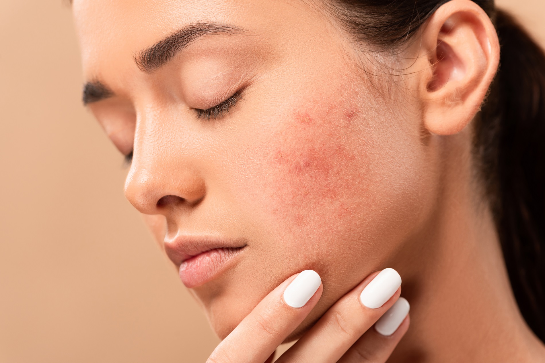
Acne vulgaris is a common skin condition characterized by the formation of various types of blemishes on the skin, such as blackheads, whiteheads, papules, pustules, and cysts or nodules. These lesions typically appear on areas of the body that have a high concentration of sebaceous glands, including the face, neck, chest, back, and shoulders.
Acne vulgaris occurs when hair follicles become clogged with dead skin cells and excess oil (sebum) produced by the sebaceous glands. This blockage provides an ideal environment for bacteria, particularly Propionibacterium acnes, to multiply, leading to inflammation and infection. The severity of acne vulgaris can range from mild with only a few scattered comedones (blackheads or whiteheads) to severe cystic acne, which can cause significant scarring and emotional distress.
The exact causes of acne vulgaris are not fully understood, but several factors contribute to its development, including:
1. Hormonal changes during puberty, menstruation, pregnancy, or due to conditions like polycystic ovary syndrome (PCOS)
2. Genetic predisposition
3. Use of certain medications, such as corticosteroids and lithium
4. Excessive production of sebum due to overactive sebaceous glands
5. Accumulation of dead skin cells that clog pores
6. Bacterial infection (particularly Propionibacterium acnes)
7. Inflammation caused by the body's immune response to bacterial infection and clogged pores
Treatment for acne vulgaris depends on its severity and can include over-the-counter or prescription topical treatments, oral medications, chemical peels, light therapies, or even hormonal therapies in some cases. It is essential to seek professional medical advice from a dermatologist or healthcare provider to determine the most appropriate treatment plan for individual needs.
Adenocarcinoma, sebaceous is a type of cancer that develops from the sebaceous glands, which are glands in the skin that produce an oily substance called sebum. This type of cancer is a malignant tumor that forms in the glandular cells and can spread to other parts of the body. It most commonly occurs in the glands found in the eyelids (known as meibomian glands), but it can also occur in other areas of the body such as the genitals, breasts, and skin.
Sebaceous adenocarcinoma is a rare type of cancer, accounting for less than 1% of all skin cancers. It typically affects older adults and has been linked to exposure to radiation and certain genetic mutations. Treatment usually involves surgical removal of the tumor, along with radiation therapy or chemotherapy in some cases.
It is important to note that while I strive to provide accurate and up-to-date information, this definition may not be complete or fully comprehensive. If you have any concerns about your health or a medical condition, it is always best to consult with a qualified healthcare professional for personalized advice and treatment.
A hair follicle is a part of the human skin from which hair grows. It is a complex organ that consists of several layers, including an outer root sheath, inner root sheath, and matrix. The hair follicle is located in the dermis, the second layer of the skin, and is surrounded by sebaceous glands and erector pili muscles.
The hair growth cycle includes three phases: anagen (growth phase), catagen (transitional phase), and telogen (resting phase). During the anagen phase, cells in the matrix divide rapidly to produce new hair fibers that grow out of the follicle. The hair fiber is made up of a protein called keratin, which also makes up the outer layers of the skin and nails.
Hair follicles are important for various biological functions, including thermoregulation, sensory perception, and social communication. They also play a role in wound healing and can serve as a source of stem cells that can differentiate into other cell types.
Apocrine glands are a type of sweat gland found in mammals, including humans. They are most concentrated in areas with dense hair follicles, such as the axillae (armpits) and genital region. These glands release their secretions into the hair follicle, which then reaches the skin surface through the pores.
Apocrine glands become active during puberty and are associated with the production of odorous sweat. The sweat produced by apocrine glands is initially odorless but can acquire a smell when it comes into contact with bacteria on the skin surface, which break down the organic compounds in the sweat. This can contribute to body odor.
It's important to note that while apocrine glands are often associated with body odor, they do not cause body odor directly. The odor is produced when the sweat from apocrine glands mixes with bacteria on the skin surface.
The epidermis is the outermost layer of the skin, composed mainly of stratified squamous epithelium. It forms a protective barrier that prevents water loss and inhibits the entry of microorganisms. The epidermis contains no blood vessels, and its cells are nourished by diffusion from the underlying dermis. The bottom-most layer of the epidermis, called the stratum basale, is responsible for generating new skin cells that eventually move up to replace dead cells on the surface. This process of cell turnover takes about 28 days in adults.
The most superficial part of the epidermis consists of dead cells called squames, which are constantly shed and replaced. The exact rate at which this happens varies depending on location; for example, it's faster on the palms and soles than elsewhere. Melanocytes, the pigment-producing cells, are also located in the epidermis, specifically within the stratum basale layer.
In summary, the epidermis is a vital part of our integumentary system, providing not only physical protection but also playing a crucial role in immunity and sensory perception through touch receptors called Pacinian corpuscles.
Alopecia is a medical term that refers to the loss of hair or baldness. It can occur in various parts of the body, but it's most commonly used to describe hair loss from the scalp. Alopecia can have several causes, including genetics, hormonal changes, medical conditions, and aging.
There are different types of alopecia, such as:
* Alopecia Areata: It is a condition that causes round patches of hair loss on the scalp or other parts of the body. The immune system attacks the hair follicles, causing the hair to fall out.
* Androgenetic Alopecia: Also known as male pattern baldness or female pattern baldness, it's a genetic condition that causes gradual hair thinning and eventual hair loss, typically following a specific pattern.
* Telogen Effluvium: It is a temporary hair loss condition caused by stress, medication, pregnancy, or other factors that can cause the hair follicles to enter a resting phase, leading to shedding and thinning of the hair.
The treatment for alopecia depends on the underlying cause. In some cases, such as with telogen effluvium, hair growth may resume without any treatment. However, other forms of alopecia may require medical intervention, including topical treatments, oral medications, or even hair transplant surgery in severe cases.
Salivary glands are exocrine glands that produce saliva, which is secreted into the oral cavity to keep the mouth and throat moist, aid in digestion by initiating food breakdown, and help maintain dental health. There are three major pairs of salivary glands: the parotid glands located in the cheeks, the submandibular glands found beneath the jaw, and the sublingual glands situated under the tongue. Additionally, there are numerous minor salivary glands distributed throughout the oral cavity lining. These glands release their secretions through a system of ducts into the mouth.
Medically, hair is defined as a threadlike structure that grows from the follicles found in the skin of mammals. It is primarily made up of a protein called keratin and consists of three parts: the medulla (the innermost part or core), the cortex (middle layer containing keratin filaments) and the cuticle (outer layer of overlapping scales).
Hair growth occurs in cycles, with each cycle consisting of a growth phase (anagen), a transitional phase (catagen), and a resting phase (telogen). The length of hair is determined by the duration of the anagen phase.
While hair plays a crucial role in protecting the skin from external factors like UV radiation, temperature changes, and physical damage, it also serves as an essential aspect of human aesthetics and identity.
In medical terms, the skin is the largest organ of the human body. It consists of two main layers: the epidermis (outer layer) and dermis (inner layer), as well as accessory structures like hair follicles, sweat glands, and oil glands. The skin plays a crucial role in protecting us from external factors such as bacteria, viruses, and environmental hazards, while also regulating body temperature and enabling the sense of touch.
Sweat glands are specialized tubular structures in the skin that produce and secrete sweat, also known as perspiration. They are part of the body's thermoregulatory system, helping to maintain optimal body temperature by releasing water and heat through evaporation. There are two main types of sweat glands: eccrine and apocrine.
1. Eccrine sweat glands: These are distributed throughout the body, with a higher concentration on areas like the palms, soles, and forehead. They are responsible for producing a watery, odorless sweat that primarily helps to cool down the body through evaporation.
2. Apocrine sweat glands: These are mainly found in the axillary (armpit) region and around the anogenital area. They become active during puberty and produce a thick, milky fluid that does not have a strong odor on its own but can mix with bacteria on the skin's surface, leading to body odor.
Sweat glands are controlled by the autonomic nervous system, meaning they function involuntarily in response to various stimuli such as emotions, physical activity, or changes in environmental temperature.
Mammary glands are specialized exocrine glands found in mammals, including humans and other animals. These glands are responsible for producing milk, which is used to nurse offspring after birth. The mammary glands are located in the breast region of female mammals and are usually rudimentary or absent in males.
In animals, mammary glands can vary in number and location depending on the species. For example, humans and other primates have two mammary glands, one in each breast. Cows, goats, and sheep, on the other hand, have multiple pairs of mammary glands located in their lower abdominal region.
Mammary glands are made up of several structures, including lobules, ducts, and connective tissue. The lobules contain clusters of milk-secreting cells called alveoli, which produce and store milk. The ducts transport the milk from the lobules to the nipple, where it is released during lactation.
Mammary glands are an essential feature of mammals, as they provide a source of nutrition for newborn offspring. They also play a role in the development and maintenance of the mother-infant bond, as nursing provides opportunities for physical contact and bonding between the mother and her young.
Eccrine glands are the most numerous type of sweat glands in the human body, found in virtually all skin locations. They play a crucial role in thermoregulation by producing a watery sweat that cools the body when it evaporates on the skin surface. These glands are distributed over the entire body, with a higher concentration on the soles of the feet, palms of the hands, and forehead.
Structurally, eccrine glands consist of two main parts: the coiled secretory portion located in the dermis and the straight duct that extends through the dermis and epidermis to reach the skin surface. The secretory portion is lined with a simple cuboidal epithelium, while the duct is lined with a simple squamous or low cuboidal epithelium.
Eccrine glands are stimulated to produce sweat by the activation of the sympathetic nervous system, particularly through the release of acetylcholine at the neuro-glandular junction. The sweat produced is primarily water with small amounts of electrolytes, such as sodium, chloride, and potassium. This composition helps maintain the body's electrolyte balance while facilitating heat loss during physical exertion or in hot environments.
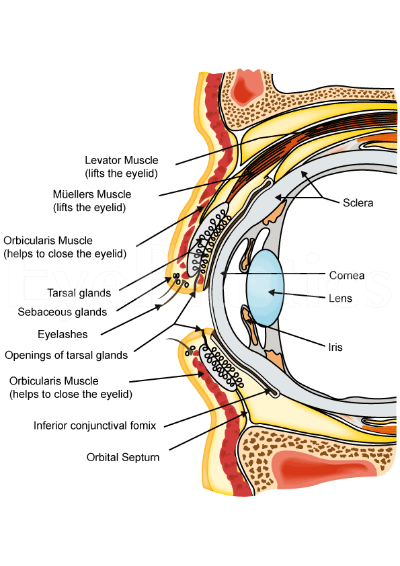
Meibomian glands are sebaceous glands located in the eyelids, specifically at the rim of the eyelid near the lashes. They produce an oily substance called meibum that forms the outermost layer of the tear film, helping to prevent evaporation and keep the eye surface lubricated. The Meibomian glands play a crucial role in maintaining the health and comfort of the eyes by providing stability to the tear film and protecting the eye from irritants and dryness.
Exocrine glands are a type of gland in the human body that produce and release substances through ducts onto an external or internal surface. These glands are responsible for secreting various substances such as enzymes, hormones, and lubricants that help in digestion, protection, and other bodily functions.
Exocrine glands can be further classified into three types based on their mode of secretion:
1. Merocrine glands: These glands release their secretions by exocytosis, where the secretory product is enclosed in a vesicle that fuses with the cell membrane and releases its contents outside the cell. Examples include sweat glands and mucous glands.
2. Apocrine glands: These glands release their secretions by pinching off a portion of the cytoplasm along with the secretory product. An example is the apocrine sweat gland found in the armpits and genital area.
3. Holocrine glands: These glands release their secretions by disintegrating and releasing the entire cell, including its organelles and secretory products. An example is the sebaceous gland found in the skin, which releases an oily substance called sebum.
The submandibular glands are one of the major salivary glands in the human body. They are located beneath the mandible (jawbone) and produce saliva that helps in digestion, lubrication, and protection of the oral cavity. The saliva produced by the submandibular glands contains enzymes like amylase and mucin, which aid in the digestion of carbohydrates and provide moisture to the mouth and throat. Any medical condition or disease that affects the submandibular gland may impact its function and could lead to problems such as dry mouth (xerostomia), swelling, pain, or infection.
Neoplasms, adnexal and skin appendage refer to abnormal growths or tumors that develop in the sweat glands, hair follicles, or other structures associated with the skin. These growths can be benign (non-cancerous) or malignant (cancerous), and they can occur anywhere on the body.
Adnexal neoplasms are tumors that arise from the sweat glands or hair follicles, including the sebaceous glands, eccrine glands, and apocrine glands. These tumors can range in size and severity, and they may cause symptoms such as pain, itching, or changes in the appearance of the skin.
Skin appendage neoplasms are similar to adnexal neoplasms, but they specifically refer to tumors that arise from structures such as hair follicles, nails, and sweat glands. Examples of skin appendage neoplasms include pilomatricomas (tumors of the hair follicle), trichilemmomas (tumors of the outer root sheath of the hair follicle), and sebaceous adenomas (tumors of the sebaceous glands).
It is important to note that while many adnexal and skin appendage neoplasms are benign, some can be malignant and may require aggressive treatment. If you notice any unusual growths or changes in your skin, it is important to consult with a healthcare professional for further evaluation and care.
The scalp is the anatomical region located at the upper part of the human head, covering the skull except for the face and the ears. It is made up of several layers: the skin, the connective tissue, the galea aponeurotica (a strong, flat, tendinous sheet), loose areolar tissue, and the periosteum (the highly vascularized innermost layer that attaches directly to the skull bones). The scalp has a rich blood supply and is home to numerous sensory receptors, including those for touch, pain, and temperature. It also contains hair follicles, sebaceous glands, and sweat glands.
"Skin physiological phenomena" is not a standard medical term with a specific definition. However, I can provide some information about the general concepts that might be encompassed by this term.
Physiological phenomena refer to the functions and processes that occur in living organisms. When it comes to the skin, there are many different physiological phenomena that take place, including:
1. Barrier function: The skin acts as a barrier to protect the body from external elements such as bacteria, viruses, chemicals, and UV radiation.
2. Temperature regulation: The skin helps regulate body temperature through sweat production and blood flow.
3. Sensation: The skin contains nerve endings that allow us to feel touch, pressure, pain, and temperature.
4. Vitamin D synthesis: The skin can produce vitamin D when exposed to sunlight.
5. Moisture regulation: The skin helps maintain the body's moisture balance by producing sweat and preventing water loss.
6. Immunological function: The skin plays a role in the immune system by providing a physical barrier and containing immune cells that help fight off infections.
7. Excretion: The skin eliminates waste products through sweat.
8. Wound healing: The skin has the ability to repair itself after injury, through a complex process involving inflammation, tissue regeneration, and remodeling.
Therefore, "skin physiological phenomena" could refer to any or all of these functions and processes that take place in the skin.
The integumentary system is the largest organ system in the human body, encompassing the skin, hair, nails, and various glands. Its primary function is to act as a barrier, protecting the body from external damage, radiation, and pathogens while also helping regulate body temperature, prevent water loss, and maintain fluid balance. The integumentary system plays crucial roles in sensory perception through nerve endings in the skin, synthesizing vitamin D via sunlight exposure, and excreting waste products through sweat. Overall, it serves as a vital organ system that ensures the body's integrity and homeostasis.
Seborrheic dermatitis is a common, inflammatory skin condition that mainly affects the scalp, face, and upper part of the body. It causes skin irritation, flaking, and redness, often in areas where the skin is oily or greasy. The exact cause of seborrheic dermatitis is not fully understood, but it appears to be related to a combination of genetic, environmental, and microbial factors.
The symptoms of seborrheic dermatitis can vary in severity and may include:
* Greasy or flaky scales on the scalp, eyebrows, eyelashes, ears, or beard
* Redness and inflammation of the skin
* Itching, burning, or stinging sensations
* Yellow or white crusty patches on the scalp or other affected areas
* Hair loss (in severe cases)
Seborrheic dermatitis is a chronic condition that tends to flare up and then subside over time. While there is no cure for seborrheic dermatitis, various treatments can help manage the symptoms and prevent complications. These may include medicated shampoos, topical creams or ointments, and lifestyle changes such as stress reduction and avoiding triggers that worsen symptoms.
It is important to note that seborrheic dermatitis should not be confused with other skin conditions, such as psoriasis or eczema, which may have similar symptoms. A healthcare professional can provide a proper diagnosis and recommend appropriate treatment options based on the individual's specific needs.
Hyperplasia is a medical term that refers to an abnormal increase in the number of cells in an organ or tissue, leading to an enlargement of the affected area. It's a response to various stimuli such as hormones, chronic irritation, or inflammation. Hyperplasia can be physiological, like the growth of breast tissue during pregnancy, or pathological, like in the case of benign or malignant tumors. The process is generally reversible if the stimulus is removed. It's important to note that hyperplasia itself is not cancerous, but some forms of hyperplasia can increase the risk of developing cancer over time.
Eyelid neoplasms refer to abnormal growths or tumors that develop in the tissues of the eyelids. These growths can be benign (non-cancerous) or malignant (cancerous). Common types of benign eyelid neoplasms include papillomas, hemangiomas, and nevi. Malignant eyelid neoplasms are typically classified as basal cell carcinomas, squamous cell carcinomas, or melanomas. These malignant tumors can be aggressive and may spread to other parts of the body if left untreated. Treatment options for eyelid neoplasms depend on the type, size, and location of the growth, as well as the patient's overall health. Surgical excision is often the preferred treatment approach, although radiation therapy and chemotherapy may also be used in some cases. Regular follow-up care is important to monitor for recurrence or new growths.
The parotid gland is the largest of the major salivary glands. It is a bilobed, accessory digestive organ that secretes serous saliva into the mouth via the parotid duct (Stensen's duct), located near the upper second molar tooth. The parotid gland is primarily responsible for moistening and lubricating food to aid in swallowing and digestion.
Anatomically, the parotid gland is located in the preauricular region, extending from the zygomatic arch superiorly to the angle of the mandible inferiorly, and from the masseter muscle anteriorly to the sternocleidomastoid muscle posteriorly. It is enclosed within a fascial capsule and has a rich blood supply from the external carotid artery and a complex innervation pattern involving both parasympathetic and sympathetic fibers.
Parotid gland disorders can include salivary gland stones (sialolithiasis), infections, inflammatory conditions, benign or malignant tumors, and autoimmune diseases such as Sjögren's syndrome.
Melanocortin receptors (MCRs) are a group of G protein-coupled receptors that bind melanocortin peptides, which include α-, β-, and γ-melanocyte stimulating hormones (MSH) and adrenocorticotropic hormone (ACTH). These receptors are involved in a variety of physiological processes, including pigmentation, energy homeostasis, sexual function, and inflammation. There are five subtypes of melanocortin receptors (MCR1-5) that are expressed in different tissues and have distinct functions.
MCR1 is primarily expressed in melanocytes and plays a crucial role in skin and hair pigmentation. Activation of MCR1 by α-MSH leads to the production and distribution of eumelanin, which results in darker skin and hair.
MCR2 is widely expressed in the central nervous system (CNS) and peripheral tissues, including the adrenal gland, testis, and ovary. It is involved in various functions such as sexual function, feeding behavior, and energy homeostasis.
MCR3 is primarily expressed in the adrenal gland and plays a critical role in the regulation of steroid hormone production and release. Activation of MCR3 by ACTH leads to the synthesis and secretion of cortisol and other steroid hormones.
MCR4 is widely expressed in the CNS, peripheral tissues, and immune cells. It is involved in various functions such as energy homeostasis, feeding behavior, sexual function, and inflammation.
MCR5 is primarily expressed in the testis and plays a role in spermatogenesis and fertility.
Overall, melanocortin receptors are important regulators of various physiological processes, and dysregulation of these receptors has been implicated in several diseases, including obesity, metabolic disorders, and skin disorders.
Skin diseases, also known as dermatological conditions, refer to any medical condition that affects the skin, which is the largest organ of the human body. These diseases can affect the skin's function, appearance, or overall health. They can be caused by various factors, including genetics, infections, allergies, environmental factors, and aging.
Skin diseases can present in many different forms, such as rashes, blisters, sores, discolorations, growths, or changes in texture. Some common examples of skin diseases include acne, eczema, psoriasis, dermatitis, fungal infections, viral infections, bacterial infections, and skin cancer.
The symptoms and severity of skin diseases can vary widely depending on the specific condition and individual factors. Some skin diseases are mild and can be treated with over-the-counter medications or topical creams, while others may require more intensive treatments such as prescription medications, light therapy, or even surgery.
It is important to seek medical attention if you experience any unusual or persistent changes in your skin, as some skin diseases can be serious or indicative of other underlying health conditions. A dermatologist is a medical doctor who specializes in the diagnosis and treatment of skin diseases.
Ectodysplasins are a group of signaling proteins that play crucial roles in the development and differentiation of ectodermal tissues, including the skin, hair, nails, teeth, and sweat glands. They are involved in various signaling pathways and help regulate cell growth, migration, and pattern formation during embryogenesis. Mutations in genes encoding ectodysplasins can lead to genetic disorders characterized by abnormalities in these tissues, such as ectodermal dysplasia syndromes.
Isotretinoin is a derivative of vitamin A, used in the treatment of severe recalcitrant nodular acne that has not responded to other therapies. It is a potent inhibitor of sebaceous gland function and keratinization. Isotretinoin is also known to have anti-inflammatory properties. It is taken orally in the form of capsules and its use requires careful monitoring due to potential teratogenic effects and other side effects, such as dryness of the skin and mucous membranes, mood changes, and liver enzyme abnormalities.
Keratoacanthoma is a rapidly growing, dome-shaped, skin tumor that typically arises on sun-exposed areas such as the face, arms, and legs. It is considered a low-grade squamous cell carcinoma (a type of skin cancer) because it shares some characteristics with both benign and malignant tumors.
Keratoacanthomas usually develop over a period of several weeks to months, growing rapidly in size before eventually stabilizing and then gradually regressing on their own within a few months to a year. However, the regression process can take years, and some lesions may not regress completely, leading to cosmetic concerns or even local invasion.
Histologically, keratoacanthomas are characterized by a central keratin-filled crater surrounded by a well-differentiated layer of squamous epithelial cells. The tumor's growth pattern and histological features can make it difficult to distinguish from other types of skin cancer, such as squamous cell carcinoma.
Treatment options for keratoacanthomas include surgical excision, cryosurgery, curettage and electrodesiccation, and topical therapies like imiquimod or 5-fluorouracil. The choice of treatment depends on various factors such as the size, location, and number of lesions, as well as patient preferences and overall health status.
Hair diseases is a broad term that refers to various medical conditions affecting the hair shaft, follicle, or scalp. These conditions can be categorized into several types, including:
1. Hair shaft abnormalities: These are conditions that affect the structure and growth of the hair shaft. Examples include trichorrhexis nodosa, where the hair becomes weak and breaks easily, and pili torti, where the hair shaft is twisted and appears sparse and fragile.
2. Hair follicle disorders: These are conditions that affect the hair follicles, leading to hair loss or abnormal growth patterns. Examples include alopecia areata, an autoimmune disorder that causes patchy hair loss, and androgenetic alopecia, a genetic condition that leads to pattern baldness in both men and women.
3. Scalp disorders: These are conditions that affect the scalp, leading to symptoms such as itching, redness, scaling, or pain. Examples include seborrheic dermatitis, psoriasis, and tinea capitis (ringworm of the scalp).
4. Hair cycle abnormalities: These are conditions that affect the normal growth cycle of the hair, leading to excessive shedding or thinning. Examples include telogen effluvium, where a large number of hairs enter the resting phase and fall out, and anagen effluvium, which is typically caused by chemotherapy or radiation therapy.
5. Infectious diseases: Hair follicles can become infected with various bacteria, viruses, or fungi, leading to conditions such as folliculitis, furunculosis, and kerion.
6. Genetic disorders: Some genetic disorders can affect the hair, such as Menkes syndrome, which is a rare inherited disorder that affects copper metabolism and leads to kinky, sparse, and brittle hair.
Proper diagnosis and treatment of hair diseases require consultation with a healthcare professional, often a dermatologist or a trichologist who specializes in hair and scalp disorders.
Keratinocytes are the predominant type of cells found in the epidermis, which is the outermost layer of the skin. These cells are responsible for producing keratin, a tough protein that provides structural support and protection to the skin. Keratinocytes undergo constant turnover, with new cells produced in the basal layer of the epidermis and older cells moving upward and eventually becoming flattened and filled with keratin as they reach the surface of the skin, where they are then shed. They also play a role in the immune response and can release cytokines and other signaling molecules to help protect the body from infection and injury.
Corticotropin receptors are a type of cell surface receptor that bind to the hormone corticotropin (also known as adrenocorticotropic hormone or ACTH). These receptors are found in various tissues throughout the body, including the adrenal glands.
There are two main types of corticotropin receptors, known as melanocortin receptor 1 (MC1R) and melanocortin receptor 2 (MC2R). MC2R is the primary receptor for corticotropin in the adrenal glands. When corticotropin binds to this receptor, it stimulates the production and release of steroid hormones, such as cortisol, which help regulate metabolism, immune response, and stress response.
Abnormalities in corticotropin receptors have been implicated in several medical conditions, including certain endocrine disorders and skin pigmentation disorders.
Lipogenesis is the biological process by which fatty acids are synthesized and stored as lipids or fat in living organisms. This process occurs primarily in the liver and adipose tissue, with excess glucose being converted into fatty acids and then esterified to form triglycerides. These triglycerides are then packaged with proteins and cholesterol to form lipoproteins, which are transported throughout the body for energy storage or use. Lipogenesis is a complex process involving multiple enzymes and metabolic pathways, and it is tightly regulated by hormones such as insulin, glucagon, and adrenaline. Disorders of lipogenesis can lead to conditions such as obesity, fatty liver disease, and metabolic disorders.
A papilloma is a benign (noncancerous) tumor that grows on a stalk, often appearing as a small cauliflower-like growth. It can develop in various parts of the body, but when it occurs in the mucous membranes lining the respiratory, digestive, or genitourinary tracts, they are called squamous papillomas. The most common type is the skin papilloma, which includes warts. They are usually caused by human papillomavirus (HPV) infection and can be removed through various medical procedures if they become problematic or unsightly.
The sublingual glands are a pair of salivary glands located in the floor of the mouth, beneath the tongue. They are the smallest of the major salivary glands and produce around 5-10% of the total saliva in the mouth. The sublingual glands secrete saliva containing electrolytes, enzymes (such as amylase), and antibacterial compounds that help in digestion, lubrication, and protection against microorganisms.
The sublingual glands' secretions are released through multiple small ducts called the ducts of Rivinus or minor sublingual ducts, as well as a larger duct called the duct of Wharton, which is a common excretory duct for both sublingual and submandibular glands.
Sublingual gland dysfunction can lead to conditions such as dry mouth (xerostomia), dental caries, or oral infections.
Neurophysiology is a branch of physiology that deals with the study of the functioning of the nervous system and its components, including the neurons, neurotransmitters, and electrical signals that transmit information within the nervous system. It involves the examination of various aspects such as nerve impulse transmission, sensory processes, muscle activation, and brain function using techniques like electroencephalography (EEG), electromyography (EMG), and nerve conduction studies. The findings from neurophysiological studies can be applied to diagnose and manage neurological disorders and injuries.
Vernix caseosa is a medical term that refers to the white, cheesy, protective substance covering the skin of a newborn baby. It is composed of sebum (oil produced by the baby's sebaceous glands), dead skin cells, and water. This natural emollient provides a barrier against bacterial invasion and helps keep the baby's skin moisturized and supple. Vernix caseosa begins to form around the 20th week of gestation and is more abundant in premature infants than those born at term. It is typically washed off after birth, but some hospitals and midwives recommend leaving it on as long as possible due to its protective properties.
An encyclopedia is a comprehensive reference work containing articles on various topics, usually arranged in alphabetical order. In the context of medicine, a medical encyclopedia is a collection of articles that provide information about a wide range of medical topics, including diseases and conditions, treatments, tests, procedures, and anatomy and physiology. Medical encyclopedias may be published in print or electronic formats and are often used as a starting point for researching medical topics. They can provide reliable and accurate information on medical subjects, making them useful resources for healthcare professionals, students, and patients alike. Some well-known examples of medical encyclopedias include the Merck Manual and the Stedman's Medical Dictionary.
Eyelid diseases refer to a variety of medical conditions that affect the function and/or appearance of the eyelids. These can include structural abnormalities, such as entropion (inward turning of the eyelid) or ectropion (outward turning of the eyelid), as well as functional issues like ptosis (drooping of the upper eyelid). Other common eyelid diseases include blepharitis (inflammation of the eyelid margin), chalazion (a blocked oil gland in the eyelid), and cancerous or benign growths on the eyelid. Symptoms of eyelid diseases can vary widely, but often include redness, swelling, pain, itching, tearing, and sensitivity to light. Treatment for these conditions depends on the specific diagnosis and may range from self-care measures and medications to surgical intervention.
Eyelashes are defined in medical terms as the slender, hair-like growths that originate from the edges of the eyelids. They are made up of keratin and follicles, and their primary function is to protect the eyes from debris, sweat, and other irritants by acting as a physical barrier. Additionally, they play a role in enhancing the aesthetic appeal of the eyes and can also serve as a sensory organ, helping to detect potential threats near the eye area.
Keratins are a type of fibrous structural proteins that constitute the main component of the integumentary system, which includes the hair, nails, and skin of vertebrates. They are also found in other tissues such as horns, hooves, feathers, and reptilian scales. Keratins are insoluble proteins that provide strength, rigidity, and protection to these structures.
Keratins are classified into two types: soft keratins (Type I) and hard keratins (Type II). Soft keratins are found in the skin and simple epithelial tissues, while hard keratins are present in structures like hair, nails, horns, and hooves.
Keratin proteins have a complex structure consisting of several domains, including an alpha-helical domain, beta-pleated sheet domain, and a non-repetitive domain. These domains provide keratin with its unique properties, such as resistance to heat, chemicals, and mechanical stress.
In summary, keratins are fibrous structural proteins that play a crucial role in providing strength, rigidity, and protection to various tissues in the body.
I'm sorry for any confusion, but "Hoof and Claw" is not a medical term or condition. The term "hoof" refers to the hard covering on the toes of animals such as horses, cows, and other ungulates, while "claw" refers to the sharp nail-like structure found on the toes of animals such as cats, dogs, and birds.
If you have any questions related to medical terminology or health concerns, I would be happy to try to help answer them for you.
Perissodactyla is not a medical term, but rather a taxonomic order in zoology. It includes mammals with an odd number of toes on each foot and a particular type of digestive system called "hindgut fermentation." The order Perissodactyla includes horses, rhinos, and tapirs.
Intermediate filaments (IFs) are a type of cytoskeletal filament found in the cytoplasm of eukaryotic cells, including animal cells. They are called "intermediate" because they are smaller in diameter than microfilaments and larger than microtubules, two other types of cytoskeletal structures.
Intermediate filaments are composed of fibrous proteins that form long, unbranched, and flexible filaments. These filaments provide structural support to the cell and help maintain its shape. They also play a role in cell-to-cell adhesion, intracellular transport, and protection against mechanical stress.
Intermediate filaments are classified into six types based on their protein composition: Type I (acidic keratins), Type II (neutral/basic keratins), Type III (vimentin, desmin, peripherin), Type IV (neurofilaments), Type V (lamins), and Type VI (nestin). Each type of intermediate filament has a specific function and is expressed in different cell types. For example, Type I and II keratins are found in epithelial cells, while vimentin is expressed in mesenchymal cells.
Overall, intermediate filaments play an essential role in maintaining the structural integrity of cells and tissues, and their dysfunction has been implicated in various human diseases, including cancer, neurodegenerative disorders, and genetic disorders.
I'm sorry for any confusion, but "Reptiles" is not a medical term. It is a term used in biology to describe a class of cold-blooded, scaly-skinned animals that include snakes, lizards, alligators, crocodiles, turtles, and tortoises. They are characterized by having lungs for breathing, laying eggs on land, and having a three-chambered heart. If you have any medical questions or terms, I'd be happy to help clarify those!
Type II keratins are a group of intermediate filament proteins that are primarily expressed in epithelial cells. They are part of the keratin family, which is divided into two types (Type I and Type II) based on their acidic or basic isoelectric point. Type II keratins have a basic isoelectric point and include several subtypes such as KRT2, KRT3, KRT4, KRT10, KRT12, and others.
Type II keratins form heteropolymers with Type I keratins to provide structural support and integrity to epithelial cells. They are essential for the maintenance of cell shape, polarity, and mechanical resistance to stress. Mutations in type II keratin genes have been associated with several human genetic disorders, including epidermolysis bullosa simplex, a blistering skin disorder, and some forms of hair loss.
In summary, Type II keratins are a group of basic intermediate filament proteins that form heteropolymers with Type I keratins to provide structural support and integrity to epithelial cells.
 Sebaceous gland
Testosterone
Fordyce spots
Acne
Nasal sebum
Preputial gland
Sebacic acid
Gland
Skin secretions (human)
Mammary gland
Exocrine gland
Uropygial gland
Cutibacterium acnes
Scalp
Bovidae
Woolly mammoth
Toker cell
Androsterone
Pattern hair loss
Jamaican fruit bat
Demodex folliculorum
Sebaceous carcinoma
Santosh G. Honavar
Human anus
Pimple
Head and neck anatomy
Baleen whale
Vermilion border
Estrogen (medication)
Isotretinoin
Sebaceous gland - Wikipedia
 Sebaceous Gland Carcinoma: Background, History of the Procedure, Epidemiology
 View source for Sebaceous gland - wikidoc
 Sebaceous Cysts - An Infected Gland on your cat or dog
Mitomycin C in sebaceous gland carcinoma with pagetoid spread | British Journal of Ophthalmology
 "Sebaceous Gland Adenoma in a Dog" by M. ÖZGÜR ÖZYİĞİT, AHMET AKKOÇ et al.
 Sebaceous Glands | Profiles RNS
 Sebaceous Gland Suppression With Ethynyl Estradiol and Diethylstilbestrol | JAMA Dermatology | JAMA Network
 Clinical Trials : Sebaceous Gland Diseases
 Understanding Sebaceous Glands: A Key To Unlocking Hair Growth Potential | FullyVital
 Planteome: Term Details for "sebaceous gland development" (GO:0048733)
 Sebaceous Cyst Natural Herbal Remedies for Skin Glands - Herbal Care Products
 How Often Should You Wash Your Hair Sebaceous Glands in your Skin
 Tumors of the Skin in Dogs - Dog Owners - Merck Veterinary Manual
Keratin - Wikipedia
 Diagnosing Skin Tumors in Dogs
 Catalan Institute of Dermatology Incade in Barcelona, Spain
 Celine | Difference Between
 Acne and Genetics | SpringerLink
Cytomorphology and diagnostic pitfalls of sebaceous and nonsebaceous salivary gland lymphadenoma: A multi-institutional study<...
 Sebaceous adenoma: MedlinePlus Medical Encyclopedia
Plus it
 Page 1 | Search Results | Skin Pharmacology and Physiology | Karger Publishers
 Positive Health Online | Article - Natural Treatment Options for Acne Vulgaris
 Chronic unilateral blepharoconjunctivitis caused by sebaceous carcinoma
 Registration Dossier - ECHA
 How to get rid of large pores: The top 8 ways
Table 1 - Histopathologic Improvement with Lymphedema Management, Léogâne, Haiti - Volume 10, Number 11-November 2004 -...
Sebum32
- A sebaceous gland, or oil gland, is a microscopic exocrine gland in the skin that opens into a hair follicle to secrete an oily or waxy matter, called sebum, which lubricates the hair and skin of mammals. (wikipedia.org)
- In the eyelids, meibomian glands, also called tarsal glands, are a type of sebaceous gland that secrete a special type of sebum into tears. (wikipedia.org)
- The glands deposit sebum on the hairs and bring it to the skin surface along the hair shaft. (wikipedia.org)
- Relative to keratinocytes that make up the hair follicle, sebaceous glands are composed of huge cells with many large vesicles that contain the sebum. (wikipedia.org)
- Sebaceous glands secrete the oily, waxy substance called sebum (Latin: fat, tallow) that is made of triglycerides, wax esters, squalene, and metabolites of fat-producing cells. (wikipedia.org)
- Sebum is produced in a holocrine process, in which cells within the sebaceous gland rupture and disintegrate as they release the sebum and the cell remnants are secreted together with the sebum. (wikipedia.org)
- Sebum is secreted by the sebaceous gland in humans. (wikipedia.org)
- The prescription drug [[isotretinoin]] significantly reduces the amount of sebum produced by the sebaceous glands, and is used to treat acne. (wikidoc.org)
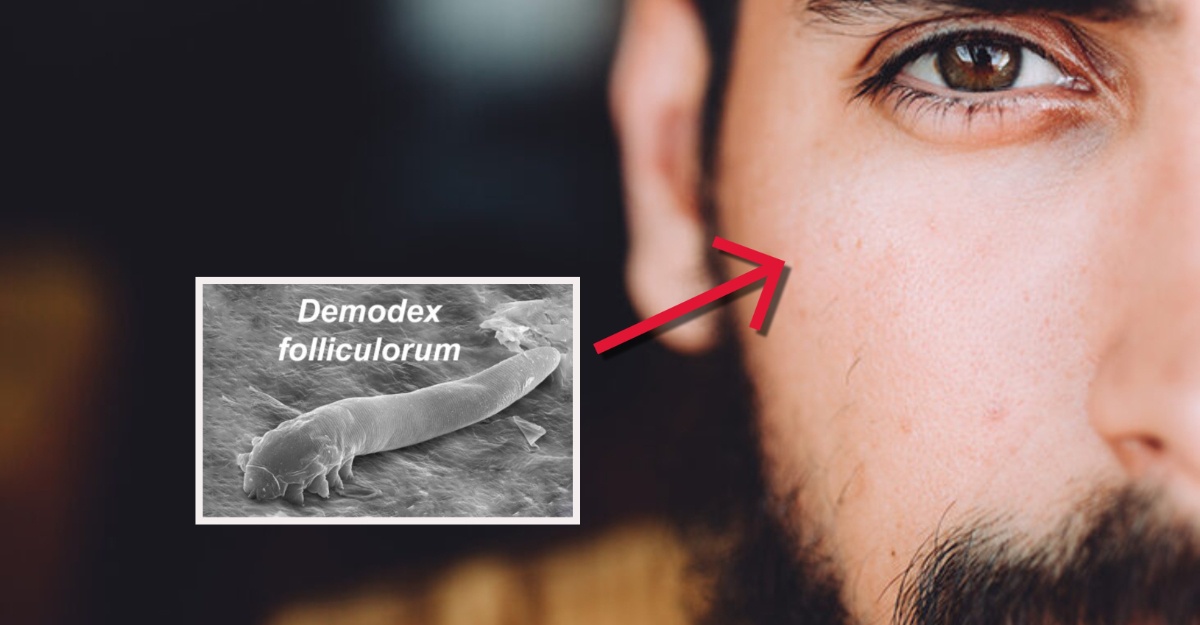
- Importance to other animals== Certain species of [[Demodex mite]]s feed on sebum and are commonly found in the sebaceous glands of mammals, including those of humans. (wikidoc.org)
- Our pets, like us, have microscopic oil glands that produce sebum. (pethealthnetwork.com)
- Sebaceous glands secrete SEBUM. (wakehealth.edu)
- Sebum, the oil secreted by sebaceous glands, acts as a natural conditioner for your hair, preventing dryness and breakage. (fullyvital.com)
- These glands continuously produce sebum, which travels up hair follicles to reach the skin's surface. (fullyvital.com)
- Sebaceous glands are typically found near the hair follicles, where they release sebum, an oily substance. (fullyvital.com)
- Sebaceous glands in your skin are connected to individual hair follicles on your scalp and they secrete oily sebum which serves to moisturize as well as waterproof your skin and hair. (beautyzoomin.net)
- Higher correlations of sebum excretion and the proportion of branched fatty acids in the fraction of sebaceous wax esters were found in monozygotic vs. dizygotic twins [8, 9]. (springer.com)
- Howard I. Maibach Oily skin (seborrhea) is a common cosmetic problem that occurs when oversized sebaceous glands produce excessive amounts of sebum giving the appearance of shiny and greasy skin. (karger.com)
- These glands respond to circulating androgens (male hormones, which both men and women have) by producing sebum (fat). (positivehealth.com)
- The sebaceous gland is an example of a holocrine gland because its product of secretion (sebum) is released with remnants of dead cells. (coursehero.com)
- The infundibulum is part of the pilosebaceous canal, the one responsible for discharging sebum and one that is composed of the infundibulum and the short duct of the sebaceous gland itself. (coursehero.com)
- Regardless, the secretion of sebum out of the gland is helped along by the contraction of the arrector pili muscle. (coursehero.com)
- Under each of your pores is a sebaceous gland that produces a natural oil called sebum, keeping your skin hydrated and healthy. (healthline.com)
- GT20029 could also effectively inhibit sebaceous gland development and sebum secretion. (biospace.com)
- They develop from sebaceous glands and are filled with keratin and sebum. (bestfinance-blog.com)
- It does this by causing the sebaceous glands to enlarge and produce excess sebum and by stimulating the overproduction of keratin, the main component of the outer layer of the skin. (positivehealth.com)
- These glands make the oil sebum (SEE-bum), which softens the skin and makes it waterproof. (childrensmn.org)
- Produced in glands called sebaceous glands, which surround hair follicles, sebum moisturizes and waterproofs the skin, protects it against cold and microorganisms, and distributes antioxidants and hormones on the surface of the skin. (acne.org)
- Sebaceous glands are already formed and actively producing sebum while we are still in the womb. (acne.org)
- Only mammals have sebaceous glands, and thus, sebum. (acne.org)
- These hormones prompt your sebaceous (oil) glands to make lots of a fatty substance called sebum . (howstuffworks.com)
- When your revved-up hormones prompt your sebaceous glands to produce lots of sebum, that oil has to go somewhere. (howstuffworks.com)
- Sebaceous glands, or holocrine glands, secrete sebum, which serves to lubricate the skin and make it more impervious to moisture. (medscape.com)
Hyperplasia3
- Neoplasms of the sebaceous glands may be benign, such as sebaceous hyperplasia or sebaceous gland adenomas. (medscape.com)
- If you have several small bumps of the sebaceous glands, this is called sebaceous hyperplasia. (medlineplus.gov)
- Acne is one of the most common multifactorial chronic inflammatory diseases of the pilosebaceous follicles involving androgen induced sebaceous hyperplasia, altered follicular keratinisation, hormonal imbalance, immune hypersensitivity, and bacterial ( Propionibacterium acnes ) colonisation [ 2 , 3 ]. (hindawi.com)
Develop from sebaceous glands1
- vulvar epidermal cysts develop from sebaceous glands. (msdmanuals.com)
Benign5
- Fordyce spots are benign, visible, sebaceous glands found usually on the lips, gums and inner cheeks, and genitals. (wikipedia.org)
- Sebaceous cell carcinoma is a lethal eyelid malignancy and can masquerade as benign conditions. (medscape.com)
- sebaceous adenoma is a more benign [[neoplasm]] of the sebaceous glands. (wikidoc.org)
- While sebaceous cysts are benign, they may be difficult to distinguish from other lumps and bumps that could be cancerous. (pethealthnetwork.com)
- Background: Salivary gland lymphadenoma (LAD) is a rare benign neoplasm comprising sebaceous (SLAD) and nonsebaceous (NSLAD) types. (johnshopkins.edu)
Epidermal5
- The structure, consisting of hair, hair follicle, arrector pili muscles, and sebaceous gland, is an epidermal invagination known as a pilosebaceous unit. (wikipedia.org)
- Sebaceous glands are part of epidermal appendages. (medscape.com)
- Dilutional effect of increased sebaceous gland activity on the proportion of linoleic acid in sebaceous wax esters and in epidermal acylceramides. (springer.com)
- Epidermal nevi(EN)represent a heterogeneous group of mosaic skin lesions frequently following the lines of Blaschko. (karger.com)
- Epidermal cysts (sebaceous cysts) result from obstruction of sebaceous gland ducts. (msdmanuals.com)
Secretion2
- The effect of various dosages of ethinyl estradiol or diethylstilbestrol on sebaceous gland secretion was studied in 54 women, 15 of whom had acne. (jamanetwork.com)
- Vitamin B5 also influences the adrenal cortex to stabilize the output of androgens, hormones that control sebaceous gland secretion, and normal keratin production. (positivehealth.com)
Cysts14
- Sebaceous cysts are basically very large pimples that are usually harmless to your pet. (pethealthnetwork.com)
- All dogs and cats can get sebaceous cysts, whether purebred or not. (pethealthnetwork.com)
- Caring for your pet's skin and coat as recommended by your veterinarian can help reduce the chance of sebaceous cysts forming. (pethealthnetwork.com)
- Sebaceous cysts are small, painless and noncancerous growths on the skin. (herbal-care-products.com)
- Sebaceous cysts form when the passage where the oil leaves from the glands gets blocked. (herbal-care-products.com)
- Reasons for the development of sebaceous cysts are deformed duct, damage to the cells, genetic conditions or surgical trauma. (herbal-care-products.com)
- It is one of the safe and best methods to get rid of sebaceous cysts. (herbal-care-products.com)
- Wash the sebaceous cysts with mild soap and water. (herbal-care-products.com)
- When warm heat is applied on the sebaceous cysts, it speeds up the healing and draining process. (herbal-care-products.com)
- Wring out the excess and place the cloth on the sebaceous cysts. (herbal-care-products.com)
- It also helps to reduce the inflammation and itching caused by sebaceous cysts. (herbal-care-products.com)
- Repeat the process daily for few days to remove sebaceous cysts. (herbal-care-products.com)
- Sebaceous cysts are the most common type of cysts found on the skin. (bestfinance-blog.com)
- There was also a thickening and enlargement of the eyelids due to cartilaginous hypertrophy, dystrophic changes of the conjunctiva, and atrophy of the Meibomian glands, with the formation of multiple cysts and granulomas. (medscape.com)
Carcinoma23
- The malignant sebaceous gland carcinoma most commonly arises in the periocular area. (medscape.com)
- Fewer than 120 cases of sebaceous cell carcinoma have been reported at extraocular sites. (medscape.com)
- The most common site of origin is the meibomian glands of the eyelids, leading to the term meibomian gland carcinoma. (medscape.com)
- The incidence of sebaceous cell carcinoma is 3.2% among malignant tumors and 0.8% of all eyelid tumors. (medscape.com)
- In a retrospective study, 31 patients were diagnosed with sebaceous cell carcinoma of the ocular adnexa on histopathology. (medscape.com)
- Sebaceous cell carcinoma may mimic either chalazion or chronic blepharitis. (medscape.com)
- Foamy cytoplasm is seen only in sebaceous carcinoma, but it is absent in conjunctival or cutaneous squamous cell carcinoma. (medscape.com)
- Either fresh tissue or formalin-fixed tissue not exposed to alcohol can be frozen, and positive fat stains, such as oil red O, can confirm the diagnosis of sebaceous carcinoma. (medscape.com)
- The clinical appearance of sebaceous gland carcinoma is highly variable. (medscape.com)
- Both the history and the presentation of sebaceous cell carcinoma are variable. (medscape.com)
- Sebaceous cell carcinoma also can mimic unilateral blepharoconjunctivitis, meibomitis, basal or squamous cell carcinoma, conjunctival or corneal carcinoma in situ, orbital inflammation, or superior limbic keratoconjunctivitis. (medscape.com)
- Sebaceous cell carcinoma is shown in the images below. (medscape.com)
- A 63-year-old white man with lower eyelid sebaceous cell carcinoma and lash loss is shown. (medscape.com)
- Gross pathology slide of sebaceous cell carcinoma, from same man as seen in image above. (medscape.com)
- Sebaceous gland carcinoma is a rare eyelid tumour comprising less than 1% of all eyelid malignancies. (bmj.com)
- Topical application of mitomycin C, a non-cell cycle specific alkylating agent, has been advocated for pagetoid spread of sebaceous gland carcinoma. (bmj.com)
- 3 We report the use of mitomycin C as adjuvant therapy in a patient with completely excised sebaceous gland carcinoma and pagetoid spread. (bmj.com)
- The biopsy confirmed sebaceous gland carcinoma with pagetoid invasion of the conjunctival epithelium (fig 2). (bmj.com)
- These showed sebaceous gland carcinoma to the margin of the excision with pagetoid invasion of the conjunctiva and epidermis of the lid margin. (bmj.com)
- Intraepithelial invasion in sebaceous gland carcinoma is noted to occur in 41-80% of cases. (bmj.com)
- 8 This is only the second reported article where mitomycin C has been used in the treatment of sebaceous gland carcinoma. (bmj.com)
- A patient with chronic unilateral conjunctivitis of six years' duration was discovered to have sebaceous carcinoma of the eyelid. (nih.gov)
- Hair follicle and skin glands and appendages, Steroid synthesis and function in skin, hair follicles and sebaceous glands, Basal cell carcinoma Gli and Sonic Hedgehog. (qmul.ac.uk)
Meibomian gland3
- At the rim of the eyelids, [[meibomian gland]]s are a specialized form of sebaceous gland. (wikidoc.org)
- Anterior blepharitis refers to inflammation mainly centered around the skin, eyelashes, and lash follicles, whereas the posterior variant involves the meibomian gland orifices, meibomian glands, tarsal plate, and blepharo-conjunctival junction. (medscape.com)
- Colonization of the lid margin is increased in the presence of seborrheic dermatitis or meibomian gland dysfunction. (medscape.com)
Pilosebaceous4
- One or more glands may surround each hair follicle, and the glands themselves are surrounded by arrector pili muscles, forming a pilosebaceous unit. (wikipedia.org)
- Base of pilosebaceous unit Insertion of sebaceous glands into hair shaft Sagittal section through the upper eyelid. (wikipedia.org)
- Acne is a hormonally driven condition which affects your pilosebaceous glands. (positivehealth.com)
- All patients were interviewed and com- inflammatory disease of the pilosebaceous pleted a written consent and a questionnaire glands located on the face, chest, and upper form that contained information about their back. (who.int)
Adenoma4
- Sebaceous Gland Adenoma in a Dog" by M. ÖZGÜR ÖZYİĞİT, AHMET AKKOÇ et al. (tubitak.gov.tr)
- A 14-year-old male cocker spaniel was diagnosed with sebaceous gland adenoma in the right external ear canal. (tubitak.gov.tr)
- A sebaceous adenoma is a noncancerous tumor of an oil-producing gland in the skin. (medlineplus.gov)
- A sebaceous adenoma is a small bump. (medlineplus.gov)
Holocrine glands1
- Sebaceous glands are classified as holocrine glands. (coursehero.com)
Acne5
- There was a significantly positive correlation between the degree of sebaceous gland inhibition and acne improvement. (jamanetwork.com)
- 6] Acne arises if the level of these circulating androgens is very high or the gland becomes overly sensitive to circulating androgens. (positivehealth.com)
- Acne occurs when dead skin cells, hair follicles, and sebaceous glands (oil glands) clog together, forming a plug. (remedypost.com)
- Acne results from blockages in the sebaceous glands causing whiteheads, blackheads and inflammation. (positivehealth.com)
- Tom's face, back and neck, where the sebaceous glands are highly concentrated, were covered with acne, and his doctor had suggested steroid cream, which made the problem worse. (positivehealth.com)
Cyst13
- A sebaceous cyst can develop when a hair follicle or skin pore gets blocked by dirt, debris, or scar tissue, or as the result of an infection. (pethealthnetwork.com)
- If your pet has a sebaceous cyst, you will notice a raised bump. (pethealthnetwork.com)
- Sebaceous cyst can be treated using some simple home remedies like Tea Tree Oil, warm compress, apple cider vinegar, castor oil and many others sebaceous cyst diet . (herbal-care-products.com)
- That's all info posted by Herbal Care Products online company also treat sebaceous cyst pain . (herbal-care-products.com)
- In our herbal skin care products have Banical for patients sebaceous cyst natural herbal treatment at your home place. (herbal-care-products.com)
- sebaceous cyst natural treatment treat by nature and you get your skin back. (herbal-care-products.com)
- You can get best sebaceous cyst herbal treatment only from herbal skin products store. (herbal-care-products.com)
- This post have your natural herbal treatment for sebaceous cyst . (herbal-care-products.com)
- Natural treatment for sebaceous cyst you can use in all type of age. (herbal-care-products.com)
- If you want online treatment then come here and get your herbal treatment for sebaceous cyst . (herbal-care-products.com)
- There are various tried and tested remedies to get rid of sebaceous cyst . (herbal-care-products.com)
- Place it on the sebaceous cyst and secure it with a bandage. (herbal-care-products.com)
- Dip a small washcloth in castor oil and place it on the sebaceous cyst. (herbal-care-products.com)
Eyelids2
- Sebaceous glands are also found in hairless areas (glabrous skin) of the eyelids, nose, penis, labia minora, the inner mucosal membrane of the cheek, and nipples. (wikipedia.org)
- Sebaceous glands on the lip and mucosa of the cheek, and on the genitalia, are known as Fordyce spots, and glands on the eyelids are known as meibomian glands. (wikipedia.org)
Ducts4
- The ducts from most sebaceous glands open into a HAIR FOLLICLE, but some open on the general surface of the SKIN. (wakehealth.edu)
- They are typically larger than eccrine glands and their ducts tend to open into hair follicles instead of hairless areas of skin. (coursehero.com)
- Sebaceous gland ducts thus usually open up into the upper part of a hair follicle, called the infundibulum. (coursehero.com)
- However, some sebaceous gland ducts open directly onto our skin surface such as at the corner of the mouth and the glans penis. (coursehero.com)
Apocrine glands4
- Sebaceous secretions in conjunction with apocrine glands also play an important thermoregulatory role. (wikipedia.org)
- The other kind of sweat glands are known as apocrine glands. (coursehero.com)
- The apocrine glands are found in places like the armpits, scrotum, anus, and labia majora. (coursehero.com)
- Unlike eccrine glands, the exact function of apocrine glands is unknown and debated. (coursehero.com)
Soles of t2
- In humans, sebaceous glands occur in the greatest number on the face and scalp, but also on all parts of the skin except the palms of the hands and soles of the feet. (wikipedia.org)
- Sebaceous glands are found throughout all areas of the skin, except the palms of the hands and soles of the feet. (wikipedia.org)
Hair27
- There are two types of sebaceous glands, those connected to hair follicles and those that exist independently. (wikipedia.org)
- Sebaceous glands are found in hair-covered areas, where they are connected to hair follicles. (wikipedia.org)
- Sebaceous glands are first visible from the 13th to the 16th week of fetal development, as bulgings off hair follicles. (wikipedia.org)
- A hair follicle with associated structures Scalp cross section showing hair follicle with sebaceous glands. (wikipedia.org)
- Caption = Schematic view of a hair follicle with sebaceous gland. (wikidoc.org)
- Fear not, for in this article, we'll demystify the sebaceous gland, its significance in the realm of hair growth , and everything you need to know. (fullyvital.com)
- Why Is The Sebaceous Gland Important For Hair Growth? (fullyvital.com)
- The sebaceous gland plays a pivotal role in the quest for hair growth. (fullyvital.com)
- A healthy scalp is essential for robust hair growth, and sebaceous glands contribute by keeping the scalp nourished and hydrated. (fullyvital.com)
- Overactive sebaceous glands can lead to oily hair and scalp, which may require more frequent washing. (fullyvital.com)
- What Are The Sebaceous Glands In Hair Structure? (fullyvital.com)
- What Role Do Sebaceous Glands Play In Hair Maintenance? (fullyvital.com)
- Sebaceous glands are the unsung heroes of hair maintenance, tirelessly working to keep your tresses in top condition. (fullyvital.com)
- Cellular infiltrate around hair, sweat, and sebaceous glands. (cdc.gov)
- A sebaceous (oil) gland, adjacent to each hair canal, secretes oil to waterproof the fur. (nps.gov)
- Apocrine sweat glands are coiled tubular glands that discharge in the canals of hair follicles. (coursehero.com)
- The sebaceous glands are located in the dermis, the middle layer of the skin, and they develop from the epithelial cells of the hair follicle itself (the external root sheath of the hair follicle). (coursehero.com)
- It happens when oil secreted by sebaceous glands clogs the hair follicle. (massgeneral.org)
- The upper dermis showed underdeveloped hair follicles with sebaceous glands, consistent with sebaceous nevus. (mendeley.com)
- This is where our blood vessels, nerve endings, sweat glands, and hair follicles are. (childrensmn.org)
- A bonus: Each strand of hair has its own sebaceous gland. (howstuffworks.com)
- Biology of the hair follicle and sebaceous gland. (qmul.ac.uk)
- Immunohistochemical staining demonstrated TDI-hp localization in the stratum corneum, hair follicles, and sebaceous glands. (cdc.gov)
- TDI-hp were co-localized with CD11b+ (integrin alphaM/Mac-1), CD207+ (langerin), and CD103+ (integrin alphaE) cells in the hair follicles and in sebaceous glands. (cdc.gov)
- The results of this study demonstrate that the stratum corneum, hair follicles, and associated sebaceous glands in mice are dendritic cell accessible reservoirs for TDI-hp and thus identify a mechanism for immune recognition following epicutaneous exposure to TDI. (cdc.gov)
- These intradermal epithelial structures, such as sebaceous glands, sweat glands, and hair follicles, are lined with epithelial cells with the potential for division and differentiation. (medscape.com)
- It is an atypical lesion that presents with skin appendages (sebaceous glands, sweat glands and hair follicles) on its walls, mimicking skin. (bvsalud.org)
Integumentary System1
- Sebaceous glands are part of the body's integumentary system and serve to protect the body against microorganisms. (wikipedia.org)
Tumors3
- Sebaceous tumors of the eyelid may arise from meibomian glands, Zeis glands, or glands associated with the caruncle. (medscape.com)
- Among these, papillomas, sebaceous gland tumors, mast cell tumors and lipomas are the most frequent in dogs. (vetinfo.com)
- Tumors and related lesions of the sebaceous glands. (medlineplus.gov)
Salivary Glands1
- Numerous minor salivary glands can be observed on a histologic section of the lip. (medscape.com)
Overactive1
- If one of your parents has oily skin, you're likely to have overactive sebaceous glands, too. (healthline.com)
Oily substance1
- A thick oily substance, secreted by the sebaceous glands of the skin, that consists of fat and cellular debris. (coursehero.com)
Secrete a substance called1
- The sebaceous glands of a human fetus secrete a substance called vernix caseosa, a waxy, translucent white substance coating the skin of newborns. (wikipedia.org)
Occur2
- However, this neoplasm can occur in other sebaceous glands, such as in the caruncle, the glands of Zeis, and in the eyebrow. (medscape.com)
- Scarring may occur all over the body but is most profuse on the face, which has the highest concentration of sebaceous glands. (cdc.gov)
Eccrine5
- In hot conditions, the secretions emulsify the sweat produced by the eccrine sweat glands and this produces a sheet of sweat that is not readily lost in drops of sweat. (wikipedia.org)
- The two main types of sweat glands are eccrine sweat glands and apocrine sweat glands. (coursehero.com)
- Eccrine sweat glands are smaller sweat glands. (coursehero.com)
- The most numerous types of sweat glands in our skin, found almost everywhere on the body, are called eccrine glands. (coursehero.com)
- These glands, unlike the eccrine glands, serve virtually no role in the regulation of body temperature. (coursehero.com)
Androgens2
- After birth, activity of the glands decreases until there is almost no activity during ages 2-6 years, and then increases to a peak of activity during puberty, due to heightened levels of androgens. (wikipedia.org)
- Sebaceous gland development and function are controlled throughout life by androgens , which are male hormones present in both males and females. (acne.org)
Anatomy1
- This paper overviews the main concepts of sebaceous gland anatomy and physiology. (karger.com)
Adrenal glands2
- It is produced in the adrenal glands, the ovaries in women and testes in men. (positivehealth.com)
- Oral isotretinoin is administered at higher doses of 160 mg/day for 14 consecutive days for children with neuroblastoma, a tumor of the adrenal glands that most commonly affects children 5 years of age and younger. (degruyter.com)
Carcinomas3
- Sebaceous cell carcinomas are typically found in women, more often in the seventh decade of life, and they usually are on the upper eyelid margin. (medscape.com)
- Although sebaceous cell carcinomas are more common in elderly patients, they may be seen in younger patients with a history of radiation to the face. (medscape.com)
- Sebaceous cell carcinomas may grow in nests with central necrosis. (medscape.com)
Duct2
- The glands have an acinar structure (like a many-lobed berry), in which multiple glands branch off a central duct. (wikipedia.org)
- Each gland has a single duct that emerges from a cluster of oval alveoli. (wakehealth.edu)
Caruncle1
- 1 It commonly arises from the meibomian glands of the tarsus, but may also arise from the glands of Zeis or from the sebaceous glands of caruncle. (bmj.com)
Disintegrate1
- Secretory cells in the dermal layer of skin absorb body fats and enter the sebaceous gland and then disintegrate. (beautyzoomin.net)
Pores1
- That allows the oil from the sebaceous glands to penetrate more deeply into the skin, rather than staying in the pores on the surface. (medicalnewstoday.com)
Nevus2
- Xin H, Matt D, Qin JZ, Burg G, Boni R: The sebaceous nevus: a nevus with deletions of the PTCH gene. (karger.com)
- Following a diagnosis of cerebriform nevus sebaceous, complete excision was performed with excellent aesthetic results. (mendeley.com)
Inflammation1
- Beyond the Adrenal Gland: Dysfunctional Skin-Derived Glucocorticoid Synthesis Promotes Inflammation in Psoriasis: J Invest Dermatol. (qmul.ac.uk)
Eyelid1
- The meibomian glands originate in the tarsus and number approximately 25 in the upper eyelid and 20 in the lower eyelid. (medscape.com)
Sweat gland4
- The type of sweat gland that is least responsible for thermoregulation and most responsible for body odor. (coursehero.com)
- The secretory portion of a sweat gland is a twisted and coiled tube that has an opening at its very top. (coursehero.com)
- It is in the coiled secretory portion of the sweat gland where the sweat is actually produced. (coursehero.com)
- Cross sectional image of skin showing a sweat gland and a sebaceous gland. (coursehero.com)
Lipids1
- Lipogenesis was assayed by determining 14 C incorporation into sebaceous lipids extracted from the sebaceous glands which were preincubated with 14 C-acetate. (karger.com)
Found4
- Sebaceous glands are found on most parts of the body, except for the palms of your hands, soles and dorsum of the feet. (positivehealth.com)
- Sebaceous glands are mostly found on the face and scalp, but also on chest and back. (positivehealth.com)
- The major sweat glands of the human body, found in virtually all skin, produce a clear, odorless substance, consisting primarily of water and NaCl. (coursehero.com)
- Sweat glands, also called sudoriferous glands, are simple tubular glands found almost everywhere on our body. (coursehero.com)
Malignant1
- The sebaceous adenocarcinomas are malignant and are red, ulcerated and solid. (vetinfo.com)
Bacteria1
- These are also the glands largely responsible for body smells, as their excretions are converted by skin bacteria into various chemicals we associated with body odor. (coursehero.com)
Hormones1
- That causes the pituitary gland to release luteinizing hormones (LH) and follicle-stimulating hormones (FSH) . (howstuffworks.com)
Epithelium1
- BDS: Lectures on Cell cycle, epithelium, skin and glands. (qmul.ac.uk)
Necrosis1
- Pockmarks and scarring are the most common sequelae.They are a consequence of virus-mediated necrosis and destruction of sebaceous glands. (cdc.gov)