Serous Membrane
A thin lining of closed cavities of the body, consisting of a single layer of squamous epithelial cells (MESOTHELIUM) resting on a thin layer of CONNECTIVE TISSUE, and covered with secreted clear fluid from blood and lymph vessels. Major serous membranes in the body include PERICARDIUM; PERITONEUM; and PLEURA.
Ca-sensitive Na transport in sheep omasum. (1/160)
Na transport across a preparation of sheep omasum was studied. All tissues exhibited a serosa-positive short-circuit current (Isc), with a range of 1-4 microeq. h-1. cm-2. A Michaelis-Menten-type kinetic was found between the Na concentration and the Isc (Michaelis-Menten constant for transport of Na = 6.7 mM; maximal transport capacity of Na = 4.16 microeq. h-1. cm-2). Mucosal amiloride (1 mM), phenamil (1 or 10 microM), or serosal aldosterone (1 microM for 6 h) did not change Isc. Removal of divalent cations (Ca and Mg) enhanced Isc considerably from 2.61 +/- 0.24 to a peak value of 11.18 +/- 1.1 microeq. h-1. cm-2. The peak Isc (overshoot) immediately declined to a plateau Isc of approximately 6-7 microeq. h-1. cm-2. Na flux measurements showed a close correlation between changes in Isc and Na transport. Transepithelial studies demonstrated that K, Cs, Rb, and Li are transported, indicating putative nonselective cation channels, which are inhibited by divalent cations (including Ca, Mg, Sr, Ba) and by (trivalent) La. Intracellular microelectrode recordings from the luminal side clearly showed changes of voltage divider ratio when mucosal divalent cations were removed. The obtained data support the assumption of a distinct electrogenic Na transport mechanism in sheep omasum. (+info)Borrmann's type IV gastric cancer: clinicopathologic analysis. (2/160)
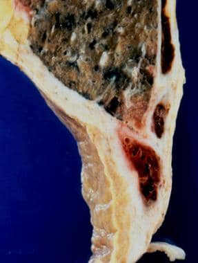
OBJECTIVE: To determine whether there is a specific pattern of clinicopathological features that could distinguish Borrmann's type IV gastric cancer from other types of gastric cancer. DESIGN: A retrospective study of patients with advanced gastric cancer treated between 1985 and 1995. SETTING: The Department of Surgery, Sendai National Hospital, a 716-bed teaching hospital. PATIENTS: The clinicopathologic features of 88 patients with Borrmann's type IV carcinoma of the stomach were reviewed from the database of gastric cancer. The results were compared with those of 309 patients with other types of gastric carcinoma. MAIN OUTCOME MEASURES: Gender, age, tumour size, depth of invasion, histologic type, cancer-stromal relationship, histologic growth pattern, nodal involvement, lymphatic and vascular invasion, type of operation, cause of death and 5-year survival. RESULTS: Women were afflicted as commonly as men in the Borrmann's type IV group. These patients tended to be younger and to have larger tumours involving the entire stomach than patients with other types of cancer. Histologic type was commonly diffuse and scirrhous, and serosal invasion was prominent with infiltrative growth. Nodal involvement and lymphatic invasion were more common in patients with Borrmann's type IV than in those with other types of gastric cancer. The disease was advanced in most instances and a total gastrectomy was performed in 55% of the patients. The survival rate of patients with Borrmann's type IV tumour was lower than for patients with other types of gastric cancer (p < 0.005, log-rank test). CONCLUSIONS: In Borrmann's type IV gastric cancer, early detection and curative resection are crucial to extend the patient's survival. Aggressive postoperative chemotherapy is recommended when a noncurative resection is performed. (+info)The serous demilune of rat sublingual gland is an artificial structure produced by conventional fixation. (3/160)
The ultrastructure of the secretory end-piece of the rat sublingual gland was examined in samples prepared by rapid freezing and freeze-substitution method, and results were analyzed in combination with 3-D images reconstructed by computer graphics from light micrographs of serial sections. Fixation by rapid freezing followed by freeze-substitution preserved cellular ultrastructures, especially the membrane structure, in perfect condition, and demonstrated the terminal portion of the sublingual gland to be a compound branched tubulo-alveolar gland with serous cells distributed throughout the end-pieces. All the serous cells aligned with mucous cells to surround a common lumen, leaving no demilune structure. In contrast, samples fixed by the conventional immersion method showed distended mucous cells displacing the serous cells toward the basal portion of the acinus to form the demilune structure. The luminal space was also compressed and appeared disconnected from the serous cells. From these observations, the serous demilune that for more than 130 years has been believed to be an actual histological entity was proved to be an artificial structure produced through compression by the hydrated and expanded mucous cells during immersion fixation. (+info)Inducible expression of the gp49B inhibitory receptor on NK cells. (4/160)
Murine NK cells express inhibitory receptors belonging to the C-type lectin-like (Ly-49, CD94/NKG2) and Ig superfamily-related (gp49) receptors. The murine gp49B receptor displays structural homology with human killer inhibitory receptors, and was previously identified to be a receptor on mast cells and activated NK cells. The gp49B receptor is highly related to gp49A, a receptor with unknown function. In this study, using a novel mAb produced against soluble gp49B molecules that cross-reacts with gp49A, we examined the cellular distribution and function of these receptors. gp49 is constitutively expressed on cells of the myeloid lineage throughout development, as well as on mature cells. Importantly, gp49 is not expressed on spleen- and liver-derived lymphocytes, including NK cells, but its expression is induced in vitro on NK cells following IL-2 stimulation, or in vivo by infection with murine CMV. Molecular studies revealed that both the immunoreceptor tyrosine-based inhibitory motif-containing gp49B as well as immunoreceptor tyrosine-based inhibitory motif-less gp49A receptors are up-regulated on NK cells following murine CMV infection. When co-cross-linked with NK1.1, gp49B can inhibit NK1.1-mediated cytokine release by NK cells. Taken together, these studies demonstrate that the expression of gp49B on NK cells is regulated, providing the first example of an in vivo activation-induced NK cell inhibitory receptor, in contrast to the constitutively expressed Ly49 family. (+info)Mist1 expression is a common link among serous exocrine cells exhibiting regulated exocytosis. (5/160)
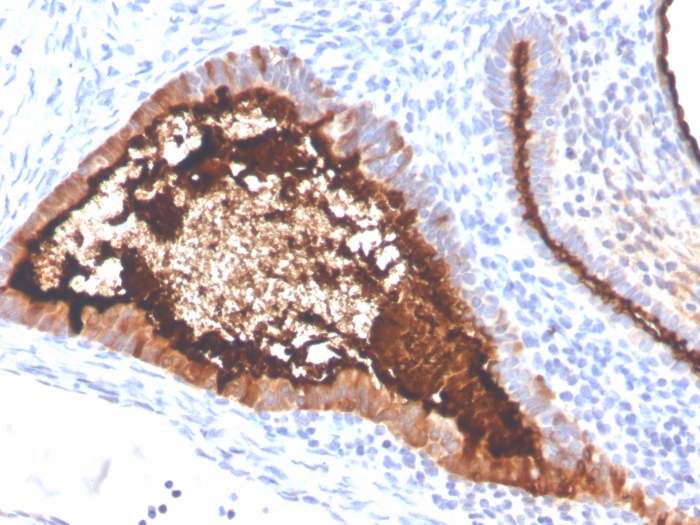
Mist1 is a basic helix-loop-helix transcription factor that represses E-box-mediated transcription. Previous studies have suggested that the Mist1 gene is expressed in a wide range of tissues, although a complete characterization of Mist1 protein accumulation in the adult organism has not been described. In an effort to identify specific cell types that contain the Mist1 protein, antibodies specific for Mist1 were generated and used in Western blot and immunohistochemical assays. Our studies show that the Mist1 protein is present in many different tissues but that it is restricted to cell types that are exclusively secretory in nature. Pancreatic acinar cells, serous or seromucous cells of the salivary glands, chief cells of the stomach, and secretory cells of the prostate and seminal vesicle show high levels of Mist1 protein, whereas nonserous exocrine cells, including the mucus-producing cells of the salivary glands, remain Mist1 negative. These results identify Mist1 as the first transcription factor that exhibits this unique serous-specific expression pattern and suggest that Mist1 may have a key role in establishing and maintaining a pathway responsible for the exocytosis of serous secretions. (+info)Comparative morphology and histochemistry of glands associated with the vomeronasal organ in humans, mouse lemurs, and voles. (6/160)
The vomeronasal organ (VNO) is a chemosensory structure of the vertebrate nasal septum that has been recently shown to exist in nearly all adult humans. Although its link to reproductive behaviors has been shown in some primates, its functionality in humans is still debated. Some authors have suggested that the human VNO has the capacity to detect pheromones, while others described it as little more than a glandular pit. However, no studies have utilized histochemical techniques that would reveal whether the human VNO functions as a generalized gland duct or a specialized chemosensory organ. Nasal septal tissue from 13 humans (2-86 years old) were compared to that of two adult lemurs (Microcebus murinus) and eight adult voles (four Microtus pennsylvanicus and four Microtus ochrogaster). Sections at selected intervals of the VNO were stained with periodic acid-Schiff (PAS), alcian blue (AB), AB-PAS, and PAS-hematoxylin procedures. Results revealed typical well-developed VNOs with tubuloacinar glands in Microtus and Microcebus. VNO glands were AB-negative and PAS-positive in voles and mouse lemurs. Homo differed from Microtus and Microcebus in having more branched, AB and PAS-positive glands that emptied into the VNO lumen. Furthermore, the human VNO epithelium had unicellular mucous glands (AB and PAS-positive) and cilia, similar to respiratory epithelia. These results demonstrate unique characteristics of the human VNO which at once differs from glandular ducts (e.g., cilia) and also from the VNOs of mammals possessing demonstrably functional VNO. (+info)Functional and molecular characterization of an anion exchanger in airway serous epithelial cells. (7/160)
Serous cells secrete Cl(-) and HCO(3)(-) and play an important role in airway function. Recent studies suggest that a Cl(-)/HCO(3)(-) anion exchanger (AE) may contribute to Cl(-) secretion by airway epithelial cells. However, the molecular identity, the cellular location, and the contribution of AEs to Cl(-) secretion in serous epithelial cells in tracheal submucosal glands are unknown. The goal of the present study was to determine the molecular identity, the cellular location, and the role of AEs in the function of serous epithelial cells. To this end, Calu-3 cells, a human airway cell line with a serous-cell phenotype, were studied by RT-PCR, immunoblot, and electrophysiological analysis to examine the role of AEs in Cl(-) secretion. In addition, the subcellular location of AE proteins was examined by immunofluorescence microscopy. Calu-3 cells expressed mRNA and protein for AE2 as determined by RT-PCR and Western blot analysis, respectively. Immunofluorescence microscopy identified AE2 in the basolateral membrane of Calu-3 cells in culture and rat tracheal serous cells in situ. In Cl(-)/HCO(3)(-)/Na(+)-containing media, the 8-(4-chlorophenylthio)adenosine 3',5'-cyclic monophosphate (CPT-cAMP)-stimulated short-circuit anion current (I(sc)) was reduced by basolateral but not by apical application of 4, 4'-diisothiocyanostilbene-2,2'-disulfonic acid (50 microM) and 4, 4'-dinitrostilbene-2,2'-disulfonic acid [DNDS (500 microM)], inhibitors of AEs. In the absence of Na(+) in the bath solutions, to eliminate the contributions of the Na(+)/HCO(3)(-) and Na(+)/K(+)/2Cl(-) cotransporters to I(sc), CPT-cAMP stimulated a small DNDS-sensitive I(sc). Taken together with previous studies, these observations suggest that a small component of cAMP-stimulated I(sc) across serous cells may be referable to Cl(-) secretion and that uptake of Cl(-) across the basolateral membrane may be mediated by AE2. (+info)Rapid activation of basolateral potassium transport in human colon by oestradiol. (8/160)
1. We investigated the effect of oestradiol on basolateral potassium channels in human colonic epithelium. 2. Ion transport was quantified using short circuit current (I:(sc)) measurements of samples mounted in Ussing chambers. Serosal K transport was studied using nystatin permeabilization of the apical membrane. Intracellular pH changes were quantified using spectroflouresence techniques. 3. Experiments were performed with either 10 nM or 1 microM Ca(2+) in the apical bathing solution. With 10 nM Ca(2+) in the apical bathing solution addition of oestradiol (1 nM) to the basolateral bath produced a rapid increase in current (delta I(K)=11.2+/-1.2 microA.cm(-2), n=6). This response was prevented by treatment of the serosal membrane with tolbutamide (1 microM). With 1 microM Ca(2+) in the apical bathing solution addition of oestradiol produced a rapid fall in current (delta I(K)=-12.8+/-1.4 microA.cm(-2)), this response was prevented by treatment of the basolateral membrane with tetra-pentyl-ammonium (TPeA). These responses were rapid and occurred independently of protein synthesis. 4. Inhibition of basolateral Na(+)/H(+) exchange with either amiloride or a low sodium bathing solution prevented this response. These responses were prevented by inhibition of protein kinase C (PKC) with bis-indolyl-maleimide. 5. Oestradiol (1 nM) produced a rapid intracellular alkanization (mean increase=0.11 pH units; n=6; P<0.01). 6. These results suggest that oestradiol rapidly modulates serosal K transport in human colon. These effects depend upon intact Na(+)/H(+) exchange and protein kinase C. We propose a non-classical, possibly membrane linked, mechanism for oestradiol action in human colonic epithelium. (+info)A serous membrane, also known as serosa, is a smooth, moist, and thin tissue that forms the lining of the body's cavities and covers the outer surface of organs, secreting a lubricating serous fluid for ease of movement and reduction of friction.
A serous membrane is a type of thin, smooth tissue that lines the inside of body cavities and surrounds certain organs. It consists of two layers: an outer parietal layer that lines the cavity wall, and an inner visceral layer that covers the organ. Between these two layers is a small amount of fluid called serous fluid, which reduces friction and allows for easy movement of the organs within the body cavity.
Serous membranes are found in several areas of the body, including the pleural cavity (around the lungs), the pericardial cavity (around the heart), and the peritoneal cavity (around the abdominal organs). They play an important role in protecting these organs and allowing them to move smoothly within their respective cavities.
 Serous membrane - Wikipedia
In the human body, there are three serous cavities with associated serous membranes: A serous membrane lines the pericardial ... The serous membrane covering the heart and lining the mediastinum is referred to as the pericardium, the serous membrane lining ... The tunica vaginalis is the serous membrane, which surrounds the male gonad, the testis. The two layers of serous membranes are ... Mesotheliomas are neoplasias that are relatively specific for serous membranes. The modified Mullerian-derived serous membranes ...
 Serous membrane Definition & Meaning | Dictionary.com
... any of various thin membranes, as the peritoneum, that line certain cavities of the body and exude a serous fluid. See more. ... serous membrane. in a sentence. *. By the time that these changes are effected, the serous membrane and amnion are both very ... Each lung is covered, except at one point, with an elastic serous membrane in a double layer, called the pleura. ... any of various thin membranes, as the peritoneum, that line certain cavities of the body and exude a serous fluid. ...
 What is the role of serous membranes in the body quizlet?
It allows membranes to slide past each other without friction as it secretes a thin watery serous fluid. ... Does serous membrane cover organs?. The serous membrane that covers internal organs is called a visceral membrane; while the ... Serous fluid.. What does the serous membrane cover?. Serous membranes line body cavities that do not open directly to the ... What is the function of serous membrane quizlet?. What is the function of serous membrane? It allows membranes to slide past ...
 Category:Pleura - Wikimedia Commons
serous membrane that lines the wall of thoracic cavity and the surface of lung ... serous membrane that lines the wall of thoracic cavity and the surface of lung; serozo de origino mezoderma, kiu kovras ambaŭ ... membrane séreuse délimitant un espace virtuel situé entre les poumons en dedans et la cage thoracique en dehors; אנטומיה; ...
 Laparoscopic or open distal gastrectomy after neoadjuvant chemotherapy for operable gastric cancer, a randomized Phase II trial...
 Pathology of Nonmesothelial Cancers of the Pleura: Definition, Etiology, Epidemiology
Angiosarcoma of serous membranes. Arch Pathol Lab Med. 1983. 107:304-306. ... Malignant vascular tumors of the serous membranes mimicking mesothelioma. Am J Surg Pathol. 1996. 20:1431-1439. ... 30, 31] There may also be an association between angiosarcoma of the serosal membranes and prior radiation exposure. [32] ... Cytokeratin and epithelial membrane antigen expression in angiosarcomas. An Immunohistochemical study of 33 cases. Arch Pathol ...
Pathology of Nonmesothelial Cancers of the Pleura: Definition, Etiology, Epidemiology
Angiosarcoma of serous membranes. Arch Pathol Lab Med. 1983. 107:304-306. ... Malignant vascular tumors of the serous membranes mimicking mesothelioma. Am J Surg Pathol. 1996. 20:1431-1439. ... 30, 31] There may also be an association between angiosarcoma of the serosal membranes and prior radiation exposure. [32] ... Cytokeratin and epithelial membrane antigen expression in angiosarcomas. An Immunohistochemical study of 33 cases. Arch Pathol ...
 Advanced Search Results - Public Health Image Library(PHIL)
 thoracic cavity
The chest cavity is lined with a serous membrane, which exudes a thin fluid. That portion of the chest membrane is called the ... The membrane continues over the lung, where it is called the visceral pleura, and over part of the esophagus, the heart, and ... The membrane is well supplied with blood vessels, nerves, and lymph channels. The vessels of the visceral part of the pleura ... The heart is covered by a fibrous membrane sac called the pericardium that blends with the trunks of the vessels running to and ...
 Colloidal Definition And Terms | A Colloidal Silver Glossary
serum - 1) The clear portion of any body fluid; the clear fluid moistening serous membranes. 2) Blood serum; the clear liquid ... osmosis - The difussion of a fluid through a semi permeable membrane.. osmotic pressure - The pressure in atmospheres or mm of ... Ion-selective electrodes are often membrane type electrodes.. isoelectric point - The point on a pH vs zeta potential plot ...
 Peritoneal cancer: What it is, who it affects, and more
The Cardiovascular System | Encyclopedia.com
Serous fluid (SIR-us):. Clear, watery, lubricating fluid produced by serous membranes, which line body cavities and cover ... Diffusion of water through a semipermeable membrane.. Pericardium (pair-i-CAR-dee-um):. Tough, fibrous, two-layered membrane ... Osmosis is the diffusion of water through a semipermeable membrane (a membrane that allows some materials but not others to ... Thin membrane lining the interior of the heart.. Epicardium (ep-i-CAR-dee-um):. Lubricating outer layer of the heart wall and ...
 A&P Chapterr 1 Cheat Sheet by Xyloswagg96 - Download free from Cheatography - Cheatography.com: Cheat Sheets For Every Occasion
 Newcastle Disease in Poultry - Poultry - Merck Veterinary Manual
H5N1 Influenza Virus, Domestic Birds, Western Siberia, Russia - Volume 12, Number 7-July 2006 - Emerging Infectious Diseases...
 Bryonia - George Vithoulkas
The mucous membranes, serous membranes or skin can dry up to a tremendous extent. Further examples of such dryness include: ... Bryonia has a marked action on all the serous membranes and the viscera they contain, causing inflammation and exudation. It ... Dropsical swellings into synovial and serous membranes, painful to touch, which increase as the day goes on and disappear ... Dryness of the mucous membranes; scanty discharges. Dry, burning heat, as if blood were burning in the veins or one part cold ...
 Anatomy and Physiology: Internal Male Reproductive Anatomy
Housed inside the tunica vaginalis, a serous membrane, the testis consists of lobules where sperm develop. The testis is ... Each ~7.5 cm sac is lined with a mucous membrane, and the cells within the membrane secrete a pale fluid containing sugars, ... There are two of them (most things come in pairs in the body). The duct lies between the peritoneal membrane and the lateral ...
 Bryonia alba | Homeopathy | Spirit India
 Serous fluid: Metastatic sarcomas, melanoma, and other non-epithelial neoplasms - CytoJournal
Serous fluid: Metastatic sarcomas, melanoma, and other non-epithelial neoplasms ... Most tumors metastatic to the serous membranes are of epithelial origin. Sarcomas account for only 3-6% of malignant effusions ... Involvement of serous membranes as well as intraperitoneal spread can occur. Rhabdomyosarcomas can also occur in the setting of ... While most tumors metastatic to the serous membranes are of epithelial origin, cytologists should be aware that non-epithelial ...
 Natur 3 - Moisturizing and Revitalizing Cream - Breast 75 Milliliters
- Bayho
Natur 3 - Moisturizing and Revitalizing Cream - Breast 75 Milliliters
- Bayho
 DailyMed - PROPRANOLOL HYDROCHLORIDE solution
Oculomucocutaneous syndrome involving the skin, serous membranes and conjunctivae reported for a beta-blocker (practolol) have ... Skin and Mucous Membranes: Stevens-Johnson Syndrome, toxic epidermal necrolysis, dry eyes, exfoliative dermatitis, erythema ... At dosages greater than required for beta-blockade, propranolol also exerts a quinidine-like or anesthetic-like membrane action ... In dosages greater than required for beta-blockade, propranolol also exerts a quinidine-like or anesthetic-like membrane action ...
 Pericardium - Structure & Function | GetBodySmart
... which is secreted by the serous membranes. The fluids reduce friction between membranes as they glide past one another during ... Under the fibrous pericardium is a thin layer of serous membrane known as the parietal pericardium. ... Between the walls of the serous pericardium is the pericardial cavity. This narrow space is normally filled with a few (10-50) ... Together, the parietal and visceral pericardial layers are also called the serous pericardium. ...
 Frontiers | Case Report: Primary NK/T Cell Lymphoma Nasal Type of the Colon With Multiple Intestinal Perforations
 Pathology Outlines - Anatomy, history, grossing & features to report
 IUPAC - serum (ST06843)
 Registration Dossier - ECHA
 Chylothorax (Proceedings)
 lamina of omasum - Ontology Browser - Rat Genome Database
Muscular system Cheat Sheet by jonahenry - Download free from Cheatography - Cheatography.com: Cheat Sheets For Every Occasion
Subserous fascia is a connective tissue layer of the serous membranes covering organs in various body cavities.. -Myofibrilsare ... Movement can occur up or down a cell membrane A cell membrane is a boundary wall surrou-nding cytoplasm of a cell. Muscle ... A membrane is permeable when materials can pass through it.. Diffusion is the movement form an area of high concen-tration to ... Molecules, gas ions, nutrients, and waste are able to pass through the cell membrane Muscle cells provide movement Nerve cell ...
 JCM | Special Issue : Advances in Clinical and Translational Research of Oral Surgery, Biomaterials, and Oral Disease Management
Histology revealed that the Schneiderian membrane had atrophied with loss of cilia and serous glands in both groups at 4 weeks ... Collagenated Synthetic Bone Substitute Material for Sinus Floor Elevation at Sites with a Perforated Schneiderian Membrane by ... Schneiderian membrane perforation (SMP) is the most common complication during sinus floor elevation (SFE). Conventional ... Schneiderian membrane perforation (SMP) is the most common complication during sinus floor elevation (SFE). Conventional ...
Mucous membranes4
- The sensation of dryness of the mucous membranes is most frequently reported, but the dryness of Bryonia extends to the emotional and mental levels as well. (hpathy.com)
- Symbiopathic - This remedy is indicated for symptoms associated with inflammation of serous and mucous membranes, bone infections and joint problems according to traditional homeopathic practice. (forresthealth.com)
- Pénfigo foliáceo, without affecting the mucous membranes. (bvsalud.org)
- Blood pressure of 120/70 mmHg, heart plaques, with a seborrheic appearance and rate of 76 beats per minute, respiratory rate distribution (face, neck, and trunk), where of 16 per minute, oxygen saturation of 97 %, the mucous membranes are generally not temperature of 37.1 degrees Celsius. (bvsalud.org)
Serosa2
- The serous membrane (or serosa) is a smooth tissue membrane of mesothelium lining the contents and inner walls of body cavities, which secrete serous fluid to allow lubricated sliding movements between opposing surfaces. (wikipedia.org)
- A serous membrane, or serosa, lines the three internal cavities that do not open to the exterior: pleural, pericardial, and peritoneal cavities. (digitalhistology.org)
Fluid18
- Between the two opposing serosal surfaces is often a potential space, mostly empty except for the small amount of serous fluid. (wikipedia.org)
- Serous membranes line and enclose several body cavities, also known as serous cavities, where they secrete a lubricating fluid which reduces friction from movements. (wikipedia.org)
- Between the parietal and visceral layers is a very thin, fluid-filled serous space, or cavity. (wikipedia.org)
- The epithelial layer, known as mesothelium, consists of a single layer of avascular flat nucleated cells (simple squamous epithelium) which produce the lubricating serous fluid. (wikipedia.org)
- The fluid is produced by the serous membranes and stays between the two layers to reduce friction between the walls of the cavities and the internal organs when they move with respect to one another, such as when the lungs inflate or the heart beats. (wikipedia.org)
- any of various thin membranes, as the peritoneum, that line certain cavities of the body and exude a serous fluid. (dictionary.com)
- Cells of the serous layer secrete a serous fluid that provides lubrication to reduce friction. (urhelpmate.com)
- The secreted fluid is called serous fluid. (urhelpmate.com)
- Serous fluid. (urhelpmate.com)
- Serous membranes are covered by a thin layer of serous fluid that is secreted by the epithelium. (urhelpmate.com)
- The chest cavity is lined with a serous membrane, which exudes a thin fluid. (britannica.com)
- Because the atmospheric pressure between the parietal pleura and the visceral pleura is less than that of the outer atmosphere, the two surfaces tend to touch, friction between the two during the respiratory movements of the lung being eliminated by the lubricating actions of the serous fluid. (britannica.com)
- Each ~7.5 cm sac is lined with a mucous membrane, and the cells within the membrane secrete a pale fluid containing sugars, prostaglandins , and other substances. (visiblebody.com)
- This narrow space is normally filled with a few (10-50) millilitres of pericardial fluid, which is secreted by the serous membranes. (getbodysmart.com)
- Clear watery fluid especially that moistening the surface of serous membranes or that exuded through inflammation of any of these membranes. (iupac.org)
- Synovial fluid, aqueous humor, cerebrospinal fluid and serous body cavity fluids are all routinely evaluated in veterinary medicine. (dvm360.com)
- Friis) from affected tissues (e.g., fibrin from serous membranes, synovial tissue or joint fluid) or, more commonly, detection of M. hyorhinis by PCR in these same sample types. (nationalhogfarmer.com)
- An air-fluid level or bubbles of air may be visible through the tympanic membrane. (msdmanuals.com)
Pleura5
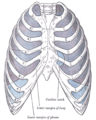
- The serous membrane covering the heart and lining the mediastinum is referred to as the pericardium, the serous membrane lining the thoracic cavity and surrounding the lungs is referred to as the pleura, and that lining the abdominopelvic cavity and the viscera is referred to as the peritoneum. (wikipedia.org)
- Each lung is covered, except at one point, with an elastic serous membrane in a double layer, called the pleura. (dictionary.com)
- That portion of the chest membrane is called the parietal pleura . (britannica.com)
- The membrane continues over the lung, where it is called the visceral pleura, and over part of the esophagus, the heart, and the great vessels, as the mediastinal pleura, the mediastinum being the space and the tissues and structures between the two lungs. (britannica.com)
- The thoracic cavity is lined entirely by a serous membrane known as pleura. (dvm360.com)
Inflammation1
- Inflammation of a serous membrane. (bvsalud.org)
Connective tissue8
- Each serous membrane is composed of a secretory epithelial layer and a connective tissue layer underneath. (wikipedia.org)
- The connective tissue layer provides the blood vessels and nerves for the overlying secretory cells, and also serves as the binding layer which allows the whole serous membrane to adhere to organs and other structures. (wikipedia.org)
- Subserous fascia is a connective tissue layer of the serous membranes covering organs in various body cavities. (cheatography.com)
- EN)do-mysium- connective tissue that covers the muscle fiber. (cheatography.com)
- All surfaces of the body are covered and protected by a membrane, usually consisting of an epithelium and an underlying connective tissue. (digitalhistology.org)
- The cutaneous membrane, or skin, covers the exterior of the body and is composed of a stratified squamous keratinized epithelium, called the epidermis, and a connective tissue layer, the dermis. (digitalhistology.org)
- This membrane is composed of simple squamous epithelium (mesothelium) and an underlying connective tissue. (digitalhistology.org)
- The peritoneal cavity is covered by a serous membrane of mesothelial cells overlying a connective tissue stroma. (dvm360.com)
Organs15
- The visceral layer of the membrane covers the organs (the viscera). (wikipedia.org)
- The pericardial cavity (surrounding the heart), pleural cavity (surrounding the lungs) and peritoneal cavity (surrounding most organs of the abdomen) are the three serous cavities within the human body. (wikipedia.org)
- The peritoneum is the serous membrane that surrounds several organs in the abdominopelvic cavity. (wikipedia.org)
- Does serous membrane cover organs? (urhelpmate.com)
- How do serous membranes protect organs? (urhelpmate.com)
- How do serous membranes protect organs from infection? (urhelpmate.com)
- Thin double layered membrane that lines the body cavities and covers organs. (urhelpmate.com)
- A thin double-layered membrane that surrounds organs in the ventral body cavity. (urhelpmate.com)
- The part of the membrane that lines the organs within the cavity. (urhelpmate.com)
- What membrane covers abdominal organs? (urhelpmate.com)
- How can serous membranes protect the organs of the body? (urhelpmate.com)
- The serous membrane has a number of functions related to protection of the organs and body cavities which it encloses. (urhelpmate.com)
- What is the role of serous membranes in the body and how can it protect the organs of the body involved? (urhelpmate.com)
- What is the role of serous membrane in the body and how can it protect the organs of the body involved? (urhelpmate.com)
- The mucous membrane, or mucosa, lines the lumens of all organs opening directly or indirectly to the exterior of the body, such as the stomach, intestines, trachea, and ureter. (digitalhistology.org)
Called the parietal2
- while the one that covers the cavity wall is called the parietal membrane. (wikipedia.org)
- For the heart, the layers of the serous membrane are called the parietal pericardium, and the visceral pericardium (sometimes called the epicardium). (wikipedia.org)
Fibrous membrane1
- The heart is covered by a fibrous membrane sac called the pericardium that blends with the trunks of the vessels running to and from the heart. (britannica.com)
Pericardial2
- In the human body, there are three serous cavities with associated serous membranes: A serous membrane lines the pericardial cavity of the heart, and reflects back to cover the heart, much like an under-inflated balloon would form two layers surrounding a fist. (wikipedia.org)
- Together, the parietal and visceral pericardial layers are also called the serous pericardium . (getbodysmart.com)
Lymph2
- The membrane is well supplied with blood vessels , nerves, and lymph channels. (britannica.com)
- Gross lesions in stillborn or weak, infected piglets include hydrocephalus, subcutaneous edema, ascites, hydrothorax, hemorrhages on serous membranes, congestion of lymph nodes and necrotic foci in the liver and spleen. (iastate.edu)
Cavity7
- The parietal layers of the membranes line the walls of the body cavity (pariet- refers to a cavity wall). (wikipedia.org)
- While serous membranes have a lubricative role to play in all three cavities, in the pleural cavity it has a greater role to play in the function of breathing. (wikipedia.org)
- Therefore, each organ becomes surrounded by serous membrane - they do not lie within the serous cavity. (wikipedia.org)
- The part of the membrane that lines the walls of the cavity. (urhelpmate.com)
- In peritoneal cancer, cancer cells grow within the serous membrane, a component of the peritoneal cavity. (medicalnewstoday.com)
- The lower tip of the heart, called the apex, points toward the left hip and rests on the diaphragm (a membrane of muscle separating the chest cavity from the abdominal cavity). (encyclopedia.com)
- Membrane of muscle separating the chest cavity from the abdominal cavity. (encyclopedia.com)
Reduce friction1
- The fluids reduce friction between membranes as they glide past one another during heartbeats. (getbodysmart.com)
Peritoneum1
- The outer coat is the serous membrane which lines the abdomen,--the peritoneum (note, p. 135). (dictionary.com)
Fluids1
- It is modified slightly from the chapter by the initial authors in the first edition of Diagnostic Cytopathology of Serous Fluids. (cytojournal.com)
Lungs1
- Approximately 95%-100% of the lungs were affected, and all serous membranes showed petechial and confluent hemorrhages. (cdc.gov)
Spinal cord1
- This is an infection of the membranes covering the brain and spinal cord. (medlineplus.gov)
Epithelial2
- While most tumors metastatic to the serous membranes are of epithelial origin, cytologists should be aware that non-epithelial neoplasms can also cause malignant effusions including sarcomas, melanomas, germ cell tumors, and, more rarely, brain tumors. (cytojournal.com)
- Most tumors metastatic to the serous membranes are of epithelial origin. (cytojournal.com)
Layer1
- SD-OCT (Heidelberg Engineering, Vista, CA / Germany), of the left eye revealed an elevated RPE corresponding to the scrolled margin of the tear [Figure 2] B, D. Fibrovascular membrane beneath the outer segments was seen as hyporeflective, due to the lack of RPE layer. (ijo.in)
Effusion2
- Serous otitis media is an effusion in the middle ear resulting from incomplete resolution of acute otitis media or obstruction of the eustachian tube without infection. (msdmanuals.com)
- may be done to confirm middle ear effusion (by showing reduced mobility of the tympanic membrane). (msdmanuals.com)
Abdomen2
- This diagrammatic cross section through the abdomen illustrates the location of the three types of body membranes: cutaneous, mucous and serous. (digitalhistology.org)
- A thin, smooth, serous membrane investing the whole internal surface of the abdomen, and more or less completely, all the viscera contained in it. (byu.edu)
Tunica1
- The tunica vaginalis is the serous membrane, which surrounds the male gonad, the testis. (wikipedia.org)
Body5
- The serous cavities are formed from the intraembryonic coelom and are basically an empty space within the body surrounded by serous membrane. (wikipedia.org)
- All serous membranes found in the human body formed ultimately from the mesoderm of the trilaminar embryo. (wikipedia.org)
- What is the role of serous membranes in the body quizlet? (urhelpmate.com)
- Portion of the serous membrane that lines the inner surface of the body wall chamber. (urhelpmate.com)
- What is the role of serous membrane in the body? (urhelpmate.com)
Chest1
- Pentachloride (great soreness of mucous membrane of EYES and nose, throat and chest sore). (abchomeopathy.com)
Thin2
- By the time that these changes are effected, the serous membrane and amnion are both very thin and not easily separable. (dictionary.com)
- Thin membrane lining the interior of the heart. (encyclopedia.com)
Diagnosis2
- Diagnosis is based on appearance of the tympanic membrane and sometimes on tympanometry. (msdmanuals.com)
- Diagnosis of serous otitis media is clinical plus pneumatic otoscopy. (msdmanuals.com)
Edema1
- The acute uveitic stage is heralded by the onset of sequential blurring of vision in both eyes, 1-2 days after the onset of CNS signs, and is marked by bilateral granulomatous anterior uveitis, a variable degree of vitritis, thickening of the posterior choroid, edema of the optic nerve, and multiple serous retinal detachments (Fig 9-52). (aao.org)
Action3
- Secretions are constantly maintained, during life, from the serous membrane , by the action of the internal exhalants. (dictionary.com)
- At dosages greater than required for beta-blockade, propranolol also exerts a quinidine-like or anesthetic-like membrane action, which affects the cardiac action potential. (nih.gov)
- The significance of the membrane action in the treatment of arrhythmias is uncertain. (nih.gov)