Salivary Gland Diseases
Salivary Gland Calculi
Salivary Ducts
Sialadenitis
Parotid Diseases
Salivary Gland Fistula
Parotid Gland
Submandibular Gland
Oral Surgical Procedures
Submandibular Gland Diseases
Sjogren's Syndrome
Iatrogenic Disease
Salivary Glands, Minor
Radiographic Image Enhancement
Cone-Beam Computed Tomography
Salivary Glands
Subtraction Technique
Sjogren's syndrome complicated with autoimmune hepatitis and antiphospholipid antibody syndrome. (1/35)
A 56-year-old Japanese female simultaneously developed thrombocytopenia, sicca symptoms, and an elevation of transaminase. Antiphospholipid antibodies were detected in her serum. The presence of anti-SS-A antibodies in the serum and sialectasis, disclosed by sialography, suggested the presence of primary Sjogren's syndrome (SjS). The laboratory data and the biopsy of the liver showed compatible findings with autoimmune hepatitis (AIH). Thrombocytopenia and liver dysfunction satisfactorily responded to corticosteroid. To our knowledge, this is the first reported case of SjS with AIH and antiphospholipid antibody syndrome (APAS). Analysis of serum cytokine levels showed a predominance of Th0-Th1 response, which is not compatible with AIH, in this complicated autoimmune state. (+info)Sjogren's syndrome: comparison of assessments with quantitative salivary gland scintigraphy and contrast sialography. (2/35)
This study compared the quantitative parameters of salivary gland scintigraphy and the sialographic stages in patients with Sjogren's syndrome. METHODS: One hundred sixteen patients suspected of having Sjogren's syndrome were examined with salivary gland scintigraphy and contrast sialography. When contrast sialography was used as the gold standard, Sjogren's syndrome was diagnosed in 50 of these 116 patients; Sjogren's syndrome was not seen in the other 66 patients. After injection of 370 MBq 99mTc-sodium pertechnetate, dynamic salivary gland scintigraphy with lemon juice stimulation was performed for 50 min. Functional parameters for the parotid and submandibular glands were calculated, and scintigraphic and sialographic results were compared. RESULTS: With the progression of sialographic stages from 0 to 4, the quantity of tracer accumulation decreased in the submandibular gland (P < 0.0001), and the quantity of tracer secretion decreased in the parotid gland (P < 0.0001). The sialographic stage in patients with Sjogren's syndrome was correlated with these scintigraphic parameters (P < 0.0001): sialographic stage = 3.243 - 0.337 x (submandibular gland uptake ratio) - 0.026 x (parotid gland maximum secretion). CONCLUSION: The decreased accumulation in the submandibular gland and the decreased secretion in the parotid gland were highly sensitive indicators of salivary gland disease in Sjogren's syndrome. The sialographic stage was correlated with these scintigraphic parameters. (+info)Scintigraphic study of the major salivary glands in pediatric bone marrow transplant recipients. (3/35)
Total body irradiation (TBI) at bone marrow transplantation (BMT) is shown to cause salivary gland dysfunction in children. The aim of the investigation was to study the function of major salivary glands in long-term surviving children following treatment with TBI, using salivary gland scintigraphy (SGS). Thirteen patients (seven male, six female), who had received TBI before the age of 13 years and survived more than 4 years, participated in the study. A reference group of 10 patients (nine male, one female) was examined shortly before they were to undergo BMT. The mean age was 14.1 +/- 4.1 years in the TBI-treated group and 12.8 +/- 5.9 years in the reference group. Unstimulated and stimulated whole salivary secretion rates were measured for 15 and 5 min, respectively, before SGS was performed. The percentage of stimulated secretion was 44.7 +/- 18.1% in the TBI-treated group compared to 58.4 +/- 13.0% in the reference group (P = 0.0438). Slower reaccumulation after excretion was found in the TBI-treated patients compared to the reference group (P = 0. 0300). The function of the major salivary glands in long-term survivors treated with TBI at BMT before the age of 13 years was found to be diminished, as shown by the reduced trapping rate and reduced emptying capacity, compared to prior to BMT. Bone Marrow Transplantation (2000) 26, 775-779. (+info)Blunt trauma to the parotid gland. (4/35)
Trauma involving the parotid gland is rare and is usually caused by penetrating injuries or fractures of the facial skeleton. A unique case is presented of rupture of the parotid gland after a minor external force in a 14 year old boy. The radiological findings are presented, which pointed to diffuse rupture of the gland parenchyma with an intact duct system. A conservative management policy using antibiotics and anticholinergics was effective, with complete resolution of symptoms. A literature review of more serious parotid injuries supported the conservative management policy. (+info)Comparative study of MR sialography and digital subtraction sialography for benign salivary gland disorders. (5/35)
BACKGROUND AND PURPOSE: MR sialography has become an alternative imaging technique for ductal salivary gland diseases. We compared the diagnostic accuracies of MR sialography and digital subtraction sialography in patients with successful completion of both examinations and benign salivary gland disorders. METHODS: In a prospective study, we attempted to examine salivary glands in 80 patients with clinically suspected diagnoses of sialadenitis and/or sialolithiasis. Each patient underwent digital subtraction sialography and MR sialography. MR sialography was obtained with a T2-weighted single-shot turbo spin-echo sequence (TR/TE 2800/1100 msec, acquisition time 7 seconds), with use of a quadrature head coil. Final diagnoses were confirmed by clinical follow-up and results of biopsy (n = 9) or surgery (n = 19). RESULTS: Failure rate was 5% (four of 80) for MR sialography and 14% (11 of 80) for digital subtraction sialography. Eighty-one salivary glands (48 parotid, 33 submandibular) in 65 patients were successfully visualized with both modalities. MR sialography depicted the main ductal system and first- and second-order branches, whereas digital subtraction sialography was able to depict third-order branches. Sensitivity and specificity to diagnose chronic sialadenitis were 70% and 98% with MR and 96% and 100% with digital subtraction sialography. MR sialography enabled diagnosis of sialolithiasis with a sensitivity of 80% and a specificity of 98% versus 90% and 98% for each with digital subtraction sialography. CONCLUSION: MR sialography with a heavily T2-weighted sequence is highly successful in the noninvasive visualization of the ductal system of major salivary glands. It is useful for diagnosing sialolithiasis and sialadenitis. Digital subtraction sialography, an invasive technique, had a substantial procedural failure rate, particularly for the submandibular duct. However, because of its higher spatial resolution, successfully completed digital subtraction sialography achieved superior diagnostic information compared with that of MR sialography. (+info)SIALADENITIS AND MAJOR SALIVARY GLAND TUMORS IN CHILDREN: EXPERIENCE AT LOS ANGELES CHILDRENS HOSPITAL AND A REVIEW OF THE LITERATURE. (6/35)
Except for mumps, the benign lesions most frequently seen in the salivary glands of a child are parotitis, hemangioendotheliomas and mixed tumors. Carcinoma and sarcoma are uncommon. Rapid growth and pain are features of malignant change. Chronic parotitis usually subsides under conservative treatment. If operation is necessary, total parotidectomy is advisable. Scout x-ray films and sialangiographic examination are useful in differentiating an inflammatory lesion from a neoplastic growth.The treatment of choice for a non-inflammatory tumor is surgical excision, for most parotid tumors are radioresistant. Small masses should be completely excised for pathological evaluation. Since operation entails risk of damage to the seventh nerve, incisional biopsy may occasionally be indicated in the case of a large diffuse lesion for it is very likely to be benign and operation unnecessary. The risks of seventh nerve damage are magnified in a child as the anatomic structures are smaller and the nerve lies in a more superficial position. (+info)Ultrasonography of salivary glands in primary Sjogren's syndrome. A comparison with magnetic resonance imaging and magnetic resonance sialography of parotid glands. (7/35)
OBJECTIVES: To evaluate ultrasonography (US) of salivary glands in primary Sjogren's syndrome (SS) and to compare US with parotid magnetic resonance (MR) imaging and MR sialography. METHODS: US examination of parotid, submandibular and sublingual glands was performed on 27 patients with primary SS, 27 healthy controls and 27 symptomatic controls without SS. The results were compared with parotid MR imaging and MR sialography and the clinical features of the patients. RESULTS: Salivary gland abnormalities, parenchymal inhomogeneity or adipose degeneration, were visualized in 21 (78%) SS patients, in one healthy control and in two symptomatic controls by US. Eighteen (67%) patients had changes in the parotid and submandibular glands and 8 (30%) changes in the sublingual glands. In the comparison, MR sialography was found to be the most sensitive method (96%), followed by MR imaging (81%) and US (78%), in detecting glandular changes. The specificity of US was 94%. The US and MR results were related to anti-Ro/SSA positivity but not to saliva secretion. The focus scores were related only to parotid MR imaging findings. CONCLUSIONS: US, MR imaging and MR sialography with modern technology have reached such a good accuracy in visualizing glandular structural changes that they are promising alternatives to the conventional invasive examinations in the diagnostics of SS. (+info)Parotid sialolithiasis in Stensen's duct. (8/35)
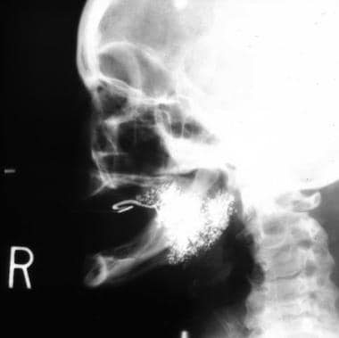
Salivary duct lithiasis is a condition characterized by the obstruction of a salivary gland or its excretory duct due to the formation of calcareous concretions or sialoliths resulting in salivary ectasia and even provoking the subsequent dilation of the salivary gland. Sialolithiasis accounts for 30% of salivary diseases and most commonly involves the submaxillary gland (83 to 94%) and less frequently the parotid (4 to 10%) and sublingual glands (1 to 7%). The present study reports the case of a 45-year-old male patient complaining of bad breath and foul-tasting mouth at meal times and presenting with a salivary calculus in left Stensen's duct. Once the patient was diagnosed, the sialolith was surgically removed using local anesthesia. In this paper we have also updated a series of concepts related to the etiology, diagnosis and treatment of sialolithiasis. (+info)Sialography is a medical imaging technique used to examine the ducts (salivary glands) that carry saliva from the salivary glands to the mouth. In this procedure, a radiopaque contrast material is injected into the salivary gland, and then X-rays or other forms of imaging are taken to visualize the shape and any abnormalities in the ducts.
The contrast material outlines the ducts on the images, allowing healthcare professionals to identify any blockages, narrowing, dilations, stones, or other abnormalities that may be present in the salivary glands. Sialography is typically used to diagnose and manage conditions such as salivary gland inflammation, obstruction, or infection.
It's worth noting that sialography has been largely replaced by newer imaging techniques, such as ultrasound, CT scans, and MRI, which do not require the injection of a contrast material and are generally considered safer and more comfortable for patients. However, sialography may still be used in certain cases where these other methods are not sufficient to make an accurate diagnosis.
Salivary gland diseases refer to a group of conditions that affect the function and structure of the salivary glands. These glands are responsible for producing saliva, which helps in digestion, lubrication, and protection of the mouth and throat. The major salivary glands include the parotid, submandibular, and sublingual glands.
There are several types of salivary gland diseases, including:
1. Salivary Gland Infections: These are usually caused by bacteria or viruses that infect the gland, ducts, or surrounding tissues. The most common infection is called sialadenitis, which can cause pain, swelling, redness, and difficulty swallowing.
2. Salivary Gland Stones (Sialolithiasis): These are small, hard deposits that form in the ducts of the salivary glands, causing blockages and leading to swelling, pain, and infection.
3. Salivary Gland Tumors: Both benign and malignant tumors can develop in the salivary glands. Benign tumors are usually slow-growing and cause localized swelling, while malignant tumors may be more aggressive and spread to other parts of the body.
4. Salivary Gland Dysfunction: This refers to conditions that affect the production or flow of saliva, such as Sjogren's syndrome, radiation therapy, dehydration, or certain medications.
5. Autoimmune Disorders: Conditions like Sjogren's syndrome, lupus, and rheumatoid arthritis can affect the salivary glands and cause inflammation, dry mouth, and other symptoms.
6. Salivary Gland Trauma: Injuries to the face or neck can damage the salivary glands and lead to swelling, bleeding, or decreased function.
Proper diagnosis and treatment of salivary gland diseases require a thorough evaluation by a healthcare professional, often involving imaging studies, laboratory tests, and biopsies. Treatment options may include antibiotics, surgery, radiation therapy, or changes in medication or lifestyle.
Salivary gland calculi, also known as salivary duct stones or sialoliths, are small, hard deposits that form in the salivary glands or their ducts. These calculi typically consist of calcium salts and other minerals, and can vary in size from a few millimeters to over a centimeter in diameter.
Salivary gland calculi can cause a range of symptoms, including pain, swelling, and difficulty swallowing, particularly during meals. The obstruction of the salivary duct by the calculus can lead to infection or inflammation of the salivary gland (sialadenitis).
The most common location for salivary gland calculi is in the submandibular gland and its duct, followed by the parotid gland and then the sublingual gland. Treatment options for salivary gland calculi include conservative management with hydration, massage, and warm compresses, as well as more invasive procedures such as extracorporeal shock wave lithotripsy, sialendoscopy, or surgical removal of the calculus.
Salivary ducts are the excretory tubules that transport saliva from the major and minor salivary glands to the oral cavity. The main function of these ducts is to convey the salivary secretions, which contain enzymes and lubricants, into the mouth to aid in digestion, speech, and swallowing.
There are two pairs of major salivary glands: the parotid glands and the submandibular glands. Each pair has its own set of ducts. The parotid gland's saliva is drained through the parotid duct, also known as Stensen's duct, which opens into the oral cavity opposite the upper second molar tooth. The submandibular gland's saliva is transported through the submandibular duct, or Wharton's duct, which empties into the floor of the mouth near the base of the tongue.
Minor salivary glands are scattered throughout the oral cavity and pharynx, and their secretions are drained via small ducts directly into the oral mucosa.
Sialadenitis is a medical condition characterized by inflammation of the salivary gland. It can occur in any of the major salivary glands, including the parotid, submandibular, and sublingual glands. The inflammation may result from bacterial or viral infections, autoimmune disorders, or obstruction of the salivary ducts.
Acute sialadenitis is often caused by bacterial infections and can lead to symptoms such as pain, swelling, redness, and difficulty swallowing. Chronic sialadenitis, on the other hand, may be caused by recurrent infections, autoimmune disorders like Sjogren's syndrome, or stones in the salivary ducts. Symptoms of chronic sialadenitis can include intermittent swelling, pain, and dry mouth.
Treatment for sialadenitis depends on the underlying cause but may include antibiotics, anti-inflammatory medications, hydration, and massage of the salivary glands. In some cases, surgery may be necessary to remove obstructions or damaged tissue in the salivary gland.
Parotitis is the medical term for inflammation of the parotid gland, which is one of the major salivary glands located in the face, near the ear. The condition can result from various causes, including bacterial or viral infections, autoimmune disorders, or obstruction of the salivary ducts.
Parotitis can cause symptoms such as pain, swelling, redness, and difficulty swallowing. In some cases, it may also lead to fever, chills, and general malaise. The diagnosis of parotitis typically involves a physical examination, medical history, and sometimes imaging studies or laboratory tests to identify the underlying cause. Treatment depends on the specific cause but may include antibiotics, pain relievers, hydration, and measures to improve salivary flow.
Parotid diseases refer to conditions that affect the parotid glands, which are the largest of the salivary glands and are located in front of each ear. These glands produce saliva that helps in digestion and keeps the mouth moist. Parotid diseases can cause swelling, pain, dry mouth, or difficulty swallowing, among other symptoms. Some common parotid diseases include:
1. Parotid gland infection (also called parotitis) - an inflammation of the parotid gland due to bacterial or viral infections.
2. Salivary gland stones (also called sialolithiasis) - calcified deposits that form in the salivary ducts and can block the flow of saliva.
3. Salivary gland tumors - abnormal growths that can be benign or malignant, and may require surgical removal.
4. Parotid gland inflammation (also called sialadenitis) - an inflammation of the parotid gland due to autoimmune disorders, radiation therapy, or dehydration.
5. Parotid gland cysts (also called ranula or mucocele) - fluid-filled sacs that form in the salivary gland or duct.
Proper diagnosis and treatment of parotid diseases require a thorough evaluation by a healthcare professional, often involving imaging studies, laboratory tests, and biopsies.
A salivary gland fistula is an abnormal connection or duct between a salivary gland and the skin surface or another epithelial-lined structure, such as the mouth or the neck. This condition typically results from trauma, surgery, or infection that causes damage to the salivary gland or its ducts, leading to leakage of saliva into surrounding tissues.
Salivary gland fistulas can cause symptoms such as swelling, pain, redness, and discharge of saliva from the affected area. They may also increase the risk of infection and affect a person's quality of life. Treatment options for salivary gland fistulas include pressure dressings, antibiotics, and surgical intervention to repair the damaged duct or remove the affected salivary gland.
The parotid gland is the largest of the major salivary glands. It is a bilobed, accessory digestive organ that secretes serous saliva into the mouth via the parotid duct (Stensen's duct), located near the upper second molar tooth. The parotid gland is primarily responsible for moistening and lubricating food to aid in swallowing and digestion.
Anatomically, the parotid gland is located in the preauricular region, extending from the zygomatic arch superiorly to the angle of the mandible inferiorly, and from the masseter muscle anteriorly to the sternocleidomastoid muscle posteriorly. It is enclosed within a fascial capsule and has a rich blood supply from the external carotid artery and a complex innervation pattern involving both parasympathetic and sympathetic fibers.
Parotid gland disorders can include salivary gland stones (sialolithiasis), infections, inflammatory conditions, benign or malignant tumors, and autoimmune diseases such as Sjögren's syndrome.
The submandibular glands are one of the major salivary glands in the human body. They are located beneath the mandible (jawbone) and produce saliva that helps in digestion, lubrication, and protection of the oral cavity. The saliva produced by the submandibular glands contains enzymes like amylase and mucin, which aid in the digestion of carbohydrates and provide moisture to the mouth and throat. Any medical condition or disease that affects the submandibular gland may impact its function and could lead to problems such as dry mouth (xerostomia), swelling, pain, or infection.
Oral surgical procedures refer to various types of surgeries performed in the oral cavity and maxillofacial region, which includes the mouth, jaws, face, and skull. These procedures are typically performed by oral and maxillofacial surgeons, who are dental specialists with extensive training in surgical procedures involving the mouth, jaws, and face.
Some common examples of oral surgical procedures include:
1. Tooth extractions: This involves removing a tooth that is damaged beyond repair or causing problems for the surrounding teeth. Wisdom tooth removal is a common type of tooth extraction.
2. Dental implant placement: This procedure involves placing a small titanium post in the jawbone to serve as a replacement root for a missing tooth. A dental crown is then attached to the implant, creating a natural-looking and functional replacement tooth.
3. Jaw surgery: Also known as orthognathic surgery, this procedure involves repositioning the jaws to correct bite problems or facial asymmetry.
4. Biopsy: This procedure involves removing a small sample of tissue from the oral cavity for laboratory analysis, often to diagnose suspicious lesions or growths.
5. Lesion removal: This procedure involves removing benign or malignant growths from the oral cavity, such as tumors or cysts.
6. Temporomandibular joint (TMJ) surgery: This procedure involves treating disorders of the TMJ, which connects the jawbone to the skull and allows for movement when eating, speaking, and yawning.
7. Facial reconstruction: This procedure involves rebuilding or reshaping the facial bones after trauma, cancer surgery, or other conditions that affect the face.
Overall, oral surgical procedures are an important part of dental and medical care, helping to diagnose and treat a wide range of conditions affecting the mouth, jaws, and face.
Submandibular gland diseases refer to a group of disorders that affect the function or structure of the submandibular glands, which are salivary glands located beneath the jaw and produce saliva. These diseases can be categorized into inflammatory, infectious, obstructive, neoplastic (benign or malignant), and autoimmune disorders.
Some common submandibular gland diseases include:
1. Submandibular sialadenitis: Inflammation of the submandibular gland due to bacterial or viral infections, stones, or autoimmune conditions.
2. Salivary gland stones (sialolithiasis): Calcified deposits that obstruct the ducts leading from the submandibular gland, causing swelling and pain, especially during meals.
3. Submandibular gland tumors: Abnormal growths in the submandibular gland, which can be benign or malignant (cancerous). Malignant tumors may invade surrounding tissues and spread to other parts of the body.
4. Sjögren's syndrome: An autoimmune disorder that affects the exocrine glands, including the submandibular gland, leading to dry mouth and eyes.
5. IgG4-related disease: A systemic inflammatory condition characterized by the infiltration of IgG4-positive plasma cells into various organs, including the submandibular gland, causing swelling and damage.
6. Mikulicz's disease: A rare benign lymphoepithelial lesion that affects the salivary and lacrimal glands, including the submandibular gland, leading to enlargement and dryness of the affected glands.
7. Salivary gland dysfunction: Reduced or impaired saliva production due to aging, medications, radiation therapy, or systemic diseases, which can affect the submandibular gland.
Proper diagnosis and treatment of submandibular gland diseases require a thorough clinical evaluation, imaging studies, and sometimes biopsy or surgical intervention.
Sjögren's syndrome is a chronic autoimmune disorder in which the body's immune system mistakenly attacks its own moisture-producing glands, particularly the tear and salivary glands. This can lead to symptoms such as dry eyes, dry mouth, and dryness in other areas of the body. In some cases, it may also affect other organs, leading to a variety of complications.
There are two types of Sjögren's syndrome: primary and secondary. Primary Sjögren's syndrome occurs when the condition develops on its own, while secondary Sjögren's syndrome occurs when it develops in conjunction with another autoimmune disease, such as rheumatoid arthritis or lupus.
The exact cause of Sjögren's syndrome is not fully understood, but it is believed to involve a combination of genetic and environmental factors. Treatment typically focuses on relieving symptoms and may include artificial tears, saliva substitutes, medications to stimulate saliva production, and immunosuppressive drugs in more severe cases.
Iatrogenic disease refers to any condition or illness that is caused, directly or indirectly, by medical treatment or intervention. This can include adverse reactions to medications, infections acquired during hospitalization, complications from surgical procedures, or injuries caused by medical equipment. It's important to note that iatrogenic diseases are unintended and often preventable with proper care and precautions.
Minor salivary glands are numerous small exocrine glands that produce saliva and are distributed throughout the oral cavity, nasal cavity, pharynx, larynx, and paranasal sinuses. They are classified as "minor" due to their smaller size compared to the three pairs of major salivary glands (parotid, submandibular, and sublingual). The minor salivary glands are primarily mucous glands, although some contain serous cells. They are responsible for producing approximately 5-10% of the total saliva in the mouth. These glands help moisten the oral cavity, protect the mucosal lining, and facilitate speaking, chewing, and swallowing.
Radiographic image enhancement refers to the process of improving the quality and clarity of radiographic images, such as X-rays, CT scans, or MRI images, through various digital techniques. These techniques may include adjusting contrast, brightness, and sharpness, as well as removing noise and artifacts that can interfere with image interpretation.
The goal of radiographic image enhancement is to provide medical professionals with clearer and more detailed images, which can help in the diagnosis and treatment of medical conditions. This process may be performed using specialized software or hardware tools, and it requires a strong understanding of imaging techniques and the specific needs of medical professionals.
Cone-beam computed tomography (CBCT) is a medical imaging technique that uses a cone-shaped X-ray beam to create detailed, cross-sectional images of the body. In dental and maxillofacial radiology, CBCT is used to produce three-dimensional images of the teeth, jaws, and surrounding bones.
CBCT differs from traditional computed tomography (CT) in that it uses a cone-shaped X-ray beam instead of a fan-shaped beam, which allows for a faster scan time and lower radiation dose. The X-ray beam is rotated around the patient's head, capturing data from multiple angles, which is then reconstructed into a three-dimensional image using specialized software.
CBCT is commonly used in dental implant planning, orthodontic treatment planning, airway analysis, and the diagnosis and management of jaw pathologies such as tumors and fractures. It provides detailed information about the anatomy of the teeth, jaws, and surrounding structures, which can help clinicians make more informed decisions about patient care.
However, it is important to note that CBCT should only be used when necessary, as it still involves exposure to ionizing radiation. The benefits of using CBCT must be weighed against the potential risks associated with radiation exposure.
Salivary glands are exocrine glands that produce saliva, which is secreted into the oral cavity to keep the mouth and throat moist, aid in digestion by initiating food breakdown, and help maintain dental health. There are three major pairs of salivary glands: the parotid glands located in the cheeks, the submandibular glands found beneath the jaw, and the sublingual glands situated under the tongue. Additionally, there are numerous minor salivary glands distributed throughout the oral cavity lining. These glands release their secretions through a system of ducts into the mouth.
The "subtraction technique" is not a widely recognized or established term in medical terminology. It may refer to various methods used in different medical contexts that involve subtracting or comparing measurements, values, or observations to diagnose, monitor, or treat medical conditions. However, without more specific context, it's difficult to provide an accurate medical definition of the term.
In radiology, for example, the subtraction technique is a method used in imaging to enhance the visibility of certain structures by digitally subtracting one image from another. This technique is often used in angiography to visualize blood vessels more clearly.
Therefore, it's essential to provide more context or specify the medical field when using the term "subtraction technique" to ensure accurate communication and understanding.
A cyst is a closed sac, having a distinct membrane and division between the sac and its surrounding tissue, that contains fluid, air, or semisolid material. Cysts can occur in various parts of the body, including the skin, internal organs, and bones. They can be caused by various factors, such as infection, genetic predisposition, or blockage of a duct or gland. Some cysts may cause symptoms, such as pain or discomfort, while others may not cause any symptoms at all. Treatment for cysts depends on the type and location of the cyst, as well as whether it is causing any problems. Some cysts may go away on their own, while others may need to be drained or removed through a surgical procedure.
 Sialography
Salivary gland disease
Sialodochitis
Salivary duct stricture
Stafne defect
Oral medicine
Xerostomia
Pneumoparotitis
List of MeSH codes (E01)
List of MeSH codes (E06)
Parotid gland
Sialography - Wikipedia
 Rabinov Sialography Catheter | Cook Medical
 PLAN SIALOGRAPHY localizer - mrimaster
 MRI Sialography Test Near You in Delhi | Book Test at Low Price
 Sialogram: MedlinePlus Medical Encyclopedia
 Benign Parotid Tumors: Practice Essentials, Anatomy, Incidence and Etiology
 When You Have Lupus and Sjogren's Syndrome| HSS Rheumatology
 Ultrasound Elastography in Diffuse and Focal Parotid Gland Lesions | ORL | Karger Publishers
 Classification criteria for Sjögren's syndrome: a revised version of the European criteria proposed by the American-European...
Sjogren Syndrome: Practice Essentials, Etiology, Epidemiology
 Fluoroscopy - CHFT
 Ear pain in children - wikidoc
Parotitis-Like Symptoms Associated with COVID-19, France, March-April 2020 - Volume 26, Number 9-September 2020 - Emerging...
Parotitis: Practice Essentials, Background, Pathophysiology
Sjögren's syndrome MRI - wikidoc
 Shop Cook Medical Imaging by Manufacturer Name - McKesson Medical-Surgical
fm5 - San Diego Musical Theatre
Parotitis Workup: Laboratory Studies, Imaging Studies, Procedures
 Additional Tools - Bonameda
 dcadmin
Hernia repair - Wikipedia
 Digital X Ray Centre in Chandigarh, Mohali & Panchkula - - Kior Healthcare
Reproscan Imaging Centre - Parel W, Mumbai | 1Stop4Health
 INICET MDS Preparation - 100 Days Study Plan
 E-ORAL MEDICINE & RADIOLOGY | IDS Barielly
 sialoangiography | Taber's Medical Dictionary
 Medical Terminology Ch. 9 Flashcards - Easy Notecards
Sjogren's Syndrome Treatment, Surgery, Complication - Health | RXharun
Ultrasound2
- He also performs sialography, and diagnostic and interventional ultrasound. (proconferences.com)
- Diagnostic imaging techniques such as ultrasound, sialography, or CT scans are valuable in confirming the diagnosis. (barw.krd)
Salivary flow1
- Noninvasive procedures include salivary flow rate, sialography, or salivary scintigraphy. (rheumaknowledgy.com)
Diagnosis3
- Diagnosis is made clinically or with CT, ultrasonography, or sialography. (msdmanuals.com)
- CT, ultrasonography, and sialography are highly sensitive and are used if clinical diagnosis is equivocal. (msdmanuals.com)
- Sialography is the preferred method for diagnosis and treatment planning in obstructive sialadenitis. (elsevierpure.com)
Examination2
- Sialography (also termed radiosialography) is the radiographic examination of the salivary glands. (wikipedia.org)
- Sialography is the examination of the salivary glands after a dye has been injected. (toothfairy.co.in)
Parotid2
- Used during sialography procedures involving the submandibular duct or the parotid duct. (cookmedical.com)
- The sialography is indicated in cases of signs of sialodenitis, radiolucent stones, submandibular stones, deep parotid stones, and contraindicated in cases of infection, patients allergic to the radiograph contrast solution. (bvsalud.org)
Catheter1
- Contrast sialography may be done through a catheter inserted into the duct and can differentiate between stone, stenosis, and tumor. (msdmanuals.com)
Ultrasonography3
- Sialography has largely been replaced by sialoendoscopy and cross-sectional imaging, such as CT, MRI and ultrasonography. (wikipedia.org)
- Ultrasonography is much easier to perform than sialography and seems to be replacing sialography in many institutions. (medscape.com)
- 90% and MRI appears to be more sensitive in detecting small stones and distal duct stones than ultrasonography or contrast sialography. (msdmanuals.com)
Duct1
- Sialography revealed an oval-shaped filling defect in the dilated left Wharton's duct, which could suggest radiolucent calculus. (annalsafrmed.org)
Projections1
- Other examples of extraoral X-rays include sialography and cephalometric projections among others. (houstonoralhealthcarespecialists.com)
Involves1
- Sialography involves visualization of the salivary glands following the injection of a dye. (mountainoakdental.com)
Magnetic1
- The Magnetic resonance imaging (MRI) cost for the Sialography Test in Delhi starts at INR 8,000. (ganeshdiagnostic.com)
Clinically1
- It is not as sensitive as sialography, but this is probably not clinically significant. (medscape.com)
Procedure1
- What is the procedure for the Sialography MRI Screening Test? (ganeshdiagnostic.com)
Stones1
- MRI sialography is done when infections, tumours, stones, or certain types of cancer are suspected. (ganeshdiagnostic.com)
Test1
- Sialography is used to demonstrate the anatomy of the drainage system and is a very useful test. (medscape.com)
Subtraction sialography1
- Reduced radiation exposure, short examination time and almost painless examinations with good patient tolerance proved to be the major advantages of digital subtraction sialography as a diagnostic tool. (acibadem.edu.tr)
Computed tomography1
- Recently, diagnostic performance of computed tomography (CT) sialography is being explored and has been reported to have high sensitivity in detection of small sialoliths and allows differentiation of sialoliths from other calcifications in glandular ductal system. (nih.gov)
Sjogren1
- This study was conducted to assess the utility of diffusion-weighted (DW) MR imaging with use of SPLICE technique and dynamic MR sialography after administration of tartaric acid, and to estimate the salivary gland function in patients with Sjogren syndrome (SS). (nii.ac.jp)
Contrast4
- Contrast sialography may be done through a catheter inserted into the duct and can differentiate between stone, stenosis, and tumor. (msdmanuals.com)
- 90% and MRI appears to be more sensitive in detecting small stones and distal duct stones than ultrasonography or contrast sialography. (msdmanuals.com)
- Empty bags can be assessed by radio contrast sialography. (familytreecounseling.com)
- The sialography is indicated in cases of signs of sialodenitis, radiolucent stones, submandibular stones, deep parotid stones, and contraindicated in cases of infection, patients allergic to the radiograph contrast solution. (bvsalud.org)
Ducts4
- Sialography may be performed when a disorder of the salivary ducts and/or glands is suspected. (medlineplus.gov)
- Primarily sialography is used as a diagnostic tool, additionally it plays an important therapeutic role as salivary gland lavage in cases of recurrent salivary gland infections and in obstructive salivary gland disorders by helping in clearance of mucous plugs or small sialoliths within the ducts. (nih.gov)
- Sialography reveals irregular dilations and strictures of the glandular ducts (a "cherry blossom" appearance). (empendium.com)
- Sialography may suggest a space-occupying mass when the ducts are compressed or smoothly displaced around the lesion (the "ball-in-hand" appearance). (dentaldevotee.com)
Radiographic1
- Sialography (also termed radiosialography) is the radiographic examination of the salivary glands. (wikipedia.org)
Technique1
- Conventional sialography is considered as a useful and reliable technique in evaluation of salivary glands especially intrinsic and acquired abnormalities involving the ductal system and is useful for detection of non-radiopaque sialoliths which are invisible on routine plain radiographs. (nih.gov)
Dentist1
- Sialography usually used by the dentist to check or see the salivary gland problems, such as blockages or Sjögren's syndrome. (dentalhealthcarecenter.net)
Imaging4
- Diagnostic and technical advantages of the digital imaging and subtraction for sialography were investigated. (acibadem.edu.tr)
- Imaging parameters of dynamic MR sialography were: echo spacing=11. (nii.ac.jp)
- We conclude that DW MR imaging and dynamic MR sialography are extremely useful for detecting the early disease condition of SS. (nii.ac.jp)
- Sialography is an imaging test wherein the salivary glands are examined for stones and masses. (targetwoman.com)
Biopsy1
- Calculated ADC values and morphologic pattern of dynamic MR sialography were compared with the results of labial gland biopsy and Saxon test. (nii.ac.jp)
Test2
- Sialography is used to demonstrate the anatomy of the drainage system and is a very useful test. (medscape.com)
- Significant correlation was present between the results of dynamic MR sialography and Saxon test. (nii.ac.jp)
Sequence2
Significant1
- Even in patients with early disease condition in whom static MR sialography showed no significant morphologic abnormalities, dynamic MR sialography showed abnormal response to acid stimulation. (nii.ac.jp)
Perform2
- Most dentists can perform sialography in the office. (medscape.com)
- When something is suspected of having gone awry, they may opt to perform a sialography. (floridassmiles.com)
Patients1
- In all patients with SS, lack or reduced response to acid stimulation was seen on dynamic MR sialography. (nii.ac.jp)