Survival Rate
Cell Survival
Disease-Free Survival
Survival Analysis
Treatment Outcome
Survival
Graft Survival
Retrospective Studies
Prognosis
Kaplan-Meier Estimate
Neoplasm Staging
Follow-Up Studies
Combined Modality Therapy
Proportional Hazards Models
Antineoplastic Combined Chemotherapy Protocols
Neoplasm Recurrence, Local
Multivariate Analysis
Lymphatic Metastasis
Actuarial Analysis
Risk Factors
Chemotherapy, Adjuvant
Prospective Studies
Tumor Markers, Biological
Liver Transplantation
Apoptosis
Cisplatin
Carcinoma, Hepatocellular
Carcinoma, Squamous Cell
Neoplasm Metastasis
Age Factors
Immunohistochemistry
Cohort Studies
Postoperative Complications
Registries
Radiotherapy, Adjuvant
Disease Progression
Carcinoma, Non-Small-Cell Lung
Doxorubicin
Cyclophosphamide
Reoperation
Fluorouracil
Brain Neoplasms
Pancreatic Neoplasms
Risk Assessment
Drug Administration Schedule
Life Tables
Remission Induction
Graft Rejection
Carcinoma
Colorectal Neoplasms
Lymph Node Excision
Predictive Value of Tests
Signal Transduction
Transplantation, Homologous
Mice, Inbred C57BL
Gastrectomy
Neoadjuvant Therapy
Neoplasms
Prednisone
Incidence
Disease Models, Animal
Etoposide
Chemoembolization, Therapeutic
Tissue Donors
Cardiopulmonary Resuscitation
SEER Program
Heart Arrest
Japan
Salvage Therapy
Sarcoma
Gene Expression Regulation, Neoplastic
Immunosuppressive Agents
Chi-Square Distribution
Dose Fractionation
Cryopreservation
Melanoma
Mice, Inbred BALB C
Deoxycytidine
Cells, Cultured
Cause of Death
Ovarian Neoplasms
Patient Selection
Head and Neck Neoplasms
Dose-Response Relationship, Drug
Treatment Failure
Prosthesis Failure
Mutation
Analysis of Variance
Osteosarcoma
Reverse Transcriptase Polymerase Chain Reaction
Paclitaxel
Methotrexate
Kidney Failure, Chronic
Bile Ducts, Intrahepatic
Radiotherapy, High-Energy
Glioblastoma
Neoplasm Grading
United States
Lymphoma, Non-Hodgkin
Mice, Knockout
Brachytherapy
Survivors
Sex Factors
Bleomycin
Microbial Viability
Liver Failure
Carcinoma, Small Cell
Preoperative Care
Severity of Illness Index
Tumor Burden
Cryoprotective Agents
RNA, Messenger
Transplantation, Autologous
Blotting, Western
Lymph Nodes
Clinical Trials as Topic
Carcinoma, Renal Cell
Dacarbazine
Regression Analysis
Cytarabine
Glioma
Gene Expression Profiling
Drug Resistance, Neoplasm
Survival of Motor Neuron 1 Protein
Soft Tissue Neoplasms
Ifosfamide
Hodgkin Disease
Proto-Oncogene Proteins
Hematopoietic Stem Cell Transplantation
Infusions, Intravenous
Multiple Myeloma
Tumor Suppressor Protein p53
Precursor Cell Lymphoblastic Leukemia-Lymphoma
Tomography, X-Ray Computed
Mice, Transgenic
Leucovorin
Postoperative Care
Renal Dialysis
Taxoids
Neoplasm Proteins
Cell Death
Pregnancy
Out-of-Hospital Cardiac Arrest
alpha-Fetoproteins
Bone Marrow Transplantation
Lymphoma, Large B-Cell, Diffuse
Dose-Response Relationship, Radiation
Ethylene Glycol
Vitrification
Randomized Controlled Trials as Topic
Leukemia, Myeloid, Acute
Antineoplastic Agents, Alkylating
Statistics, Nonparametric
France
Sepsis
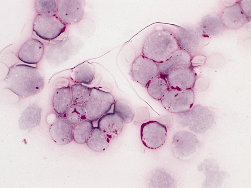
The role of alternative splicing of the adhesion molecule, CD44, in lymphoid malignancy. (1/23200)
AIM: To investigate the expression of CD44 isoforms containing variant exon 6 (v6) in a well characterised cohort of patients with non-Hodgkin's lymphoma (NHL) and chronic lymphocytic leukaemia (CLL), and to correlate this with phenotype and disease course. METHODS: Cryostat sections of OCT embedded diagnostic nodal material from NHL patients and cryopreserved mononuclear preparations from CLL patients were used as sources of RNA. After reverse transcription, PCR was carried out with amplimers positioned at either side of the variant exon insertion site to amplify all possible CD44 isoforms. Those isoforms containing v6 were identified after Southern blotting and hybridisation with a radiolabelled oligonucleotide. RESULTS: Of 32 NHL samples analysed, 16 did not express CD44 isoforms containing v6, six expressed an isoform containing exon v6 alone, and 10 expressed v6 long isoforms which contained exon v6 in addition to other variant exons. These data did not correlate with lymphoma classification, disease staging, or the presence or absence of extranodal disease. However, those patients expressing v6 long CD44 isoforms had a worse overall survival than those that did not. The plateau of the survival curves was 50% compared with 82%. No v6 long isoforms were detected in the 21 CLL samples investigated. CONCLUSIONS: The expression of v6 long CD44 isoforms is associated with aggressive disease in NHL, independent of grade, stage, or presence of extranodal disease. (+info)Expression of extracellular matrix proteins in cervical squamous cell carcinoma--a clinicopathological study. (2/23200)
AIM: To evaluate the intracellular and peritumoral expression of matrix proteins in squamous cell carcinoma of the uterine cervix using immunohistochemistry. METHODS: 71 squamous cell carcinomas and 10 controls were stained for laminin, fibronectin, and collagen IV. Cytoplasmic staining in tumour cells and peritumoral deposition of matrix proteins were evaluated. The association between staining results and patient age, tumour stage, histological grade, and survival was studied. RESULTS: Positive cytoplasmic staining for laminin, fibronectin, and collagen IV was observed in 17 (23.9%), 27 (38%), and 10 (14.1%) cases, respectively. Staining for laminin was most pronounced in the invasive front of tumour islands, while for fibronectin and collagen IV it appeared to be diffuse. Peritumoral staining for laminin and collagen IV was detected in 12 cases (16.9%). Early stage (Ia1-Ia2) tumours were uniformly negative for all three proteins. Cytoplasmic staining for laminin correlated with positive staining for fibronectin and collagen IV, and with the presence of a peritumoral deposition of collagen IV and laminin. There was no correlation with any of the three markers between staining results and patient age, stage, grade, or survival. CONCLUSIONS: Expression of extracellular matrix proteins in some cervical squamous cell carcinomas might reflect the enhanced ability of these tumours to modify the peritumoral stroma. This ability seems to be absent in early stage tumours. The correlation between intracytoplasmic and peritumoral expression of matrix proteins supports the evidence of their synthesis by tumour cells. However, this property did not correlate with disease outcome in this study. (+info)Is hospital care involved in inequalities in coronary heart disease mortality? Results from the French WHO-MONICA Project in men aged 30-64. (3/23200)
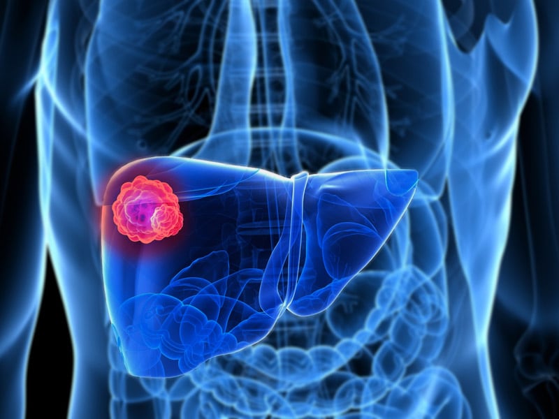
OBJECTIVES: The goal of the study was to assess whether possible disparities in coronary heart disease (CHD) management between occupational categories (OC) in men might be observed and contribute to the increasing inequalities in CHD morbidity and mortality reported in France. METHODS: The data from the three registers of the French MONICA Collaborative Centres (MCC-Lille, MCC-Strasbourg, and MCC-Toulouse) were analysed during two period: 1985-87 and 1989-91. Acute myocardial infarctions and coronary deaths concerning men, aged 30-64 years, were included. Non-professionally active and retired men were excluded. Results were adjusted for age and MCC, using a logistic regression analysis. RESULTS: 605 and 695 events were analysed for 1985-87 and 1989-91, respectively. Out of hospital cardiac arrests, with or without cardiac resuscitation, and 28 day case fatality rates were lower among upper executives in both periods. A coronarography before the acute event had been performed more frequently in men of this category and the proportion of events that could be hospitalised was higher among them. In both periods, the management of acute myocardial infarctions in hospital and prescriptions on discharge were similar among occupational categories. CONCLUSIONS: For patients who could be admitted to hospital, the management was found to be similar among OCs, as was the 28 day case fatality rate among the hospitalised patients. In contrast, lower prognosis and higher probability of being hospitalised after the event among some categories suggest that pre-hospital care and the patient's conditions before the event are the primary factors involved. (+info)Intrahepatic recurrence after curative resection of hepatocellular carcinoma: long-term results of treatment and prognostic factors. (4/23200)
OBJECTIVE: This study aimed to evaluate the long-term results of treatment and prognostic factors in patients with intrahepatic recurrence after curative resection of hepatocellular carcinoma (HCC). SUMMARY BACKGROUND DATA: Recent studies have demonstrated the usefulness of re-resection, transarterial oily chemoembolization (TOCE), or percutaneous ethanol injection therapy (PEIT) in selected patients with intrahepatic recurrent HCC. The overall results of a treatment strategy combining these modalities have not been fully evaluated, and the prognostic factors determining survival in these patients remain to be clarified. METHODS: Two hundred and forty-four patients who underwent curative resection for HCC were followed for intrahepatic recurrence, which was treated aggressively with a strategy including different modalities. Survival results after recurrence and from initial hepatectomy were analyzed, and prognostic factors were determined by univariate and multivariate analysis using 27 clinicopathologic variables. RESULTS: One hundred and five patients (43%) with intrahepatic recurrence were treated with re-resection (11), TOCE (71), PEIT (6), systemic chemotherapy (8) or conservatively (9). The overall 1-year, 3-year, and 5-year survival rates from the time of recurrence were 65.5%, 34.9%, and 19.7%, respectively, and from the time of initial hepatectomy were 78.4%, 47.2%, and 30.9%, respectively. The re-resection group had the best survival, followed by the TOCE group. Multivariate analysis revealed Child's B or C grading, serum albumin < or = 40 g/l, multiple recurrent tumors, recurrence < or = 1 year after hepatectomy, and concurrent extrahepatic recurrence to be independent adverse prognostic factors. CONCLUSIONS: Aggressive treatment with a multimodality strategy could result in prolonged survival in patients with intrahepatic recurrence after curative resection for HCC. Prognosis was determined by the liver function status, interval to recurrence, number of recurrent tumors, any concurrent extrahepatic recurrence, and type of treatment. (+info)Surgery-related factors and local recurrence of Wilms tumor in National Wilms Tumor Study 4. (5/23200)
OBJECTIVE: To assess the prognostic factors for local recurrence in Wilms tumor. SUMMARY BACKGROUND DATA: Current therapy for Wilms tumor has evolved through four studies of the National Wilms Tumor Study Group. As adverse prognostic factors were identified, treatment of children with Wilms tumor has been tailored based on these factors. Two-year relapse-free survival of children in the fourth study (NWTS-4) exceeded 91%. Factors once of prognostic import for local recurrence may lose their significance as more effective therapeutic regimens are devised. METHODS: Children evaluated were drawn from the records of NWTS-4. A total of 2482 randomized or followed patients were identified. Local recurrence, defined as recurrence in the original tumor bed, retroperitoneum, or within the abdominal cavity or pelvis, occurred in 100 children. Using a nested case-control study design, 182 matched controls were selected. Factors were analyzed for their association with local failure. Relative risks and 95% confidence intervals were calculated, taking into account the matching. RESULTS: The largest relative risks for local recurrence were observed in patients with stage III disease, those with unfavorable histology (especially diffuse anaplasia), and those reported to have tumor spillage during surgery. Multiple regression analysis adjusting for the combined effects of histology, lymph node involvement, and age revealed that tumor spillage remained significant. The relative risk of local recurrence from spill was largest in children with stage II disease. The absence of lymph node biopsy was also associated with an increased relative risk of recurrence, which was largest in children with stage I disease. The survival of children after local recurrence is poor, with an average survival rate at 2 years after relapse of 43%. Survival was dependent on initial stage: those who received more therapy before relapse had a worse prognosis. CONCLUSIONS: This study has demonstrated that surgical rupture of the tumor must be prevented by the surgeon, because spills produce an increased risk of local relapse. Both local and diffuse spills produce this risk. Stage II children with local spill appear to require more aggressive therapy than that used in NWTS-4. The continued critical importance of lymph node sampling in conjunction with nephrectomy for Wilms tumor is also established. Absence of lymph node biopsy may result in understaging and inadequate treatment of the child and may produce an increased risk of local recurrence. (+info)Serum triglyceride: a possible risk factor for ruptured abdominal aortic aneurysm. (6/23200)
BACKGROUND: We aimed to determine the relationship between ruptured abdominal aortic aneurysm (AAA) and serum concentrations of lipids and apolipoproteins. METHODS: A cohort of 21 520 men, aged 35-64 years, was recruited from men attending the British United Provident Association (BUPA) clinic in London for a routine medical examination in 1975-1982. Smoking habits, weight, height and blood pressure were recorded at entry. Lipids and apolipoproteins were measured in stored serum samples from the 30 men who subsequently died of ruptured AAA and 150 matched controls. RESULTS: Triglyceride was strongly related to risk of ruptured AAA. In univariate analyses the risk in men on the 90th centile of the distribution relative to the risk in men on the 10th (RO10-90) was 12 (95% confidence interval [CI] : 3.8-37) for triglyceride, 5.5 (95% CI: 1.8-17) for apolipoprotein B (apoB) (the protein component of low density lipoprotein [LDL]), 0.15 (95% CI : 0.04-0.56) for apo A1 (the protein component of high density lipoprotein [HDL]), 3.7 (95% CI: 1.4-9.4) for body mass index and 3.0 (95% CI: 1.1-8.5) for systolic blood pressure. Lipoprotein (a) (Lp(a)) was not a significant risk factor (RO10-90 = 1.6, 95% CI: 0.6-3.0). In multivariate analysis triglyceride retained its strong association. CONCLUSION: Triglyceride appears to be a strong risk factor for ruptured AAA, although further studies are required to clarify this. If this and other associations are cause and effect, then changing the distribution of risk factors in the population (by many people stopping smoking and adopting a lower saturated fat diet and by lowering blood pressure) could achieve an important reduction in mortality from ruptured AAA. (+info)Respiratory symptoms and long-term risk of death from cardiovascular disease, cancer and other causes in Swedish men. (7/23200)
BACKGROUND: Depressed respiratory function and respiratory symptoms are associated with impaired survival. The present study was undertaken to assess the relation between respiratory symptoms and mortality from cardiovascular causes, cancer and all causes in a large population of middle-aged men. METHODS: Prospective population study of 6442 men aged 51-59 at baseline, free of clinical angina pectoris and prior myocardial infarction. RESULTS: During 16 years there were 1804 deaths (786 from cardiovascular disease, 608 from cancer, 103 from pulmonary disease and 307 from any other cause). Men with effort-related breathlessness had increased risk of dying from all of the examined diseases. After adjustment for age, smoking habit and other risk factors, the relative risk (RR) associated with breathlessness of dying from coronary disease was 1.43 (95% CI : 1.16-1.77), from stroke 1.77 (95% CI: 1.07-2.93), from any cardiovascular disease 1.48 (95% CI : 1.24-1.76), cancer 1.36 (95% CI : 1.11-1.67) and from any cause 1.62 (95% CI: 1.44-1.81). An independent effect of breathlessness on cardiovascular death, cancer death and mortality from all causes was found in life-time non-smokers, and also if men with chest pain not considered to be angina were excluded. An independent effect was also found if all deaths during the first half of the follow-up were excluded. Men with cough and phlegm, without breathlessness, also had an elevated risk of dying from cardiovascular disease and cancer, but after adjustment for smoking and other risk factors this was no longer significant. However, a slightly elevated independent risk of dying from any cause was found (RR = 1.18 [95% CI: 1.02-1.36]). CONCLUSION: A positive response to a simple question about effort related breathlessness predicted subsequent mortality from several causes during a follow-up period of 16 years, independently of smoking and other risk factors. (+info)Socioeconomic inequalities and disability pension in middle-aged men. (8/23200)
BACKGROUND: The issue of inequalities in health has generated much discussion and socioeconomic status is considered an important variable in studies of health. It is frequently used in epidemiological studies, either as a possible risk factor or a confounder and the aim of this study was to analyse the relation between socioeconomic status and risk of disability pension. METHODS: Five complete birth year cohorts of middle-aged male residents in Malmo were invited to a health survey and 5782 with complete data constituted the cohort in this prospective study. Each subject was followed for approximately 11 years and nationwide Swedish data registers were used for surveillance. RESULTS: Among the 715 men (12%), granted disability pension during follow-up, three groups were distinguished. The cumulative incidence of disability pension among blue collar workers was 17% and among lower and higher level white collar workers, 11% and 6% respectively. With simultaneous adjustment for biological risk factors and job conditions, the relative risk for being granted a disability pension (using higher level white collar workers as reference) was 2.5 among blue collar workers and 1.6 among lower level white collar workers. CONCLUSIONS: Socioeconomic status, as defined by occupation, is a risk factor for being granted disability pension even after adjusting for work conditions and other risk factors for disease. (+info)Medical survival rate is a statistical measure used to determine the percentage of patients who are still alive for a specific period of time after their diagnosis or treatment for a certain condition or disease. It is often expressed as a five-year survival rate, which refers to the proportion of people who are alive five years after their diagnosis. Survival rates can be affected by many factors, including the stage of the disease at diagnosis, the patient's age and overall health, the effectiveness of treatment, and other health conditions that the patient may have. It is important to note that survival rates are statistical estimates and do not necessarily predict an individual patient's prognosis.
Cell survival refers to the ability of a cell to continue living and functioning normally, despite being exposed to potentially harmful conditions or treatments. This can include exposure to toxins, radiation, chemotherapeutic drugs, or other stressors that can damage cells or interfere with their normal processes.
In scientific research, measures of cell survival are often used to evaluate the effectiveness of various therapies or treatments. For example, researchers may expose cells to a particular drug or treatment and then measure the percentage of cells that survive to assess its potential therapeutic value. Similarly, in toxicology studies, measures of cell survival can help to determine the safety of various chemicals or substances.
It's important to note that cell survival is not the same as cell proliferation, which refers to the ability of cells to divide and multiply. While some treatments may promote cell survival, they may also inhibit cell proliferation, making them useful for treating diseases such as cancer. Conversely, other treatments may be designed to specifically target and kill cancer cells, even if it means sacrificing some healthy cells in the process.
Disease-free survival (DFS) is a term used in medical research and clinical practice, particularly in the field of oncology. It refers to the length of time after primary treatment for a cancer during which no evidence of the disease can be found. This means that the patient shows no signs or symptoms of the cancer, and any imaging studies or other tests do not reveal any tumors or other indications of the disease.
DFS is often used as an important endpoint in clinical trials to evaluate the effectiveness of different treatments for cancer. By measuring the length of time until the cancer recurs or a new cancer develops, researchers can get a better sense of how well a particular treatment is working and whether it is improving patient outcomes.
It's important to note that DFS is not the same as overall survival (OS), which refers to the length of time from primary treatment until death from any cause. While DFS can provide valuable information about the effectiveness of cancer treatments, it does not necessarily reflect the impact of those treatments on patients' overall survival.
Survival analysis is a branch of statistics that deals with the analysis of time to event data. It is used to estimate the time it takes for a certain event of interest to occur, such as death, disease recurrence, or treatment failure. The event of interest is called the "failure" event, and survival analysis estimates the probability of not experiencing the failure event until a certain point in time, also known as the "survival" probability.
Survival analysis can provide important information about the effectiveness of treatments, the prognosis of patients, and the identification of risk factors associated with the event of interest. It can handle censored data, which is common in medical research where some participants may drop out or be lost to follow-up before the event of interest occurs.
Survival analysis typically involves estimating the survival function, which describes the probability of surviving beyond a certain time point, as well as hazard functions, which describe the instantaneous rate of failure at a given time point. Other important concepts in survival analysis include median survival times, restricted mean survival times, and various statistical tests to compare survival curves between groups.
Treatment outcome is a term used to describe the result or effect of medical treatment on a patient's health status. It can be measured in various ways, such as through symptoms improvement, disease remission, reduced disability, improved quality of life, or survival rates. The treatment outcome helps healthcare providers evaluate the effectiveness of a particular treatment plan and make informed decisions about future care. It is also used in clinical research to compare the efficacy of different treatments and improve patient care.
In a medical context, "survival" generally refers to the continuation of life following a serious illness, injury, or dangerous event. It is often used in research and clinical settings to describe the length and quality of life after a specific treatment or diagnosis. For example, survival rate might refer to the percentage of patients who are still alive after a certain period of time following a cancer diagnosis or surgery. Survival can also be used more broadly to describe an individual's ability to adapt and persist in the face of adversity or challenge, whether that's due to medical conditions or other life circumstances.
Graft survival, in medical terms, refers to the success of a transplanted tissue or organ in continuing to function and integrate with the recipient's body over time. It is the opposite of graft rejection, which occurs when the recipient's immune system recognizes the transplanted tissue as foreign and attacks it, leading to its failure.
Graft survival depends on various factors, including the compatibility between the donor and recipient, the type and location of the graft, the use of immunosuppressive drugs to prevent rejection, and the overall health of the recipient. A successful graft survival implies that the transplanted tissue or organ has been accepted by the recipient's body and is functioning properly, providing the necessary physiological support for the recipient's survival and improved quality of life.
Retrospective studies, also known as retrospective research or looking back studies, are a type of observational study that examines data from the past to draw conclusions about possible causal relationships between risk factors and outcomes. In these studies, researchers analyze existing records, medical charts, or previously collected data to test a hypothesis or answer a specific research question.
Retrospective studies can be useful for generating hypotheses and identifying trends, but they have limitations compared to prospective studies, which follow participants forward in time from exposure to outcome. Retrospective studies are subject to biases such as recall bias, selection bias, and information bias, which can affect the validity of the results. Therefore, retrospective studies should be interpreted with caution and used primarily to generate hypotheses for further testing in prospective studies.
Prognosis is a medical term that refers to the prediction of the likely outcome or course of a disease, including the chances of recovery or recurrence, based on the patient's symptoms, medical history, physical examination, and diagnostic tests. It is an important aspect of clinical decision-making and patient communication, as it helps doctors and patients make informed decisions about treatment options, set realistic expectations, and plan for future care.
Prognosis can be expressed in various ways, such as percentages, categories (e.g., good, fair, poor), or survival rates, depending on the nature of the disease and the available evidence. However, it is important to note that prognosis is not an exact science and may vary depending on individual factors, such as age, overall health status, and response to treatment. Therefore, it should be used as a guide rather than a definitive forecast.
The Kaplan-Meier estimate is a statistical method used to calculate the survival probability over time in a population. It is commonly used in medical research to analyze time-to-event data, such as the time until a patient experiences a specific event like disease progression or death. The Kaplan-Meier estimate takes into account censored data, which occurs when some individuals are lost to follow-up before experiencing the event of interest.
The method involves constructing a survival curve that shows the proportion of subjects still surviving at different time points. At each time point, the survival probability is calculated as the product of the conditional probabilities of surviving from one time point to the next. The Kaplan-Meier estimate provides an unbiased and consistent estimator of the survival function, even when censoring is present.
In summary, the Kaplan-Meier estimate is a crucial tool in medical research for analyzing time-to-event data and estimating survival probabilities over time while accounting for censored observations.
Neoplasm staging is a systematic process used in medicine to describe the extent of spread of a cancer, including the size and location of the original (primary) tumor and whether it has metastasized (spread) to other parts of the body. The most widely accepted system for this purpose is the TNM classification system developed by the American Joint Committee on Cancer (AJCC) and the Union for International Cancer Control (UICC).
In this system, T stands for tumor, and it describes the size and extent of the primary tumor. N stands for nodes, and it indicates whether the cancer has spread to nearby lymph nodes. M stands for metastasis, and it shows whether the cancer has spread to distant parts of the body.
Each letter is followed by a number that provides more details about the extent of the disease. For example, a T1N0M0 cancer means that the primary tumor is small and has not spread to nearby lymph nodes or distant sites. The higher the numbers, the more advanced the cancer.
Staging helps doctors determine the most appropriate treatment for each patient and estimate the patient's prognosis. It is an essential tool for communication among members of the healthcare team and for comparing outcomes of treatments in clinical trials.
Follow-up studies are a type of longitudinal research that involve repeated observations or measurements of the same variables over a period of time, in order to understand their long-term effects or outcomes. In medical context, follow-up studies are often used to evaluate the safety and efficacy of medical treatments, interventions, or procedures.
In a typical follow-up study, a group of individuals (called a cohort) who have received a particular treatment or intervention are identified and then followed over time through periodic assessments or data collection. The data collected may include information on clinical outcomes, adverse events, changes in symptoms or functional status, and other relevant measures.
The results of follow-up studies can provide important insights into the long-term benefits and risks of medical interventions, as well as help to identify factors that may influence treatment effectiveness or patient outcomes. However, it is important to note that follow-up studies can be subject to various biases and limitations, such as loss to follow-up, recall bias, and changes in clinical practice over time, which must be carefully considered when interpreting the results.
In the field of medicine, "time factors" refer to the duration of symptoms or time elapsed since the onset of a medical condition, which can have significant implications for diagnosis and treatment. Understanding time factors is crucial in determining the progression of a disease, evaluating the effectiveness of treatments, and making critical decisions regarding patient care.
For example, in stroke management, "time is brain," meaning that rapid intervention within a specific time frame (usually within 4.5 hours) is essential to administering tissue plasminogen activator (tPA), a clot-busting drug that can minimize brain damage and improve patient outcomes. Similarly, in trauma care, the "golden hour" concept emphasizes the importance of providing definitive care within the first 60 minutes after injury to increase survival rates and reduce morbidity.
Time factors also play a role in monitoring the progression of chronic conditions like diabetes or heart disease, where regular follow-ups and assessments help determine appropriate treatment adjustments and prevent complications. In infectious diseases, time factors are crucial for initiating antibiotic therapy and identifying potential outbreaks to control their spread.
Overall, "time factors" encompass the significance of recognizing and acting promptly in various medical scenarios to optimize patient outcomes and provide effective care.
Combined modality therapy (CMT) is a medical treatment approach that utilizes more than one method or type of therapy simultaneously or in close succession, with the goal of enhancing the overall effectiveness of the treatment. In the context of cancer care, CMT often refers to the combination of two or more primary treatment modalities, such as surgery, radiation therapy, and systemic therapies (chemotherapy, immunotherapy, targeted therapy, etc.).
The rationale behind using combined modality therapy is that each treatment method can target cancer cells in different ways, potentially increasing the likelihood of eliminating all cancer cells and reducing the risk of recurrence. The specific combination and sequence of treatments will depend on various factors, including the type and stage of cancer, patient's overall health, and individual preferences.
For example, a common CMT approach for locally advanced rectal cancer may involve preoperative (neoadjuvant) chemoradiation therapy, followed by surgery to remove the tumor, and then postoperative (adjuvant) chemotherapy. This combined approach allows for the reduction of the tumor size before surgery, increases the likelihood of complete tumor removal, and targets any remaining microscopic cancer cells with systemic chemotherapy.
It is essential to consult with a multidisciplinary team of healthcare professionals to determine the most appropriate CMT plan for each individual patient, considering both the potential benefits and risks associated with each treatment method.
Proportional hazards models are a type of statistical analysis used in medical research to investigate the relationship between covariates (predictor variables) and survival times. The most common application of proportional hazards models is in the Cox regression model, which is named after its developer, Sir David Cox.
In a proportional hazards model, the hazard rate or risk of an event occurring at a given time is assumed to be proportional to the hazard rate of a reference group, after adjusting for the covariates. This means that the ratio of the hazard rates between any two individuals remains constant over time, regardless of their survival times.
Mathematically, the hazard function h(t) at time t for an individual with a set of covariates X can be expressed as:
h(t|X) = h0(t) \* exp(β1X1 + β2X2 + ... + βpXp)
where h0(t) is the baseline hazard function, X1, X2, ..., Xp are the covariates, and β1, β2, ..., βp are the regression coefficients that represent the effect of each covariate on the hazard rate.
The assumption of proportionality is crucial in the interpretation of the results from a Cox regression model. If the assumption is violated, then the estimated regression coefficients may be biased and misleading. Therefore, it is important to test for the proportional hazards assumption before interpreting the results of a Cox regression analysis.
Antineoplastic combined chemotherapy protocols refer to a treatment plan for cancer that involves the use of more than one antineoplastic (chemotherapy) drug given in a specific sequence and schedule. The combination of drugs is used because they may work better together to destroy cancer cells compared to using a single agent alone. This approach can also help to reduce the likelihood of cancer cells becoming resistant to the treatment.
The choice of drugs, dose, duration, and frequency are determined by various factors such as the type and stage of cancer, patient's overall health, and potential side effects. Combination chemotherapy protocols can be used in various settings, including as a primary treatment, adjuvant therapy (given after surgery or radiation to kill any remaining cancer cells), neoadjuvant therapy (given before surgery or radiation to shrink the tumor), or palliative care (to alleviate symptoms and prolong survival).
It is important to note that while combined chemotherapy protocols can be effective in treating certain types of cancer, they can also cause significant side effects, including nausea, vomiting, hair loss, fatigue, and an increased risk of infection. Therefore, patients undergoing such treatment should be closely monitored and managed by a healthcare team experienced in administering chemotherapy.
Local neoplasm recurrence is the return or regrowth of a tumor in the same location where it was originally removed or treated. This means that cancer cells have survived the initial treatment and started to grow again in the same area. It's essential to monitor and detect any local recurrence as early as possible, as it can affect the prognosis and may require additional treatment.
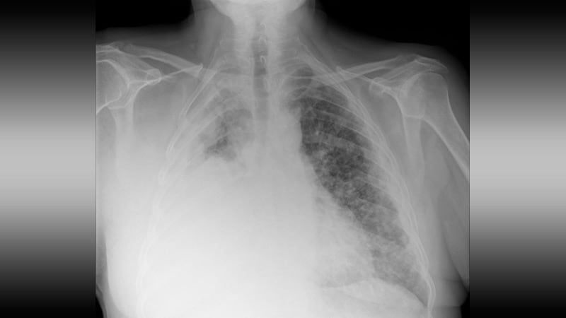
Lung neoplasms refer to abnormal growths or tumors in the lung tissue. These tumors can be benign (non-cancerous) or malignant (cancerous). Malignant lung neoplasms are further classified into two main types: small cell lung carcinoma and non-small cell lung carcinoma. Lung neoplasms can cause symptoms such as cough, chest pain, shortness of breath, and weight loss. They are often caused by smoking or exposure to secondhand smoke, but can also occur due to genetic factors, radiation exposure, and other environmental carcinogens. Early detection and treatment of lung neoplasms is crucial for improving outcomes and survival rates.
Liver neoplasms refer to abnormal growths in the liver that can be benign or malignant. Benign liver neoplasms are non-cancerous tumors that do not spread to other parts of the body, while malignant liver neoplasms are cancerous tumors that can invade and destroy surrounding tissue and spread to other organs.
Liver neoplasms can be primary, meaning they originate in the liver, or secondary, meaning they have metastasized (spread) to the liver from another part of the body. Primary liver neoplasms can be further classified into different types based on their cell of origin and behavior, including hepatocellular carcinoma, cholangiocarcinoma, and hepatic hemangioma.
The diagnosis of liver neoplasms typically involves a combination of imaging studies, such as ultrasound, CT scan, or MRI, and biopsy to confirm the type and stage of the tumor. Treatment options depend on the type and extent of the neoplasm and may include surgery, radiation therapy, chemotherapy, or liver transplantation.
Multivariate analysis is a statistical method used to examine the relationship between multiple independent variables and a dependent variable. It allows for the simultaneous examination of the effects of two or more independent variables on an outcome, while controlling for the effects of other variables in the model. This technique can be used to identify patterns, associations, and interactions among multiple variables, and is commonly used in medical research to understand complex health outcomes and disease processes. Examples of multivariate analysis methods include multiple regression, factor analysis, cluster analysis, and discriminant analysis.
Adenocarcinoma is a type of cancer that arises from glandular epithelial cells. These cells line the inside of many internal organs, including the breasts, prostate, colon, and lungs. Adenocarcinomas can occur in any of these organs, as well as in other locations where glands are present.
The term "adenocarcinoma" is used to describe a cancer that has features of glandular tissue, such as mucus-secreting cells or cells that produce hormones. These cancers often form glandular structures within the tumor mass and may produce mucus or other substances.
Adenocarcinomas are typically slow-growing and tend to spread (metastasize) to other parts of the body through the lymphatic system or bloodstream. They can be treated with surgery, radiation therapy, chemotherapy, targeted therapy, or a combination of these treatments. The prognosis for adenocarcinoma depends on several factors, including the location and stage of the cancer, as well as the patient's overall health and age.
Lymphatic metastasis is the spread of cancer cells from a primary tumor to distant lymph nodes through the lymphatic system. It occurs when malignant cells break away from the original tumor, enter the lymphatic vessels, and travel to nearby or remote lymph nodes. Once there, these cancer cells can multiply and form new tumors, leading to further progression of the disease. Lymphatic metastasis is a common way for many types of cancer to spread and can have significant implications for prognosis and treatment strategies.
Actuarial analysis is a process used in the field of actuarial science to evaluate and manage risk, typically for financial or insurance purposes. It involves the use of statistical modeling, mathematical calculations, and data analysis to estimate the probability and potential financial impact of various events or outcomes.
In a medical context, actuarial analysis may be used to assess the risks and costs associated with different health conditions, treatments, or patient populations. For example, an actuary might use data on morbidity rates, mortality rates, and healthcare utilization patterns to estimate the expected costs of providing coverage to a group of patients with a particular medical condition.
Actuarial analysis can help healthcare organizations, insurers, and policymakers make informed decisions about resource allocation, pricing, and risk management. It can also be used to develop predictive models that identify high-risk populations or forecast future trends in healthcare utilization and costs.
Medical Definition:
"Risk factors" are any attribute, characteristic or exposure of an individual that increases the likelihood of developing a disease or injury. They can be divided into modifiable and non-modifiable risk factors. Modifiable risk factors are those that can be changed through lifestyle choices or medical treatment, while non-modifiable risk factors are inherent traits such as age, gender, or genetic predisposition. Examples of modifiable risk factors include smoking, alcohol consumption, physical inactivity, and unhealthy diet, while non-modifiable risk factors include age, sex, and family history. It is important to note that having a risk factor does not guarantee that a person will develop the disease, but rather indicates an increased susceptibility.
Adjuvant chemotherapy is a medical treatment that is given in addition to the primary therapy, such as surgery or radiation, to increase the chances of a cure or to reduce the risk of recurrence in patients with cancer. It involves the use of chemicals (chemotherapeutic agents) to destroy any remaining cancer cells that may not have been removed by the primary treatment. This type of chemotherapy is typically given after the main treatment has been completed, and its goal is to kill any residual cancer cells that may be present in the body and reduce the risk of the cancer coming back. The specific drugs used and the duration of treatment will depend on the type and stage of cancer being treated.
Prospective studies, also known as longitudinal studies, are a type of cohort study in which data is collected forward in time, following a group of individuals who share a common characteristic or exposure over a period of time. The researchers clearly define the study population and exposure of interest at the beginning of the study and follow up with the participants to determine the outcomes that develop over time. This type of study design allows for the investigation of causal relationships between exposures and outcomes, as well as the identification of risk factors and the estimation of disease incidence rates. Prospective studies are particularly useful in epidemiology and medical research when studying diseases with long latency periods or rare outcomes.
Tumor markers are substances that can be found in the body and their presence can indicate the presence of certain types of cancer or other conditions. Biological tumor markers refer to those substances that are produced by cancer cells or by other cells in response to cancer or certain benign (non-cancerous) conditions. These markers can be found in various bodily fluids such as blood, urine, or tissue samples.
Examples of biological tumor markers include:
1. Proteins: Some tumor markers are proteins that are produced by cancer cells or by other cells in response to the presence of cancer. For example, prostate-specific antigen (PSA) is a protein produced by normal prostate cells and in higher amounts by prostate cancer cells.
2. Genetic material: Tumor markers can also include genetic material such as DNA, RNA, or microRNA that are shed by cancer cells into bodily fluids. For example, circulating tumor DNA (ctDNA) is genetic material from cancer cells that can be found in the bloodstream.
3. Metabolites: Tumor markers can also include metabolic products produced by cancer cells or by other cells in response to cancer. For example, lactate dehydrogenase (LDH) is an enzyme that is released into the bloodstream when cancer cells break down glucose for energy.
It's important to note that tumor markers are not specific to cancer and can be elevated in non-cancerous conditions as well. Therefore, they should not be used alone to diagnose cancer but rather as a tool in conjunction with other diagnostic tests and clinical evaluations.
Antineoplastic agents are a class of drugs used to treat malignant neoplasms or cancer. These agents work by inhibiting the growth and proliferation of cancer cells, either by killing them or preventing their division and replication. Antineoplastic agents can be classified based on their mechanism of action, such as alkylating agents, antimetabolites, topoisomerase inhibitors, mitotic inhibitors, and targeted therapy agents.
Alkylating agents work by adding alkyl groups to DNA, which can cause cross-linking of DNA strands and ultimately lead to cell death. Antimetabolites interfere with the metabolic processes necessary for DNA synthesis and replication, while topoisomerase inhibitors prevent the relaxation of supercoiled DNA during replication. Mitotic inhibitors disrupt the normal functioning of the mitotic spindle, which is essential for cell division. Targeted therapy agents are designed to target specific molecular abnormalities in cancer cells, such as mutated oncogenes or dysregulated signaling pathways.
It's important to note that antineoplastic agents can also affect normal cells and tissues, leading to various side effects such as nausea, vomiting, hair loss, and myelosuppression (suppression of bone marrow function). Therefore, the use of these drugs requires careful monitoring and management of their potential adverse effects.
Liver transplantation is a surgical procedure in which a diseased or failing liver is replaced with a healthy one from a deceased donor or, less commonly, a portion of a liver from a living donor. The goal of the procedure is to restore normal liver function and improve the patient's overall health and quality of life.
Liver transplantation may be recommended for individuals with end-stage liver disease, acute liver failure, certain genetic liver disorders, or liver cancers that cannot be treated effectively with other therapies. The procedure involves complex surgery to remove the diseased liver and implant the new one, followed by a period of recovery and close medical monitoring to ensure proper function and minimize the risk of complications.
The success of liver transplantation has improved significantly in recent years due to advances in surgical techniques, immunosuppressive medications, and post-transplant care. However, it remains a major operation with significant risks and challenges, including the need for lifelong immunosuppression to prevent rejection of the new liver, as well as potential complications such as infection, bleeding, and organ failure.
Apoptosis is a programmed and controlled cell death process that occurs in multicellular organisms. It is a natural process that helps maintain tissue homeostasis by eliminating damaged, infected, or unwanted cells. During apoptosis, the cell undergoes a series of morphological changes, including cell shrinkage, chromatin condensation, and fragmentation into membrane-bound vesicles called apoptotic bodies. These bodies are then recognized and engulfed by neighboring cells or phagocytic cells, preventing an inflammatory response. Apoptosis is regulated by a complex network of intracellular signaling pathways that involve proteins such as caspases, Bcl-2 family members, and inhibitors of apoptosis (IAPs).
Cisplatin is a chemotherapeutic agent used to treat various types of cancers, including testicular, ovarian, bladder, head and neck, lung, and cervical cancers. It is an inorganic platinum compound that contains a central platinum atom surrounded by two chloride atoms and two ammonia molecules in a cis configuration.
Cisplatin works by forming crosslinks between DNA strands, which disrupts the structure of DNA and prevents cancer cells from replicating. This ultimately leads to cell death and slows down or stops the growth of tumors. However, cisplatin can also cause damage to normal cells, leading to side effects such as nausea, vomiting, hearing loss, and kidney damage. Therefore, it is essential to monitor patients closely during treatment and manage any adverse effects promptly.
Hepatocellular carcinoma (HCC) is the most common type of primary liver cancer in adults. It originates from the hepatocytes, which are the main functional cells of the liver. This type of cancer is often associated with chronic liver diseases such as cirrhosis caused by hepatitis B or C virus infection, alcohol abuse, non-alcoholic fatty liver disease (NAFLD), and aflatoxin exposure.
The symptoms of HCC can vary but may include unexplained weight loss, lack of appetite, abdominal pain or swelling, jaundice, and fatigue. The diagnosis of HCC typically involves imaging tests such as ultrasound, CT scan, or MRI, as well as blood tests to measure alpha-fetoprotein (AFP) levels. Treatment options for Hepatocellular carcinoma depend on the stage and extent of the cancer, as well as the patient's overall health and liver function. Treatment options may include surgery, radiation therapy, chemotherapy, targeted therapy, or liver transplantation.
Squamous cell carcinoma is a type of skin cancer that begins in the squamous cells, which are flat, thin cells that form the outer layer of the skin (epidermis). It commonly occurs on sun-exposed areas such as the face, ears, lips, and backs of the hands. Squamous cell carcinoma can also develop in other areas of the body including the mouth, lungs, and cervix.
This type of cancer usually develops slowly and may appear as a rough or scaly patch of skin, a red, firm nodule, or a sore or ulcer that doesn't heal. While squamous cell carcinoma is not as aggressive as some other types of cancer, it can metastasize (spread) to other parts of the body if left untreated, making early detection and treatment important.
Risk factors for developing squamous cell carcinoma include prolonged exposure to ultraviolet (UV) radiation from the sun or tanning beds, fair skin, a history of sunburns, a weakened immune system, and older age. Prevention measures include protecting your skin from the sun by wearing protective clothing, using a broad-spectrum sunscreen with an SPF of at least 30, avoiding tanning beds, and getting regular skin examinations.
Neoplasm metastasis is the spread of cancer cells from the primary site (where the original or primary tumor formed) to other places in the body. This happens when cancer cells break away from the original (primary) tumor and enter the bloodstream or lymphatic system. The cancer cells can then travel to other parts of the body and form new tumors, called secondary tumors or metastases.
Metastasis is a key feature of malignant neoplasms (cancers), and it is one of the main ways that cancer can cause harm in the body. The metastatic tumors may continue to grow and may cause damage to the organs and tissues where they are located. They can also release additional cancer cells into the bloodstream or lymphatic system, leading to further spread of the cancer.
The metastatic tumors are named based on the location where they are found, as well as the type of primary cancer. For example, if a patient has a primary lung cancer that has metastasized to the liver, the metastatic tumor would be called a liver metastasis from lung cancer.
It is important to note that the presence of metastases can significantly affect a person's prognosis and treatment options. In general, metastatic cancer is more difficult to treat than cancer that has not spread beyond its original site. However, there are many factors that can influence a person's prognosis and response to treatment, so it is important for each individual to discuss their specific situation with their healthcare team.
Breast neoplasms refer to abnormal growths in the breast tissue that can be benign or malignant. Benign breast neoplasms are non-cancerous tumors or growths, while malignant breast neoplasms are cancerous tumors that can invade surrounding tissues and spread to other parts of the body.
Breast neoplasms can arise from different types of cells in the breast, including milk ducts, milk sacs (lobules), or connective tissue. The most common type of breast cancer is ductal carcinoma, which starts in the milk ducts and can spread to other parts of the breast and nearby structures.
Breast neoplasms are usually detected through screening methods such as mammography, ultrasound, or MRI, or through self-examination or clinical examination. Treatment options for breast neoplasms depend on several factors, including the type and stage of the tumor, the patient's age and overall health, and personal preferences. Treatment may include surgery, radiation therapy, chemotherapy, hormone therapy, or targeted therapy.
"Age factors" refer to the effects, changes, or differences that age can have on various aspects of health, disease, and medical care. These factors can encompass a wide range of issues, including:
1. Physiological changes: As people age, their bodies undergo numerous physical changes that can affect how they respond to medications, illnesses, and medical procedures. For example, older adults may be more sensitive to certain drugs or have weaker immune systems, making them more susceptible to infections.
2. Chronic conditions: Age is a significant risk factor for many chronic diseases, such as heart disease, diabetes, cancer, and arthritis. As a result, age-related medical issues are common and can impact treatment decisions and outcomes.
3. Cognitive decline: Aging can also lead to cognitive changes, including memory loss and decreased decision-making abilities. These changes can affect a person's ability to understand and comply with medical instructions, leading to potential complications in their care.
4. Functional limitations: Older adults may experience physical limitations that impact their mobility, strength, and balance, increasing the risk of falls and other injuries. These limitations can also make it more challenging for them to perform daily activities, such as bathing, dressing, or cooking.
5. Social determinants: Age-related factors, such as social isolation, poverty, and lack of access to transportation, can impact a person's ability to obtain necessary medical care and affect their overall health outcomes.
Understanding age factors is critical for healthcare providers to deliver high-quality, patient-centered care that addresses the unique needs and challenges of older adults. By taking these factors into account, healthcare providers can develop personalized treatment plans that consider a person's age, physical condition, cognitive abilities, and social circumstances.
Immunohistochemistry (IHC) is a technique used in pathology and laboratory medicine to identify specific proteins or antigens in tissue sections. It combines the principles of immunology and histology to detect the presence and location of these target molecules within cells and tissues. This technique utilizes antibodies that are specific to the protein or antigen of interest, which are then tagged with a detection system such as a chromogen or fluorophore. The stained tissue sections can be examined under a microscope, allowing for the visualization and analysis of the distribution and expression patterns of the target molecule in the context of the tissue architecture. Immunohistochemistry is widely used in diagnostic pathology to help identify various diseases, including cancer, infectious diseases, and immune-mediated disorders.
A cohort study is a type of observational study in which a group of individuals who share a common characteristic or exposure are followed up over time to determine the incidence of a specific outcome or outcomes. The cohort, or group, is defined based on the exposure status (e.g., exposed vs. unexposed) and then monitored prospectively to assess for the development of new health events or conditions.
Cohort studies can be either prospective or retrospective in design. In a prospective cohort study, participants are enrolled and followed forward in time from the beginning of the study. In contrast, in a retrospective cohort study, researchers identify a cohort that has already been assembled through medical records, insurance claims, or other sources and then look back in time to assess exposure status and health outcomes.
Cohort studies are useful for establishing causality between an exposure and an outcome because they allow researchers to observe the temporal relationship between the two. They can also provide information on the incidence of a disease or condition in different populations, which can be used to inform public health policy and interventions. However, cohort studies can be expensive and time-consuming to conduct, and they may be subject to bias if participants are not representative of the population or if there is loss to follow-up.
Postoperative complications refer to any unfavorable condition or event that occurs during the recovery period after a surgical procedure. These complications can vary in severity and may include, but are not limited to:
1. Infection: This can occur at the site of the incision or inside the body, such as pneumonia or urinary tract infection.
2. Bleeding: Excessive bleeding (hemorrhage) can lead to a drop in blood pressure and may require further surgical intervention.
3. Blood clots: These can form in the deep veins of the legs (deep vein thrombosis) and can potentially travel to the lungs (pulmonary embolism).
4. Wound dehiscence: This is when the surgical wound opens up, which can lead to infection and further complications.
5. Pulmonary issues: These include atelectasis (collapsed lung), pneumonia, or respiratory failure.
6. Cardiovascular problems: These include abnormal heart rhythms (arrhythmias), heart attack, or stroke.
7. Renal failure: This can occur due to various reasons such as dehydration, blood loss, or the use of certain medications.
8. Pain management issues: Inadequate pain control can lead to increased stress, anxiety, and decreased mobility.
9. Nausea and vomiting: These can be caused by anesthesia, opioid pain medication, or other factors.
10. Delirium: This is a state of confusion and disorientation that can occur in the elderly or those with certain medical conditions.
Prompt identification and management of these complications are crucial to ensure the best possible outcome for the patient.
Recurrence, in a medical context, refers to the return of symptoms or signs of a disease after a period of improvement or remission. It indicates that the condition has not been fully eradicated and may require further treatment. Recurrence is often used to describe situations where a disease such as cancer comes back after initial treatment, but it can also apply to other medical conditions. The likelihood of recurrence varies depending on the type of disease and individual patient factors.
Hepatectomy is a surgical procedure that involves the removal of part or all of the liver. This procedure can be performed for various reasons, such as removing cancerous or non-cancerous tumors, treating liver trauma, or donating a portion of the liver to another person in need of a transplant (live donor hepatectomy). The extent of the hepatectomy depends on the medical condition and overall health of the patient. It is a complex procedure that requires significant expertise and experience from the surgical team due to the liver's unique anatomy, blood supply, and regenerative capabilities.
A registry in the context of medicine is a collection or database of standardized information about individuals who share a certain condition or attribute, such as a disease, treatment, exposure, or demographic group. These registries are used for various purposes, including:
* Monitoring and tracking the natural history of diseases and conditions
* Evaluating the safety and effectiveness of medical treatments and interventions
* Conducting research and generating hypotheses for further study
* Providing information to patients, clinicians, and researchers
* Informing public health policy and decision-making
Registries can be established for a wide range of purposes, including disease-specific registries (such as cancer or diabetes registries), procedure-specific registries (such as joint replacement or cardiac surgery registries), and population-based registries (such as birth defects or cancer registries). Data collected in registries may include demographic information, clinical data, laboratory results, treatment details, and outcomes.
Registries can be maintained by a variety of organizations, including hospitals, clinics, academic medical centers, professional societies, government agencies, and industry. Participation in registries is often voluntary, although some registries may require informed consent from participants. Data collected in registries are typically de-identified to protect the privacy of individuals.
Adjuvant radiotherapy is a type of cancer treatment that uses radiation therapy as an adjunct to a primary surgical procedure. The goal of adjuvant radiotherapy is to eliminate any remaining microscopic cancer cells that may be present in the surrounding tissues after surgery, thereby reducing the risk of local recurrence and improving the chances of cure.
Radiotherapy involves the use of high-energy radiation to destroy cancer cells and shrink tumors. In adjuvant radiotherapy, the radiation is usually delivered to the tumor bed and regional lymph nodes in order to target any potential sites of residual disease. The timing and dosing of adjuvant radiotherapy may vary depending on the type and stage of cancer being treated, as well as other factors such as patient age and overall health status.
Adjuvant radiotherapy is commonly used in the treatment of various types of cancer, including breast, colorectal, lung, head and neck, and gynecologic cancers. Its use has been shown to improve survival rates and reduce the risk of recurrence in many cases, making it an important component of comprehensive cancer care.
Stomach neoplasms refer to abnormal growths in the stomach that can be benign or malignant. They include a wide range of conditions such as:
1. Gastric adenomas: These are benign tumors that develop from glandular cells in the stomach lining.
2. Gastrointestinal stromal tumors (GISTs): These are rare tumors that can be found in the stomach and other parts of the digestive tract. They originate from the stem cells in the wall of the digestive tract.
3. Leiomyomas: These are benign tumors that develop from smooth muscle cells in the stomach wall.
4. Lipomas: These are benign tumors that develop from fat cells in the stomach wall.
5. Neuroendocrine tumors (NETs): These are tumors that develop from the neuroendocrine cells in the stomach lining. They can be benign or malignant.
6. Gastric carcinomas: These are malignant tumors that develop from the glandular cells in the stomach lining. They are the most common type of stomach neoplasm and include adenocarcinomas, signet ring cell carcinomas, and others.
7. Lymphomas: These are malignant tumors that develop from the immune cells in the stomach wall.
Stomach neoplasms can cause various symptoms such as abdominal pain, nausea, vomiting, weight loss, and difficulty swallowing. The diagnosis of stomach neoplasms usually involves a combination of imaging tests, endoscopy, and biopsy. Treatment options depend on the type and stage of the neoplasm and may include surgery, chemotherapy, radiation therapy, or targeted therapy.
Disease progression is the worsening or advancement of a medical condition over time. It refers to the natural course of a disease, including its development, the severity of symptoms and complications, and the impact on the patient's overall health and quality of life. Understanding disease progression is important for developing appropriate treatment plans, monitoring response to therapy, and predicting outcomes.
The rate of disease progression can vary widely depending on the type of medical condition, individual patient factors, and the effectiveness of treatment. Some diseases may progress rapidly over a short period of time, while others may progress more slowly over many years. In some cases, disease progression may be slowed or even halted with appropriate medical interventions, while in other cases, the progression may be inevitable and irreversible.
In clinical practice, healthcare providers closely monitor disease progression through regular assessments, imaging studies, and laboratory tests. This information is used to guide treatment decisions and adjust care plans as needed to optimize patient outcomes and improve quality of life.
Carcinoma, non-small-cell lung (NSCLC) is a type of lung cancer that includes several subtypes of malignant tumors arising from the epithelial cells of the lung. These subtypes are classified based on the appearance of the cancer cells under a microscope and include adenocarcinoma, squamous cell carcinoma, and large cell carcinoma. NSCLC accounts for about 85% of all lung cancers and tends to grow and spread more slowly than small-cell lung cancer (SCLC).
NSCLC is often asymptomatic in its early stages, but as the tumor grows, symptoms such as coughing, chest pain, shortness of breath, hoarseness, and weight loss may develop. Treatment options for NSCLC depend on the stage and location of the cancer, as well as the patient's overall health and lung function. Common treatments include surgery, radiation therapy, chemotherapy, targeted therapy, or a combination of these approaches.
Doxorubicin is a type of chemotherapy medication known as an anthracycline. It works by interfering with the DNA in cancer cells, which prevents them from growing and multiplying. Doxorubicin is used to treat a wide variety of cancers, including leukemia, lymphoma, breast cancer, lung cancer, ovarian cancer, and many others. It may be given alone or in combination with other chemotherapy drugs.
Doxorubicin is usually administered through a vein (intravenously) and can cause side effects such as nausea, vomiting, hair loss, mouth sores, and increased risk of infection. It can also cause damage to the heart muscle, which can lead to heart failure in some cases. For this reason, doctors may monitor patients' heart function closely while they are receiving doxorubicin treatment.
It is important for patients to discuss the potential risks and benefits of doxorubicin therapy with their healthcare provider before starting treatment.
Vincristine is an antineoplastic agent, specifically a vinca alkaloid. It is derived from the Madagascar periwinkle plant (Catharanthus roseus). Vincristine binds to tubulin, a protein found in microtubules, and inhibits their polymerization, which results in disruption of mitotic spindles leading to cell cycle arrest and apoptosis (programmed cell death). It is used in the treatment of various types of cancer including leukemias, lymphomas, and solid tumors. Common side effects include peripheral neuropathy, constipation, and alopecia.
Cyclophosphamide is an alkylating agent, which is a type of chemotherapy medication. It works by interfering with the DNA of cancer cells, preventing them from dividing and growing. This helps to stop the spread of cancer in the body. Cyclophosphamide is used to treat various types of cancer, including lymphoma, leukemia, multiple myeloma, and breast cancer. It can be given orally as a tablet or intravenously as an injection.
Cyclophosphamide can also have immunosuppressive effects, which means it can suppress the activity of the immune system. This makes it useful in treating certain autoimmune diseases, such as rheumatoid arthritis and lupus. However, this immunosuppression can also increase the risk of infections and other side effects.
Like all chemotherapy medications, cyclophosphamide can cause a range of side effects, including nausea, vomiting, hair loss, fatigue, and increased susceptibility to infections. It is important for patients receiving cyclophosphamide to be closely monitored by their healthcare team to manage these side effects and ensure the medication is working effectively.
A reoperation is a surgical procedure that is performed again on a patient who has already undergone a previous operation for the same or related condition. Reoperations may be required due to various reasons, such as inadequate initial treatment, disease recurrence, infection, or complications from the first surgery. The nature and complexity of a reoperation can vary widely depending on the specific circumstances, but it often carries higher risks and potential complications compared to the original operation.
Fluorouracil is a antineoplastic medication, which means it is used to treat cancer. It is a type of chemotherapy drug known as an antimetabolite. Fluorouracil works by interfering with the growth of cancer cells and ultimately killing them. It is often used to treat colon, esophageal, stomach, and breast cancers, as well as skin conditions such as actinic keratosis and superficial basal cell carcinoma. Fluorouracil may be given by injection or applied directly to the skin in the form of a cream.
It is important to note that fluorouracil can have serious side effects, including suppression of bone marrow function, mouth sores, stomach and intestinal ulcers, and nerve damage. It should only be used under the close supervision of a healthcare professional.
Neoplasm invasiveness is a term used in pathology and oncology to describe the aggressive behavior of cancer cells as they invade surrounding tissues and organs. This process involves the loss of cell-to-cell adhesion, increased motility and migration, and the ability of cancer cells to degrade the extracellular matrix (ECM) through the production of enzymes such as matrix metalloproteinases (MMPs).
Invasive neoplasms are cancers that have spread beyond the original site where they first developed and have infiltrated adjacent tissues or structures. This is in contrast to non-invasive or in situ neoplasms, which are confined to the epithelial layer where they originated and have not yet invaded the underlying basement membrane.
The invasiveness of a neoplasm is an important prognostic factor in cancer diagnosis and treatment, as it can indicate the likelihood of metastasis and the potential effectiveness of various therapies. In general, more invasive cancers are associated with worse outcomes and require more aggressive treatment approaches.
Kidney transplantation is a surgical procedure where a healthy kidney from a deceased or living donor is implanted into a patient with end-stage renal disease (ESRD) or permanent kidney failure. The new kidney takes over the functions of filtering waste and excess fluids from the blood, producing urine, and maintaining the body's electrolyte balance.
The transplanted kidney is typically placed in the lower abdomen, with its blood vessels connected to the recipient's iliac artery and vein. The ureter of the new kidney is then attached to the recipient's bladder to ensure proper urine flow. Following the surgery, the patient will require lifelong immunosuppressive therapy to prevent rejection of the transplanted organ by their immune system.
A cell line that is derived from tumor cells and has been adapted to grow in culture. These cell lines are often used in research to study the characteristics of cancer cells, including their growth patterns, genetic changes, and responses to various treatments. They can be established from many different types of tumors, such as carcinomas, sarcomas, and leukemias. Once established, these cell lines can be grown and maintained indefinitely in the laboratory, allowing researchers to conduct experiments and studies that would not be feasible using primary tumor cells. It is important to note that tumor cell lines may not always accurately represent the behavior of the original tumor, as they can undergo genetic changes during their time in culture.
Brain neoplasms, also known as brain tumors, are abnormal growths of cells within the brain. These growths can be benign (non-cancerous) or malignant (cancerous). Benign brain tumors typically grow slowly and do not spread to other parts of the body. However, they can still cause serious problems if they press on sensitive areas of the brain. Malignant brain tumors, on the other hand, are cancerous and can grow quickly, invading surrounding brain tissue and spreading to other parts of the brain or spinal cord.
Brain neoplasms can arise from various types of cells within the brain, including glial cells (which provide support and insulation for nerve cells), neurons (nerve cells that transmit signals in the brain), and meninges (the membranes that cover the brain and spinal cord). They can also result from the spread of cancer cells from other parts of the body, known as metastatic brain tumors.
Symptoms of brain neoplasms may vary depending on their size, location, and growth rate. Common symptoms include headaches, seizures, weakness or paralysis in the limbs, difficulty with balance and coordination, changes in speech or vision, confusion, memory loss, and changes in behavior or personality.
Treatment for brain neoplasms depends on several factors, including the type, size, location, and grade of the tumor, as well as the patient's age and overall health. Treatment options may include surgery, radiation therapy, chemotherapy, targeted therapy, or a combination of these approaches. Regular follow-up care is essential to monitor for recurrence and manage any long-term effects of treatment.
Pancreatic neoplasms refer to abnormal growths in the pancreas that can be benign or malignant. The pancreas is a gland located behind the stomach that produces hormones and digestive enzymes. Pancreatic neoplasms can interfere with the normal functioning of the pancreas, leading to various health complications.
Benign pancreatic neoplasms are non-cancerous growths that do not spread to other parts of the body. They are usually removed through surgery to prevent any potential complications, such as blocking the bile duct or causing pain.
Malignant pancreatic neoplasms, also known as pancreatic cancer, are cancerous growths that can invade and destroy surrounding tissues and organs. They can also spread (metastasize) to other parts of the body, such as the liver, lungs, or bones. Pancreatic cancer is often aggressive and difficult to treat, with a poor prognosis.
There are several types of pancreatic neoplasms, including adenocarcinomas, neuroendocrine tumors, solid pseudopapillary neoplasms, and cystic neoplasms. The specific type of neoplasm is determined through various diagnostic tests, such as imaging studies, biopsies, and blood tests. Treatment options depend on the type, stage, and location of the neoplasm, as well as the patient's overall health and preferences.
Radiotherapy dosage refers to the total amount of radiation energy that is absorbed by tissues or organs, typically measured in units of Gray (Gy), during a course of radiotherapy treatment. It is the product of the dose rate (the amount of radiation delivered per unit time) and the duration of treatment. The prescribed dosage for cancer treatments can range from a few Gray to more than 70 Gy, depending on the type and location of the tumor, the patient's overall health, and other factors. The goal of radiotherapy is to deliver a sufficient dosage to destroy the cancer cells while minimizing damage to surrounding healthy tissues.
Risk assessment in the medical context refers to the process of identifying, evaluating, and prioritizing risks to patients, healthcare workers, or the community related to healthcare delivery. It involves determining the likelihood and potential impact of adverse events or hazards, such as infectious diseases, medication errors, or medical devices failures, and implementing measures to mitigate or manage those risks. The goal of risk assessment is to promote safe and high-quality care by identifying areas for improvement and taking action to minimize harm.
A "Drug Administration Schedule" refers to the plan for when and how a medication should be given to a patient. It includes details such as the dose, frequency (how often it should be taken), route (how it should be administered, such as orally, intravenously, etc.), and duration (how long it should be taken) of the medication. This schedule is often created and prescribed by healthcare professionals, such as doctors or pharmacists, to ensure that the medication is taken safely and effectively. It may also include instructions for missed doses or changes in the dosage.
Life tables are statistical tools used in actuarial science, demography, and public health to estimate the mortality rate and survival rates of a population. They provide a data-driven representation of the probability that individuals of a certain age will die before their next birthday (the death rate) or live to a particular age (the survival rate).
Life tables are constructed using data on the number of deaths and the size of the population in specific age groups over a given period. These tables typically include several columns representing different variables, such as:
1. Age group or interval: The age range for which the data is being presented (e.g., 0-1 year, 1-5 years, 5-10 years, etc.).
2. Number of people in the population: The size of the population within each age group.
3. Number of deaths: The number of individuals who died during the study period within each age group.
4. Death rate: The probability that an individual in a given age group will die before their next birthday. It is calculated as the number of deaths divided by the size of the population for that age group.
5. Survival rate: The probability that an individual in a given age group will survive to a specific age or older. It is calculated using the death rates from earlier age groups.
6. Life expectancy: The average number of years a person is expected to live, based on their current age and mortality rates for each subsequent age group.
Life tables are essential in various fields, including insurance, pension planning, social security administration, and healthcare policy development. They help researchers and policymakers understand the health status and demographic trends of populations, allowing them to make informed decisions about resource allocation, program development, and public health interventions.
Esophageal neoplasms refer to abnormal growths in the tissue of the esophagus, which is the muscular tube that connects the throat to the stomach. These growths can be benign (non-cancerous) or malignant (cancerous). Malignant esophageal neoplasms are typically classified as either squamous cell carcinomas or adenocarcinomas, depending on the type of cell from which they originate.
Esophageal cancer is a serious and often life-threatening condition that can cause symptoms such as difficulty swallowing, chest pain, weight loss, and coughing. Risk factors for esophageal neoplasms include smoking, heavy alcohol consumption, gastroesophageal reflux disease (GERD), and Barrett's esophagus. Treatment options may include surgery, radiation therapy, chemotherapy, or a combination of these approaches.
Remission induction is a treatment approach in medicine, particularly in the field of oncology and hematology. It refers to the initial phase of therapy aimed at reducing or eliminating the signs and symptoms of active disease, such as cancer or autoimmune disorders. The primary goal of remission induction is to achieve a complete response (disappearance of all detectable signs of the disease) or a partial response (a decrease in the measurable extent of the disease). This phase of treatment is often intensive and may involve the use of multiple drugs or therapies, including chemotherapy, immunotherapy, or targeted therapy. After remission induction, patients may receive additional treatments to maintain the remission and prevent relapse, known as consolidation or maintenance therapy.
Graft rejection is an immune response that occurs when transplanted tissue or organ (the graft) is recognized as foreign by the recipient's immune system, leading to the activation of immune cells to attack and destroy the graft. This results in the failure of the transplant and the need for additional medical intervention or another transplant. There are three types of graft rejection: hyperacute, acute, and chronic. Hyperacute rejection occurs immediately or soon after transplantation due to pre-existing antibodies against the graft. Acute rejection typically occurs within weeks to months post-transplant and is characterized by the infiltration of T-cells into the graft. Chronic rejection, which can occur months to years after transplantation, is a slow and progressive process characterized by fibrosis and tissue damage due to ongoing immune responses against the graft.
Carcinoma is a type of cancer that develops from epithelial cells, which are the cells that line the inner and outer surfaces of the body. These cells cover organs, glands, and other structures within the body. Carcinomas can occur in various parts of the body, including the skin, lungs, breasts, prostate, colon, and pancreas. They are often characterized by the uncontrolled growth and division of abnormal cells that can invade surrounding tissues and spread to other parts of the body through a process called metastasis. Carcinomas can be further classified based on their appearance under a microscope, such as adenocarcinoma, squamous cell carcinoma, and basal cell carcinoma.
Colorectal neoplasms refer to abnormal growths in the colon or rectum, which can be benign or malignant. These growths can arise from the inner lining (mucosa) of the colon or rectum and can take various forms such as polyps, adenomas, or carcinomas.
Benign neoplasms, such as hyperplastic polyps and inflammatory polyps, are not cancerous but may need to be removed to prevent the development of malignant tumors. Adenomas, on the other hand, are precancerous lesions that can develop into colorectal cancer if left untreated.
Colorectal cancer is a malignant neoplasm that arises from the uncontrolled growth and division of cells in the colon or rectum. It is one of the most common types of cancer worldwide and can spread to other parts of the body through the bloodstream or lymphatic system.
Regular screening for colorectal neoplasms is recommended for individuals over the age of 50, as early detection and removal of precancerous lesions can significantly reduce the risk of developing colorectal cancer.
Lymph node excision is a surgical procedure in which one or more lymph nodes are removed from the body for the purpose of examination. This procedure is often conducted to help diagnose or stage various types of cancer, as malignant cells may spread to the lymphatic system and eventually accumulate within nearby lymph nodes.
During a lymph node excision, an incision is made in the skin overlying the affected lymph node(s). The surgeon carefully dissects the tissue surrounding the lymph node(s) to isolate them from adjacent structures before removing them. In some cases, a sentinel lymph node biopsy may be performed instead, where only the sentinel lymph node (the first lymph node to which cancer cells are likely to spread) is removed and examined.
The excised lymph nodes are then sent to a laboratory for histopathological examination, which involves staining and microscopic evaluation of the tissue to determine whether it contains any malignant cells. The results of this examination can help guide further treatment decisions and provide valuable prognostic information.
The Predictive Value of Tests, specifically the Positive Predictive Value (PPV) and Negative Predictive Value (NPV), are measures used in diagnostic tests to determine the probability that a positive or negative test result is correct.
Positive Predictive Value (PPV) is the proportion of patients with a positive test result who actually have the disease. It is calculated as the number of true positives divided by the total number of positive results (true positives + false positives). A higher PPV indicates that a positive test result is more likely to be a true positive, and therefore the disease is more likely to be present.
Negative Predictive Value (NPV) is the proportion of patients with a negative test result who do not have the disease. It is calculated as the number of true negatives divided by the total number of negative results (true negatives + false negatives). A higher NPV indicates that a negative test result is more likely to be a true negative, and therefore the disease is less likely to be present.
The predictive value of tests depends on the prevalence of the disease in the population being tested, as well as the sensitivity and specificity of the test. A test with high sensitivity and specificity will generally have higher predictive values than a test with low sensitivity and specificity. However, even a highly sensitive and specific test can have low predictive values if the prevalence of the disease is low in the population being tested.
Signal transduction is the process by which a cell converts an extracellular signal, such as a hormone or neurotransmitter, into an intracellular response. This involves a series of molecular events that transmit the signal from the cell surface to the interior of the cell, ultimately resulting in changes in gene expression, protein activity, or metabolism.
The process typically begins with the binding of the extracellular signal to a receptor located on the cell membrane. This binding event activates the receptor, which then triggers a cascade of intracellular signaling molecules, such as second messengers, protein kinases, and ion channels. These molecules amplify and propagate the signal, ultimately leading to the activation or inhibition of specific cellular responses.
Signal transduction pathways are highly regulated and can be modulated by various factors, including other signaling molecules, post-translational modifications, and feedback mechanisms. Dysregulation of these pathways has been implicated in a variety of diseases, including cancer, diabetes, and neurological disorders.
Homologous transplantation is a type of transplant surgery where organs or tissues are transferred between two genetically non-identical individuals of the same species. The term "homologous" refers to the similarity in structure and function of the donated organ or tissue to the recipient's own organ or tissue.
For example, a heart transplant from one human to another is an example of homologous transplantation because both organs are hearts and perform the same function. Similarly, a liver transplant, kidney transplant, lung transplant, and other types of organ transplants between individuals of the same species are also considered homologous transplantations.
Homologous transplantation is in contrast to heterologous or xenogeneic transplantation, where organs or tissues are transferred from one species to another, such as a pig heart transplanted into a human. Homologous transplantation is more commonly performed than heterologous transplantation due to the increased risk of rejection and other complications associated with xenogeneic transplants.
C57BL/6 (C57 Black 6) is an inbred strain of laboratory mouse that is widely used in biomedical research. The term "inbred" refers to a strain of animals where matings have been carried out between siblings or other closely related individuals for many generations, resulting in a population that is highly homozygous at most genetic loci.
The C57BL/6 strain was established in 1920 by crossing a female mouse from the dilute brown (DBA) strain with a male mouse from the black strain. The resulting offspring were then interbred for many generations to create the inbred C57BL/6 strain.
C57BL/6 mice are known for their robust health, longevity, and ease of handling, making them a popular choice for researchers. They have been used in a wide range of biomedical research areas, including studies of cancer, immunology, neuroscience, cardiovascular disease, and metabolism.
One of the most notable features of the C57BL/6 strain is its sensitivity to certain genetic modifications, such as the introduction of mutations that lead to obesity or impaired glucose tolerance. This has made it a valuable tool for studying the genetic basis of complex diseases and traits.
Overall, the C57BL/6 inbred mouse strain is an important model organism in biomedical research, providing a valuable resource for understanding the genetic and molecular mechanisms underlying human health and disease.
A Gastrectomy is a surgical procedure involving the removal of all or part of the stomach. This procedure can be total (complete resection of the stomach), partial (removal of a portion of the stomach), or sleeve (removal of a portion of the stomach to create a narrow sleeve-shaped pouch).
Gastrectomies are typically performed to treat conditions such as gastric cancer, benign tumors, severe peptic ulcers, and in some cases, for weight loss in individuals with morbid obesity. The type of gastrectomy performed depends on the patient's medical condition and the extent of the disease.
Following a gastrectomy, patients may require adjustments to their diet and lifestyle, as well as potential supplementation of vitamins and minerals that would normally be absorbed in the stomach. In some cases, further reconstructive surgery might be necessary to reestablish gastrointestinal continuity.
Neoadjuvant therapy is a treatment regimen that is administered to patients before they undergo definitive or curative surgery for their cancer. The main goal of neoadjuvant therapy is to reduce the size and extent of the tumor, making it easier to remove surgically and increasing the likelihood of complete resection. This type of therapy often involves the use of chemotherapy, radiation therapy, or targeted therapy, and it can help improve treatment outcomes by reducing the risk of recurrence and improving overall survival rates. Neoadjuvant therapy is commonly used in the treatment of various types of cancer, including breast, lung, esophageal, rectal, and bladder cancer.
Neoplasms are abnormal growths of cells or tissues in the body that serve no physiological function. They can be benign (non-cancerous) or malignant (cancerous). Benign neoplasms are typically slow growing and do not spread to other parts of the body, while malignant neoplasms are aggressive, invasive, and can metastasize to distant sites.
Neoplasms occur when there is a dysregulation in the normal process of cell division and differentiation, leading to uncontrolled growth and accumulation of cells. This can result from genetic mutations or other factors such as viral infections, environmental exposures, or hormonal imbalances.
Neoplasms can develop in any organ or tissue of the body and can cause various symptoms depending on their size, location, and type. Treatment options for neoplasms include surgery, radiation therapy, chemotherapy, immunotherapy, and targeted therapy, among others.
Prednisone is a synthetic glucocorticoid, which is a type of corticosteroid hormone. It is primarily used to reduce inflammation in various conditions such as asthma, allergies, arthritis, and autoimmune disorders. Prednisone works by mimicking the effects of natural hormones produced by the adrenal glands, suppressing the immune system's response and reducing the release of substances that cause inflammation.
It is available in oral tablet form and is typically prescribed to be taken at specific times during the day, depending on the condition being treated. Common side effects of prednisone include increased appetite, weight gain, mood changes, insomnia, and easy bruising. Long-term use or high doses can lead to more serious side effects such as osteoporosis, diabetes, cataracts, and increased susceptibility to infections.
Healthcare providers closely monitor patients taking prednisone for extended periods to minimize the risk of adverse effects. It is essential to follow the prescribed dosage regimen and not discontinue the medication abruptly without medical supervision, as this can lead to withdrawal symptoms or a rebound of the underlying condition.
In epidemiology, the incidence of a disease is defined as the number of new cases of that disease within a specific population over a certain period of time. It is typically expressed as a rate, with the number of new cases in the numerator and the size of the population at risk in the denominator. Incidence provides information about the risk of developing a disease during a given time period and can be used to compare disease rates between different populations or to monitor trends in disease occurrence over time.
Animal disease models are specialized animals, typically rodents such as mice or rats, that have been genetically engineered or exposed to certain conditions to develop symptoms and physiological changes similar to those seen in human diseases. These models are used in medical research to study the pathophysiology of diseases, identify potential therapeutic targets, test drug efficacy and safety, and understand disease mechanisms.
The genetic modifications can include knockout or knock-in mutations, transgenic expression of specific genes, or RNA interference techniques. The animals may also be exposed to environmental factors such as chemicals, radiation, or infectious agents to induce the disease state.
Examples of animal disease models include:
1. Mouse models of cancer: Genetically engineered mice that develop various types of tumors, allowing researchers to study cancer initiation, progression, and metastasis.
2. Alzheimer's disease models: Transgenic mice expressing mutant human genes associated with Alzheimer's disease, which exhibit amyloid plaque formation and cognitive decline.
3. Diabetes models: Obese and diabetic mouse strains like the NOD (non-obese diabetic) or db/db mice, used to study the development of type 1 and type 2 diabetes, respectively.
4. Cardiovascular disease models: Atherosclerosis-prone mice, such as ApoE-deficient or LDLR-deficient mice, that develop plaque buildup in their arteries when fed a high-fat diet.
5. Inflammatory bowel disease models: Mice with genetic mutations affecting intestinal barrier function and immune response, such as IL-10 knockout or SAMP1/YitFc mice, which develop colitis.
Animal disease models are essential tools in preclinical research, but it is important to recognize their limitations. Differences between species can affect the translatability of results from animal studies to human patients. Therefore, researchers must carefully consider the choice of model and interpret findings cautiously when applying them to human diseases.
Etoposide is a chemotherapy medication used to treat various types of cancer, including lung cancer, testicular cancer, and certain types of leukemia. It works by inhibiting the activity of an enzyme called topoisomerase II, which is involved in DNA replication and transcription. By doing so, etoposide can interfere with the growth and multiplication of cancer cells.
Etoposide is often administered intravenously in a hospital or clinic setting, although it may also be given orally in some cases. The medication can cause a range of side effects, including nausea, vomiting, hair loss, and an increased risk of infection. It can also have more serious side effects, such as bone marrow suppression, which can lead to anemia, bleeding, and a weakened immune system.
Like all chemotherapy drugs, etoposide is not without risks and should only be used under the close supervision of a qualified healthcare provider. It is important for patients to discuss the potential benefits and risks of this medication with their doctor before starting treatment.
Bile duct neoplasms, also known as cholangiocarcinomas, refer to a group of malignancies that arise from the bile ducts. These are the tubes that carry bile from the liver to the gallbladder and small intestine. Bile duct neoplasms can be further classified based on their location as intrahepatic (within the liver), perihilar (at the junction of the left and right hepatic ducts), or distal (in the common bile duct).
These tumors are relatively rare, but their incidence has been increasing in recent years. They can cause a variety of symptoms, including jaundice, abdominal pain, weight loss, and fever. The diagnosis of bile duct neoplasms typically involves imaging studies such as CT or MRI scans, as well as blood tests to assess liver function. In some cases, a biopsy may be necessary to confirm the diagnosis.
Treatment options for bile duct neoplasms depend on several factors, including the location and stage of the tumor, as well as the patient's overall health. Surgical resection is the preferred treatment for early-stage tumors, while chemotherapy and radiation therapy may be used in more advanced cases. For patients who are not candidates for surgery, palliative treatments such as stenting or bypass procedures may be recommended to relieve symptoms and improve quality of life.
Chemoembolization, therapeutic is a medical procedure that involves the delivery of chemotherapy drugs directly to a tumor through its blood supply, followed by the blocking of the blood vessel leading to the tumor. This approach allows for a higher concentration of the chemotherapy drug to be delivered directly to the tumor while minimizing exposure to the rest of the body. The embolization component of the procedure involves blocking the blood vessel with various substances such as microspheres, gel foam, or coils, which can help to starve the tumor of oxygen and nutrients.
Therapeutic chemoembolization is typically used in the treatment of liver cancer, including primary liver cancer (hepatocellular carcinoma) and metastatic liver cancer. It may also be used in other types of cancer that have spread to the liver. The procedure can help to reduce the size of the tumor, relieve symptoms, and improve survival rates in some patients. However, like all medical procedures, it carries a risk of complications such as infection, bleeding, and damage to surrounding tissues.
A tissue donor is an individual who has agreed to allow organs and tissues to be removed from their body after death for the purpose of transplantation to restore the health or save the life of another person. The tissues that can be donated include corneas, heart valves, skin, bone, tendons, ligaments, veins, and cartilage. These tissues can enhance the quality of life for many recipients and are often used in reconstructive surgeries. It is important to note that tissue donation does not interfere with an open casket funeral or other cultural or religious practices related to death and grieving.
Cell proliferation is the process by which cells increase in number, typically through the process of cell division. In the context of biology and medicine, it refers to the reproduction of cells that makes up living tissue, allowing growth, maintenance, and repair. It involves several stages including the transition from a phase of quiescence (G0 phase) to an active phase (G1 phase), DNA replication in the S phase, and mitosis or M phase, where the cell divides into two daughter cells.
Abnormal or uncontrolled cell proliferation is a characteristic feature of many diseases, including cancer, where deregulated cell cycle control leads to excessive and unregulated growth of cells, forming tumors that can invade surrounding tissues and metastasize to distant sites in the body.
Bone neoplasms are abnormal growths or tumors that develop in the bone. They can be benign (non-cancerous) or malignant (cancerous). Benign bone neoplasms do not spread to other parts of the body and are rarely a threat to life, although they may cause problems if they grow large enough to press on surrounding tissues or cause fractures. Malignant bone neoplasms, on the other hand, can invade and destroy nearby tissue and may spread (metastasize) to other parts of the body.
There are many different types of bone neoplasms, including:
1. Osteochondroma - a benign tumor that develops from cartilage and bone
2. Enchondroma - a benign tumor that forms in the cartilage that lines the inside of the bones
3. Chondrosarcoma - a malignant tumor that develops from cartilage
4. Osteosarcoma - a malignant tumor that develops from bone cells
5. Ewing sarcoma - a malignant tumor that develops in the bones or soft tissues around the bones
6. Giant cell tumor of bone - a benign or occasionally malignant tumor that develops from bone tissue
7. Fibrosarcoma - a malignant tumor that develops from fibrous tissue in the bone
The symptoms of bone neoplasms vary depending on the type, size, and location of the tumor. They may include pain, swelling, stiffness, fractures, or limited mobility. Treatment options depend on the type and stage of the tumor but may include surgery, radiation therapy, chemotherapy, or a combination of these treatments.
Cardiopulmonary resuscitation (CPR) is a lifesaving procedure that is performed when someone's breathing or heartbeat has stopped. It involves a series of steps that are designed to manually pump blood through the body and maintain the flow of oxygen to the brain until advanced medical treatment can be provided.
CPR typically involves a combination of chest compressions and rescue breaths, which are delivered in a specific rhythm and frequency. The goal is to maintain circulation and oxygenation of vital organs, particularly the brain, until advanced life support measures such as defibrillation or medication can be administered.
Chest compressions are used to manually pump blood through the heart and into the rest of the body. This is typically done by placing both hands on the lower half of the chest and pressing down with enough force to compress the chest by about 2 inches. The compressions should be delivered at a rate of at least 100-120 compressions per minute.
Rescue breaths are used to provide oxygen to the lungs and maintain oxygenation of the body's tissues. This is typically done by pinching the nose shut, creating a seal around the person's mouth with your own, and blowing in enough air to make the chest rise. The breath should be delivered over about one second, and this process should be repeated until the person begins to breathe on their own or advanced medical help arrives.
CPR can be performed by trained laypeople as well as healthcare professionals. It is an important skill that can help save lives in emergency situations where a person's breathing or heartbeat has stopped.
The Surveillance, Epidemiology, and End Results (SEER) Program is not a medical condition or diagnosis, but rather a research program run by the National Cancer Institute (NCI), which is part of the National Institutes of Health (NIH). The SEER Program collects and publishes cancer incidence and survival data from population-based cancer registries covering approximately 34.6% of the U.S. population.
The primary goal of the SEER Program is to provide reliable, up-to-date, and accessible information about cancer incidence and survival in the United States. This information is used by researchers, clinicians, policymakers, and the public to monitor cancer trends, identify factors that influence cancer risk, inform cancer prevention and control efforts, and improve cancer care.
The SEER Program collects data on patient demographics, primary tumor site, morphology, stage at diagnosis, first course of treatment, and survival. The program also supports research on the causes and effects of cancer, as well as the development of new methods for cancer surveillance and data analysis.
Rectal neoplasms refer to abnormal growths in the tissues of the rectum, which can be benign or malignant. They are characterized by uncontrolled cell division and can invade nearby tissues or spread to other parts of the body (metastasis). The most common type of rectal neoplasm is rectal cancer, which often begins as a small polyp or growth in the lining of the rectum. Other types of rectal neoplasms include adenomas, carcinoids, and gastrointestinal stromal tumors (GISTs). Regular screenings are recommended for early detection and treatment of rectal neoplasms.
Cardiac arrest, also known as heart arrest, is a medical condition where the heart suddenly stops beating or functioning properly. This results in the cessation of blood flow to the rest of the body, including the brain, leading to loss of consciousness and pulse. Cardiac arrest is often caused by electrical disturbances in the heart that disrupt its normal rhythm, known as arrhythmias. If not treated immediately with cardiopulmonary resuscitation (CPR) and defibrillation, it can lead to death or permanent brain damage due to lack of oxygen supply. It's important to note that a heart attack is different from cardiac arrest; a heart attack occurs when blood flow to a part of the heart is blocked, often by a clot, causing damage to the heart muscle, but the heart continues to beat. However, a heart attack can sometimes trigger a cardiac arrest.
I'm sorry for any confusion, but "Japan" is not a medical term. Japan is the name of a country, officially known as Nippon-koku or Nihon-koku in Japanese, and is located in East Asia. It is an island nation in the Pacific Ocean with a population of about 126 million people.
If you have any medical questions or terms that you would like me to define, please let me know!
Salvage therapy, in the context of medical oncology, refers to the use of treatments that are typically considered less desirable or more aggressive, often due to greater side effects or lower efficacy, when standard treatment options have failed. These therapies are used to attempt to salvage a response or delay disease progression in patients with refractory or relapsed cancers.
In other words, salvage therapy is a last-resort treatment approach for patients who have not responded to first-line or subsequent lines of therapy. It may involve the use of different drug combinations, higher doses of chemotherapy, immunotherapy, targeted therapy, or radiation therapy. The goal of salvage therapy is to extend survival, improve quality of life, or achieve disease stabilization in patients with limited treatment options.
Sarcoma is a type of cancer that develops from certain types of connective tissue (such as muscle, fat, fibrous tissue, blood vessels, or nerves) found throughout the body. It can occur in any part of the body, but it most commonly occurs in the arms, legs, chest, and abdomen.
Sarcomas are classified into two main groups: bone sarcomas and soft tissue sarcomas. Bone sarcomas develop in the bones, while soft tissue sarcomas develop in the soft tissues of the body, such as muscles, tendons, ligaments, fat, blood vessels, and nerves.
Sarcomas can be further classified into many subtypes based on their specific characteristics, such as the type of tissue they originate from, their genetic makeup, and their appearance under a microscope. The different subtypes of sarcoma have varying symptoms, prognoses, and treatment options.
Overall, sarcomas are relatively rare cancers, accounting for less than 1% of all cancer diagnoses in the United States each year. However, they can be aggressive and may require intensive treatment, such as surgery, radiation therapy, and chemotherapy.
Neoplastic gene expression regulation refers to the processes that control the production of proteins and other molecules from genes in neoplastic cells, or cells that are part of a tumor or cancer. In a normal cell, gene expression is tightly regulated to ensure that the right genes are turned on or off at the right time. However, in cancer cells, this regulation can be disrupted, leading to the overexpression or underexpression of certain genes.
Neoplastic gene expression regulation can be affected by a variety of factors, including genetic mutations, epigenetic changes, and signals from the tumor microenvironment. These changes can lead to the activation of oncogenes (genes that promote cancer growth and development) or the inactivation of tumor suppressor genes (genes that prevent cancer).
Understanding neoplastic gene expression regulation is important for developing new therapies for cancer, as targeting specific genes or pathways involved in this process can help to inhibit cancer growth and progression.
Radiotherapy, also known as radiation therapy, is a medical treatment that uses ionizing radiation to kill cancer cells, shrink tumors, and prevent the growth and spread of cancer. The radiation can be delivered externally using machines or internally via radioactive substances placed in or near the tumor. Radiotherapy works by damaging the DNA of cancer cells, which prevents them from dividing and growing. Normal cells are also affected by radiation, but they have a greater ability to repair themselves compared to cancer cells. The goal of radiotherapy is to destroy as many cancer cells as possible while minimizing damage to healthy tissue.
Immunosuppressive agents are medications that decrease the activity of the immune system. They are often used to prevent the rejection of transplanted organs and to treat autoimmune diseases, where the immune system mistakenly attacks the body's own tissues. These drugs work by interfering with the immune system's normal responses, which helps to reduce inflammation and damage to tissues. However, because they suppress the immune system, people who take immunosuppressive agents are at increased risk for infections and other complications. Examples of immunosuppressive agents include corticosteroids, azathioprine, cyclophosphamide, mycophenolate mofetil, tacrolimus, and sirolimus.
The Chi-square distribution is a continuous probability distribution that is often used in statistical hypothesis testing. It is the distribution of a sum of squares of k independent standard normal random variables. The resulting quantity follows a chi-square distribution with k degrees of freedom, denoted as χ²(k).
The probability density function (pdf) of the Chi-square distribution with k degrees of freedom is given by:
f(x; k) = (1/ (2^(k/2) * Γ(k/2))) \* x^((k/2)-1) \* e^(-x/2), for x > 0 and 0, otherwise.
Where Γ(k/2) is the gamma function evaluated at k/2. The mean and variance of a Chi-square distribution with k degrees of freedom are k and 2k, respectively.
The Chi-square distribution has various applications in statistical inference, including testing goodness-of-fit, homogeneity of variances, and independence in contingency tables.
Cholangiocarcinoma is a type of cancer that arises from the cells that line the bile ducts, which are small tubes that carry digestive enzymes from the liver to the small intestine. It can occur in different parts of the bile duct system, including the bile ducts inside the liver (intrahepatic), the bile ducts outside the liver (extrahepatic), and the area where the bile ducts join the pancreas and small intestine (ampulla of Vater).
Cholangiocarcinoma is a relatively rare cancer, but its incidence has been increasing in recent years. It can be difficult to diagnose because its symptoms are often nonspecific and similar to those of other conditions, such as gallstones or pancreatitis. Treatment options depend on the location and stage of the cancer, and may include surgery, radiation therapy, chemotherapy, or a combination of these approaches.
Dose fractionation is a medical term that refers to the practice of dividing the total dose of radiation therapy or chemotherapy into smaller doses, which are given over a longer period. This approach allows for the delivery of a higher total dose of treatment while minimizing damage to healthy tissues and reducing side effects.
In radiation therapy, fractionation is used to target cancer cells while sparing surrounding normal tissues. By delivering smaller doses of radiation over several treatments, healthy tissue has time to recover between treatments, reducing the risk of complications. The number and size of fractions can vary depending on the type and location of the tumor, as well as other factors such as the patient's overall health.
Similarly, in chemotherapy, dose fractionation is used to maximize the effectiveness of the treatment while minimizing toxicity. By administering smaller doses of chemotherapy over time, the body has a chance to recover between treatments, reducing side effects and allowing for higher total doses to be given. The schedule and duration of chemotherapy fractionation may vary depending on the type of drug used, the type and stage of cancer, and other factors.
Overall, dose fractionation is an important technique in both radiation therapy and chemotherapy that allows for more effective treatment while minimizing harm to healthy tissues.
Cryopreservation is a medical procedure that involves the preservation of cells, tissues, or organs by cooling them to very low temperatures, typically below -150°C. This is usually achieved using liquid nitrogen. The low temperature slows down or stops biological activity, including chemical reactions and cellular metabolism, which helps to prevent damage and decay.
The cells, tissues, or organs that are being cryopreserved must be treated with a cryoprotectant solution before cooling to prevent the formation of ice crystals, which can cause significant damage. Once cooled, the samples are stored in specialized containers or tanks until they are needed for use.
Cryopreservation is commonly used in assisted reproductive technologies, such as the preservation of sperm, eggs, and embryos for fertility treatments. It is also used in research, including the storage of cell lines and stem cells, and in clinical settings, such as the preservation of skin grafts and corneas for transplantation.
Melanoma is defined as a type of cancer that develops from the pigment-containing cells known as melanocytes. It typically occurs in the skin but can rarely occur in other parts of the body, including the eyes and internal organs. Melanoma is characterized by the uncontrolled growth and multiplication of melanocytes, which can form malignant tumors that invade and destroy surrounding tissue.
Melanoma is often caused by exposure to ultraviolet (UV) radiation from the sun or tanning beds, but it can also occur in areas of the body not exposed to the sun. It is more likely to develop in people with fair skin, light hair, and blue or green eyes, but it can affect anyone, regardless of their skin type.
Melanoma can be treated effectively if detected early, but if left untreated, it can spread to other parts of the body and become life-threatening. Treatment options for melanoma include surgery, radiation therapy, chemotherapy, immunotherapy, and targeted therapy, depending on the stage and location of the cancer. Regular skin examinations and self-checks are recommended to detect any changes or abnormalities in moles or other pigmented lesions that may indicate melanoma.
BALB/c is an inbred strain of laboratory mouse that is widely used in biomedical research. The strain was developed at the Institute of Cancer Research in London by Henry Baldwin and his colleagues in the 1920s, and it has since become one of the most commonly used inbred strains in the world.
BALB/c mice are characterized by their black coat color, which is determined by a recessive allele at the tyrosinase locus. They are also known for their docile and friendly temperament, making them easy to handle and work with in the laboratory.
One of the key features of BALB/c mice that makes them useful for research is their susceptibility to certain types of tumors and immune responses. For example, they are highly susceptible to developing mammary tumors, which can be induced by chemical carcinogens or viral infection. They also have a strong Th2-biased immune response, which makes them useful models for studying allergic diseases and asthma.
BALB/c mice are also commonly used in studies of genetics, neuroscience, behavior, and infectious diseases. Because they are an inbred strain, they have a uniform genetic background, which makes it easier to control for genetic factors in experiments. Additionally, because they have been bred in the laboratory for many generations, they are highly standardized and reproducible, making them ideal subjects for scientific research.
Deoxycytidine is a chemical compound that is a component of DNA, one of the nucleic acids in living organisms. It is a nucleoside, consisting of the sugar deoxyribose and the base cytosine. Deoxycytidine pairs with guanine via hydrogen bonds to form base pairs in the double helix structure of DNA.
In biochemistry, deoxycytidine can also exist as a free nucleoside, not bound to other molecules. It is involved in various cellular processes related to DNA metabolism and replication. Deoxycytidine can be phosphorylated to form deoxycytidine monophosphate (dCMP), which is an important intermediate in the synthesis of DNA.
It's worth noting that while deoxycytidine is a component of DNA, its counterpart in RNA is cytidine, which contains ribose instead of deoxyribose as the sugar component.
"Cells, cultured" is a medical term that refers to cells that have been removed from an organism and grown in controlled laboratory conditions outside of the body. This process is called cell culture and it allows scientists to study cells in a more controlled and accessible environment than they would have inside the body. Cultured cells can be derived from a variety of sources, including tissues, organs, or fluids from humans, animals, or cell lines that have been previously established in the laboratory.
Cell culture involves several steps, including isolation of the cells from the tissue, purification and characterization of the cells, and maintenance of the cells in appropriate growth conditions. The cells are typically grown in specialized media that contain nutrients, growth factors, and other components necessary for their survival and proliferation. Cultured cells can be used for a variety of purposes, including basic research, drug development and testing, and production of biological products such as vaccines and gene therapies.
It is important to note that cultured cells may behave differently than they do in the body, and results obtained from cell culture studies may not always translate directly to human physiology or disease. Therefore, it is essential to validate findings from cell culture experiments using additional models and ultimately in clinical trials involving human subjects.
The "cause of death" is a medical determination of the disease, injury, or event that directly results in a person's death. This information is typically documented on a death certificate and may be used for public health surveillance, research, and legal purposes. The cause of death is usually determined by a physician based on their clinical judgment and any available medical evidence, such as laboratory test results, autopsy findings, or eyewitness accounts. In some cases, the cause of death may be uncertain or unknown, and the death may be classified as "natural," "accidental," "homicide," or "suicide" based on the available information.
Ovarian neoplasms refer to abnormal growths or tumors in the ovary, which can be benign (non-cancerous) or malignant (cancerous). These growths can originate from various cell types within the ovary, including epithelial cells, germ cells, and stromal cells. Ovarian neoplasms are often classified based on their cell type of origin, histological features, and potential for invasive or metastatic behavior.
Epithelial ovarian neoplasms are the most common type and can be further categorized into several subtypes, such as serous, mucinous, endometrioid, clear cell, and Brenner tumors. Some of these epithelial tumors have a higher risk of becoming malignant and spreading to other parts of the body.
Germ cell ovarian neoplasms arise from the cells that give rise to eggs (oocytes) and can include teratomas, dysgerminomas, yolk sac tumors, and embryonal carcinomas. Stromal ovarian neoplasms develop from the connective tissue cells supporting the ovary and can include granulosa cell tumors, thecomas, and fibromas.
It is essential to diagnose and treat ovarian neoplasms promptly, as some malignant forms can be aggressive and potentially life-threatening if not managed appropriately. Regular gynecological exams, imaging studies, and tumor marker tests are often used for early detection and monitoring of ovarian neoplasms. Treatment options may include surgery, chemotherapy, or radiation therapy, depending on the type, stage, and patient's overall health condition.
Chemoradiotherapy is a medical treatment that combines chemotherapy and radiotherapy. Chemotherapy involves the use of drugs to kill or damage cancer cells, while radiotherapy uses ionizing radiation to achieve the same goal. In chemoradiotherapy, these two modalities are used simultaneously or sequentially to treat a malignancy.
The aim of chemoradiotherapy is to increase the effectiveness of treatment by targeting cancer cells with both chemotherapy and radiation therapy. This approach can be particularly effective in treating certain types of cancer, such as head and neck cancer, lung cancer, esophageal cancer, cervical cancer, anal cancer, and rectal cancer.
The specific drugs used in chemoradiotherapy and the doses and schedules of both chemotherapy and radiotherapy vary depending on the type and stage of cancer being treated. The side effects of chemoradiotherapy can be significant and may include fatigue, skin reactions, mucositis, nausea, vomiting, diarrhea, and myelosuppression. However, these side effects are usually manageable with appropriate supportive care.
Patient selection, in the context of medical treatment or clinical research, refers to the process of identifying and choosing appropriate individuals who are most likely to benefit from a particular medical intervention or who meet specific criteria to participate in a study. This decision is based on various factors such as the patient's diagnosis, stage of disease, overall health status, potential risks, and expected benefits. The goal of patient selection is to ensure that the selected individuals will receive the most effective and safe care possible while also contributing to meaningful research outcomes.
Head and neck neoplasms refer to abnormal growths or tumors in the head and neck region, which can be benign (non-cancerous) or malignant (cancerous). These tumors can develop in various sites, including the oral cavity, nasopharynx, oropharynx, larynx, hypopharynx, paranasal sinuses, salivary glands, and thyroid gland.
Benign neoplasms are slow-growing and generally do not spread to other parts of the body. However, they can still cause problems if they grow large enough to press on surrounding tissues or structures. Malignant neoplasms, on the other hand, can invade nearby tissues and organs and may also metastasize (spread) to other parts of the body.
Head and neck neoplasms can have various symptoms depending on their location and size. Common symptoms include difficulty swallowing, speaking, or breathing; pain in the mouth, throat, or ears; persistent coughing or hoarseness; and swelling or lumps in the neck or face. Early detection and treatment of head and neck neoplasms are crucial for improving outcomes and reducing the risk of complications.
A dose-response relationship in the context of drugs refers to the changes in the effects or symptoms that occur as the dose of a drug is increased or decreased. Generally, as the dose of a drug is increased, the severity or intensity of its effects also increases. Conversely, as the dose is decreased, the effects of the drug become less severe or may disappear altogether.
The dose-response relationship is an important concept in pharmacology and toxicology because it helps to establish the safe and effective dosage range for a drug. By understanding how changes in the dose of a drug affect its therapeutic and adverse effects, healthcare providers can optimize treatment plans for their patients while minimizing the risk of harm.
The dose-response relationship is typically depicted as a curve that shows the relationship between the dose of a drug and its effect. The shape of the curve may vary depending on the drug and the specific effect being measured. Some drugs may have a steep dose-response curve, meaning that small changes in the dose can result in large differences in the effect. Other drugs may have a more gradual dose-response curve, where larger changes in the dose are needed to produce significant effects.
In addition to helping establish safe and effective dosages, the dose-response relationship is also used to evaluate the potential therapeutic benefits and risks of new drugs during clinical trials. By systematically testing different doses of a drug in controlled studies, researchers can identify the optimal dosage range for the drug and assess its safety and efficacy.
In the context of medicine and healthcare, 'probability' does not have a specific medical definition. However, in general terms, probability is a branch of mathematics that deals with the study of numerical quantities called probabilities, which are assigned to events or sets of events. Probability is a measure of the likelihood that an event will occur. It is usually expressed as a number between 0 and 1, where 0 indicates that the event is impossible and 1 indicates that the event is certain to occur.
In medical research and statistics, probability is often used to quantify the uncertainty associated with statistical estimates or hypotheses. For example, a p-value is a probability that measures the strength of evidence against a hypothesis. A small p-value (typically less than 0.05) suggests that the observed data are unlikely under the assumption of the null hypothesis, and therefore provides evidence in favor of an alternative hypothesis.
Probability theory is also used to model complex systems and processes in medicine, such as disease transmission dynamics or the effectiveness of medical interventions. By quantifying the uncertainty associated with these models, researchers can make more informed decisions about healthcare policies and practices.
Antimetabolites are a class of antineoplastic (chemotherapy) drugs that interfere with the metabolism of cancer cells and inhibit their growth and proliferation. These agents are structurally similar to naturally occurring metabolites, such as amino acids, nucleotides, and folic acid, which are essential for cellular replication and growth. Antimetabolites act as false analogs and get incorporated into the growing cells' DNA or RNA, causing disruption of the normal synthesis process, leading to cell cycle arrest and apoptosis (programmed cell death).
Examples of antimetabolite drugs include:
1. Folate antagonists: Methotrexate, Pemetrexed
2. Purine analogs: Mercaptopurine, Thioguanine, Fludarabine, Cladribine
3. Pyrimidine analogs: 5-Fluorouracil (5-FU), Capecitabine, Cytarabine, Gemcitabine
These drugs are used to treat various types of cancers, such as leukemias, lymphomas, breast, ovarian, and gastrointestinal cancers. Due to their mechanism of action, antimetabolites can also affect normal, rapidly dividing cells in the body, leading to side effects like myelosuppression (decreased production of blood cells), mucositis (inflammation and ulceration of the gastrointestinal tract), and alopecia (hair loss).
Tissue survival, in the context of medical and surgical sciences, refers to the ability of tissues to maintain their structural and functional integrity after being subjected to various stressors such as injury, surgery, ischemia (restriction in blood supply), or disease. The maintenance of tissue survival is crucial for ensuring proper healing, reducing the risk of complications, and preserving organ function.
Factors that contribute to tissue survival include adequate blood flow, sufficient oxygen and nutrient supply, removal of waste products, maintenance of a healthy cellular environment (pH, temperature, etc.), and minimal exposure to harmful substances or damaging agents. In some cases, therapeutic interventions such as hypothermia, pharmacological treatments, or tissue engineering strategies may be employed to enhance tissue survival in challenging clinical scenarios.
Vinblastine is an alkaloid derived from the Madagascar periwinkle plant (Catharanthus roseus) and is primarily used in cancer chemotherapy. It is classified as a vinca alkaloid, along with vincristine, vinorelbine, and others.
Medically, vinblastine is an antimicrotubule agent that binds to tubulin, a protein involved in the formation of microtubules during cell division. By binding to tubulin, vinblastine prevents the assembly of microtubules, which are essential for mitosis (cell division). This leads to the inhibition of cell division and ultimately results in the death of rapidly dividing cells, such as cancer cells.
Vinblastine is used to treat various types of cancers, including Hodgkin's lymphoma, non-Hodgkin's lymphoma, testicular cancer, breast cancer, and others. It is often administered intravenously in a healthcare setting and may be given as part of a combination chemotherapy regimen with other anticancer drugs.
As with any medication, vinblastine can have side effects, including bone marrow suppression (leading to an increased risk of infection, anemia, and bleeding), neurotoxicity (resulting in peripheral neuropathy, constipation, and jaw pain), nausea, vomiting, hair loss, and mouth sores. Regular monitoring by a healthcare professional is necessary during vinblastine treatment to manage side effects and ensure the safe and effective use of this medication.
Kidney neoplasms refer to abnormal growths or tumors in the kidney tissues that can be benign (non-cancerous) or malignant (cancerous). These growths can originate from various types of kidney cells, including the renal tubules, glomeruli, and the renal pelvis.
Malignant kidney neoplasms are also known as kidney cancers, with renal cell carcinoma being the most common type. Benign kidney neoplasms include renal adenomas, oncocytomas, and angiomyolipomas. While benign neoplasms are generally not life-threatening, they can still cause problems if they grow large enough to compromise kidney function or if they undergo malignant transformation.
Early detection and appropriate management of kidney neoplasms are crucial for improving patient outcomes and overall prognosis. Regular medical check-ups, imaging studies, and urinalysis can help in the early identification of these growths, allowing for timely intervention and treatment.
Treatment failure is a term used in medicine to describe the situation when a prescribed treatment or intervention is not achieving the desired therapeutic goals or objectives. This may occur due to various reasons, such as:
1. Development of drug resistance by the pathogen or disease being treated.
2. Inadequate dosage or frequency of the medication.
3. Poor adherence or compliance to the treatment regimen by the patient.
4. The presence of underlying conditions or comorbidities that may affect the efficacy of the treatment.
5. The severity or progression of the disease despite appropriate treatment.
When treatment failure occurs, healthcare providers may need to reassess the patient's condition and modify the treatment plan accordingly, which may include adjusting the dosage, changing the medication, adding new medications, or considering alternative treatments.
Prosthesis failure is a term used to describe a situation where a prosthetic device, such as an artificial joint or limb, has stopped functioning or failed to meet its intended purpose. This can be due to various reasons, including mechanical failure, infection, loosening of the device, or a reaction to the materials used in the prosthesis.
Mechanical failure can occur due to wear and tear, manufacturing defects, or improper use of the prosthetic device. Infection can also lead to prosthesis failure, particularly in cases where the prosthesis is implanted inside the body. The immune system may react to the presence of the foreign material, leading to inflammation and infection.
Loosening of the prosthesis can also cause it to fail over time, as the device becomes less stable and eventually stops working properly. Additionally, some people may have a reaction to the materials used in the prosthesis, leading to tissue damage or other complications that can result in prosthesis failure.
In general, prosthesis failure can lead to decreased mobility, pain, and the need for additional surgeries or treatments to correct the problem. It is important for individuals with prosthetic devices to follow their healthcare provider's instructions carefully to minimize the risk of prosthesis failure and ensure that the device continues to function properly over time.
A newborn infant is a baby who is within the first 28 days of life. This period is also referred to as the neonatal period. Newborns require specialized care and attention due to their immature bodily systems and increased vulnerability to various health issues. They are closely monitored for signs of well-being, growth, and development during this critical time.
A living donor is a person who voluntarily donates an organ or part of an organ to another person while they are still alive. This can include donations such as a kidney, liver lobe, lung, or portion of the pancreas or intestines. The donor and recipient typically undergo medical evaluation and compatibility testing to ensure the best possible outcome for the transplantation procedure. Living donation is regulated by laws and ethical guidelines to ensure that donors are fully informed and making a voluntary decision.
A mutation is a permanent change in the DNA sequence of an organism's genome. Mutations can occur spontaneously or be caused by environmental factors such as exposure to radiation, chemicals, or viruses. They may have various effects on the organism, ranging from benign to harmful, depending on where they occur and whether they alter the function of essential proteins. In some cases, mutations can increase an individual's susceptibility to certain diseases or disorders, while in others, they may confer a survival advantage. Mutations are the driving force behind evolution, as they introduce new genetic variability into populations, which can then be acted upon by natural selection.
Carboplatin is a chemotherapeutic agent used to treat various types of cancers, including ovarian, lung, and head and neck cancer. It is a platinum-containing compound that works by forming crosslinks in DNA, which leads to the death of rapidly dividing cells, such as cancer cells. Carboplatin is often used in combination with other chemotherapy drugs and is administered intravenously.
The medical definition of Carboplatin is:
"A platinum-containing antineoplastic agent that forms crosslinks with DNA, inducing cell cycle arrest and apoptosis. It is used to treat a variety of cancers, including ovarian, lung, and head and neck cancer."
Analysis of Variance (ANOVA) is a statistical technique used to compare the means of two or more groups and determine whether there are any significant differences between them. It is a way to analyze the variance in a dataset to determine whether the variability between groups is greater than the variability within groups, which can indicate that the groups are significantly different from one another.
ANOVA is based on the concept of partitioning the total variance in a dataset into two components: variance due to differences between group means (also known as "between-group variance") and variance due to differences within each group (also known as "within-group variance"). By comparing these two sources of variance, ANOVA can help researchers determine whether any observed differences between groups are statistically significant, or whether they could have occurred by chance.
ANOVA is a widely used technique in many areas of research, including biology, psychology, engineering, and business. It is often used to compare the means of two or more experimental groups, such as a treatment group and a control group, to determine whether the treatment had a significant effect. ANOVA can also be used to compare the means of different populations or subgroups within a population, to identify any differences that may exist between them.
A pneumonectomy is a surgical procedure in which an entire lung is removed. This type of surgery is typically performed as a treatment for certain types of lung cancer, although it may also be used to treat other conditions such as severe damage or infection in the lung that does not respond to other treatments. The surgery requires general anesthesia and can be quite complex, with potential risks including bleeding, infection, pneumonia, and air leaks. Recovery from a pneumonectomy can take several weeks, and patients may require ongoing rehabilitation to regain strength and mobility.
Osteosarcoma is defined as a type of cancerous tumor that arises from the cells that form bones (osteoblasts). It's the most common primary bone cancer, and it typically develops in the long bones of the body, such as the arms or legs, near the growth plates. Osteosarcoma can metastasize (spread) to other parts of the body, including the lungs, making it a highly malignant form of cancer. Symptoms may include bone pain, swelling, and fractures. Treatment usually involves a combination of surgery, chemotherapy, and/or radiation therapy.
Esophagectomy is a surgical procedure in which part or all of the esophagus (the muscular tube that connects the throat to the stomach) is removed. This surgery is typically performed as a treatment for esophageal cancer, although it may also be used to treat other conditions such as severe damage to the esophagus from acid reflux or benign tumors.
During an esophagectomy, the surgeon will make incisions in the neck, chest, and/or abdomen to access the esophagus. The affected portion of the esophagus is then removed, and the remaining ends are reconnected, often using a section of the stomach or colon to create a new conduit for food to pass from the throat to the stomach.
Esophagectomy is a complex surgical procedure that requires significant expertise and experience on the part of the surgeon. It carries risks such as bleeding, infection, and complications related to anesthesia. Additionally, patients who undergo esophagectomy may experience difficulty swallowing, chronic pain, and other long-term complications. However, for some patients with esophageal cancer or other serious conditions affecting the esophagus, esophagectomy may be the best available treatment option.
Reverse Transcriptase Polymerase Chain Reaction (RT-PCR) is a laboratory technique used in molecular biology to amplify and detect specific DNA sequences. This technique is particularly useful for the detection and quantification of RNA viruses, as well as for the analysis of gene expression.
The process involves two main steps: reverse transcription and polymerase chain reaction (PCR). In the first step, reverse transcriptase enzyme is used to convert RNA into complementary DNA (cDNA) by reading the template provided by the RNA molecule. This cDNA then serves as a template for the PCR amplification step.
In the second step, the PCR reaction uses two primers that flank the target DNA sequence and a thermostable polymerase enzyme to repeatedly copy the targeted cDNA sequence. The reaction mixture is heated and cooled in cycles, allowing the primers to anneal to the template, and the polymerase to extend the new strand. This results in exponential amplification of the target DNA sequence, making it possible to detect even small amounts of RNA or cDNA.
RT-PCR is a sensitive and specific technique that has many applications in medical research and diagnostics, including the detection of viruses such as HIV, hepatitis C virus, and SARS-CoV-2 (the virus that causes COVID-19). It can also be used to study gene expression, identify genetic mutations, and diagnose genetic disorders.
Palliative care is a type of medical care that focuses on relieving the pain, symptoms, and stress of serious illnesses. The goal is to improve quality of life for both the patient and their family. It is provided by a team of doctors, nurses, and other specialists who work together to address the physical, emotional, social, and spiritual needs of the patient. Palliative care can be provided at any stage of an illness, alongside curative treatments, and is not dependent on prognosis.
The World Health Organization (WHO) defines palliative care as: "an approach that improves the quality of life of patients and their families facing the problems associated with life-threatening illness, through the prevention and relief of suffering by means of early identification and impeccable assessment and treatment of pain and other problems, physical, psychological and spiritual."
Paclitaxel is a chemotherapeutic agent derived from the bark of the Pacific yew tree (Taxus brevifolia). It is an antimicrotubule agent that promotes the assembly and stabilization of microtubules, thereby interfering with the normal dynamic reorganization of the microtubule network that is essential for cell division.
Paclitaxel is used in the treatment of various types of cancer including ovarian, breast, lung, and pancreatic cancers. It works by inhibiting the disassembly of microtubules, which prevents the separation of chromosomes during mitosis, leading to cell cycle arrest and apoptosis (programmed cell death).
Common side effects of paclitaxel include neutropenia (low white blood cell count), anemia (low red blood cell count), alopecia (hair loss), peripheral neuropathy (nerve damage causing numbness or tingling in the hands and feet), myalgias (muscle pain), arthralgias (joint pain), and hypersensitivity reactions.
Methotrexate is a medication used in the treatment of certain types of cancer and autoimmune diseases. It is an antimetabolite that inhibits the enzyme dihydrofolate reductase, which is necessary for the synthesis of purines and pyrimidines, essential components of DNA and RNA. By blocking this enzyme, methotrexate interferes with cell division and growth, making it effective in treating rapidly dividing cells such as cancer cells.
In addition to its use in cancer treatment, methotrexate is also used to manage autoimmune diseases such as rheumatoid arthritis, psoriasis, and inflammatory bowel disease. In these conditions, methotrexate modulates the immune system and reduces inflammation.
It's important to note that methotrexate can have significant side effects and should be used under the close supervision of a healthcare provider. Regular monitoring of blood counts, liver function, and kidney function is necessary during treatment with methotrexate.
Chronic kidney failure, also known as chronic kidney disease (CKD) stage 5 or end-stage renal disease (ESRD), is a permanent loss of kidney function that occurs gradually over a period of months to years. It is defined as a glomerular filtration rate (GFR) of less than 15 ml/min, which means the kidneys are filtering waste and excess fluids at less than 15% of their normal capacity.
CKD can be caused by various underlying conditions such as diabetes, hypertension, glomerulonephritis, polycystic kidney disease, and recurrent kidney infections. Over time, the damage to the kidneys can lead to a buildup of waste products and fluids in the body, which can cause a range of symptoms including fatigue, weakness, shortness of breath, nausea, vomiting, and confusion.
Treatment for chronic kidney failure typically involves managing the underlying condition, making lifestyle changes such as following a healthy diet, and receiving supportive care such as dialysis or a kidney transplant to replace lost kidney function.
Intrahepatic bile ducts are the small tubular structures inside the liver that collect bile from the liver cells (hepatocytes). Bile is a digestive fluid produced by the liver that helps in the absorption of fats and fat-soluble vitamins from food. The intrahepatic bile ducts merge to form larger ducts, which eventually exit the liver and join with the cystic duct from the gallbladder to form the common bile duct. The common bile duct then empties into the duodenum, the first part of the small intestine, where bile aids in digestion. Intrahepatic bile ducts can become obstructed or damaged due to various conditions such as gallstones, tumors, or inflammation, leading to complications like jaundice, liver damage, and infection.
High-energy radiotherapy, also known as external beam radiation therapy (EBRT), is a type of cancer treatment that uses high-energy radiation beams to destroy cancer cells and shrink tumors. The radiation beams are produced by a machine called a linear accelerator (LINAC) and are directed at the tumor site from outside the body. High-energy radiotherapy can be used to treat many different types of cancer, either alone or in combination with other treatments such as surgery or chemotherapy.
The high-energy radiation beams used in this type of radiotherapy are able to penetrate deep into the body and target large areas, making it an effective treatment for cancers that have spread or are too large to be removed surgically. The dose and duration of treatment will depend on the type and stage of cancer being treated, as well as the patient's overall health.
High-energy radiotherapy works by damaging the DNA of cancer cells, which prevents them from dividing and growing. This ultimately leads to the death of the cancer cells. While radiation therapy can also damage normal cells, they are generally better able to repair themselves compared to cancer cells. Therefore, the goal of high-energy radiotherapy is to deliver a high enough dose to destroy the cancer cells while minimizing harm to surrounding healthy tissue.
It's important to note that high-energy radiotherapy requires careful planning and delivery to ensure that the radiation beams are focused on the tumor site and avoid healthy tissues as much as possible. This is typically done using imaging techniques such as CT, MRI, or PET scans to create a treatment plan that maps out the exact location and shape of the tumor. The patient will then undergo a series of treatments, usually scheduled daily over several weeks.
Glioblastoma, also known as Glioblastoma multiforme (GBM), is a highly aggressive and malignant type of brain tumor that arises from the glial cells in the brain. These tumors are characterized by their rapid growth, invasion into surrounding brain tissue, and resistance to treatment.
Glioblastomas are composed of various cell types, including astrocytes and other glial cells, which make them highly heterogeneous and difficult to treat. They typically have a poor prognosis, with a median survival rate of 14-15 months from the time of diagnosis, even with aggressive treatment.
Symptoms of glioblastoma can vary depending on the location and size of the tumor but may include headaches, seizures, nausea, vomiting, memory loss, difficulty speaking or understanding speech, changes in personality or behavior, and weakness or paralysis on one side of the body.
Standard treatment for glioblastoma typically involves surgical resection of the tumor, followed by radiation therapy and chemotherapy with temozolomide. However, despite these treatments, glioblastomas often recur, leading to a poor overall prognosis.
Nasopharyngeal neoplasms refer to abnormal growths or tumors in the nasopharynx, which is the upper part of the pharynx (throat) behind the nose. These growths can be benign (non-cancerous) or malignant (cancerous).
Malignant nasopharyngeal neoplasms are often referred to as nasopharyngeal carcinoma or cancer. There are different types of nasopharyngeal carcinomas, including keratinizing squamous cell carcinoma, non-keratinizing carcinoma, and basaloid squamous cell carcinoma.
The risk factors for developing nasopharyngeal neoplasms include exposure to the Epstein-Barr virus (EBV), consumption of certain foods, smoking, and genetic factors. Symptoms may include a lump in the neck, nosebleeds, hearing loss, ringing in the ears, and difficulty swallowing or speaking. Treatment options depend on the type, size, and stage of the neoplasm and may include surgery, radiation therapy, chemotherapy, or a combination of these treatments.
Neoplasm grading is a system used by pathologists to classify the degree of abnormality in cells that make up a tumor (neoplasm). It provides an assessment of how quickly the tumor is likely to grow and spread. The grade helps doctors predict the prognosis and determine the best treatment options.
Neoplasm grading typically involves evaluating certain cellular features under a microscope, such as:
1. Differentiation or degree of maturity: This refers to how closely the tumor cells resemble their normal counterparts in terms of size, shape, and organization. Well-differentiated tumors have cells that look more like normal cells and are usually slower growing. Poorly differentiated tumors have cells that appear very abnormal and tend to grow and spread more aggressively.
2. Mitotic count: This is the number of times the tumor cells divide (mitosis) within a given area. A higher mitotic count indicates a faster-growing tumor.
3. Necrosis: This refers to areas of dead tissue within the tumor. A significant amount of necrosis may suggest a more aggressive tumor.
Based on these and other factors, pathologists assign a grade to the tumor using a standardized system, such as the Bloom-Richardson or Scarff-Bloom-Richardson grading systems for breast cancer or the Fuhrman grading system for kidney cancer. The grade usually consists of a number or a range (e.g., G1, G2, G3, or G4) or a combination of grades (e.g., low grade, intermediate grade, and high grade).
In general, higher-grade tumors have a worse prognosis than lower-grade tumors because they are more likely to grow quickly, invade surrounding tissues, and metastasize (spread) to other parts of the body. However, neoplasm grading is just one aspect of cancer diagnosis and treatment planning. Other factors, such as the stage of the disease, location of the tumor, patient's overall health, and specific molecular markers, are also considered when making treatment decisions.
I'm sorry for any confusion, but "United States" is a geopolitical entity, specifically the name of the country consisting of 50 states, and it is not a medical term or concept. Therefore, I can't provide a medical definition for it. If you have any questions related to health, medicine, or biology, I would be happy to try to help answer those!
Non-Hodgkin lymphoma (NHL) is a type of cancer that originates in the lymphatic system, which is part of the immune system. It involves the abnormal growth and proliferation of malignant lymphocytes (a type of white blood cell), leading to the formation of tumors in lymph nodes, spleen, bone marrow, or other organs. NHL can be further classified into various subtypes based on the specific type of lymphocyte involved and its characteristics.
The symptoms of Non-Hodgkin lymphoma may include:
* Painless swelling of lymph nodes in the neck, armpits, or groin
* Persistent fatigue
* Unexplained weight loss
* Fever
* Night sweats
* Itchy skin
The exact cause of Non-Hodgkin lymphoma is not well understood, but it has been associated with certain risk factors such as age (most common in people over 60), exposure to certain chemicals, immune system deficiencies, and infection with viruses like Epstein-Barr virus or HIV.
Treatment for Non-Hodgkin lymphoma depends on the stage and subtype of the disease, as well as the patient's overall health. Treatment options may include chemotherapy, radiation therapy, immunotherapy, targeted therapy, stem cell transplantation, or a combination of these approaches. Regular follow-up care is essential to monitor the progression of the disease and manage any potential long-term side effects of treatment.
A "knockout" mouse is a genetically engineered mouse in which one or more genes have been deleted or "knocked out" using molecular biology techniques. This allows researchers to study the function of specific genes and their role in various biological processes, as well as potential associations with human diseases. The mice are generated by introducing targeted DNA modifications into embryonic stem cells, which are then used to create a live animal. Knockout mice have been widely used in biomedical research to investigate gene function, disease mechanisms, and potential therapeutic targets.
Brachytherapy is a type of cancer treatment that involves placing radioactive material directly into or near the tumor site. The term "brachy" comes from the Greek word for "short," which refers to the short distance that the radiation travels. This allows for a high dose of radiation to be delivered directly to the tumor while minimizing exposure to healthy surrounding tissue.
There are two main types of brachytherapy:
1. Intracavitary brachytherapy: The radioactive material is placed inside a body cavity, such as the uterus or windpipe.
2. Interstitial brachytherapy: The radioactive material is placed directly into the tumor or surrounding tissue using needles, seeds, or catheters.
Brachytherapy can be used alone or in combination with other cancer treatments such as surgery, external beam radiation therapy, and chemotherapy. It may be recommended for a variety of cancers, including prostate, cervical, vaginal, vulvar, head and neck, and skin cancers. The specific type of brachytherapy used will depend on the size, location, and stage of the tumor.
The advantages of brachytherapy include its ability to deliver a high dose of radiation directly to the tumor while minimizing exposure to healthy tissue, which can result in fewer side effects compared to other forms of radiation therapy. Additionally, brachytherapy is often a shorter treatment course than external beam radiation therapy, with some treatments lasting only a few minutes or hours.
However, there are also potential risks and side effects associated with brachytherapy, including damage to nearby organs and tissues, bleeding, infection, and pain. Patients should discuss the benefits and risks of brachytherapy with their healthcare provider to determine if it is an appropriate treatment option for them.
In a medical context, "survivors" typically refers to individuals who have lived through or recovered from a serious illness, injury, or life-threatening event. This may include people who have survived cancer, heart disease, trauma, or other conditions that posed a significant risk to their health and well-being. The term is often used to describe the resilience and strength of these individuals, as well as to highlight the importance of ongoing support and care for those who have faced serious medical challenges. It's important to note that the definition may vary depending on the context in which it's used.
"Sex factors" is a term used in medicine and epidemiology to refer to the differences in disease incidence, prevalence, or response to treatment that are observed between males and females. These differences can be attributed to biological differences such as genetics, hormones, and anatomy, as well as social and cultural factors related to gender.
For example, some conditions such as autoimmune diseases, depression, and osteoporosis are more common in women, while others such as cardiovascular disease and certain types of cancer are more prevalent in men. Additionally, sex differences have been observed in the effectiveness and side effects of various medications and treatments.
It is important to consider sex factors in medical research and clinical practice to ensure that patients receive appropriate and effective care.
Bleomycin is a type of chemotherapeutic agent used to treat various types of cancer, including squamous cell carcinoma, testicular cancer, and lymphomas. It works by causing DNA damage in rapidly dividing cells, which can inhibit the growth and proliferation of cancer cells.
Bleomycin is an antibiotic derived from Streptomyces verticillus and is often administered intravenously or intramuscularly. While it can be effective in treating certain types of cancer, it can also have serious side effects, including lung toxicity, which can lead to pulmonary fibrosis and respiratory failure. Therefore, bleomycin should only be used under the close supervision of a healthcare professional who is experienced in administering chemotherapy drugs.
Microbial viability is the ability of a microorganism to grow, reproduce and maintain its essential life functions. It can be determined through various methods such as cell growth in culture media, staining techniques that detect metabolic activity, or direct observation of active movement. In contrast, non-viable microorganisms are those that have been killed or inactivated and cannot replicate or cause further harm. The measurement of microbial viability is important in various fields such as medicine, food safety, water quality, and environmental monitoring to assess the effectiveness of disinfection and sterilization procedures, and to determine the presence and concentration of harmful bacteria in different environments.
Liver failure is a serious condition in which the liver is no longer able to perform its normal functions, such as removing toxins and waste products from the blood, producing bile to help digest food, and regulating blood clotting. This can lead to a buildup of toxins in the body, jaundice (yellowing of the skin and eyes), fluid accumulation in the abdomen, and an increased risk of bleeding. Liver failure can be acute (sudden) or chronic (developing over time). Acute liver failure is often caused by medication toxicity, viral hepatitis, or other sudden illnesses. Chronic liver failure is most commonly caused by long-term damage from conditions such as cirrhosis, hepatitis, alcohol abuse, and non-alcoholic fatty liver disease.
It's important to note that Liver Failure is a life threatening condition and need immediate medical attention.
Pancreas transplantation is a surgical procedure that involves implanting a healthy pancreas from a deceased donor into a recipient with diabetes. The primary goal of this procedure is to restore the recipient's insulin production and eliminate the need for insulin injections, thereby improving their quality of life and reducing the risk of long-term complications associated with diabetes.
There are three main types of pancreas transplantation:
1. Simultaneous pancreas-kidney (SPK) transplantation: This is the most common type of pancreas transplant, performed simultaneously with a kidney transplant in patients with diabetes and end-stage renal disease (ESRD). The new pancreas not only restores insulin production but also helps prevent further kidney damage.
2. Pancreas after kidney (PAK) transplantation: In this procedure, a patient receives a kidney transplant first, followed by a pancreas transplant at a later time. This is typically performed in patients who have already undergone a successful kidney transplant and wish to improve their diabetes management.
3. Pancreas transplantation alone (PTA): In rare cases, a pancreas transplant may be performed without a concurrent kidney transplant. This is usually considered for patients with brittle diabetes who experience severe hypoglycemic episodes despite optimal medical management and lifestyle modifications.
The success of pancreas transplantation has significantly improved over the years, thanks to advancements in surgical techniques, immunosuppressive medications, and post-transplant care. However, it is essential to weigh the benefits against the risks, such as potential complications related to surgery, infection, rejection, and long-term use of immunosuppressive drugs. Ultimately, the decision to undergo pancreas transplantation should be made in consultation with a multidisciplinary team of healthcare professionals, considering each patient's unique medical history and personal circumstances.
Prostatic neoplasms refer to abnormal growths in the prostate gland, which can be benign or malignant. The term "neoplasm" simply means new or abnormal tissue growth. When it comes to the prostate, neoplasms are often referred to as tumors.
Benign prostatic neoplasms, such as prostate adenomas, are non-cancerous overgrowths of prostate tissue. They usually grow slowly and do not spread to other parts of the body. While they can cause uncomfortable symptoms like difficulty urinating, they are generally not life-threatening.
Malignant prostatic neoplasms, on the other hand, are cancerous growths. The most common type of prostate cancer is adenocarcinoma, which arises from the glandular cells in the prostate. Prostate cancer often grows slowly and may not cause any symptoms for many years. However, some types of prostate cancer can be aggressive and spread quickly to other parts of the body, such as the bones or lymph nodes.
It's important to note that while prostate neoplasms can be concerning, early detection and treatment can significantly improve outcomes for many men. Regular check-ups with a healthcare provider are key to monitoring prostate health and catching any potential issues early on.
Carcinoma, small cell is a type of lung cancer that typically starts in the bronchi (the airways that lead to the lungs). It is called "small cell" because the cancer cells are small and appear round or oval in shape. This type of lung cancer is also sometimes referred to as "oat cell carcinoma" due to the distinctive appearance of the cells, which can resemble oats when viewed under a microscope.
Small cell carcinoma is a particularly aggressive form of lung cancer that tends to spread quickly to other parts of the body. It is strongly associated with smoking and is less common than non-small cell lung cancer (NSCLC), which accounts for about 85% of all lung cancers.
Like other types of lung cancer, small cell carcinoma may not cause any symptoms in its early stages. However, as the tumor grows and spreads, it can cause a variety of symptoms, including coughing, chest pain, shortness of breath, hoarseness, and weight loss. Treatment for small cell carcinoma typically involves a combination of chemotherapy, radiation therapy, and sometimes surgery.
Preoperative care refers to the series of procedures, interventions, and preparations that are conducted before a surgical operation. The primary goal of preoperative care is to ensure the patient's well-being, optimize their physical condition, reduce potential risks, and prepare them mentally and emotionally for the upcoming surgery.
Preoperative care typically includes:
1. Preoperative assessment: A thorough evaluation of the patient's overall health status, including medical history, physical examination, laboratory tests, and diagnostic imaging, to identify any potential risk factors or comorbidities that may impact the surgical procedure and postoperative recovery.
2. Informed consent: The process of ensuring the patient understands the nature of the surgery, its purpose, associated risks, benefits, and alternative treatment options. The patient signs a consent form indicating they have been informed and voluntarily agree to undergo the surgery.
3. Preoperative instructions: Guidelines provided to the patient regarding their diet, medication use, and other activities in the days leading up to the surgery. These instructions may include fasting guidelines, discontinuing certain medications, or arranging for transportation after the procedure.
4. Anesthesia consultation: A meeting with the anesthesiologist to discuss the type of anesthesia that will be used during the surgery and address any concerns related to anesthesia risks, side effects, or postoperative pain management.
5. Preparation of the surgical site: Cleaning and shaving the area where the incision will be made, as well as administering appropriate antimicrobial agents to minimize the risk of infection.
6. Medical optimization: Addressing any underlying medical conditions or correcting abnormalities that may negatively impact the surgical outcome. This may involve adjusting medications, treating infections, or managing chronic diseases such as diabetes.
7. Emotional and psychological support: Providing counseling, reassurance, and education to help alleviate anxiety, fear, or emotional distress related to the surgery.
8. Preoperative holding area: The patient is transferred to a designated area near the operating room where they are prepared for surgery by changing into a gown, having intravenous (IV) lines inserted, and receiving monitoring equipment.
By following these preoperative care guidelines, healthcare professionals aim to ensure that patients undergo safe and successful surgical procedures with optimal outcomes.
A Severity of Illness Index is a measurement tool used in healthcare to assess the severity of a patient's condition and the risk of mortality or other adverse outcomes. These indices typically take into account various physiological and clinical variables, such as vital signs, laboratory values, and co-morbidities, to generate a score that reflects the patient's overall illness severity.
Examples of Severity of Illness Indices include the Acute Physiology and Chronic Health Evaluation (APACHE) system, the Simplified Acute Physiology Score (SAPS), and the Mortality Probability Model (MPM). These indices are often used in critical care settings to guide clinical decision-making, inform prognosis, and compare outcomes across different patient populations.
It is important to note that while these indices can provide valuable information about a patient's condition, they should not be used as the sole basis for clinical decision-making. Rather, they should be considered in conjunction with other factors, such as the patient's overall clinical presentation, treatment preferences, and goals of care.
Monoclonal antibodies are a type of antibody that are identical because they are produced by a single clone of cells. They are laboratory-produced molecules that act like human antibodies in the immune system. They can be designed to attach to specific proteins found on the surface of cancer cells, making them useful for targeting and treating cancer. Monoclonal antibodies can also be used as a therapy for other diseases, such as autoimmune disorders and inflammatory conditions.
Monoclonal antibodies are produced by fusing a single type of immune cell, called a B cell, with a tumor cell to create a hybrid cell, or hybridoma. This hybrid cell is then able to replicate indefinitely, producing a large number of identical copies of the original antibody. These antibodies can be further modified and engineered to enhance their ability to bind to specific targets, increase their stability, and improve their effectiveness as therapeutic agents.
Monoclonal antibodies have several mechanisms of action in cancer therapy. They can directly kill cancer cells by binding to them and triggering an immune response. They can also block the signals that promote cancer growth and survival. Additionally, monoclonal antibodies can be used to deliver drugs or radiation directly to cancer cells, increasing the effectiveness of these treatments while minimizing their side effects on healthy tissues.
Monoclonal antibodies have become an important tool in modern medicine, with several approved for use in cancer therapy and other diseases. They are continuing to be studied and developed as a promising approach to treating a wide range of medical conditions.
Tumor burden is a term used to describe the total amount of cancer in the body. It can refer to the number of tumors, the size of the tumors, or the amount of cancer cells in the body. In research and clinical trials, tumor burden is often measured to assess the effectiveness of treatments or to monitor disease progression. High tumor burden can cause various symptoms and complications, depending on the type and location of the cancer. It can also affect a person's prognosis and treatment options.
Cryoprotective agents are substances that are used to protect biological material from damage during freezing and thawing. These agents work by reducing the amount of ice that forms in the cells, which can help to prevent the formation of damaging ice crystals. Commonly used cryoprotective agents include dimethyl sulfoxide (DMSO), glycerol, and ethylene glycol.
When biological material, such as cells or tissues, is cooled to very low temperatures for storage or transportation, the water in the cells can freeze and form ice crystals. These ice crystals can damage the cell membranes and other structures within the cell, leading to cell death. Cryoprotective agents help to prevent this by lowering the freezing point of the solution that the cells are stored in, which reduces the amount of ice that forms.
Cryoprotective agents are often used in the field of assisted reproductive technology (ART) to protect sperm, eggs, and embryos during freezing and thawing. They are also used in research settings to preserve cells and tissues for later use. It is important to note that while cryoprotective agents can help to reduce the amount of damage that occurs during freezing and thawing, they cannot completely prevent it. Therefore, it is important to carefully control the freezing and thawing process to minimize any potential harm to the biological material.
Heart transplantation is a surgical procedure where a diseased, damaged, or failing heart is removed and replaced with a healthy donor heart. This procedure is usually considered as a last resort for patients with end-stage heart failure or severe coronary artery disease who have not responded to other treatments. The donor heart typically comes from a brain-dead individual whose family has agreed to donate their loved one's organs for transplantation. Heart transplantation is a complex and highly specialized procedure that requires a multidisciplinary team of healthcare professionals, including cardiologists, cardiac surgeons, anesthesiologists, perfusionists, nurses, and other support staff. The success rates for heart transplantation have improved significantly over the past few decades, with many patients experiencing improved quality of life and increased survival rates. However, recipients of heart transplants require lifelong immunosuppressive therapy to prevent rejection of the donor heart, which can increase the risk of infections and other complications.
Messenger RNA (mRNA) is a type of RNA (ribonucleic acid) that carries genetic information copied from DNA in the form of a series of three-base code "words," each of which specifies a particular amino acid. This information is used by the cell's machinery to construct proteins, a process known as translation. After being transcribed from DNA, mRNA travels out of the nucleus to the ribosomes in the cytoplasm where protein synthesis occurs. Once the protein has been synthesized, the mRNA may be degraded and recycled. Post-transcriptional modifications can also occur to mRNA, such as alternative splicing and addition of a 5' cap and a poly(A) tail, which can affect its stability, localization, and translation efficiency.
Autologous transplantation is a medical procedure where cells, tissues, or organs are removed from a person, stored and then returned back to the same individual at a later time. This is different from allogeneic transplantation where the tissue or organ is obtained from another donor. The term "autologous" is derived from the Greek words "auto" meaning self and "logos" meaning study.
In autologous transplantation, the patient's own cells or tissues are used to replace or repair damaged or diseased ones. This reduces the risk of rejection and eliminates the need for immunosuppressive drugs, which are required in allogeneic transplants to prevent the body from attacking the foreign tissue.
Examples of autologous transplantation include:
* Autologous bone marrow or stem cell transplantation, where stem cells are removed from the patient's blood or bone marrow, stored and then reinfused back into the same individual after high-dose chemotherapy or radiation therapy to treat cancer.
* Autologous skin grafting, where a piece of skin is taken from one part of the body and transplanted to another area on the same person.
* Autologous chondrocyte implantation, where cartilage cells are harvested from the patient's own knee, cultured in a laboratory and then implanted back into the knee to repair damaged cartilage.
Skin neoplasms refer to abnormal growths or tumors in the skin that can be benign (non-cancerous) or malignant (cancerous). They result from uncontrolled multiplication of skin cells, which can form various types of lesions. These growths may appear as lumps, bumps, sores, patches, or discolored areas on the skin.
Benign skin neoplasms include conditions such as moles, warts, and seborrheic keratoses, while malignant skin neoplasms are primarily classified into melanoma, squamous cell carcinoma, and basal cell carcinoma. These three types of cancerous skin growths are collectively known as non-melanoma skin cancers (NMSCs). Melanoma is the most aggressive and dangerous form of skin cancer, while NMSCs tend to be less invasive but more common.
It's essential to monitor any changes in existing skin lesions or the appearance of new growths and consult a healthcare professional for proper evaluation and treatment if needed.
Western blotting is a laboratory technique used in molecular biology to detect and quantify specific proteins in a mixture of many different proteins. This technique is commonly used to confirm the expression of a protein of interest, determine its size, and investigate its post-translational modifications. The name "Western" blotting distinguishes this technique from Southern blotting (for DNA) and Northern blotting (for RNA).
The Western blotting procedure involves several steps:
1. Protein extraction: The sample containing the proteins of interest is first extracted, often by breaking open cells or tissues and using a buffer to extract the proteins.
2. Separation of proteins by electrophoresis: The extracted proteins are then separated based on their size by loading them onto a polyacrylamide gel and running an electric current through the gel (a process called sodium dodecyl sulfate-polyacrylamide gel electrophoresis or SDS-PAGE). This separates the proteins according to their molecular weight, with smaller proteins migrating faster than larger ones.
3. Transfer of proteins to a membrane: After separation, the proteins are transferred from the gel onto a nitrocellulose or polyvinylidene fluoride (PVDF) membrane using an electric current in a process called blotting. This creates a replica of the protein pattern on the gel but now immobilized on the membrane for further analysis.
4. Blocking: The membrane is then blocked with a blocking agent, such as non-fat dry milk or bovine serum albumin (BSA), to prevent non-specific binding of antibodies in subsequent steps.
5. Primary antibody incubation: A primary antibody that specifically recognizes the protein of interest is added and allowed to bind to its target protein on the membrane. This step may be performed at room temperature or 4°C overnight, depending on the antibody's properties.
6. Washing: The membrane is washed with a buffer to remove unbound primary antibodies.
7. Secondary antibody incubation: A secondary antibody that recognizes the primary antibody (often coupled to an enzyme or fluorophore) is added and allowed to bind to the primary antibody. This step may involve using a horseradish peroxidase (HRP)-conjugated or alkaline phosphatase (AP)-conjugated secondary antibody, depending on the detection method used later.
8. Washing: The membrane is washed again to remove unbound secondary antibodies.
9. Detection: A detection reagent is added to visualize the protein of interest by detecting the signal generated from the enzyme-conjugated or fluorophore-conjugated secondary antibody. This can be done using chemiluminescent, colorimetric, or fluorescent methods.
10. Analysis: The resulting image is analyzed to determine the presence and quantity of the protein of interest in the sample.
Western blotting is a powerful technique for identifying and quantifying specific proteins within complex mixtures. It can be used to study protein expression, post-translational modifications, protein-protein interactions, and more. However, it requires careful optimization and validation to ensure accurate and reproducible results.
Colonic neoplasms refer to abnormal growths in the large intestine, also known as the colon. These growths can be benign (non-cancerous) or malignant (cancerous). The two most common types of colonic neoplasms are adenomas and carcinomas.
Adenomas are benign tumors that can develop into cancer over time if left untreated. They are often found during routine colonoscopies and can be removed during the procedure.
Carcinomas, on the other hand, are malignant tumors that invade surrounding tissues and can spread to other parts of the body. Colorectal cancer is the third leading cause of cancer-related deaths in the United States, and colonic neoplasms are a significant risk factor for developing this type of cancer.
Regular screenings for colonic neoplasms are recommended for individuals over the age of 50 or those with a family history of colorectal cancer or other risk factors. Early detection and removal of colonic neoplasms can significantly reduce the risk of developing colorectal cancer.
Prosthesis design is a specialized field in medical device technology that involves creating and developing artificial substitutes to replace a missing body part, such as a limb, tooth, eye, or internal organ. The design process typically includes several stages: assessment of the patient's needs, selection of appropriate materials, creation of a prototype, testing and refinement, and final fabrication and fitting of the prosthesis.
The goal of prosthesis design is to create a device that functions as closely as possible to the natural body part it replaces, while also being comfortable, durable, and aesthetically pleasing for the patient. The design process may involve collaboration between medical professionals, engineers, and designers, and may take into account factors such as the patient's age, lifestyle, occupation, and overall health.
Prosthesis design can be highly complex, particularly for advanced devices such as robotic limbs or implantable organs. These devices often require sophisticated sensors, actuators, and control systems to mimic the natural functions of the body part they replace. As a result, prosthesis design is an active area of research and development in the medical field, with ongoing efforts to improve the functionality, comfort, and affordability of these devices for patients.
Lymph nodes are small, bean-shaped organs that are part of the immune system. They are found throughout the body, especially in the neck, armpits, groin, and abdomen. Lymph nodes filter lymph fluid, which carries waste and unwanted substances such as bacteria, viruses, and cancer cells. They contain white blood cells called lymphocytes that help fight infections and diseases by attacking and destroying the harmful substances found in the lymph fluid. When an infection or disease is present, lymph nodes may swell due to the increased number of immune cells and fluid accumulation as they work to fight off the invaders.
Clinical trials are research studies that involve human participants and are designed to evaluate the safety and efficacy of new medical treatments, drugs, devices, or behavioral interventions. The purpose of clinical trials is to determine whether a new intervention is safe, effective, and beneficial for patients, as well as to compare it with currently available treatments. Clinical trials follow a series of phases, each with specific goals and criteria, before a new intervention can be approved by regulatory authorities for widespread use.
Clinical trials are conducted according to a protocol, which is a detailed plan that outlines the study's objectives, design, methodology, statistical analysis, and ethical considerations. The protocol is developed and reviewed by a team of medical experts, statisticians, and ethicists, and it must be approved by an institutional review board (IRB) before the trial can begin.
Participation in clinical trials is voluntary, and participants must provide informed consent before enrolling in the study. Informed consent involves providing potential participants with detailed information about the study's purpose, procedures, risks, benefits, and alternatives, as well as their rights as research subjects. Participants can withdraw from the study at any time without penalty or loss of benefits to which they are entitled.
Clinical trials are essential for advancing medical knowledge and improving patient care. They help researchers identify new treatments, diagnostic tools, and prevention strategies that can benefit patients and improve public health. However, clinical trials also pose potential risks to participants, including adverse effects from experimental interventions, time commitment, and inconvenience. Therefore, it is important for researchers to carefully design and conduct clinical trials to minimize risks and ensure that the benefits outweigh the risks.
Carcinoma, renal cell (also known as renal cell carcinoma or RCC) is a type of cancer that originates in the lining of the tubules of the kidney. These tubules are small structures within the kidney that help filter waste and fluids from the blood to form urine.
Renal cell carcinoma is the most common type of kidney cancer in adults, accounting for about 80-85% of all cases. It can affect people of any age, but it is more commonly diagnosed in those over the age of 50.
There are several subtypes of renal cell carcinoma, including clear cell, papillary, chromophobe, and collecting duct carcinomas, among others. Each subtype has a different appearance under the microscope and may have a different prognosis and response to treatment.
Symptoms of renal cell carcinoma can vary but may include blood in the urine, flank pain, a lump or mass in the abdomen, unexplained weight loss, fatigue, and fever. Treatment options for renal cell carcinoma depend on the stage and grade of the cancer, as well as the patient's overall health and preferences. Treatment may include surgery, radiation therapy, chemotherapy, immunotherapy, or targeted therapy.
Dacarbazine is a medical term that refers to a chemotherapeutic agent used in the treatment of various types of cancer. It is an alkylating agent, which means it works by modifying the DNA of cancer cells, preventing them from dividing and growing. Dacarbazine is often used to treat malignant melanoma, Hodgkin's lymphoma, and soft tissue sarcomas.
The drug is typically administered intravenously in a hospital or clinic setting, and the dosage and schedule may vary depending on the type and stage of cancer being treated, as well as the patient's overall health and response to treatment. Common side effects of dacarbazine include nausea, vomiting, loss of appetite, and weakness or fatigue. More serious side effects, such as low white blood cell counts, anemia, and liver damage, may also occur.
It is important for patients receiving dacarbazine to follow their doctor's instructions carefully and report any unusual symptoms or side effects promptly. Regular monitoring of blood counts and other laboratory tests may be necessary to ensure safe and effective treatment.
Regression analysis is a statistical technique used in medicine, as well as in other fields, to examine the relationship between one or more independent variables (predictors) and a dependent variable (outcome). It allows for the estimation of the average change in the outcome variable associated with a one-unit change in an independent variable, while controlling for the effects of other independent variables. This technique is often used to identify risk factors for diseases or to evaluate the effectiveness of medical interventions. In medical research, regression analysis can be used to adjust for potential confounding variables and to quantify the relationship between exposures and health outcomes. It can also be used in predictive modeling to estimate the probability of a particular outcome based on multiple predictors.
Cytarabine is a chemotherapeutic agent used in the treatment of various types of cancer, including leukemias and lymphomas. Its chemical name is cytosine arabinoside, and it works by interfering with the DNA synthesis of cancer cells, which ultimately leads to their death.
Cytarabine is often used in combination with other chemotherapy drugs and may be administered through various routes, such as intravenous (IV) or subcutaneous injection, or orally. The specific dosage and duration of treatment will depend on the type and stage of cancer being treated, as well as the patient's overall health status.
Like all chemotherapy drugs, cytarabine can cause a range of side effects, including nausea, vomiting, diarrhea, hair loss, and an increased risk of infection. It may also cause more serious side effects, such as damage to the liver, kidneys, or nervous system, and it is important for patients to be closely monitored during treatment to minimize these risks.
It's important to note that medical treatments should only be administered under the supervision of a qualified healthcare professional, and this information should not be used as a substitute for medical advice.
A mouth neoplasm refers to an abnormal growth or tumor in the oral cavity, which can be benign (non-cancerous) or malignant (cancerous). Malignant mouth neoplasms are also known as oral cancer. They can develop on the lips, gums, tongue, roof and floor of the mouth, inside the cheeks, and in the oropharynx (the middle part of the throat at the back of the mouth).
Mouth neoplasms can have various causes, including genetic factors, tobacco use, alcohol consumption, and infection with human papillomavirus (HPV). Symptoms may include a lump or thickening in the oral soft tissues, white or red patches, persistent mouth sores, difficulty swallowing or speaking, and numbness in the mouth. Early detection and treatment of mouth neoplasms are crucial for improving outcomes and preventing complications.
A cadaver is a deceased body that is used for medical research or education. In the field of medicine, cadavers are often used in anatomy lessons, surgical training, and other forms of medical research. The use of cadavers allows medical professionals to gain a deeper understanding of the human body and its various systems without causing harm to living subjects. Cadavers may be donated to medical schools or obtained through other means, such as through consent of the deceased or their next of kin. It is important to handle and treat cadavers with respect and dignity, as they were once living individuals who deserve to be treated with care even in death.
A glioma is a type of tumor that originates from the glial cells in the brain. Glial cells are non-neuronal cells that provide support and protection for nerve cells (neurons) within the central nervous system, including providing nutrients, maintaining homeostasis, and insulating neurons.
Gliomas can be classified into several types based on the specific type of glial cell from which they originate. The most common types include:
1. Astrocytoma: Arises from astrocytes, a type of star-shaped glial cells that provide structural support to neurons.
2. Oligodendroglioma: Develops from oligodendrocytes, which produce the myelin sheath that insulates nerve fibers.
3. Ependymoma: Originate from ependymal cells, which line the ventricles (fluid-filled spaces) in the brain and spinal cord.
4. Glioblastoma multiforme (GBM): A highly aggressive and malignant type of astrocytoma that tends to spread quickly within the brain.
Gliomas can be further classified based on their grade, which indicates how aggressive and fast-growing they are. Lower-grade gliomas tend to grow more slowly and may be less aggressive, while higher-grade gliomas are more likely to be aggressive and rapidly growing.
Symptoms of gliomas depend on the location and size of the tumor but can include headaches, seizures, cognitive changes, and neurological deficits such as weakness or paralysis in certain parts of the body. Treatment options for gliomas may include surgery, radiation therapy, chemotherapy, or a combination of these approaches.
Longevity, in a medical context, refers to the condition of living for a long period of time. It is often used to describe individuals who have reached a advanced age, such as 85 years or older, and is sometimes associated with the study of aging and factors that contribute to a longer lifespan.
It's important to note that longevity can be influenced by various genetic and environmental factors, including family history, lifestyle choices, and access to quality healthcare. Some researchers are also studying the potential impact of certain medical interventions, such as stem cell therapies and caloric restriction, on lifespan and healthy aging.
Gene expression profiling is a laboratory technique used to measure the activity (expression) of thousands of genes at once. This technique allows researchers and clinicians to identify which genes are turned on or off in a particular cell, tissue, or organism under specific conditions, such as during health, disease, development, or in response to various treatments.
The process typically involves isolating RNA from the cells or tissues of interest, converting it into complementary DNA (cDNA), and then using microarray or high-throughput sequencing technologies to determine which genes are expressed and at what levels. The resulting data can be used to identify patterns of gene expression that are associated with specific biological states or processes, providing valuable insights into the underlying molecular mechanisms of diseases and potential targets for therapeutic intervention.
In recent years, gene expression profiling has become an essential tool in various fields, including cancer research, drug discovery, and personalized medicine, where it is used to identify biomarkers of disease, predict patient outcomes, and guide treatment decisions.
Drug resistance in neoplasms (also known as cancer drug resistance) refers to the ability of cancer cells to withstand the effects of chemotherapeutic agents or medications designed to kill or inhibit the growth of cancer cells. This can occur due to various mechanisms, including changes in the cancer cell's genetic makeup, alterations in drug targets, increased activity of drug efflux pumps, and activation of survival pathways.
Drug resistance can be intrinsic (present at the beginning of treatment) or acquired (developed during the course of treatment). It is a significant challenge in cancer therapy as it often leads to reduced treatment effectiveness, disease progression, and poor patient outcomes. Strategies to overcome drug resistance include the use of combination therapies, development of new drugs that target different mechanisms, and personalized medicine approaches that consider individual patient and tumor characteristics.
Survival of Motor Neuron 1 (SMN1) protein is a critical component for the survival of motor neurons, which are nerve cells that control muscle movements. The SMN1 protein is produced by the Survival of Motor Neuron 1 gene, located on human chromosome 5q13.
The primary function of the SMN1 protein is to assist in the biogenesis of small nuclear ribonucleoproteins (snRNPs), which are essential for spliceosomes - complex molecular machines responsible for RNA processing in the cell. The absence or significant reduction of SMN1 protein leads to defective snRNP assembly, impaired RNA splicing, and ultimately results in motor neuron degeneration.
Mutations in the SMN1 gene can cause Spinal Muscular Atrophy (SMA), a genetic disorder characterized by progressive muscle weakness, atrophy, and paralysis due to the loss of lower motor neurons in the spinal cord. The severity of SMA depends on the amount of functional SMN1 protein produced, with less protein leading to more severe symptoms.
Soft tissue neoplasms refer to abnormal growths or tumors that develop in the soft tissues of the body. Soft tissues include muscles, tendons, ligaments, fascia, nerves, blood vessels, fat, and synovial membranes (the thin layer of cells that line joints and tendons). Neoplasms can be benign (non-cancerous) or malignant (cancerous), and their behavior and potential for spread depend on the specific type of neoplasm.
Benign soft tissue neoplasms are typically slow-growing, well-circumscribed, and rarely spread to other parts of the body. They can often be removed surgically with a low risk of recurrence. Examples of benign soft tissue neoplasms include lipomas (fat tumors), schwannomas (nerve sheath tumors), and hemangiomas (blood vessel tumors).
Malignant soft tissue neoplasms, on the other hand, can grow rapidly, invade surrounding tissues, and may metastasize (spread) to distant parts of the body. They are often more difficult to treat than benign neoplasms and require a multidisciplinary approach, including surgery, radiation therapy, and chemotherapy. Examples of malignant soft tissue neoplasms include sarcomas, such as rhabdomyosarcoma (arising from skeletal muscle), leiomyosarcoma (arising from smooth muscle), and angiosarcoma (arising from blood vessels).
It is important to note that soft tissue neoplasms can occur in any part of the body, and their diagnosis and treatment require a thorough evaluation by a healthcare professional with expertise in this area.
Ifosfamide is an alkylating agent, which is a type of chemotherapy medication. It works by interfering with the DNA of cancer cells, preventing them from dividing and growing. Ifosfamide is used to treat various types of cancers, such as testicular cancer, small cell lung cancer, ovarian cancer, cervical cancer, and certain types of sarcomas.
The medical definition of Ifosfamide is:
Ifosfamide is a synthetic antineoplastic agent, an oxazaphosphorine derivative, with the chemical formula C6H15Cl2N2O2P. It is used in the treatment of various malignancies, including germ cell tumors, sarcomas, lymphomas, and testicular cancer. The drug is administered intravenously and exerts its cytotoxic effects through the alkylation and cross-linking of DNA, leading to the inhibition of DNA replication and transcription. Ifosfamide can cause significant myelosuppression and has been associated with urotoxicity, neurotoxicity, and secondary malignancies. Therefore, it is essential to monitor patients closely during treatment and manage any adverse effects promptly.
Hodgkin disease, also known as Hodgkin lymphoma, is a type of cancer that originates in the white blood cells called lymphocytes. It typically affects the lymphatic system, which is a network of vessels and glands spread throughout the body. The disease is characterized by the presence of a specific type of abnormal cell, known as a Reed-Sternberg cell, within the affected lymph nodes.
The symptoms of Hodgkin disease may include painless swelling of the lymph nodes in the neck, armpits, or groin; fever; night sweats; weight loss; and fatigue. The exact cause of Hodgkin disease is unknown, but it is thought to involve a combination of genetic, environmental, and infectious factors.
Hodgkin disease is typically treated with a combination of chemotherapy, radiation therapy, and/or immunotherapy, depending on the stage and extent of the disease. With appropriate treatment, the prognosis for Hodgkin disease is generally very good, with a high cure rate. However, long-term side effects of treatment may include an increased risk of secondary cancers and other health problems.
Proto-oncogene proteins are normal cellular proteins that play crucial roles in various cellular processes, such as signal transduction, cell cycle regulation, and apoptosis (programmed cell death). They are involved in the regulation of cell growth, differentiation, and survival under physiological conditions.
When proto-oncogene proteins undergo mutations or aberrations in their expression levels, they can transform into oncogenic forms, leading to uncontrolled cell growth and division. These altered proteins are then referred to as oncogene products or oncoproteins. Oncogenic mutations can occur due to various factors, including genetic predisposition, environmental exposures, and aging.
Examples of proto-oncogene proteins include:
1. Ras proteins: Involved in signal transduction pathways that regulate cell growth and differentiation. Activating mutations in Ras genes are found in various human cancers.
2. Myc proteins: Regulate gene expression related to cell cycle progression, apoptosis, and metabolism. Overexpression of Myc proteins is associated with several types of cancer.
3. EGFR (Epidermal Growth Factor Receptor): A transmembrane receptor tyrosine kinase that regulates cell proliferation, survival, and differentiation. Mutations or overexpression of EGFR are linked to various malignancies, such as lung cancer and glioblastoma.
4. Src family kinases: Intracellular tyrosine kinases that regulate signal transduction pathways involved in cell proliferation, survival, and migration. Dysregulation of Src family kinases is implicated in several types of cancer.
5. Abl kinases: Cytoplasmic tyrosine kinases that regulate various cellular processes, including cell growth, differentiation, and stress responses. Aberrant activation of Abl kinases, as seen in chronic myelogenous leukemia (CML), leads to uncontrolled cell proliferation.
Understanding the roles of proto-oncogene proteins and their dysregulation in cancer development is essential for developing targeted cancer therapies that aim to inhibit or modulate these aberrant signaling pathways.
Hematopoietic Stem Cell Transplantation (HSCT) is a medical procedure where hematopoietic stem cells (immature cells that give rise to all blood cell types) are transplanted into a patient. This procedure is often used to treat various malignant and non-malignant disorders affecting the hematopoietic system, such as leukemias, lymphomas, multiple myeloma, aplastic anemia, inherited immune deficiency diseases, and certain genetic metabolic disorders.
The transplantation can be autologous (using the patient's own stem cells), allogeneic (using stem cells from a genetically matched donor, usually a sibling or unrelated volunteer), or syngeneic (using stem cells from an identical twin).
The process involves collecting hematopoietic stem cells, most commonly from the peripheral blood or bone marrow. The collected cells are then infused into the patient after the recipient's own hematopoietic system has been ablated (or destroyed) using high-dose chemotherapy and/or radiation therapy. This allows the donor's stem cells to engraft, reconstitute, and restore the patient's hematopoietic system.
HSCT is a complex and potentially risky procedure with various complications, including graft-versus-host disease, infections, and organ damage. However, it offers the potential for cure or long-term remission in many patients with otherwise fatal diseases.
Intravenous (IV) infusion is a medical procedure in which liquids, such as medications, nutrients, or fluids, are delivered directly into a patient's vein through a needle or a catheter. This route of administration allows for rapid absorption and distribution of the infused substance throughout the body. IV infusions can be used for various purposes, including resuscitation, hydration, nutrition support, medication delivery, and blood product transfusion. The rate and volume of the infusion are carefully controlled to ensure patient safety and efficacy of treatment.
An acute disease is a medical condition that has a rapid onset, develops quickly, and tends to be short in duration. Acute diseases can range from minor illnesses such as a common cold or flu, to more severe conditions such as pneumonia, meningitis, or a heart attack. These types of diseases often have clear symptoms that are easy to identify, and they may require immediate medical attention or treatment.
Acute diseases are typically caused by an external agent or factor, such as a bacterial or viral infection, a toxin, or an injury. They can also be the result of a sudden worsening of an existing chronic condition. In general, acute diseases are distinct from chronic diseases, which are long-term medical conditions that develop slowly over time and may require ongoing management and treatment.
Examples of acute diseases include:
* Acute bronchitis: a sudden inflammation of the airways in the lungs, often caused by a viral infection.
* Appendicitis: an inflammation of the appendix that can cause severe pain and requires surgical removal.
* Gastroenteritis: an inflammation of the stomach and intestines, often caused by a viral or bacterial infection.
* Migraine headaches: intense headaches that can last for hours or days, and are often accompanied by nausea, vomiting, and sensitivity to light and sound.
* Myocardial infarction (heart attack): a sudden blockage of blood flow to the heart muscle, often caused by a buildup of plaque in the coronary arteries.
* Pneumonia: an infection of the lungs that can cause coughing, chest pain, and difficulty breathing.
* Sinusitis: an inflammation of the sinuses, often caused by a viral or bacterial infection.
It's important to note that while some acute diseases may resolve on their own with rest and supportive care, others may require medical intervention or treatment to prevent complications and promote recovery. If you are experiencing symptoms of an acute disease, it is always best to seek medical attention to ensure proper diagnosis and treatment.
Multiple myeloma is a type of cancer that forms in a type of white blood cell called a plasma cell. Plasma cells help your body fight infection by producing antibodies. In multiple myeloma, cancerous plasma cells accumulate in the bone marrow and crowd out healthy blood cells. Rather than producing useful antibodies, the cancer cells produce abnormal proteins that can cause complications such as kidney damage, bone pain and fractures.
Multiple myeloma is a type of cancer called a plasma cell neoplasm. Plasma cell neoplasms are diseases in which there is an overproduction of a single clone of plasma cells. In multiple myeloma, this results in the crowding out of normal plasma cells, red and white blood cells and platelets, leading to many of the complications associated with the disease.
The abnormal proteins produced by the cancer cells can also cause damage to organs and tissues in the body. These abnormal proteins can be detected in the blood or urine and are often used to monitor the progression of multiple myeloma.
Multiple myeloma is a relatively uncommon cancer, but it is the second most common blood cancer after non-Hodgkin lymphoma. It typically occurs in people over the age of 65, and men are more likely to develop multiple myeloma than women. While there is no cure for multiple myeloma, treatments such as chemotherapy, radiation therapy, and stem cell transplantation can help manage the disease and its symptoms, and improve quality of life.
A pancreatectomy is a surgical procedure in which all or part of the pancreas is removed. There are several types of pancreatectomies, including:
* **Total pancreatectomy:** Removal of the entire pancreas, as well as the spleen and nearby lymph nodes. This type of pancreatectomy is usually done for patients with cancer that has spread throughout the pancreas or for those who have had multiple surgeries to remove pancreatic tumors.
* **Distal pancreatectomy:** Removal of the body and tail of the pancreas, as well as nearby lymph nodes. This type of pancreatectomy is often done for patients with tumors in the body or tail of the pancreas.
* **Partial (or segmental) pancreatectomy:** Removal of a portion of the head or body of the pancreas, as well as nearby lymph nodes. This type of pancreatectomy is often done for patients with tumors in the head or body of the pancreas that can be removed without removing the entire organ.
* **Pylorus-preserving pancreaticoduodenectomy (PPPD):** A type of surgery used to treat tumors in the head of the pancreas, as well as other conditions such as chronic pancreatitis. In this procedure, the head of the pancreas, duodenum, gallbladder, and bile duct are removed, but the stomach and lower portion of the esophagus (pylorus) are left in place.
After a pancreatectomy, patients may experience problems with digestion and blood sugar regulation, as the pancreas plays an important role in these functions. Patients may need to take enzyme supplements to help with digestion and may require insulin therapy to manage their blood sugar levels.
Tumor suppressor protein p53, also known as p53 or tumor protein p53, is a nuclear phosphoprotein that plays a crucial role in preventing cancer development and maintaining genomic stability. It does so by regulating the cell cycle and acting as a transcription factor for various genes involved in apoptosis (programmed cell death), DNA repair, and cell senescence (permanent cell growth arrest).
In response to cellular stress, such as DNA damage or oncogene activation, p53 becomes activated and accumulates in the nucleus. Activated p53 can then bind to specific DNA sequences and promote the transcription of target genes that help prevent the proliferation of potentially cancerous cells. These targets include genes involved in cell cycle arrest (e.g., CDKN1A/p21), apoptosis (e.g., BAX, PUMA), and DNA repair (e.g., GADD45).
Mutations in the TP53 gene, which encodes p53, are among the most common genetic alterations found in human cancers. These mutations often lead to a loss or reduction of p53's tumor suppressive functions, allowing cancer cells to proliferate uncontrollably and evade apoptosis. As a result, p53 has been referred to as "the guardian of the genome" due to its essential role in preventing tumorigenesis.
Precursor Cell Lymphoblastic Leukemia-Lymphoma (previously known as Precursor T-lymphoblastic Leukemia/Lymphoma) is a type of cancer that affects the early stages of T-cell development. It is a subtype of acute lymphoblastic leukemia (ALL), which is characterized by the overproduction of immature white blood cells called lymphoblasts in the bone marrow, blood, and other organs.
In Precursor Cell Lymphoblastic Leukemia-Lymphoma, these abnormal lymphoblasts accumulate primarily in the lymphoid tissues such as the thymus and lymph nodes, leading to the enlargement of these organs. This subtype is more aggressive than other forms of ALL and has a higher risk of spreading to the central nervous system (CNS).
The medical definition of Precursor Cell Lymphoblastic Leukemia-Lymphoma includes:
1. A malignant neoplasm of immature T-cell precursors, also known as lymphoblasts.
2. Characterized by the proliferation and accumulation of these abnormal cells in the bone marrow, blood, and lymphoid tissues such as the thymus and lymph nodes.
3. Often associated with chromosomal abnormalities, genetic mutations, or aberrant gene expression that contribute to its aggressive behavior and poor prognosis.
4. Typically presents with symptoms related to bone marrow failure (anemia, neutropenia, thrombocytopenia), lymphadenopathy (swollen lymph nodes), hepatosplenomegaly (enlarged liver and spleen), and potential CNS involvement.
5. Diagnosed through a combination of clinical evaluation, imaging studies, and laboratory tests, including bone marrow aspiration and biopsy, immunophenotyping, cytogenetic analysis, and molecular genetic testing.
6. Treated with intensive multi-agent chemotherapy regimens, often combined with radiation therapy and/or stem cell transplantation to achieve remission and improve survival outcomes.
X-ray computed tomography (CT or CAT scan) is a medical imaging method that uses computer-processed combinations of many X-ray images taken from different angles to produce cross-sectional (tomographic) images (virtual "slices") of the body. These cross-sectional images can then be used to display detailed internal views of organs, bones, and soft tissues in the body.
The term "computed tomography" is used instead of "CT scan" or "CAT scan" because the machines take a series of X-ray measurements from different angles around the body and then use a computer to process these data to create detailed images of internal structures within the body.
CT scanning is a noninvasive, painless medical test that helps physicians diagnose and treat medical conditions. CT imaging provides detailed information about many types of tissue including lung, bone, soft tissue and blood vessels. CT examinations can be performed on every part of the body for a variety of reasons including diagnosis, surgical planning, and monitoring of therapeutic responses.
In computed tomography (CT), an X-ray source and detector rotate around the patient, measuring the X-ray attenuation at many different angles. A computer uses this data to construct a cross-sectional image by the process of reconstruction. This technique is called "tomography". The term "computed" refers to the use of a computer to reconstruct the images.
CT has become an important tool in medical imaging and diagnosis, allowing radiologists and other physicians to view detailed internal images of the body. It can help identify many different medical conditions including cancer, heart disease, lung nodules, liver tumors, and internal injuries from trauma. CT is also commonly used for guiding biopsies and other minimally invasive procedures.
In summary, X-ray computed tomography (CT or CAT scan) is a medical imaging technique that uses computer-processed combinations of many X-ray images taken from different angles to produce cross-sectional images of the body. It provides detailed internal views of organs, bones, and soft tissues in the body, allowing physicians to diagnose and treat medical conditions.
Transgenic mice are genetically modified rodents that have incorporated foreign DNA (exogenous DNA) into their own genome. This is typically done through the use of recombinant DNA technology, where a specific gene or genetic sequence of interest is isolated and then introduced into the mouse embryo. The resulting transgenic mice can then express the protein encoded by the foreign gene, allowing researchers to study its function in a living organism.
The process of creating transgenic mice usually involves microinjecting the exogenous DNA into the pronucleus of a fertilized egg, which is then implanted into a surrogate mother. The offspring that result from this procedure are screened for the presence of the foreign DNA, and those that carry the desired genetic modification are used to establish a transgenic mouse line.
Transgenic mice have been widely used in biomedical research to model human diseases, study gene function, and test new therapies. They provide a valuable tool for understanding complex biological processes and developing new treatments for a variety of medical conditions.
Leucovorin is the pharmaceutical name for a form of folic acid, also known as folinic acid. It is used in medicine as a medication to reduce the toxic effects of certain chemotherapy drugs, such as methotrexate, that work by blocking the action of folic acid in the body. Leucovorin is able to bypass this blockage and restore some of the necessary functions of folic acid, helping to prevent or reduce the severity of side effects like nausea, vomiting, and damage to the mucous membranes.
Leucovorin may also be used in combination with fluorouracil chemotherapy to enhance its effectiveness in treating certain types of cancer. It is important to note that leucovorin should only be used under the supervision of a healthcare professional, as it can interact with other medications and have potentially serious side effects if not used properly.
Postoperative care refers to the comprehensive medical treatment and nursing attention provided to a patient following a surgical procedure. The goal of postoperative care is to facilitate the patient's recovery, prevent complications, manage pain, ensure proper healing of the incision site, and maintain overall health and well-being until the patient can resume their normal activities.
This type of care includes monitoring vital signs, managing pain through medication or other techniques, ensuring adequate hydration and nutrition, helping the patient with breathing exercises to prevent lung complications, encouraging mobility to prevent blood clots, monitoring for signs of infection or other complications, administering prescribed medications, providing wound care, and educating the patient about postoperative care instructions.
The duration of postoperative care can vary depending on the type and complexity of the surgical procedure, as well as the individual patient's needs and overall health status. It may be provided in a hospital setting, an outpatient surgery center, or in the patient's home, depending on the level of care required.
Renal dialysis is a medical procedure that is used to artificially remove waste products, toxins, and excess fluids from the blood when the kidneys are no longer able to perform these functions effectively. This process is also known as hemodialysis.
During renal dialysis, the patient's blood is circulated through a special machine called a dialyzer or an artificial kidney, which contains a semi-permeable membrane that filters out waste products and excess fluids from the blood. The cleaned blood is then returned to the patient's body.
Renal dialysis is typically recommended for patients with advanced kidney disease or kidney failure, such as those with end-stage renal disease (ESRD). It is a life-sustaining treatment that helps to maintain the balance of fluids and electrolytes in the body, prevent the buildup of waste products and toxins, and control blood pressure.
There are two main types of renal dialysis: hemodialysis and peritoneal dialysis. Hemodialysis is the most common type and involves using a dialyzer to filter the blood outside the body. Peritoneal dialysis, on the other hand, involves placing a catheter in the abdomen and using the lining of the abdomen (peritoneum) as a natural filter to remove waste products and excess fluids from the body.
Overall, renal dialysis is an essential treatment option for patients with kidney failure, helping them to maintain their quality of life and prolong their survival.
Taxoids are a class of naturally occurring compounds that are derived from the bark of the Pacific yew tree (Taxus brevifolia) and other species of the genus Taxus. They are known for their antineoplastic (cancer-fighting) properties and have been used in chemotherapy to treat various types of cancer, including ovarian, breast, and lung cancer.
The most well-known taxoid is paclitaxel (also known by the brand name Taxol), which was first discovered in the 1960s and has since become a widely used cancer drug. Paclitaxel works by stabilizing microtubules, which are important components of the cell's skeleton, and preventing them from disassembling. This disrupts the normal function of the cell's mitotic spindle, leading to cell cycle arrest and ultimately apoptosis (programmed cell death).
Other taxoids that have been developed for clinical use include docetaxel (Taxotere), which is a semi-synthetic analogue of paclitaxel, and cabazitaxel (Jevtana), which is a second-generation taxoid. These drugs have similar mechanisms of action to paclitaxel but may have different pharmacokinetic properties or be effective against cancer cells that have developed resistance to other taxoids.
While taxoids have been successful in treating certain types of cancer, they can also cause significant side effects, including neutropenia (low white blood cell count), anemia (low red blood cell count), and peripheral neuropathy (nerve damage). As with all chemotherapy drugs, the use of taxoids must be carefully balanced against their potential benefits and risks.
A neoplasm is a tumor or growth that is formed by an abnormal and excessive proliferation of cells, which can be benign or malignant. Neoplasm proteins are therefore any proteins that are expressed or produced in these neoplastic cells. These proteins can play various roles in the development, progression, and maintenance of neoplasms.
Some neoplasm proteins may contribute to the uncontrolled cell growth and division seen in cancer, such as oncogenic proteins that promote cell cycle progression or inhibit apoptosis (programmed cell death). Others may help the neoplastic cells evade the immune system, allowing them to proliferate undetected. Still others may be involved in angiogenesis, the formation of new blood vessels that supply the tumor with nutrients and oxygen.
Neoplasm proteins can also serve as biomarkers for cancer diagnosis, prognosis, or treatment response. For example, the presence or level of certain neoplasm proteins in biological samples such as blood or tissue may indicate the presence of a specific type of cancer, help predict the likelihood of cancer recurrence, or suggest whether a particular therapy will be effective.
Overall, understanding the roles and behaviors of neoplasm proteins can provide valuable insights into the biology of cancer and inform the development of new diagnostic and therapeutic strategies.
Cell death is the process by which cells cease to function and eventually die. There are several ways that cells can die, but the two most well-known and well-studied forms of cell death are apoptosis and necrosis.
Apoptosis is a programmed form of cell death that occurs as a normal and necessary process in the development and maintenance of healthy tissues. During apoptosis, the cell's DNA is broken down into small fragments, the cell shrinks, and the membrane around the cell becomes fragmented, allowing the cell to be easily removed by phagocytic cells without causing an inflammatory response.
Necrosis, on the other hand, is a form of cell death that occurs as a result of acute tissue injury or overwhelming stress. During necrosis, the cell's membrane becomes damaged and the contents of the cell are released into the surrounding tissue, causing an inflammatory response.
There are also other forms of cell death, such as autophagy, which is a process by which cells break down their own organelles and proteins to recycle nutrients and maintain energy homeostasis, and pyroptosis, which is a form of programmed cell death that occurs in response to infection and involves the activation of inflammatory caspases.
Cell death is an important process in many physiological and pathological processes, including development, tissue homeostasis, and disease. Dysregulation of cell death can contribute to the development of various diseases, including cancer, neurodegenerative disorders, and autoimmune diseases.
Pregnancy is a physiological state or condition where a fertilized egg (zygote) successfully implants and grows in the uterus of a woman, leading to the development of an embryo and finally a fetus. This process typically spans approximately 40 weeks, divided into three trimesters, and culminates in childbirth. Throughout this period, numerous hormonal and physical changes occur to support the growing offspring, including uterine enlargement, breast development, and various maternal adaptations to ensure the fetus's optimal growth and well-being.
Out-of-hospital cardiac arrest (OHCA) is a medical condition where the heart suddenly and unexpectedly stops functioning outside of a hospital setting, leading to the cessation of blood circulation and breathing. This results in immediate unconsciousness and can be caused by various factors such as electrical disturbances in the heart, severe trauma, or suffocation. It is a serious emergency that requires immediate cardiopulmonary resuscitation (CPR) and advanced life support measures to restore spontaneous circulation and improve survival outcomes.
Alpha-fetoprotein (AFP) is a protein produced by the yolk sac and the liver during fetal development. In adults, AFP is normally present in very low levels in the blood. However, abnormal production of AFP can occur in certain medical conditions, such as:
* Liver cancer or hepatocellular carcinoma (HCC)
* Germ cell tumors, including non-seminomatous testicular cancer and ovarian cancer
* Hepatitis or liver inflammation
* Certain types of benign liver disease, such as cirrhosis or hepatic adenomas
Elevated levels of AFP in the blood can be detected through a simple blood test. This test is often used as a tumor marker to help diagnose and monitor certain types of cancer, particularly HCC. However, it's important to note that an elevated AFP level alone is not enough to diagnose cancer, and further testing is usually needed to confirm the diagnosis. Additionally, some non-cancerous conditions can also cause elevated AFP levels, so it's important to interpret the test results in the context of the individual's medical history and other diagnostic tests.
Bone marrow transplantation (BMT) is a medical procedure in which damaged or destroyed bone marrow is replaced with healthy bone marrow from a donor. Bone marrow is the spongy tissue inside bones that produces blood cells. The main types of BMT are autologous, allogeneic, and umbilical cord blood transplantation.
In autologous BMT, the patient's own bone marrow is used for the transplant. This type of BMT is often used in patients with lymphoma or multiple myeloma who have undergone high-dose chemotherapy or radiation therapy to destroy their cancerous bone marrow.
In allogeneic BMT, bone marrow from a genetically matched donor is used for the transplant. This type of BMT is often used in patients with leukemia, lymphoma, or other blood disorders who have failed other treatments.
Umbilical cord blood transplantation involves using stem cells from umbilical cord blood as a source of healthy bone marrow. This type of BMT is often used in children and adults who do not have a matched donor for allogeneic BMT.
The process of BMT typically involves several steps, including harvesting the bone marrow or stem cells from the donor, conditioning the patient's body to receive the new bone marrow or stem cells, transplanting the new bone marrow or stem cells into the patient's body, and monitoring the patient for signs of engraftment and complications.
BMT is a complex and potentially risky procedure that requires careful planning, preparation, and follow-up care. However, it can be a life-saving treatment for many patients with blood disorders or cancer.
Large B-cell lymphoma, diffuse is a type of cancer that starts in cells called B-lymphocytes, which are part of the body's immune system. "Large B-cell" refers to the size and appearance of the abnormal cells when viewed under a microscope. "Diffuse" means that the abnormal cells are spread throughout the lymph node or tissue where the cancer has started, rather than being clustered in one area.
This type of lymphoma is typically aggressive, which means it grows and spreads quickly. It can occur almost anywhere in the body, but most commonly affects the lymph nodes, spleen, and bone marrow. Symptoms may include swollen lymph nodes, fever, night sweats, weight loss, and fatigue.
Treatment for large B-cell lymphoma, diffuse typically involves chemotherapy, radiation therapy, or a combination of both. In some cases, stem cell transplantation or targeted therapy may also be recommended. The prognosis varies depending on several factors, including the stage and location of the cancer, as well as the patient's age and overall health.
A dose-response relationship in radiation refers to the correlation between the amount of radiation exposure (dose) and the biological response or adverse health effects observed in exposed individuals. As the level of radiation dose increases, the severity and frequency of the adverse health effects also tend to increase. This relationship is crucial in understanding the risks associated with various levels of radiation exposure and helps inform radiation protection standards and guidelines.
The effects of ionizing radiation can be categorized into two types: deterministic and stochastic. Deterministic effects have a threshold dose below which no effect is observed, and above this threshold, the severity of the effect increases with higher doses. Examples include radiation-induced cataracts or radiation dermatitis. Stochastic effects, on the other hand, do not have a clear threshold and are based on probability; as the dose increases, so does the likelihood of the adverse health effect occurring, such as an increased risk of cancer.
Understanding the dose-response relationship in radiation exposure is essential for setting limits on occupational and public exposure to ionizing radiation, optimizing radiation protection practices, and developing effective medical countermeasures in case of radiation emergencies.
Ethylene glycol is a colorless, odorless, syrupy liquid with a sweet taste, which makes it appealing to animals and children. It is commonly used in the manufacture of antifreeze, coolants, deicers, hydraulic brake fluids, solvents, and other industrial products. Ethylene glycol is also found in some household items such as certain types of wood stains, paints, and cosmetics.
Ingesting even small amounts of ethylene glycol can be harmful or fatal to humans and animals. It is metabolized by the body into toxic substances that can cause damage to the central nervous system, heart, kidneys, and other organs. Symptoms of ethylene glycol poisoning may include nausea, vomiting, abdominal pain, decreased level of consciousness, seizures, coma, acidosis, increased heart rate, low blood pressure, and kidney failure.
If you suspect that someone has ingested ethylene glycol, it is important to seek medical attention immediately. Treatment typically involves administering a medication called fomepizole or ethanol to inhibit the metabolism of ethylene glycol, as well as providing supportive care such as fluid replacement and dialysis to remove the toxic substances from the body.
Vitrification is a process used in cryopreservation, where a liquid or semi-liquid biological material is transformed into a glass-like solid state by cooling it to extremely low temperatures at a rate that suppresses the formation of ice crystals. This technique is often used in assisted reproductive technology (ART) for preserving oocytes (human eggs), embryos, and ovarian or testicular tissues.
During vitrification, the biological material is exposed to high concentrations of cryoprotectants, which help prevent ice crystal formation and minimize cellular damage during cooling. The sample is then rapidly cooled using liquid nitrogen, achieving temperatures below -150°C (-238°F) in a matter of seconds or minutes.
The primary advantage of vitrification over traditional slow-freezing methods is the elimination of ice crystal formation, which can cause significant damage to cellular structures and organelles. Vitrified samples maintain their structural integrity and have higher survival rates upon thawing, making them more suitable for use in ART procedures.
However, it's important to note that vitrification also has potential risks, such as the toxicity of high cryoprotectant concentrations and the possibility of cracking during cooling or warming due to thermal stress. Proper technique and careful handling are crucial to ensure successful vitrification and subsequent use in clinical applications.
A randomized controlled trial (RCT) is a type of clinical study in which participants are randomly assigned to receive either the experimental intervention or the control condition, which may be a standard of care, placebo, or no treatment. The goal of an RCT is to minimize bias and ensure that the results are due to the intervention being tested rather than other factors. This design allows for a comparison between the two groups to determine if there is a significant difference in outcomes. RCTs are often considered the gold standard for evaluating the safety and efficacy of medical interventions, as they provide a high level of evidence for causal relationships between the intervention and health outcomes.
Acute myeloid leukemia (AML) is a type of cancer that originates in the bone marrow, the soft inner part of certain bones where new blood cells are made. In AML, the immature cells, called blasts, in the bone marrow fail to mature into normal blood cells. Instead, these blasts accumulate and interfere with the production of normal blood cells, leading to a shortage of red blood cells (anemia), platelets (thrombocytopenia), and normal white blood cells (leukopenia).
AML is called "acute" because it can progress quickly and become severe within days or weeks without treatment. It is a type of myeloid leukemia, which means that it affects the myeloid cells in the bone marrow. Myeloid cells are a type of white blood cell that includes monocytes and granulocytes, which help fight infection and defend the body against foreign invaders.
In AML, the blasts can build up in the bone marrow and spread to other parts of the body, including the blood, lymph nodes, liver, spleen, and brain. This can cause a variety of symptoms, such as fatigue, fever, frequent infections, easy bruising or bleeding, and weight loss.
AML is typically treated with a combination of chemotherapy, radiation therapy, and/or stem cell transplantation. The specific treatment plan will depend on several factors, including the patient's age, overall health, and the type and stage of the leukemia.
Antineoplastic agents, alkylating, are a class of chemotherapeutic drugs that work by alkylating (adding alkyl groups) to DNA, which can lead to the death or dysfunction of cancer cells. These agents can form cross-links between strands of DNA, preventing DNA replication and transcription, ultimately leading to cell cycle arrest and apoptosis (programmed cell death). Examples of alkylating agents include cyclophosphamide, melphalan, and cisplatin. While these drugs are designed to target rapidly dividing cancer cells, they can also affect normal cells that divide quickly, such as those in the bone marrow and digestive tract, leading to side effects like anemia, neutropenia, thrombocytopenia, and nausea/vomiting.
Nonparametric statistics is a branch of statistics that does not rely on assumptions about the distribution of variables in the population from which the sample is drawn. In contrast to parametric methods, nonparametric techniques make fewer assumptions about the data and are therefore more flexible in their application. Nonparametric tests are often used when the data do not meet the assumptions required for parametric tests, such as normality or equal variances.
Nonparametric statistical methods include tests such as the Wilcoxon rank-sum test (also known as the Mann-Whitney U test) for comparing two independent groups, the Wilcoxon signed-rank test for comparing two related groups, and the Kruskal-Wallis test for comparing more than two independent groups. These tests use the ranks of the data rather than the actual values to make comparisons, which allows them to be used with ordinal or continuous data that do not meet the assumptions of parametric tests.
Overall, nonparametric statistics provide a useful set of tools for analyzing data in situations where the assumptions of parametric methods are not met, and can help researchers draw valid conclusions from their data even when the data are not normally distributed or have other characteristics that violate the assumptions of parametric tests.
I'm sorry for any confusion, but "France" is not a medical term or concept. France is the largest country in Western Europe and the third-largest in Europe after Russia and Ukraine. It has been a major player in world affairs for centuries, with a significant cultural and artistic influence. If you have any questions about medical terms or concepts, I would be happy to help answer those for you.
Sepsis is a life-threatening condition that arises when the body's response to an infection injures its own tissues and organs. It is characterized by a whole-body inflammatory state (systemic inflammation) that can lead to blood clotting issues, tissue damage, and multiple organ failure.
Sepsis happens when an infection you already have triggers a chain reaction throughout your body. Infections that lead to sepsis most often start in the lungs, urinary tract, skin, or gastrointestinal tract.
Sepsis is a medical emergency. If you suspect sepsis, seek immediate medical attention. Early recognition and treatment of sepsis are crucial to improve outcomes. Treatment usually involves antibiotics, intravenous fluids, and may require oxygen, medication to raise blood pressure, and corticosteroids. In severe cases, surgery may be required to clear the infection.
Chemoradiotherapy, adjuvant is a medical treatment approach that involves the use of both chemotherapy and radiotherapy in combination to kill any remaining cancer cells after surgery. The goal of this therapy is to reduce the risk of recurrence or spread of the cancer. Adjuvant chemoradiotherapy may be recommended for certain types of cancers, such as colon, rectal, breast, head and neck, and lung cancer, among others.
Adjuvant chemotherapy involves the use of drugs that kill cancer cells throughout the body, while adjuvant radiotherapy uses high-energy radiation to target specific areas where the cancer was removed during surgery. The combination of these two treatments can be more effective than either treatment alone in preventing cancer recurrence and improving survival rates.
The timing and duration of chemoradiotherapy, as well as the specific drugs and doses used, may vary depending on the type and stage of cancer being treated, as well as the individual patient's overall health and medical history. It is important for patients to discuss their treatment options with their healthcare team to determine the best approach for their particular situation.
Tegafur is an antineoplastic agent, which is a type of drug used to treat cancer. It is a prodrug of 5-fluorouracil (5-FU), meaning that it is converted into 5-FU in the body after administration. 5-FU is a chemotherapeutic agent that interferes with DNA and RNA synthesis, ultimately leading to the death of cancer cells.
Tegafur is used alone or in combination with other antineoplastic agents to treat various types of cancers, including colon, rectal, gastric, breast, and head and neck cancers. It works by disrupting the growth of cancer cells, which are rapidly dividing cells.
Like all chemotherapeutic agents, Tegafur has potential side effects, including nausea, vomiting, diarrhea, mouth sores, and hair loss. Additionally, it can cause myelosuppression, a condition in which the production of blood cells in the bone marrow is decreased, leading to an increased risk of infection, anemia, and bleeding. Therefore, patients receiving Tegafur require regular monitoring of their blood counts and other laboratory tests to ensure that they are tolerating the treatment well.
The portal vein is the large venous trunk that carries blood from the gastrointestinal tract, spleen, pancreas, and gallbladder to the liver. It is formed by the union of the superior mesenteric vein (draining the small intestine and a portion of the large intestine) and the splenic vein (draining the spleen and pancreas). The portal vein then divides into right and left branches within the liver, where the blood flows through the sinusoids and gets enriched with oxygen and nutrients before being drained by the hepatic veins into the inferior vena cava. This unique arrangement allows the liver to process and detoxify the absorbed nutrients, remove waste products, and regulate metabolic homeostasis.
A cell line is a culture of cells that are grown in a laboratory for use in research. These cells are usually taken from a single cell or group of cells, and they are able to divide and grow continuously in the lab. Cell lines can come from many different sources, including animals, plants, and humans. They are often used in scientific research to study cellular processes, disease mechanisms, and to test new drugs or treatments. Some common types of human cell lines include HeLa cells (which come from a cancer patient named Henrietta Lacks), HEK293 cells (which come from embryonic kidney cells), and HUVEC cells (which come from umbilical vein endothelial cells). It is important to note that cell lines are not the same as primary cells, which are cells that are taken directly from a living organism and have not been grown in the lab.
Cell differentiation is the process by which a less specialized cell, or stem cell, becomes a more specialized cell type with specific functions and structures. This process involves changes in gene expression, which are regulated by various intracellular signaling pathways and transcription factors. Differentiation results in the development of distinct cell types that make up tissues and organs in multicellular organisms. It is a crucial aspect of embryonic development, tissue repair, and maintenance of homeostasis in the body.
Urinary Bladder Neoplasms are abnormal growths or tumors in the urinary bladder, which can be benign (non-cancerous) or malignant (cancerous). Malignant neoplasms can be further classified into various types of bladder cancer, such as urothelial carcinoma, squamous cell carcinoma, and adenocarcinoma. These malignant tumors often invade surrounding tissues and organs, potentially spreading to other parts of the body (metastasis), which can lead to serious health consequences if not detected and treated promptly and effectively.
Quality of Life (QOL) is a broad, multidimensional concept that usually includes an individual's physical health, psychological state, level of independence, social relationships, personal beliefs, and their relationship to salient features of their environment. It reflects the impact of disease and treatment on a patient's overall well-being and ability to function in daily life.
The World Health Organization (WHO) defines QOL as "an individual's perception of their position in life in the context of the culture and value systems in which they live and in relation to their goals, expectations, standards and concerns." It is a subjective concept, meaning it can vary greatly from person to person.
In healthcare, QOL is often used as an outcome measure in clinical trials and other research studies to assess the impact of interventions or treatments on overall patient well-being.
Organoplatinum compounds are a group of chemical substances that contain at least one carbon-platinum bond. These compounds have been widely studied and used in the field of medicine, particularly in cancer chemotherapy. The most well-known organoplatinum compound is cisplatin, which is a platinum-based drug used to treat various types of cancers such as testicular, ovarian, bladder, and lung cancers. Cisplatin works by forming crosslinks with the DNA of cancer cells, disrupting their ability to replicate and ultimately leading to cell death. Other examples of organoplatinum compounds used in cancer treatment include carboplatin and oxaliplatin.
Hospital mortality is a term used to describe the number or rate of deaths that occur in a hospital setting during a specific period. It is often used as a measure of the quality of healthcare provided by a hospital, as a higher hospital mortality rate may indicate poorer care or more complex cases being treated. However, it's important to note that hospital mortality rates can be influenced by many factors, including the severity of illness of the patients being treated, patient demographics, and the availability of resources and specialized care. Therefore, hospital mortality rates should be interpreted with caution and in the context of other quality metrics.
Antineoplastic agents, phytogenic, also known as plant-derived anticancer drugs, are medications that are derived from plants and used to treat cancer. These agents have natural origins and work by interfering with the growth and multiplication of cancer cells, helping to slow or stop the spread of the disease. Some examples of antineoplastic agents, phytogenic include paclitaxel (Taxol), vincristine, vinblastine, and etoposide. These drugs are often used in combination with other treatments such as surgery, radiation therapy, and other medications to provide a comprehensive approach to cancer care.
Immunosuppression is a state in which the immune system's ability to mount an immune response is reduced, compromised or inhibited. This can be caused by certain medications (such as those used to prevent rejection of transplanted organs), diseases (like HIV/AIDS), or genetic disorders. As a result, the body becomes more susceptible to infections and cancer development. It's important to note that immunosuppression should not be confused with immunity, which refers to the body's ability to resist and fight off infections and diseases.
Rhabdomyosarcoma is a type of cancer that develops in the body's soft tissues, specifically in the muscle cells. It is a rare and aggressive form of sarcoma, which is a broader category of cancers that affect the connective tissues such as muscles, tendons, cartilages, bones, blood vessels, and fatty tissues.
Rhabdomyosarcomas can occur in various parts of the body, including the head, neck, arms, legs, trunk, and genitourinary system. They are more common in children than adults, with most cases diagnosed before the age of 18. The exact cause of rhabdomyosarcoma is not known, but genetic factors and exposure to radiation or certain chemicals may increase the risk.
There are several subtypes of rhabdomyosarcoma, including embryonal, alveolar, pleomorphic, and spindle cell/sclerosing. The type and stage of the cancer determine the treatment options, which may include surgery, radiation therapy, chemotherapy, or a combination of these approaches. Early diagnosis and prompt treatment are crucial for improving the prognosis and long-term survival rates.
Pancreatic ductal carcinoma (PDC) is a specific type of cancer that forms in the ducts that carry digestive enzymes out of the pancreas. It's the most common form of exocrine pancreatic cancer, making up about 90% of all cases.
The symptoms of PDC are often vague and can include abdominal pain, jaundice (yellowing of the skin and eyes), unexplained weight loss, and changes in bowel movements. These symptoms can be similar to those caused by other less serious conditions, which can make diagnosis difficult.
Pancreatic ductal carcinoma is often aggressive and difficult to treat. The prognosis for PDC is generally poor, with a five-year survival rate of only about 9%. Treatment options may include surgery, chemotherapy, radiation therapy, or a combination of these approaches. However, because PDC is often not detected until it has advanced, treatment is frequently focused on palliative care to relieve symptoms and improve quality of life.
Combination drug therapy is a treatment approach that involves the use of multiple medications with different mechanisms of action to achieve better therapeutic outcomes. This approach is often used in the management of complex medical conditions such as cancer, HIV/AIDS, and cardiovascular diseases. The goal of combination drug therapy is to improve efficacy, reduce the risk of drug resistance, decrease the likelihood of adverse effects, and enhance the overall quality of life for patients.
In combining drugs, healthcare providers aim to target various pathways involved in the disease process, which may help to:
1. Increase the effectiveness of treatment by attacking the disease from multiple angles.
2. Decrease the dosage of individual medications, reducing the risk and severity of side effects.
3. Slow down or prevent the development of drug resistance, a common problem in chronic diseases like HIV/AIDS and cancer.
4. Improve patient compliance by simplifying dosing schedules and reducing pill burden.
Examples of combination drug therapy include:
1. Antiretroviral therapy (ART) for HIV treatment, which typically involves three or more drugs from different classes to suppress viral replication and prevent the development of drug resistance.
2. Chemotherapy regimens for cancer treatment, where multiple cytotoxic agents are used to target various stages of the cell cycle and reduce the likelihood of tumor cells developing resistance.
3. Cardiovascular disease management, which may involve combining medications such as angiotensin-converting enzyme (ACE) inhibitors, beta-blockers, diuretics, and statins to control blood pressure, heart rate, fluid balance, and cholesterol levels.
4. Treatment of tuberculosis, which often involves a combination of several antibiotics to target different aspects of the bacterial life cycle and prevent the development of drug-resistant strains.
When prescribing combination drug therapy, healthcare providers must carefully consider factors such as potential drug interactions, dosing schedules, adverse effects, and contraindications to ensure safe and effective treatment. Regular monitoring of patients is essential to assess treatment response, manage side effects, and adjust the treatment plan as needed.
Cell division is the process by which a single eukaryotic cell (a cell with a true nucleus) divides into two identical daughter cells. This complex process involves several stages, including replication of DNA, separation of chromosomes, and division of the cytoplasm. There are two main types of cell division: mitosis and meiosis.
Mitosis is the type of cell division that results in two genetically identical daughter cells. It is a fundamental process for growth, development, and tissue repair in multicellular organisms. The stages of mitosis include prophase, prometaphase, metaphase, anaphase, and telophase, followed by cytokinesis, which divides the cytoplasm.
Meiosis, on the other hand, is a type of cell division that occurs in the gonads (ovaries and testes) during the production of gametes (sex cells). Meiosis results in four genetically unique daughter cells, each with half the number of chromosomes as the parent cell. This process is essential for sexual reproduction and genetic diversity. The stages of meiosis include meiosis I and meiosis II, which are further divided into prophase, prometaphase, metaphase, anaphase, and telophase.
In summary, cell division is the process by which a single cell divides into two daughter cells, either through mitosis or meiosis. This process is critical for growth, development, tissue repair, and sexual reproduction in multicellular organisms.
Gallbladder neoplasms refer to abnormal growths in the tissue of the gallbladder, which can be benign or malignant. Benign neoplasms are non-cancerous and typically do not spread to other parts of the body. Malignant neoplasms, also known as gallbladder cancer, can invade nearby tissues and organs and may metastasize (spread) to distant parts of the body. Gallbladder neoplasms can cause symptoms such as abdominal pain, jaundice, and nausea, but they are often asymptomatic until they have advanced to an advanced stage. The exact causes of gallbladder neoplasms are not fully understood, but risk factors include gallstones, chronic inflammation of the gallbladder, and certain inherited genetic conditions.
"Nude mice" is a term used in the field of laboratory research to describe a strain of mice that have been genetically engineered to lack a functional immune system. Specifically, nude mice lack a thymus gland and have a mutation in the FOXN1 gene, which results in a failure to develop a mature T-cell population. This means that they are unable to mount an effective immune response against foreign substances or organisms.
The name "nude" refers to the fact that these mice also have a lack of functional hair follicles, resulting in a hairless or partially hairless phenotype. This feature is actually a secondary consequence of the same genetic mutation that causes their immune deficiency.
Nude mice are commonly used in research because their weakened immune system makes them an ideal host for transplanted tumors, tissues, and cells from other species, including humans. This allows researchers to study the behavior of these foreign substances in a living organism without the complication of an immune response. However, it's important to note that because nude mice lack a functional immune system, they must be kept in sterile conditions and are more susceptible to infection than normal mice.
Laryngeal neoplasms refer to abnormal growths or tumors in the larynx, also known as the voice box. These growths can be benign (non-cancerous) or malignant (cancerous). Laryngeal neoplasms can affect any part of the larynx, including the vocal cords, epiglottis, and the area around the vocal cords called the ventricle.
Benign laryngeal neoplasms may include papillomas, hemangiomas, or polyps. Malignant laryngeal neoplasms are typically squamous cell carcinomas, which account for more than 95% of all malignant laryngeal tumors. Other types of malignant laryngeal neoplasms include adenocarcinoma, sarcoma, and lymphoma.
Risk factors for developing laryngeal neoplasms include smoking, alcohol consumption, exposure to industrial chemicals, and a history of acid reflux. Symptoms may include hoarseness, difficulty swallowing, sore throat, ear pain, or a lump in the neck. Treatment options depend on the type, size, location, and stage of the neoplasm but may include surgery, radiation therapy, chemotherapy, or a combination of these treatments.
Tissue Microarray (TMA) analysis is a surgical pathology technique that allows for the simultaneous analysis of multiple tissue samples (known as "cores") from different patients or even different regions of the same tumor, on a single microscope slide. This technique involves the extraction of small cylindrical samples of tissue, which are then arrayed in a grid-like pattern on a recipient paraffin block. Once the TMA is created, sections can be cut and stained with various histochemical or immunohistochemical stains to evaluate the expression of specific proteins or other molecules of interest.
Tissue Array Analysis has become an important tool in biomedical research, enabling high-throughput analysis of tissue samples for molecular markers, gene expression patterns, and other features that can help inform clinical decision making, drug development, and our understanding of disease processes. It's widely used in cancer research to study the heterogeneity of tumors, identify new therapeutic targets, and evaluate patient prognosis.
Monoclonal antibodies are laboratory-produced proteins that mimic the immune system's ability to fight off harmful antigens such as viruses and cancer cells. They are created by fusing a single B cell (the type of white blood cell responsible for producing antibodies) with a tumor cell, resulting in a hybrid cell called a hybridoma. This hybridoma can then be cloned to produce a large number of identical cells, all producing the same antibody, hence "monoclonal."
Humanized monoclonal antibodies are a type of monoclonal antibody that have been genetically engineered to include human components. This is done to reduce the risk of an adverse immune response in patients receiving the treatment. In this process, the variable region of the mouse monoclonal antibody, which contains the antigen-binding site, is grafted onto a human constant region. The resulting humanized monoclonal antibody retains the ability to bind to the target antigen while minimizing the immunogenicity associated with murine (mouse) antibodies.
In summary, "antibodies, monoclonal, humanized" refers to a type of laboratory-produced protein that mimics the immune system's ability to fight off harmful antigens, but with reduced immunogenicity due to the inclusion of human components in their structure.
Biological models, also known as physiological models or organismal models, are simplified representations of biological systems, processes, or mechanisms that are used to understand and explain the underlying principles and relationships. These models can be theoretical (conceptual or mathematical) or physical (such as anatomical models, cell cultures, or animal models). They are widely used in biomedical research to study various phenomena, including disease pathophysiology, drug action, and therapeutic interventions.
Examples of biological models include:
1. Mathematical models: These use mathematical equations and formulas to describe complex biological systems or processes, such as population dynamics, metabolic pathways, or gene regulation networks. They can help predict the behavior of these systems under different conditions and test hypotheses about their underlying mechanisms.
2. Cell cultures: These are collections of cells grown in a controlled environment, typically in a laboratory dish or flask. They can be used to study cellular processes, such as signal transduction, gene expression, or metabolism, and to test the effects of drugs or other treatments on these processes.
3. Animal models: These are living organisms, usually vertebrates like mice, rats, or non-human primates, that are used to study various aspects of human biology and disease. They can provide valuable insights into the pathophysiology of diseases, the mechanisms of drug action, and the safety and efficacy of new therapies.
4. Anatomical models: These are physical representations of biological structures or systems, such as plastic models of organs or tissues, that can be used for educational purposes or to plan surgical procedures. They can also serve as a basis for developing more sophisticated models, such as computer simulations or 3D-printed replicas.
Overall, biological models play a crucial role in advancing our understanding of biology and medicine, helping to identify new targets for therapeutic intervention, develop novel drugs and treatments, and improve human health.
Peritoneal neoplasms refer to tumors or cancerous growths that develop in the peritoneum, which is the thin, transparent membrane that lines the inner wall of the abdomen and covers the organs within it. These neoplasms can be benign (non-cancerous) or malignant (cancerous). Malignant peritoneal neoplasms are often associated with advanced stages of gastrointestinal, ovarian, or uterine cancers and can spread (metastasize) to other parts of the abdomen.
Peritoneal neoplasms can cause various symptoms such as abdominal pain, bloating, nausea, vomiting, loss of appetite, and weight loss. Diagnosis typically involves imaging tests like CT scans or MRIs, followed by a biopsy to confirm the presence of cancerous cells. Treatment options may include surgery, chemotherapy, radiation therapy, or a combination of these approaches, depending on the type, stage, and location of the neoplasm.
Tissue and organ procurement is the process of obtaining viable tissues and organs from deceased or living donors for the purpose of transplantation, research, or education. This procedure is performed by trained medical professionals in a sterile environment, adhering to strict medical standards and ethical guidelines. The tissues and organs that can be procured include hearts, lungs, livers, kidneys, pancreases, intestines, corneas, skin, bones, tendons, and heart valves. The process involves a thorough medical evaluation of the donor, as well as consent from the donor or their next of kin. After procurement, the tissues and organs are preserved and transported to recipients in need.
Amputation is defined as the surgical removal of all or part of a limb or extremity such as an arm, leg, foot, hand, toe, or finger. This procedure is typically performed to remove damaged or dead tissue due to various reasons like severe injury, infection, tumors, or chronic conditions that impair circulation, such as diabetes or peripheral arterial disease. The goal of amputation is to alleviate pain, prevent further complications, and improve the patient's quality of life. Following the surgery, patients may require rehabilitation and prosthetic devices to help them adapt to their new physical condition.
Phosphorylation is the process of adding a phosphate group (a molecule consisting of one phosphorus atom and four oxygen atoms) to a protein or other organic molecule, which is usually done by enzymes called kinases. This post-translational modification can change the function, localization, or activity of the target molecule, playing a crucial role in various cellular processes such as signal transduction, metabolism, and regulation of gene expression. Phosphorylation is reversible, and the removal of the phosphate group is facilitated by enzymes called phosphatases.
"Random allocation," also known as "random assignment" or "randomization," is a process used in clinical trials and other research studies to distribute participants into different intervention groups (such as experimental group vs. control group) in a way that minimizes selection bias and ensures the groups are comparable at the start of the study.
In random allocation, each participant has an equal chance of being assigned to any group, and the assignment is typically made using a computer-generated randomization schedule or other objective methods. This process helps to ensure that any differences between the groups are due to the intervention being tested rather than pre-existing differences in the participants' characteristics.
Liver cirrhosis is a chronic, progressive disease characterized by the replacement of normal liver tissue with scarred (fibrotic) tissue, leading to loss of function. The scarring is caused by long-term damage from various sources such as hepatitis, alcohol abuse, nonalcoholic fatty liver disease, and other causes. As the disease advances, it can lead to complications like portal hypertension, fluid accumulation in the abdomen (ascites), impaired brain function (hepatic encephalopathy), and increased risk of liver cancer. It is generally irreversible, but early detection and treatment of underlying causes may help slow down its progression.
The postoperative period is the time following a surgical procedure during which the patient's response to the surgery and anesthesia is monitored, and any complications or adverse effects are managed. This period can vary in length depending on the type of surgery and the individual patient's needs, but it typically includes the immediate recovery phase in the post-anesthesia care unit (PACU) or recovery room, as well as any additional time spent in the hospital for monitoring and management of pain, wound healing, and other aspects of postoperative care.
The goals of postoperative care are to ensure the patient's safety and comfort, promote optimal healing and rehabilitation, and minimize the risk of complications such as infection, bleeding, or other postoperative issues. The specific interventions and treatments provided during this period will depend on a variety of factors, including the type and extent of surgery performed, the patient's overall health and medical history, and any individualized care plans developed in consultation with the patient and their healthcare team.
Gene expression is the process by which the information encoded in a gene is used to synthesize a functional gene product, such as a protein or RNA molecule. This process involves several steps: transcription, RNA processing, and translation. During transcription, the genetic information in DNA is copied into a complementary RNA molecule, known as messenger RNA (mRNA). The mRNA then undergoes RNA processing, which includes adding a cap and tail to the mRNA and splicing out non-coding regions called introns. The resulting mature mRNA is then translated into a protein on ribosomes in the cytoplasm through the process of translation.
The regulation of gene expression is a complex and highly controlled process that allows cells to respond to changes in their environment, such as growth factors, hormones, and stress signals. This regulation can occur at various stages of gene expression, including transcriptional activation or repression, RNA processing, mRNA stability, and translation. Dysregulation of gene expression has been implicated in many diseases, including cancer, genetic disorders, and neurological conditions.
Lymphoma is a type of cancer that originates from the white blood cells called lymphocytes, which are part of the immune system. These cells are found in various parts of the body such as the lymph nodes, spleen, bone marrow, and other organs. Lymphoma can be classified into two main types: Hodgkin lymphoma (HL) and non-Hodgkin lymphoma (NHL).
HL is characterized by the presence of a specific type of abnormal lymphocyte called Reed-Sternberg cells, while NHL includes a diverse group of lymphomas that lack these cells. The symptoms of lymphoma may include swollen lymph nodes, fever, night sweats, weight loss, and fatigue.
The exact cause of lymphoma is not known, but it is believed to result from genetic mutations in the lymphocytes that lead to uncontrolled cell growth and division. Exposure to certain viruses, chemicals, and radiation may increase the risk of developing lymphoma. Treatment options for lymphoma depend on various factors such as the type and stage of the disease, age, and overall health of the patient. Common treatments include chemotherapy, radiation therapy, immunotherapy, and stem cell transplantation.
"Age distribution" is a term used to describe the number of individuals within a population or sample that fall into different age categories. It is often presented in the form of a graph, table, or chart, and can provide important information about the demographic structure of a population.
The age distribution of a population can be influenced by a variety of factors, including birth rates, mortality rates, migration patterns, and aging. Public health officials and researchers use age distribution data to inform policies and programs related to healthcare, social services, and other areas that affect the well-being of populations.
For example, an age distribution graph might show a larger number of individuals in the younger age categories, indicating a population with a high birth rate. Alternatively, it might show a larger number of individuals in the older age categories, indicating a population with a high life expectancy or an aging population. Understanding the age distribution of a population can help policymakers plan for future needs and allocate resources more effectively.
Supratentorial neoplasms refer to tumors that originate in the region of the brain located above the tentorium cerebelli, which is a dual layer of dura mater (the protective outer covering of the brain) that separates the cerebrum from the cerebellum. This area includes the cerebral hemispheres, basal ganglia, thalamus, hypothalamus, and pineal gland. Supratentorial neoplasms can be benign or malignant and may arise from various cell types such as neurons, glial cells, meninges, or blood vessels. They can cause a variety of neurological symptoms depending on their size, location, and rate of growth.
The liver is a large, solid organ located in the upper right portion of the abdomen, beneath the diaphragm and above the stomach. It plays a vital role in several bodily functions, including:
1. Metabolism: The liver helps to metabolize carbohydrates, fats, and proteins from the food we eat into energy and nutrients that our bodies can use.
2. Detoxification: The liver detoxifies harmful substances in the body by breaking them down into less toxic forms or excreting them through bile.
3. Synthesis: The liver synthesizes important proteins, such as albumin and clotting factors, that are necessary for proper bodily function.
4. Storage: The liver stores glucose, vitamins, and minerals that can be released when the body needs them.
5. Bile production: The liver produces bile, a digestive juice that helps to break down fats in the small intestine.
6. Immune function: The liver plays a role in the immune system by filtering out bacteria and other harmful substances from the blood.
Overall, the liver is an essential organ that plays a critical role in maintaining overall health and well-being.
Immunoenzyme techniques are a group of laboratory methods used in immunology and clinical chemistry that combine the specificity of antibody-antigen reactions with the sensitivity and amplification capabilities of enzyme reactions. These techniques are primarily used for the detection, quantitation, or identification of various analytes (such as proteins, hormones, drugs, viruses, or bacteria) in biological samples.
In immunoenzyme techniques, an enzyme is linked to an antibody or antigen, creating a conjugate. This conjugate then interacts with the target analyte in the sample, forming an immune complex. The presence and amount of this immune complex can be visualized or measured by detecting the enzymatic activity associated with it.
There are several types of immunoenzyme techniques, including:
1. Enzyme-linked Immunosorbent Assay (ELISA): A widely used method for detecting and quantifying various analytes in a sample. In ELISA, an enzyme is attached to either the capture antibody or the detection antibody. After the immune complex formation, a substrate is added that reacts with the enzyme, producing a colored product that can be measured spectrophotometrically.
2. Immunoblotting (Western blot): A method used for detecting specific proteins in a complex mixture, such as a protein extract from cells or tissues. In this technique, proteins are separated by gel electrophoresis and transferred to a membrane, where they are probed with an enzyme-conjugated antibody directed against the target protein.
3. Immunohistochemistry (IHC): A method used for detecting specific antigens in tissue sections or cells. In IHC, an enzyme-conjugated primary or secondary antibody is applied to the sample, and the presence of the antigen is visualized using a chromogenic substrate that produces a colored product at the site of the antigen-antibody interaction.
4. Immunofluorescence (IF): A method used for detecting specific antigens in cells or tissues by employing fluorophore-conjugated antibodies. The presence of the antigen is visualized using a fluorescence microscope.
5. Enzyme-linked immunosorbent assay (ELISA): A method used for detecting and quantifying specific antigens or antibodies in liquid samples, such as serum or culture supernatants. In ELISA, an enzyme-conjugated detection antibody is added after the immune complex formation, and a substrate is added that reacts with the enzyme to produce a colored product that can be measured spectrophotometrically.
These techniques are widely used in research and diagnostic laboratories for various applications, including protein characterization, disease diagnosis, and monitoring treatment responses.
The Ki-67 antigen is a cellular protein that is expressed in all active phases of the cell cycle (G1, S, G2, and M), but not in the resting phase (G0). It is often used as a marker for cell proliferation and can be found in high concentrations in rapidly dividing cells. Immunohistochemical staining for Ki-67 can help to determine the growth fraction of a group of cells, which can be useful in the diagnosis and prognosis of various malignancies, including cancer. The level of Ki-67 expression is often associated with the aggressiveness of the tumor and its response to treatment.
'Tumor cells, cultured' refers to the process of removing cancerous cells from a tumor and growing them in controlled laboratory conditions. This is typically done by isolating the tumor cells from a patient's tissue sample, then placing them in a nutrient-rich environment that promotes their growth and multiplication.
The resulting cultured tumor cells can be used for various research purposes, including the study of cancer biology, drug development, and toxicity testing. They provide a valuable tool for researchers to better understand the behavior and characteristics of cancer cells outside of the human body, which can lead to the development of more effective cancer treatments.
It is important to note that cultured tumor cells may not always behave exactly the same way as they do in the human body, so findings from cell culture studies must be validated through further research, such as animal models or clinical trials.
Procarbazine is an antineoplastic agent, specifically an alkylating agent, used in the treatment of certain types of cancer such as Hodgkin's lymphoma and brain tumors. It works by interfering with the DNA of cancer cells, preventing them from dividing and growing. Procarbazine is often used in combination with other chemotherapy drugs to increase its effectiveness.
It is important to note that procarbazine can have significant side effects, including nausea, vomiting, loss of appetite, and weakness. It can also suppress the immune system, increasing the risk of infection. Additionally, it can cause damage to cells outside of the cancerous tissue, which can result in side effects such as hair loss and mouth sores.
Procarbazine is a prescription medication that should only be used under the supervision of a healthcare professional. It is important for patients to follow their doctor's instructions carefully when taking this medication and to report any side effects or concerns promptly.
Up-regulation is a term used in molecular biology and medicine to describe an increase in the expression or activity of a gene, protein, or receptor in response to a stimulus. This can occur through various mechanisms such as increased transcription, translation, or reduced degradation of the molecule. Up-regulation can have important functional consequences, for example, enhancing the sensitivity or response of a cell to a hormone, neurotransmitter, or drug. It is a normal physiological process that can also be induced by disease or pharmacological interventions.
Limb salvage is a medical term used to describe the surgical procedures and treatments aimed at preserving and restoring the functionality of a severely injured or diseased limb, rather than amputating it. The goal of limb salvage is to improve the patient's quality of life by maintaining their mobility, independence, and overall well-being.
Limb salvage may involve various surgical techniques such as vascular reconstruction, bone realignment, muscle flap coverage, and external fixation. These procedures aim to restore blood flow, stabilize bones, cover exposed tissues, and prevent infection. Additionally, adjuvant therapies like hyperbaric oxygen treatment, physical therapy, and pain management may be employed to support the healing process and improve functional outcomes.
Limb salvage is typically considered when a limb is threatened by conditions such as severe trauma, tumors, infections, or peripheral arterial disease. The decision to pursue limb salvage over amputation depends on factors like the patient's overall health, age, and personal preferences, as well as the extent of the injury or disease, potential for recovery, and likelihood of successful rehabilitation.
Ischemia is the medical term used to describe a lack of blood flow to a part of the body, often due to blocked or narrowed blood vessels. This can lead to a shortage of oxygen and nutrients in the tissues, which can cause them to become damaged or die. Ischemia can affect many different parts of the body, including the heart, brain, legs, and intestines. Symptoms of ischemia depend on the location and severity of the blockage, but they may include pain, cramping, numbness, weakness, or coldness in the affected area. In severe cases, ischemia can lead to tissue death (gangrene) or organ failure. Treatment for ischemia typically involves addressing the underlying cause of the blocked blood flow, such as through medication, surgery, or lifestyle changes.
A feasibility study is a preliminary investigation or analysis conducted to determine the viability of a proposed project, program, or product. In the medical field, feasibility studies are often conducted before implementing new treatments, procedures, equipment, or facilities. These studies help to assess the practicality and effectiveness of the proposed intervention, as well as its potential benefits and risks.
Feasibility studies in healthcare typically involve several steps:
1. Problem identification: Clearly define the problem that the proposed project, program, or product aims to address.
2. Objectives setting: Establish specific, measurable, achievable, relevant, and time-bound (SMART) objectives for the study.
3. Literature review: Conduct a thorough review of existing research and best practices related to the proposed intervention.
4. Methodology development: Design a methodology for data collection and analysis that will help answer the research questions and achieve the study's objectives.
5. Resource assessment: Evaluate the availability and adequacy of resources, including personnel, time, and finances, required to carry out the proposed intervention.
6. Risk assessment: Identify potential risks and challenges associated with the implementation of the proposed intervention and develop strategies to mitigate them.
7. Cost-benefit analysis: Estimate the costs and benefits of the proposed intervention, including direct and indirect costs, as well as short-term and long-term benefits.
8. Stakeholder engagement: Engage relevant stakeholders, such as patients, healthcare providers, administrators, and policymakers, to gather their input and support for the proposed intervention.
9. Decision-making: Based on the findings of the feasibility study, make an informed decision about whether or not to proceed with the proposed project, program, or product.
Feasibility studies are essential in healthcare as they help ensure that resources are allocated efficiently and effectively, and that interventions are evidence-based, safe, and beneficial for patients.
Conformal radiotherapy is a type of external beam radiation therapy that uses advanced technology to conform the radiation beam to the shape of the tumor, allowing for more precise and targeted treatment while minimizing exposure to healthy surrounding tissue. This can help reduce the risk of side effects and improve the therapeutic ratio. Conformal radiotherapy techniques include 3D conformal radiation therapy (3D-CRT), intensity-modulated radiation therapy (IMRT), and volumetric modulated arc therapy (VMAT). These techniques use sophisticated imaging and treatment planning systems to create a personalized treatment plan for each patient, based on the size, shape, and location of their tumor.
I am not aware of a specific medical definition for the term "China." Generally, it is used to refer to:
1. The People's Republic of China (PRC), which is a country in East Asia. It is the most populous country in the world and the fourth largest by geographical area. Its capital city is Beijing.
2. In a historical context, "China" was used to refer to various dynasties and empires that existed in East Asia over thousands of years. The term "Middle Kingdom" or "Zhongguo" (中国) has been used by the Chinese people to refer to their country for centuries.
3. In a more general sense, "China" can also be used to describe products or goods that originate from or are associated with the People's Republic of China.
If you have a specific context in which you encountered the term "China" related to medicine, please provide it so I can give a more accurate response.
Central nervous system (CNS) neoplasms refer to a group of abnormal growths or tumors that develop within the brain or spinal cord. These tumors can be benign or malignant, and their growth can compress or disrupt the normal functioning of surrounding brain or spinal cord tissue.
Benign CNS neoplasms are slow-growing and rarely spread to other parts of the body. However, they can still cause significant problems if they grow large enough to put pressure on vital structures within the brain or spinal cord. Malignant CNS neoplasms, on the other hand, are aggressive tumors that can invade and destroy surrounding tissue. They may also spread to other parts of the CNS or, rarely, to other organs in the body.
CNS neoplasms can arise from various types of cells within the brain or spinal cord, including nerve cells, glial cells (which provide support and insulation for nerve cells), and supportive tissues such as blood vessels. The specific type of CNS neoplasm is often used to help guide treatment decisions and determine prognosis.
Symptoms of CNS neoplasms can vary widely depending on the location and size of the tumor, but may include headaches, seizures, weakness or paralysis, vision or hearing changes, balance problems, memory loss, and changes in behavior or personality. Treatment options for CNS neoplasms may include surgery, radiation therapy, chemotherapy, or a combination of these approaches.
Mechlorethamine is an antineoplastic agent, which means it is used to treat cancer. It is a type of alkylating agent, which is a class of drugs that work by interfering with the DNA of cancer cells, preventing them from dividing and growing. Mechlorethamine is used in the treatment of Hodgkin's lymphoma and non-Hodgkin's lymphoma, as well as some other types of cancer. It can be administered intravenously or topically (as a cream) to treat skin lesions caused by certain types of cancer.
Mechlorethamine is a potent drug that can have significant side effects, including nausea, vomiting, hair loss, and an increased risk of infection due to suppression of the immune system. It can also cause damage to the heart, lungs, and reproductive system with long-term use. As with all chemotherapy drugs, mechlorethamine should be administered under the close supervision of a healthcare professional.
A "second primary neoplasm" is a distinct, new cancer or malignancy that develops in a person who has already had a previous cancer. It is not a recurrence or metastasis of the original tumor, but rather an independent cancer that arises in a different location or organ system. The development of second primary neoplasms can be influenced by various factors such as genetic predisposition, environmental exposures, and previous treatments like chemotherapy or radiation therapy.
It is important to note that the definition of "second primary neoplasm" may vary slightly depending on the specific source or context. In general medical usage, it refers to a new, separate cancer; however, in some research or clinical settings, there might be more precise criteria for defining and diagnosing second primary neoplasms.
Camptothecin is a topoisomerase I inhibitor, which is a type of chemotherapeutic agent used in cancer treatment. It works by interfering with the function of an enzyme called topoisomerase I, which helps to uncoil DNA during cell division. By inhibiting this enzyme, camptothecin prevents the cancer cells from dividing and growing, ultimately leading to their death.
Camptothecin is found naturally in the bark and stem of the Camptotheca acuminata tree, also known as the "happy tree," which is native to China. It was first isolated in 1966 and has since been developed into several synthetic derivatives, including irinotecan and topotecan, which are used clinically to treat various types of cancer, such as colon, lung, and ovarian cancers.
Like other chemotherapeutic agents, camptothecin can have significant side effects, including nausea, vomiting, diarrhea, and myelosuppression (suppression of bone marrow function). It is important for patients receiving camptothecin-based therapies to be closely monitored by their healthcare team to manage these side effects effectively.
Melphalan is an antineoplastic agent, specifically an alkylating agent. It is used in the treatment of multiple myeloma and other types of cancer. The medical definition of Melphalan is:
A nitrogen mustard derivative that is used as an alkylating agent in the treatment of cancer, particularly multiple myeloma and ovarian cancer. Melphalan works by forming covalent bonds with DNA, resulting in cross-linking of the double helix and inhibition of DNA replication and transcription. This ultimately leads to cell cycle arrest and apoptosis (programmed cell death) in rapidly dividing cells, such as cancer cells.
Melphalan is administered orally or intravenously, and its use is often accompanied by other anticancer therapies, such as radiation therapy or chemotherapy. Common side effects of Melphalan include nausea, vomiting, diarrhea, and bone marrow suppression, which can lead to anemia, neutropenia, and thrombocytopenia. Other potential side effects include hair loss, mucositis, and secondary malignancies.
It is important to note that Melphalan should be used under the close supervision of a healthcare professional, as it can cause serious adverse reactions if not administered correctly.
Intra-arterial infusion is a medical procedure in which a liquid medication or fluid is delivered directly into an artery. This technique is used to deliver drugs directly to a specific organ or region of the body, bypassing the usual systemic circulation and allowing for higher concentrations of the drug to reach the target area. It is often used in cancer treatment to deliver chemotherapeutic agents directly to tumors, as well as in other conditions such as severe infections or inflammation.
Intra-arterial infusions are typically administered through a catheter that is inserted into an artery, usually under the guidance of imaging techniques such as fluoroscopy, CT, or MRI. The procedure requires careful monitoring and precise control to ensure proper placement of the catheter and accurate delivery of the medication.
It's important to note that intra-arterial infusions are different from intra venous (IV) infusions, where medications are delivered into a vein instead of an artery. The choice between intra-arterial and intra-venous infusion depends on various factors such as the type of medication being used, the location of the target area, and the patient's overall medical condition.
Small interfering RNA (siRNA) is a type of short, double-stranded RNA molecule that plays a role in the RNA interference (RNAi) pathway. The RNAi pathway is a natural cellular process that regulates gene expression by targeting and destroying specific messenger RNA (mRNA) molecules, thereby preventing the translation of those mRNAs into proteins.
SiRNAs are typically 20-25 base pairs in length and are generated from longer double-stranded RNA precursors called hairpin RNAs or dsRNAs by an enzyme called Dicer. Once generated, siRNAs associate with a protein complex called the RNA-induced silencing complex (RISC), which uses one strand of the siRNA (the guide strand) to recognize and bind to complementary sequences in the target mRNA. The RISC then cleaves the target mRNA, leading to its degradation and the inhibition of protein synthesis.
SiRNAs have emerged as a powerful tool for studying gene function and have shown promise as therapeutic agents for a variety of diseases, including viral infections, cancer, and genetic disorders. However, their use as therapeutics is still in the early stages of development, and there are challenges associated with delivering siRNAs to specific cells and tissues in the body.
In the context of medicine, risk is the probability or likelihood of an adverse health effect or the occurrence of a negative event related to treatment or exposure to certain hazards. It is usually expressed as a ratio or percentage and can be influenced by various factors such as age, gender, lifestyle, genetics, and environmental conditions. Risk assessment involves identifying, quantifying, and prioritizing risks to make informed decisions about prevention, mitigation, or treatment strategies.
"ErbB-2" is also known as "HER2" or "human epidermal growth factor receptor 2." It is a type of receptor tyrosine kinase (RTK) found on the surface of some cells. ErbB-2 does not bind to any known ligands, but it can form heterodimers with other ErbB family members, such as ErbB-3 and ErbB-4, which do have identified ligands. When a ligand binds to one of these receptors, it causes a conformational change that allows the ErbB-2 receptor to become activated through transphosphorylation. This activation triggers a signaling cascade that regulates cell growth, differentiation, and survival.
Overexpression or amplification of the ERBB2 gene, which encodes the ErbB-2 protein, is observed in approximately 20-30% of breast cancers and is associated with a more aggressive disease phenotype and poorer prognosis. Therefore, ErbB-2 has become an important target for cancer therapy, and several drugs that target this receptor have been developed, including trastuzumab (Herceptin), lapatinib (Tykerb), and pertuzumab (Perjeta).
Stem cell transplantation is a medical procedure where stem cells, which are immature and unspecialized cells with the ability to differentiate into various specialized cell types, are introduced into a patient. The main purpose of this procedure is to restore the function of damaged or destroyed tissues or organs, particularly in conditions that affect the blood and immune systems, such as leukemia, lymphoma, aplastic anemia, and inherited metabolic disorders.
There are two primary types of stem cell transplantation: autologous and allogeneic. In autologous transplantation, the patient's own stem cells are collected, stored, and then reinfused back into their body after high-dose chemotherapy or radiation therapy to destroy the diseased cells. In allogeneic transplantation, stem cells are obtained from a donor (related or unrelated) whose human leukocyte antigen (HLA) type closely matches that of the recipient.
The process involves several steps: first, the patient undergoes conditioning therapy to suppress their immune system and make space for the new stem cells. Then, the harvested stem cells are infused into the patient's bloodstream, where they migrate to the bone marrow and begin to differentiate and produce new blood cells. This procedure requires close monitoring and supportive care to manage potential complications such as infections, graft-versus-host disease, and organ damage.
Cranial irradiation is a medical treatment that involves the use of radiation therapy to target the brain. It is often used to treat various conditions affecting the brain, such as brain tumors, leukemia, and certain neurological disorders. The radiation is directed at the skull and can be focused on specific areas of the brain or delivered more broadly, depending on the nature and location of the condition being treated.
The goal of cranial irradiation may be to destroy cancer cells, reduce the size of tumors, prevent the spread of cancer, or provide symptomatic relief for patients with advanced disease. However, it is important to note that cranial irradiation can have side effects, including hair loss, fatigue, memory problems, and cognitive changes, among others. These side effects can vary in severity and duration depending on the individual patient and the specific treatment regimen.
Neuroblastoma is defined as a type of cancer that develops from immature nerve cells found in the fetal or early postnatal period, called neuroblasts. It typically occurs in infants and young children, with around 90% of cases diagnosed before age five. The tumors often originate in the adrenal glands but can also arise in the neck, chest, abdomen, or spine. Neuroblastoma is characterized by its ability to spread (metastasize) to other parts of the body, including bones, bone marrow, lymph nodes, and skin. The severity and prognosis of neuroblastoma can vary widely, depending on factors such as the patient's age at diagnosis, stage of the disease, and specific genetic features of the tumor.
The digestive system is a series of organs that work together to convert food into nutrients and energy. Digestive system surgical procedures involve operations on any part of the digestive system, including the esophagus, stomach, small intestine, large intestine, liver, pancreas, and gallbladder. These procedures can be performed for a variety of reasons, such as to treat diseases, repair damage, or remove cancerous growths.
Some common digestive system surgical procedures include:
1. Gastric bypass surgery: A procedure in which the stomach is divided into two parts and the smaller part is connected directly to the small intestine, bypassing a portion of the stomach and upper small intestine. This procedure is used to treat severe obesity.
2. Colonoscopy: A procedure in which a flexible tube with a camera on the end is inserted into the rectum and colon to examine the lining for polyps, cancer, or other abnormalities.
3. Colectomy: A procedure in which all or part of the colon is removed, often due to cancer, inflammatory bowel disease, or diverticulitis.
4. Gastrostomy: A procedure in which a hole is made through the abdominal wall and into the stomach to create an opening for feeding. This is often done for patients who have difficulty swallowing.
5. Esophagectomy: A procedure in which all or part of the esophagus is removed, often due to cancer. The remaining esophagus is then reconnected to the stomach or small intestine.
6. Liver resection: A procedure in which a portion of the liver is removed, often due to cancer or other diseases.
7. Pancreatectomy: A procedure in which all or part of the pancreas is removed, often due to cancer or chronic pancreatitis.
8. Cholecystectomy: A procedure in which the gallbladder is removed, often due to gallstones or inflammation.
These are just a few examples of digestive system surgical procedures. There are many other types of operations that can be performed on the digestive system depending on the specific needs and condition of each patient.
Neoplasms, germ cell and embryonal are types of tumors that originate from the abnormal growth of cells. Here's a brief medical definition for each:
1. Neoplasms: Neoplasms refer to abnormal tissue growths or masses, which can be benign (non-cancerous) or malignant (cancerous). They result from uncontrolled cell division and may invade surrounding tissues or spread to other parts of the body through a process called metastasis.
2. Germ Cell Tumors: These are rare tumors that develop from the germ cells, which give rise to sperm and eggs in the reproductive organs (ovaries and testes). They can be benign or malignant and may occur in both children and adults. Germ cell tumors can also arise outside of the reproductive organs, a condition known as extragonadal germ cell tumors.
3. Embryonal Tumors: These are a type of malignant neoplasm that primarily affects infants and young children. They develop from embryonic cells, which are immature cells present during fetal development. Embryonal tumors can occur in various organs, including the brain (medulloblastomas), nervous system (primitive neuroectodermal tumors or PNETs), and other areas like the kidneys and liver.
It is essential to note that these conditions require professional medical evaluation and treatment by healthcare professionals with expertise in oncology and related fields.
A waiting list, in the context of healthcare and medicine, refers to a list of patients who are awaiting a particular medical service or procedure, such as surgery, consultation with a specialist, or therapy. These lists are often established when the demand for certain services exceeds the immediate supply of resources, including physician time, hospital beds, or specialized equipment.
Patients on waiting lists are typically ranked based on factors like the severity of their condition, the urgency of their need for treatment, and the date they were placed on the list. The goal is to ensure that those with the most pressing medical needs receive care as soon as possible, while also providing a fair and transparent system for allocating limited resources.
However, it's important to note that extended waiting times can have negative consequences for patients, including worsening of symptoms, decreased quality of life, and potential complications. As such, healthcare systems strive to minimize wait times through various strategies, such as increasing resource allocation, improving efficiency, and implementing alternative service delivery models.
Mitomycin is an antineoplastic antibiotic derived from Streptomyces caespitosus. It is primarily used in cancer chemotherapy, particularly in the treatment of various carcinomas including gastrointestinal tract malignancies and breast cancer. Mitomycin works by forming cross-links in DNA, thereby inhibiting its replication and transcription, which ultimately leads to cell death.
In addition to its systemic use, mitomycin is also used topically in ophthalmology for the treatment of certain eye conditions such as glaucoma and various ocular surface disorders. The topical application of mitomycin can help reduce scarring and fibrosis by inhibiting the proliferation of fibroblasts.
It's important to note that mitomycin has a narrow therapeutic index, meaning there is only a small range between an effective dose and a toxic one. Therefore, its use should be closely monitored to minimize side effects, which can include myelosuppression, mucositis, alopecia, and potential secondary malignancies.
Gastrointestinal (GI) neoplasms refer to abnormal growths in the gastrointestinal tract, which can be benign or malignant. The gastrointestinal tract includes the mouth, esophagus, stomach, small intestine, large intestine, rectum, and anus.
Benign neoplasms are non-cancerous growths that do not invade nearby tissues or spread to other parts of the body. They can sometimes be removed completely and may not cause any further health problems.
Malignant neoplasms, on the other hand, are cancerous growths that can invade nearby tissues and organs and spread to other parts of the body through the bloodstream or lymphatic system. These types of neoplasms can be life-threatening if not diagnosed and treated promptly.
GI neoplasms can cause various symptoms, including abdominal pain, bloating, changes in bowel habits, nausea, vomiting, weight loss, and anemia. The specific symptoms may depend on the location and size of the neoplasm.
There are many types of GI neoplasms, including adenocarcinomas, gastrointestinal stromal tumors (GISTs), lymphomas, and neuroendocrine tumors. The diagnosis of GI neoplasms typically involves a combination of medical history, physical examination, imaging studies, and biopsy. Treatment options may include surgery, radiation therapy, chemotherapy, targeted therapy, or immunotherapy.
Recombinant proteins are artificially created proteins produced through the use of recombinant DNA technology. This process involves combining DNA molecules from different sources to create a new set of genes that encode for a specific protein. The resulting recombinant protein can then be expressed, purified, and used for various applications in research, medicine, and industry.
Recombinant proteins are widely used in biomedical research to study protein function, structure, and interactions. They are also used in the development of diagnostic tests, vaccines, and therapeutic drugs. For example, recombinant insulin is a common treatment for diabetes, while recombinant human growth hormone is used to treat growth disorders.
The production of recombinant proteins typically involves the use of host cells, such as bacteria, yeast, or mammalian cells, which are engineered to express the desired protein. The host cells are transformed with a plasmid vector containing the gene of interest, along with regulatory elements that control its expression. Once the host cells are cultured and the protein is expressed, it can be purified using various chromatography techniques.
Overall, recombinant proteins have revolutionized many areas of biology and medicine, enabling researchers to study and manipulate proteins in ways that were previously impossible.
Dental restoration failure refers to the breakdown or loss of functionality of a dental restoration, which is a procedure performed to restore the function, integrity, and morphology of a tooth that has been damaged due to decay, trauma, or wear. The restoration can include fillings, crowns, veneers, bridges, and implants. Failure of dental restorations can occur due to various reasons such as recurrent decay, fracture, poor fit, or material failure, leading to further damage or loss of the tooth.
Life expectancy is a statistical measure that indicates the average amount of time a person is expected to live, based on their current age and other demographic factors such as sex, health status, and geographical location. It is often calculated using data from population studies and represents the number of years of life remaining at a given age, assuming that current mortality rates continue to apply.
For example, if the life expectancy at birth in a particular population is 80 years, it means that on average, newborns in that population are expected to live to be 80 years old. However, it's important to note that life expectancy is a statistical measure and does not predict the exact lifespan of any individual person.
Medulloblastoma is a type of malignant brain tumor that originates in the cerebellum, which is the part of the brain located at the back of the skull and controls coordination and balance. It is one of the most common types of pediatric brain tumors, although it can also occur in adults.
Medulloblastomas are typically made up of small, round cancer cells that grow quickly and can spread to other parts of the central nervous system, such as the spinal cord. They are usually treated with a combination of surgery, radiation therapy, and chemotherapy. The exact cause of medulloblastoma is not known, but it is thought to be related to genetic mutations or abnormalities that occur during development.
Biliary atresia is a rare, progressive liver disease in infants and children, characterized by the inflammation, fibrosis, and obstruction of the bile ducts. This results in the impaired flow of bile from the liver to the intestine, leading to cholestasis (accumulation of bile in the liver), jaundice (yellowing of the skin and eyes), and eventually liver cirrhosis and failure if left untreated.
The exact cause of biliary atresia is not known, but it is believed to be a combination of genetic and environmental factors. It can occur as an isolated condition or in association with other congenital anomalies. The diagnosis of biliary atresia is typically made through imaging studies, such as ultrasound and cholangiography, and confirmed by liver biopsy.
The standard treatment for biliary atresia is a surgical procedure called the Kasai portoenterostomy, which aims to restore bile flow from the liver to the intestine. In this procedure, the damaged bile ducts are removed and replaced with a loop of intestine that is connected directly to the liver. The success of the Kasai procedure depends on several factors, including the age at diagnosis and surgery, the extent of liver damage, and the skill and experience of the surgeon.
Despite successful Kasai surgery, many children with biliary atresia will eventually develop cirrhosis and require liver transplantation. The prognosis for children with biliary atresia has improved significantly over the past few decades due to earlier diagnosis, advances in surgical techniques, and better postoperative care. However, it remains a challenging condition that requires close monitoring and multidisciplinary management by pediatric hepatologists, surgeons, and other healthcare professionals.
Pyrimidines are heterocyclic aromatic organic compounds similar to benzene and pyridine, containing two nitrogen atoms at positions 1 and 3 of the six-member ring. They are one of the two types of nucleobases found in nucleic acids, the other being purines. The pyrimidine bases include cytosine (C) and thymine (T) in DNA, and uracil (U) in RNA, which pair with guanine (G) and adenine (A), respectively, through hydrogen bonding to form the double helix structure of nucleic acids. Pyrimidines are also found in many other biomolecules and have various roles in cellular metabolism and genetic regulation.
A phenotype is the physical or biochemical expression of an organism's genes, or the observable traits and characteristics resulting from the interaction of its genetic constitution (genotype) with environmental factors. These characteristics can include appearance, development, behavior, and resistance to disease, among others. Phenotypes can vary widely, even among individuals with identical genotypes, due to differences in environmental influences, gene expression, and genetic interactions.
I'm sorry for any confusion, but "Italy" is not a medical term or concept. It is a country located in Southern Europe. If you have any questions related to medical terminology or health-related topics, I would be happy to try and help answer those for you.
A residual neoplasm is a term used in pathology and oncology to describe the remaining abnormal tissue or cancer cells after a surgical procedure or treatment aimed at completely removing a tumor. This means that some cancer cells have been left behind and continue to persist in the body. The presence of residual neoplasm can increase the risk of recurrence or progression of the disease, as these remaining cells may continue to grow and divide.
Residual neoplasm is often assessed during follow-up appointments and monitoring, using imaging techniques like CT scans, MRIs, or PET scans, and sometimes through biopsies. The extent of residual neoplasm can influence the choice of further treatment options, such as additional surgery, radiation therapy, chemotherapy, or targeted therapies, to eliminate the remaining cancer cells and reduce the risk of recurrence.
Induction chemotherapy is a type of cancer treatment that involves the use of cytotoxic drugs to reduce the size of tumors prior to administering other forms of therapy, such as radiation therapy or surgery. The goal of induction chemotherapy is to eliminate as many cancer cells as possible and shrink the tumor to improve the chances of a successful outcome with subsequent treatments.
This approach is often used in the treatment of certain types of cancer, including lymphoma, leukemia, and testicular cancer, among others. The specific drugs used and the duration of treatment may vary depending on the type and stage of cancer being treated.
It's important to note that induction chemotherapy is a complex medical procedure that should be administered under the close supervision of an experienced oncologist. Patients undergoing this treatment may experience side effects, such as nausea, vomiting, fatigue, and hair loss, among others. However, these side effects can often be managed with supportive care and medications.
"Freezing" is a term used in the medical field to describe a phenomenon that can occur in certain neurological conditions, most notably in Parkinson's disease. It refers to a sudden and temporary inability to move or initiate movement, often triggered by environmental factors such as narrow spaces, turning, or approaching a destination. This can increase the risk of falls and make daily activities challenging for affected individuals.
Freezing is also known as "freezing of gait" (FOG) when it specifically affects a person's ability to walk. During FOG episodes, the person may feel like their feet are glued to the ground, making it difficult to take steps forward. This can be very distressing and debilitating for those affected.
It is important to note that "freezing" has different meanings in different medical contexts, such as in the field of orthopedics, where it may refer to a loss of joint motion due to stiffness or inflammation. Always consult with a healthcare professional for accurate information tailored to your specific situation.
Comorbidity is the presence of one or more additional health conditions or diseases alongside a primary illness or condition. These co-occurring health issues can have an impact on the treatment plan, prognosis, and overall healthcare management of an individual. Comorbidities often interact with each other and the primary condition, leading to more complex clinical situations and increased healthcare needs. It is essential for healthcare professionals to consider and address comorbidities to provide comprehensive care and improve patient outcomes.
Phosphatidylinositol 3-Kinases (PI3Ks) are a family of enzymes that play a crucial role in intracellular signal transduction. They phosphorylate the 3-hydroxyl group of the inositol ring in phosphatidylinositol and its derivatives, which results in the production of second messengers that regulate various cellular processes such as cell growth, proliferation, differentiation, motility, and survival.
PI3Ks are divided into three classes based on their structure and substrate specificity. Class I PI3Ks are further subdivided into two categories: class IA and class IB. Class IA PI3Ks are heterodimers consisting of a catalytic subunit (p110α, p110β, or p110δ) and a regulatory subunit (p85α, p85β, p55γ, or p50γ). They are primarily activated by receptor tyrosine kinases and G protein-coupled receptors. Class IB PI3Ks consist of a catalytic subunit (p110γ) and a regulatory subunit (p101 or p84/87). They are mainly activated by G protein-coupled receptors.
Dysregulation of PI3K signaling has been implicated in various human diseases, including cancer, diabetes, and autoimmune disorders. Therefore, PI3Ks have emerged as important targets for drug development in these areas.
Neoplasm transplantation is not a recognized or established medical procedure in the field of oncology. The term "neoplasm" refers to an abnormal growth of cells, which can be benign or malignant (cancerous). "Transplantation" typically refers to the surgical transfer of living cells, tissues, or organs from one part of the body to another or between individuals.
The concept of neoplasm transplantation may imply the transfer of cancerous cells or tissues from a donor to a recipient, which is not a standard practice due to ethical considerations and the potential harm it could cause to the recipient. In some rare instances, researchers might use laboratory animals to study the transmission and growth of human cancer cells, but this is done for scientific research purposes only and under strict regulatory guidelines.
In summary, there is no medical definition for 'Neoplasm Transplantation' as it does not represent a standard or ethical medical practice.
Epirubicin is an anthracycline antibiotic used in cancer chemotherapy. It works by interfering with the DNA in cancer cells and preventing them from dividing and growing. Epirubicin is often used to treat breast cancer, lung cancer, stomach cancer, and ovarian cancer.
Like other anthracyclines, epirubicin can cause side effects such as hair loss, nausea and vomiting, mouth sores, and increased risk of infection due to damage to the bone marrow. It can also cause heart problems, including congestive heart failure, especially when given in high doses or when combined with other chemotherapy drugs that can also harm the heart.
Epirubicin is usually given by injection into a vein (intravenously) and is typically administered in cycles, with breaks between treatment periods to allow the body to recover from any side effects. The dose and schedule of epirubicin may vary depending on the type and stage of cancer being treated, as well as other factors such as the patient's overall health and any other medical conditions they may have.
Logistic models, specifically logistic regression models, are a type of statistical analysis used in medical and epidemiological research to identify the relationship between the risk of a certain health outcome or disease (dependent variable) and one or more independent variables, such as demographic factors, exposure variables, or other clinical measurements.
In contrast to linear regression models, logistic regression models are used when the dependent variable is binary or dichotomous in nature, meaning it can only take on two values, such as "disease present" or "disease absent." The model uses a logistic function to estimate the probability of the outcome based on the independent variables.
Logistic regression models are useful for identifying risk factors and estimating the strength of associations between exposures and health outcomes, adjusting for potential confounders, and predicting the probability of an outcome given certain values of the independent variables. They can also be used to develop clinical prediction rules or scores that can aid in decision-making and patient care.
A lung is a pair of spongy, elastic organs in the chest that work together to enable breathing. They are responsible for taking in oxygen and expelling carbon dioxide through the process of respiration. The left lung has two lobes, while the right lung has three lobes. The lungs are protected by the ribcage and are covered by a double-layered membrane called the pleura. The trachea divides into two bronchi, which further divide into smaller bronchioles, leading to millions of tiny air sacs called alveoli, where the exchange of gases occurs.
Monoclonal murine-derived antibodies are a type of laboratory-produced antibody that is identical in structure, having been derived from a single clone of cells. These antibodies are created using mouse cells and are therefore composed entirely of mouse immune proteins. They are designed to bind specifically to a particular target protein or antigen, making them useful tools for research, diagnostic testing, and therapeutic applications.
Monoclonal antibodies offer several advantages over polyclonal antibodies (which are derived from multiple clones of cells and can recognize multiple epitopes on an antigen). Monoclonal antibodies have a consistent and uniform structure, making them more reliable for research and diagnostic purposes. They also have higher specificity and affinity for their target antigens, allowing for more sensitive detection and measurement.
However, there are some limitations to using monoclonal murine-derived antibodies in therapeutic applications. Because they are composed entirely of mouse proteins, they can elicit an immune response in humans, leading to the production of human anti-mouse antibodies (HAMA) that can neutralize their effectiveness. To overcome this limitation, researchers have developed chimeric and humanized monoclonal antibodies that incorporate human protein sequences, reducing the risk of an immune response.
Hypopharyngeal neoplasms refer to abnormal growths or tumors in the hypopharynx, which is the lower part of the pharynx or throat. These growths can be benign (non-cancerous) or malignant (cancerous). Malignant hypopharyngeal neoplasms are often squamous cell carcinomas and are aggressive with a poor prognosis due to their location and tendency to spread early. They can cause symptoms such as difficulty swallowing, pain when swallowing, sore throat, ear pain, and neck masses. Risk factors for hypopharyngeal cancer include smoking, heavy alcohol consumption, and poor nutrition.
Hyperthermia, induced, is a medically controlled increase in core body temperature beyond the normal range (36.5-37.5°C or 97.7-99.5°F) to a target temperature typically between 38-42°C (100.4-107.6°F). This therapeutic intervention is used in various medical fields, including oncology and critical care medicine. Induced hyperthermia can be achieved through different methods such as whole-body heating or localized heat application, often combined with chemotherapy or radiation therapy to enhance treatment efficacy.
In the context of oncology, hyperthermia is used as a sensitizer for cancer treatments by increasing blood flow to tumors, enhancing drug delivery, and directly damaging cancer cells through protein denaturation and apoptosis at higher temperatures. In critical care settings, induced hyperthermia may be applied in therapeutic hypothermia protocols to protect the brain after cardiac arrest or other neurological injuries by decreasing metabolic demand and reducing oxidative stress.
It is essential to closely monitor patients undergoing induced hyperthermia for potential adverse effects, including cardiovascular instability, electrolyte imbalances, and infections, and manage these complications promptly to ensure patient safety during the procedure.
A mastectomy is a surgical procedure where the entire breast tissue along with the nipple and areola is removed. This is usually performed to treat or prevent breast cancer. There are different types of mastectomies, such as simple (total) mastectomy, skin-sparing mastectomy, and nipple-sparing mastectomy. The choice of procedure depends on various factors including the type and stage of cancer, patient's preference, and the recommendation of the surgical team.
Acute liver failure is a sudden and severe loss of liver function that occurs within a few days or weeks. It can be caused by various factors such as drug-induced liver injury, viral hepatitis, or metabolic disorders. In acute liver failure, the liver cannot perform its vital functions, including protein synthesis, detoxification, and metabolism of carbohydrates, fats, and proteins.
The symptoms of acute liver failure include jaundice (yellowing of the skin and eyes), coagulopathy (bleeding disorders), hepatic encephalopathy (neurological symptoms such as confusion, disorientation, and coma), and elevated levels of liver enzymes in the blood. Acute liver failure is a medical emergency that requires immediate hospitalization and treatment, which may include medications, supportive care, and liver transplantation.
Neutropenia is a condition characterized by an abnormally low concentration (less than 1500 cells/mm3) of neutrophils, a type of white blood cell that plays a crucial role in fighting off bacterial and fungal infections. Neutrophils are essential components of the innate immune system, and their main function is to engulf and destroy microorganisms that can cause harm to the body.
Neutropenia can be classified as mild, moderate, or severe based on the severity of the neutrophil count reduction:
* Mild neutropenia: Neutrophil count between 1000-1500 cells/mm3
* Moderate neutropenia: Neutrophil count between 500-1000 cells/mm3
* Severe neutropenia: Neutrophil count below 500 cells/mm3
Severe neutropenia significantly increases the risk of developing infections, as the body's ability to fight off microorganisms is severely compromised. Common causes of neutropenia include viral infections, certain medications (such as chemotherapy or antibiotics), autoimmune disorders, and congenital conditions affecting bone marrow function. Treatment for neutropenia typically involves addressing the underlying cause, administering granulocyte-colony stimulating factors to boost neutrophil production, and providing appropriate antimicrobial therapy to prevent or treat infections.
Sprague-Dawley rats are a strain of albino laboratory rats that are widely used in scientific research. They were first developed by researchers H.H. Sprague and R.C. Dawley in the early 20th century, and have since become one of the most commonly used rat strains in biomedical research due to their relatively large size, ease of handling, and consistent genetic background.
Sprague-Dawley rats are outbred, which means that they are genetically diverse and do not suffer from the same limitations as inbred strains, which can have reduced fertility and increased susceptibility to certain diseases. They are also characterized by their docile nature and low levels of aggression, making them easier to handle and study than some other rat strains.
These rats are used in a wide variety of research areas, including toxicology, pharmacology, nutrition, cancer, and behavioral studies. Because they are genetically diverse, Sprague-Dawley rats can be used to model a range of human diseases and conditions, making them an important tool in the development of new drugs and therapies.
Histocompatibility testing, also known as tissue typing, is a medical procedure that determines the compatibility of tissues between two individuals, usually a potential donor and a recipient for organ or bone marrow transplantation. The test identifies specific antigens, called human leukocyte antigens (HLAs), found on the surface of most cells in the body. These antigens help the immune system distinguish between "self" and "non-self" cells.
The goal of histocompatibility testing is to find a donor whose HLA markers closely match those of the recipient, reducing the risk of rejection of the transplanted organ or tissue. The test involves taking blood samples from both the donor and the recipient and analyzing them for the presence of specific HLA antigens using various laboratory techniques such as molecular typing or serological testing.
A high degree of histocompatibility between the donor and recipient is crucial to ensure the success of the transplantation procedure, minimize complications, and improve long-term outcomes.
Cystectomy is a surgical procedure in which all or part of the urinary bladder is removed. This procedure is often used to treat bladder cancer, but it may also be necessary in cases of severe bladder damage, infection, or inflammation that do not respond to other treatments.
There are several types of cystectomy, including:
1. Radical cystectomy: This is the most common type of cystectomy performed for bladder cancer. It involves removing the entire bladder, as well as nearby lymph nodes, the prostate gland in men, and the uterus, ovaries, fallopian tubes, and a portion of the vagina in women.
2. Partial cystectomy: In this procedure, only a part of the bladder is removed. This may be an option for patients with early-stage bladder cancer that has not spread deeply into the bladder muscle or to other parts of the body.
3. Urinary diversion: After a cystectomy, the surgeon must create a new way for urine to leave the body. This may involve creating a urostomy, in which a piece of intestine is used to form a stoma (an opening) on the abdominal wall, through which urine can be collected in a bag. Alternatively, the surgeon may create an internal pouch using a segment of intestine, which can then be connected to the ureters and allowed to drain into the rectum or vagina.
As with any surgical procedure, cystectomy carries risks such as bleeding, infection, and reactions to anesthesia. Patients may also experience long-term complications such as urinary incontinence, sexual dysfunction, and changes in bowel habits. However, for many patients with bladder cancer or other severe bladder conditions, cystectomy can be a life-saving procedure.
Cytokines are a broad and diverse category of small signaling proteins that are secreted by various cells, including immune cells, in response to different stimuli. They play crucial roles in regulating the immune response, inflammation, hematopoiesis, and cellular communication.
Cytokines mediate their effects by binding to specific receptors on the surface of target cells, which triggers intracellular signaling pathways that ultimately result in changes in gene expression, cell behavior, and function. Some key functions of cytokines include:
1. Regulating the activation, differentiation, and proliferation of immune cells such as T cells, B cells, natural killer (NK) cells, and macrophages.
2. Coordinating the inflammatory response by recruiting immune cells to sites of infection or tissue damage and modulating their effector functions.
3. Regulating hematopoiesis, the process of blood cell formation in the bone marrow, by controlling the proliferation, differentiation, and survival of hematopoietic stem and progenitor cells.
4. Modulating the development and function of the nervous system, including neuroinflammation, neuroprotection, and neuroregeneration.
Cytokines can be classified into several categories based on their structure, function, or cellular origin. Some common types of cytokines include interleukins (ILs), interferons (IFNs), tumor necrosis factors (TNFs), chemokines, colony-stimulating factors (CSFs), and transforming growth factors (TGFs). Dysregulation of cytokine production and signaling has been implicated in various pathological conditions, such as autoimmune diseases, chronic inflammation, cancer, and neurodegenerative disorders.
I'm sorry for any confusion, but "Taiwan" is not a medical term or concept. It is the name of an island nation located in East Asia. The official name of the country is the Republic of China (ROC). If you have any medical questions or inquiries, I would be happy to help answer those for you!
A hip prosthesis, also known as a total hip replacement, is a surgical implant designed to replace the damaged or diseased components of the human hip joint. The procedure involves replacing the femoral head (the ball at the top of the thigh bone) and the acetabulum (the socket in the pelvis) with artificial parts, typically made from materials such as metal, ceramic, or plastic.
The goal of a hip prosthesis is to relieve pain, improve joint mobility, and restore function, allowing patients to return to their normal activities and enjoy an improved quality of life. The procedure is most commonly performed in individuals with advanced osteoarthritis, rheumatoid arthritis, or other degenerative conditions that have caused significant damage to the hip joint.
There are several different types of hip prostheses available, each with its own unique design and set of benefits and risks. The choice of prosthesis will depend on a variety of factors, including the patient's age, activity level, overall health, and specific medical needs. In general, however, all hip prostheses are designed to provide a durable, long-lasting solution for patients suffering from debilitating joint pain and stiffness.
Whole-Body Irradiation (WBI) is a medical procedure that involves the exposure of the entire body to a controlled dose of ionizing radiation, typically used in the context of radiation therapy for cancer treatment. The purpose of WBI is to destroy cancer cells or suppress the immune system prior to a bone marrow transplant. It can be delivered using various sources of radiation, such as X-rays, gamma rays, or electrons, and is carefully planned and monitored to minimize harm to healthy tissues while maximizing the therapeutic effect on cancer cells. Potential side effects include nausea, vomiting, fatigue, and an increased risk of infection due to decreased white blood cell counts.
Vindesine is a type of chemotherapy medication known as a vinca alkaloid. It is derived from the Madagascar periwinkle plant and works by interfering with the formation of microtubules, which are necessary for cell division. This causes the cancer cells to stop growing and dividing, ultimately leading to their death.
Vindesine is used to treat several types of cancer, including lung cancer, Kaposi's sarcoma, and certain types of leukemia. It may be given alone or in combination with other chemotherapy drugs. The medication is typically administered intravenously (through an IV) in a healthcare setting.
Like all chemotherapy drugs, vindesine can cause side effects, including nausea, vomiting, hair loss, and increased risk of infection. It may also cause peripheral neuropathy, which is damage to the nerves that can result in numbness, tingling, or pain in the hands and feet. Vindesine can also affect blood cell production, leading to anemia, bleeding, or bruising.
It's important for patients receiving vindesine to be closely monitored by their healthcare team to manage any side effects and adjust the dosage as needed.
Cyclosporine is a medication that belongs to a class of drugs called immunosuppressants. It is primarily used to prevent the rejection of transplanted organs, such as kidneys, livers, and hearts. Cyclosporine works by suppressing the activity of the immune system, which helps to reduce the risk of the body attacking the transplanted organ.
In addition to its use in organ transplantation, cyclosporine may also be used to treat certain autoimmune diseases, such as rheumatoid arthritis and psoriasis. It does this by suppressing the overactive immune response that contributes to these conditions.
Cyclosporine is available in capsule, oral solution, and injectable forms. Common side effects of the medication include kidney problems, high blood pressure, tremors, headache, and nausea. Long-term use of cyclosporine can also increase the risk of certain types of cancer and infections.
It is important to note that cyclosporine should only be used under the close supervision of a healthcare provider, as it requires regular monitoring of blood levels and kidney function.
Radiosurgery is a non-invasive surgical procedure that uses precisely focused beams of radiation to treat various medical conditions, primarily in the field of neurosurgery and oncology. It allows for the destruction of targeted tissue while minimizing damage to surrounding healthy structures. Unlike traditional surgery, radiosurgery does not require any incisions, as it delivers radiation through the skin to reach the intended target.
The term "stereotactic" is often associated with radiosurgery, which refers to the use of a three-dimensional coordinate system to precisely locate and target the affected area. This technique enables high doses of radiation to be delivered accurately and efficiently, maximizing therapeutic effectiveness while minimizing side effects.
Radiosurgery can be used to treat various conditions such as brain tumors (both malignant and benign), arteriovenous malformations (AVMs), trigeminal neuralgia, acoustic neuromas, pituitary adenomas, and spinal cord tumors. Common radiosurgery platforms include the Gamma Knife, CyberKnife, and linear accelerator-based systems like Novalis Tx or TrueBeam.
It is essential to note that although it is called "surgery," radiosurgery does not involve any physical incisions or removal of tissue. Instead, it relies on the destructive effects of high-dose radiation to ablate or damage targeted cells over time, leading to their eventual death and resolution of symptoms or tumor control.
Nephrectomy is a surgical procedure in which all or part of a kidney is removed. It may be performed due to various reasons such as severe kidney damage, kidney cancer, or living donor transplantation. The type of nephrectomy depends on the reason for the surgery - a simple nephrectomy involves removing only the affected portion of the kidney, while a radical nephrectomy includes removal of the whole kidney along with its surrounding tissues like the adrenal gland and lymph nodes.
Myocardial infarction (MI), also known as a heart attack, is a medical condition characterized by the death of a segment of heart muscle (myocardium) due to the interruption of its blood supply. This interruption is most commonly caused by the blockage of a coronary artery by a blood clot formed on the top of an atherosclerotic plaque, which is a buildup of cholesterol and other substances in the inner lining of the artery.
The lack of oxygen and nutrients supply to the heart muscle tissue results in damage or death of the cardiac cells, causing the affected area to become necrotic. The extent and severity of the MI depend on the size of the affected area, the duration of the occlusion, and the presence of collateral circulation.
Symptoms of a myocardial infarction may include chest pain or discomfort, shortness of breath, nausea, lightheadedness, and sweating. Immediate medical attention is necessary to restore blood flow to the affected area and prevent further damage to the heart muscle. Treatment options for MI include medications, such as thrombolytics, antiplatelet agents, and pain relievers, as well as procedures such as percutaneous coronary intervention (PCI) or coronary artery bypass grafting (CABG).
A case-control study is an observational research design used to identify risk factors or causes of a disease or health outcome. In this type of study, individuals with the disease or condition (cases) are compared with similar individuals who do not have the disease or condition (controls). The exposure history or other characteristics of interest are then compared between the two groups to determine if there is an association between the exposure and the disease.
Case-control studies are often used when it is not feasible or ethical to conduct a randomized controlled trial, as they can provide valuable insights into potential causes of diseases or health outcomes in a relatively short period of time and at a lower cost than other study designs. However, because case-control studies rely on retrospective data collection, they are subject to biases such as recall bias and selection bias, which can affect the validity of the results. Therefore, it is important to carefully design and conduct case-control studies to minimize these potential sources of bias.
ICR (Institute of Cancer Research) is a strain of albino Swiss mice that are widely used in scientific research. They are an outbred strain, which means that they have been bred to maintain maximum genetic heterogeneity. However, it is also possible to find inbred strains of ICR mice, which are genetically identical individuals produced by many generations of brother-sister mating.
Inbred ICR mice are a specific type of ICR mouse that has been inbred for at least 20 generations. This means that they have a high degree of genetic uniformity and are essentially genetically identical to one another. Inbred strains of mice are often used in research because their genetic consistency makes them more reliable models for studying biological phenomena and testing new therapies or treatments.
It is important to note that while inbred ICR mice may be useful for certain types of research, they do not necessarily represent the genetic diversity found in human populations. Therefore, it is important to consider the limitations of using any animal model when interpreting research findings and applying them to human health.
A metastasectomy is a surgical procedure to remove metastatic tumors, which are cancerous growths that have spread from the primary site of the cancer to other locations in the body. This procedure is typically performed when there is a reasonable expectation that all of the metastatic lesions can be removed, and it is often used in conjunction with other treatments such as chemotherapy or radiation therapy.
The goal of a metastasectomy is to improve the patient's prognosis by reducing the overall burden of cancer in their body. This may help to alleviate symptoms caused by the metastatic tumors, improve quality of life, and potentially increase survival time. However, the decision to perform a metastasectomy must be carefully weighed against the risks of surgery and other factors such as the patient's overall health and the extent of their cancer.
The hepatic artery is a branch of the celiac trunk or abdominal aorta that supplies oxygenated blood to the liver. It typically divides into two main branches, the right and left hepatic arteries, which further divide into smaller vessels to supply different regions of the liver. The hepatic artery also gives off branches to supply other organs such as the gallbladder, pancreas, and duodenum.
It's worth noting that there is significant variability in the anatomy of the hepatic artery, with some individuals having additional branches or variations in the origin of the vessel. This variability can have implications for surgical procedures involving the liver and surrounding organs.
Protein-Serine-Threonine Kinases (PSTKs) are a type of protein kinase that catalyzes the transfer of a phosphate group from ATP to the hydroxyl side chains of serine or threonine residues on target proteins. This phosphorylation process plays a crucial role in various cellular signaling pathways, including regulation of metabolism, gene expression, cell cycle progression, and apoptosis. PSTKs are involved in many physiological and pathological processes, and their dysregulation has been implicated in several diseases, such as cancer, diabetes, and neurodegenerative disorders.
Piperazines are a class of heterocyclic organic compounds that contain a seven-membered ring with two nitrogen atoms at positions 1 and 4. They have the molecular formula N-NRR' where R and R' can be alkyl or aryl groups. Piperazines have a wide range of uses in pharmaceuticals, agrochemicals, and as building blocks in organic synthesis.
In a medical context, piperazines are used in the manufacture of various drugs, including some antipsychotics, antidepressants, antihistamines, and anti-worm medications. For example, the antipsychotic drug trifluoperazine and the antidepressant drug nefazodone both contain a piperazine ring in their chemical structure.
However, it's important to note that some piperazines are also used as recreational drugs due to their stimulant and euphoric effects. These include compounds such as BZP (benzylpiperazine) and TFMPP (trifluoromethylphenylpiperazine), which have been linked to serious health risks, including addiction, seizures, and death. Therefore, the use of these substances should be avoided.
Oligonucleotide Array Sequence Analysis is a type of microarray analysis that allows for the simultaneous measurement of the expression levels of thousands of genes in a single sample. In this technique, oligonucleotides (short DNA sequences) are attached to a solid support, such as a glass slide, in a specific pattern. These oligonucleotides are designed to be complementary to specific target mRNA sequences from the sample being analyzed.
During the analysis, labeled RNA or cDNA from the sample is hybridized to the oligonucleotide array. The level of hybridization is then measured and used to determine the relative abundance of each target sequence in the sample. This information can be used to identify differences in gene expression between samples, which can help researchers understand the underlying biological processes involved in various diseases or developmental stages.
It's important to note that this technique requires specialized equipment and bioinformatics tools for data analysis, as well as careful experimental design and validation to ensure accurate and reproducible results.
Carcinoma, large cell is a type of lung cancer that is characterized by the presence of large, abnormal-looking cells when viewed under a microscope. These cells have a large nucleus and a significant amount of cytoplasm. This type of lung cancer can be further divided into subtypes based on the appearance of the cells and the presence or absence of specific genetic mutations.
Large cell carcinoma is often aggressive and tends to grow and spread quickly. It is typically treated with a combination of surgery, chemotherapy, and/or radiation therapy. The prognosis for large cell carcinoma varies depending on the stage at diagnosis and the individual's overall health.
I'm sorry for any confusion, but the term "Europe" is a geographical and political designation, rather than a medical one. It refers to the continent located entirely in the Northern Hemisphere and mostly in the Eastern Hemisphere. It is bordered by the Arctic Ocean to the north, the Atlantic Ocean to the west, and the Mediterranean Sea to the south. Europe is made up of approximately 50 countries, depending on how one defines a "country."
If you have any questions related to medical terminology or health-related topics, I'd be happy to help answer them!
Neurons, also known as nerve cells or neurocytes, are specialized cells that constitute the basic unit of the nervous system. They are responsible for receiving, processing, and transmitting information and signals within the body. Neurons have three main parts: the dendrites, the cell body (soma), and the axon. The dendrites receive signals from other neurons or sensory receptors, while the axon transmits these signals to other neurons, muscles, or glands. The junction between two neurons is called a synapse, where neurotransmitters are released to transmit the signal across the gap (synaptic cleft) to the next neuron. Neurons vary in size, shape, and structure depending on their function and location within the nervous system.
A hepatic portoenterostomy, also known as Kasai procedure, is a surgical operation performed on infants with extrahepatic biliary atresia. This condition is characterized by the absence or abnormal formation of the bile ducts that carry bile from the liver to the small intestine, leading to obstruction and damage to the liver.
During a hepatic portoenterostomy, the surgeon creates an anastomosis (connection) between the portal vein, which brings blood to the liver, and a loop of intestine. This connection allows bile to flow directly from the liver into the intestine, bypassing the blocked or absent bile ducts. The goal of the procedure is to restore bile flow and prevent further damage to the liver.
The success of the procedure varies, but it can help improve the child's quality of life and delay or prevent the need for a liver transplant in some cases. However, many children with biliary atresia will eventually require a liver transplant as the disease progresses.
Phase III clinical trials are a type of medical research study that involves testing the safety and efficacy of a new drug, device, or treatment in a large group of people. These studies typically enroll hundreds to thousands of participants, who are randomly assigned to receive either the experimental treatment or a standard of care comparison group.
The primary goal of Phase III clinical trials is to determine whether the new treatment works better than existing treatments and to assess its safety and side effects in a larger population. The data collected from these studies can help regulatory agencies like the U.S. Food and Drug Administration (FDA) decide whether to approve the new treatment for use in the general population.
Phase III clinical trials are usually conducted at multiple centers, often across different countries, to ensure that the results are generalizable to a wide range of patients. Participants may be followed for several years to assess long-term safety and efficacy outcomes.
Overall, Phase III clinical trials play a critical role in ensuring that new treatments are safe and effective before they become widely available to patients.
Immunotherapy is a type of medical treatment that uses the body's own immune system to fight against diseases, such as cancer. It involves the use of substances (like vaccines, medications, or immune cells) that stimulate or suppress the immune system to help it recognize and destroy harmful disease-causing cells or agents, like tumor cells.
Immunotherapy can work in several ways:
1. Activating the immune system: Certain immunotherapies boost the body's natural immune responses, helping them recognize and attack cancer cells more effectively.
2. Suppressing immune system inhibitors: Some immunotherapies target and block proteins or molecules that can suppress the immune response, allowing the immune system to work more efficiently against diseases.
3. Replacing or enhancing specific immune cells: Immunotherapy can also involve administering immune cells (like T-cells) that have been genetically engineered or modified to recognize and destroy cancer cells.
Immunotherapies have shown promising results in treating various types of cancer, autoimmune diseases, and allergies. However, they can also cause side effects, as an overactive immune system may attack healthy tissues and organs. Therefore, careful monitoring is necessary during immunotherapy treatment.
In the medical field, cementation refers to the process of using a type of dental cement or bonding agent to attach a dental restoration (such as a crown, bridge, or false tooth) to a natural tooth or implant. The cement helps to create a strong and secure attachment, while also helping to seal the restoration and prevent the entry of bacteria and saliva.
Dental cement can be made from various materials, including glass ionomers, resin-modified glass ionomers, zinc phosphate, and polycarboxylate cements. The choice of cement depends on several factors, such as the type of restoration being attached, the location in the mouth, and the patient's individual needs and preferences.
Cementation is an important step in many dental procedures, as it helps to ensure the longevity and success of the restoration. Proper technique and material selection are crucial for achieving a successful cementation that will last for years to come.
A factual database in the medical context is a collection of organized and structured data that contains verified and accurate information related to medicine, healthcare, or health sciences. These databases serve as reliable resources for various stakeholders, including healthcare professionals, researchers, students, and patients, to access evidence-based information for making informed decisions and enhancing knowledge.
Examples of factual medical databases include:
1. PubMed: A comprehensive database of biomedical literature maintained by the US National Library of Medicine (NLM). It contains citations and abstracts from life sciences journals, books, and conference proceedings.
2. MEDLINE: A subset of PubMed, MEDLINE focuses on high-quality, peer-reviewed articles related to biomedicine and health. It is the primary component of the NLM's database and serves as a critical resource for healthcare professionals and researchers worldwide.
3. Cochrane Library: A collection of systematic reviews and meta-analyses focused on evidence-based medicine. The library aims to provide unbiased, high-quality information to support clinical decision-making and improve patient outcomes.
4. OVID: A platform that offers access to various medical and healthcare databases, including MEDLINE, Embase, and PsycINFO. It facilitates the search and retrieval of relevant literature for researchers, clinicians, and students.
5. ClinicalTrials.gov: A registry and results database of publicly and privately supported clinical studies conducted around the world. The platform aims to increase transparency and accessibility of clinical trial data for healthcare professionals, researchers, and patients.
6. UpToDate: An evidence-based, physician-authored clinical decision support resource that provides information on diagnosis, treatment, and prevention of medical conditions. It serves as a point-of-care tool for healthcare professionals to make informed decisions and improve patient care.
7. TRIP Database: A search engine designed to facilitate evidence-based medicine by providing quick access to high-quality resources, including systematic reviews, clinical guidelines, and practice recommendations.
8. National Guideline Clearinghouse (NGC): A database of evidence-based clinical practice guidelines and related documents developed through a rigorous review process. The NGC aims to provide clinicians, healthcare providers, and policymakers with reliable guidance for patient care.
9. DrugBank: A comprehensive, freely accessible online database containing detailed information about drugs, their mechanisms, interactions, and targets. It serves as a valuable resource for researchers, healthcare professionals, and students in the field of pharmacology and drug discovery.
10. Genetic Testing Registry (GTR): A database that provides centralized information about genetic tests, test developers, laboratories offering tests, and clinical validity and utility of genetic tests. It serves as a resource for healthcare professionals, researchers, and patients to make informed decisions regarding genetic testing.
Heart failure is a pathophysiological state in which the heart is unable to pump sufficient blood to meet the metabolic demands of the body or do so only at the expense of elevated filling pressures. It can be caused by various cardiac disorders, including coronary artery disease, hypertension, valvular heart disease, cardiomyopathy, and arrhythmias. Symptoms may include shortness of breath, fatigue, and fluid retention. Heart failure is often classified based on the ejection fraction (EF), which is the percentage of blood that is pumped out of the left ventricle during each contraction. A reduced EF (less than 40%) is indicative of heart failure with reduced ejection fraction (HFrEF), while a preserved EF (greater than or equal to 50%) is indicative of heart failure with preserved ejection fraction (HFpEF). There is also a category of heart failure with mid-range ejection fraction (HFmrEF) for those with an EF between 40-49%.
Transitional cell carcinoma (TCC) is a type of cancer that develops in the transitional epithelium, which is the tissue that lines the inner surface of the urinary tract. This includes the renal pelvis, ureters, bladder, and urethra. Transitional cell carcinoma is the most common type of bladder cancer and can also occur in other parts of the urinary system.
Transitional cells are specialized epithelial cells that can stretch and change shape as the organs they line expand or contract. These cells normally have a flat, squamous appearance when at rest but become more cuboidal and columnar when the organ is full. Transitional cell carcinomas typically start in the urothelium, which is the innermost lining of the urinary tract.
Transitional cell carcinoma can be classified as non-invasive (also called papillary or superficial), invasive, or both. Non-invasive TCCs are confined to the urothelium and have not grown into the underlying connective tissue. Invasive TCCs have grown through the urothelium and invaded the lamina propria (a layer of connective tissue beneath the urothelium) or the muscle wall of the bladder.
Transitional cell carcinoma can also be categorized as low-grade or high-grade, depending on how abnormal the cancer cells look under a microscope and how likely they are to grow and spread. Low-grade TCCs tend to have a better prognosis than high-grade TCCs.
Treatment for transitional cell carcinoma depends on the stage and grade of the cancer, as well as other factors such as the patient's overall health. Treatment options may include surgery, radiation therapy, chemotherapy, or immunotherapy.
Antibiotics are a type of medication used to treat infections caused by bacteria. They work by either killing the bacteria or inhibiting their growth.
Antineoplastics, also known as chemotherapeutic agents, are a class of drugs used to treat cancer. These medications target and destroy rapidly dividing cells, such as cancer cells, although they can also affect other quickly dividing cells in the body, such as those in the hair follicles or digestive tract, which can lead to side effects.
Antibiotics and antineoplastics are two different classes of drugs with distinct mechanisms of action and uses. It is important to use them appropriately and under the guidance of a healthcare professional.
A kidney, in medical terms, is one of two bean-shaped organs located in the lower back region of the body. They are essential for maintaining homeostasis within the body by performing several crucial functions such as:
1. Regulation of water and electrolyte balance: Kidneys help regulate the amount of water and various electrolytes like sodium, potassium, and calcium in the bloodstream to maintain a stable internal environment.
2. Excretion of waste products: They filter waste products from the blood, including urea (a byproduct of protein metabolism), creatinine (a breakdown product of muscle tissue), and other harmful substances that result from normal cellular functions or external sources like medications and toxins.
3. Endocrine function: Kidneys produce several hormones with important roles in the body, such as erythropoietin (stimulates red blood cell production), renin (regulates blood pressure), and calcitriol (activated form of vitamin D that helps regulate calcium homeostasis).
4. pH balance regulation: Kidneys maintain the proper acid-base balance in the body by excreting either hydrogen ions or bicarbonate ions, depending on whether the blood is too acidic or too alkaline.
5. Blood pressure control: The kidneys play a significant role in regulating blood pressure through the renin-angiotensin-aldosterone system (RAAS), which constricts blood vessels and promotes sodium and water retention to increase blood volume and, consequently, blood pressure.
Anatomically, each kidney is approximately 10-12 cm long, 5-7 cm wide, and 3 cm thick, with a weight of about 120-170 grams. They are surrounded by a protective layer of fat and connected to the urinary system through the renal pelvis, ureters, bladder, and urethra.
Reproduction, in the context of biology and medicine, refers to the process by which organisms produce offspring. It is a complex process that involves the creation, development, and growth of new individuals from parent organisms. In sexual reproduction, this process typically involves the combination of genetic material from two parents through the fusion of gametes (sex cells) such as sperm and egg cells. This results in the formation of a zygote, which then develops into a new individual with a unique genetic makeup.
In contrast, asexual reproduction does not involve the fusion of gametes and can occur through various mechanisms such as budding, fragmentation, or parthenogenesis. Asexual reproduction results in offspring that are genetically identical to the parent organism.
Reproduction is a fundamental process that ensures the survival and continuation of species over time. It is also an area of active research in fields such as reproductive medicine, where scientists and clinicians work to understand and address issues related to human fertility, contraception, and genetic disorders.
Fetal viability is the point in pregnancy at which a fetus is considered capable of surviving outside the uterus, given appropriate medical support. Although there is no precise gestational age that defines fetal viability, it is generally considered to occur between 24 and 28 weeks of gestation. At this stage, the fetus has developed sufficient lung maturity and body weight, and the risk of neonatal mortality and morbidity significantly decreases. However, the exact definition of fetal viability may vary depending on regional standards, medical facilities, and individual clinical assessments.
Bcl-x is a protein that belongs to the Bcl-2 family, which regulates programmed cell death (apoptosis). Specifically, Bcl-x has both pro-survival and pro-apoptotic functions, depending on its splice variants. The long form of Bcl-x (Bcl-xL) is a potent inhibitor of apoptosis, while the short form (Bcl-xS) promotes cell death. Bcl-x plays critical roles in various cellular processes, including development, homeostasis, and stress responses, by controlling the mitochondrial outer membrane permeabilization and the release of cytochrome c, which eventually leads to caspase activation and apoptosis. Dysregulation of Bcl-x has been implicated in several diseases, such as cancer and neurodegenerative disorders.
Testicular neoplasms are abnormal growths or tumors in the testicle that can be benign (non-cancerous) or malignant (cancerous). They are a type of genitourinary cancer, which affects the reproductive and urinary systems. Testicular neoplasms can occur in men of any age but are most commonly found in young adults between the ages of 15 and 40.
Testicular neoplasms can be classified into two main categories: germ cell tumors and non-germ cell tumors. Germ cell tumors, which arise from the cells that give rise to sperm, are further divided into seminomas and non-seminomas. Seminomas are typically slow-growing and have a good prognosis, while non-seminomas tend to grow more quickly and can spread to other parts of the body.
Non-germ cell tumors are less common than germ cell tumors and include Leydig cell tumors, Sertoli cell tumors, and lymphomas. These tumors can have a variety of clinical behaviors, ranging from benign to malignant.
Testicular neoplasms often present as a painless mass or swelling in the testicle. Other symptoms may include a feeling of heaviness or discomfort in the scrotum, a dull ache in the lower abdomen or groin, and breast enlargement (gynecomastia).
Diagnosis typically involves a physical examination, imaging studies such as ultrasound or CT scan, and blood tests to detect tumor markers. Treatment options depend on the type and stage of the neoplasm but may include surgery, radiation therapy, chemotherapy, or a combination of these modalities. Regular self-examinations of the testicles are recommended for early detection and improved outcomes.
A xenograft model antitumor assay is a type of preclinical cancer research study that involves transplanting human tumor cells or tissues into an immunodeficient mouse. This model allows researchers to study the effects of various treatments, such as drugs or immune therapies, on human tumors in a living organism.
In this assay, human tumor cells or tissues are implanted into the mouse, typically under the skin or in another organ, where they grow and form a tumor. Once the tumor has established, the mouse is treated with the experimental therapy, and the tumor's growth is monitored over time. The response of the tumor to the treatment is then assessed by measuring changes in tumor size or weight, as well as other parameters such as survival rate and metastasis.
Xenograft model antitumor assays are useful for evaluating the efficacy and safety of new cancer therapies before they are tested in human clinical trials. They provide valuable information on how the tumors respond to treatment, drug pharmacokinetics, and toxicity, which can help researchers optimize dosing regimens and identify potential side effects. However, it is important to note that xenograft models have limitations, such as differences in tumor biology between mice and humans, and may not always predict how well a therapy will work in human patients.
A biopsy is a medical procedure in which a small sample of tissue is taken from the body to be examined under a microscope for the presence of disease. This can help doctors diagnose and monitor various medical conditions, such as cancer, infections, or autoimmune disorders. The type of biopsy performed will depend on the location and nature of the suspected condition. Some common types of biopsies include:
1. Incisional biopsy: In this procedure, a surgeon removes a piece of tissue from an abnormal area using a scalpel or other surgical instrument. This type of biopsy is often used when the lesion is too large to be removed entirely during the initial biopsy.
2. Excisional biopsy: An excisional biopsy involves removing the entire abnormal area, along with a margin of healthy tissue surrounding it. This technique is typically employed for smaller lesions or when cancer is suspected.
3. Needle biopsy: A needle biopsy uses a thin, hollow needle to extract cells or fluid from the body. There are two main types of needle biopsies: fine-needle aspiration (FNA) and core needle biopsy. FNA extracts loose cells, while a core needle biopsy removes a small piece of tissue.
4. Punch biopsy: In a punch biopsy, a round, sharp tool is used to remove a small cylindrical sample of skin tissue. This type of biopsy is often used for evaluating rashes or other skin abnormalities.
5. Shave biopsy: During a shave biopsy, a thin slice of tissue is removed from the surface of the skin using a sharp razor-like instrument. This technique is typically used for superficial lesions or growths on the skin.
After the biopsy sample has been collected, it is sent to a laboratory where a pathologist will examine the tissue under a microscope and provide a diagnosis based on their findings. The results of the biopsy can help guide further treatment decisions and determine the best course of action for managing the patient's condition.
Estrogen receptors (ERs) are a type of nuclear receptor protein that are expressed in various tissues and cells throughout the body. They play a critical role in the regulation of gene expression and cellular responses to the hormone estrogen. There are two main subtypes of ERs, ERα and ERβ, which have distinct molecular structures, expression patterns, and functions.
ERs function as transcription factors that bind to specific DNA sequences called estrogen response elements (EREs) in the promoter regions of target genes. When estrogen binds to the ER, it causes a conformational change in the receptor that allows it to recruit co-activator proteins and initiate transcription of the target gene. This process can lead to a variety of cellular responses, including changes in cell growth, differentiation, and metabolism.
Estrogen receptors are involved in a wide range of physiological processes, including the development and maintenance of female reproductive tissues, bone homeostasis, cardiovascular function, and cognitive function. They have also been implicated in various pathological conditions, such as breast cancer, endometrial cancer, and osteoporosis. As a result, ERs are an important target for therapeutic interventions in these diseases.
Protein kinase inhibitors (PKIs) are a class of drugs that work by interfering with the function of protein kinases. Protein kinases are enzymes that play a crucial role in many cellular processes by adding a phosphate group to specific proteins, thereby modifying their activity, localization, or interaction with other molecules. This process of adding a phosphate group is known as phosphorylation and is a key mechanism for regulating various cellular functions, including signal transduction, metabolism, and cell division.
In some diseases, such as cancer, protein kinases can become overactive or mutated, leading to uncontrolled cell growth and division. Protein kinase inhibitors are designed to block the activity of these dysregulated kinases, thereby preventing or slowing down the progression of the disease. These drugs can be highly specific, targeting individual protein kinases or families of kinases, making them valuable tools for targeted therapy in cancer and other diseases.
Protein kinase inhibitors can work in various ways to block the activity of protein kinases. Some bind directly to the active site of the enzyme, preventing it from interacting with its substrates. Others bind to allosteric sites, changing the conformation of the enzyme and making it inactive. Still, others target upstream regulators of protein kinases or interfere with their ability to form functional complexes.
Examples of protein kinase inhibitors include imatinib (Gleevec), which targets the BCR-ABL kinase in chronic myeloid leukemia, and gefitinib (Iressa), which inhibits the EGFR kinase in non-small cell lung cancer. These drugs have shown significant clinical benefits in treating these diseases and have become important components of modern cancer therapy.
A "colony count" is a method used to estimate the number of viable microorganisms, such as bacteria or fungi, in a sample. In this technique, a known volume of the sample is spread onto the surface of a solid nutrient medium in a petri dish and then incubated under conditions that allow the microorganisms to grow and form visible colonies. Each colony that grows on the plate represents an individual cell (or small cluster of cells) from the original sample that was able to divide and grow under the given conditions. By counting the number of colonies that form, researchers can make a rough estimate of the concentration of microorganisms in the original sample.
The term "microbial" simply refers to microscopic organisms, such as bacteria, fungi, or viruses. Therefore, a "colony count, microbial" is a general term that encompasses the use of colony counting techniques to estimate the number of any type of microorganism in a sample.
Colony counts are used in various fields, including medical research, food safety testing, and environmental monitoring, to assess the levels of contamination or the effectiveness of disinfection procedures. However, it is important to note that colony counts may not always provide an accurate measure of the total number of microorganisms present in a sample, as some cells may be injured or unable to grow under the conditions used for counting. Additionally, some microorganisms may form clusters or chains that can appear as single colonies, leading to an overestimation of the true cell count.
"Edentulous jaw" is a medical term used to describe a jaw that is missing all of its natural teeth. The term "edentulous" is derived from the Latin word "edentulus," which means "without teeth." This condition can affect either the upper jaw (maxilla) or the lower jaw (mandible), or both, resulting in a significant impact on an individual's ability to eat, speak, and maintain proper facial structure.
Edentulism is often associated with aging, as tooth loss becomes more common in older adults due to factors like gum disease, tooth decay, and injury. However, it can also affect younger individuals who have lost their teeth due to various reasons. Dental professionals typically recommend the use of dentures or dental implants to restore oral function and aesthetics for patients with edentulous jaws.
Pancreaticoduodenectomy, also known as the Whipple procedure, is a complex surgical operation that involves the removal of the head of the pancreas, the duodenum (the first part of the small intestine), the gallbladder, and the distal common bile duct. In some cases, a portion of the stomach may also be removed. The remaining parts of the pancreas, bile duct, and intestines are then reconnected to allow for the digestion of food and drainage of bile.
This procedure is typically performed as a treatment for various conditions affecting the pancreas, such as tumors (including pancreatic cancer), chronic pancreatitis, or traumatic injuries. It is a major surgical operation that requires significant expertise and experience to perform safely and effectively.
SCID mice is an acronym for Severe Combined Immunodeficiency mice. These are genetically modified mice that lack a functional immune system due to the mutation or knockout of several key genes required for immunity. This makes them ideal for studying the human immune system, infectious diseases, and cancer, as well as testing new therapies and treatments in a controlled environment without the risk of interference from the mouse's own immune system. SCID mice are often used in xenotransplantation studies, where human cells or tissues are transplanted into the mouse to study their behavior and interactions with the human immune system.
Cerebellar neoplasms refer to abnormal growths or tumors that develop in the cerebellum, which is the part of the brain responsible for coordinating muscle movements and maintaining balance. These tumors can be benign (non-cancerous) or malignant (cancerous), and they can arise from various types of cells within the cerebellum.
The most common type of cerebellar neoplasm is a medulloblastoma, which arises from primitive nerve cells in the cerebellum. Other types of cerebellar neoplasms include astrocytomas, ependymomas, and brain stem gliomas. Symptoms of cerebellar neoplasms may include headaches, vomiting, unsteady gait, coordination problems, and visual disturbances. Treatment options depend on the type, size, and location of the tumor, as well as the patient's overall health and age. Treatment may involve surgery, radiation therapy, chemotherapy, or a combination of these approaches.
Graft-versus-host disease (GVHD) is a condition that can occur after an allogeneic hematopoietic stem cell transplantation (HSCT), where the donated immune cells (graft) recognize the recipient's tissues (host) as foreign and attack them. This results in inflammation and damage to various organs, particularly the skin, gastrointestinal tract, and liver.
Acute GVHD typically occurs within 100 days of transplantation and is characterized by symptoms such as rash, diarrhea, and liver dysfunction. Chronic GVHD, on the other hand, can occur after 100 days or even years post-transplant and may present with a wider range of symptoms, including dry eyes and mouth, skin changes, lung involvement, and issues with mobility and flexibility in joints.
GVHD is a significant complication following allogeneic HSCT and can have a substantial impact on the patient's quality of life and overall prognosis. Preventative measures, such as immunosuppressive therapy, are often taken to reduce the risk of GVHD, but its management remains a challenge in transplant medicine.
Mortality, in medical terms, refers to the state or condition of being mortal; the quality or fact of being subject to death. It is often used in reference to the mortality rate, which is the number of deaths in a specific population, divided by the size of that population, per a given time period. This can be used as a measure of the risk of death among a population.
The Epidermal Growth Factor Receptor (EGFR) is a type of receptor found on the surface of many cells in the body, including those of the epidermis or outer layer of the skin. It is a transmembrane protein that has an extracellular ligand-binding domain and an intracellular tyrosine kinase domain.
EGFR plays a crucial role in various cellular processes such as proliferation, differentiation, migration, and survival. When EGF (Epidermal Growth Factor) or other ligands bind to the extracellular domain of EGFR, it causes the receptor to dimerize and activate its intrinsic tyrosine kinase activity. This leads to the autophosphorylation of specific tyrosine residues on the receptor, which in turn recruits and activates various downstream signaling molecules, resulting in a cascade of intracellular signaling events that ultimately regulate gene expression and cell behavior.
Abnormal activation of EGFR has been implicated in several human diseases, including cancer. Overexpression or mutation of EGFR can lead to uncontrolled cell growth and division, angiogenesis, and metastasis, making it an important target for cancer therapy.
Tumor Necrosis Factor-alpha (TNF-α) is a cytokine, a type of small signaling protein involved in immune response and inflammation. It is primarily produced by activated macrophages, although other cell types such as T-cells, natural killer cells, and mast cells can also produce it.
TNF-α plays a crucial role in the body's defense against infection and tissue injury by mediating inflammatory responses, activating immune cells, and inducing apoptosis (programmed cell death) in certain types of cells. It does this by binding to its receptors, TNFR1 and TNFR2, which are found on the surface of many cell types.
In addition to its role in the immune response, TNF-α has been implicated in the pathogenesis of several diseases, including autoimmune disorders such as rheumatoid arthritis, inflammatory bowel disease, and psoriasis, as well as cancer, where it can promote tumor growth and metastasis.
Therapeutic agents that target TNF-α, such as infliximab, adalimumab, and etanercept, have been developed to treat these conditions. However, these drugs can also increase the risk of infections and other side effects, so their use must be carefully monitored.
Antineoplastic agents, hormonal, are a class of drugs used to treat cancers that are sensitive to hormones. These agents work by interfering with the production or action of hormones in the body. They can be used to slow down or stop the growth of cancer cells and may also help to relieve symptoms caused by the spread of cancer.
Hormonal therapies can work in one of two ways: they can either block the production of hormones or prevent their action on cancer cells. For example, some hormonal therapies work by blocking the action of estrogen or testosterone, which are hormones that can stimulate the growth of certain types of cancer cells.
Examples of hormonal agents used to treat cancer include:
* Aromatase inhibitors (such as letrozole, anastrozole, and exemestane), which block the production of estrogen in postmenopausal women
* Selective estrogen receptor modulators (such as tamoxifen and raloxifene), which block the action of estrogen on cancer cells
* Luteinizing hormone-releasing hormone agonists (such as leuprolide, goserelin, and triptorelin), which block the production of testosterone in men
* Antiandrogens (such as bicalutamide, flutamide, and enzalutamide), which block the action of testosterone on cancer cells
Hormonal therapies are often used in combination with other treatments, such as surgery or radiation therapy. They may be used to shrink tumors before surgery, to kill any remaining cancer cells after surgery, or to help control the spread of cancer that cannot be removed by surgery. Hormonal therapies can also be used to relieve symptoms and improve quality of life in people with advanced cancer.
It's important to note that hormonal therapies are not effective for all types of cancer. They are most commonly used to treat breast, prostate, and endometrial cancers, which are known to be sensitive to hormones. Hormonal therapies may also be used to treat other types of cancer in certain situations.
Like all medications, hormonal therapies can have side effects. These can vary depending on the specific drug and the individual person. Common side effects of hormonal therapies include hot flashes, fatigue, mood changes, and sexual dysfunction. Some hormonal therapies can also cause more serious side effects, such as an increased risk of osteoporosis or blood clots. It's important to discuss the potential risks and benefits of hormonal therapy with a healthcare provider before starting treatment.
Radiation-protective agents, also known as radioprotectors, are substances that help in providing protection against the harmful effects of ionizing radiation. They can be used to prevent or reduce damage to biological tissues, including DNA, caused by exposure to radiation. These agents work through various mechanisms such as scavenging free radicals, modulating cellular responses to radiation-induced damage, and enhancing DNA repair processes.
Radiation-protective agents can be categorized into two main groups:
1. Radiosensitizers: These are substances that make cancer cells more sensitive to the effects of radiation therapy, increasing their susceptibility to damage and potentially improving treatment outcomes. However, radiosensitizers do not provide protection to normal tissues against radiation exposure.
2. Radioprotectors: These agents protect both normal and cancerous cells from radiation-induced damage. They can be further divided into two categories: direct and indirect radioprotectors. Direct radioprotectors interact directly with radiation, absorbing or scattering it away from sensitive tissues. Indirect radioprotectors work by neutralizing free radicals and reactive oxygen species generated during radiation exposure, which would otherwise cause damage to cellular structures and DNA.
Examples of radiation-protective agents include antioxidants like vitamins C and E, chemical compounds such as amifostine and cysteamine, and various natural substances found in plants and foods. It is important to note that while some radiation-protective agents have shown promise in preclinical studies, their efficacy and safety in humans require further investigation before they can be widely used in clinical settings.
Adenosquamous carcinoma is a rare type of cancer that contains two types of cells: glandular (adeno) and squamous. This mixed composition leads to a unique microscopic appearance and more aggressive behavior compared to other types of carcinomas. Adenosquamous carcinoma can occur in various organs, such as the lung, pancreas, cervix, and skin.
The glandular (adeno) component is made up of columnar epithelial cells that form glands or tubular structures. These cells produce mucus or other secretions. The squamous component consists of flat, scale-like cells that resemble the cells found in the outer layer of the skin.
The presence of both adeno and squamous components in a single tumor can lead to more rapid growth, increased likelihood of metastasis (spreading to other parts of the body), and poorer prognosis compared to carcinomas with only one cell type. Treatment typically involves surgical resection, radiation therapy, chemotherapy, or a combination of these approaches, depending on the location and stage of the cancer.
Vascular surgical procedures are operations that are performed to treat conditions and diseases related to the vascular system, which includes the arteries, veins, and capillaries. These procedures can be invasive or minimally invasive and are often used to treat conditions such as peripheral artery disease, carotid artery stenosis, aortic aneurysms, and venous insufficiency.
Some examples of vascular surgical procedures include:
* Endarterectomy: a procedure to remove plaque buildup from the inside of an artery
* Bypass surgery: creating a new path for blood to flow around a blocked or narrowed artery
* Angioplasty and stenting: using a balloon to open a narrowed artery and placing a stent to keep it open
* Aneurysm repair: surgically repairing an aneurysm, a weakened area in the wall of an artery that has bulged out and filled with blood
* Embolectomy: removing a blood clot from a blood vessel
* Thrombectomy: removing a blood clot from a vein
These procedures are typically performed by vascular surgeons, who are trained in the diagnosis and treatment of vascular diseases.
Astrocytoma is a type of brain tumor that arises from astrocytes, which are star-shaped glial cells in the brain. These tumors can occur in various parts of the brain and can have different grades of malignancy, ranging from low-grade (I or II) to high-grade (III or IV). Low-grade astrocytomas tend to grow slowly and may not cause any symptoms for a long time, while high-grade astrocytomas are more aggressive and can grow quickly, causing neurological problems.
Symptoms of astrocytoma depend on the location and size of the tumor but may include headaches, seizures, weakness or numbness in the limbs, difficulty speaking or swallowing, changes in vision or behavior, and memory loss. Treatment options for astrocytomas include surgery, radiation therapy, chemotherapy, or a combination of these approaches. The prognosis for astrocytoma varies widely depending on the grade and location of the tumor, as well as the age and overall health of the patient.
Cell movement, also known as cell motility, refers to the ability of cells to move independently and change their location within tissue or inside the body. This process is essential for various biological functions, including embryonic development, wound healing, immune responses, and cancer metastasis.
There are several types of cell movement, including:
1. **Crawling or mesenchymal migration:** Cells move by extending and retracting protrusions called pseudopodia or filopodia, which contain actin filaments. This type of movement is common in fibroblasts, immune cells, and cancer cells during tissue invasion and metastasis.
2. **Amoeboid migration:** Cells move by changing their shape and squeezing through tight spaces without forming protrusions. This type of movement is often observed in white blood cells (leukocytes) as they migrate through the body to fight infections.
3. **Pseudopodial extension:** Cells extend pseudopodia, which are temporary cytoplasmic projections containing actin filaments. These protrusions help the cell explore its environment and move forward.
4. **Bacterial flagellar motion:** Bacteria use a whip-like structure called a flagellum to propel themselves through their environment. The rotation of the flagellum is driven by a molecular motor in the bacterial cell membrane.
5. **Ciliary and ependymal movement:** Ciliated cells, such as those lining the respiratory tract and fallopian tubes, have hair-like structures called cilia that beat in coordinated waves to move fluids or mucus across the cell surface.
Cell movement is regulated by a complex interplay of signaling pathways, cytoskeletal rearrangements, and adhesion molecules, which enable cells to respond to environmental cues and navigate through tissues.
Benzamides are a class of organic compounds that consist of a benzene ring (a aromatic hydrocarbon) attached to an amide functional group. The amide group can be bound to various substituents, leading to a variety of benzamide derivatives with different biological activities.
In a medical context, some benzamides have been developed as drugs for the treatment of various conditions. For example, danzol (a benzamide derivative) is used as a hormonal therapy for endometriosis and breast cancer. Additionally, other benzamides such as sulpiride and amisulpride are used as antipsychotic medications for the treatment of schizophrenia and related disorders.
It's important to note that while some benzamides have therapeutic uses, others may be toxic or have adverse effects, so they should only be used under the supervision of a medical professional.
Pleural neoplasms refer to abnormal growths or tumors that develop in the pleura, which is the thin, double layered membrane that surrounds the lungs and lines the inside of the chest wall. These neoplasms can be benign (non-cancerous) or malignant (cancerous).
Malignant pleural neoplasms are often associated with lung cancer, mesothelioma, or metastasis from other types of cancer. They can cause symptoms such as chest pain, cough, shortness of breath, and weight loss. Diagnosis typically involves imaging tests like X-rays or CT scans, followed by biopsy to confirm the type of tumor. Treatment options may include surgery, radiation therapy, chemotherapy, or a combination of these approaches.
DNA-binding proteins are a type of protein that have the ability to bind to DNA (deoxyribonucleic acid), the genetic material of organisms. These proteins play crucial roles in various biological processes, such as regulation of gene expression, DNA replication, repair and recombination.
The binding of DNA-binding proteins to specific DNA sequences is mediated by non-covalent interactions, including electrostatic, hydrogen bonding, and van der Waals forces. The specificity of binding is determined by the recognition of particular nucleotide sequences or structural features of the DNA molecule.
DNA-binding proteins can be classified into several categories based on their structure and function, such as transcription factors, histones, and restriction enzymes. Transcription factors are a major class of DNA-binding proteins that regulate gene expression by binding to specific DNA sequences in the promoter region of genes and recruiting other proteins to modulate transcription. Histones are DNA-binding proteins that package DNA into nucleosomes, the basic unit of chromatin structure. Restriction enzymes are DNA-binding proteins that recognize and cleave specific DNA sequences, and are widely used in molecular biology research and biotechnology applications.
Medical oncology is a branch of medicine that deals with the prevention, diagnosis, and treatment of cancer using systemic medications, including chemotherapy, hormonal therapy, targeted therapy, and immunotherapy. Medical oncologists are specialized physicians who manage cancer patients throughout their illness, from diagnosis to survivorship or end-of-life care. They work closely with other healthcare professionals, such as surgeons, radiation oncologists, radiologists, pathologists, and nurses, to provide comprehensive cancer care for their patients. The primary goal of medical oncology is to improve the quality of life and overall survival of cancer patients while minimizing side effects and toxicities associated with cancer treatments.
Down-regulation is a process that occurs in response to various stimuli, where the number or sensitivity of cell surface receptors or the expression of specific genes is decreased. This process helps maintain homeostasis within cells and tissues by reducing the ability of cells to respond to certain signals or molecules.
In the context of cell surface receptors, down-regulation can occur through several mechanisms:
1. Receptor internalization: After binding to their ligands, receptors can be internalized into the cell through endocytosis. Once inside the cell, these receptors may be degraded or recycled back to the cell surface in smaller numbers.
2. Reduced receptor synthesis: Down-regulation can also occur at the transcriptional level, where the expression of genes encoding for specific receptors is decreased, leading to fewer receptors being produced.
3. Receptor desensitization: Prolonged exposure to a ligand can lead to a decrease in receptor sensitivity or affinity, making it more difficult for the cell to respond to the signal.
In the context of gene expression, down-regulation refers to the decreased transcription and/or stability of specific mRNAs, leading to reduced protein levels. This process can be induced by various factors, including microRNA (miRNA)-mediated regulation, histone modification, or DNA methylation.
Down-regulation is an essential mechanism in many physiological processes and can also contribute to the development of several diseases, such as cancer and neurodegenerative disorders.
Cobalt radioisotopes are radioactive forms of the element cobalt, which are used in various medical applications. The most commonly used cobalt radioisotope is Cobalt-60 (Co-60), which has a half-life of 5.27 years.
Co-60 emits gamma rays and beta particles, making it useful for radiation therapy to treat cancer, as well as for sterilizing medical equipment and food irradiation. In radiation therapy, Co-60 is used in teletherapy machines to deliver a focused beam of radiation to tumors, helping to destroy cancer cells while minimizing damage to surrounding healthy tissue.
It's important to note that handling and disposal of cobalt radioisotopes require strict safety measures due to their radioactive nature, as they can pose risks to human health and the environment if not managed properly.
Vascular Endothelial Growth Factor A (VEGFA) is a specific isoform of the vascular endothelial growth factor (VEGF) family. It is a well-characterized signaling protein that plays a crucial role in angiogenesis, the process of new blood vessel formation from pre-existing vessels. VEGFA stimulates the proliferation and migration of endothelial cells, which line the interior surface of blood vessels, thereby contributing to the growth and development of new vasculature. This protein is essential for physiological processes such as embryonic development and wound healing, but it has also been implicated in various pathological conditions, including cancer, age-related macular degeneration, and diabetic retinopathy. The regulation of VEGFA expression and activity is critical to maintaining proper vascular function and homeostasis.
Tacrolimus is an immunosuppressant drug that is primarily used to prevent the rejection of transplanted organs. It works by inhibiting the activity of T-cells, which are a type of white blood cell that plays a central role in the body's immune response. By suppressing the activity of these cells, tacrolimus helps to reduce the risk of an immune response being mounted against the transplanted organ.
Tacrolimus is often used in combination with other immunosuppressive drugs, such as corticosteroids and mycophenolate mofetil, to provide a comprehensive approach to preventing organ rejection. It is available in various forms, including capsules, oral solution, and intravenous injection.
The drug was first approved for use in the United States in 1994 and has since become a widely used immunosuppressant in transplant medicine. Tacrolimus is also being studied as a potential treatment for a variety of other conditions, including autoimmune diseases and cancer.
Molecular sequence data refers to the specific arrangement of molecules, most commonly nucleotides in DNA or RNA, or amino acids in proteins, that make up a biological macromolecule. This data is generated through laboratory techniques such as sequencing, and provides information about the exact order of the constituent molecules. This data is crucial in various fields of biology, including genetics, evolution, and molecular biology, allowing for comparisons between different organisms, identification of genetic variations, and studies of gene function and regulation.
A diaphragmatic hernia is a type of hernia that occurs when the abdominal organs (such as the stomach, intestines, or liver) protrude through an opening in the diaphragm, the thin muscle that separates the chest and abdominal cavities. This condition can be present at birth (congenital) or acquired due to injury or surgery.
There are two main types of diaphragmatic hernias:
1. Bochdalek hernia: This is a congenital defect that occurs when the posterior portion of the diaphragm fails to close properly during fetal development, creating an opening through which abdominal organs can move into the chest cavity. It is more common on the left side and can lead to pulmonary hypoplasia (underdevelopment of the lungs) and other complications if not detected and treated early.
2. Morgagni hernia: This is a less common type of congenital diaphragmatic hernia that occurs when there is an opening in the anterior portion of the diaphragm, allowing abdominal organs to move into the chest cavity near the sternum. It tends to be asymptomatic and may not be discovered until adulthood.
Acquired diaphragmatic hernias can result from trauma, such as a car accident or penetrating injury, which causes a tear in the diaphragm. In some cases, surgical procedures involving the abdomen or chest can also lead to a diaphragmatic hernia.
Symptoms of a diaphragmatic hernia may include difficulty breathing, chest pain, vomiting, and bowel obstruction. Treatment typically involves surgery to repair the defect in the diaphragm and return the abdominal organs to their proper position.
The spleen is an organ in the upper left side of the abdomen, next to the stomach and behind the ribs. It plays multiple supporting roles in the body:
1. It fights infection by acting as a filter for the blood. Old red blood cells are recycled in the spleen, and platelets and white blood cells are stored there.
2. The spleen also helps to control the amount of blood in the body by removing excess red blood cells and storing platelets.
3. It has an important role in immune function, producing antibodies and removing microorganisms and damaged red blood cells from the bloodstream.
The spleen can be removed without causing any significant problems, as other organs take over its functions. This is known as a splenectomy and may be necessary if the spleen is damaged or diseased.
Radiation tolerance, in the context of medicine and particularly radiation oncology, refers to the ability of tissues or organs to withstand and recover from exposure to ionizing radiation without experiencing significant damage or loss of function. It is often used to describe the maximum dose of radiation that can be safely delivered to a specific area of the body during radiotherapy treatments.
Radiation tolerance varies depending on the type and location of the tissue or organ. For example, some tissues such as the brain, spinal cord, and lungs have lower radiation tolerance than others like the skin or bone. Factors that can affect radiation tolerance include the total dose of radiation, the fractionation schedule (the number and size of radiation doses), the volume of tissue treated, and the individual patient's overall health and genetic factors.
Assessing radiation tolerance is critical in designing safe and effective radiotherapy plans for cancer patients, as excessive radiation exposure can lead to serious side effects such as radiation-induced injury, fibrosis, or even secondary malignancies.
Transplantation conditioning, also known as preparative regimen or immunoablative therapy, refers to the use of various treatments prior to transplantation of cells, tissues or organs. The main goal of transplantation conditioning is to suppress the recipient's immune system, allowing for successful engraftment and minimizing the risk of rejection of the donor tissue.
There are two primary types of transplantation conditioning: myeloablative and non-myeloablative.
1. Myeloablative conditioning is a more intensive regimen that involves the use of high-dose chemotherapy, radiation therapy or both. This approach eliminates not only immune cells but also stem cells in the bone marrow, requiring the recipient to receive a hematopoietic cell transplant (HCT) from the donor to reconstitute their blood and immune system.
2. Non-myeloablative conditioning is a less intensive regimen that primarily targets immune cells while sparing the stem cells in the bone marrow. This approach allows for mixed chimerism, where both recipient and donor immune cells coexist, reducing the risk of severe complications associated with myeloablative conditioning.
The choice between these two types of transplantation conditioning depends on various factors, including the type of transplant, patient's age, overall health, and comorbidities. Both approaches carry risks and benefits, and the decision should be made carefully by a multidisciplinary team of healthcare professionals in consultation with the patient.
Enzyme inhibitors are substances that bind to an enzyme and decrease its activity, preventing it from catalyzing a chemical reaction in the body. They can work by several mechanisms, including blocking the active site where the substrate binds, or binding to another site on the enzyme to change its shape and prevent substrate binding. Enzyme inhibitors are often used as drugs to treat various medical conditions, such as high blood pressure, abnormal heart rhythms, and bacterial infections. They can also be found naturally in some foods and plants, and can be used in research to understand enzyme function and regulation.
Mesothelioma is a rare and aggressive form of cancer that develops in the mesothelial cells, which are the thin layers of tissue that cover many of the internal organs. The most common site for mesothelioma to occur is in the pleura, the membrane that surrounds the lungs. This type is called pleural mesothelioma. Other types include peritoneal mesothelioma (which occurs in the lining of the abdominal cavity) and pericardial mesothelioma (which occurs in the lining around the heart).
Mesothelioma is almost always caused by exposure to asbestos, a group of naturally occurring minerals that were widely used in construction, insulation, and other industries because of their heat resistance and insulating properties. When asbestos fibers are inhaled or ingested, they can become lodged in the mesothelium, leading to inflammation, scarring, and eventually cancerous changes in the cells.
The symptoms of mesothelioma can take many years to develop after exposure to asbestos, and they may include chest pain, coughing, shortness of breath, fatigue, and weight loss. Treatment options for mesothelioma depend on the stage and location of the cancer, but may include surgery, radiation therapy, chemotherapy, or a combination of these approaches. Unfortunately, the prognosis for mesothelioma is often poor, with a median survival time of around 12-18 months after diagnosis.
Heterologous transplantation is a type of transplantation where an organ or tissue is transferred from one species to another. This is in contrast to allogeneic transplantation, where the donor and recipient are of the same species, or autologous transplantation, where the donor and recipient are the same individual.
In heterologous transplantation, the immune systems of the donor and recipient are significantly different, which can lead to a strong immune response against the transplanted organ or tissue. This is known as a graft-versus-host disease (GVHD), where the immune cells in the transplanted tissue attack the recipient's body.
Heterologous transplantation is not commonly performed in clinical medicine due to the high risk of rejection and GVHD. However, it may be used in research settings to study the biology of transplantation and to develop new therapies for transplant rejection.
In situ nick-end labeling (ISEL, also known as TUNEL) is a technique used in pathology and molecular biology to detect DNA fragmentation, which is a characteristic of apoptotic cells (cells undergoing programmed cell death). The method involves labeling the 3'-hydroxyl termini of double or single stranded DNA breaks in situ (within tissue sections or individual cells) using modified nucleotides that are coupled to a detectable marker, such as a fluorophore or an enzyme. This technique allows for the direct visualization and quantification of apoptotic cells within complex tissues or cell populations.
Litter size is a term used in veterinary medicine, particularly in relation to breeding of animals. It refers to the number of offspring that are born to an animal during one pregnancy. For example, in the case of dogs or cats, it would be the number of kittens or puppies born in a single litter. The size of the litter can vary widely depending on the species, breed, age, and health status of the parent animals.
A biological marker, often referred to as a biomarker, is a measurable indicator that reflects the presence or severity of a disease state, or a response to a therapeutic intervention. Biomarkers can be found in various materials such as blood, tissues, or bodily fluids, and they can take many forms, including molecular, histologic, radiographic, or physiological measurements.
In the context of medical research and clinical practice, biomarkers are used for a variety of purposes, such as:
1. Diagnosis: Biomarkers can help diagnose a disease by indicating the presence or absence of a particular condition. For example, prostate-specific antigen (PSA) is a biomarker used to detect prostate cancer.
2. Monitoring: Biomarkers can be used to monitor the progression or regression of a disease over time. For instance, hemoglobin A1c (HbA1c) levels are monitored in diabetes patients to assess long-term blood glucose control.
3. Predicting: Biomarkers can help predict the likelihood of developing a particular disease or the risk of a negative outcome. For example, the presence of certain genetic mutations can indicate an increased risk for breast cancer.
4. Response to treatment: Biomarkers can be used to evaluate the effectiveness of a specific treatment by measuring changes in the biomarker levels before and after the intervention. This is particularly useful in personalized medicine, where treatments are tailored to individual patients based on their unique biomarker profiles.
It's important to note that for a biomarker to be considered clinically valid and useful, it must undergo rigorous validation through well-designed studies, including demonstrating sensitivity, specificity, reproducibility, and clinical relevance.
"Cell count" is a medical term that refers to the process of determining the number of cells present in a given volume or sample of fluid or tissue. This can be done through various laboratory methods, such as counting individual cells under a microscope using a specialized grid called a hemocytometer, or using automated cell counters that use light scattering and electrical impedance techniques to count and classify different types of cells.
Cell counts are used in a variety of medical contexts, including hematology (the study of blood and blood-forming tissues), microbiology (the study of microscopic organisms), and pathology (the study of diseases and their causes). For example, a complete blood count (CBC) is a routine laboratory test that includes a white blood cell (WBC) count, red blood cell (RBC) count, hemoglobin level, hematocrit value, and platelet count. Abnormal cell counts can indicate the presence of various medical conditions, such as infections, anemia, or leukemia.
Coronary artery bypass surgery, also known as coronary artery bypass grafting (CABG), is a surgical procedure used to improve blood flow to the heart in patients with severe coronary artery disease. This condition occurs when the coronary arteries, which supply oxygen-rich blood to the heart muscle, become narrowed or blocked due to the buildup of fatty deposits, called plaques.
During CABG surgery, a healthy blood vessel from another part of the body is grafted, or attached, to the coronary artery, creating a new pathway for oxygen-rich blood to flow around the blocked or narrowed portion of the artery and reach the heart muscle. This bypass helps to restore normal blood flow and reduce the risk of angina (chest pain), shortness of breath, and other symptoms associated with coronary artery disease.
There are different types of CABG surgery, including traditional on-pump CABG, off-pump CABG, and minimally invasive CABG. The choice of procedure depends on various factors, such as the patient's overall health, the number and location of blocked arteries, and the presence of other medical conditions.
It is important to note that while CABG surgery can significantly improve symptoms and quality of life in patients with severe coronary artery disease, it does not cure the underlying condition. Lifestyle modifications, such as regular exercise, a healthy diet, smoking cessation, and medication therapy, are essential for long-term management and prevention of further progression of the disease.
Survival of Motor Neuron 2 (SMN2) protein is a functional copy of the Survival of Motor Neuron (SMN) protein, which is produced from the SMN2 gene. The SMN protein is crucial for the survival of motor neurons, the nerve cells that control muscle movement. In people with spinal muscular atrophy (SMA), a genetic disorder that causes progressive muscle weakness and loss of movement, there is a mutation in the main SMN1 gene that leads to reduced levels of functional SMN protein.
The SMN2 gene can also produce some functional SMN protein, but it mainly produces an unstable, truncated form of the protein due to a critical difference in its exon 7 splicing pattern. However, a small percentage (about 10-15%) of SMN2 transcripts can be correctly spliced and produce full-length, functional SMN protein. The amount of functional SMN protein produced from the SMN2 gene is directly related to the severity of SMA; more SMN protein production from SMN2 leads to less severe symptoms. Therefore, therapies aimed at increasing SMN2-derived SMN protein levels are being developed and tested for the treatment of SMA.
"Newborn animals" refers to the very young offspring of animals that have recently been born. In medical terminology, newborns are often referred to as "neonates," and they are classified as such from birth until about 28 days of age. During this time period, newborn animals are particularly vulnerable and require close monitoring and care to ensure their survival and healthy development.
The specific needs of newborn animals can vary widely depending on the species, but generally, they require warmth, nutrition, hydration, and protection from harm. In many cases, newborns are unable to regulate their own body temperature or feed themselves, so they rely heavily on their mothers for care and support.
In medical settings, newborn animals may be examined and treated by veterinarians to ensure that they are healthy and receiving the care they need. This can include providing medical interventions such as feeding tubes, antibiotics, or other treatments as needed to address any health issues that arise. Overall, the care and support of newborn animals is an important aspect of animal medicine and conservation efforts.
B-cell lymphoma is a type of cancer that originates from the B-lymphocytes, which are a part of the immune system and play a crucial role in fighting infections. These cells can develop mutations in their DNA, leading to uncontrolled growth and division, resulting in the formation of a tumor.
B-cell lymphomas can be classified into two main categories: Hodgkin's lymphoma and non-Hodgkin's lymphoma. B-cell lymphomas are further divided into subtypes based on their specific characteristics, such as the appearance of the cells under a microscope, the genetic changes present in the cancer cells, and the aggressiveness of the disease.
Some common types of B-cell lymphomas include diffuse large B-cell lymphoma, follicular lymphoma, mantle cell lymphoma, and Burkitt lymphoma. Treatment options for B-cell lymphomas depend on the specific subtype, stage of the disease, and other individual factors. Treatment may include chemotherapy, radiation therapy, immunotherapy, targeted therapy, or stem cell transplantation.
Resuscitation is a medical term that refers to the process of reversing cardiopulmonary arrest or preventing further deterioration of someone in cardiac or respiratory arrest. It involves a series of interventions aimed at restoring spontaneous blood circulation and breathing, thereby preventing or minimizing tissue damage due to lack of oxygen.
The most common form of resuscitation is cardiopulmonary resuscitation (CPR), which combines chest compressions to manually pump blood through the body with rescue breaths to provide oxygen to the lungs. In a hospital setting, more advanced techniques such as defibrillation, medication administration, and intubation may also be used as part of the resuscitation process.
The goal of resuscitation is to stabilize the patient's condition and prevent further harm while treating the underlying cause of the arrest. Successful resuscitation can lead to a full recovery or, in some cases, result in varying degrees of neurological impairment depending on the severity and duration of the cardiac or respiratory arrest.
Blood vessel prosthesis implantation is a surgical procedure in which an artificial blood vessel, also known as a vascular graft or prosthetic graft, is inserted into the body to replace a damaged or diseased native blood vessel. The prosthetic graft can be made from various materials such as Dacron (polyester), PTFE (polytetrafluoroethylene), or bovine/human tissue.
The implantation of a blood vessel prosthesis is typically performed to treat conditions that cause narrowing or blockage of the blood vessels, such as atherosclerosis, aneurysms, or traumatic injuries. The procedure may be used to bypass blocked arteries in the legs (peripheral artery disease), heart (coronary artery bypass surgery), or neck (carotid endarterectomy). It can also be used to replace damaged veins for hemodialysis access in patients with kidney failure.
The success of blood vessel prosthesis implantation depends on various factors, including the patient's overall health, the location and extent of the vascular disease, and the type of graft material used. Possible complications include infection, bleeding, graft thrombosis (clotting), and graft failure, which may require further surgical intervention or endovascular treatments.
Radioisotope teletherapy is a type of cancer treatment that uses high-energy radiation from a radioisotope to destroy cancer cells. In this procedure, the radioisotope is placed outside the body and aimed at the tumor site, rather than being inserted into the body like in brachytherapy. The radiation travels through space and penetrates the tissue to reach the tumor, where it damages the DNA of cancer cells and inhibits their ability to divide and grow. This type of radiotherapy is often used for larger or more difficult-to-reach tumors, as well as for palliative care in advanced stages of cancer. Examples of radioisotopes commonly used in teletherapy include cobalt-60 and cesium-137.
'Gene expression regulation' refers to the processes that control whether, when, and where a particular gene is expressed, meaning the production of a specific protein or functional RNA encoded by that gene. This complex mechanism can be influenced by various factors such as transcription factors, chromatin remodeling, DNA methylation, non-coding RNAs, and post-transcriptional modifications, among others. Proper regulation of gene expression is crucial for normal cellular function, development, and maintaining homeostasis in living organisms. Dysregulation of gene expression can lead to various diseases, including cancer and genetic disorders.
Death is the cessation of all biological functions that sustain a living organism. It is characterized by the loss of brainstem reflexes, unresponsiveness, and apnea (no breathing). In medical terms, death can be defined as:
1. Cardiopulmonary Death: The irreversible cessation of circulatory and respiratory functions.
2. Brain Death: The irreversible loss of all brain function, including the brainstem. This is often used as a definition of death when performing organ donation.
It's important to note that the exact definition of death can vary somewhat based on cultural, religious, and legal perspectives.
Gene deletion is a type of mutation where a segment of DNA, containing one or more genes, is permanently lost or removed from a chromosome. This can occur due to various genetic mechanisms such as homologous recombination, non-homologous end joining, or other types of genomic rearrangements.
The deletion of a gene can have varying effects on the organism, depending on the function of the deleted gene and its importance for normal physiological processes. If the deleted gene is essential for survival, the deletion may result in embryonic lethality or developmental abnormalities. However, if the gene is non-essential or has redundant functions, the deletion may not have any noticeable effects on the organism's phenotype.
Gene deletions can also be used as a tool in genetic research to study the function of specific genes and their role in various biological processes. For example, researchers may use gene deletion techniques to create genetically modified animal models to investigate the impact of gene deletion on disease progression or development.
Prednisolone is a synthetic glucocorticoid drug, which is a class of steroid hormones. It is commonly used in the treatment of various inflammatory and autoimmune conditions due to its potent anti-inflammatory and immunosuppressive effects. Prednisolone works by binding to specific receptors in cells, leading to changes in gene expression that reduce the production of substances involved in inflammation, such as cytokines and prostaglandins.
Prednisolone is available in various forms, including tablets, syrups, and injectable solutions. It can be used to treat a wide range of medical conditions, including asthma, rheumatoid arthritis, inflammatory bowel disease, allergies, skin conditions, and certain types of cancer.
Like other steroid medications, prednisolone can have significant side effects if used in high doses or for long periods of time. These may include weight gain, mood changes, increased risk of infections, osteoporosis, diabetes, and adrenal suppression. As a result, the use of prednisolone should be closely monitored by a healthcare professional to ensure that its benefits outweigh its risks.
Pathologic neovascularization is the abnormal growth of new blood vessels in previously avascular tissue or excessive growth within existing vasculature, which occurs as a result of hypoxia, inflammation, or angiogenic stimuli. These newly formed vessels are often disorganized, fragile, and lack proper vessel hierarchy, leading to impaired blood flow and increased vascular permeability. Pathologic neovascularization can be observed in various diseases such as cancer, diabetic retinopathy, age-related macular degeneration, and chronic inflammation. This process contributes to disease progression by promoting tumor growth, metastasis, and edema formation, ultimately leading to tissue damage and organ dysfunction.
Inbred strains of mice are defined as lines of mice that have been brother-sister mated for at least 20 consecutive generations. This results in a high degree of homozygosity, where the mice of an inbred strain are genetically identical to one another, with the exception of spontaneous mutations.
Inbred strains of mice are widely used in biomedical research due to their genetic uniformity and stability, which makes them useful for studying the genetic basis of various traits, diseases, and biological processes. They also provide a consistent and reproducible experimental system, as compared to outbred or genetically heterogeneous populations.
Some commonly used inbred strains of mice include C57BL/6J, BALB/cByJ, DBA/2J, and 129SvEv. Each strain has its own unique genetic background and phenotypic characteristics, which can influence the results of experiments. Therefore, it is important to choose the appropriate inbred strain for a given research question.
Transfection is a term used in molecular biology that refers to the process of deliberately introducing foreign genetic material (DNA, RNA or artificial gene constructs) into cells. This is typically done using chemical or physical methods, such as lipofection or electroporation. Transfection is widely used in research and medical settings for various purposes, including studying gene function, producing proteins, developing gene therapies, and creating genetically modified organisms. It's important to note that transfection is different from transduction, which is the process of introducing genetic material into cells using viruses as vectors.
I'm sorry for any confusion, but "Germany" is not a medical term or concept. It is the name of a country in central Europe. If you have any questions related to medical topics, I would be happy to try and help answer those for you!
Emergency Medical Services (EMS) is a system that provides immediate and urgent medical care, transportation, and treatment to patients who are experiencing an acute illness or injury that poses an immediate threat to their health, safety, or life. EMS is typically composed of trained professionals, such as emergency medical technicians (EMTs), paramedics, and first responders, who work together to assess a patient's condition, administer appropriate medical interventions, and transport the patient to a hospital or other medical facility for further treatment.
The goal of EMS is to quickly and effectively stabilize patients in emergency situations, prevent further injury or illness, and ensure that they receive timely and appropriate medical care. This may involve providing basic life support (BLS) measures such as cardiopulmonary resuscitation (CPR), controlling bleeding, and managing airway obstructions, as well as more advanced interventions such as administering medications, establishing intravenous lines, and performing emergency procedures like intubation or defibrillation.
EMS systems are typically organized and managed at the local or regional level, with coordination and oversight provided by public health agencies, hospitals, and other healthcare organizations. EMS providers may work for private companies, non-profit organizations, or government agencies, and they may be dispatched to emergencies via 911 or other emergency response systems.
In summary, Emergency Medical Services (EMS) is a critical component of the healthcare system that provides urgent medical care and transportation to patients who are experiencing acute illnesses or injuries. EMS professionals work together to quickly assess, stabilize, and transport patients to appropriate medical facilities for further treatment.
Follicular lymphoma is a specific type of low-grade or indolent non-Hodgkin lymphoma (NHL). It develops from the B-lymphocytes, a type of white blood cell found in the lymphatic system. This lymphoma is characterized by the presence of abnormal follicles or nodules in the lymph nodes and other organs. The neoplastic cells in this subtype exhibit a distinct growth pattern that resembles normal follicular centers, hence the name "follicular lymphoma."
The majority of cases involve a translocation between chromosomes 14 and 18 [t(14;18)], leading to an overexpression of the BCL-2 gene. This genetic alteration contributes to the cancer cells' resistance to programmed cell death, allowing them to accumulate in the body.
Follicular lymphoma is typically slow-growing and may not cause symptoms for a long time. Common manifestations include painless swelling of lymph nodes, fatigue, weight loss, and night sweats. Treatment options depend on various factors such as the stage of the disease, patient's age, and overall health. Watchful waiting, chemotherapy, immunotherapy, targeted therapy, radiation therapy, or a combination of these approaches may be used to manage follicular lymphoma.
Macrophages are a type of white blood cell that are an essential part of the immune system. They are large, specialized cells that engulf and destroy foreign substances, such as bacteria, viruses, parasites, and fungi, as well as damaged or dead cells. Macrophages are found throughout the body, including in the bloodstream, lymph nodes, spleen, liver, lungs, and connective tissues. They play a critical role in inflammation, immune response, and tissue repair and remodeling.
Macrophages originate from monocytes, which are a type of white blood cell produced in the bone marrow. When monocytes enter the tissues, they differentiate into macrophages, which have a larger size and more specialized functions than monocytes. Macrophages can change their shape and move through tissues to reach sites of infection or injury. They also produce cytokines, chemokines, and other signaling molecules that help coordinate the immune response and recruit other immune cells to the site of infection or injury.
Macrophages have a variety of surface receptors that allow them to recognize and respond to different types of foreign substances and signals from other cells. They can engulf and digest foreign particles, bacteria, and viruses through a process called phagocytosis. Macrophages also play a role in presenting antigens to T cells, which are another type of immune cell that helps coordinate the immune response.
Overall, macrophages are crucial for maintaining tissue homeostasis, defending against infection, and promoting wound healing and tissue repair. Dysregulation of macrophage function has been implicated in a variety of diseases, including cancer, autoimmune disorders, and chronic inflammatory conditions.
Oral administration is a route of giving medications or other substances by mouth. This can be in the form of tablets, capsules, liquids, pastes, or other forms that can be swallowed. Once ingested, the substance is absorbed through the gastrointestinal tract and enters the bloodstream to reach its intended target site in the body. Oral administration is a common and convenient route of medication delivery, but it may not be appropriate for all substances or in certain situations, such as when rapid onset of action is required or when the patient has difficulty swallowing.
Polymerase Chain Reaction (PCR) is a laboratory technique used to amplify specific regions of DNA. It enables the production of thousands to millions of copies of a particular DNA sequence in a rapid and efficient manner, making it an essential tool in various fields such as molecular biology, medical diagnostics, forensic science, and research.
The PCR process involves repeated cycles of heating and cooling to separate the DNA strands, allow primers (short sequences of single-stranded DNA) to attach to the target regions, and extend these primers using an enzyme called Taq polymerase, resulting in the exponential amplification of the desired DNA segment.
In a medical context, PCR is often used for detecting and quantifying specific pathogens (viruses, bacteria, fungi, or parasites) in clinical samples, identifying genetic mutations or polymorphisms associated with diseases, monitoring disease progression, and evaluating treatment effectiveness.
Radiation injuries refer to the damages that occur to living tissues as a result of exposure to ionizing radiation. These injuries can be acute, occurring soon after exposure to high levels of radiation, or chronic, developing over a longer period after exposure to lower levels of radiation. The severity and type of injury depend on the dose and duration of exposure, as well as the specific tissues affected.
Acute radiation syndrome (ARS), also known as radiation sickness, is the most severe form of acute radiation injury. It can cause symptoms such as nausea, vomiting, diarrhea, fatigue, fever, and skin burns. In more severe cases, it can lead to neurological damage, hemorrhage, infection, and death.
Chronic radiation injuries, on the other hand, may not appear until months or even years after exposure. They can cause a range of symptoms, including fatigue, weakness, skin changes, cataracts, reduced fertility, and an increased risk of cancer.
Radiation injuries can be treated with supportive care, such as fluids and electrolytes replacement, antibiotics, wound care, and blood transfusions. In some cases, surgery may be necessary to remove damaged tissue or control bleeding. Prevention is the best approach to radiation injuries, which includes limiting exposure through proper protective measures and monitoring radiation levels in the environment.
Vascular patency is a term used in medicine to describe the state of a blood vessel (such as an artery or vein) being open, unobstructed, and allowing for the normal flow of blood. It is an important concept in the treatment and management of various cardiovascular conditions, such as peripheral artery disease, coronary artery disease, and deep vein thrombosis.
Maintaining vascular patency can help prevent serious complications like tissue damage, organ dysfunction, or even death. This may involve medical interventions such as administering blood-thinning medications to prevent clots, performing procedures to remove blockages, or using devices like stents to keep vessels open. Regular monitoring of vascular patency is also crucial for evaluating the effectiveness of treatments and adjusting care plans accordingly.
Caspase-3 is a type of protease enzyme that plays a central role in the execution-phase of cell apoptosis, or programmed cell death. It's also known as CPP32 (CPP for ced-3 protease precursor) or apopain. Caspase-3 is produced as an inactive protein that is activated when cleaved by other caspases during the early stages of apoptosis. Once activated, it cleaves a variety of cellular proteins, including structural proteins, enzymes, and signal transduction proteins, leading to the characteristic morphological and biochemical changes associated with apoptotic cell death. Caspase-3 is often referred to as the "death protease" because of its crucial role in executing the cell death program.
Genetic therapy, also known as gene therapy, is a medical intervention that involves the use of genetic material, such as DNA or RNA, to treat or prevent diseases. It works by introducing functional genes into cells to replace missing or faulty ones caused by genetic disorders or mutations. The introduced gene is incorporated into the recipient's genome, allowing for the production of a therapeutic protein that can help manage the disease symptoms or even cure the condition.
There are several approaches to genetic therapy, including:
1. Replacing a faulty gene with a healthy one
2. Inactivating or "silencing" a dysfunctional gene causing a disease
3. Introducing a new gene into the body to help fight off a disease, such as cancer
Genetic therapy holds great promise for treating various genetic disorders, including cystic fibrosis, muscular dystrophy, hemophilia, and certain types of cancer. However, it is still an evolving field with many challenges, such as efficient gene delivery, potential immune responses, and ensuring the safety and long-term effectiveness of the therapy.
I'm sorry for any confusion, but "Iran" is not a medical term or concept. It is the name of a country located in southwest Asia. If you have any questions about medical terminology or health-related topics, I would be happy to try and help answer them!
Genotype, in genetics, refers to the complete heritable genetic makeup of an individual organism, including all of its genes. It is the set of instructions contained in an organism's DNA for the development and function of that organism. The genotype is the basis for an individual's inherited traits, and it can be contrasted with an individual's phenotype, which refers to the observable physical or biochemical characteristics of an organism that result from the expression of its genes in combination with environmental influences.
It is important to note that an individual's genotype is not necessarily identical to their genetic sequence. Some genes have multiple forms called alleles, and an individual may inherit different alleles for a given gene from each parent. The combination of alleles that an individual inherits for a particular gene is known as their genotype for that gene.
Understanding an individual's genotype can provide important information about their susceptibility to certain diseases, their response to drugs and other treatments, and their risk of passing on inherited genetic disorders to their offspring.
The term "European Continental Ancestry Group" is a medical/ethnic classification that refers to individuals who trace their genetic ancestry to the continent of Europe. This group includes people from various ethnic backgrounds and nationalities, such as Northern, Southern, Eastern, and Western European descent. It is often used in research and medical settings for population studies or to identify genetic patterns and predispositions to certain diseases that may be more common in specific ancestral groups. However, it's important to note that this classification can oversimplify the complex genetic diversity within and between populations, and should be used with caution.
Transcription factors are proteins that play a crucial role in regulating gene expression by controlling the transcription of DNA to messenger RNA (mRNA). They function by binding to specific DNA sequences, known as response elements, located in the promoter region or enhancer regions of target genes. This binding can either activate or repress the initiation of transcription, depending on the properties and interactions of the particular transcription factor. Transcription factors often act as part of a complex network of regulatory proteins that determine the precise spatiotemporal patterns of gene expression during development, differentiation, and homeostasis in an organism.
A larva is a distinct stage in the life cycle of various insects, mites, and other arthropods during which they undergo significant metamorphosis before becoming adults. In a medical context, larvae are known for their role in certain parasitic infections. Specifically, some helminth (parasitic worm) species use larval forms to infect human hosts. These invasions may lead to conditions such as cutaneous larva migrans, visceral larva migrans, or gnathostomiasis, depending on the specific parasite involved and the location of the infection within the body.
The larval stage is characterized by its markedly different morphology and behavior compared to the adult form. Larvae often have a distinct appearance, featuring unsegmented bodies, simple sense organs, and undeveloped digestive systems. They are typically adapted for a specific mode of life, such as free-living or parasitic existence, and rely on external sources of nutrition for their development.
In the context of helminth infections, larvae may be transmitted to humans through various routes, including ingestion of contaminated food or water, direct skin contact with infective stages, or transmission via an intermediate host (such as a vector). Once inside the human body, these parasitic larvae can cause tissue damage and provoke immune responses, leading to the clinical manifestations of disease.
It is essential to distinguish between the medical definition of 'larva' and its broader usage in biology and zoology. In those fields, 'larva' refers to any juvenile form that undergoes metamorphosis before reaching adulthood, regardless of whether it is parasitic or not.
A chronic disease is a long-term medical condition that often progresses slowly over a period of years and requires ongoing management and care. These diseases are typically not fully curable, but symptoms can be managed to improve quality of life. Common chronic diseases include heart disease, stroke, cancer, diabetes, arthritis, and COPD (chronic obstructive pulmonary disease). They are often associated with advanced age, although they can also affect children and younger adults. Chronic diseases can have significant impacts on individuals' physical, emotional, and social well-being, as well as on healthcare systems and society at large.
Inhibitor of Apoptosis Proteins (IAPs) are a family of proteins that play a crucial role in regulating programmed cell death, also known as apoptosis. These proteins function by binding to and inhibiting the activity of caspases, which are enzymes that drive the execution phase of apoptosis.
There are eight known human IAPs, including X-linked IAP (XIAP), cellular IAP1 (cIAP1), cIAP2, survivin, melanoma IAP (ML-IAP), ILP-2, NAIP, and Bruce. Each IAP contains at least one baculoviral IAP repeat (BIR) domain, which is responsible for binding to caspases and other regulatory proteins.
In addition to inhibiting caspases, some IAPs have been shown to regulate other cellular processes, such as inflammation, innate immunity, and cell cycle progression. Dysregulation of IAP function has been implicated in various diseases, including cancer, neurodegenerative disorders, and autoimmune diseases. Therefore, IAPs are considered important targets for the development of new therapeutic strategies aimed at modulating apoptosis and other cellular processes.
Lomustine is a medical term for a specific antineoplastic agent, which is a type of medication used to treat cancer. It's a nitrosourea compound that is classified as an alkylating agent, meaning it works by preventing the reproduction of cancer cells. Lomustine is used in the treatment of various types of cancer, including brain tumors, Hodgkin's lymphoma, and non-Hodgkin's lymphoma. It's usually administered orally in the form of a capsule. As with any medication, it can have side effects, which can include nausea, vomiting, and lowered blood cell counts.
Peritonitis is a medical condition characterized by inflammation of the peritoneum, which is the serous membrane that lines the inner wall of the abdominal cavity and covers the abdominal organs. The peritoneum has an important role in protecting the abdominal organs and providing a smooth surface for them to move against each other.
Peritonitis can occur as a result of bacterial or fungal infection, chemical irritation, or trauma to the abdomen. The most common cause of peritonitis is a rupture or perforation of an organ in the abdominal cavity, such as the appendix, stomach, or intestines, which allows bacteria from the gut to enter the peritoneal cavity.
Symptoms of peritonitis may include abdominal pain and tenderness, fever, nausea and vomiting, loss of appetite, and decreased bowel movements. In severe cases, peritonitis can lead to sepsis, a life-threatening condition characterized by widespread inflammation throughout the body.
Treatment for peritonitis typically involves antibiotics to treat the infection, as well as surgical intervention to repair any damage to the abdominal organs and remove any infected fluid or tissue from the peritoneal cavity. In some cases, a temporary or permanent drain may be placed in the abdomen to help remove excess fluid and promote healing.
The cell cycle is a series of events that take place in a cell leading to its division and duplication. It consists of four main phases: G1 phase, S phase, G2 phase, and M phase.
During the G1 phase, the cell grows in size and synthesizes mRNA and proteins in preparation for DNA replication. In the S phase, the cell's DNA is copied, resulting in two complete sets of chromosomes. During the G2 phase, the cell continues to grow and produces more proteins and organelles necessary for cell division.
The M phase is the final stage of the cell cycle and consists of mitosis (nuclear division) and cytokinesis (cytoplasmic division). Mitosis results in two genetically identical daughter nuclei, while cytokinesis divides the cytoplasm and creates two separate daughter cells.
The cell cycle is regulated by various checkpoints that ensure the proper completion of each phase before progressing to the next. These checkpoints help prevent errors in DNA replication and division, which can lead to mutations and cancer.
A multicenter study is a type of clinical research study that involves multiple centers or institutions. These studies are often conducted to increase the sample size and diversity of the study population, which can improve the generalizability of the study results. In a multicenter study, data is collected from participants at multiple sites and then analyzed together to identify patterns, trends, and relationships in the data. This type of study design can be particularly useful for researching rare diseases or conditions, or for testing new treatments or interventions that require a large number of participants.
Multicenter studies can be either interventional (where participants are randomly assigned to receive different treatments or interventions) or observational (where researchers collect data on participants' characteristics and outcomes without intervening). In both cases, it is important to ensure standardization of data collection and analysis procedures across all study sites to minimize bias and ensure the validity and reliability of the results.
Multicenter studies can provide valuable insights into the effectiveness and safety of new treatments or interventions, as well as contribute to our understanding of disease mechanisms and risk factors. However, they can also be complex and expensive to conduct, requiring careful planning, coordination, and management to ensure their success.
T-lymphocytes, also known as T-cells, are a type of white blood cell that plays a key role in the adaptive immune system's response to infection. They are produced in the bone marrow and mature in the thymus gland. There are several different types of T-cells, including CD4+ helper T-cells, CD8+ cytotoxic T-cells, and regulatory T-cells (Tregs).
CD4+ helper T-cells assist in activating other immune cells, such as B-lymphocytes and macrophages. They also produce cytokines, which are signaling molecules that help coordinate the immune response. CD8+ cytotoxic T-cells directly kill infected cells by releasing toxic substances. Regulatory T-cells help maintain immune tolerance and prevent autoimmune diseases by suppressing the activity of other immune cells.
T-lymphocytes are important in the immune response to viral infections, cancer, and other diseases. Dysfunction or depletion of T-cells can lead to immunodeficiency and increased susceptibility to infections. On the other hand, an overactive T-cell response can contribute to autoimmune diseases and chronic inflammation.
Surgical anastomosis is a medical procedure that involves the connection of two tubular structures, such as blood vessels or intestines, to create a continuous passage. This technique is commonly used in various types of surgeries, including vascular, gastrointestinal, and orthopedic procedures.
During a surgical anastomosis, the ends of the two tubular structures are carefully prepared by removing any damaged or diseased tissue. The ends are then aligned and joined together using sutures, staples, or other devices. The connection must be secure and leak-free to ensure proper function and healing.
The success of a surgical anastomosis depends on several factors, including the patient's overall health, the location and condition of the structures being joined, and the skill and experience of the surgeon. Complications such as infection, bleeding, or leakage can occur, which may require additional medical intervention or surgery.
Proper postoperative care is also essential to ensure the success of a surgical anastomosis. This may include monitoring for signs of complications, administering medications to prevent infection and promote healing, and providing adequate nutrition and hydration.
Oropharyngeal neoplasms refer to abnormal growths or tumors in the oropharynx, which is the middle part of the pharynx (throat) that includes the back one-third of the tongue, the soft palate, the side and back walls of the throat, and the tonsils. These neoplasms can be benign (non-cancerous) or malignant (cancerous). Oropharyngeal cancer is a significant global health concern, with squamous cell carcinoma being the most common type of malignant neoplasm in this region. The primary risk factors for oropharyngeal cancers include tobacco use, alcohol consumption, and human papillomavirus (HPV) infection. Early detection and treatment are crucial for improving outcomes and survival rates.
Thymus neoplasms are abnormal growths in the thymus gland that result from uncontrolled cell division. The term "neoplasm" refers to any new and abnormal growth of tissue, also known as a tumor. Thymus neoplasms can be benign or malignant (cancerous).
Malignant thymus neoplasms are called thymomas or thymic carcinomas. Thymomas are the most common type and tend to grow slowly, invading nearby tissues and organs. They can also spread (metastasize) to other parts of the body. Thymic carcinomas are rarer and more aggressive, growing and spreading more quickly than thymomas.
Symptoms of thymus neoplasms may include coughing, chest pain, difficulty breathing, or swelling in the neck or upper chest. Treatment options for thymus neoplasms depend on the type, size, location, and stage of the tumor, as well as the patient's overall health. Treatment may include surgery, radiation therapy, chemotherapy, or a combination of these approaches.
Neck dissection is a surgical procedure that involves the removal of lymph nodes and other tissues from the neck. It is typically performed as part of cancer treatment, particularly in cases of head and neck cancer, to help determine the stage of the cancer, prevent the spread of cancer, or treat existing metastases. There are several types of neck dissections, including radical, modified radical, and selective neck dissection, which vary based on the extent of tissue removal. The specific type of neck dissection performed depends on the location and extent of the cancer.
Body weight is the measure of the force exerted on a scale or balance by an object's mass, most commonly expressed in units such as pounds (lb) or kilograms (kg). In the context of medical definitions, body weight typically refers to an individual's total weight, which includes their skeletal muscle, fat, organs, and bodily fluids.
Healthcare professionals often use body weight as a basic indicator of overall health status, as it can provide insights into various aspects of a person's health, such as nutritional status, metabolic function, and risk factors for certain diseases. For example, being significantly underweight or overweight can increase the risk of developing conditions like malnutrition, diabetes, heart disease, and certain types of cancer.
It is important to note that body weight alone may not provide a complete picture of an individual's health, as it does not account for factors such as muscle mass, bone density, or body composition. Therefore, healthcare professionals often use additional measures, such as body mass index (BMI), waist circumference, and blood tests, to assess overall health status more comprehensively.
"Sex distribution" is a term used to describe the number of males and females in a study population or sample. It can be presented as a simple count, a percentage, or a ratio. This information is often used in research to identify any differences in health outcomes, disease prevalence, or response to treatment between males and females. Additionally, understanding sex distribution can help researchers ensure that their studies are representative of the general population and can inform the design of future studies.
A needle biopsy is a medical procedure in which a thin, hollow needle is used to remove a small sample of tissue from a suspicious or abnormal area of the body. The tissue sample is then examined under a microscope to check for cancer cells or other abnormalities. Needle biopsies are often used to diagnose lumps or masses that can be felt through the skin, but they can also be guided by imaging techniques such as ultrasound, CT scan, or MRI to reach areas that cannot be felt. There are several types of needle biopsy procedures, including fine-needle aspiration (FNA) and core needle biopsy. FNA uses a thin needle and gentle suction to remove fluid and cells from the area, while core needle biopsy uses a larger needle to remove a small piece of tissue. The type of needle biopsy used depends on the location and size of the abnormal area, as well as the reason for the procedure.
A dental prosthesis that is supported by dental implants is an artificial replacement for one or more missing teeth. It is a type of dental restoration that is anchored to the jawbone using one or more titanium implant posts, which are surgically placed into the bone. The prosthesis is then attached to the implants, providing a stable and secure fit that closely mimics the function and appearance of natural teeth.
There are several types of implant-supported dental prostheses, including crowns, bridges, and dentures. A single crown may be used to replace a single missing tooth, while a bridge or denture can be used to replace multiple missing teeth. The specific type of prosthesis used will depend on the number and location of the missing teeth, as well as the patient's individual needs and preferences.
Implant-supported dental prostheses offer several advantages over traditional removable dentures, including improved stability, comfort, and functionality. They also help to preserve jawbone density and prevent facial sagging that can occur when teeth are missing. However, they do require a surgical procedure to place the implants, and may not be suitable for all patients due to factors such as bone density or overall health status.
A germinoma is a type of tumor that develops in the brain or the spine, primarily in the pituitary gland or pineal gland. It is a rare form of primary central nervous system (CNS) cancer and is classified as a type of germ cell tumor. These tumors arise from cells that normally develop into sperm or eggs, which can migrate to unusual locations during embryonic development.
Germinomas are highly sensitive to radiation therapy and chemotherapy, making them generally treatable and curable with appropriate medical intervention. Symptoms of a germinoma may include headaches, nausea, vomiting, visual disturbances, hormonal imbalances, and neurological deficits, depending on the location and size of the tumor. Diagnosis typically involves imaging studies like MRI or CT scans, followed by a biopsy to confirm the presence of malignant cells.
RNA interference (RNAi) is a biological process in which RNA molecules inhibit the expression of specific genes. This process is mediated by small RNA molecules, including microRNAs (miRNAs) and small interfering RNAs (siRNAs), that bind to complementary sequences on messenger RNA (mRNA) molecules, leading to their degradation or translation inhibition.
RNAi plays a crucial role in regulating gene expression and defending against foreign genetic elements, such as viruses and transposons. It has also emerged as an important tool for studying gene function and developing therapeutic strategies for various diseases, including cancer and viral infections.
Cancer care facilities are healthcare institutions that provide medical and supportive services to patients diagnosed with cancer. These facilities offer a range of treatments, including surgery, radiation therapy, chemotherapy, immunotherapy, and hormone therapy. They also provide diagnostic services, pain management, rehabilitation, palliative care, and psychosocial support to help patients cope with the physical and emotional challenges of cancer and its treatment.
Cancer care facilities can vary in size and scope, from large academic medical centers that offer cutting-edge clinical trials and specialized treatments, to community hospitals and outpatient clinics that provide more routine cancer care. Some cancer care facilities specialize in specific types of cancer or treatments, while others offer a comprehensive range of services for all types of cancer.
In addition to medical treatment, cancer care facilities may also provide complementary therapies such as acupuncture, massage, and yoga to help patients manage symptoms and improve their quality of life during and after treatment. They may also offer support groups, counseling, and other resources to help patients and their families cope with the challenges of cancer.
Overall, cancer care facilities play a critical role in diagnosing, treating, and supporting patients with cancer, helping them to achieve the best possible outcomes and quality of life.
The odds ratio (OR) is a statistical measure used in epidemiology and research to estimate the association between an exposure and an outcome. It represents the odds that an event will occur in one group versus the odds that it will occur in another group, assuming that all other factors are held constant.
In medical research, the odds ratio is often used to quantify the strength of the relationship between a risk factor (exposure) and a disease outcome. An OR of 1 indicates no association between the exposure and the outcome, while an OR greater than 1 suggests that there is a positive association between the two. Conversely, an OR less than 1 implies a negative association.
It's important to note that the odds ratio is not the same as the relative risk (RR), which compares the incidence rates of an outcome in two groups. While the OR can approximate the RR when the outcome is rare, they are not interchangeable and can lead to different conclusions about the association between an exposure and an outcome.
Radiation-sensitizing agents are drugs that make cancer cells more sensitive to radiation therapy. These agents work by increasing the ability of radiation to damage the DNA of cancer cells, which can lead to more effective tumor cell death. This means that lower doses of radiation may be required to achieve the same therapeutic effect, reducing the potential for damage to normal tissues surrounding the tumor.
Radiation-sensitizing agents are often used in conjunction with radiation therapy to improve treatment outcomes for patients with various types of cancer. They can be given either systemically (through the bloodstream) or locally (directly to the tumor site). The choice of agent and the timing of administration depend on several factors, including the type and stage of cancer, the patient's overall health, and the specific radiation therapy protocol being used.
It is important to note that while radiation-sensitizing agents can enhance the effectiveness of radiation therapy, they may also increase the risk of side effects. Therefore, careful monitoring and management of potential toxicities are essential during treatment.
Enzyme activation refers to the process by which an enzyme becomes biologically active and capable of carrying out its specific chemical or biological reaction. This is often achieved through various post-translational modifications, such as proteolytic cleavage, phosphorylation, or addition of cofactors or prosthetic groups to the enzyme molecule. These modifications can change the conformation or structure of the enzyme, exposing or creating a binding site for the substrate and allowing the enzymatic reaction to occur.
For example, in the case of proteolytic cleavage, an inactive precursor enzyme, known as a zymogen, is cleaved into its active form by a specific protease. This is seen in enzymes such as trypsin and chymotrypsin, which are initially produced in the pancreas as inactive precursors called trypsinogen and chymotrypsinogen, respectively. Once they reach the small intestine, they are activated by enteropeptidase, a protease that cleaves a specific peptide bond, releasing the active enzyme.
Phosphorylation is another common mechanism of enzyme activation, where a phosphate group is added to a specific serine, threonine, or tyrosine residue on the enzyme by a protein kinase. This modification can alter the conformation of the enzyme and create a binding site for the substrate, allowing the enzymatic reaction to occur.
Enzyme activation is a crucial process in many biological pathways, as it allows for precise control over when and where specific reactions take place. It also provides a mechanism for regulating enzyme activity in response to various signals and stimuli, such as hormones, neurotransmitters, or changes in the intracellular environment.
Liver diseases refer to a wide range of conditions that affect the normal functioning of the liver. The liver is a vital organ responsible for various critical functions such as detoxification, protein synthesis, and production of biochemicals necessary for digestion.
Liver diseases can be categorized into acute and chronic forms. Acute liver disease comes on rapidly and can be caused by factors like viral infections (hepatitis A, B, C, D, E), drug-induced liver injury, or exposure to toxic substances. Chronic liver disease develops slowly over time, often due to long-term exposure to harmful agents or inherent disorders of the liver.
Common examples of liver diseases include hepatitis, cirrhosis (scarring of the liver tissue), fatty liver disease, alcoholic liver disease, autoimmune liver diseases, genetic/hereditary liver disorders (like Wilson's disease and hemochromatosis), and liver cancers. Symptoms may vary widely depending on the type and stage of the disease but could include jaundice, abdominal pain, fatigue, loss of appetite, nausea, and weight loss.
Early diagnosis and treatment are essential to prevent progression and potential complications associated with liver diseases.
Ploidy is a term used in genetics to describe the number of sets of chromosomes in a cell or an organism. The ploidy level can have important implications for genetic inheritance and expression, as well as for evolutionary processes such as speciation and hybridization.
In most animals, including humans, the normal ploidy level is diploid, meaning that each cell contains two sets of chromosomes - one set inherited from each parent. However, there are also many examples of polyploidy, in which an organism has more than two sets of chromosomes.
Polyploidy can arise through various mechanisms, such as genome duplication or hybridization between different species. In some cases, polyploidy may confer evolutionary advantages, such as increased genetic diversity and adaptability to new environments. However, it can also lead to reproductive isolation and the formation of new species.
In plants, polyploidy is relatively common and has played a significant role in their evolution and diversification. Many crop plants are polyploids, including wheat, cotton, and tobacco. In some cases, artificial induction of polyploidy has been used to create new varieties with desirable traits for agriculture and horticulture.
Overall, ploidy is an important concept in genetics and evolution, with implications for a wide range of biological processes and phenomena.
Leukemia is a type of cancer that originates from the bone marrow - the soft, inner part of certain bones where new blood cells are made. It is characterized by an abnormal production of white blood cells, known as leukocytes or blasts. These abnormal cells accumulate in the bone marrow and interfere with the production of normal blood cells, leading to a decrease in red blood cells (anemia), platelets (thrombocytopenia), and healthy white blood cells (leukopenia).
There are several types of leukemia, classified based on the specific type of white blood cell affected and the speed at which the disease progresses:
1. Acute Leukemias - These types of leukemia progress rapidly, with symptoms developing over a few weeks or months. They involve the rapid growth and accumulation of immature, nonfunctional white blood cells (blasts) in the bone marrow and peripheral blood. The two main categories are:
- Acute Lymphoblastic Leukemia (ALL) - Originates from lymphoid progenitor cells, primarily affecting children but can also occur in adults.
- Acute Myeloid Leukemia (AML) - Develops from myeloid progenitor cells and is more common in older adults.
2. Chronic Leukemias - These types of leukemia progress slowly, with symptoms developing over a period of months to years. They involve the production of relatively mature, but still abnormal, white blood cells that can accumulate in large numbers in the bone marrow and peripheral blood. The two main categories are:
- Chronic Lymphocytic Leukemia (CLL) - Affects B-lymphocytes and is more common in older adults.
- Chronic Myeloid Leukemia (CML) - Originates from myeloid progenitor cells, characterized by the presence of a specific genetic abnormality called the Philadelphia chromosome. It can occur at any age but is more common in middle-aged and older adults.
Treatment options for leukemia depend on the type, stage, and individual patient factors. Treatments may include chemotherapy, targeted therapy, immunotherapy, stem cell transplantation, or a combination of these approaches.
Carmustine is a chemotherapy drug used to treat various types of cancer, including brain tumors, multiple myeloma, and Hodgkin's lymphoma. It belongs to a class of drugs called alkylating agents, which work by damaging the DNA in cancer cells, preventing them from dividing and growing.
Carmustine is available as an injectable solution that is administered intravenously (into a vein) or as implantable wafers that are placed directly into the brain during surgery. The drug can cause side effects such as nausea, vomiting, hair loss, and low blood cell counts, among others. It may also increase the risk of certain infections and bleeding complications.
As with all chemotherapy drugs, carmustine can have serious and potentially life-threatening side effects, and it should only be administered under the close supervision of a qualified healthcare professional. Patients receiving carmustine treatment should be closely monitored for signs of toxicity and other adverse reactions.
Carcinoma, signet ring cell is a type of adenocarcinoma, which is a cancer that begins in glandular cells. In signet ring cell carcinoma, the cancer cells have a characteristic appearance when viewed under a microscope. They contain large amounts of mucin, a substance that causes the nucleus of the cell to be pushed to one side, giving the cell a crescent or "signet ring" shape.
Signet ring cell carcinoma can occur in various organs, including the stomach, colon, rectum, and breast. It is often aggressive and has a poor prognosis, as it tends to grow and spread quickly. Treatment options may include surgery, chemotherapy, and radiation therapy, depending on the location and extent of the cancer.
A confidence interval (CI) is a range of values that is likely to contain the true value of a population parameter with a certain level of confidence. It is commonly used in statistical analysis to express the uncertainty associated with estimates derived from sample data.
For example, if we calculate a 95% confidence interval for the mean height of a population based on a sample of individuals, we can say that we are 95% confident that the true population mean height falls within the calculated range. The width of the confidence interval gives us an idea of how precise our estimate is - narrower intervals indicate more precise estimates, while wider intervals suggest greater uncertainty.
Confidence intervals are typically calculated using statistical formulas that take into account the sample size, standard deviation, and level of confidence desired. They can be used to compare different groups or to evaluate the effectiveness of interventions in medical research.
Phase II clinical trials are a type of medical research study that aims to assess the safety and effectiveness of a new drug or intervention in a specific patient population. These studies typically follow successful completion of Phase I clinical trials, which focus primarily on evaluating the safety and dosage of the treatment in a small group of healthy volunteers.
In Phase II clinical trials, the treatment is tested in a larger group of patients (usually several hundred) who have the condition or disease that the treatment is intended to treat. The main goals of these studies are to:
1. Determine the optimal dosage range for the treatment
2. Evaluate the safety and side effects of the treatment at different doses
3. Assess how well the treatment works in treating the target condition or disease
Phase II clinical trials are typically randomized, controlled studies, meaning that participants are randomly assigned to receive either the new treatment or a comparison group, such as a placebo or standard of care. The study is also often blinded, meaning that neither the participants nor the researchers know who is receiving which treatment. This helps to minimize bias and ensure that the results are due to the treatment itself rather than other factors.
Overall, Phase II clinical trials play an important role in determining whether a new drug or intervention is safe and effective enough to move on to larger, more expensive Phase III clinical trials, which involve even larger groups of patients and are designed to confirm and expand upon the results of Phase II studies.
Apoptosis regulatory proteins are a group of proteins that play an essential role in the regulation and execution of apoptosis, also known as programmed cell death. This process is a normal part of development and tissue homeostasis, allowing for the elimination of damaged or unnecessary cells. The balance between pro-apoptotic and anti-apoptotic proteins determines whether a cell will undergo apoptosis.
Pro-apoptotic proteins, such as BAX, BID, and PUMA, promote apoptosis by neutralizing or counteracting the effects of anti-apoptotic proteins or by directly activating the apoptotic pathway. These proteins can be activated in response to various stimuli, including DNA damage, oxidative stress, and activation of the death receptor pathway.
Anti-apoptotic proteins, such as BCL-2, BCL-XL, and MCL-1, inhibit apoptosis by binding and neutralizing pro-apoptotic proteins or by preventing the release of cytochrome c from the mitochondria, which is a key step in the intrinsic apoptotic pathway.
Dysregulation of apoptosis regulatory proteins has been implicated in various diseases, including cancer, neurodegenerative disorders, and autoimmune diseases. Therefore, understanding the role of these proteins in apoptosis regulation is crucial for developing new therapeutic strategies to treat these conditions.
The term "DNA, neoplasm" is not a standard medical term or concept. DNA refers to deoxyribonucleic acid, which is the genetic material present in the cells of living organisms. A neoplasm, on the other hand, is a tumor or growth of abnormal tissue that can be benign (non-cancerous) or malignant (cancerous).
In some contexts, "DNA, neoplasm" may refer to genetic alterations found in cancer cells. These genetic changes can include mutations, amplifications, deletions, or rearrangements of DNA sequences that contribute to the development and progression of cancer. Identifying these genetic abnormalities can help doctors diagnose and treat certain types of cancer more effectively.
However, it's important to note that "DNA, neoplasm" is not a term that would typically be used in medical reports or research papers without further clarification. If you have any specific questions about DNA changes in cancer cells or neoplasms, I would recommend consulting with a healthcare professional or conducting further research on the topic.
Adenocarcinoma, mucinous is a type of cancer that begins in the glandular cells that line certain organs and produce mucin, a substance that lubricates and protects tissues. This type of cancer is characterized by the presence of abundant pools of mucin within the tumor. It typically develops in organs such as the colon, rectum, lungs, pancreas, and ovaries.
Mucinous adenocarcinomas tend to have a distinct appearance under the microscope, with large pools of mucin pushing aside the cancer cells. They may also have a different clinical behavior compared to other types of adenocarcinomas, such as being more aggressive or having a worse prognosis in some cases.
It is important to note that while a diagnosis of adenocarcinoma, mucinous can be serious, the prognosis and treatment options may vary depending on several factors, including the location of the cancer, the stage at which it was diagnosed, and the individual's overall health.
"Length of Stay" (LOS) is a term commonly used in healthcare to refer to the amount of time a patient spends receiving care in a hospital, clinic, or other healthcare facility. It is typically measured in hours, days, or weeks and can be used as a metric for various purposes such as resource planning, quality assessment, and reimbursement. The length of stay can vary depending on the type of illness or injury, the severity of the condition, the patient's response to treatment, and other factors. It is an important consideration in healthcare management and can have significant implications for both patients and providers.
A mammalian embryo is the developing offspring of a mammal, from the time of implantation of the fertilized egg (blastocyst) in the uterus until the end of the eighth week of gestation. During this period, the embryo undergoes rapid cell division and organ differentiation to form a complex structure with all the major organs and systems in place. This stage is followed by fetal development, which continues until birth. The study of mammalian embryos is important for understanding human development, evolution, and reproductive biology.
Extrahepatic bile ducts refer to the portion of the biliary system that lies outside the liver. The biliary system is responsible for producing, storing, and transporting bile, a digestive fluid produced by the liver.
The extrahepatic bile ducts include:
1. The common hepatic duct: This duct is formed by the union of the right and left hepatic ducts, which drain bile from the corresponding lobes of the liver.
2. The cystic duct: This short duct connects the gallbladder to the common hepatic duct, allowing bile to flow into the gallbladder for storage and concentration.
3. The common bile duct: This is the result of the fusion of the common hepatic duct and the cystic duct. It transports bile from the liver and gallbladder to the duodenum, the first part of the small intestine, where it aids in fat digestion.
4. The ampulla of Vater (or hepatopancreatic ampulla): This is a dilated area where the common bile duct and the pancreatic duct join and empty their contents into the duodenum through a shared opening called the major duodenal papilla.
Extrahepatic bile ducts can be affected by various conditions, such as gallstones, inflammation (cholangitis), strictures, or tumors, which may require medical or surgical intervention.
Neuroendocrine tumors (NETs) are a diverse group of neoplasms that arise from cells of the neuroendocrine system, which is composed of dispersed neuroendocrine cells throughout the body, often in close association with nerves and blood vessels. These cells have the ability to produce and secrete hormones or hormone-like substances in response to various stimuli. NETs can occur in a variety of organs, including the lungs, pancreas, small intestine, colon, rectum, stomach, and thyroid gland, as well as in some less common sites such as the thymus, adrenal glands, and nervous system.
NETs can be functional or nonfunctional, depending on whether they produce and secrete hormones or hormone-like substances that cause specific symptoms related to hormonal excess. Functional NETs may give rise to a variety of clinical syndromes, such as carcinoid syndrome, Zollinger-Ellison syndrome, pancreatic neuroendocrine tumor syndrome (also known as Verner-Morrison or WDHA syndrome), and others. Nonfunctional NETs are more likely to present with symptoms related to the size and location of the tumor, such as abdominal pain, intestinal obstruction, or bleeding.
The diagnosis of NETs typically involves a combination of imaging studies, biochemical tests (e.g., measurement of serum hormone levels), and histopathological examination of tissue samples obtained through biopsy or surgical resection. Treatment options depend on the type, location, stage, and grade of the tumor, as well as the presence or absence of functional symptoms. They may include surgery, radiation therapy, chemotherapy, targeted therapy, and/or peptide receptor radionuclide therapy (PRRT).
Hip arthroplasty, also known as hip replacement surgery, is a medical procedure where the damaged or diseased joint surfaces of the hip are removed and replaced with artificial components. These components typically include a metal or ceramic ball that replaces the head of the femur (thigh bone), and a polyethylene or ceramic socket that replaces the acetabulum (hip socket) in the pelvis.
The goal of hip arthroplasty is to relieve pain, improve joint mobility, and restore function to the hip joint. This procedure is commonly performed in patients with advanced osteoarthritis, rheumatoid arthritis, hip fractures, or other conditions that cause significant damage to the hip joint.
There are several types of hip replacement surgeries, including traditional total hip arthroplasty, partial (hemi) hip arthroplasty, and resurfacing hip arthroplasty. The choice of procedure depends on various factors, such as the patient's age, activity level, overall health, and the extent of joint damage.
After surgery, patients typically require rehabilitation to regain strength, mobility, and function in the affected hip. With proper care and follow-up, most patients can expect significant pain relief and improved quality of life following hip arthroplasty.
Small Cell Lung Carcinoma (SCLC) is a type of lung cancer that typically originates in the central part of the lungs. It is called "small cell" because the tumor cells appear small and round under a microscope. SCLC is an aggressive form of lung cancer that tends to spread rapidly to other parts of the body, such as the lymph nodes, liver, bones, and brain.
SCLC is strongly associated with smoking and is relatively uncommon in people who have never smoked. It accounts for about 10-15% of all lung cancer cases. SCLC is often diagnosed at a later stage because it can grow quickly and cause symptoms such as coughing, chest pain, shortness of breath, and weight loss.
Treatment for SCLC typically involves a combination of chemotherapy and radiation therapy. Surgery is not usually an option due to the advanced stage of the disease at diagnosis. The prognosis for SCLC is generally poor, with a five-year survival rate of less than 7%. However, early detection and treatment can improve outcomes in some cases.
A drug combination refers to the use of two or more drugs in combination for the treatment of a single medical condition or disease. The rationale behind using drug combinations is to achieve a therapeutic effect that is superior to that obtained with any single agent alone, through various mechanisms such as:
* Complementary modes of action: When different drugs target different aspects of the disease process, their combined effects may be greater than either drug used alone.
* Synergistic interactions: In some cases, the combination of two or more drugs can result in a greater-than-additive effect, where the total response is greater than the sum of the individual responses to each drug.
* Antagonism of adverse effects: Sometimes, the use of one drug can mitigate the side effects of another, allowing for higher doses or longer durations of therapy.
Examples of drug combinations include:
* Highly active antiretroviral therapy (HAART) for HIV infection, which typically involves a combination of three or more antiretroviral drugs to suppress viral replication and prevent the development of drug resistance.
* Chemotherapy regimens for cancer treatment, where combinations of cytotoxic agents are used to target different stages of the cell cycle and increase the likelihood of tumor cell death.
* Fixed-dose combination products, such as those used in the treatment of hypertension or type 2 diabetes, which combine two or more active ingredients into a single formulation for ease of administration and improved adherence to therapy.
However, it's important to note that drug combinations can also increase the risk of adverse effects, drug-drug interactions, and medication errors. Therefore, careful consideration should be given to the selection of appropriate drugs, dosing regimens, and monitoring parameters when using drug combinations in clinical practice.
Oxidative stress is defined as an imbalance between the production of reactive oxygen species (free radicals) and the body's ability to detoxify them or repair the damage they cause. This imbalance can lead to cellular damage, oxidation of proteins, lipids, and DNA, disruption of cellular functions, and activation of inflammatory responses. Prolonged or excessive oxidative stress has been linked to various health conditions, including cancer, cardiovascular diseases, neurodegenerative disorders, and aging-related diseases.
Nerve Growth Factors (NGFs) are a family of proteins that play an essential role in the growth, maintenance, and survival of certain neurons (nerve cells). They were first discovered by Rita Levi-Montalcini and Stanley Cohen in 1956. NGF is particularly crucial for the development and function of the peripheral nervous system, which connects the central nervous system to various organs and tissues throughout the body.
NGF supports the differentiation and survival of sympathetic and sensory neurons during embryonic development. In adults, NGF continues to regulate the maintenance and repair of these neurons, contributing to neuroplasticity – the brain's ability to adapt and change over time. Additionally, NGF has been implicated in pain transmission and modulation, as well as inflammatory responses.
Abnormal levels or dysfunctional NGF signaling have been associated with various medical conditions, including neurodegenerative diseases (e.g., Alzheimer's and Parkinson's), chronic pain disorders, and certain cancers (e.g., small cell lung cancer). Therefore, understanding the role of NGF in physiological and pathological processes may provide valuable insights into developing novel therapeutic strategies for these conditions.
Necrosis is the premature death of cells or tissues due to damage or injury, such as from infection, trauma, infarction (lack of blood supply), or toxic substances. It's a pathological process that results in the uncontrolled and passive degradation of cellular components, ultimately leading to the release of intracellular contents into the extracellular space. This can cause local inflammation and may lead to further tissue damage if not treated promptly.
There are different types of necrosis, including coagulative, liquefactive, caseous, fat, fibrinoid, and gangrenous necrosis, each with distinct histological features depending on the underlying cause and the affected tissues or organs.
Tumor suppressor proteins are a type of regulatory protein that helps control the cell cycle and prevent cells from dividing and growing in an uncontrolled manner. They work to inhibit tumor growth by preventing the formation of tumors or slowing down their progression. These proteins can repair damaged DNA, regulate gene expression, and initiate programmed cell death (apoptosis) if the damage is too severe for repair.
Mutations in tumor suppressor genes, which provide the code for these proteins, can lead to a decrease or loss of function in the resulting protein. This can result in uncontrolled cell growth and division, leading to the formation of tumors and cancer. Examples of tumor suppressor proteins include p53, Rb (retinoblastoma), and BRCA1/2.
Lung transplantation is a surgical procedure where one or both diseased lungs are removed and replaced with healthy lungs from a deceased donor. It is typically considered as a treatment option for patients with end-stage lung diseases, such as chronic obstructive pulmonary disease (COPD), cystic fibrosis, idiopathic pulmonary fibrosis, and alpha-1 antitrypsin deficiency, who have exhausted all other medical treatments and continue to suffer from severe respiratory failure.
The procedure involves several steps, including evaluating the patient's eligibility for transplantation, matching the donor's lung size and blood type with the recipient, and performing the surgery under general anesthesia. After the surgery, patients require close monitoring and lifelong immunosuppressive therapy to prevent rejection of the new lungs.
Lung transplantation can significantly improve the quality of life and survival rates for some patients with end-stage lung disease, but it is not without risks, including infection, bleeding, and rejection. Therefore, careful consideration and thorough evaluation are necessary before pursuing this treatment option.
CD (cluster of differentiation) antigens are cell-surface proteins that are expressed on leukocytes (white blood cells) and can be used to identify and distinguish different subsets of these cells. They are important markers in the field of immunology and hematology, and are commonly used to diagnose and monitor various diseases, including cancer, autoimmune disorders, and infectious diseases.
CD antigens are designated by numbers, such as CD4, CD8, CD19, etc., which refer to specific proteins found on the surface of different types of leukocytes. For example, CD4 is a protein found on the surface of helper T cells, while CD8 is found on cytotoxic T cells.
CD antigens can be used as targets for immunotherapy, such as monoclonal antibody therapy, in which antibodies are designed to bind to specific CD antigens and trigger an immune response against cancer cells or infected cells. They can also be used as markers to monitor the effectiveness of treatments and to detect minimal residual disease (MRD) after treatment.
It's important to note that not all CD antigens are exclusive to leukocytes, some can be found on other cell types as well, and their expression can vary depending on the activation state or differentiation stage of the cells.
Gene knockdown techniques are methods used to reduce the expression or function of specific genes in order to study their role in biological processes. These techniques typically involve the use of small RNA molecules, such as siRNAs (small interfering RNAs) or shRNAs (short hairpin RNAs), which bind to and promote the degradation of complementary mRNA transcripts. This results in a decrease in the production of the protein encoded by the targeted gene.
Gene knockdown techniques are often used as an alternative to traditional gene knockout methods, which involve completely removing or disrupting the function of a gene. Knockdown techniques allow for more subtle and reversible manipulation of gene expression, making them useful for studying genes that are essential for cell survival or have redundant functions.
These techniques are widely used in molecular biology research to investigate gene function, genetic interactions, and disease mechanisms. However, it is important to note that gene knockdown can have off-target effects and may not completely eliminate the expression of the targeted gene, so results should be interpreted with caution.
Granulocyte Colony-Stimulating Factor (G-CSF) is a type of growth factor that specifically stimulates the production and survival of granulocytes, a type of white blood cell crucial for fighting off infections. G-CSF works by promoting the proliferation and differentiation of hematopoietic stem cells into mature granulocytes, primarily neutrophils, in the bone marrow.
Recombinant forms of G-CSF are used clinically as a medication to boost white blood cell production in patients undergoing chemotherapy or radiation therapy for cancer, those with congenital neutropenia, and those who have had a bone marrow transplant. By increasing the number of circulating neutrophils, G-CSF helps reduce the risk of severe infections during periods of intense immune suppression.
Examples of recombinant G-CSF medications include filgrastim (Neupogen), pegfilgrastim (Neulasta), and lipegfilgrastim (Lonquex).
Chronic lymphocytic leukemia (CLL) is a type of cancer that starts from cells that become certain white blood cells (called lymphocytes) in the bone marrow. The cancer (leukemia) cells start in the bone marrow but then go into the blood.
In CLL, the leukemia cells often build up slowly. Many people don't have any symptoms for at least a few years. But over time, the cells can spread to other parts of the body, including the lymph nodes, liver, and spleen.
The "B-cell" part of the name refers to the fact that the cancer starts in a type of white blood cell called a B lymphocyte or B cell. The "chronic" part means that this leukemia usually progresses more slowly than other types of leukemia.
It's important to note that chronic lymphocytic leukemia is different from chronic myelogenous leukemia (CML). Although both are cancers of the white blood cells, they start in different types of white blood cells and progress differently.
Tamoxifen is a selective estrogen receptor modulator (SERM) medication that is primarily used in the treatment and prevention of breast cancer. It works by blocking the action of estrogen in the body, particularly in breast tissue. This can help to stop or slow the growth of hormone-sensitive tumors.
Tamoxifen has been approved by the U.S. Food and Drug Administration (FDA) for use in both men and women. It is often used as a part of adjuvant therapy, which is treatment given after surgery to reduce the risk of cancer recurrence. Tamoxifen may also be used to treat metastatic breast cancer that has spread to other parts of the body.
Common side effects of tamoxifen include hot flashes, vaginal discharge, and changes in mood or vision. Less commonly, tamoxifen can increase the risk of blood clots, stroke, and endometrial cancer (cancer of the lining of the uterus). However, for many women with breast cancer, the benefits of taking tamoxifen outweigh the risks.
It's important to note that while tamoxifen can be an effective treatment option for some types of breast cancer, it is not appropriate for all patients. A healthcare professional will consider a variety of factors when determining whether tamoxifen is the right choice for an individual patient.
Hematologic diseases, also known as hematological disorders, refer to a group of conditions that affect the production, function, or destruction of blood cells or blood-related components, such as plasma. These diseases can affect erythrocytes (red blood cells), leukocytes (white blood cells), and platelets (thrombocytes), as well as clotting factors and hemoglobin.
Hematologic diseases can be broadly categorized into three main types:
1. Anemia: A condition characterized by a decrease in the total red blood cell count, hemoglobin, or hematocrit, leading to insufficient oxygen transport to tissues and organs. Examples include iron deficiency anemia, sickle cell anemia, and aplastic anemia.
2. Leukemia and other disorders of white blood cells: These conditions involve the abnormal production or function of leukocytes, which can lead to impaired immunity and increased susceptibility to infections. Examples include leukemias (acute lymphoblastic leukemia, chronic myeloid leukemia), lymphomas, and myelodysplastic syndromes.
3. Platelet and clotting disorders: These diseases affect the production or function of platelets and clotting factors, leading to abnormal bleeding or clotting tendencies. Examples include hemophilia, von Willebrand disease, thrombocytopenia, and disseminated intravascular coagulation (DIC).
Hematologic diseases can have various causes, including genetic defects, infections, autoimmune processes, environmental factors, or malignancies. Proper diagnosis and management of these conditions often require the expertise of hematologists, who specialize in diagnosing and treating disorders related to blood and its components.
Donor selection is the process of evaluating and choosing potential organ, tissue, or stem cell donors based on various medical and non-medical criteria to ensure the safety and efficacy of the transplantation. The goal of donor selection is to identify a compatible donor with minimal risk of rejection and transmission of infectious diseases while also considering ethical and legal considerations.
Medical criteria for donor selection may include:
1. Age: Donors are typically required to be within a certain age range, depending on the type of organ or tissue being donated.
2. Blood type and human leukocyte antigen (HLA) typing: Compatibility between the donor's and recipient's blood types and HLA markers is crucial to reduce the risk of rejection.
3. Medical history: Donors must undergo a thorough medical evaluation, including a review of their medical history, physical examination, and laboratory tests to assess their overall health and identify any potential risks or contraindications for donation.
4. Infectious disease screening: Donors are tested for various infectious diseases, such as HIV, hepatitis B and C, syphilis, and cytomegalovirus (CMV), among others, to ensure they do not transmit infections to the recipient.
5. Tissue typing: For organ transplants, tissue typing is performed to assess the compatibility of the donor's and recipient's major histocompatibility complex (MHC) antigens, which play a significant role in the immune response and rejection risk.
Non-medical criteria for donor selection may include:
1. Consent: Donors must provide informed consent for organ or tissue donation, and their next of kin or legal representative may be involved in the decision-making process for deceased donors.
2. Legal considerations: There are specific laws and regulations governing organ and tissue donation that must be followed, such as age restrictions, geographical proximity between the donor and recipient, and cultural or religious beliefs.
3. Ethical considerations: Donor selection should adhere to ethical principles, such as fairness, respect for autonomy, and non-maleficence, to ensure that the process is transparent, equitable, and free from coercion or exploitation.
Interferon-alpha (IFN-α) is a type I interferon, which is a group of signaling proteins made and released by host cells in response to the presence of viruses, parasites, and tumor cells. It plays a crucial role in the immune response against viral infections. IFN-α has antiviral, immunomodulatory, and anti-proliferative effects.
IFN-α is produced naturally by various cell types, including leukocytes (white blood cells), fibroblasts, and epithelial cells, in response to viral or bacterial stimulation. It binds to specific receptors on the surface of nearby cells, triggering a signaling cascade that leads to the activation of genes involved in the antiviral response. This results in the production of proteins that inhibit viral replication and promote the presentation of viral antigens to the immune system, enhancing its ability to recognize and eliminate infected cells.
In addition to its role in the immune response, IFN-α has been used as a therapeutic agent for various medical conditions, including certain types of cancer, chronic hepatitis B and C, and multiple sclerosis. However, its use is often limited by side effects such as flu-like symptoms, depression, and neuropsychiatric disorders.
The ampulla of Vater, also known as hepatopancreatic ampulla, is a dilated portion of the common bile duct where it joins the main pancreatic duct and empties into the second part of the duodenum. It serves as a conduit for both bile from the liver and digestive enzymes from the pancreas to reach the small intestine, facilitating the digestion and absorption of nutrients. The ampulla of Vater is surrounded by a muscular sphincter, the sphincter of Oddi, which controls the flow of these secretions into the duodenum.
Brain stem neoplasms refer to tumors that originate in the brainstem, which is the lower part of the brain that connects to the spinal cord. These tumors can be benign or malignant and can arise from various types of cells within the brainstem, such as nerve cells, glial cells (which support and protect nerve cells), or cells that make up blood vessels.
Brain stem neoplasms are relatively rare, accounting for about 2% of all primary brain tumors. They can cause a variety of symptoms depending on their size and location, including headache, vomiting, double vision, difficulty swallowing, facial weakness, and problems with balance and coordination. Treatment options may include surgery, radiation therapy, and chemotherapy, depending on the type, location, and extent of the tumor.
I'm sorry for any confusion, but "Oxonic Acid" is not a recognized term in medical or clinical sciences. It is, however, a chemical compound with the formula C5H7NO5. It is a type of organic acid that is used in research and industrial settings, particularly as a precursor in the synthesis of other chemicals.
If you have any questions related to medical terminology or health-related topics, I would be happy to help answer them for you!
Membrane proteins are a type of protein that are embedded in the lipid bilayer of biological membranes, such as the plasma membrane of cells or the inner membrane of mitochondria. These proteins play crucial roles in various cellular processes, including:
1. Cell-cell recognition and signaling
2. Transport of molecules across the membrane (selective permeability)
3. Enzymatic reactions at the membrane surface
4. Energy transduction and conversion
5. Mechanosensation and signal transduction
Membrane proteins can be classified into two main categories: integral membrane proteins, which are permanently associated with the lipid bilayer, and peripheral membrane proteins, which are temporarily or loosely attached to the membrane surface. Integral membrane proteins can further be divided into three subcategories based on their topology:
1. Transmembrane proteins, which span the entire width of the lipid bilayer with one or more alpha-helices or beta-barrels.
2. Lipid-anchored proteins, which are covalently attached to lipids in the membrane via a glycosylphosphatidylinositol (GPI) anchor or other lipid modifications.
3. Monotopic proteins, which are partially embedded in the membrane and have one or more domains exposed to either side of the bilayer.
Membrane proteins are essential for maintaining cellular homeostasis and are targets for various therapeutic interventions, including drug development and gene therapy. However, their structural complexity and hydrophobicity make them challenging to study using traditional biochemical methods, requiring specialized techniques such as X-ray crystallography, nuclear magnetic resonance (NMR) spectroscopy, and single-particle cryo-electron microscopy (cryo-EM).
Peritoneal dialysis is a type of renal replacement therapy used to treat patients with severe kidney dysfunction or end-stage renal disease. It is a process that utilizes the peritoneum, a membranous sac lining the abdominal cavity, as a natural semipermeable membrane for filtering waste products, excess fluids, and electrolytes from the bloodstream.
In peritoneal dialysis, a sterile dialysate solution is infused into the peritoneal cavity via a permanently implanted catheter. The dialysate contains various substances such as glucose or other osmotic agents, electrolytes, and buffer solutions that facilitate the diffusion of waste products and fluids from the blood vessels surrounding the peritoneum into the dialysate.
There are two primary types of peritoneal dialysis: continuous ambulatory peritoneal dialysis (CAPD) and automated peritoneal dialysis (APD). CAPD is performed manually, several times a day, while APD is carried out using a cycler machine overnight.
Peritoneal dialysis offers certain advantages over hemodialysis, such as better preservation of residual renal function, fewer dietary restrictions, and greater flexibility in scheduling treatments. However, it also has potential complications, including peritonitis (inflammation of the peritoneum), catheter-related infections, fluid imbalances, and membrane failure over time.
NF-κB (Nuclear Factor kappa-light-chain-enhancer of activated B cells) is a protein complex that plays a crucial role in regulating the immune response to infection and inflammation, as well as in cell survival, differentiation, and proliferation. It is composed of several subunits, including p50, p52, p65 (RelA), c-Rel, and RelB, which can form homodimers or heterodimers that bind to specific DNA sequences called κB sites in the promoter regions of target genes.
Under normal conditions, NF-κB is sequestered in the cytoplasm by inhibitory proteins known as IκBs (inhibitors of κB). However, upon stimulation by various signals such as cytokines, bacterial or viral products, and stress, IκBs are phosphorylated, ubiquitinated, and degraded, leading to the release and activation of NF-κB. Activated NF-κB then translocates to the nucleus, where it binds to κB sites and regulates the expression of target genes involved in inflammation, immunity, cell survival, and proliferation.
Dysregulation of NF-κB signaling has been implicated in various pathological conditions such as cancer, chronic inflammation, autoimmune diseases, and neurodegenerative disorders. Therefore, targeting NF-κB signaling has emerged as a potential therapeutic strategy for the treatment of these diseases.
Thoracotomy is a surgical procedure that involves making an incision on the chest wall to gain access to the thoracic cavity, which contains the lungs, heart, esophagus, trachea, and other vital organs. The incision can be made on the side (lateral thoracotomy), back (posterolateral thoracotomy), or front (median sternotomy) of the chest wall, depending on the specific surgical indication.
Thoracotomy is performed for various indications, including lung biopsy, lung resection, esophagectomy, heart surgery, and mediastinal mass removal. The procedure allows the surgeon to directly visualize and access the organs within the thoracic cavity, perform necessary procedures, and control bleeding if needed.
After the procedure, the incision is typically closed with sutures or staples, and a chest tube may be placed to drain any accumulated fluid or air from the pleural space around the lungs. The patient will require postoperative care and monitoring in a hospital setting until their condition stabilizes.
Mitoxantrone is a synthetic antineoplastic anthracenedione drug, which means it is used to treat cancer. Its medical definition can be found in various authoritative sources such as the Merck Manual or Stedman's Medical Dictionary. Here's a brief version of the definition from MedlinePlus, a service of the US National Library of Medicine:
"Mitoxantrone is used to treat certain types of cancer (e.g., breast cancer, leukemia, non-Hodgkin's lymphoma). It works by slowing or stopping the growth of cancer cells. Mitoxantrone belongs to a class of drugs known as antitumor antibiotics."
Please note that this is a simplified definition meant for general information purposes and does not include all the details that might be present in a comprehensive medical definition. Always consult a healthcare professional or refer to authoritative resources for accurate, detailed, and up-to-date information.
Real-Time Polymerase Chain Reaction (RT-PCR) is a laboratory technique used in molecular biology to amplify and detect specific DNA sequences in real-time. It is a sensitive and specific method that allows for the quantification of target nucleic acids, such as DNA or RNA, through the use of fluorescent reporter molecules.
The RT-PCR process involves several steps: first, the template DNA is denatured to separate the double-stranded DNA into single strands. Then, primers (short sequences of DNA) specific to the target sequence are added and allowed to anneal to the template DNA. Next, a heat-stable enzyme called Taq polymerase adds nucleotides to the annealed primers, extending them along the template DNA until a new double-stranded DNA molecule is formed.
During each amplification cycle, fluorescent reporter molecules are added that bind specifically to the newly synthesized DNA. As more and more copies of the target sequence are generated, the amount of fluorescence increases in proportion to the number of copies present. This allows for real-time monitoring of the PCR reaction and quantification of the target nucleic acid.
RT-PCR is commonly used in medical diagnostics, research, and forensics to detect and quantify specific DNA or RNA sequences. It has been widely used in the diagnosis of infectious diseases, genetic disorders, and cancer, as well as in the identification of microbial pathogens and the detection of gene expression.
Pharyngeal neoplasms refer to abnormal growths or tumors in the pharynx, which is the part of the throat that lies behind the nasal cavity and mouth, and above the esophagus and larynx. These growths can be benign (non-cancerous) or malignant (cancerous).
Pharyngeal neoplasms can occur in any part of the pharynx, which is divided into three regions: the nasopharynx, oropharynx, and hypopharynx. The most common type of pharyngeal cancer is squamous cell carcinoma, which arises from the flat cells that line the mucosal surface of the pharynx.
Risk factors for developing pharyngeal neoplasms include tobacco use, heavy alcohol consumption, and infection with human papillomavirus (HPV). Symptoms may include sore throat, difficulty swallowing, ear pain, neck masses, and changes in voice or speech. Treatment options depend on the type, size, location, and stage of the neoplasm, and may include surgery, radiation therapy, chemotherapy, or a combination of these approaches.
Extracorporeal Membrane Oxygenation (ECMO) is a medical procedure that uses a machine to take over the function of the lungs and sometimes also the heart, by pumping and oxygenating the patient's blood outside of their body. This technique is used when a patient's lungs or heart are unable to provide adequate gas exchange or circulation, despite other forms of treatment.
During ECMO, blood is removed from the body through a large catheter or cannula, passed through a membrane oxygenator that adds oxygen and removes carbon dioxide, and then returned to the body through another catheter. This process helps to rest and heal the lungs and/or heart while maintaining adequate oxygenation and circulation to the rest of the body.
ECMO is typically used as a last resort in patients with severe respiratory or cardiac failure who have not responded to other treatments, such as mechanical ventilation or medication. It can be a life-saving procedure, but it also carries risks, including bleeding, infection, and damage to blood vessels or organs.
Antilymphocyte serum (ALS) is a type of immune serum that contains antibodies against human lymphocytes. It is produced by immunizing animals, such as horses or rabbits, with human lymphocytes to stimulate an immune response and the production of anti-lymphocyte antibodies. The resulting serum is then collected and can be used as a therapeutic agent to suppress the activity of the immune system in certain medical conditions.
ALS is primarily used in the treatment of transplant rejection, particularly in organ transplantation, where it helps to prevent the recipient's immune system from attacking and rejecting the transplanted organ. It can also be used in the management of autoimmune diseases, such as rheumatoid arthritis and lupus, to suppress the overactive immune response that contributes to these conditions.
It is important to note that the use of ALS carries a risk of side effects, including allergic reactions, fever, and decreased white blood cell counts. Close monitoring and appropriate management of these potential adverse events are essential during treatment with ALS.
Progesterone receptors (PRs) are a type of nuclear receptor proteins that are expressed in the nucleus of certain cells and play a crucial role in the regulation of various physiological processes, including the menstrual cycle, embryo implantation, and maintenance of pregnancy. These receptors bind to the steroid hormone progesterone, which is produced primarily in the ovaries during the second half of the menstrual cycle and during pregnancy.
Once progesterone binds to the PRs, it triggers a series of molecular events that lead to changes in gene expression, ultimately resulting in the modulation of various cellular functions. Progesterone receptors exist in two main isoforms, PR-A and PR-B, which differ in their size, structure, and transcriptional activity. Both isoforms are expressed in a variety of tissues, including the female reproductive tract, breast, brain, and bone.
Abnormalities in progesterone receptor expression or function have been implicated in several pathological conditions, such as uterine fibroids, endometriosis, breast cancer, and osteoporosis. Therefore, understanding the molecular mechanisms underlying PR signaling is essential for developing novel therapeutic strategies to treat these disorders.
In medical terms, "breeding" is not a term that is commonly used. It is more frequently used in the context of animal husbandry to refer to the process of mating animals in order to produce offspring with specific desired traits or characteristics. In human medicine, the term is not typically applied to people and instead, related concepts such as reproduction, conception, or pregnancy are used.
Ewing sarcoma is a type of cancer that originates in bones or the soft tissues surrounding them, such as muscles and tendons. It primarily affects children and adolescents, although it can occur in adults as well. The disease is characterized by small, round tumor cells that typically grow quickly and are prone to metastasize (spread) to other parts of the body, most commonly the lungs, bones, and bone marrow.
Ewing sarcoma is caused by a genetic abnormality, specifically a chromosomal translocation that results in the fusion of two genes, EWSR1 and FLI1. This gene fusion leads to the formation of an abnormal protein that disrupts normal cell growth and division processes, ultimately resulting in cancer.
Symptoms of Ewing sarcoma can vary depending on the location and size of the tumor but may include pain or swelling in the affected area, fever, fatigue, and weight loss. Diagnosis typically involves imaging studies such as X-rays, CT scans, or MRI scans to locate the tumor, followed by a biopsy to confirm the presence of cancer cells. Treatment may involve surgery, radiation therapy, chemotherapy, or a combination of these approaches, depending on the stage and location of the disease.
Carcinoma, ductal, breast is a type of breast cancer that begins in the milk ducts (the tubes that carry milk from the lobules of the breast to the nipple). It is called "ductal" because it starts in the cells that line the milk ducts. Ductal carcinoma can be further classified as either non-invasive or invasive, based on whether the cancer cells are confined to the ducts or have spread beyond them into the surrounding breast tissue.
Non-invasive ductal carcinoma (also known as intraductal carcinoma or ductal carcinoma in situ) is a condition where abnormal cells have been found in the lining of the milk ducts, but they have not spread outside of the ducts. These cells have the potential to become invasive and spread to other parts of the breast or body if left untreated.
Invasive ductal carcinoma (IDC) is a type of breast cancer that starts in a milk duct and then grows into the surrounding breast tissue. From there, it can spread to other parts of the body through the bloodstream and lymphatic system. IDC is the most common form of breast cancer, accounting for about 80% of all cases.
Symptoms of ductal carcinoma may include a lump or thickening in the breast, changes in the size or shape of the breast, dimpling or puckering of the skin on the breast, nipple discharge (especially if it is clear or bloody), and/or redness or scaling of the nipple or breast skin. However, many cases of ductal carcinoma are detected through mammography before any symptoms develop.
Treatment for ductal carcinoma depends on several factors, including the stage and grade of the cancer, as well as the patient's overall health and personal preferences. Treatment options may include surgery (such as a lumpectomy or mastectomy), radiation therapy, chemotherapy, hormone therapy, and/or targeted therapies.
Propylene glycol is not a medical term, but rather a chemical compound. However, it does have various applications in the medical field. Medically, propylene glycol can be used as a:
1. Vehicle for intravenous (IV) medications: Propylene glycol helps dissolve drugs that are not water-soluble and allows them to be administered intravenously. It is used in the preparation of some IV medications, including certain antibiotics, antivirals, and chemotherapeutic agents.
2. Preservative: Propylene glycol acts as a preservative in various medical products, such as topical ointments, eye drops, and injectable solutions, to prevent bacterial growth and increase shelf life.
3. Humectant: In some medical devices and pharmaceutical formulations, propylene glycol is used as a humectant, which means it helps maintain moisture and prevent dryness in the skin or mucous membranes.
The chemical definition of propylene glycol (C3H8O2) is:
A colorless, nearly odorless, viscous liquid belonging to the alcohol family. It is a diol, meaning it contains two hydroxyl groups (-OH), and its molecular formula is C3H8O2. Propylene glycol is miscible with water and most organic solvents and has applications in various industries, including pharmaceuticals, food processing, cosmetics, and industrial manufacturing.
Hemorrhagic shock is a type of shock that occurs when there is significant blood loss leading to inadequate perfusion of tissues and organs. It is characterized by hypovolemia (low blood volume), hypotension (low blood pressure), tachycardia (rapid heart rate), and decreased urine output. Hemorrhagic shock can be classified into four stages based on the amount of blood loss and hemodynamic changes. In severe cases, it can lead to multi-organ dysfunction and death if not treated promptly and effectively.
A knee prosthesis, also known as a knee replacement or artificial knee joint, is a medical device used to replace the damaged or diseased weight-bearing surfaces of the knee joint. It typically consists of three components: the femoral component (made of metal) that fits over the end of the thighbone (femur), the tibial component (often made of metal and plastic) that fits into the top of the shinbone (tibia), and a patellar component (usually made of plastic) that replaces the damaged surface of the kneecap.
The primary goal of knee prosthesis is to relieve pain, restore function, and improve quality of life for individuals with advanced knee joint damage due to conditions such as osteoarthritis, rheumatoid arthritis, or traumatic injuries. The procedure to implant a knee prosthesis is called knee replacement surgery or total knee arthroplasty (TKA).
Dexamethasone is a type of corticosteroid medication, which is a synthetic version of a natural hormone produced by the adrenal glands. It is often used to reduce inflammation and suppress the immune system in a variety of medical conditions, including allergies, asthma, rheumatoid arthritis, and certain skin conditions.
Dexamethasone works by binding to specific receptors in cells, which triggers a range of anti-inflammatory effects. These include reducing the production of chemicals that cause inflammation, suppressing the activity of immune cells, and stabilizing cell membranes.
In addition to its anti-inflammatory effects, dexamethasone can also be used to treat other medical conditions, such as certain types of cancer, brain swelling, and adrenal insufficiency. It is available in a variety of forms, including tablets, liquids, creams, and injectable solutions.
Like all medications, dexamethasone can have side effects, particularly if used for long periods of time or at high doses. These may include mood changes, increased appetite, weight gain, acne, thinning skin, easy bruising, and an increased risk of infections. It is important to follow the instructions of a healthcare provider when taking dexamethasone to minimize the risk of side effects.
I'm sorry for any confusion, but "pyrazines" is not a term commonly used in medical definitions. Pyrazines are actually a class of organic compounds that contain a ring made up of two nitrogen atoms and three carbon atoms. They are found in various foods and contribute to their flavors. If you have any questions about organic chemistry or food science, I'd be happy to try to help!
Tongue neoplasms refer to abnormal growths or tumors that develop in the tongue tissue. These growths can be benign (non-cancerous) or malignant (cancerous).
Benign tongue neoplasms may include entities such as papillomas, fibromas, or granular cell tumors. They are typically slow growing and less likely to spread to other parts of the body.
Malignant tongue neoplasms, on the other hand, are cancers that can invade surrounding tissues and spread to other parts of the body. The most common type of malignant tongue neoplasm is squamous cell carcinoma, which arises from the thin, flat cells (squamous cells) that line the surface of the tongue.
Tongue neoplasms can cause various symptoms such as a lump or thickening on the tongue, pain or burning sensation in the mouth, difficulty swallowing or speaking, and unexplained bleeding from the mouth. Early detection and treatment are crucial for improving outcomes and preventing complications.
A laryngectomy is a surgical procedure that involves the removal of the larynx, also known as the voice box. This is typically performed in cases of advanced laryngeal cancer or other severe diseases of the larynx. After the surgery, the patient will have a permanent stoma (opening) in the neck to allow for breathing. The ability to speak after a total laryngectomy can be restored through various methods such as esophageal speech, tracheoesophageal puncture with a voice prosthesis, or electronic devices.
Quinazolines are not a medical term per se, but they are a class of organic compounds that have been widely used in the development of various pharmaceutical drugs. Therefore, I will provide you with a chemical definition of quinazolines:
Quinazolines are heterocyclic aromatic organic compounds consisting of a benzene ring fused to a pyrazine ring. The structure can be represented as follows:
Quinazoline
They are often used as building blocks in the synthesis of various drugs, including those used for treating cancer, cardiovascular diseases, and microbial infections. Some examples of FDA-approved drugs containing a quinazoline core include the tyrosine kinase inhibitors gefitinib (Iressa) and erlotinib (Tarceva), which are used to treat non-small cell lung cancer, and the calcium channel blocker verapamil (Calan, Isoptin), which is used to treat hypertension and angina.
Fertility is the natural ability to conceive or to cause conception of offspring. In humans, it is the capacity of a woman and a man to reproduce through sexual reproduction. For women, fertility usually takes place during their reproductive years, which is from adolescence until menopause. A woman's fertility depends on various factors including her age, overall health, and the health of her reproductive system.
For men, fertility can be affected by a variety of factors such as age, genetics, general health, sexual function, and environmental factors that may affect sperm production or quality. Factors that can negatively impact male fertility include exposure to certain chemicals, radiation, smoking, alcohol consumption, drug use, and sexually transmitted infections (STIs).
Infertility is a common medical condition affecting about 10-15% of couples trying to conceive. Infertility can be primary or secondary. Primary infertility refers to the inability to conceive after one year of unprotected sexual intercourse, while secondary infertility refers to the inability to conceive following a previous pregnancy.
Infertility can be treated with various medical and surgical interventions depending on the underlying cause. These may include medications to stimulate ovulation, intrauterine insemination (IUI), in vitro fertilization (IVF), or surgery to correct anatomical abnormalities.
"Intraperitoneal injection" is a medical term that refers to the administration of a substance or medication directly into the peritoneal cavity, which is the space between the lining of the abdominal wall and the organs contained within it. This type of injection is typically used in clinical settings for various purposes, such as delivering chemotherapy drugs, anesthetics, or other medications directly to the abdominal organs.
The procedure involves inserting a needle through the abdominal wall and into the peritoneal cavity, taking care to avoid any vital structures such as blood vessels or nerves. Once the needle is properly positioned, the medication can be injected slowly and carefully to ensure even distribution throughout the cavity.
It's important to note that intraperitoneal injections are typically reserved for situations where other routes of administration are not feasible or effective, as they carry a higher risk of complications such as infection, bleeding, or injury to surrounding organs. As with any medical procedure, it should only be performed by trained healthcare professionals under appropriate clinical circumstances.
Carcinoembryonic antigen (CEA) is a protein that is normally produced in small amounts during fetal development. In adults, low levels of CEA can be found in the blood, but elevated levels are typically associated with various types of cancer, particularly colon, rectal, and breast cancer.
Measurement of CEA levels in the blood is sometimes used as a tumor marker to monitor response to treatment, detect recurrence, or screen for secondary cancers in patients with a history of certain types of cancer. However, it's important to note that CEA is not a specific or sensitive indicator of cancer and can be elevated in various benign conditions such as inflammation, smoking, and some gastrointestinal diseases. Therefore, the test should be interpreted in conjunction with other clinical and diagnostic findings.
Fetal death, also known as stillbirth or intrauterine fetal demise, is defined as the death of a fetus at 20 weeks of gestation or later. The criteria for defining fetal death may vary slightly by country and jurisdiction, but in general, it refers to the loss of a pregnancy after the point at which the fetus is considered viable outside the womb.
Fetal death can occur for a variety of reasons, including chromosomal abnormalities, placental problems, maternal health conditions, infections, and umbilical cord accidents. In some cases, the cause of fetal death may remain unknown.
The diagnosis of fetal death is typically made through ultrasound or other imaging tests, which can confirm the absence of a heartbeat or movement in the fetus. Once fetal death has been diagnosed, medical professionals will work with the parents to determine the best course of action for managing the pregnancy and delivering the fetus. This may involve waiting for labor to begin naturally, inducing labor, or performing a cesarean delivery.
Experiencing a fetal death can be a very difficult and emotional experience for parents, and it is important for them to receive supportive care from their healthcare providers, family members, and friends. Grief counseling and support groups may also be helpful in coping with the loss.
Sensitivity and specificity are statistical measures used to describe the performance of a diagnostic test or screening tool in identifying true positive and true negative results.
* Sensitivity refers to the proportion of people who have a particular condition (true positives) who are correctly identified by the test. It is also known as the "true positive rate" or "recall." A highly sensitive test will identify most or all of the people with the condition, but may also produce more false positives.
* Specificity refers to the proportion of people who do not have a particular condition (true negatives) who are correctly identified by the test. It is also known as the "true negative rate." A highly specific test will identify most or all of the people without the condition, but may also produce more false negatives.
In medical testing, both sensitivity and specificity are important considerations when evaluating a diagnostic test. High sensitivity is desirable for screening tests that aim to identify as many cases of a condition as possible, while high specificity is desirable for confirmatory tests that aim to rule out the condition in people who do not have it.
It's worth noting that sensitivity and specificity are often influenced by factors such as the prevalence of the condition in the population being tested, the threshold used to define a positive result, and the reliability and validity of the test itself. Therefore, it's important to consider these factors when interpreting the results of a diagnostic test.
Endometrial neoplasms refer to abnormal growths or tumors in the endometrium, which is the innermost lining of the uterus. These neoplasms can be benign (non-cancerous) or malignant (cancerous). The two main types of endometrial cancer are type I, also known as endometrioid adenocarcinoma, and type II, which includes serous carcinoma, clear cell carcinoma, and carcinosarcoma.
Type I endometrial cancers are usually estrogen-dependent and associated with risk factors such as obesity, diabetes, and prolonged exposure to estrogen without progesterone. They tend to grow more slowly and have a better prognosis than type II cancers.
Type II endometrial cancers are less common but more aggressive, often presenting at an advanced stage and having a worse prognosis. They are not typically associated with hormonal factors and may occur in women who have gone through menopause.
Endometrial neoplasms can also include benign growths such as polyps, hyperplasia, and endometriosis. While these conditions are not cancerous, they can increase the risk of developing endometrial cancer and should be monitored closely by a healthcare provider.
Bacterial proteins are a type of protein that are produced by bacteria as part of their structural or functional components. These proteins can be involved in various cellular processes, such as metabolism, DNA replication, transcription, and translation. They can also play a role in bacterial pathogenesis, helping the bacteria to evade the host's immune system, acquire nutrients, and multiply within the host.
Bacterial proteins can be classified into different categories based on their function, such as:
1. Enzymes: Proteins that catalyze chemical reactions in the bacterial cell.
2. Structural proteins: Proteins that provide structural support and maintain the shape of the bacterial cell.
3. Signaling proteins: Proteins that help bacteria to communicate with each other and coordinate their behavior.
4. Transport proteins: Proteins that facilitate the movement of molecules across the bacterial cell membrane.
5. Toxins: Proteins that are produced by pathogenic bacteria to damage host cells and promote infection.
6. Surface proteins: Proteins that are located on the surface of the bacterial cell and interact with the environment or host cells.
Understanding the structure and function of bacterial proteins is important for developing new antibiotics, vaccines, and other therapeutic strategies to combat bacterial infections.
Longitudinal studies are a type of research design where data is collected from the same subjects repeatedly over a period of time, often years or even decades. These studies are used to establish patterns of changes and events over time, and can help researchers identify causal relationships between variables. They are particularly useful in fields such as epidemiology, psychology, and sociology, where the focus is on understanding developmental trends and the long-term effects of various factors on health and behavior.
In medical research, longitudinal studies can be used to track the progression of diseases over time, identify risk factors for certain conditions, and evaluate the effectiveness of treatments or interventions. For example, a longitudinal study might follow a group of individuals over several decades to assess their exposure to certain environmental factors and their subsequent development of chronic diseases such as cancer or heart disease. By comparing data collected at multiple time points, researchers can identify trends and correlations that may not be apparent in shorter-term studies.
Longitudinal studies have several advantages over other research designs, including their ability to establish temporal relationships between variables, track changes over time, and reduce the impact of confounding factors. However, they also have some limitations, such as the potential for attrition (loss of participants over time), which can introduce bias and affect the validity of the results. Additionally, longitudinal studies can be expensive and time-consuming to conduct, requiring significant resources and a long-term commitment from both researchers and study participants.
L-Lactate Dehydrogenase (LDH) is an enzyme found in various tissues within the body, including the heart, liver, kidneys, muscles, and brain. It plays a crucial role in the process of energy production, particularly during anaerobic conditions when oxygen levels are low.
In the presence of the coenzyme NADH, LDH catalyzes the conversion of pyruvate to lactate, generating NAD+ as a byproduct. Conversely, in the presence of NAD+, LDH can convert lactate back to pyruvate using NADH. This reversible reaction is essential for maintaining the balance between lactate and pyruvate levels within cells.
Elevated blood levels of LDH may indicate tissue damage or injury, as this enzyme can be released into the circulation following cellular breakdown. As a result, LDH is often used as a nonspecific biomarker for various medical conditions, such as myocardial infarction (heart attack), liver disease, muscle damage, and certain types of cancer. However, it's important to note that an isolated increase in LDH does not necessarily pinpoint the exact location or cause of tissue damage, and further diagnostic tests are usually required for confirmation.
Statistical models are mathematical representations that describe the relationship between variables in a given dataset. They are used to analyze and interpret data in order to make predictions or test hypotheses about a population. In the context of medicine, statistical models can be used for various purposes such as:
1. Disease risk prediction: By analyzing demographic, clinical, and genetic data using statistical models, researchers can identify factors that contribute to an individual's risk of developing certain diseases. This information can then be used to develop personalized prevention strategies or early detection methods.
2. Clinical trial design and analysis: Statistical models are essential tools for designing and analyzing clinical trials. They help determine sample size, allocate participants to treatment groups, and assess the effectiveness and safety of interventions.
3. Epidemiological studies: Researchers use statistical models to investigate the distribution and determinants of health-related events in populations. This includes studying patterns of disease transmission, evaluating public health interventions, and estimating the burden of diseases.
4. Health services research: Statistical models are employed to analyze healthcare utilization, costs, and outcomes. This helps inform decisions about resource allocation, policy development, and quality improvement initiatives.
5. Biostatistics and bioinformatics: In these fields, statistical models are used to analyze large-scale molecular data (e.g., genomics, proteomics) to understand biological processes and identify potential therapeutic targets.
In summary, statistical models in medicine provide a framework for understanding complex relationships between variables and making informed decisions based on data-driven insights.
Catheter ablation is a medical procedure in which specific areas of heart tissue that are causing arrhythmias (irregular heartbeats) are destroyed or ablated using heat energy (radiofrequency ablation), cold energy (cryoablation), or other methods. The procedure involves threading one or more catheters through the blood vessels to the heart, where the tip of the catheter can be used to selectively destroy the problematic tissue. Catheter ablation is often used to treat atrial fibrillation, atrial flutter, and other types of arrhythmias that originate in the heart's upper chambers (atria). It may also be used to treat certain types of arrhythmias that originate in the heart's lower chambers (ventricles), such as ventricular tachycardia.
The goal of catheter ablation is to eliminate or reduce the frequency and severity of arrhythmias, thereby improving symptoms and quality of life. In some cases, it may also help to reduce the risk of stroke and other complications associated with arrhythmias. Catheter ablation is typically performed by a specialist in heart rhythm disorders (electrophysiologist) in a hospital or outpatient setting under local anesthesia and sedation. The procedure can take several hours to complete, depending on the complexity of the arrhythmia being treated.
It's important to note that while catheter ablation is generally safe and effective, it does carry some risks, such as bleeding, infection, damage to nearby structures, and the possibility of recurrent arrhythmias. Patients should discuss the potential benefits and risks of the procedure with their healthcare provider before making a decision about treatment.
Desiccation is a medical term that refers to the process of extreme dryness or the state of being dried up. It is the removal of water or moisture from an object or tissue, which can lead to its dehydration and preservation. In medicine, desiccation may be used as a therapeutic technique for treating certain conditions, such as drying out wet wounds or preventing infection in surgical instruments. However, desiccation can also have harmful effects on living tissues, leading to cell damage or death.
In a broader context, desiccation is also used to describe the process of drying up of an organ, tissue, or body part due to various reasons such as exposure to air, heat, or certain medical conditions that affect moisture regulation in the body. For example, diabetic patients may experience desiccation of their skin due to decreased moisture production and increased evaporation caused by high blood sugar levels. Similarly, people living in dry climates or using central heating systems may experience desiccation of their mucous membranes, leading to dryness of the eyes, nose, and throat.
Bone marrow is the spongy tissue found inside certain bones in the body, such as the hips, thighs, and vertebrae. It is responsible for producing blood-forming cells, including red blood cells, white blood cells, and platelets. There are two types of bone marrow: red marrow, which is involved in blood cell production, and yellow marrow, which contains fatty tissue.
Red bone marrow contains hematopoietic stem cells, which can differentiate into various types of blood cells. These stem cells continuously divide and mature to produce new blood cells that are released into the circulation. Red blood cells carry oxygen throughout the body, white blood cells help fight infections, and platelets play a crucial role in blood clotting.
Bone marrow also serves as a site for immune cell development and maturation. It contains various types of immune cells, such as lymphocytes, macrophages, and dendritic cells, which help protect the body against infections and diseases.
Abnormalities in bone marrow function can lead to several medical conditions, including anemia, leukopenia, thrombocytopenia, and various types of cancer, such as leukemia and multiple myeloma. Bone marrow aspiration and biopsy are common diagnostic procedures used to evaluate bone marrow health and function.
Retroperitoneal neoplasms refer to abnormal growths or tumors that develop in the retroperitoneal space. This is the area located behind the peritoneum, which is the membrane that lines the abdominal cavity and covers the abdominal organs. The retroperitoneal space contains several vital structures such as the kidneys, adrenal glands, pancreas, aorta, and lymphatic vessels.
Retroperitoneal neoplasms can be benign or malignant (cancerous). Malignant retroperitoneal neoplasms are often aggressive and can invade surrounding tissues and organs, leading to various complications. Common types of retroperitoneal neoplasms include lymphomas, sarcomas, and metastatic tumors from other primary sites. Symptoms may vary depending on the size and location of the tumor but can include abdominal or back pain, weight loss, and swelling in the legs. Diagnosis typically involves imaging studies such as CT scans or MRI, followed by a biopsy to determine the type and grade of the tumor. Treatment options may include surgery, radiation therapy, chemotherapy, or a combination of these approaches.
'C3H' is the name of an inbred strain of laboratory mice that was developed at the Jackson Laboratory in Bar Harbor, Maine. The mice are characterized by their uniform genetic background and have been widely used in biomedical research for many decades.
The C3H strain is particularly notable for its susceptibility to certain types of cancer, including mammary tumors and lymphomas. It also has a high incidence of age-related macular degeneration and other eye diseases. The strain is often used in studies of immunology, genetics, and carcinogenesis.
Like all inbred strains, the C3H mice are the result of many generations of brother-sister matings, which leads to a high degree of genetic uniformity within the strain. This makes them useful for studying the effects of specific genes or environmental factors on disease susceptibility and other traits. However, it also means that they may not always be representative of the genetic diversity found in outbred populations, including humans.
Skin transplantation, also known as skin grafting, is a surgical procedure that involves the removal of healthy skin from one part of the body (donor site) and its transfer to another site (recipient site) that has been damaged or lost due to various reasons such as burns, injuries, infections, or diseases. The transplanted skin can help in healing wounds, restoring functionality, and improving the cosmetic appearance of the affected area. There are different types of skin grafts, including split-thickness grafts, full-thickness grafts, and composite grafts, which vary in the depth and size of the skin removed and transplanted. The success of skin transplantation depends on various factors, including the size and location of the wound, the patient's overall health, and the availability of suitable donor sites.
Myelodysplastic syndromes (MDS) are a group of diverse bone marrow disorders characterized by dysplasia (abnormal development or maturation) of one or more types of blood cells or by ineffective hematopoiesis, resulting in cytopenias (lower than normal levels of one or more types of blood cells). MDS can be classified into various subtypes based on the number and type of cytopenias, the degree of dysplasia, the presence of ring sideroblasts, and cytogenetic abnormalities.
The condition primarily affects older adults, with a median age at diagnosis of around 70 years. MDS can evolve into acute myeloid leukemia (AML) in approximately 30-40% of cases. The pathophysiology of MDS involves genetic mutations and chromosomal abnormalities that lead to impaired differentiation and increased apoptosis of hematopoietic stem and progenitor cells, ultimately resulting in cytopenias and an increased risk of developing AML.
The diagnosis of MDS typically requires a bone marrow aspiration and biopsy, along with cytogenetic and molecular analyses to identify specific genetic mutations and chromosomal abnormalities. Treatment options for MDS depend on the subtype, severity of cytopenias, and individual patient factors. These may include supportive care measures, such as transfusions and growth factor therapy, or more aggressive treatments, such as chemotherapy and stem cell transplantation.
A colectomy is a surgical procedure in which all or part of the large intestine (colon) is removed. This surgery may be performed to treat or prevent various medical conditions, including colon cancer, inflammatory bowel disease, diverticulitis, and severe obstructions or injuries of the colon.
There are several types of colectomies, depending on how much of the colon is removed:
* Total colectomy: Removal of the entire colon.
* Partial colectomy: Removal of a portion of the colon.
* Hemicolectomy: Removal of one half of the colon.
* Sigmoidectomy: Removal of the sigmoid colon, which is the part of the colon that is closest to the rectum.
After the affected portion of the colon is removed, the remaining ends of the intestine are reconnected, allowing stool to pass through the digestive system as usual. In some cases, a temporary or permanent colostomy may be necessary, in which a surgical opening (stoma) is created in the abdominal wall and the end of the colon is attached to it, allowing stool to be collected in a pouch outside the body.
Colectomies are major surgeries that require general anesthesia and hospitalization. The recovery time can vary depending on the type of colectomy performed and the individual's overall health, but typically ranges from several weeks to a few months. Complications of colectomy may include bleeding, infection, leakage from the surgical site, bowel obstruction, and changes in bowel habits or function.
'Digestive System Neoplasms' refer to new and abnormal growths of tissue in the digestive system that can be benign or malignant. These growths are also known as tumors, and they can occur in any part of the digestive system, including the esophagus, stomach, small intestine, large intestine (colon and rectum), liver, bile ducts, pancreas, and gallbladder. Neoplasms in the digestive system can interfere with normal digestion and absorption of nutrients, cause bleeding, obstruct the digestive tract, and spread to other parts of the body (metastasis) if they are malignant.
Benign neoplasms are not cancerous and do not usually spread to other parts of the body. They can often be removed surgically and may not require further treatment. Malignant neoplasms, on the other hand, are cancerous and can invade nearby tissues and organs and spread to other parts of the body. Treatment for malignant neoplasms in the digestive system typically involves a combination of surgery, radiation therapy, and chemotherapy.
The causes of digestive system neoplasms are varied and include genetic factors, environmental exposures, lifestyle factors (such as diet and smoking), and infectious agents. Prevention strategies may include maintaining a healthy diet, avoiding tobacco and excessive alcohol consumption, practicing safe sex, getting vaccinated against certain viral infections, and undergoing regular screenings for certain types of neoplasms (such as colonoscopies for colorectal cancer).
Nervous system neoplasms are abnormal growths or tumors that occur within the nervous system, which includes the brain, spinal cord, and peripheral nerves. These tumors can be benign (non-cancerous) or malignant (cancerous), and their growth can compress or infiltrate surrounding tissues, leading to various neurological symptoms. The causes of nervous system neoplasms are not fully understood but may involve genetic factors, exposure to certain chemicals or radiation, and certain viral infections. Treatment options depend on the type, location, and size of the tumor and can include surgery, radiation therapy, chemotherapy, or a combination of these approaches.
An Enzyme-Linked Immunosorbent Assay (ELISA) is a type of analytical biochemistry assay used to detect and quantify the presence of a substance, typically a protein or peptide, in a liquid sample. It takes its name from the enzyme-linked antibodies used in the assay.
In an ELISA, the sample is added to a well containing a surface that has been treated to capture the target substance. If the target substance is present in the sample, it will bind to the surface. Next, an enzyme-linked antibody specific to the target substance is added. This antibody will bind to the captured target substance if it is present. After washing away any unbound material, a substrate for the enzyme is added. If the enzyme is present due to its linkage to the antibody, it will catalyze a reaction that produces a detectable signal, such as a color change or fluorescence. The intensity of this signal is proportional to the amount of target substance present in the sample, allowing for quantification.
ELISAs are widely used in research and clinical settings to detect and measure various substances, including hormones, viruses, and bacteria. They offer high sensitivity, specificity, and reproducibility, making them a reliable choice for many applications.
Embryo transfer is a medical procedure that involves the transfer of an embryo, which is typically created through in vitro fertilization (IVF), into the uterus of a woman with the aim of establishing a pregnancy. The embryo may be created using the intended parent's own sperm and eggs or those from donors. After fertilization and early cell division, the resulting embryo is transferred into the uterus of the recipient mother through a thin catheter that is inserted through the cervix. This procedure is typically performed under ultrasound guidance to ensure proper placement of the embryo. Embryo transfer is a key step in assisted reproductive technology (ART) and is often used as a treatment for infertility.
Prostate-Specific Antigen (PSA) is a glycoprotein enzyme produced by the epithelial cells of the prostate gland. It is primarily involved in liquefying semen after ejaculation, allowing sperm mobility.
In clinical medicine, PSA is used as a tumor marker, mainly for monitoring the treatment and recurrence of prostate cancer. Elevated levels of PSA can indicate inflammation, infection, benign prostatic hyperplasia (BPH), or prostate cancer. However, it's important to note that an elevated PSA level does not necessarily confirm cancer; further diagnostic tests like digital rectal examination, transrectal ultrasound, and prostate biopsy are often required for definitive diagnosis.
Doctors may also use PSA isoforms or derivatives, such as free PSA, total PSA, and PSA density, to help improve the specificity of cancer detection and differentiate between malignant and benign conditions.
Organ preservation is a medical technique used to maintain the viability and functionality of an organ outside the body for a certain period, typically for transplantation purposes. This process involves cooling the organ to slow down its metabolic activity and prevent tissue damage, while using specialized solutions that help preserve the organ's structure and function. Commonly preserved organs include hearts, livers, kidneys, lungs, and pancreases. The goal of organ preservation is to ensure that the transplanted organ remains in optimal condition until it can be successfully implanted into a recipient.
I'm sorry for any confusion, but "pilot projects" is not a medical term per se. Instead, it is a general term used in various fields, including healthcare and medicine, to describe a small-scale initiative that is implemented on a temporary basis to evaluate its feasibility, effectiveness, or impact before deciding whether to expand or continue it.
In the context of healthcare, pilot projects might involve testing new treatment protocols, implementing innovative care models, or introducing technology solutions in a limited setting to assess their potential benefits and drawbacks. The results of these projects can help inform decisions about broader implementation and provide valuable insights for improving the quality and efficiency of healthcare services.
Nose neoplasms refer to abnormal growths or tumors in the nasal cavity or paranasal sinuses. These growths can be benign (non-cancerous) or malignant (cancerous). Benign neoplasms are typically slow-growing and do not spread to other parts of the body, while malignant neoplasms can invade surrounding tissues and have the potential to metastasize.
Nose neoplasms can cause various symptoms such as nasal congestion, nosebleeds, difficulty breathing through the nose, loss of smell, facial pain or numbness, and visual changes if they affect the eye. The diagnosis of nose neoplasms usually involves a combination of physical examination, imaging studies (such as CT or MRI scans), and biopsy to determine the type and extent of the growth. Treatment options depend on the type, size, location, and stage of the neoplasm and may include surgery, radiation therapy, chemotherapy, or a combination of these approaches.
"Recovery of function" is a term used in medical rehabilitation to describe the process in which an individual regains the ability to perform activities or tasks that were previously difficult or impossible due to injury, illness, or disability. This can involve both physical and cognitive functions. The goal of recovery of function is to help the person return to their prior level of independence and participation in daily activities, work, and social roles as much as possible.
Recovery of function may be achieved through various interventions such as physical therapy, occupational therapy, speech-language therapy, and other rehabilitation strategies. The specific approach used will depend on the individual's needs and the nature of their impairment. Recovery of function can occur spontaneously as the body heals, or it may require targeted interventions to help facilitate the process.
It is important to note that recovery of function does not always mean a full return to pre-injury or pre-illness levels of ability. Instead, it often refers to the person's ability to adapt and compensate for any remaining impairments, allowing them to achieve their maximum level of functional independence and quality of life.
Caspases are a family of protease enzymes that play essential roles in programmed cell death, also known as apoptosis. These enzymes are produced as inactive precursors and are activated when cells receive signals to undergo apoptosis. Once activated, caspases cleave specific protein substrates, leading to the characteristic morphological changes and DNA fragmentation associated with apoptotic cell death. Caspases also play roles in other cellular processes, including inflammation and differentiation. There are two types of caspases: initiator caspases (caspase-2, -8, -9, and -10) and effector caspases (caspase-3, -6, and -7). Initiator caspases are activated in response to various apoptotic signals and then activate the effector caspases, which carry out the proteolytic cleavage of cellular proteins. Dysregulation of caspase activity has been implicated in a variety of diseases, including neurodegenerative disorders, ischemic injury, and cancer.
A leukocyte count, also known as a white blood cell (WBC) count, is a laboratory test that measures the number of leukocytes in a sample of blood. Leukocytes are a vital part of the body's immune system and help fight infection and inflammation. A high or low leukocyte count may indicate an underlying medical condition, such as an infection, inflammation, or a bone marrow disorder. The normal range for a leukocyte count in adults is typically between 4,500 and 11,000 cells per microliter (mcL) of blood. However, the normal range can vary slightly depending on the laboratory and the individual's age and sex.
Creatinine is a waste product that's produced by your muscles and removed from your body by your kidneys. Creatinine is a breakdown product of creatine, a compound found in meat and fish, as well as in the muscles of vertebrates, including humans.
In healthy individuals, the kidneys filter out most of the creatinine and eliminate it through urine. However, when the kidneys are not functioning properly, creatinine levels in the blood can rise. Therefore, measuring the amount of creatinine in the blood or urine is a common way to test how well the kidneys are working. High creatinine levels in the blood may indicate kidney damage or kidney disease.
An abdominal aortic aneurysm (AAA) is a localized dilatation or bulging of the abdominal aorta, which is the largest artery in the body that supplies oxygenated blood to the trunk and lower extremities. Normally, the diameter of the abdominal aorta measures about 2 centimeters (cm) in adults. However, when the diameter of the aorta exceeds 3 cm, it is considered an aneurysm.
AAA can occur anywhere along the length of the abdominal aorta, but it most commonly occurs below the renal arteries and above the iliac bifurcation. The exact cause of AAA remains unclear, but several risk factors have been identified, including smoking, hypertension, advanced age, male gender, family history, and certain genetic disorders such as Marfan syndrome and Ehlers-Danlos syndrome.
The main concern with AAA is the risk of rupture, which can lead to life-threatening internal bleeding. The larger the aneurysm, the greater the risk of rupture. Symptoms of AAA may include abdominal or back pain, a pulsating mass in the abdomen, or symptoms related to compression of surrounding structures such as the kidneys, ureters, or nerves. However, many AAAs are asymptomatic and are discovered incidentally during imaging studies performed for other reasons.
Diagnosis of AAA typically involves imaging tests such as ultrasound, computed tomography (CT) scan, or magnetic resonance imaging (MRI). Treatment options depend on the size and location of the aneurysm, as well as the patient's overall health status. Small AAAs that are not causing symptoms may be monitored with regular imaging studies to assess for growth. Larger AAAs or those that are growing rapidly may require surgical repair, either through open surgery or endovascular repair using a stent graft.
Thymoma is a type of tumor that originates from the thymus gland, which is a part of the immune system located in the chest behind the breastbone. Thymomas are typically slow-growing and often do not cause any symptoms until they have grown quite large or spread to other parts of the body.
Thymomas can be classified into different types based on their appearance under a microscope, such as type A, AB, B1, B2, and B3. These classifications are important because they can help predict how aggressive the tumor is likely to be and how it should be treated.
Symptoms of thymoma may include cough, chest pain, difficulty breathing, or swelling in the face or neck. Thymomas can also be associated with autoimmune disorders such as myasthenia gravis, which affects muscle strength and mobility. Treatment for thymoma typically involves surgical removal of the tumor, often followed by radiation therapy or chemotherapy to help prevent recurrence.
Neoplasm antigens, also known as tumor antigens, are substances that are produced by cancer cells (neoplasms) and can stimulate an immune response. These antigens can be proteins, carbohydrates, or other molecules that are either unique to the cancer cells or are overexpressed or mutated versions of normal cellular proteins.
Neoplasm antigens can be classified into two main categories: tumor-specific antigens (TSAs) and tumor-associated antigens (TAAs). TSAs are unique to cancer cells and are not expressed by normal cells, while TAAs are present at low levels in normal cells but are overexpressed or altered in cancer cells.
TSAs can be further divided into viral antigens and mutated antigens. Viral antigens are produced when cancer is caused by a virus, such as human papillomavirus (HPV) in cervical cancer. Mutated antigens are the result of genetic mutations that occur during cancer development and are unique to each patient's tumor.
Neoplasm antigens play an important role in the immune response against cancer. They can be recognized by the immune system, leading to the activation of immune cells such as T cells and natural killer (NK) cells, which can then attack and destroy cancer cells. However, cancer cells often develop mechanisms to evade the immune response, allowing them to continue growing and spreading.
Understanding neoplasm antigens is important for the development of cancer immunotherapies, which aim to enhance the body's natural immune response against cancer. These therapies include checkpoint inhibitors, which block proteins that inhibit T cell activation, and therapeutic vaccines, which stimulate an immune response against specific tumor antigens.
Drug synergism is a pharmacological concept that refers to the interaction between two or more drugs, where the combined effect of the drugs is greater than the sum of their individual effects. This means that when these drugs are administered together, they produce an enhanced therapeutic response compared to when they are given separately.
Drug synergism can occur through various mechanisms, such as:
1. Pharmacodynamic synergism - When two or more drugs interact with the same target site in the body and enhance each other's effects.
2. Pharmacokinetic synergism - When one drug affects the metabolism, absorption, distribution, or excretion of another drug, leading to an increased concentration of the second drug in the body and enhanced therapeutic effect.
3. Physiochemical synergism - When two drugs interact physically, such as when one drug enhances the solubility or permeability of another drug, leading to improved absorption and bioavailability.
It is important to note that while drug synergism can result in enhanced therapeutic effects, it can also increase the risk of adverse reactions and toxicity. Therefore, healthcare providers must carefully consider the potential benefits and risks when prescribing combinations of drugs with known or potential synergistic effects.
I'm sorry for any confusion, but "Romania" is not a medical term. It is a country located in southeastern Europe. If you have any questions about medical terminology or health-related topics, I would be happy to help. Could you please clarify your question?
A base sequence in the context of molecular biology refers to the specific order of nucleotides in a DNA or RNA molecule. In DNA, these nucleotides are adenine (A), guanine (G), cytosine (C), and thymine (T). In RNA, uracil (U) takes the place of thymine. The base sequence contains genetic information that is transcribed into RNA and ultimately translated into proteins. It is the exact order of these bases that determines the genetic code and thus the function of the DNA or RNA molecule.
Common bile duct neoplasms refer to abnormal growths that can occur in the common bile duct, which is a tube that carries bile from the liver and gallbladder into the small intestine. These growths can be benign or malignant (cancerous).
Benign neoplasms of the common bile duct include papillomas, adenomas, and leiomyomas. Malignant neoplasms are typically adenocarcinomas, which arise from the glandular cells lining the duct. Other types of malignancies that can affect the common bile duct include cholangiocarcinoma, gallbladder carcinoma, and metastatic cancer from other sites.
Symptoms of common bile duct neoplasms may include jaundice (yellowing of the skin and eyes), abdominal pain, dark urine, and light-colored stools. Diagnosis may involve imaging tests such as CT scans or MRCP (magnetic resonance cholangiopancreatography) and biopsy to confirm the type of neoplasm. Treatment options depend on the type and stage of the neoplasm and may include surgery, radiation therapy, chemotherapy, or a combination of these approaches.
Reproducibility of results in a medical context refers to the ability to obtain consistent and comparable findings when a particular experiment or study is repeated, either by the same researcher or by different researchers, following the same experimental protocol. It is an essential principle in scientific research that helps to ensure the validity and reliability of research findings.
In medical research, reproducibility of results is crucial for establishing the effectiveness and safety of new treatments, interventions, or diagnostic tools. It involves conducting well-designed studies with adequate sample sizes, appropriate statistical analyses, and transparent reporting of methods and findings to allow other researchers to replicate the study and confirm or refute the results.
The lack of reproducibility in medical research has become a significant concern in recent years, as several high-profile studies have failed to produce consistent findings when replicated by other researchers. This has led to increased scrutiny of research practices and a call for greater transparency, rigor, and standardization in the conduct and reporting of medical research.
Corneal transplantation, also known as keratoplasty, is a surgical procedure in which all or part of a damaged or diseased cornea is replaced with healthy corneal tissue from a deceased donor. The cornea is the clear, dome-shaped surface at the front of the eye that plays an important role in focusing vision. When it becomes cloudy or misshapen due to injury, infection, or inherited conditions, vision can become significantly impaired.
During the procedure, the surgeon carefully removes a circular section of the damaged cornea and replaces it with a similarly sized piece of donor tissue. The new cornea is then stitched into place using very fine sutures that are typically removed several months after surgery.
Corneal transplantation has a high success rate, with more than 90% of procedures resulting in improved vision. However, as with any surgical procedure, there are risks involved, including infection, rejection of the donor tissue, and bleeding. Regular follow-up care is essential to monitor for any signs of complications and ensure proper healing.
Physiological stress is a response of the body to a demand or threat that disrupts homeostasis and activates the autonomic nervous system and hypothalamic-pituitary-adrenal (HPA) axis. This results in the release of stress hormones such as adrenaline, cortisol, and noradrenaline, which prepare the body for a "fight or flight" response. Increased heart rate, rapid breathing, heightened sensory perception, and increased alertness are some of the physiological changes that occur during this response. Chronic stress can have negative effects on various bodily functions, including the immune, cardiovascular, and nervous systems.
Septic shock is a serious condition that occurs as a complication of an infection that has spread throughout the body. It's characterized by a severe drop in blood pressure and abnormalities in cellular metabolism, which can lead to organ failure and death if not promptly treated.
In septic shock, the immune system overreacts to an infection, releasing an overwhelming amount of inflammatory chemicals into the bloodstream. This leads to widespread inflammation, blood vessel dilation, and leaky blood vessels, which can cause fluid to leak out of the blood vessels and into surrounding tissues. As a result, the heart may not be able to pump enough blood to vital organs, leading to organ failure.
Septic shock is often caused by bacterial infections, but it can also be caused by fungal or viral infections. It's most commonly seen in people with weakened immune systems, such as those who have recently undergone surgery, have chronic medical conditions, or are taking medications that suppress the immune system.
Prompt diagnosis and treatment of septic shock is critical to prevent long-term complications and improve outcomes. Treatment typically involves aggressive antibiotic therapy, intravenous fluids, vasopressors to maintain blood pressure, and supportive care in an intensive care unit (ICU).
I'm sorry for any confusion, but "Korea" is not a medical term. It refers to a region in East Asia that is divided into two distinct sovereign states: North Korea (officially the Democratic People's Republic of Korea) and South Korea (officially the Republic of Korea).
If you're looking for medical terms, I'd be happy to help. Could you please provide more context?
In medical terms, "retreatment" refers to the process of providing additional treatment or courses of therapy to an individual who has previously undergone a medical intervention but has not achieved the desired outcomes or has experienced a recurrence of symptoms. This may apply to various medical conditions and treatments, including dental procedures, cancer therapies, mental health treatments, and more.
In the context of dentistry, specifically endodontics (root canal treatment), retreatment is the process of repeating the root canal procedure on a tooth that has already been treated before. This may be necessary if the initial treatment was not successful in eliminating infection or if reinfection has occurred. The goal of retreatment is to preserve the natural tooth and alleviate any persistent pain or discomfort.
Nuclear proteins are a category of proteins that are primarily found in the nucleus of a eukaryotic cell. They play crucial roles in various nuclear functions, such as DNA replication, transcription, repair, and RNA processing. This group includes structural proteins like lamins, which form the nuclear lamina, and regulatory proteins, such as histones and transcription factors, that are involved in gene expression. Nuclear localization signals (NLS) often help target these proteins to the nucleus by interacting with importin proteins during active transport across the nuclear membrane.
An animal model in medicine refers to the use of non-human animals in experiments to understand, predict, and test responses and effects of various biological and chemical interactions that may also occur in humans. These models are used when studying complex systems or processes that cannot be easily replicated or studied in human subjects, such as genetic manipulation or exposure to harmful substances. The choice of animal model depends on the specific research question being asked and the similarities between the animal's and human's biological and physiological responses. Examples of commonly used animal models include mice, rats, rabbits, guinea pigs, and non-human primates.
Sirolimus is a medication that belongs to a class of drugs called immunosuppressants. It is also known as rapamycin. Sirolimus works by inhibiting the mammalian target of rapamycin (mTOR), which is a protein that plays a key role in cell growth and division.
Sirolimus is primarily used to prevent rejection of transplanted organs, such as kidneys, livers, and hearts. It works by suppressing the activity of the immune system, which can help to reduce the risk of the body rejecting the transplanted organ. Sirolimus is often used in combination with other immunosuppressive drugs, such as corticosteroids and calcineurin inhibitors.
Sirolimus is also being studied for its potential therapeutic benefits in a variety of other conditions, including cancer, tuberous sclerosis complex, and lymphangioleiomyomatosis. However, more research is needed to fully understand the safety and efficacy of sirolimus in these contexts.
It's important to note that sirolimus can have significant side effects, including increased risk of infections, mouth sores, high blood pressure, and kidney damage. Therefore, it should only be used under the close supervision of a healthcare provider.
Autophagy is a fundamental cellular process that involves the degradation and recycling of damaged or unnecessary cellular components, such as proteins and organelles. The term "autophagy" comes from the Greek words "auto" meaning self and "phagy" meaning eating. It is a natural process that occurs in all types of cells and helps maintain cellular homeostasis by breaking down and recycling these components.
There are several different types of autophagy, including macroautophagy, microautophagy, and chaperone-mediated autophagy (CMA). Macroautophagy is the most well-known form and involves the formation of a double-membraned vesicle called an autophagosome, which engulfs the cellular component to be degraded. The autophagosome then fuses with a lysosome, an organelle containing enzymes that break down and recycle the contents of the autophagosome.
Autophagy plays important roles in various cellular processes, including adaptation to starvation, removal of damaged organelles, clearance of protein aggregates, and regulation of programmed cell death (apoptosis). Dysregulation of autophagy has been implicated in a number of diseases, including cancer, neurodegenerative disorders, and infectious diseases.
Regional perfusion chemotherapy for cancer is a medical treatment in which a specific area or region of the body is infused with high concentrations of cancer-killing (cytotoxic) drugs via a temporary isolation and perfusion of that region. This technique is typically used to treat isolated areas of cancer that are locally advanced, recurrent, or cannot be removed surgically.
The procedure involves isolating the regional blood circulation by cannulating the artery and vein that supply blood to the target area, often the limbs (such as in melanoma or sarcoma) or the liver (for liver tumors). The chemotherapeutic drugs are then introduced into the isolated arterial circulation, allowing for a high concentration of the drug to be delivered directly to the cancerous tissue while minimizing systemic exposure and toxicity.
After the infusion, the region is rinsed with a blood-substitute solution to remove any residual chemotherapeutic agents before reconnecting the circulation. This procedure can be repeated multiple times if necessary.
Regional perfusion chemotherapy has been shown to improve local control and potentially increase survival rates in certain types of cancer, while reducing systemic side effects compared to traditional intravenous chemotherapy. However, it is a complex and invasive procedure that requires specialized medical expertise and facilities.
Infection is defined medically as the invasion and multiplication of pathogenic microorganisms such as bacteria, viruses, fungi, or parasites within the body, which can lead to tissue damage, illness, and disease. This process often triggers an immune response from the host's body in an attempt to eliminate the infectious agents and restore homeostasis. Infections can be transmitted through various routes, including airborne particles, direct contact with contaminated surfaces or bodily fluids, sexual contact, or vector-borne transmission. The severity of an infection may range from mild and self-limiting to severe and life-threatening, depending on factors such as the type and quantity of pathogen, the host's immune status, and any underlying health conditions.
The Mitotic Index (MI) is a measure of cell proliferation that reflects the percentage of cells in a population or sample that are undergoing mitosis, which is the process of cell division. It is often expressed as the number of mitotic figures (dividing cells) per 100 or 1,000 cells counted in a microscopic field. The Mitotic Index is used in various fields, including pathology and research, to assess the growth fraction of cells in tissues or cultures, and to monitor the effects of treatments that affect cell division, such as chemotherapy or radiation therapy.
A segmental mastectomy, also known as a partial mastectomy, is a surgical procedure that involves the removal of a portion of the breast tissue. This type of mastectomy is typically used to treat breast cancer that is limited to a specific area of the breast. During the procedure, the surgeon removes the cancerous tumor along with some surrounding healthy tissue, as well as the lining of the chest wall below the tumor and the lymph nodes in the underarm area.
In a segmental mastectomy, the goal is to remove the cancer while preserving as much of the breast tissue as possible. This approach can help to achieve a more cosmetic outcome compared to a total or simple mastectomy, which involves removing the entire breast. However, the extent of the surgery will depend on the size and location of the tumor, as well as other factors such as the patient's overall health and personal preferences.
It is important to note that while a segmental mastectomy can be an effective treatment option for breast cancer, it may not be appropriate for all patients or tumors. The decision to undergo this procedure should be made in consultation with a healthcare provider, taking into account the individual patient's medical history, diagnosis, and treatment goals.
"Swine" is a common term used to refer to even-toed ungulates of the family Suidae, including domestic pigs and wild boars. However, in a medical context, "swine" often appears in the phrase "swine flu," which is a strain of influenza virus that typically infects pigs but can also cause illness in humans. The 2009 H1N1 pandemic was caused by a new strain of swine-origin influenza A virus, which was commonly referred to as "swine flu." It's important to note that this virus is not transmitted through eating cooked pork products; it spreads from person to person, mainly through respiratory droplets produced when an infected person coughs or sneezes.
End-stage liver disease (ESLD) is a term used to describe advanced and irreversible liver damage, usually caused by chronic liver conditions such as cirrhosis, hepatitis, or alcoholic liver disease. At this stage, the liver can no longer function properly, leading to a range of serious complications.
The symptoms of ESLD may include:
* Jaundice (yellowing of the skin and eyes)
* Ascites (accumulation of fluid in the abdomen)
* Encephalopathy (confusion, drowsiness, or coma caused by the buildup of toxins in the brain)
* Bleeding from the gastrointestinal tract
* Infections
* Kidney failure
Treatment for ESLD typically focuses on managing symptoms and preventing complications. In some cases, a liver transplant may be necessary to improve survival. However, due to the shortage of available donor livers, many people with ESLD are not eligible for transplantation. The prognosis for individuals with ESLD is generally poor, with a median survival time of less than one year.
Ependymoma is a type of brain or spinal cord tumor that develops from the ependymal cells that line the ventricles (fluid-filled spaces) in the brain, or the central canal of the spinal cord. These tumors can be benign or malignant, and they can cause various symptoms depending on their location and size.
Ependymomas are relatively rare, accounting for about 2-3% of all primary brain and central nervous system tumors. They most commonly occur in children and young adults, but they can also affect older individuals. Treatment typically involves surgical removal of the tumor, followed by radiation therapy or chemotherapy, depending on the grade and location of the tumor. The prognosis for ependymomas varies widely, with some patients experiencing long-term survival and others having more aggressive tumors that are difficult to treat.
Vidarabine is an antiviral medication used to treat herpes simplex infections, particularly severe cases such as herpes encephalitis (inflammation of the brain caused by the herpes simplex virus). It works by interfering with the DNA replication of the virus.
In medical terms, vidarabine is a nucleoside analogue that is phosphorylated intracellularly to the active form, vidarabine triphosphate. This compound inhibits viral DNA polymerase and incorporates into viral DNA, causing termination of viral DNA synthesis.
Vidarabine was previously used as an injectable medication but has largely been replaced by more modern antiviral drugs such as acyclovir due to its greater efficacy and lower toxicity.
Multiple primary neoplasms refer to the occurrence of more than one primary malignant tumor in an individual, where each tumor is unrelated to the other and originates from separate cells or organs. This differs from metastatic cancer, where a single malignancy spreads to multiple sites in the body. Multiple primary neoplasms can be synchronous (occurring at the same time) or metachronous (occurring at different times). The risk of developing multiple primary neoplasms increases with age and is associated with certain genetic predispositions, environmental factors, and lifestyle choices such as smoking and alcohol consumption.
Thalidomide is a pharmaceutical drug that was initially developed and marketed as a sedative and treatment for morning sickness in pregnant women. However, it was later found to cause severe birth defects when given during pregnancy, particularly damage to the limbs, ears, and eyes of the developing fetus. As a result, thalidomide was banned in many countries in the 1960s.
In recent years, thalidomide has been reintroduced as a treatment for certain medical conditions, including multiple myeloma (a type of cancer that affects plasma cells) and leprosy. It is also being studied as a potential treatment for other diseases, such as rheumatoid arthritis and Crohn's disease.
Thalidomide works by suppressing the immune system and inhibiting the formation of new blood vessels (angiogenesis). However, its use is tightly regulated due to its teratogenic effects, meaning it can cause birth defects if taken during pregnancy. Women who are pregnant or planning to become pregnant should not take thalidomide, and healthcare providers must follow strict guidelines when prescribing the drug to ensure that it is used safely and effectively.
A heart valve prosthesis is a medical device that is implanted in the heart to replace a damaged or malfunctioning heart valve. The prosthetic valve can be made of biological tissue (such as from a pig or cow) or artificial materials (such as carbon or polyester). Its function is to allow for the proper directional flow of blood through the heart, opening and closing with each heartbeat to prevent backflow of blood.
There are several types of heart valve prostheses, including:
1. Mechanical valves: These are made entirely of artificial materials and have a longer lifespan than biological valves. However, they require the patient to take blood-thinning medication for the rest of their life to prevent blood clots from forming on the valve.
2. Bioprosthetic valves: These are made of biological tissue and typically last 10-15 years before needing replacement. They do not require the patient to take blood-thinning medication, but there is a higher risk of reoperation due to degeneration of the tissue over time.
3. Homografts or allografts: These are human heart valves that have been donated and preserved for transplantation. They have similar longevity to bioprosthetic valves and do not require blood-thinning medication.
4. Autografts: In this case, the patient's own pulmonary valve is removed and used to replace the damaged aortic valve. This procedure is called the Ross procedure and has excellent long-term results, but it requires advanced surgical skills and is not widely available.
The choice of heart valve prosthesis depends on various factors, including the patient's age, overall health, lifestyle, and personal preferences.
Daunorubicin is an anthracycline antibiotic used in the treatment of various types of cancer, including leukemia, Hodgkin's lymphoma, and breast cancer. It works by intercalating with DNA and inhibiting topoisomerase II, which results in DNA damage and ultimately cell death.
The drug is administered intravenously and may cause side effects such as nausea, vomiting, hair loss, mouth sores, and damage to the heart muscle (cardiotoxicity) with long-term use. Regular monitoring of cardiac function is recommended during treatment with daunorubicin.
It's important to note that this medication should only be used under the supervision of a qualified healthcare professional, as it can have serious and potentially life-threatening consequences if not used correctly.
"Wistar rats" are a strain of albino rats that are widely used in laboratory research. They were developed at the Wistar Institute in Philadelphia, USA, and were first introduced in 1906. Wistar rats are outbred, which means that they are genetically diverse and do not have a fixed set of genetic characteristics like inbred strains.
Wistar rats are commonly used as animal models in biomedical research because of their size, ease of handling, and relatively low cost. They are used in a wide range of research areas, including toxicology, pharmacology, nutrition, cancer, cardiovascular disease, and behavioral studies. Wistar rats are also used in safety testing of drugs, medical devices, and other products.
Wistar rats are typically larger than many other rat strains, with males weighing between 500-700 grams and females weighing between 250-350 grams. They have a lifespan of approximately 2-3 years. Wistar rats are also known for their docile and friendly nature, making them easy to handle and work with in the laboratory setting.
I'm sorry for any confusion, but "Republic of Korea" is a geopolitical term referring to a country located in East Asia, also known as South Korea. It does not have a specific medical definition. The term refers to the political, social, and cultural aspects of the country, rather than medical conditions or health-related concepts. If you have any questions related to medical definitions or health, I'd be happy to try to help answer those!
Chronic myelogenous leukemia (CML), BCR-ABL positive is a specific subtype of leukemia that originates in the bone marrow and involves the excessive production of mature granulocytes, a type of white blood cell. It is characterized by the presence of the Philadelphia chromosome, which is formed by a genetic translocation between chromosomes 9 and 22, resulting in the formation of the BCR-ABL fusion gene. This gene encodes for an abnormal protein with increased tyrosine kinase activity, leading to uncontrolled cell growth and division. The presence of this genetic abnormality is used to confirm the diagnosis and guide treatment decisions.
CA 19-9 antigen, also known as carbohydrate antigen 19-9, is a tumor marker that is commonly found in the blood. It is a type of sialylated Lewis blood group antigen, which is a complex carbohydrate molecule found on the surface of many cells in the body.
CA 19-9 antigen is often elevated in people with certain types of cancer, particularly pancreatic cancer, bile duct cancer, and colon cancer. However, it can also be elevated in noncancerous conditions such as pancreatitis, liver cirrhosis, and cholestasis. Therefore, CA 19-9 antigen is not a specific or sensitive marker for cancer, and its use as a screening test for cancer is not recommended.
Instead, CA 19-9 antigen is often used as a tumor marker to monitor the response to treatment in people with known cancers, particularly pancreatic cancer. A decrease in CA 19-9 antigen levels may indicate that the cancer is responding to treatment, while an increase may suggest that the cancer is growing or has recurred. However, it is important to note that CA 19-9 antigen levels can also be affected by other factors, such as the size and location of the tumor, the presence of obstructive jaundice, and the patient's overall health status. Therefore, CA 19-9 antigen should always be interpreted in conjunction with other clinical and diagnostic findings.
I'm sorry for any confusion, but "Sweden" is not a medical term. It is a country located in northern Europe. If you have any questions about medical terminology or concepts, I would be happy to help answer those!
Boronic acids are organic compounds that contain a boron atom bonded to two carbon atoms and a hydroxyl group. The general formula for a boronic acid is RB(OH)2, where R represents a organic group. Boronic acids are important reagents in organic synthesis and have been used in the preparation of pharmaceuticals, agrochemicals, and materials science. They can also form stable complexes with many diols and phenols, which is the basis for their use in the detection and quantification of sugars, as well as in the design of boronic acid-based drugs that target diseases such as cancer and diabetes.
Epidemiologic methods are systematic approaches used to investigate and understand the distribution, determinants, and outcomes of health-related events or diseases in a population. These methods are applied to study the patterns of disease occurrence and transmission, identify risk factors and causes, and evaluate interventions for prevention and control. The core components of epidemiologic methods include:
1. Descriptive Epidemiology: This involves the systematic collection and analysis of data on the who, what, when, and where of health events to describe their distribution in a population. It includes measures such as incidence, prevalence, mortality, and morbidity rates, as well as geographic and temporal patterns.
2. Analytical Epidemiology: This involves the use of statistical methods to examine associations between potential risk factors and health outcomes. It includes observational studies (cohort, case-control, cross-sectional) and experimental studies (randomized controlled trials). The goal is to identify causal relationships and quantify the strength of associations.
3. Experimental Epidemiology: This involves the design and implementation of interventions or experiments to test hypotheses about disease prevention and control. It includes randomized controlled trials, community trials, and other experimental study designs.
4. Surveillance and Monitoring: This involves ongoing systematic collection, analysis, and interpretation of health-related data for early detection, tracking, and response to health events or diseases.
5. Ethical Considerations: Epidemiologic studies must adhere to ethical principles such as respect for autonomy, beneficence, non-maleficence, and justice. This includes obtaining informed consent, ensuring confidentiality, and minimizing harm to study participants.
Overall, epidemiologic methods provide a framework for investigating and understanding the complex interplay between host, agent, and environmental factors that contribute to the occurrence of health-related events or diseases in populations.
Abdominal neoplasms refer to abnormal growths or tumors in the abdomen that can be benign (non-cancerous) or malignant (cancerous). These growths can occur in any of the organs within the abdominal cavity, including the stomach, small intestine, large intestine, liver, pancreas, spleen, and kidneys.
Abdominal neoplasms can cause various symptoms depending on their size, location, and type. Some common symptoms include abdominal pain or discomfort, bloating, changes in bowel habits, unexplained weight loss, fatigue, and fever. In some cases, abdominal neoplasms may not cause any symptoms until they have grown quite large or spread to other parts of the body.
The diagnosis of abdominal neoplasms typically involves a combination of physical exam, medical history, imaging studies such as CT scans or MRIs, and sometimes biopsy to confirm the type of tumor. Treatment options depend on the type, stage, and location of the neoplasm but may include surgery, radiation therapy, chemotherapy, targeted therapy, or a combination of these approaches.
Molecular targeted therapy is a type of treatment that targets specific molecules involved in the growth, progression, and spread of cancer. These molecules can be proteins, genes, or other molecules that contribute to the development of cancer. By targeting these specific molecules, molecular targeted therapy aims to block the abnormal signals that promote cancer growth and progression, thereby inhibiting or slowing down the growth of cancer cells while minimizing harm to normal cells.
Examples of molecular targeted therapies include monoclonal antibodies, tyrosine kinase inhibitors, angiogenesis inhibitors, and immunotherapies that target specific immune checkpoints. These therapies can be used alone or in combination with other cancer treatments such as chemotherapy, radiation therapy, or surgery. The goal of molecular targeted therapy is to improve the effectiveness of cancer treatment while reducing side effects and improving quality of life for patients.
Interleukin-6 (IL-6) is a cytokine, a type of protein that plays a crucial role in communication between cells, especially in the immune system. It is produced by various cells including T-cells, B-cells, fibroblasts, and endothelial cells in response to infection, injury, or inflammation.
IL-6 has diverse effects on different cell types. In the immune system, it stimulates the growth and differentiation of B-cells into plasma cells that produce antibodies. It also promotes the activation and survival of T-cells. Moreover, IL-6 plays a role in fever induction by acting on the hypothalamus to raise body temperature during an immune response.
In addition to its functions in the immune system, IL-6 has been implicated in various physiological processes such as hematopoiesis (the formation of blood cells), bone metabolism, and neural development. However, abnormal levels of IL-6 have also been associated with several diseases, including autoimmune disorders, chronic inflammation, and cancer.
Intestinal neoplasms refer to abnormal growths in the tissues of the intestines, which can be benign or malignant. These growths are called neoplasms and they result from uncontrolled cell division. In the case of intestinal neoplasms, these growths occur in the small intestine, large intestine (colon), rectum, or appendix.
Benign intestinal neoplasms are not cancerous and often do not invade surrounding tissues or spread to other parts of the body. However, they can still cause problems if they grow large enough to obstruct the intestines or cause bleeding. Common types of benign intestinal neoplasms include polyps, leiomyomas, and lipomas.
Malignant intestinal neoplasms, on the other hand, are cancerous and can invade surrounding tissues and spread to other parts of the body. The most common type of malignant intestinal neoplasm is adenocarcinoma, which arises from the glandular cells lining the inside of the intestines. Other types of malignant intestinal neoplasms include lymphomas, sarcomas, and carcinoid tumors.
Symptoms of intestinal neoplasms can vary depending on their size, location, and type. Common symptoms include abdominal pain, bloating, changes in bowel habits, rectal bleeding, weight loss, and fatigue. If you experience any of these symptoms, it is important to seek medical attention promptly.
A stent is a small mesh tube that's used to treat narrow or weak arteries. Arteries are blood vessels that carry blood away from your heart to other parts of your body. A stent is placed in an artery as part of a procedure called angioplasty. Angioplasty restores blood flow through narrowed or blocked arteries by inflating a tiny balloon inside the blocked artery to widen it.
The stent is then inserted into the widened artery to keep it open. The stent is usually made of metal, but some are coated with medication that is slowly and continuously released to help prevent the formation of scar tissue in the artery. This can reduce the chance of the artery narrowing again.
Stents are also used in other parts of the body, such as the neck (carotid artery) and kidneys (renal artery), to help maintain blood flow and prevent blockages. They can also be used in the urinary system to treat conditions like ureteropelvic junction obstruction or narrowing of the urethra.
The term "extremities" in a medical context refers to the most distant parts of the body, including the hands and feet (both fingers and toes), as well as the arms and legs. These are the farthest parts from the torso and head. Medical professionals may examine a patient's extremities for various reasons, such as checking circulation, assessing nerve function, or looking for injuries or abnormalities.
Gestational age is the length of time that has passed since the first day of the last menstrual period (LMP) in pregnant women. It is the standard unit used to estimate the age of a pregnancy and is typically expressed in weeks. This measure is used because the exact date of conception is often not known, but the start of the last menstrual period is usually easier to recall.
It's important to note that since ovulation typically occurs around two weeks after the start of the LMP, gestational age is approximately two weeks longer than fetal age, which is the actual time elapsed since conception. Medical professionals use both gestational and fetal age to track the development and growth of the fetus during pregnancy.
Aortic rupture is a medical emergency that refers to the tearing or splitting of the aorta, which is the largest and main artery in the body. The aorta carries oxygenated blood from the heart to the rest of the body. An aortic rupture can lead to life-threatening internal bleeding and requires immediate medical attention.
There are two types of aortic ruptures:
1. Aortic dissection: This occurs when there is a tear in the inner lining of the aorta, allowing blood to flow between the layers of the aortic wall. This can cause the aorta to bulge or split, leading to a rupture.
2. Thoracic aortic aneurysm rupture: An aneurysm is a weakened and bulging area in the aortic wall. When an aneurysm in the thoracic aorta (the part of the aorta that runs through the chest) ruptures, it can cause severe bleeding and other complications.
Risk factors for aortic rupture include high blood pressure, smoking, aging, family history of aortic disease, and certain genetic conditions such as Marfan syndrome or Ehlers-Danlos syndrome. Symptoms of an aortic rupture may include sudden severe chest or back pain, difficulty breathing, weakness, sweating, and loss of consciousness. Treatment typically involves emergency surgery to repair the aorta and control bleeding.
Experimental neoplasms refer to abnormal growths or tumors that are induced and studied in a controlled laboratory setting, typically in animals or cell cultures. These studies are conducted to understand the fundamental mechanisms of cancer development, progression, and potential treatment strategies. By manipulating various factors such as genetic mutations, environmental exposures, and pharmacological interventions, researchers can gain valuable insights into the complex processes underlying neoplasm formation and identify novel targets for cancer therapy. It is important to note that experimental neoplasms may not always accurately represent human cancers, and further research is needed to translate these findings into clinically relevant applications.
Pulmonary hypertension is a medical condition characterized by increased blood pressure in the pulmonary arteries, which are the blood vessels that carry blood from the right side of the heart to the lungs. This results in higher than normal pressures in the pulmonary circulation and can lead to various symptoms and complications.
Pulmonary hypertension is typically defined as a mean pulmonary artery pressure (mPAP) greater than or equal to 25 mmHg at rest, as measured by right heart catheterization. The World Health Organization (WHO) classifies pulmonary hypertension into five groups based on the underlying cause:
1. Pulmonary arterial hypertension (PAH): This group includes idiopathic PAH, heritable PAH, drug-induced PAH, and associated PAH due to conditions such as connective tissue diseases, HIV infection, portal hypertension, congenital heart disease, and schistosomiasis.
2. Pulmonary hypertension due to left heart disease: This group includes conditions that cause elevated left atrial pressure, such as left ventricular systolic or diastolic dysfunction, valvular heart disease, and congenital cardiovascular shunts.
3. Pulmonary hypertension due to lung diseases and/or hypoxia: This group includes chronic obstructive pulmonary disease (COPD), interstitial lung disease, sleep-disordered breathing, alveolar hypoventilation disorders, and high altitude exposure.
4. Chronic thromboembolic pulmonary hypertension (CTEPH): This group includes persistent obstruction of the pulmonary arteries due to organized thrombi or emboli.
5. Pulmonary hypertension with unclear and/or multifactorial mechanisms: This group includes hematologic disorders, systemic disorders, metabolic disorders, and other conditions that can cause pulmonary hypertension but do not fit into the previous groups.
Symptoms of pulmonary hypertension may include shortness of breath, fatigue, chest pain, lightheadedness, and syncope (fainting). Diagnosis typically involves a combination of medical history, physical examination, imaging studies, and invasive testing such as right heart catheterization. Treatment depends on the underlying cause but may include medications, oxygen therapy, pulmonary rehabilitation, and, in some cases, surgical intervention.
Culture media is a substance that is used to support the growth of microorganisms or cells in an artificial environment, such as a petri dish or test tube. It typically contains nutrients and other factors that are necessary for the growth and survival of the organisms being cultured. There are many different types of culture media, each with its own specific formulation and intended use. Some common examples include blood agar, which is used to culture bacteria; Sabouraud dextrose agar, which is used to culture fungi; and Eagle's minimum essential medium, which is used to culture animal cells.
Intensity-modulated radiotherapy (IMRT) is a type of external beam radiation therapy that uses advanced technology to precisely target tumors while minimizing exposure to healthy tissues. In IMRT, the intensity of the radiation beam is modulated or varied during treatment, allowing for more conformal dose distributions and better sparing of normal structures. This is achieved through the use of computer-controlled linear accelerators that shape the radiation beam to match the three-dimensional shape of the tumor. The result is improved treatment accuracy, reduced side effects, and potentially higher cure rates compared to conventional radiotherapy techniques.
A laparotomy is a surgical procedure that involves making an incision in the abdominal wall to gain access to the abdominal cavity. This procedure is typically performed to diagnose and treat various conditions such as abdominal trauma, tumors, infections, or inflammatory diseases. The size of the incision can vary depending on the reason for the surgery and the extent of the condition being treated. Once the procedure is complete, the incision is closed with sutures or staples.
The term "laparotomy" comes from the Greek words "lapara," which means "flank" or "side," and "tome," which means "to cut." Together, they describe the surgical procedure that involves cutting into the abdomen to examine its contents.
BCL-associated death protein, often referred to as BAD, is a type of protein that belongs to the BCL-2 family. These proteins play a crucial role in regulating programmed cell death, also known as apoptosis. Specifically, BAD is a pro-apoptotic protein, meaning it promotes cell death under certain conditions.
The function of BAD is tightly regulated through various post-translational modifications and interactions with other BCL-2 family members. When activated, BAD can bind to and inhibit anti-apoptotic proteins like BCL-2 or BCL-XL, thereby releasing pro-apoptotic proteins such as BAX and BAK, which form pores in the mitochondrial membrane and initiate the apoptotic cascade.
Dysregulation of BAD and other BCL-2 family members has been implicated in several diseases, including cancer and neurodegenerative disorders. For instance, overexpression of anti-apoptotic proteins or downregulation of pro-apoptotic proteins like BAD can contribute to tumor development and resistance to chemotherapy. Therefore, understanding the role of BAD and other BCL-2 family members in apoptosis regulation is essential for developing novel therapeutic strategies in cancer and other diseases.
The term "African Continental Ancestry Group" is a racial category used in the field of genetics and population health to describe individuals who have ancestral origins in the African continent. This group includes people from diverse ethnic backgrounds, cultures, and languages across the African continent. It's important to note that this term is used for genetic and epidemiological research purposes and should not be used to make assumptions about an individual's personal identity, culture, or experiences.
It's also worth noting that there is significant genetic diversity within Africa, and using a single category to describe all individuals with African ancestry can oversimplify this diversity. Therefore, it's more accurate and informative to specify the particular population or region of African ancestry when discussing genetic research or health outcomes.
Leiomyosarcoma is a type of cancer that arises from the smooth muscle cells, which are responsible for the involuntary contractions of various organs and blood vessels. It most commonly occurs in the uterus, soft tissues (such as muscles and fat), and the gastrointestinal tract.
Leiomyosarcomas can vary in their aggressiveness and may spread to other parts of the body (metastasize) through the bloodstream or lymphatic system. The prognosis for leiomyosarcoma depends on several factors, including the location and size of the tumor, the patient's age and overall health, and the extent of metastasis. Treatment typically involves surgical removal of the tumor, along with radiation therapy and/or chemotherapy to help prevent recurrence or spread of the cancer.
A blood vessel prosthesis is a medical device that is used as a substitute for a damaged or diseased natural blood vessel. It is typically made of synthetic materials such as polyester, Dacron, or ePTFE (expanded polytetrafluoroethylene) and is designed to mimic the function of a native blood vessel by allowing the flow of blood through it.
Blood vessel prostheses are used in various surgical procedures, including coronary artery bypass grafting, peripheral arterial reconstruction, and the creation of arteriovenous fistulas for dialysis access. The choice of material and size of the prosthesis depends on several factors, such as the location and diameter of the vessel being replaced, the patient's age and overall health status, and the surgeon's preference.
It is important to note that while blood vessel prostheses can be effective in restoring blood flow, they may also carry risks such as infection, thrombosis (blood clot formation), and graft failure over time. Therefore, careful patient selection, surgical technique, and postoperative management are crucial for the success of these procedures.
Virulence, in the context of medicine and microbiology, refers to the degree or severity of damage or harm that a pathogen (like a bacterium, virus, fungus, or parasite) can cause to its host. It is often associated with the ability of the pathogen to invade and damage host tissues, evade or suppress the host's immune response, replicate within the host, and spread between hosts.
Virulence factors are the specific components or mechanisms that contribute to a pathogen's virulence, such as toxins, enzymes, adhesins, and capsules. These factors enable the pathogen to establish an infection, cause tissue damage, and facilitate its transmission between hosts. The overall virulence of a pathogen can be influenced by various factors, including host susceptibility, environmental conditions, and the specific strain or species of the pathogen.
Adenoid cystic carcinoma (AdCC) is a rare type of cancer that can occur in various glands and tissues of the body, most commonly in the salivary glands. AdCC is characterized by its slow growth and tendency to spread along nerves. It typically forms solid, cystic, or mixed tumors with distinct histological features, including epithelial cells arranged in tubular, cribriform, or solid patterns.
The term "carcinoma" refers to a malignant tumor originating from the epithelial cells lining various organs and glands. In this case, adenoid cystic carcinoma is a specific type of carcinoma that arises in the salivary glands or other glandular tissues.
The primary treatment options for AdCC include surgical resection, radiation therapy, and sometimes chemotherapy. Despite its slow growth, adenoid cystic carcinoma has a propensity to recur locally and metastasize to distant sites such as the lungs, bones, and liver. Long-term follow-up is essential due to the risk of late recurrences.
A transplant is a medical procedure where an organ or tissue is removed from one person (the donor) and placed into another person (the recipient) for the purpose of replacing the recipient's damaged or failing organ or tissue with a healthy functioning one. The transplanted organ or tissue can come from a deceased donor, a living donor who is genetically related to the recipient, or a living donor who is not genetically related to the recipient.
Transplantation is an important medical intervention for many patients with end-stage organ failure or severe tissue damage, and it can significantly improve their quality of life and longevity. However, transplantation is a complex and risky procedure that requires careful matching of donor and recipient, rigorous evaluation and preparation of the recipient, and close monitoring and management of the transplanted organ or tissue to prevent rejection and other complications.
T-cell lymphoma is a type of cancer that affects the T-cells, which are a specific type of white blood cell responsible for immune function. These lymphomas develop from mature T-cells and can be classified into various subtypes based on their clinical and pathological features.
T-cell lymphomas can arise in many different organs, including the lymph nodes, skin, and other soft tissues. They often present with symptoms such as enlarged lymph nodes, fever, night sweats, and weight loss. The diagnosis of T-cell lymphoma typically involves a biopsy of the affected tissue, followed by immunophenotyping and genetic analysis to determine the specific subtype.
Treatment for T-cell lymphomas may include chemotherapy, radiation therapy, immunotherapy, or stem cell transplantation, depending on the stage and aggressiveness of the disease. The prognosis for T-cell lymphoma varies widely depending on the subtype and individual patient factors.
Positron-Emission Tomography (PET) is a type of nuclear medicine imaging that uses small amounts of radioactive material, called a radiotracer, to produce detailed, three-dimensional images. This technique measures metabolic activity within the body, such as sugar metabolism, to help distinguish between healthy and diseased tissue, identify cancerous cells, or examine the function of organs.
During a PET scan, the patient is injected with a radiotracer, typically a sugar-based compound labeled with a positron-emitting radioisotope, such as fluorine-18 (^18^F). The radiotracer accumulates in cells that are metabolically active, like cancer cells. As the radiotracer decays, it emits positrons, which then collide with electrons in nearby tissue, producing gamma rays. A special camera, called a PET scanner, detects these gamma rays and uses this information to create detailed images of the body's internal structures and processes.
PET is often used in conjunction with computed tomography (CT) or magnetic resonance imaging (MRI) to provide both functional and anatomical information, allowing for more accurate diagnosis and treatment planning. Common applications include detecting cancer recurrence, staging and monitoring cancer, evaluating heart function, and assessing brain function in conditions like dementia and epilepsy.
Leukemia, myeloid is a type of cancer that originates in the bone marrow, where blood cells are produced. Myeloid leukemia affects the myeloid cells, which include red blood cells, platelets, and most types of white blood cells. In this condition, the bone marrow produces abnormal myeloid cells that do not mature properly and accumulate in the bone marrow and blood. These abnormal cells hinder the production of normal blood cells, leading to various symptoms such as anemia, fatigue, increased risk of infections, and easy bruising or bleeding.
There are several types of myeloid leukemias, including acute myeloid leukemia (AML) and chronic myeloid leukemia (CML). AML progresses rapidly and requires immediate treatment, while CML tends to progress more slowly. The exact causes of myeloid leukemia are not fully understood, but risk factors include exposure to radiation or certain chemicals, smoking, genetic disorders, and a history of chemotherapy or other cancer treatments.
In situ hybridization, fluorescence (FISH) is a type of molecular cytogenetic technique used to detect and localize the presence or absence of specific DNA sequences on chromosomes through the use of fluorescent probes. This technique allows for the direct visualization of genetic material at a cellular level, making it possible to identify chromosomal abnormalities such as deletions, duplications, translocations, and other rearrangements.
The process involves denaturing the DNA in the sample to separate the double-stranded molecules into single strands, then adding fluorescently labeled probes that are complementary to the target DNA sequence. The probe hybridizes to the complementary sequence in the sample, and the location of the probe is detected by fluorescence microscopy.
FISH has a wide range of applications in both clinical and research settings, including prenatal diagnosis, cancer diagnosis and monitoring, and the study of gene expression and regulation. It is a powerful tool for identifying genetic abnormalities and understanding their role in human disease.
Fetoscopy is a minimally invasive surgical procedure that allows direct visualization of the fetus and the intrauterine environment through the use of a fiber-optic scope. It is typically performed during the second trimester of pregnancy to diagnose or treat various fetal conditions, such as twin-to-twin transfusion syndrome, congenital diaphragmatic hernia, or spina bifida. The procedure involves inserting a thin tube called a fetoscope through the mother's abdomen and uterus to access the fetus. Fetoscopy can also be used for taking fetal tissue samples for genetic testing.
It is important to note that while fetoscopy can provide valuable information and treatment options, it does carry some risks, including preterm labor, premature rupture of membranes, infection, and bleeding. Therefore, the decision to undergo fetoscopy should be made carefully, in consultation with a medical professional, and based on a thorough evaluation of the potential benefits and risks.
Iodized oil is a type of oil, often sesame or soybean oil, that has been artificially enriched with the essential micromineral iodine. It is typically used as a medical treatment for iodine deficiency disorders, such as goiter and cretinism, and for preventing their occurrence.
The iodization process involves binding iodine to the oil molecules, which allows the iodine to be slowly released and absorbed by the body over an extended period of time. This makes it an effective long-term supplement for maintaining adequate iodine levels in the body. Iodized oil is usually administered via intramuscular injection, and its effects can last for several months to a year.
It's important to note that while iodized oil is a valuable tool in addressing iodine deficiency on an individual level, global public health initiatives have focused on adding iodine to table salt (known as iodization of salt) as a more widespread and sustainable solution for eliminating iodine deficiency disorders.
Carcinoma, papillary is a type of cancer that begins in the cells that line the glandular structures or the lining of organs. In a papillary carcinoma, the cancerous cells grow and form small finger-like projections, called papillae, within the tumor. This type of cancer most commonly occurs in the thyroid gland, but can also be found in other organs such as the lung, breast, and kidney. Papillary carcinoma of the thyroid gland is usually slow-growing and has a good prognosis, especially when it is diagnosed at an early stage.
A genetic vector is a vehicle, often a plasmid or a virus, that is used to introduce foreign DNA into a host cell as part of genetic engineering or gene therapy techniques. The vector contains the desired gene or genes, along with regulatory elements such as promoters and enhancers, which are needed for the expression of the gene in the target cells.
The choice of vector depends on several factors, including the size of the DNA to be inserted, the type of cell to be targeted, and the efficiency of uptake and expression required. Commonly used vectors include plasmids, adenoviruses, retroviruses, and lentiviruses.
Plasmids are small circular DNA molecules that can replicate independently in bacteria. They are often used as cloning vectors to amplify and manipulate DNA fragments. Adenoviruses are double-stranded DNA viruses that infect a wide range of host cells, including human cells. They are commonly used as gene therapy vectors because they can efficiently transfer genes into both dividing and non-dividing cells.
Retroviruses and lentiviruses are RNA viruses that integrate their genetic material into the host cell's genome. This allows for stable expression of the transgene over time. Lentiviruses, a subclass of retroviruses, have the advantage of being able to infect non-dividing cells, making them useful for gene therapy applications in post-mitotic tissues such as neurons and muscle cells.
Overall, genetic vectors play a crucial role in modern molecular biology and medicine, enabling researchers to study gene function, develop new therapies, and modify organisms for various purposes.
Chromosome aberrations refer to structural and numerical changes in the chromosomes that can occur spontaneously or as a result of exposure to mutagenic agents. These changes can affect the genetic material encoded in the chromosomes, leading to various consequences such as developmental abnormalities, cancer, or infertility.
Structural aberrations include deletions, duplications, inversions, translocations, and rings, which result from breaks and rearrangements of chromosome segments. Numerical aberrations involve changes in the number of chromosomes, such as aneuploidy (extra or missing chromosomes) or polyploidy (multiples of a complete set of chromosomes).
Chromosome aberrations can be detected and analyzed using various cytogenetic techniques, including karyotyping, fluorescence in situ hybridization (FISH), and comparative genomic hybridization (CGH). These methods allow for the identification and characterization of chromosomal changes at the molecular level, providing valuable information for genetic counseling, diagnosis, and research.
Angiogenesis inhibitors are a class of drugs that block the growth of new blood vessels (angiogenesis). They work by targeting specific molecules involved in the process of angiogenesis, such as vascular endothelial growth factor (VEGF) and its receptors. By blocking these molecules, angiogenesis inhibitors can prevent the development of new blood vessels that feed tumors, thereby slowing or stopping their growth.
Angiogenesis inhibitors are used in the treatment of various types of cancer, including colon, lung, breast, kidney, and ovarian cancer. They may be given alone or in combination with other cancer treatments, such as chemotherapy or radiation therapy. Some examples of angiogenesis inhibitors include bevacizumab (Avastin), sorafenib (Nexavar), sunitinib (Sutent), and pazopanib (Votrient).
It's important to note that while angiogenesis inhibitors can be effective in treating cancer, they can also have serious side effects, such as high blood pressure, bleeding, and damage to the heart or kidneys. Therefore, it's essential that patients receive careful monitoring and management of these potential side effects while undergoing treatment with angiogenesis inhibitors.
Pelvic neoplasms refer to abnormal growths or tumors located in the pelvic region. These growths can be benign (non-cancerous) or malignant (cancerous). They can originate from various tissues within the pelvis, including the reproductive organs (such as ovaries, uterus, cervix, vagina, and vulva in women; and prostate, testicles, and penis in men), the urinary system (kidneys, ureters, bladder, and urethra), the gastrointestinal tract (colon, rectum, and anus), as well as the muscles, nerves, blood vessels, and other connective tissues.
Malignant pelvic neoplasms can invade surrounding tissues and spread to distant parts of the body (metastasize). The symptoms of pelvic neoplasms may vary depending on their location, size, and type but often include abdominal or pelvic pain, bloating, changes in bowel or bladder habits, unusual vaginal bleeding or discharge, and unintentional weight loss. Early detection and prompt treatment are crucial for improving the prognosis of malignant pelvic neoplasms.
Biological preservation is the process of preventing decomposition or decay of biological materials, such as tissues, cells, organs, or organisms, in order to maintain their structural and functional integrity for further studies, research, education, or conservation purposes. This can be achieved through various methods, including fixation, freezing, drying, or the use of chemical preservatives. The goal is to maintain the samples in a stable state so that they can be examined, analyzed, or used in experiments at a later time.
Mediastinal neoplasms refer to abnormal growths or tumors located in the mediastinum, which is the central compartment of the thoracic cavity that lies between the lungs and contains various vital structures such as the heart, esophagus, trachea, blood vessels, lymph nodes, and nerves. Mediastinal neoplasms can be benign (non-cancerous) or malignant (cancerous), and they can arise from any of the tissues or organs within the mediastinum.
Benign mediastinal neoplasms may include thymomas, lipomas, neurofibromas, or teratomas, among others. These tumors are typically slow-growing and rarely spread to other parts of the body. However, they can still cause symptoms or complications by compressing adjacent structures within the mediastinum, such as the airways, blood vessels, or nerves.
Malignant mediastinal neoplasms are cancerous tumors that can invade and destroy surrounding tissues and may spread (metastasize) to other parts of the body. Common types of malignant mediastinal neoplasms include thymic carcinomas, lymphomas, germ cell tumors, and neuroendocrine tumors. These tumors often require aggressive treatment, such as surgery, radiation therapy, and chemotherapy, to control their growth and spread.
It is important to note that mediastinal neoplasms can present with various symptoms depending on their location, size, and type. Some patients may be asymptomatic, while others may experience cough, chest pain, difficulty breathing, hoarseness, or swallowing difficulties. A thorough diagnostic workup, including imaging studies and biopsies, is necessary to confirm the diagnosis and determine the best course of treatment for mediastinal neoplasms.
Chondrosarcoma is a type of cancer that develops in the cartilaginous tissue, which is the flexible and smooth connective tissue found in various parts of the body such as the bones, ribs, and nose. It is characterized by the production of malignant cartilage cells that can invade surrounding tissues and spread to other parts of the body (metastasis).
Chondrosarcomas are typically slow-growing tumors but can be aggressive in some cases. They usually occur in adults over the age of 40, and men are more commonly affected than women. The most common sites for chondrosarcoma development include the bones of the pelvis, legs, and arms.
Treatment for chondrosarcoma typically involves surgical removal of the tumor, along with radiation therapy or chemotherapy in some cases. The prognosis for chondrosarcoma depends on several factors, including the size and location of the tumor, the grade of malignancy, and whether it has spread to other parts of the body.
Delayed graft function (DGF) is a term used in the medical field, particularly in transplant medicine. It refers to a situation where a transplanted organ, most commonly a kidney, fails to function normally immediately after the transplantation procedure. This failure to function occurs within the first week after the transplant and is usually associated with poor urine output and elevated levels of creatinine in the blood.
DGF can be caused by several factors, including pre-existing conditions in the recipient, such as diabetes or hypertension, poor quality of the donor organ, or complications during the surgery. It may also result from the immune system's reaction to the transplanted organ, known as rejection.
In many cases, DGF can be managed with medical interventions, such as administering medications to help reduce inflammation and improve blood flow to the organ. However, in some instances, it may lead to more severe complications, including acute or chronic rejection of the transplanted organ, which could require additional treatments or even another transplant.
It's important to note that not all cases of DGF lead to long-term complications, and many patients with DGF can still go on to have successful transplants with proper management and care.
p53 is a tumor suppressor gene that encodes a protein responsible for controlling cell growth and division. The p53 protein plays a crucial role in preventing the development of cancer by regulating the cell cycle and activating DNA repair processes when genetic damage is detected. If the damage is too severe to be repaired, p53 can trigger apoptosis, or programmed cell death, to prevent the propagation of potentially cancerous cells. Mutations in the TP53 gene, which encodes the p53 protein, are among the most common genetic alterations found in human cancers and are often associated with a poor prognosis.
Morbidity, in medical terms, refers to the state or condition of being diseased or unhealthy. It is used to describe the incidence or prevalence of a particular disease or health condition within a population, or the presence of multiple diseases or health conditions in an individual. Morbidity can also refer to the complications or symptoms associated with a disease or injury. In clinical settings, morbidity may be used to assess a patient's overall health status and their response to treatment.
Inflammation is a complex biological response of tissues to harmful stimuli, such as pathogens, damaged cells, or irritants. It is characterized by the following signs: rubor (redness), tumor (swelling), calor (heat), dolor (pain), and functio laesa (loss of function). The process involves the activation of the immune system, recruitment of white blood cells, and release of inflammatory mediators, which contribute to the elimination of the injurious stimuli and initiation of the healing process. However, uncontrolled or chronic inflammation can also lead to tissue damage and diseases.
Thoracic neoplasms refer to abnormal growths or tumors that develop in the thorax, which is the area of the body that includes the chest and lungs. These neoplasms can be benign (non-cancerous) or malignant (cancerous). Malignant thoracic neoplasms are often referred to as lung cancer, but they can also include other types of cancer such as mesothelioma, thymoma, and esophageal cancer.
Thoracic neoplasms can cause various symptoms depending on their location and size. Common symptoms include coughing, chest pain, shortness of breath, hoarseness, and difficulty swallowing. Treatment options for thoracic neoplasms depend on the type, stage, and location of the tumor, as well as the patient's overall health. Treatment may include surgery, radiation therapy, chemotherapy, targeted therapy, or a combination of these approaches.
Edentulous partially refers to a condition where some teeth are missing in the jaw but not all. In other words, it is a state of having fewer teeth than normal for that particular dental arch. A dental arch can be either the upper or lower jaw.
In medical terms, "edentulous" means lacking teeth. So, when we say "jaw, edentulous, partially," it indicates a jaw that has some missing teeth. This condition is different from being completely edentulous, which refers to having no teeth at all in the dental arch.
Being edentulous or partially edentulous can impact an individual's ability to eat, speak, and affect their overall quality of life. Dental professionals often recommend various treatment options, such as dentures, bridges, or implants, to restore functionality and aesthetics for those who are partially edentulous.
A defibrillator is a medical device that delivers a therapeutic dose of electrical energy to the heart. The aim of the treatment is to restore the normal rhythm of the heart in cases where it has started to beat irregularly, or in a chaotic and unsynchronized manner, which can be life-threatening.
There are two main types of defibrillators: external and implantable. External defibrillators are typically used in emergency situations and are often found in public places such as airports, casinos, and sports arenas. These devices have pads that are placed on the chest of the patient, and they deliver an electrical shock to the heart through the chest wall.
Implantable cardioverter-defibrillators (ICDs) are small devices that are implanted in the chest of patients who are at risk of sudden cardiac death due to life-threatening arrhythmias. ICDs constantly monitor the heart's rhythm and deliver an electrical shock if they detect a dangerous arrhythmia, such as ventricular fibrillation or ventricular tachycardia.
Defibrillators are important medical devices that can save lives in emergency situations. They are often used in conjunction with other treatments, such as medications and cardiac procedures, to manage heart conditions and prevent sudden cardiac death.
According to the National Institutes of Health (NIH), stem cells are "initial cells" or "precursor cells" that have the ability to differentiate into many different cell types in the body. They can also divide without limit to replenish other cells for as long as the person or animal is still alive.
There are two main types of stem cells: embryonic stem cells, which come from human embryos, and adult stem cells, which are found in various tissues throughout the body. Embryonic stem cells have the ability to differentiate into all cell types in the body, while adult stem cells have more limited differentiation potential.
Stem cells play an essential role in the development and repair of various tissues and organs in the body. They are currently being studied for their potential use in the treatment of a wide range of diseases and conditions, including cancer, diabetes, heart disease, and neurological disorders. However, more research is needed to fully understand the properties and capabilities of these cells before they can be used safely and effectively in clinical settings.
Bone cements are medical-grade materials used in orthopedic and trauma surgery to fill gaps between bone surfaces and implants, such as artificial joints or screws. They serve to mechanically stabilize the implant and provide a smooth, load-bearing surface. The two most common types of bone cement are:
1. Polymethylmethacrylate (PMMA) cement: This is a two-component system consisting of powdered PMMA and liquid methyl methacrylate monomer. When mixed together, they form a dough-like consistency that hardens upon exposure to air. PMMA cement has been widely used for decades in joint replacement surgeries, such as hip or knee replacements.
2. Calcium phosphate (CP) cement: This is a two-component system consisting of a powdered CP compound and an aqueous solution. When mixed together, they form a paste that hardens through a chemical reaction at body temperature. CP cement has lower mechanical strength compared to PMMA but demonstrates better biocompatibility, bioactivity, and the ability to resorb over time.
Both types of bone cements have advantages and disadvantages, and their use depends on the specific surgical indication and patient factors.
Gamma rays are a type of ionizing radiation that is released from the nucleus of an atom during radioactive decay. They are high-energy photons, with wavelengths shorter than 0.01 nanometers and frequencies greater than 3 x 10^19 Hz. Gamma rays are electromagnetic radiation, similar to X-rays, but with higher energy levels and the ability to penetrate matter more deeply. They can cause damage to living tissue and are used in medical imaging and cancer treatment.
DNA primers are short single-stranded DNA molecules that serve as a starting point for DNA synthesis. They are typically used in laboratory techniques such as the polymerase chain reaction (PCR) and DNA sequencing. The primer binds to a complementary sequence on the DNA template through base pairing, providing a free 3'-hydroxyl group for the DNA polymerase enzyme to add nucleotides and synthesize a new strand of DNA. This allows for specific and targeted amplification or analysis of a particular region of interest within a larger DNA molecule.
Liver function tests (LFTs) are a group of blood tests that are used to assess the functioning and health of the liver. These tests measure the levels of various enzymes, proteins, and waste products that are produced or metabolized by the liver. Some common LFTs include:
1. Alanine aminotransferase (ALT): An enzyme found primarily in the liver, ALT is released into the bloodstream in response to liver cell damage. Elevated levels of ALT may indicate liver injury or disease.
2. Aspartate aminotransferase (AST): Another enzyme found in various tissues, including the liver, heart, and muscles. Like ALT, AST is released into the bloodstream following tissue damage. High AST levels can be a sign of liver damage or other medical conditions.
3. Alkaline phosphatase (ALP): An enzyme found in several organs, including the liver, bile ducts, and bones. Elevated ALP levels may indicate a blockage in the bile ducts, liver disease, or bone disorders.
4. Gamma-glutamyl transferase (GGT): An enzyme found mainly in the liver, pancreas, and biliary system. Increased GGT levels can suggest liver disease, alcohol consumption, or the use of certain medications.
5. Bilirubin: A yellowish pigment produced when hemoglobin from red blood cells is broken down. Bilirubin is processed by the liver and excreted through bile. High bilirubin levels can indicate liver dysfunction, bile duct obstruction, or certain types of anemia.
6. Albumin: A protein produced by the liver that helps maintain fluid balance in the body and transports various substances in the blood. Low albumin levels may suggest liver damage, malnutrition, or kidney disease.
7. Total protein: A measure of all proteins present in the blood, including albumin and other types of proteins produced by the liver. Decreased total protein levels can indicate liver dysfunction or other medical conditions.
These tests are often ordered together as part of a routine health checkup or when evaluating symptoms related to liver function or disease. The results should be interpreted in conjunction with clinical findings, medical history, and other diagnostic tests.
"Endpoint determination" is a medical term that refers to the process of deciding when a clinical trial or study should be stopped or concluded based on the outcomes or results that have been observed. The endpoint of a study is the primary outcome or result that the study is designed to investigate and measure.
In endpoint determination, researchers use pre-specified criteria, such as statistical significance levels or safety concerns, to evaluate whether the study has met its objectives or if there are any significant benefits or risks associated with the intervention being studied. The decision to end a study early can be based on various factors, including the achievement of a predefined level of efficacy, the emergence of unexpected safety issues, or the realization that the study is unlikely to achieve its intended goals.
Endpoint determination is an important aspect of clinical trial design and conduct, as it helps ensure that studies are conducted in an ethical and scientifically rigorous manner, and that their results can be used to inform medical practice and policy.
Nerve tissue proteins are specialized proteins found in the nervous system that provide structural and functional support to nerve cells, also known as neurons. These proteins include:
1. Neurofilaments: These are type IV intermediate filaments that provide structural support to neurons and help maintain their shape and size. They are composed of three subunits - NFL (light), NFM (medium), and NFH (heavy).
2. Neuronal Cytoskeletal Proteins: These include tubulins, actins, and spectrins that provide structural support to the neuronal cytoskeleton and help maintain its integrity.
3. Neurotransmitter Receptors: These are specialized proteins located on the postsynaptic membrane of neurons that bind neurotransmitters released by presynaptic neurons, triggering a response in the target cell.
4. Ion Channels: These are transmembrane proteins that regulate the flow of ions across the neuronal membrane and play a crucial role in generating and transmitting electrical signals in neurons.
5. Signaling Proteins: These include enzymes, receptors, and adaptor proteins that mediate intracellular signaling pathways involved in neuronal development, differentiation, survival, and death.
6. Adhesion Proteins: These are cell surface proteins that mediate cell-cell and cell-matrix interactions, playing a crucial role in the formation and maintenance of neural circuits.
7. Extracellular Matrix Proteins: These include proteoglycans, laminins, and collagens that provide structural support to nerve tissue and regulate neuronal migration, differentiation, and survival.
Temperature, in a medical context, is a measure of the degree of hotness or coldness of a body or environment. It is usually measured using a thermometer and reported in degrees Celsius (°C), degrees Fahrenheit (°F), or kelvin (K). In the human body, normal core temperature ranges from about 36.5-37.5°C (97.7-99.5°F) when measured rectally, and can vary slightly depending on factors such as time of day, physical activity, and menstrual cycle. Elevated body temperature is a common sign of infection or inflammation, while abnormally low body temperature can indicate hypothermia or other medical conditions.
Heart valve prosthesis implantation is a surgical procedure where an artificial heart valve is inserted to replace a damaged or malfunctioning native heart valve. This can be necessary for patients with valvular heart disease, including stenosis (narrowing) or regurgitation (leaking), who do not respond to medical management and are at risk of heart failure or other complications.
There are two main types of artificial heart valves used in prosthesis implantation: mechanical valves and biological valves. Mechanical valves are made of synthetic materials, such as carbon and metal, and can last a long time but require lifelong anticoagulation therapy to prevent blood clots from forming. Biological valves, on the other hand, are made from animal or human tissue and typically do not require anticoagulation therapy but may have a limited lifespan and may need to be replaced in the future.
The decision to undergo heart valve prosthesis implantation is based on several factors, including the patient's age, overall health, type and severity of valvular disease, and personal preferences. The procedure can be performed through traditional open-heart surgery or minimally invasive techniques, such as robotic-assisted surgery or transcatheter aortic valve replacement (TAVR). Recovery time varies depending on the approach used and individual patient factors.
Histocompatibility is the compatibility between tissues or organs from different individuals in terms of their histological (tissue) structure and antigenic properties. The term is most often used in the context of transplantation, where it refers to the degree of match between the human leukocyte antigens (HLAs) and other proteins on the surface of donor and recipient cells.
A high level of histocompatibility reduces the risk of rejection of a transplanted organ or tissue by the recipient's immune system, as their immune cells are less likely to recognize the donated tissue as foreign and mount an attack against it. Conversely, a low level of histocompatibility increases the likelihood of rejection, as the recipient's immune system recognizes the donated tissue as foreign and attacks it.
Histocompatibility testing is therefore an essential part of organ and tissue transplantation, as it helps to identify the best possible match between donor and recipient and reduces the risk of rejection.
Esophageal varices and gastric varices are abnormal, enlarged veins in the lower part of the esophagus (the tube that connects the throat to the stomach) and in the stomach lining, respectively. They occur as a result of increased pressure in the portal vein, which is the large blood vessel that carries blood from the digestive organs to the liver. This condition is known as portal hypertension.
Esophageal varices are more common than gastric varices and tend to be more symptomatic. They can cause bleeding, which can be life-threatening if not treated promptly. Gastric varices may also bleed, but they are often asymptomatic until they rupture.
The most common causes of esophageal and gastric varices are cirrhosis (scarring of the liver) and portal hypertension due to other liver diseases such as schistosomiasis or Budd-Chiari syndrome. Treatment options for esophageal and gastric varices include medications to reduce bleeding, endoscopic therapies to treat active bleeding or prevent recurrent bleeding, and surgical procedures to relieve portal hypertension.
Niacinamide, also known as nicotinamide, is a form of vitamin B3 (niacin). It is a water-soluble vitamin that is involved in energy production and DNA repair in the body. Niacinamide can be found in various foods such as meat, fish, milk, eggs, green vegetables, and cereal grains.
As a medical definition, niacinamide is a nutritional supplement and medication used to prevent or treat pellagra, a disease caused by niacin deficiency. It can also be used to improve skin conditions such as acne, rosacea, and hyperpigmentation, and has been studied for its potential benefits in treating diabetes, cancer, and Alzheimer's disease.
Niacinamide works by acting as a precursor to nicotinamide adenine dinucleotide (NAD), a coenzyme involved in many cellular processes such as energy metabolism, DNA repair, and gene expression. Niacinamide has anti-inflammatory properties and can help regulate the immune system, making it useful for treating inflammatory skin conditions.
It is important to note that niacinamide should not be confused with niacin (also known as nicotinic acid), which is another form of vitamin B3 that has different effects on the body. Niacin can cause flushing and other side effects at higher doses, while niacinamide does not have these effects.
Spinal neoplasms refer to abnormal growths or tumors found within the spinal column, which can be benign (non-cancerous) or malignant (cancerous). These tumors can originate in the spine itself, called primary spinal neoplasms, or they can spread to the spine from other parts of the body, known as secondary or metastatic spinal neoplasms. Spinal neoplasms can cause various symptoms, such as back pain, neurological deficits, and even paralysis, depending on their location and size. Early diagnosis and treatment are crucial to prevent or minimize long-term complications and improve the patient's prognosis.
Heart disease is a broad term for a class of diseases that involve the heart or blood vessels. It's often used to refer to conditions that include:
1. Coronary artery disease (CAD): This is the most common type of heart disease. It occurs when the arteries that supply blood to the heart become hardened and narrowed due to the buildup of cholesterol and other substances, which can lead to chest pain (angina), shortness of breath, or a heart attack.
2. Heart failure: This condition occurs when the heart is unable to pump blood efficiently to meet the body's needs. It can be caused by various conditions, including coronary artery disease, high blood pressure, and cardiomyopathy.
3. Arrhythmias: These are abnormal heart rhythms, which can be too fast, too slow, or irregular. They can lead to symptoms such as palpitations, dizziness, and fainting.
4. Valvular heart disease: This involves damage to one or more of the heart's four valves, which control blood flow through the heart. Damage can be caused by various conditions, including infection, rheumatic fever, and aging.
5. Cardiomyopathy: This is a disease of the heart muscle that makes it harder for the heart to pump blood efficiently. It can be caused by various factors, including genetics, viral infections, and drug abuse.
6. Pericardial disease: This involves inflammation or other problems with the sac surrounding the heart (pericardium). It can cause chest pain and other symptoms.
7. Congenital heart defects: These are heart conditions that are present at birth, such as a hole in the heart or abnormal blood vessels. They can range from mild to severe and may require medical intervention.
8. Heart infections: The heart can become infected by bacteria, viruses, or parasites, leading to various symptoms and complications.
It's important to note that many factors can contribute to the development of heart disease, including genetics, lifestyle choices, and certain medical conditions. Regular check-ups and a healthy lifestyle can help reduce the risk of developing heart disease.
'Neoplasms, Unknown Primary' is a medical term used to describe a condition where cancerous growths or tumors are found in the body, but the origin or primary site where the cancer started cannot be identified despite extensive diagnostic tests. This situation can occur when cancer cells spread (metastasize) to other parts of the body and form new tumors before the original (primary) tumor grows large enough to be detected or causes any symptoms. In some cases, the primary tumor may regress or become dormant, making it even more challenging to locate.
Healthcare professionals use various diagnostic techniques, such as imaging tests, biopsies, and laboratory analyses of tumor tissue samples, to identify the origin of metastatic cancer. However, when these methods fail to pinpoint the primary source, the condition is classified as 'Neoplasms, Unknown Primary.' Treatment for this condition typically involves addressing the symptoms and controlling the growth of the metastatic tumors, often involving a combination of surgery, radiation therapy, chemotherapy, or targeted therapies.
Carcinoma, bronchogenic is a medical term that refers to a type of lung cancer that originates in the bronchi, which are the branching tubes that carry air into the lungs. It is the most common form of lung cancer and can be further classified into different types based on the specific cell type involved, such as squamous cell carcinoma, adenocarcinoma, or large cell carcinoma.
Bronchogenic carcinomas are often associated with smoking and exposure to environmental pollutants, although they can also occur in non-smokers. Symptoms may include coughing, chest pain, shortness of breath, wheezing, hoarseness, or unexplained weight loss. Treatment options depend on the stage and location of the cancer, as well as the patient's overall health and may include surgery, radiation therapy, chemotherapy, targeted therapy, or a combination of these approaches.
Physiological adaptation refers to the changes or modifications that occur in an organism's biological functions or structures as a result of environmental pressures or changes. These adaptations enable the organism to survive and reproduce more successfully in its environment. They can be short-term, such as the constriction of blood vessels in response to cold temperatures, or long-term, such as the evolution of longer limbs in animals that live in open environments.
In the context of human physiology, examples of physiological adaptation include:
1. Acclimatization: The process by which the body adjusts to changes in environmental conditions, such as altitude or temperature. For example, when a person moves to a high-altitude location, their body may produce more red blood cells to compensate for the lower oxygen levels, leading to improved oxygen delivery to tissues.
2. Exercise adaptation: Regular physical activity can lead to various physiological adaptations, such as increased muscle strength and endurance, enhanced cardiovascular function, and improved insulin sensitivity.
3. Hormonal adaptation: The body can adjust hormone levels in response to changes in the environment or internal conditions. For instance, during prolonged fasting, the body releases stress hormones like cortisol and adrenaline to help maintain energy levels and prevent muscle wasting.
4. Sensory adaptation: Our senses can adapt to different stimuli over time. For example, when we enter a dark room after being in bright sunlight, it takes some time for our eyes to adjust to the new light level. This process is known as dark adaptation.
5. Aging-related adaptations: As we age, various physiological changes occur that help us adapt to the changing environment and maintain homeostasis. These include changes in body composition, immune function, and cognitive abilities.
Thrombocytopenia is a medical condition characterized by an abnormally low platelet count (thrombocytes) in the blood. Platelets are small cell fragments that play a crucial role in blood clotting, helping to stop bleeding when a blood vessel is damaged. A healthy adult typically has a platelet count between 150,000 and 450,000 platelets per microliter of blood. Thrombocytopenia is usually diagnosed when the platelet count falls below 150,000 platelets/µL.
Thrombocytopenia can be classified into three main categories based on its underlying cause:
1. Immune thrombocytopenia (ITP): An autoimmune disorder where the immune system mistakenly attacks and destroys its own platelets, leading to a decreased platelet count. ITP can be further divided into primary or secondary forms, depending on whether it occurs alone or as a result of another medical condition or medication.
2. Decreased production: Thrombocytopenia can occur when there is insufficient production of platelets in the bone marrow due to various causes, such as viral infections, chemotherapy, radiation therapy, leukemia, aplastic anemia, or vitamin B12 or folate deficiency.
3. Increased destruction or consumption: Thrombocytopenia can also result from increased platelet destruction or consumption due to conditions like disseminated intravascular coagulation (DIC), thrombotic thrombocytopenic purpura (TTP), hemolytic uremic syndrome (HUS), or severe bacterial infections.
Symptoms of thrombocytopenia may include easy bruising, prolonged bleeding from cuts, spontaneous nosebleeds, bleeding gums, blood in urine or stools, and skin rashes like petechiae (small red or purple spots) or purpura (larger patches). The severity of symptoms can vary depending on the degree of thrombocytopenia and the presence of any underlying conditions. Treatment for thrombocytopenia depends on the cause and may include medications, transfusions, or addressing the underlying condition.
Lymphatic irradiation is a medical procedure that involves the use of radiation therapy to target and treat the lymphatic system. This type of treatment is often used in cancer care, specifically in cases where cancer has spread to the lymph nodes. The goal of lymphatic irradiation is to destroy any remaining cancer cells in the lymphatic system and reduce the risk of cancer recurrence.
The procedure typically involves the use of a linear accelerator, which directs high-energy X-rays or electrons at the affected area. The radiation oncologist will determine the appropriate dose and duration of treatment based on the location and extent of the cancer, as well as the patient's overall health and medical history.
It is important to note that lymphatic irradiation can have side effects, including fatigue, skin changes, and swelling in the affected area. Patients may also experience longer-term side effects, such as lymphedema, which is a chronic swelling of the limbs due to damage to the lymphatic system.
Overall, lymphatic irradiation is an important tool in cancer care and can help improve outcomes for patients with cancer that has spread to the lymphatic system. However, it should be administered by trained medical professionals and accompanied by appropriate supportive care to manage side effects and optimize patient outcomes.
Cytoprotection refers to the protection of cells, particularly from harmful agents or damaging conditions. This can be achieved through various mechanisms, such as:
1. Activation of cellular defense pathways that help cells resist damage.
2. Inhibition of oxidative stress and inflammation, which can cause cellular damage.
3. Enhancement of cell repair processes, enabling cells to recover from damage more effectively.
4. Prevention of apoptosis (programmed cell death) or promotion of cell survival signals.
In the medical context, cytoprotective agents are often used to protect tissues and organs from injury due to various factors like chemotherapy, radiation therapy, ischemia-reperfusion injury, or inflammation. These agents can include antioxidants, anti-inflammatory drugs, growth factors, and other compounds that help maintain cellular integrity and function.
An aortic aneurysm is a medical condition characterized by the abnormal widening or bulging of the wall of the aorta, which is the largest artery in the body. The aorta carries oxygenated blood from the heart to the rest of the body. When the aortic wall weakens, it can stretch and balloon out, forming an aneurysm.
Aortic aneurysms can occur anywhere along the aorta but are most commonly found in the abdominal section (abdominal aortic aneurysm) or the chest area (thoracic aortic aneurysm). The size and location of the aneurysm, as well as the patient's overall health, determine the risk of rupture and associated complications.
Aneurysms often do not cause symptoms until they become large or rupture. Symptoms may include:
* Pain in the chest, back, or abdomen
* Pulsating sensation in the abdomen
* Difficulty breathing
* Hoarseness
* Coughing or vomiting
Risk factors for aortic aneurysms include age, smoking, high blood pressure, family history, and certain genetic conditions. Treatment options depend on the size and location of the aneurysm and may include monitoring, medication, or surgical repair.
B-lymphocytes, also known as B-cells, are a type of white blood cell that plays a key role in the immune system's response to infection. They are responsible for producing antibodies, which are proteins that help to neutralize or destroy pathogens such as bacteria and viruses.
When a B-lymphocyte encounters a pathogen, it becomes activated and begins to divide and differentiate into plasma cells, which produce and secrete large amounts of antibodies specific to the antigens on the surface of the pathogen. These antibodies bind to the pathogen, marking it for destruction by other immune cells such as neutrophils and macrophages.
B-lymphocytes also have a role in presenting antigens to T-lymphocytes, another type of white blood cell involved in the immune response. This helps to stimulate the activation and proliferation of T-lymphocytes, which can then go on to destroy infected cells or help to coordinate the overall immune response.
Overall, B-lymphocytes are an essential part of the adaptive immune system, providing long-lasting immunity to previously encountered pathogens and helping to protect against future infections.
Respiratory insufficiency is a condition characterized by the inability of the respiratory system to maintain adequate gas exchange, resulting in an inadequate supply of oxygen and/or removal of carbon dioxide from the body. This can occur due to various causes, such as lung diseases (e.g., chronic obstructive pulmonary disease, pneumonia), neuromuscular disorders (e.g., muscular dystrophy, spinal cord injury), or other medical conditions that affect breathing mechanics and/or gas exchange.
Respiratory insufficiency can manifest as hypoxemia (low oxygen levels in the blood) and/or hypercapnia (high carbon dioxide levels in the blood). Symptoms of respiratory insufficiency may include shortness of breath, rapid breathing, fatigue, confusion, and in severe cases, loss of consciousness or even death. Treatment depends on the underlying cause and severity of the condition and may include oxygen therapy, mechanical ventilation, medications, and/or other supportive measures.
Oxygen is a colorless, odorless, tasteless gas that constitutes about 21% of the earth's atmosphere. It is a crucial element for human and most living organisms as it is vital for respiration. Inhaled oxygen enters the lungs and binds to hemoglobin in red blood cells, which carries it to tissues throughout the body where it is used to convert nutrients into energy and carbon dioxide, a waste product that is exhaled.
Medically, supplemental oxygen therapy may be provided to patients with conditions such as chronic obstructive pulmonary disease (COPD), pneumonia, heart failure, or other medical conditions that impair the body's ability to extract sufficient oxygen from the air. Oxygen can be administered through various devices, including nasal cannulas, face masks, and ventilators.
A blastocyst is a stage in the early development of a fertilized egg, or embryo, in mammals. It occurs about 5-6 days after fertilization and consists of an outer layer of cells called trophoblasts, which will eventually form the placenta, and an inner cell mass, which will give rise to the fetus. The blastocyst is characterized by a fluid-filled cavity called the blastocoel. This stage is critical for the implantation of the embryo into the uterine lining.
Bone marrow cells are the types of cells found within the bone marrow, which is the spongy tissue inside certain bones in the body. The main function of bone marrow is to produce blood cells. There are two types of bone marrow: red and yellow. Red bone marrow is where most blood cell production takes place, while yellow bone marrow serves as a fat storage site.
The three main types of bone marrow cells are:
1. Hematopoietic stem cells (HSCs): These are immature cells that can differentiate into any type of blood cell, including red blood cells, white blood cells, and platelets. They have the ability to self-renew, meaning they can divide and create more hematopoietic stem cells.
2. Red blood cell progenitors: These are immature cells that will develop into mature red blood cells, also known as erythrocytes. Red blood cells carry oxygen from the lungs to the body's tissues and carbon dioxide back to the lungs.
3. Myeloid and lymphoid white blood cell progenitors: These are immature cells that will develop into various types of white blood cells, which play a crucial role in the body's immune system by fighting infections and diseases. Myeloid progenitors give rise to granulocytes (neutrophils, eosinophils, and basophils), monocytes, and megakaryocytes (which eventually become platelets). Lymphoid progenitors differentiate into B cells, T cells, and natural killer (NK) cells.
Bone marrow cells are essential for maintaining a healthy blood cell count and immune system function. Abnormalities in bone marrow cells can lead to various medical conditions, such as anemia, leukopenia, leukocytosis, thrombocytopenia, or thrombocytosis, depending on the specific type of blood cell affected. Additionally, bone marrow cells are often used in transplantation procedures to treat patients with certain types of cancer, such as leukemia and lymphoma, or other hematologic disorders.
A hysterectomy is a surgical procedure that involves the removal of the uterus (womb). Depending on the specific medical condition and necessity, a hysterectomy may also include the removal of the ovaries, fallopian tubes, and surrounding tissues. There are different types of hysterectomies, including:
1. Total hysterectomy: The uterus and cervix are removed.
2. Supracervical (or subtotal) hysterectomy: Only the upper part of the uterus is removed, leaving the cervix intact.
3. Radical hysterectomy: This procedure involves removing the uterus, cervix, surrounding tissues, and the upper part of the vagina. It is typically performed in cases of cervical cancer.
4. Oophorectomy: The removal of one or both ovaries can be performed along with a hysterectomy depending on the patient's medical condition and age.
5. Salpingectomy: The removal of one or both fallopian tubes can also be performed along with a hysterectomy if needed.
The reasons for performing a hysterectomy may include but are not limited to: uterine fibroids, heavy menstrual bleeding, endometriosis, adenomyosis, pelvic prolapse, cervical or uterine cancer, and chronic pelvic pain. The choice of the type of hysterectomy depends on the patient's medical condition, age, and personal preferences.
Anti-bacterial agents, also known as antibiotics, are a type of medication used to treat infections caused by bacteria. These agents work by either killing the bacteria or inhibiting their growth and reproduction. There are several different classes of anti-bacterial agents, including penicillins, cephalosporins, fluoroquinolones, macrolides, and tetracyclines, among others. Each class of antibiotic has a specific mechanism of action and is used to treat certain types of bacterial infections. It's important to note that anti-bacterial agents are not effective against viral infections, such as the common cold or flu. Misuse and overuse of antibiotics can lead to antibiotic resistance, which is a significant global health concern.
A Receiver Operating Characteristic (ROC) curve is a graphical representation used in medical decision-making and statistical analysis to illustrate the performance of a binary classifier system, such as a diagnostic test or a machine learning algorithm. It's a plot that shows the tradeoff between the true positive rate (sensitivity) and the false positive rate (1 - specificity) for different threshold settings.
The x-axis of an ROC curve represents the false positive rate (the proportion of negative cases incorrectly classified as positive), while the y-axis represents the true positive rate (the proportion of positive cases correctly classified as positive). Each point on the curve corresponds to a specific decision threshold, with higher points indicating better performance.
The area under the ROC curve (AUC) is a commonly used summary measure that reflects the overall performance of the classifier. An AUC value of 1 indicates perfect discrimination between positive and negative cases, while an AUC value of 0.5 suggests that the classifier performs no better than chance.
ROC curves are widely used in healthcare to evaluate diagnostic tests, predictive models, and screening tools for various medical conditions, helping clinicians make informed decisions about patient care based on the balance between sensitivity and specificity.
Circulating neoplastic cells (CNCs) are defined as malignant cancer cells that have detached from the primary tumor site and are found circulating in the peripheral blood. These cells have undergone genetic and epigenetic changes, leading to uncontrolled cell growth and division, and can form new tumors at distant sites in the body, a process known as metastasis.
The presence of CNCs has been shown to be a prognostic factor for poor outcomes in various types of cancer, including breast, colon, and prostate cancer. The detection and characterization of CNCs can provide valuable information about the tumor's biology, aggressiveness, and response to therapy, allowing for more personalized treatment approaches.
However, the detection of CNCs is challenging due to their rarity in the bloodstream, with only a few cells present among billions of normal blood cells. Therefore, highly sensitive methods such as flow cytometry, polymerase chain reaction (PCR), and next-generation sequencing are used for their identification and quantification.
Dental implants are artificial tooth roots that are surgically placed into the jawbone to replace missing or extracted teeth. They are typically made of titanium, a biocompatible material that can fuse with the bone over time in a process called osseointegration. Once the implant has integrated with the bone, a dental crown, bridge, or denture can be attached to it to restore function and aesthetics to the mouth.
Dental implants are a popular choice for tooth replacement because they offer several advantages over traditional options like dentures or bridges. They are more stable and comfortable, as they do not rely on adjacent teeth for support and do not slip or move around in the mouth. Additionally, dental implants can help to preserve jawbone density and prevent facial sagging that can occur when teeth are missing.
The process of getting dental implants typically involves several appointments with a dental specialist called a prosthodontist or an oral surgeon. During the first appointment, the implant is placed into the jawbone, and the gum tissue is stitched closed. Over the next few months, the implant will fuse with the bone. Once this process is complete, a second surgery may be necessary to expose the implant and attach an abutment, which connects the implant to the dental restoration. Finally, the crown, bridge, or denture is attached to the implant, providing a natural-looking and functional replacement for the missing tooth.
A blood transfusion is a medical procedure in which blood or its components are transferred from one individual (donor) to another (recipient) through a vein. The donated blood can be fresh whole blood, packed red blood cells, platelets, plasma, or cryoprecipitate, depending on the recipient's needs. Blood transfusions are performed to replace lost blood due to severe bleeding, treat anemia, support patients undergoing major surgeries, or manage various medical conditions such as hemophilia, thalassemia, and leukemia. The donated blood must be carefully cross-matched with the recipient's blood type to minimize the risk of transfusion reactions.
Adenoviridae is a family of viruses that includes many species that can cause various types of illnesses in humans and animals. These viruses are non-enveloped, meaning they do not have a lipid membrane, and have an icosahedral symmetry with a diameter of approximately 70-90 nanometers.
The genome of Adenoviridae is composed of double-stranded DNA, which contains linear chromosomes ranging from 26 to 45 kilobases in length. The family is divided into five genera: Mastadenovirus, Aviadenovirus, Atadenovirus, Siadenovirus, and Ichtadenovirus.
Human adenoviruses are classified under the genus Mastadenovirus and can cause a wide range of illnesses, including respiratory infections, conjunctivitis, gastroenteritis, and upper respiratory tract infections. Some serotypes have also been associated with more severe diseases such as hemorrhagic cystitis, hepatitis, and meningoencephalitis.
Adenoviruses are highly contagious and can be transmitted through respiratory droplets, fecal-oral route, or by contact with contaminated surfaces. They can also be spread through contaminated water sources. Infections caused by adenoviruses are usually self-limiting, but severe cases may require hospitalization and supportive care.
DNA damage refers to any alteration in the structure or composition of deoxyribonucleic acid (DNA), which is the genetic material present in cells. DNA damage can result from various internal and external factors, including environmental exposures such as ultraviolet radiation, tobacco smoke, and certain chemicals, as well as normal cellular processes such as replication and oxidative metabolism.
Examples of DNA damage include base modifications, base deletions or insertions, single-strand breaks, double-strand breaks, and crosslinks between the two strands of the DNA helix. These types of damage can lead to mutations, genomic instability, and chromosomal aberrations, which can contribute to the development of diseases such as cancer, neurodegenerative disorders, and aging-related conditions.
The body has several mechanisms for repairing DNA damage, including base excision repair, nucleotide excision repair, mismatch repair, and double-strand break repair. However, if the damage is too extensive or the repair mechanisms are impaired, the cell may undergo apoptosis (programmed cell death) to prevent the propagation of potentially harmful mutations.
Carrier proteins, also known as transport proteins, are a type of protein that facilitates the movement of molecules across cell membranes. They are responsible for the selective and active transport of ions, sugars, amino acids, and other molecules from one side of the membrane to the other, against their concentration gradient. This process requires energy, usually in the form of ATP (adenosine triphosphate).
Carrier proteins have a specific binding site for the molecule they transport, and undergo conformational changes upon binding, which allows them to move the molecule across the membrane. Once the molecule has been transported, the carrier protein returns to its original conformation, ready to bind and transport another molecule.
Carrier proteins play a crucial role in maintaining the balance of ions and other molecules inside and outside of cells, and are essential for many physiological processes, including nerve impulse transmission, muscle contraction, and nutrient uptake.
I'm sorry for any confusion, but "England" is not a medical term or concept. It is one of the four constituent countries that make up the United Kingdom, along with Wales, Scotland, and Northern Ireland. England is located in the southern part of Great Britain, which is the largest island of the British Isles.
If you have any questions related to medicine or healthcare, I would be happy to try to help answer them for you!
Tumor-infiltrating lymphocytes (TILs) are a type of immune cell that have migrated from the bloodstream into a tumor. They are primarily composed of T cells, B cells, and natural killer (NK) cells. TILs can be found in various types of solid tumors, and their presence and composition have been shown to correlate with patient prognosis and response to certain therapies.
TILs play a crucial role in the immune response against cancer, as they are able to recognize and kill cancer cells. They can also release cytokines and chemokines that attract other immune cells to the tumor site, enhancing the anti-tumor immune response. However, tumors can develop mechanisms to evade or suppress the immune response, including the suppression of TILs.
TILs have emerged as a promising target for cancer immunotherapy, with adoptive cell transfer (ACT) being one of the most widely studied approaches. In ACT, TILs are isolated from a patient's tumor, expanded in the laboratory, and then reinfused back into the patient to enhance their anti-tumor immune response. This approach has shown promising results in clinical trials for several types of cancer, including melanoma and cervical cancer.
The preoperative period is the time period before a surgical procedure during which various preparations are made to ensure the best possible outcome for the surgery. This includes evaluating the patient's overall health status, identifying and managing any underlying medical conditions that could increase the risk of complications, obtaining informed consent from the patient, and providing preoperative instructions regarding medications, food and drink intake, and other aspects of preparation for the surgery.
The specific activities that occur during the preoperative period may vary depending on the type and complexity of the surgical procedure, as well as the individual needs and medical history of the patient. However, some common elements of the preoperative period include:
* A thorough medical history and physical examination to assess the patient's overall health status and identify any potential risk factors for complications
* Diagnostic tests such as blood tests, imaging studies, or electrocardiograms (ECGs) to provide additional information about the patient's health status
* Consultation with anesthesia providers to determine the appropriate type and dosage of anesthesia for the procedure
* Preoperative teaching to help the patient understand what to expect before, during, and after the surgery
* Management of any underlying medical conditions such as diabetes, heart disease, or lung disease to reduce the risk of complications
* Administration of medications such as antibiotics or anti-coagulants to prevent infection or bleeding
* Fasting instructions to ensure that the stomach is empty during the surgery and reduce the risk of aspiration (inhalation of stomach contents into the lungs)
Overall, the preoperative period is a critical time for ensuring the safety and success of surgical procedures. By taking a thorough and systematic approach to preparing patients for surgery, healthcare providers can help to minimize the risks of complications and ensure the best possible outcomes for their patients.
Hematologic neoplasms, also known as hematological malignancies, are a group of diseases characterized by the uncontrolled growth and accumulation of abnormal blood cells or bone marrow cells. These disorders can originate from the myeloid or lymphoid cell lines, which give rise to various types of blood cells, including red blood cells, white blood cells, and platelets.
Hematologic neoplasms can be broadly classified into three categories:
1. Leukemias: These are cancers that primarily affect the bone marrow and blood-forming tissues. They result in an overproduction of abnormal white blood cells, which interfere with the normal functioning of the blood and immune system. There are several types of leukemia, including acute lymphoblastic leukemia (ALL), chronic lymphocytic leukemia (CLL), acute myeloid leukemia (AML), and chronic myeloid leukemia (CML).
2. Lymphomas: These are cancers that develop from the lymphatic system, which is a part of the immune system responsible for fighting infections. Lymphomas can affect lymph nodes, spleen, bone marrow, and other organs. The two main types of lymphoma are Hodgkin lymphoma (HL) and non-Hodgkin lymphoma (NHL).
3. Myelomas: These are cancers that arise from the plasma cells, a type of white blood cell responsible for producing antibodies. Multiple myeloma is the most common type of myeloma, characterized by an excessive proliferation of malignant plasma cells in the bone marrow, leading to the production of abnormal amounts of monoclonal immunoglobulins (M proteins) and bone destruction.
Hematologic neoplasms can have various symptoms, such as fatigue, weakness, frequent infections, easy bruising or bleeding, weight loss, swollen lymph nodes, and bone pain. The diagnosis typically involves a combination of medical history, physical examination, laboratory tests, imaging studies, and sometimes bone marrow biopsy. Treatment options depend on the type and stage of the disease and may include chemotherapy, radiation therapy, targeted therapy, immunotherapy, stem cell transplantation, or a combination of these approaches.
Promoter regions in genetics refer to specific DNA sequences located near the transcription start site of a gene. They serve as binding sites for RNA polymerase and various transcription factors that regulate the initiation of gene transcription. These regulatory elements help control the rate of transcription and, therefore, the level of gene expression. Promoter regions can be composed of different types of sequences, such as the TATA box and CAAT box, and their organization and composition can vary between different genes and species.
Nimustine is a medical term for a specific anti-cancer drug, also known as a cytotoxic chemotherapeutic agent. Its chemical name is nimustine hydrochloride and it belongs to the class of alkylating agents. It works by interfering with the DNA of cancer cells, preventing them from dividing and growing. Nimustine is used in the treatment of various types of cancers, including brain tumors and Hodgkin's lymphoma.
The drug is administered intravenously under the supervision of a healthcare professional, as it can have serious side effects, such as bone marrow suppression, nausea, vomiting, and hair loss. It is important for patients to be closely monitored during treatment with nimustine and to receive appropriate supportive care to manage these side effects.
It's worth noting that the use of nimustine should be based on a thorough evaluation of the patient's medical condition, the type and stage of cancer, and other factors. The decision to use this drug should be made by a qualified healthcare professional in consultation with the patient.
In a medical context, "hot temperature" is not a standard medical term with a specific definition. However, it is often used in relation to fever, which is a common symptom of illness. A fever is typically defined as a body temperature that is higher than normal, usually above 38°C (100.4°F) for adults and above 37.5-38°C (99.5-101.3°F) for children, depending on the source.
Therefore, when a medical professional talks about "hot temperature," they may be referring to a body temperature that is higher than normal due to fever or other causes. It's important to note that a high environmental temperature can also contribute to an elevated body temperature, so it's essential to consider both the body temperature and the environmental temperature when assessing a patient's condition.
Oligodendroglioma is a type of brain tumor that originates from the glial cells, specifically the oligodendrocytes, which normally provide support and protection for the nerve cells (neurons) within the brain. This type of tumor is typically slow-growing and located in the cerebrum, particularly in the frontal or temporal lobes.
Oligodendrogliomas are characterized by their distinct appearance under a microscope, where the tumor cells have a round nucleus with a clear halo around it, resembling a "fried egg." They often contain calcifications and have a tendency to infiltrate the brain tissue, making them difficult to completely remove through surgery.
Oligodendrogliomas are classified based on their genetic profile, which includes the presence or absence of certain chromosomal abnormalities like 1p/19q co-deletion. This genetic information can help predict the tumor's behavior and response to specific treatments. Overall, oligodendrogliomas tend to have a better prognosis compared to other types of brain tumors, but their treatment and management depend on various factors, including the patient's age, overall health, and the extent of the tumor.
"Intralesional injection" is a medical term that refers to the administration of a medication directly into a lesion or skin abnormality, such as a tumor, cyst, or blister. This technique is used to deliver the medication directly to the site of action, allowing for higher local concentrations and potentially reducing systemic side effects. Common examples include the injection of corticosteroids into inflamed tissues to reduce swelling and pain, or the injection of chemotherapeutic agents directly into tumors to shrink them.
Uterine cervical neoplasms, also known as cervical cancer or cervical dysplasia, refer to abnormal growths or lesions on the lining of the cervix that have the potential to become cancerous. These growths are usually caused by human papillomavirus (HPV) infection and can be detected through routine Pap smears.
Cervical neoplasms are classified into different grades based on their level of severity, ranging from mild dysplasia (CIN I) to severe dysplasia or carcinoma in situ (CIN III). In some cases, cervical neoplasms may progress to invasive cancer if left untreated.
Risk factors for developing cervical neoplasms include early sexual activity, multiple sexual partners, smoking, and a weakened immune system. Regular Pap smears and HPV testing are recommended for early detection and prevention of cervical cancer.
Membrane glycoproteins are proteins that contain oligosaccharide chains (glycans) covalently attached to their polypeptide backbone. They are integral components of biological membranes, spanning the lipid bilayer and playing crucial roles in various cellular processes.
The glycosylation of these proteins occurs in the endoplasmic reticulum (ER) and Golgi apparatus during protein folding and trafficking. The attached glycans can vary in structure, length, and composition, which contributes to the diversity of membrane glycoproteins.
Membrane glycoproteins can be classified into two main types based on their orientation within the lipid bilayer:
1. Type I (N-linked): These glycoproteins have a single transmembrane domain and an extracellular N-terminus, where the oligosaccharides are predominantly attached via asparagine residues (Asn-X-Ser/Thr sequon).
2. Type II (C-linked): These glycoproteins possess two transmembrane domains and an intracellular C-terminus, with the oligosaccharides linked to tryptophan residues via a mannose moiety.
Membrane glycoproteins are involved in various cellular functions, such as:
* Cell adhesion and recognition
* Receptor-mediated signal transduction
* Enzymatic catalysis
* Transport of molecules across membranes
* Cell-cell communication
* Immunological responses
Some examples of membrane glycoproteins include cell surface receptors (e.g., growth factor receptors, cytokine receptors), adhesion molecules (e.g., integrins, cadherins), and transporters (e.g., ion channels, ABC transporters).
"Time" is not a medical term or concept. It is a fundamental concept in physics that refers to the ongoing sequence of events taking place. While there are medical terms that include the word "time," such as "reaction time" or "pregnancy due date," these refer to specific measurements or periods within a medical context, rather than the concept of time itself.
A "University Hospital" is a type of hospital that is often affiliated with a medical school or university. These hospitals serve as major teaching institutions where medical students, residents, and fellows receive their training and education. They are equipped with advanced medical technology and resources to provide specialized and tertiary care services. University hospitals also conduct research and clinical trials to advance medical knowledge and practices. Additionally, they often treat complex and rare cases and provide a wide range of medical services to the community.
The aortic valve is the valve located between the left ventricle (the lower left chamber of the heart) and the aorta (the largest artery in the body, which carries oxygenated blood from the heart to the rest of the body). It is made up of three thin flaps or leaflets that open and close to regulate blood flow. During a heartbeat, the aortic valve opens to allow blood to be pumped out of the left ventricle into the aorta, and then closes to prevent blood from flowing back into the ventricle when it relaxes. Any abnormality or damage to this valve can lead to various cardiovascular conditions such as aortic stenosis, aortic regurgitation, or infective endocarditis.
Coronary balloon angioplasty is a minimally invasive medical procedure used to widen narrowed or obstructed coronary arteries (the blood vessels that supply oxygen-rich blood to the heart muscle) and improve blood flow to the heart. This procedure is typically performed in conjunction with the insertion of a stent, a small mesh tube that helps keep the artery open.
During coronary balloon angioplasty, a thin, flexible catheter with a deflated balloon at its tip is inserted into a blood vessel, usually through a small incision in the groin or arm. The catheter is then guided to the narrowed or obstructed section of the coronary artery. Once in position, the balloon is inflated to compress the plaque against the artery wall and widen the lumen (the inner space) of the artery. This helps restore blood flow to the heart muscle.
The procedure is typically performed under local anesthesia and conscious sedation to minimize discomfort. Coronary balloon angioplasty is a relatively safe and effective treatment for many people with coronary artery disease, although complications such as bleeding, infection, or re-narrowing of the artery (restenosis) can occur in some cases.
Wilms tumor, also known as nephroblastoma, is a type of kidney cancer that primarily affects children. It occurs in the cells of the developing kidneys and is named after Dr. Max Wilms, who first described this type of tumor in 1899. Wilms tumor typically develops before the age of 5, with most cases occurring in children under the age of 3.
The medical definition of Wilms tumor is:
A malignant, embryonal kidney tumor originating from the metanephric blastema, which is a mass of undifferentiated cells in the developing kidney. Wilms tumor is characterized by its rapid growth and potential for spread (metastasis) to other parts of the body, particularly the lungs and liver. The tumor usually presents as a large, firm, and irregular mass in the abdomen, and it may be associated with various symptoms such as abdominal pain, swelling, or blood in the urine.
Wilms tumor is typically treated with a combination of surgery, chemotherapy, and radiation therapy. The prognosis for children with Wilms tumor has improved significantly over the past few decades due to advances in treatment methods and early detection.
In medical terms, the leg refers to the lower portion of the human body that extends from the knee down to the foot. It includes the thigh (femur), lower leg (tibia and fibula), foot, and ankle. The leg is primarily responsible for supporting the body's weight and enabling movements such as standing, walking, running, and jumping.
The leg contains several important structures, including bones, muscles, tendons, ligaments, blood vessels, nerves, and joints. These structures work together to provide stability, support, and mobility to the lower extremity. Common medical conditions that can affect the leg include fractures, sprains, strains, infections, peripheral artery disease, and neurological disorders.
The brain is the central organ of the nervous system, responsible for receiving and processing sensory information, regulating vital functions, and controlling behavior, movement, and cognition. It is divided into several distinct regions, each with specific functions:
1. Cerebrum: The largest part of the brain, responsible for higher cognitive functions such as thinking, learning, memory, language, and perception. It is divided into two hemispheres, each controlling the opposite side of the body.
2. Cerebellum: Located at the back of the brain, it is responsible for coordinating muscle movements, maintaining balance, and fine-tuning motor skills.
3. Brainstem: Connects the cerebrum and cerebellum to the spinal cord, controlling vital functions such as breathing, heart rate, and blood pressure. It also serves as a relay center for sensory information and motor commands between the brain and the rest of the body.
4. Diencephalon: A region that includes the thalamus (a major sensory relay station) and hypothalamus (regulates hormones, temperature, hunger, thirst, and sleep).
5. Limbic system: A group of structures involved in emotional processing, memory formation, and motivation, including the hippocampus, amygdala, and cingulate gyrus.
The brain is composed of billions of interconnected neurons that communicate through electrical and chemical signals. It is protected by the skull and surrounded by three layers of membranes called meninges, as well as cerebrospinal fluid that provides cushioning and nutrients.
Cancer vaccines are a type of immunotherapy that stimulate the body's own immune system to recognize and destroy cancer cells. They can be prophylactic (preventive) or therapeutic (treatment) in nature. Prophylactic cancer vaccines, such as the human papillomavirus (HPV) vaccine, are designed to prevent the initial infection that can lead to certain types of cancer. Therapeutic cancer vaccines, on the other hand, are used to treat existing cancer by boosting the immune system's ability to identify and eliminate cancer cells. These vaccines typically contain specific antigens (proteins or sugars) found on the surface of cancer cells, which help the immune system to recognize and target them.
It is important to note that cancer vaccines are different from vaccines used to prevent infectious diseases, such as measles or influenza. While traditional vaccines introduce a weakened or inactivated form of a virus or bacteria to stimulate an immune response, cancer vaccines focus on training the immune system to recognize and attack cancer cells specifically.
There are several types of cancer vaccines under investigation, including:
1. Autologous cancer vaccines: These vaccines use the patient's own tumor cells, which are processed and then reintroduced into the body to stimulate an immune response.
2. Peptide-based cancer vaccines: These vaccines contain specific pieces (peptides) of proteins found on the surface of cancer cells. They are designed to trigger an immune response against cells that express these proteins.
3. Dendritic cell-based cancer vaccines: Dendritic cells are a type of immune cell responsible for presenting antigens to other immune cells, activating them to recognize and destroy infected or cancerous cells. In this approach, dendritic cells are isolated from the patient's blood, exposed to cancer antigens in the lab, and then reintroduced into the body to stimulate an immune response.
4. DNA-based cancer vaccines: These vaccines use pieces of DNA that code for specific cancer antigens. Once inside the body, these DNA fragments are taken up by cells, leading to the production of the corresponding antigen and triggering an immune response.
5. Viral vector-based cancer vaccines: In this approach, a harmless virus is modified to carry genetic material encoding cancer antigens. When introduced into the body, the virus infects cells, causing them to produce the cancer antigen and stimulating an immune response.
While some cancer vaccines have shown promising results in clinical trials, none have yet been approved for widespread use by regulatory authorities such as the US Food and Drug Administration (FDA). Researchers continue to explore and refine various vaccine strategies to improve their efficacy and safety.
Androgen antagonists are a class of drugs that block the action of androgens, which are hormones that contribute to male sexual development and characteristics. They work by binding to androgen receptors in cells, preventing the natural androgens from attaching and exerting their effects. This can be useful in treating conditions that are caused or worsened by androgens, such as prostate cancer, hirsutism (excessive hair growth in women), and acne. Examples of androgen antagonists include flutamide, bicalutamide, and spironolactone.
Cell hypoxia, also known as cellular hypoxia or tissue hypoxia, refers to a condition in which the cells or tissues in the body do not receive an adequate supply of oxygen. Oxygen is essential for the production of energy in the form of ATP (adenosine triphosphate) through a process called oxidative phosphorylation. When the cells are deprived of oxygen, they switch to anaerobic metabolism, which produces lactic acid as a byproduct and can lead to acidosis.
Cell hypoxia can result from various conditions, including:
1. Low oxygen levels in the blood (hypoxemia) due to lung diseases such as chronic obstructive pulmonary disease (COPD), pneumonia, or high altitude.
2. Reduced blood flow to tissues due to cardiovascular diseases such as heart failure, peripheral artery disease, or shock.
3. Anemia, which reduces the oxygen-carrying capacity of the blood.
4. Carbon monoxide poisoning, which binds to hemoglobin and prevents it from carrying oxygen.
5. Inadequate ventilation due to trauma, drug overdose, or other causes that can lead to respiratory failure.
Cell hypoxia can cause cell damage, tissue injury, and organ dysfunction, leading to various clinical manifestations depending on the severity and duration of hypoxia. Treatment aims to correct the underlying cause and improve oxygen delivery to the tissues.
Electric countershock, also known as defibrillation, is a medical procedure that uses an electric current to restore normal heart rhythm in certain types of cardiac arrhythmias, such as ventricular fibrillation or pulseless ventricular tachycardia. The procedure involves delivering a therapeutic dose of electrical energy to the heart through electrodes placed on the chest wall or directly on the heart. This electric current helps to depolarize a large number of cardiac cells simultaneously, which can help to interrupt the abnormal electrical activity in the heart and allow the normal conduction system to regain control and restore a normal rhythm. Electric countershock is typically delivered using an automated external defibrillator (AED) or a manual defibrillator, and it is a critical component of advanced cardiac life support (ACLS).
Ascites is an abnormal accumulation of fluid in the peritoneal cavity, which is the space between the lining of the abdominal wall and the organs within it. This buildup of fluid can cause the belly to swell and become distended. Ascites can be caused by various medical conditions, including liver cirrhosis, cancer, heart failure, and kidney disease. The accumulation of fluid in the peritoneal cavity can lead to complications such as infection, reduced mobility, and difficulty breathing. Treatment for ascites depends on the underlying cause and may include diuretics, paracentesis (a procedure to remove excess fluid from the abdomen), or treatment of the underlying medical condition.
A carcinoid tumor is a type of slow-growing neuroendocrine tumor that usually originates in the digestive tract, particularly in the small intestine. These tumors can also arise in other areas such as the lungs, appendix, and rarely in other organs. Carcinoid tumors develop from cells of the diffuse endocrine system (also known as the neuroendocrine system) that are capable of producing hormones or biologically active amines.
Carcinoid tumors can produce and release various hormones and bioactive substances, such as serotonin, histamine, bradykinins, prostaglandins, and tachykinins, which can lead to a variety of symptoms. The most common syndrome associated with carcinoid tumors is the carcinoid syndrome, characterized by flushing, diarrhea, abdominal cramping, and wheezing or difficulty breathing.
Carcinoid tumors are typically classified as functional or nonfunctional based on whether they produce and secrete hormones that cause symptoms. Functional carcinoid tumors account for approximately 30% of cases and can lead to the development of carcinoid syndrome, while nonfunctional tumors do not produce significant amounts of hormones and are often asymptomatic until they grow large enough to cause local or distant complications.
Treatment options for carcinoid tumors depend on the location, size, and extent of the tumor, as well as whether it is functional or nonfunctional. Treatment may include surgery, medications (such as somatostatin analogs, chemotherapy, or targeted therapies), and radiation therapy. Regular follow-up with imaging studies and biochemical tests is essential to monitor for recurrence and assess treatment response.
Ras proteins are a group of small GTPases that play crucial roles as regulators of intracellular signaling pathways in cells. They are involved in various cellular processes, such as cell growth, differentiation, and survival. Ras proteins cycle between an inactive GDP-bound state and an active GTP-bound state to transmit signals from membrane receptors to downstream effectors. Mutations in Ras genes can lead to constitutive activation of Ras proteins, which has been implicated in various human cancers and developmental disorders.
The esophagogastric junction (EGJ) is the region of the gastrointestinal tract where the esophagus (the tube that carries food from the mouth to the stomach) meets the stomach. It serves as a physiological sphincter, which helps control the direction of flow and prevent reflux of gastric contents back into the esophagus. The EGJ is also known as the gastroesophageal junction or cardia.
Cytogenetic analysis is a laboratory technique used to identify and study the structure and function of chromosomes, which are the structures in the cell that contain genetic material. This type of analysis involves examining the number, size, shape, and banding pattern of chromosomes in cells, typically during metaphase when they are at their most condensed state.
There are several methods used for cytogenetic analysis, including karyotyping, fluorescence in situ hybridization (FISH), and comparative genomic hybridization (CGH). Karyotyping involves staining the chromosomes with a dye to visualize their banding patterns and then arranging them in pairs based on their size and shape. FISH uses fluorescent probes to label specific DNA sequences, allowing for the detection of genetic abnormalities such as deletions, duplications, or translocations. CGH compares the DNA content of two samples to identify differences in copy number, which can be used to detect chromosomal imbalances.
Cytogenetic analysis is an important tool in medical genetics and is used for a variety of purposes, including prenatal diagnosis, cancer diagnosis and monitoring, and the identification of genetic disorders.
Chlorambucil is a medication that belongs to a class of drugs called alkylating agents. It is an antineoplastic drug, which means it is used to treat cancer. Chlorambucil works by interfering with the DNA in cells, which prevents them from dividing and growing. This makes it useful for treating certain types of cancer, such as chronic lymphocytic leukemia (CLL) and Hodgkin's lymphoma.
Chlorambucil is available in tablet form and is typically taken once a day. It is important to take chlorambucil exactly as directed by your healthcare provider, as the dosage and schedule will depend on your individual medical condition and response to treatment.
Like all medications, chlorambucil can cause side effects. Common side effects of chlorambucil include nausea, vomiting, diarrhea, and loss of appetite. It can also cause more serious side effects, such as a decrease in the number of white blood cells (which can increase the risk of infection), anemia (low red blood cell count), and thrombocytopenia (low platelet count). Chlorambucil may also increase the risk of certain types of cancer, such as acute myeloid leukemia (AML) and solid tumors.
It is important to discuss the potential risks and benefits of chlorambucil with your healthcare provider before starting treatment. They can help you understand the potential side effects and how to manage them, as well as any other precautions you should take while taking this medication.
Seminoma is a type of germ cell tumor that develops in the testicle. It is a malignant tumor, meaning it can spread to other parts of the body if left untreated. Seminomas are typically slow-growing and tend to remain localized to the testicle for a longer period compared to other types of testicular cancer. They usually occur in men between the ages of 25 and 45 but can develop at any age.
Seminomas can be classified into two main subtypes: classical seminoma and spermatocytic seminoma. Classical seminoma is more common and typically responds well to treatment, while spermatocytic seminoma is rarer and tends to have a better prognosis with a lower risk of spreading.
Seminomas are usually treated with surgery to remove the affected testicle (orchiectomy), followed by radiation therapy or chemotherapy to kill any remaining cancer cells. The prognosis for seminoma is generally good, especially when caught and treated early. Regular self-examinations of the testicles can help detect any lumps or abnormalities that may indicate the presence of a seminoma or other type of testicular cancer.
Pelvic exenteration is a major surgical procedure that involves the removal of all or most of the organs within the pelvic cavity. This procedure is typically performed as a last resort to treat recurrent or advanced pelvic cancers, such as rectal, colon, bladder, cervical, endometrial, or vulvar cancer, that have not responded to other treatments like chemotherapy or radiation therapy.
During the surgery, the surgeon removes the reproductive organs (ovaries, fallopian tubes, uterus, and vagina), bladder, rectum, and sometimes parts of the colon. In some cases, nearby lymph nodes and other tissues may also be removed. After removing these organs, the surgeon creates new pathways for urine and stool to leave the body.
Pelvic exenteration is a complex and extensive surgery that carries significant risks, including bleeding, infection, damage to nearby nerves or blood vessels, and complications related to the creation of new pathways for waste elimination. However, it can be an effective treatment option for some patients with advanced pelvic cancers who have exhausted other treatment options.
Anthracyclines are a class of chemotherapeutic agents that are derived from the bacterium Streptomyces peucetius var. caesius. These drugs include daunorubicin, doxorubicin, epirubicin, and idarubicin. They work by intercalating into DNA and inhibiting the enzyme topoisomerase II, which leads to DNA damage and ultimately cell death. Anthracyclines are used in the treatment of a variety of cancers, including leukemias, lymphomas, breast cancer, and sarcomas. However, they can also cause cardiotoxicity, which limits their long-term use.
Demography is the statistical study of populations, particularly in terms of size, distribution, and characteristics such as age, race, gender, and occupation. In medical contexts, demography is often used to analyze health-related data and trends within specific populations. This can include studying the prevalence of certain diseases or conditions, identifying disparities in healthcare access and outcomes, and evaluating the effectiveness of public health interventions. Demographic data can also be used to inform policy decisions and allocate resources to address population health needs.
Biliary tract neoplasms refer to abnormal growths or tumors that develop in the biliary system, which includes the gallbladder, bile ducts inside and outside the liver, and the ducts that connect the liver to the small intestine. These neoplasms can be benign (non-cancerous) or malignant (cancerous).
Malignant biliary tract neoplasms are often referred to as cholangiocarcinoma if they originate in the bile ducts, or gallbladder cancer if they arise in the gallbladder. These cancers are relatively rare but can be aggressive and difficult to treat. They can cause symptoms such as jaundice (yellowing of the skin and eyes), abdominal pain, weight loss, and dark urine.
Risk factors for biliary tract neoplasms include chronic inflammation of the biliary system, primary sclerosing cholangitis, liver cirrhosis, hepatitis B or C infection, parasitic infections, and certain genetic conditions. Early detection and treatment can improve outcomes for patients with these neoplasms.
Aging is a complex, progressive and inevitable process of bodily changes over time, characterized by the accumulation of cellular damage and degenerative changes that eventually lead to increased vulnerability to disease and death. It involves various biological, genetic, environmental, and lifestyle factors that contribute to the decline in physical and mental functions. The medical field studies aging through the discipline of gerontology, which aims to understand the underlying mechanisms of aging and develop interventions to promote healthy aging and extend the human healthspan.
Mycophenolic Acid (MPA) is an immunosuppressive drug that is primarily used to prevent rejection in organ transplantation. It works by inhibiting the enzyme inosine monophosphate dehydrogenase, which is a key enzyme for the de novo synthesis of guanosine nucleotides, an essential component for the proliferation of T and B lymphocytes. By doing this, MPA reduces the activity of the immune system, thereby preventing it from attacking the transplanted organ.
Mycophenolic Acid is available in two forms: as the sodium salt (Mycophenolate Sodium) and as the morpholinoethyl ester (Mycophenolate Mofetil), which is rapidly hydrolyzed to Mycophenolic Acid after oral administration. Common side effects of MPA include gastrointestinal symptoms such as diarrhea, nausea, and vomiting, as well as an increased risk of infections due to its immunosuppressive effects.
Kidney disease, also known as nephropathy or renal disease, refers to any functional or structural damage to the kidneys that impairs their ability to filter blood, regulate electrolytes, produce hormones, and maintain fluid balance. This damage can result from a wide range of causes, including diabetes, hypertension, glomerulonephritis, polycystic kidney disease, lupus, infections, drugs, toxins, and congenital or inherited disorders.
Depending on the severity and progression of the kidney damage, kidney diseases can be classified into two main categories: acute kidney injury (AKI) and chronic kidney disease (CKD). AKI is a sudden and often reversible loss of kidney function that occurs over hours to days, while CKD is a progressive and irreversible decline in kidney function that develops over months or years.
Symptoms of kidney diseases may include edema, proteinuria, hematuria, hypertension, electrolyte imbalances, metabolic acidosis, anemia, and decreased urine output. Treatment options depend on the underlying cause and severity of the disease and may include medications, dietary modifications, dialysis, or kidney transplantation.
Cell culture is a technique used in scientific research to grow and maintain cells from plants, animals, or humans in a controlled environment outside of their original organism. This environment typically consists of a sterile container called a cell culture flask or plate, and a nutrient-rich liquid medium that provides the necessary components for the cells' growth and survival, such as amino acids, vitamins, minerals, and hormones.
There are several different types of cell culture techniques used in research, including:
1. Adherent cell culture: In this technique, cells are grown on a flat surface, such as the bottom of a tissue culture dish or flask. The cells attach to the surface and spread out, forming a monolayer that can be observed and manipulated under a microscope.
2. Suspension cell culture: In suspension culture, cells are grown in liquid medium without any attachment to a solid surface. These cells remain suspended in the medium and can be agitated or mixed to ensure even distribution of nutrients.
3. Organoid culture: Organoids are three-dimensional structures that resemble miniature organs and are grown from stem cells or other progenitor cells. They can be used to study organ development, disease processes, and drug responses.
4. Co-culture: In co-culture, two or more different types of cells are grown together in the same culture dish or flask. This technique is used to study cell-cell interactions and communication.
5. Conditioned medium culture: In this technique, cells are grown in a medium that has been conditioned by previous cultures of other cells. The conditioned medium contains factors secreted by the previous cells that can influence the growth and behavior of the new cells.
Cell culture techniques are widely used in biomedical research to study cellular processes, develop drugs, test toxicity, and investigate disease mechanisms. However, it is important to note that cell cultures may not always accurately represent the behavior of cells in a living organism, and results from cell culture experiments should be validated using other methods.
Cryosurgery is a medical procedure that uses extreme cold, such as liquid nitrogen or argon gas, to destroy abnormal or unwanted tissue. The intense cold causes the water inside the cells to freeze and form ice crystals, which can rupture the cell membrane and cause the cells to die. Cryosurgery is often used to treat a variety of conditions including skin growths such as warts and tumors, precancerous lesions, and some types of cancer. The procedure is typically performed in a doctor's office or outpatient setting and may require local anesthesia.
A prostatectomy is a surgical procedure where all or part of the prostate gland is removed. This surgery can be performed through various approaches such as open surgery, laparoscopic surgery, or robotic-assisted surgery. The type of prostatectomy performed depends on the reason for the surgery and the patient's individual circumstances.
There are two main types of prostatectomies: radical and simple. A radical prostatectomy is a surgical procedure to remove the entire prostate gland, seminal vesicles, and surrounding lymph nodes. This type of prostatectomy is typically performed as a treatment for prostate cancer.
A simple prostatectomy, on the other hand, involves removing only the inner part of the prostate gland that is causing symptoms such as difficulty urinating or bladder obstruction. Simple prostatectomies are usually performed to alleviate benign prostatic hyperplasia (BPH), which is a non-cancerous enlargement of the prostate gland.
Regardless of the type of prostatectomy, potential risks and complications include bleeding, infection, urinary incontinence, erectile dysfunction, and changes in sexual function. It is important for patients to discuss these risks with their healthcare provider before undergoing surgery.
The hip joint, also known as the coxal joint, is a ball-and-socket type synovial joint that connects the femur (thigh bone) to the pelvis. The "ball" is the head of the femur, while the "socket" is the acetabulum, a concave surface on the pelvic bone.
The hip joint is surrounded by a strong fibrous capsule and is reinforced by several ligaments, including the iliofemoral, ischiofemoral, and pubofemoral ligaments. The joint allows for flexion, extension, abduction, adduction, medial and lateral rotation, and circumduction movements, making it one of the most mobile joints in the body.
The hip joint is also supported by various muscles, including the gluteus maximus, gluteus medius, gluteus minimus, iliopsoas, and other hip flexors and extensors. These muscles provide stability and strength to the joint, allowing for weight-bearing activities such as walking, running, and jumping.
BCL-2-associated X protein, often abbreviated as BAX, is a type of protein belonging to the BCL-2 family. The BCL-2 family of proteins plays a crucial role in regulating programmed cell death, also known as apoptosis. Specifically, BAX is a pro-apoptotic protein, which means that it promotes cell death.
BAX is encoded by the BAX gene, and it functions by forming pores in the outer membrane of the mitochondria, leading to the release of cytochrome c and other pro-apoptotic factors into the cytosol. This triggers a cascade of events that ultimately leads to cell death.
Dysregulation of BAX and other BCL-2 family proteins has been implicated in various diseases, including cancer and neurodegenerative disorders. For example, reduced levels of BAX have been observed in some types of cancer, which may contribute to tumor growth and resistance to chemotherapy. On the other hand, excessive activation of BAX has been linked to neuronal death in conditions such as Alzheimer's disease and Parkinson's disease.
TOR (Target Of Rapamycin) Serine-Threonine Kinases are a family of conserved protein kinases that play crucial roles in the regulation of cell growth, proliferation, and metabolism in response to various environmental cues such as nutrients, growth factors, and energy status. They are named after their ability to phosphorylate serine and threonine residues on target proteins.
Mammalian cells express two distinct TOR kinases, mTORC1 and mTORC2, which have different protein compositions and functions. mTORC1 is rapamycin-sensitive and regulates cell growth, proliferation, and metabolism by phosphorylating downstream targets such as p70S6 kinase and 4E-BP1, thereby controlling protein synthesis, autophagy, and lysosome biogenesis. mTORC2 is rapamycin-insensitive and regulates cell survival, cytoskeleton organization, and metabolism by phosphorylating AGC kinases such as AKT and PKCα.
Dysregulation of TOR Serine-Threonine Kinases has been implicated in various human diseases, including cancer, diabetes, and neurological disorders. Therefore, targeting TOR kinases has emerged as a promising therapeutic strategy for the treatment of these diseases.
Busulfan is a chemotherapy medication used to treat various types of cancer, including chronic myelogenous leukemia (CML) and acute myeloid leukemia (AML). It is an alkylating agent that works by damaging the DNA of cancer cells, which prevents them from dividing and growing.
The medical definition of Busulfan is:
A white crystalline powder used in chemotherapy to treat various types of cancer. Busulfan works by alkylating and cross-linking DNA, which inhibits DNA replication and transcription, leading to cell cycle arrest and apoptosis (programmed cell death) in rapidly dividing cells, including cancer cells. It is administered orally or intravenously and is often used in combination with other chemotherapy agents. Common side effects include nausea, vomiting, diarrhea, and bone marrow suppression, which can lead to anemia, neutropenia, thrombocytopenia, and increased susceptibility to infection. Long-term use of busulfan has been associated with pulmonary fibrosis, infertility, and an increased risk of secondary malignancies.
Ligation, in the context of medical terminology, refers to the process of tying off a part of the body, usually blood vessels or tissue, with a surgical suture or another device. The goal is to stop the flow of fluids such as blood or other substances within the body. It is commonly used during surgeries to control bleeding or to block the passage of fluids, gases, or solids in various parts of the body.
Cost-benefit analysis (CBA) is a systematic process used to compare the costs and benefits of different options to determine which one provides the greatest net benefit. In a medical context, CBA can be used to evaluate the value of medical interventions, treatments, or policies by estimating and monetizing all the relevant costs and benefits associated with each option.
The costs included in a CBA may include direct costs such as the cost of the intervention or treatment itself, as well as indirect costs such as lost productivity or time away from work. Benefits may include improved health outcomes, reduced morbidity or mortality, and increased quality of life.
Once all the relevant costs and benefits have been identified and quantified, they are typically expressed in monetary terms to allow for a direct comparison. The option with the highest net benefit (i.e., the difference between total benefits and total costs) is considered the most cost-effective.
It's important to note that CBA has some limitations and can be subject to various biases and assumptions, so it should be used in conjunction with other evaluation methods to ensure a comprehensive understanding of the value of medical interventions or policies.
Stroke volume is a term used in cardiovascular physiology and medicine. It refers to the amount of blood that is pumped out of the left ventricle of the heart during each contraction (systole). Specifically, it is the difference between the volume of blood in the left ventricle at the end of diastole (when the ventricle is filled with blood) and the volume at the end of systole (when the ventricle has contracted and ejected its contents into the aorta).
Stroke volume is an important measure of heart function, as it reflects the ability of the heart to pump blood effectively to the rest of the body. A low stroke volume may indicate that the heart is not pumping efficiently, while a high stroke volume may suggest that the heart is working too hard. Stroke volume can be affected by various factors, including heart disease, high blood pressure, and physical fitness level.
The formula for calculating stroke volume is:
Stroke Volume = End-Diastolic Volume - End-Systolic Volume
Where end-diastolic volume (EDV) is the volume of blood in the left ventricle at the end of diastole, and end-systolic volume (ESV) is the volume of blood in the left ventricle at the end of systole.
Subcutaneous injection is a route of administration where a medication or vaccine is delivered into the subcutaneous tissue, which lies between the skin and the muscle. This layer contains small blood vessels, nerves, and connective tissues that help to absorb the medication slowly and steadily over a period of time. Subcutaneous injections are typically administered using a short needle, at an angle of 45-90 degrees, and the dose is injected slowly to minimize discomfort and ensure proper absorption. Common sites for subcutaneous injections include the abdomen, thigh, or upper arm. Examples of medications that may be given via subcutaneous injection include insulin, heparin, and some vaccines.
Medical Definition:
Magnetic Resonance Imaging (MRI) is a non-invasive diagnostic imaging technique that uses a strong magnetic field and radio waves to create detailed cross-sectional or three-dimensional images of the internal structures of the body. The patient lies within a large, cylindrical magnet, and the scanner detects changes in the direction of the magnetic field caused by protons in the body. These changes are then converted into detailed images that help medical professionals to diagnose and monitor various medical conditions, such as tumors, injuries, or diseases affecting the brain, spinal cord, heart, blood vessels, joints, and other internal organs. MRI does not use radiation like computed tomography (CT) scans.
Radiopharmaceuticals are defined as pharmaceutical preparations that contain radioactive isotopes and are used for diagnosis or therapy in nuclear medicine. These compounds are designed to interact specifically with certain biological targets, such as cells, tissues, or organs, and emit radiation that can be detected and measured to provide diagnostic information or used to destroy abnormal cells or tissue in therapeutic applications.
The radioactive isotopes used in radiopharmaceuticals have carefully controlled half-lives, which determine how long they remain radioactive and how long the pharmaceutical preparation remains effective. The choice of radioisotope depends on the intended use of the radiopharmaceutical, as well as factors such as its energy, range of emission, and chemical properties.
Radiopharmaceuticals are used in a wide range of medical applications, including imaging, cancer therapy, and treatment of other diseases and conditions. Examples of radiopharmaceuticals include technetium-99m for imaging the heart, lungs, and bones; iodine-131 for treating thyroid cancer; and samarium-153 for palliative treatment of bone metastases.
The use of radiopharmaceuticals requires specialized training and expertise in nuclear medicine, as well as strict adherence to safety protocols to minimize radiation exposure to patients and healthcare workers.
Orbital neoplasms refer to abnormal growths or tumors that develop in the orbit, which is the bony cavity that contains the eyeball, muscles, nerves, fat, and blood vessels. These neoplasms can be benign (non-cancerous) or malignant (cancerous), and they can arise from various types of cells within the orbit.
Orbital neoplasms can cause a variety of symptoms depending on their size, location, and rate of growth. Common symptoms include protrusion or displacement of the eyeball, double vision, limited eye movement, pain, swelling, and numbness in the face. In some cases, orbital neoplasms may not cause any noticeable symptoms, especially if they are small and slow-growing.
There are many different types of orbital neoplasms, including:
1. Optic nerve glioma: a rare tumor that arises from the optic nerve's supportive tissue.
2. Orbital meningioma: a tumor that originates from the membranes covering the brain and extends into the orbit.
3. Lacrimal gland tumors: benign or malignant growths that develop in the lacrimal gland, which produces tears.
4. Orbital lymphangioma: a non-cancerous tumor that arises from the lymphatic vessels in the orbit.
5. Rhabdomyosarcoma: a malignant tumor that develops from the skeletal muscle cells in the orbit.
6. Metastatic tumors: cancerous growths that spread to the orbit from other parts of the body, such as the breast, lung, or prostate.
The diagnosis and treatment of orbital neoplasms depend on several factors, including the type, size, location, and extent of the tumor. Imaging tests, such as CT scans and MRI, are often used to visualize the tumor and determine its extent. A biopsy may also be performed to confirm the diagnosis and determine the tumor's type and grade. Treatment options include surgery, radiation therapy, chemotherapy, or a combination of these approaches.
Platinum compounds refer to chemical substances that contain platinum in its positive valence state combined with other negatively charged ions or molecules. They are commonly used in medicine, particularly in the treatment of cancer. The most well-known platinum compound is cisplatin, which is a platinum-containing drug used to treat various types of tumors, including testicular, ovarian, and bladder cancers.
Cisplatin works by binding to the DNA of cancer cells, causing cross-linking of the DNA strands and preventing DNA replication and transcription. This leads to cell cycle arrest and apoptosis (programmed cell death) of the cancer cells. Other platinum compounds used in cancer therapy include carboplatin and oxaliplatin, which have similar mechanisms of action but differ in their chemical structures and toxicity profiles.
It is important to note that while platinum compounds can be effective in treating certain types of cancer, they can also cause significant side effects, including kidney damage, nerve damage, and hearing loss. Therefore, careful monitoring and management of these side effects are necessary during treatment with platinum compounds.
Fibroblasts are specialized cells that play a critical role in the body's immune response and wound healing process. They are responsible for producing and maintaining the extracellular matrix (ECM), which is the non-cellular component present within all tissues and organs, providing structural support and biochemical signals for surrounding cells.
Fibroblasts produce various ECM proteins such as collagens, elastin, fibronectin, and laminins, forming a complex network of fibers that give tissues their strength and flexibility. They also help in the regulation of tissue homeostasis by controlling the turnover of ECM components through the process of remodeling.
In response to injury or infection, fibroblasts become activated and start to proliferate rapidly, migrating towards the site of damage. Here, they participate in the inflammatory response, releasing cytokines and chemokines that attract immune cells to the area. Additionally, they deposit new ECM components to help repair the damaged tissue and restore its functionality.
Dysregulation of fibroblast activity has been implicated in several pathological conditions, including fibrosis (excessive scarring), cancer (where they can contribute to tumor growth and progression), and autoimmune diseases (such as rheumatoid arthritis).
Radiation dosage, in the context of medical physics, refers to the amount of radiation energy that is absorbed by a material or tissue, usually measured in units of Gray (Gy), where 1 Gy equals an absorption of 1 Joule of radiation energy per kilogram of matter. In the clinical setting, radiation dosage is used to plan and assess the amount of radiation delivered to a patient during treatments such as radiotherapy. It's important to note that the biological impact of radiation also depends on other factors, including the type and energy level of the radiation, as well as the sensitivity of the irradiated tissues or organs.
According to the medical definition, ultraviolet (UV) rays are invisible radiations that fall in the range of the electromagnetic spectrum between 100-400 nanometers. UV rays are further divided into three categories: UVA (320-400 nm), UVB (280-320 nm), and UVC (100-280 nm).
UV rays have various sources, including the sun and artificial sources like tanning beds. Prolonged exposure to UV rays can cause damage to the skin, leading to premature aging, eye damage, and an increased risk of skin cancer. UVA rays penetrate deeper into the skin and are associated with skin aging, while UVB rays primarily affect the outer layer of the skin and are linked to sunburns and skin cancer. UVC rays are the most harmful but fortunately, they are absorbed by the Earth's atmosphere and do not reach the surface.
Healthcare professionals recommend limiting exposure to UV rays, wearing protective clothing, using broad-spectrum sunscreen with an SPF of at least 30, and avoiding tanning beds to reduce the risk of UV-related health problems.
Anemia is a medical condition characterized by a lower than normal number of red blood cells or lower than normal levels of hemoglobin in the blood. Hemoglobin is an important protein in red blood cells that carries oxygen from the lungs to the rest of the body. Anemia can cause fatigue, weakness, shortness of breath, and a pale complexion because the body's tissues are not getting enough oxygen.
Anemia can be caused by various factors, including nutritional deficiencies (such as iron, vitamin B12, or folate deficiency), blood loss, chronic diseases (such as kidney disease or rheumatoid arthritis), inherited genetic disorders (such as sickle cell anemia or thalassemia), and certain medications.
There are different types of anemia, classified based on the underlying cause, size and shape of red blood cells, and the level of hemoglobin in the blood. Treatment for anemia depends on the underlying cause and may include dietary changes, supplements, medication, or blood transfusions.
HLA (Human Leukocyte Antigen) antigens are a group of proteins found on the surface of cells in our body. They play a crucial role in the immune system's ability to differentiate between "self" and "non-self." HLA antigens are encoded by a group of genes located on chromosome 6, known as the major histocompatibility complex (MHC).
There are three types of HLA antigens: HLA class I, HLA class II, and HLA class III. HLA class I antigens are found on the surface of almost all cells in the body and help the immune system recognize and destroy virus-infected or cancerous cells. They consist of three components: HLA-A, HLA-B, and HLA-C.
HLA class II antigens are primarily found on the surface of immune cells, such as macrophages, B cells, and dendritic cells. They assist in the presentation of foreign particles (like bacteria and viruses) to CD4+ T cells, which then activate other parts of the immune system. HLA class II antigens include HLA-DP, HLA-DQ, and HLA-DR.
HLA class III antigens consist of various molecules involved in immune responses, such as cytokines and complement components. They are not directly related to antigen presentation.
The genetic diversity of HLA antigens is extensive, with thousands of variations or alleles. This diversity allows for a better ability to recognize and respond to a wide range of pathogens. However, this variation can also lead to compatibility issues in organ transplantation, as the recipient's immune system may recognize the donor's HLA antigens as foreign and attack the transplanted organ.
Mesenteric vascular occlusion refers to the blockage or obstruction of the blood vessels that supply the intestines, specifically the mesenteric arteries and veins. This condition can result in insufficient blood flow to the intestines, leading to ischemia (inadequate oxygen supply) and potential necrosis (tissue death).
There are two primary types of mesenteric vascular occlusion:
1. Mesenteric arterial occlusion: This occurs when the mesenteric artery, which carries oxygenated blood from the heart to the intestines, becomes blocked. The most common causes include atherosclerosis (plaque buildup in the arteries), embolism (a clot or particle that travels from another part of the body and lodges in the artery), and thrombosis (a blood clot forming directly in the artery).
2. Mesenteric venous occlusion: This happens when the mesenteric vein, which returns deoxygenated blood from the intestines to the heart, becomes obstructed. The most common causes include thrombophlebitis (inflammation and clot formation in the vein), tumors, or abdominal trauma.
Symptoms of mesenteric vascular occlusion may include severe abdominal pain, nausea, vomiting, diarrhea, and bloody stools. Rapid diagnosis and treatment are crucial to prevent intestinal tissue damage and potential life-threatening complications such as sepsis or shock. Treatment options typically involve surgical intervention, anticoagulation therapy, or endovascular procedures to restore blood flow.
Uracil is not a medical term, but it is a biological molecule. Medically or biologically, uracil can be defined as one of the four nucleobases in the nucleic acid of RNA (ribonucleic acid) that is linked to a ribose sugar by an N-glycosidic bond. It forms base pairs with adenine in double-stranded RNA and DNA. Uracil is a pyrimidine derivative, similar to thymine found in DNA, but it lacks the methyl group (-CH3) that thymine has at the 5 position of its ring.
Interferon-gamma (IFN-γ) is a soluble cytokine that is primarily produced by the activation of natural killer (NK) cells and T lymphocytes, especially CD4+ Th1 cells and CD8+ cytotoxic T cells. It plays a crucial role in the regulation of the immune response against viral and intracellular bacterial infections, as well as tumor cells. IFN-γ has several functions, including activating macrophages to enhance their microbicidal activity, increasing the presentation of major histocompatibility complex (MHC) class I and II molecules on antigen-presenting cells, stimulating the proliferation and differentiation of T cells and NK cells, and inducing the production of other cytokines and chemokines. Additionally, IFN-γ has direct antiproliferative effects on certain types of tumor cells and can enhance the cytotoxic activity of immune cells against infected or malignant cells.
Neutrophils are a type of white blood cell that are part of the immune system's response to infection. They are produced in the bone marrow and released into the bloodstream where they circulate and are able to move quickly to sites of infection or inflammation in the body. Neutrophils are capable of engulfing and destroying bacteria, viruses, and other foreign substances through a process called phagocytosis. They are also involved in the release of inflammatory mediators, which can contribute to tissue damage in some cases. Neutrophils are characterized by the presence of granules in their cytoplasm, which contain enzymes and other proteins that help them carry out their immune functions.
I'm sorry for any confusion, but "Netherlands" is not a medical term. It is a country located in Western Europe, known for its artistic heritage, elaborate canal system, and legalized marijuana and prostitution. If you have any questions about medical terms or concepts, I would be happy to help with those!
Interleukin-2 (IL-2) is a type of cytokine, which are signaling molecules that mediate and regulate immunity, inflammation, and hematopoiesis. Specifically, IL-2 is a growth factor for T cells, a type of white blood cell that plays a central role in the immune response. It is primarily produced by CD4+ T cells (also known as T helper cells) and stimulates the proliferation and differentiation of activated T cells, including effector T cells and regulatory T cells. IL-2 also has roles in the activation and function of other immune cells, such as B cells, natural killer cells, and dendritic cells. Dysregulation of IL-2 production or signaling can contribute to various pathological conditions, including autoimmune diseases, chronic infections, and cancer.
Replantation is a surgical procedure in which a body part that has been completely detached or amputated is reattached to the body. This procedure involves careful reattachment of bones, muscles, tendons, nerves, and blood vessels to restore function and sensation to the greatest extent possible. The success of replantation depends on various factors such as the level of injury, the condition of the amputated part, and the patient's overall health.
Equipment failure is a term used in the medical field to describe the malfunction or breakdown of medical equipment, devices, or systems that are essential for patient care. This can include simple devices like syringes and thermometers, as well as complex machines such as ventilators, infusion pumps, and imaging equipment.
Equipment failure can have serious consequences for patients, including delayed or inappropriate treatment, injury, or even death. It is therefore essential that medical equipment is properly maintained, tested, and repaired to ensure its safe and effective operation.
There are many potential causes of equipment failure, including:
* Wear and tear from frequent use
* Inadequate cleaning or disinfection
* Improper handling or storage
* Power supply issues
* Software glitches or bugs
* Mechanical failures or defects
* Human error or misuse
To prevent equipment failure, healthcare facilities should have established policies and procedures for the acquisition, maintenance, and disposal of medical equipment. Staff should be trained in the proper use and handling of equipment, and regular inspections and testing should be performed to identify and address any potential issues before they lead to failure.
Bcl-2 is a family of proteins that play a crucial role in regulating cell death (apoptosis), which is a normal process that eliminates damaged or unnecessary cells from the body. Specifically, Bcl-2 proteins are involved in controlling the mitochondrial pathway of apoptosis.
The bcl-2 gene provides instructions for making one member of this protein family, called B-cell lymphoma 2 protein. This protein is located primarily on the outer membrane of mitochondria and helps to prevent apoptosis by inhibiting the release of cytochrome c from the mitochondria into the cytoplasm.
In healthy cells, the balance between pro-apoptotic (promoting cell death) and anti-apoptotic (inhibiting cell death) proteins is critical for maintaining normal tissue homeostasis. However, in some cancers, including certain types of leukemia and lymphoma, the bcl-2 gene is abnormally overexpressed, leading to an excess of Bcl-2 protein that disrupts this balance and allows cancer cells to survive and proliferate.
Therefore, understanding the role of bcl-2 in apoptosis has important implications for developing new therapies for cancer and other diseases associated with abnormal cell death regulation.
Population dynamics, in the context of public health and epidemiology, refers to the study of the changes in size and structure of a population over time, as well as the factors that contribute to those changes. This can include birth rates, death rates, migration patterns, aging, and other demographic characteristics. Understanding population dynamics is crucial for planning and implementing public health interventions, such as vaccination programs or disease prevention strategies, as they allow researchers and policymakers to identify vulnerable populations, predict future health trends, and evaluate the impact of public health initiatives.
Asparaginase is a medication that is used in the treatment of certain types of cancer, such as acute lymphoblastic leukemia (ALL) and non-Hodgkin lymphoma (NHL). It is an enzyme that breaks down the amino acid asparagine, which is a building block of proteins. Some cancer cells are unable to produce their own asparagine and rely on obtaining it from the bloodstream. By reducing the amount of asparagine in the blood, asparaginase can help to slow or stop the growth of these cancer cells.
Asparaginase is usually given as an injection into a muscle (intramuscularly) or into a vein (intravenously). It may be given alone or in combination with other chemotherapy drugs. The specific dosage and duration of treatment will depend on the individual's medical history, the type and stage of cancer being treated, and how well the person tolerates the medication.
Like all medications, asparaginase can cause side effects. Common side effects include nausea, vomiting, loss of appetite, and changes in liver function tests. Less common but more serious side effects may include allergic reactions, pancreatitis, and blood clotting problems. It is important for patients to discuss the potential risks and benefits of asparaginase with their healthcare provider before starting treatment.
Azathioprine is an immunosuppressive medication that is used to prevent the rejection of transplanted organs and to treat autoimmune diseases such as rheumatoid arthritis, lupus, and inflammatory bowel disease. It works by suppressing the activity of the immune system, which helps to reduce inflammation and prevent the body from attacking its own tissues.
Azathioprine is a prodrug that is converted into its active form, 6-mercaptopurine, in the body. This medication can have significant side effects, including decreased white blood cell count, increased risk of infection, and liver damage. It may also increase the risk of certain types of cancer, particularly skin cancer and lymphoma.
Healthcare professionals must carefully monitor patients taking azathioprine for these potential side effects. They may need to adjust the dosage or stop the medication altogether if serious side effects occur. Patients should also take steps to reduce their risk of infection and skin cancer, such as practicing good hygiene, avoiding sun exposure, and using sunscreen.
Tissue preservation is the process of preventing decomposition or autolysis (self-digestion) of tissues after they have been removed from a living organism. This is typically achieved through the use of fixatives, such as formaldehyde or glutaraldehyde, which stabilize proteins and other cellular structures by creating cross-links between them. Other methods of tissue preservation include freezing, dehydration, and embedding in paraffin or plastic resins. Properly preserved tissues can be stored for long periods of time and used for various research and diagnostic purposes, such as histology, immunohistochemistry, and molecular biology studies.
Medical Definition:
Microtubule-associated proteins (MAPs) are a diverse group of proteins that bind to microtubules, which are key components of the cytoskeleton in eukaryotic cells. MAPs play crucial roles in regulating microtubule dynamics and stability, as well as in mediating interactions between microtubules and other cellular structures. They can be classified into several categories based on their functions, including:
1. Microtubule stabilizers: These MAPs promote the assembly of microtubules and protect them from disassembly by enhancing their stability. Examples include tau proteins and MAP2.
2. Microtubule dynamics regulators: These MAPs modulate the rate of microtubule polymerization and depolymerization, allowing for dynamic reorganization of the cytoskeleton during cell division and other processes. Examples include stathmin and XMAP215.
3. Microtubule motor proteins: These MAPs use energy from ATP hydrolysis to move along microtubules, transporting various cargoes within the cell. Examples include kinesin and dynein.
4. Adapter proteins: These MAPs facilitate interactions between microtubules and other cellular structures, such as membranes, organelles, or signaling molecules. Examples include MAP4 and CLASPs.
Dysregulation of MAPs has been implicated in several diseases, including neurodegenerative disorders like Alzheimer's disease (where tau proteins form abnormal aggregates called neurofibrillary tangles) and cancer (where altered microtubule dynamics can contribute to uncontrolled cell division).
Bilirubin is a yellowish pigment that is produced by the liver when it breaks down old red blood cells. It is a normal byproduct of hemoglobin metabolism and is usually conjugated (made water-soluble) in the liver before being excreted through the bile into the digestive system. Elevated levels of bilirubin can cause jaundice, a yellowing of the skin and eyes. Increased bilirubin levels may indicate liver disease or other medical conditions such as gallstones or hemolysis. It is also measured to assess liver function and to help diagnose various liver disorders.
I apologize, but "Canada" is not a medical term or concept. It is a country located in North America, known for its vast wilderness, multicultural cities, and significant natural resources. If you have any questions related to healthcare or medical terms, I would be happy to help answer those!
Cell adhesion refers to the binding of cells to extracellular matrices or to other cells, a process that is fundamental to the development, function, and maintenance of multicellular organisms. Cell adhesion is mediated by various cell surface receptors, such as integrins, cadherins, and immunoglobulin-like cell adhesion molecules (Ig-CAMs), which interact with specific ligands in the extracellular environment. These interactions lead to the formation of specialized junctions, such as tight junctions, adherens junctions, and desmosomes, that help to maintain tissue architecture and regulate various cellular processes, including proliferation, differentiation, migration, and survival. Disruptions in cell adhesion can contribute to a variety of diseases, including cancer, inflammation, and degenerative disorders.
Neoplastic cell transformation is a process in which a normal cell undergoes genetic alterations that cause it to become cancerous or malignant. This process involves changes in the cell's DNA that result in uncontrolled cell growth and division, loss of contact inhibition, and the ability to invade surrounding tissues and metastasize (spread) to other parts of the body.
Neoplastic transformation can occur as a result of various factors, including genetic mutations, exposure to carcinogens, viral infections, chronic inflammation, and aging. These changes can lead to the activation of oncogenes or the inactivation of tumor suppressor genes, which regulate cell growth and division.
The transformation of normal cells into cancerous cells is a complex and multi-step process that involves multiple genetic and epigenetic alterations. It is characterized by several hallmarks, including sustained proliferative signaling, evasion of growth suppressors, resistance to cell death, enabling replicative immortality, induction of angiogenesis, activation of invasion and metastasis, reprogramming of energy metabolism, and evading immune destruction.
Neoplastic cell transformation is a fundamental concept in cancer biology and is critical for understanding the molecular mechanisms underlying cancer development and progression. It also has important implications for cancer diagnosis, prognosis, and treatment, as identifying the specific genetic alterations that underlie neoplastic transformation can help guide targeted therapies and personalized medicine approaches.
Leukopenia is a medical term used to describe an abnormally low white blood cell (WBC) count in the blood. White blood cells are crucial components of the body's immune system, helping to fight infections and diseases. A normal WBC count ranges from 4,500 to 11,000 cells per microliter (μL) of blood in most laboratories. Leukopenia is typically diagnosed when the WBC count falls below 4,500 cells/μL.
There are several types of white blood cells, including neutrophils, lymphocytes, monocytes, eosinophils, and basophils. Neutropenia, a specific type of leukopenia, refers to an abnormally low neutrophil count (less than 1,500 cells/μL). Neutropenia increases the risk of bacterial and fungal infections since neutrophils play a significant role in combating these types of pathogens.
Leukopenia can result from various factors, such as viral infections, certain medications (like chemotherapy or radiation therapy), bone marrow disorders, autoimmune diseases, or congenital conditions affecting white blood cell production. It is essential to identify the underlying cause of leukopenia to provide appropriate treatment and prevent complications.
Organ sparing treatments refer to medical interventions that are designed to preserve the structure and function of an organ, while still effectively treating the underlying disease or condition. These treatments can include surgical techniques, radiation therapy, or medications that aim to target specific cells or processes involved in the disease, while minimizing damage to healthy tissues.
Organ sparing treatments may be used in a variety of medical contexts, such as cancer treatment, where the goal is to eliminate malignant cells while preserving as much normal tissue as possible. For example, radiation therapy may be delivered with precise techniques that limit exposure to surrounding organs, or medications may be used to target specific receptors on cancer cells, reducing the need for more extensive surgical interventions.
Similarly, in the context of kidney disease, organ sparing treatments may include medications that help control blood pressure and reduce proteinuria (protein in the urine), which can help slow the progression of kidney damage and potentially delay or prevent the need for dialysis or transplantation.
Overall, organ sparing treatments represent an important area of medical research and practice, as they offer the potential to improve patient outcomes, reduce treatment-related morbidity, and maintain quality of life.
Reperfusion injury is a complex pathophysiological process that occurs when blood flow is restored to previously ischemic tissues, leading to further tissue damage. This phenomenon can occur in various clinical settings such as myocardial infarction (heart attack), stroke, or peripheral artery disease after an intervention aimed at restoring perfusion.
The restoration of blood flow leads to the generation of reactive oxygen species (ROS) and inflammatory mediators, which can cause oxidative stress, cellular damage, and activation of the immune system. This results in a cascade of events that may lead to microvascular dysfunction, capillary leakage, and tissue edema, further exacerbating the injury.
Reperfusion injury is an important consideration in the management of ischemic events, as interventions aimed at restoring blood flow must be carefully balanced with potential harm from reperfusion injury. Strategies to mitigate reperfusion injury include ischemic preconditioning (exposing the tissue to short periods of ischemia before a prolonged ischemic event), ischemic postconditioning (applying brief periods of ischemia and reperfusion after restoring blood flow), remote ischemic preconditioning (ischemia applied to a distant organ or tissue to protect the target organ), and pharmacological interventions that scavenge ROS, reduce inflammation, or improve microvascular function.
MicroRNAs (miRNAs) are a class of small non-coding RNAs, typically consisting of around 20-24 nucleotides, that play crucial roles in post-transcriptional regulation of gene expression. They primarily bind to the 3' untranslated region (3' UTR) of target messenger RNAs (mRNAs), leading to mRNA degradation or translational repression. MicroRNAs are involved in various biological processes, including development, differentiation, proliferation, and apoptosis, and have been implicated in numerous diseases, such as cancers and neurological disorders. They can be found in various organisms, from plants to animals, and are often conserved across species. MicroRNAs are usually transcribed from DNA sequences located in introns or exons of protein-coding genes or in intergenic regions. After transcription, they undergo a series of processing steps, including cleavage by ribonucleases Drosha and Dicer, to generate mature miRNA molecules capable of binding to their target mRNAs.
An amino acid sequence is the specific order of amino acids in a protein or peptide molecule, formed by the linking of the amino group (-NH2) of one amino acid to the carboxyl group (-COOH) of another amino acid through a peptide bond. The sequence is determined by the genetic code and is unique to each type of protein or peptide. It plays a crucial role in determining the three-dimensional structure and function of proteins.
The cell nucleus is a membrane-bound organelle found in the eukaryotic cells (cells with a true nucleus). It contains most of the cell's genetic material, organized as DNA molecules in complex with proteins, RNA molecules, and histones to form chromosomes.
The primary function of the cell nucleus is to regulate and control the activities of the cell, including growth, metabolism, protein synthesis, and reproduction. It also plays a crucial role in the process of mitosis (cell division) by separating and protecting the genetic material during this process. The nuclear membrane, or nuclear envelope, surrounding the nucleus is composed of two lipid bilayers with numerous pores that allow for the selective transport of molecules between the nucleoplasm (nucleus interior) and the cytoplasm (cell exterior).
The cell nucleus is a vital structure in eukaryotic cells, and its dysfunction can lead to various diseases, including cancer and genetic disorders.
Lipopolysaccharides (LPS) are large molecules found in the outer membrane of Gram-negative bacteria. They consist of a hydrophilic polysaccharide called the O-antigen, a core oligosaccharide, and a lipid portion known as Lipid A. The Lipid A component is responsible for the endotoxic activity of LPS, which can trigger a powerful immune response in animals, including humans. This response can lead to symptoms such as fever, inflammation, and septic shock, especially when large amounts of LPS are introduced into the bloodstream.
Reconstructive surgical procedures are a type of surgery aimed at restoring the form and function of body parts that are defective or damaged due to various reasons such as congenital abnormalities, trauma, infection, tumors, or disease. These procedures can involve the transfer of tissue from one part of the body to another, manipulation of bones, muscles, and tendons, or use of prosthetic materials to reconstruct the affected area. The goal is to improve both the physical appearance and functionality of the body part, thereby enhancing the patient's quality of life. Examples include breast reconstruction after mastectomy, cleft lip and palate repair, and treatment of severe burns.
Intravenous injections are a type of medical procedure where medication or fluids are administered directly into a vein using a needle and syringe. This route of administration is also known as an IV injection. The solution injected enters the patient's bloodstream immediately, allowing for rapid absorption and onset of action. Intravenous injections are commonly used to provide quick relief from symptoms, deliver medications that are not easily absorbed by other routes, or administer fluids and electrolytes in cases of dehydration or severe illness. It is important that intravenous injections are performed using aseptic technique to minimize the risk of infection.
Renal replacement therapy (RRT) is a medical treatment that takes over the normal function of the kidneys when they fail. The main objectives of RRT are to remove waste products and excess fluid, correct electrolyte imbalances, and maintain acid-base balance in the body. There are several types of RRT, including hemodialysis, peritoneal dialysis, and kidney transplantation.
Hemodialysis involves circulating the patient's blood through an external filter called a dialyzer, which removes waste products and excess fluid. The cleaned blood is then returned to the patient's body. Hemodialysis can be performed in a hospital or dialysis center, or at home with appropriate training.
Peritoneal dialysis involves instilling a special solution called dialysate into the patient's abdominal cavity, where it remains for a period of time to allow waste products and excess fluid to move from the bloodstream into the dialysate through a membrane in the peritoneum. The used dialysate is then drained out of the body and replaced with fresh dialysate. Peritoneal dialysis can be performed continuously or intermittently, and it can also be done at home.
Kidney transplantation involves surgically implanting a healthy kidney from a donor into the patient's body to replace the failed kidneys. This is usually the most effective form of RRT, but it requires major surgery and long-term immunosuppressive therapy to prevent rejection of the transplanted organ.
Overall, RRT is a life-sustaining treatment for patients with end-stage kidney disease, and it can significantly improve their quality of life and longevity.
A splenectomy is a surgical procedure in which the spleen is removed from the body. The spleen is an organ located in the upper left quadrant of the abdomen, near the stomach and behind the ribs. It plays several important roles in the body, including fighting certain types of infections, removing old or damaged red blood cells from the circulation, and storing platelets and white blood cells.
There are several reasons why a splenectomy may be necessary, including:
* Trauma to the spleen that cannot be repaired
* Certain types of cancer, such as Hodgkin's lymphoma or non-Hodgkin's lymphoma
* Sickle cell disease, which can cause the spleen to enlarge and become damaged
* A ruptured spleen, which can be life-threatening if not treated promptly
* Certain blood disorders, such as idiopathic thrombocytopenic purpura (ITP) or hemolytic anemia
A splenectomy is typically performed under general anesthesia and may be done using open surgery or laparoscopically. After the spleen is removed, the incision(s) are closed with sutures or staples. Recovery time varies depending on the individual and the type of surgery performed, but most people are able to return to their normal activities within a few weeks.
It's important to note that following a splenectomy, individuals may be at increased risk for certain types of infections, so it's recommended that they receive vaccinations to help protect against these infections. They should also seek medical attention promptly if they develop fever, chills, or other signs of infection.
Prevalence, in medical terms, refers to the total number of people in a given population who have a particular disease or condition at a specific point in time, or over a specified period. It is typically expressed as a percentage or a ratio of the number of cases to the size of the population. Prevalence differs from incidence, which measures the number of new cases that develop during a certain period.
Organ size refers to the volume or physical measurement of an organ in the body of an individual. It can be described in terms of length, width, and height or by using specialized techniques such as imaging studies (like CT scans or MRIs) to determine the volume. The size of an organ can vary depending on factors such as age, sex, body size, and overall health status. Changes in organ size may indicate various medical conditions, including growths, inflammation, or atrophy.
African Americans are defined as individuals who have ancestry from any of the black racial groups of Africa. This term is often used to describe people living in the United States who have total or partial descent from enslaved African peoples. The term does not refer to a single ethnicity but is a broad term that includes various ethnic groups with diverse cultures, languages, and traditions. It's important to note that some individuals may prefer to identify as Black or of African descent rather than African American, depending on their personal identity and background.
A "mutant strain of mice" in a medical context refers to genetically engineered mice that have specific genetic mutations introduced into their DNA. These mutations can be designed to mimic certain human diseases or conditions, allowing researchers to study the underlying biological mechanisms and test potential therapies in a controlled laboratory setting.
Mutant strains of mice are created through various techniques, including embryonic stem cell manipulation, gene editing technologies such as CRISPR-Cas9, and radiation-induced mutagenesis. These methods allow scientists to introduce specific genetic changes into the mouse genome, resulting in mice that exhibit altered physiological or behavioral traits.
These strains of mice are widely used in biomedical research because their short lifespan, small size, and high reproductive rate make them an ideal model organism for studying human diseases. Additionally, the mouse genome has been well-characterized, and many genetic tools and resources are available to researchers working with these animals.
Examples of mutant strains of mice include those that carry mutations in genes associated with cancer, neurodegenerative disorders, metabolic diseases, and immunological conditions. These mice provide valuable insights into the pathophysiology of human diseases and help advance our understanding of potential therapeutic interventions.
Gene amplification is a process in molecular biology where a specific gene or set of genes are copied multiple times, leading to an increased number of copies of that gene within the genome. This can occur naturally in cells as a response to various stimuli, such as stress or exposure to certain chemicals, but it can also be induced artificially through laboratory techniques for research purposes.
In cancer biology, gene amplification is often associated with tumor development and progression, where the amplified genes can contribute to increased cell growth, survival, and drug resistance. For example, the overamplification of the HER2/neu gene in breast cancer has been linked to more aggressive tumors and poorer patient outcomes.
In diagnostic and research settings, gene amplification techniques like polymerase chain reaction (PCR) are commonly used to detect and analyze specific genes or genetic sequences of interest. These methods allow researchers to quickly and efficiently generate many copies of a particular DNA sequence, facilitating downstream analysis and detection of low-abundance targets.
I'm sorry for any confusion, but the term "Great Britain" is not a medical concept or condition. It is a geographical and political term referring to the largest island in the British Isles, on which the majority of England, Scotland, and Wales are located. It's also used to refer to the political union of these three countries, which is called the United Kingdom. Therefore, it doesn't have a medical definition.
Phenylurea compounds are a class of chemical compounds that contain a phenyl group (a functional group consisting of a six-membered aromatic ring with a hydrogen atom and a single bond to a carbon atom or other group) linked to a urea moiety. Urea is an organic compound that contains a carbonyl functional group connected to two amine groups.
Phenylurea compounds are commonly used as herbicides, fungicides, and insecticides in agriculture due to their ability to inhibit certain enzymes and disrupt plant growth processes. Some examples of phenylurea compounds include chlorotoluron, diuron, linuron, and monuron.
It is important to note that some phenylurea compounds have been found to be toxic to non-target organisms, including mammals, birds, and fish, and can pose environmental risks if not used properly. Therefore, it is essential to follow the recommended guidelines for their use and disposal to minimize potential health and ecological impacts.
I'm happy to help! "Rats, Inbred Lew" is a specific strain of laboratory rats that have been inbred for research purposes. The "Lew" part of the name refers to the location where they were first developed, the Lewis Institute in Lake Bluff, Illinois, USA.
Inbreeding is a process of mating closely related individuals over many generations to create a genetically homogeneous population. This results in a high degree of genetic similarity among members of the strain, making them ideal for use as experimental models because any differences observed between individuals are more likely to be due to the experimental manipulation rather than genetic variation.
Inbred Lew rats have been widely used in biomedical research, particularly in studies related to hypertension and cardiovascular disease. They exhibit a number of unique characteristics that make them useful for these types of studies, including their susceptibility to developing high blood pressure when fed a high-salt diet or given certain drugs.
It's important to note that while inbred strains like Lew rats can be very useful tools for researchers, they are not perfect models for human disease. Because they have been bred in a controlled environment and selected for specific traits, they may not respond to experimental manipulations in the same way that humans or other animals would. Therefore, it's important to interpret findings from these studies with caution and consider multiple lines of evidence before drawing any firm conclusions.
Cold ischemia is a medical term that refers to the loss of blood flow and subsequent lack of oxygen delivery to an organ or tissue, which is then cooled and stored in a solution at temperatures between 0-4°C (32-39°F) for the purpose of transplantation. The term "cold" indicates the temperature range, while "ischemia" refers to the lack of blood flow and oxygen delivery to the tissue.
During cold ischemia, the metabolic activity of the organ or tissue slows down significantly, which helps to reduce the rate of cellular damage that would otherwise occur due to the absence of oxygen and nutrients. However, even with cold storage, there is still some degree of injury to the organ or tissue, and this can affect its function after transplantation.
The duration of cold ischemia time is an important factor in determining the success of a transplant procedure. Prolonged cold ischemia times are associated with increased risk of poor organ function and rejection, as well as decreased graft survival rates. Therefore, it is essential to minimize the cold ischemia time as much as possible during organ transplantation to ensure optimal outcomes for the recipient.
CD8-positive T-lymphocytes, also known as CD8+ T cells or cytotoxic T cells, are a type of white blood cell that plays a crucial role in the adaptive immune system. They are named after the CD8 molecule found on their surface, which is a protein involved in cell signaling and recognition.
CD8+ T cells are primarily responsible for identifying and destroying virus-infected cells or cancerous cells. When activated, they release cytotoxic granules that contain enzymes capable of inducing apoptosis (programmed cell death) in the target cells. They also produce cytokines such as interferon-gamma, which can help coordinate the immune response and activate other immune cells.
CD8+ T cells are generated in the thymus gland and are a type of T cell, which is a lymphocyte that matures in the thymus and plays a central role in cell-mediated immunity. They recognize and respond to specific antigens presented on the surface of infected or cancerous cells in conjunction with major histocompatibility complex (MHC) class I molecules.
Overall, CD8+ T cells are an essential component of the immune system's defense against viral infections and cancer.
Amphotericin B is an antifungal medication used to treat serious and often life-threatening fungal infections. It works by binding to the ergosterol in the fungal cell membrane, creating pores that lead to the loss of essential cell components and ultimately cell death.
The medical definition of Amphotericin B is:
A polyene antifungal agent derived from Streptomyces nodosus, with a broad spectrum of activity against various fungi, including Candida, Aspergillus, Cryptococcus, and Histoplasma capsulatum. Amphotericin B is used to treat systemic fungal infections, such as histoplasmosis, cryptococcosis, candidiasis, and aspergillosis, among others. It may be administered intravenously or topically, depending on the formulation and the site of infection.
Adverse effects associated with Amphotericin B include infusion-related reactions (such as fever, chills, and hypotension), nephrotoxicity, electrolyte imbalances, and anemia. These side effects are often dose-dependent and may be managed through careful monitoring and adjustment of the dosing regimen.
'Radiation injuries, experimental' is not a widely recognized medical term. However, in the field of radiation biology and medicine, it may refer to the study and understanding of radiation-induced damage using various experimental models (e.g., cell cultures, animal models) before applying this knowledge to human health situations. These experiments aim to investigate the effects of ionizing radiation on living organisms' biological processes, tissue responses, and potential therapeutic interventions. The findings from these studies contribute to the development of medical countermeasures, diagnostic tools, and treatment strategies for accidental or intentional radiation exposures in humans.
Chinese herbal drugs, also known as traditional Chinese medicine (TCM), refer to a system of medicine that has been practiced in China for thousands of years. It is based on the belief that the body's vital energy, called Qi, must be balanced and flowing freely for good health. TCM uses various techniques such as herbal therapy, acupuncture, dietary therapy, and exercise to restore balance and promote healing.
Chinese herbal drugs are usually prescribed in the form of teas, powders, pills, or tinctures and may contain one or a combination of herbs. The herbs used in Chinese medicine are typically derived from plants, minerals, or animal products. Some commonly used Chinese herbs include ginseng, astragalus, licorice root, and cinnamon bark.
It is important to note that the use of Chinese herbal drugs should be under the guidance of a qualified practitioner, as some herbs can interact with prescription medications or have side effects. Additionally, the quality and safety of Chinese herbal products can vary widely depending on the source and manufacturing process.
Ventricular Fibrillation (VF) is a type of cardiac arrhythmia, which is an abnormal heart rhythm. In VF, the ventricles, which are the lower chambers of the heart, beat in a rapid and unorganized manner. This results in the heart being unable to pump blood effectively to the rest of the body, leading to immediate circulatory collapse and cardiac arrest if not treated promptly. It is often caused by underlying heart conditions such as coronary artery disease, structural heart problems, or electrolyte imbalances. VF is a medical emergency that requires immediate defibrillation to restore a normal heart rhythm.
Immunophenotyping is a medical laboratory technique used to identify and classify cells, usually in the context of hematologic (blood) disorders and malignancies (cancers), based on their surface or intracellular expression of various proteins and antigens. This technique utilizes specific antibodies tagged with fluorochromes, which bind to the target antigens on the cell surface or within the cells. The labeled cells are then analyzed using flow cytometry, allowing for the detection and quantification of multiple antigenic markers simultaneously.
Immunophenotyping helps in understanding the distribution of different cell types, their subsets, and activation status, which can be crucial in diagnosing various hematological disorders, immunodeficiencies, and distinguishing between different types of leukemias, lymphomas, and other malignancies. Additionally, it can also be used to monitor the progression of diseases, evaluate the effectiveness of treatments, and detect minimal residual disease (MRD) during follow-up care.
Catheterization is a medical procedure in which a catheter (a flexible tube) is inserted into the body to treat various medical conditions or for diagnostic purposes. The specific definition can vary depending on the area of medicine and the particular procedure being discussed. Here are some common types of catheterization:
1. Urinary catheterization: This involves inserting a catheter through the urethra into the bladder to drain urine. It is often performed to manage urinary retention, monitor urine output in critically ill patients, or assist with surgical procedures.
2. Cardiac catheterization: A procedure where a catheter is inserted into a blood vessel, usually in the groin or arm, and guided to the heart. This allows for various diagnostic tests and treatments, such as measuring pressures within the heart chambers, assessing blood flow, or performing angioplasty and stenting of narrowed coronary arteries.
3. Central venous catheterization: A catheter is inserted into a large vein, typically in the neck, chest, or groin, to administer medications, fluids, or nutrition, or to monitor central venous pressure.
4. Peritoneal dialysis catheterization: A catheter is placed into the abdominal cavity for individuals undergoing peritoneal dialysis, a type of kidney replacement therapy.
5. Neurological catheterization: In some cases, a catheter may be inserted into the cerebrospinal fluid space (lumbar puncture) or the brain's ventricular system (ventriculostomy) to diagnose or treat various neurological conditions.
These are just a few examples of catheterization procedures in medicine. The specific definition and purpose will depend on the medical context and the particular organ or body system involved.
Hepatorenal syndrome (HRS) is a serious complication that primarily affects people with advanced liver disease, particularly those with cirrhosis. It's characterized by functional renal failure in the absence of structural kidney damage. This means that the kidneys stop working properly, but if they were to be removed and examined, there would be no obvious physical reason for their failure.
The medical definition of hepatorenal syndrome includes specific diagnostic criteria:
1. Presence of liver cirrhosis or fulminant hepatic failure.
2. Evidence of impaired liver function, such as ascites (accumulation of fluid in the abdomen) and elevated levels of bilirubin in the blood.
3. Functional renal failure, defined as a serum creatinine level greater than 1.5 mg/dL or a doubling of the baseline creatinine to a level above 1.5 mg/dL in patients with previously normal renal function.
4. Absence of structural kidney damage, confirmed by a normal urinalysis (no protein or red blood cells in the urine), a high urine sodium concentration (greater than 10 mEq/L), and a low fractional excretion of sodium (less than 1%).
5. No alternative explanation for renal failure, such as sepsis, hypovolemia, or use of nephrotoxic medications.
Hepatorenal syndrome is further divided into two types:
- Type 1 HRS: This form is characterized by a rapid and severe decline in kidney function, with a doubling of the serum creatinine to a level greater than 2.5 mg/dL within two weeks. Type 1 HRS has a poor prognosis, with a median survival time of about two weeks if left untreated.
- Type 2 HRS: This form is characterized by a more gradual and modest decline in kidney function, with a serum creatinine level persistently above 1.5 mg/dL. Type 2 HRS has a better prognosis than type 1, but it still significantly worsens the overall survival of patients with liver cirrhosis.
Hepatorenal syndrome is a serious complication of liver cirrhosis and other forms of advanced liver disease. It requires prompt recognition and treatment to improve outcomes and prevent further deterioration of kidney function.
Epithelial cells are types of cells that cover the outer surfaces of the body, line the inner surfaces of organs and glands, and form the lining of blood vessels and body cavities. They provide a protective barrier against the external environment, regulate the movement of materials between the internal and external environments, and are involved in the sense of touch, temperature, and pain. Epithelial cells can be squamous (flat and thin), cuboidal (square-shaped and of equal height), or columnar (tall and narrow) in shape and are classified based on their location and function.
A dental prosthesis known as an "overlay denture" is a type of removable restoration that covers and restores only the occlusal (biting) surfaces of the natural teeth, while leaving the remaining tooth structure and surrounding soft tissues intact. This type of denture is typically used when there are still sufficient healthy tooth structures present to provide support and stability for the prosthesis.
Overlay dentures can be made from various materials such as acrylic resin or metal alloys, and they can be fabricated to fit over the natural teeth with precision, ensuring optimal comfort and functionality. These dentures are designed to improve the patient's ability to chew and speak properly, while also enhancing their smile and overall oral esthetics.
It is important to note that proper dental hygiene and regular check-ups with a dental professional are essential for maintaining good oral health and ensuring the longevity of an overlay denture.
Polytetrafluoroethylene (PTFE) is not inherently a medical term, but it is a chemical compound with significant uses in the medical field. Medically, PTFE is often referred to by its brand name, Teflon. It is a synthetic fluoropolymer used in various medical applications due to its unique properties such as high resistance to heat, electrical and chemical interaction, and exceptional non-reactivity with body tissues.
PTFE can be found in medical devices like catheters, where it reduces friction, making insertion easier and minimizing trauma. It is also used in orthopedic and dental implants, drug delivery systems, and sutures due to its biocompatibility and non-adhesive nature.
Gastrointestinal Stromal Tumors (GISTs) are rare, but potentially aggressive neoplasms that arise from the interstitial cells of Cajal or their precursors in the gastrointestinal tract. These tumors can be found anywhere along the digestive tract, including the stomach, small intestine, colon, and rectum. They are usually characterized by the presence of specific genetic mutations, most commonly involving the KIT (CD117) or PDGFRA genes. GISTs can vary in size and may present with a range of symptoms, such as abdominal pain, bleeding, or obstruction, depending on their location and size. Treatment typically involves surgical resection, and in some cases, targeted therapy with kinase inhibitors.
Indole is not strictly a medical term, but it is a chemical compound that can be found in the human body and has relevance to medical and biological research. Indoles are organic compounds that contain a bicyclic structure consisting of a six-membered benzene ring fused to a five-membered pyrrole ring.
In the context of medicine, indoles are particularly relevant due to their presence in certain hormones and other biologically active molecules. For example, the neurotransmitter serotonin contains an indole ring, as does the hormone melatonin. Indoles can also be found in various plant-based foods, such as cruciferous vegetables (e.g., broccoli, kale), and have been studied for their potential health benefits.
Some indoles, like indole-3-carbinol and diindolylmethane, are found in these vegetables and can have anti-cancer properties by modulating estrogen metabolism, reducing inflammation, and promoting cell death (apoptosis) in cancer cells. However, it is essential to note that further research is needed to fully understand the potential health benefits and risks associated with indoles.
Hemodynamics is the study of how blood flows through the cardiovascular system, including the heart and the vascular network. It examines various factors that affect blood flow, such as blood volume, viscosity, vessel length and diameter, and pressure differences between different parts of the circulatory system. Hemodynamics also considers the impact of various physiological and pathological conditions on these variables, and how they in turn influence the function of vital organs and systems in the body. It is a critical area of study in fields such as cardiology, anesthesiology, and critical care medicine.
Artificial respiration is an emergency procedure that can be used to provide oxygen to a person who is not breathing or is breathing inadequately. It involves manually forcing air into the lungs, either by compressing the chest or using a device to deliver breaths. The goal of artificial respiration is to maintain adequate oxygenation of the body's tissues and organs until the person can breathe on their own or until advanced medical care arrives. Artificial respiration may be used in conjunction with cardiopulmonary resuscitation (CPR) in cases of cardiac arrest.
Immunologic factors refer to the elements of the immune system that contribute to the body's defense against foreign substances, infectious agents, and cancerous cells. These factors include various types of white blood cells (such as lymphocytes, neutrophils, monocytes, and eosinophils), antibodies, complement proteins, cytokines, and other molecules involved in the immune response.
Immunologic factors can be categorized into two main types: innate immunity and adaptive immunity. Innate immunity is the non-specific defense mechanism that provides immediate protection against pathogens through physical barriers (e.g., skin, mucous membranes), chemical barriers (e.g., stomach acid, enzymes), and inflammatory responses. Adaptive immunity, on the other hand, is a specific defense mechanism that develops over time as the immune system learns to recognize and respond to particular pathogens or antigens.
Abnormalities in immunologic factors can lead to various medical conditions, such as autoimmune disorders, immunodeficiency diseases, and allergies. Therefore, understanding immunologic factors is crucial for diagnosing and treating these conditions.
The term "Asian Continental Ancestry Group" is a medical/ethnic classification used to describe a person's genetic background and ancestry. According to this categorization, individuals with origins in the Asian continent are grouped together. This includes populations from regions such as East Asia (e.g., China, Japan, Korea), South Asia (e.g., India, Pakistan, Bangladesh), Southeast Asia (e.g., Philippines, Indonesia, Thailand), and Central Asia (e.g., Kazakhstan, Uzbekistan, Tajikistan). It is important to note that this broad categorization may not fully capture the genetic diversity within these regions or accurately reflect an individual's specific ancestral origins.
Protein-Tyrosine Kinases (PTKs) are a type of enzyme that plays a crucial role in various cellular functions, including signal transduction, cell growth, differentiation, and metabolism. They catalyze the transfer of a phosphate group from ATP to the tyrosine residues of proteins, thereby modifying their activity, localization, or interaction with other molecules.
PTKs can be divided into two main categories: receptor tyrosine kinases (RTKs) and non-receptor tyrosine kinases (NRTKs). RTKs are transmembrane proteins that become activated upon binding to specific ligands, such as growth factors or hormones. NRTKs, on the other hand, are intracellular enzymes that can be activated by various signals, including receptor-mediated signaling and intracellular messengers.
Dysregulation of PTK activity has been implicated in several diseases, such as cancer, diabetes, and inflammatory disorders. Therefore, PTKs are important targets for drug development and therapy.
F344 is a strain code used to designate an outbred stock of rats that has been inbreeded for over 100 generations. The F344 rats, also known as Fischer 344 rats, were originally developed at the National Institutes of Health (NIH) and are now widely used in biomedical research due to their consistent and reliable genetic background.
Inbred strains, like the F344, are created by mating genetically identical individuals (siblings or parents and offspring) for many generations until a state of complete homozygosity is reached, meaning that all members of the strain have identical genomes. This genetic uniformity makes inbred strains ideal for use in studies where consistent and reproducible results are important.
F344 rats are known for their longevity, with a median lifespan of around 27-31 months, making them useful for aging research. They also have a relatively low incidence of spontaneous tumors compared to other rat strains. However, they may be more susceptible to certain types of cancer and other diseases due to their inbred status.
It's important to note that while F344 rats are often used as a standard laboratory rat strain, there can still be some genetic variation between individual animals within the same strain, particularly if they come from different suppliers or breeding colonies. Therefore, it's always important to consider the source and history of any animal model when designing experiments and interpreting results.
A modified radical mastectomy is a surgical procedure that involves the removal of the whole breast tissue (including the nipple and areola), some of the axillary lymph nodes, and the lining over the chest muscles. However, unlike a radical mastectomy, the underlying major chest muscle (the pectoralis major) is left intact unless it is directly involved by cancer. This type of mastectomy is often performed for breast cancer staging, particularly in cases where there's confirmation or suspicion of cancer in the lymph nodes, but the tumor is too large to be treated with breast-conserving surgery (lumpectomy).
STAT3 (Signal Transducer and Activator of Transcription 3) is a transcription factor protein that plays a crucial role in signal transduction and gene regulation. It is activated through phosphorylation by various cytokines and growth factors, which leads to its dimerization, nuclear translocation, and binding to specific DNA sequences. Once bound to the DNA, STAT3 regulates the expression of target genes involved in various cellular processes such as proliferation, differentiation, survival, and angiogenesis. Dysregulation of STAT3 has been implicated in several diseases, including cancer, autoimmune disorders, and inflammatory conditions.
Reactive Oxygen Species (ROS) are highly reactive molecules containing oxygen, including peroxides, superoxide, hydroxyl radical, and singlet oxygen. They are naturally produced as byproducts of normal cellular metabolism in the mitochondria, and can also be generated by external sources such as ionizing radiation, tobacco smoke, and air pollutants. At low or moderate concentrations, ROS play important roles in cell signaling and homeostasis, but at high concentrations, they can cause significant damage to cell structures, including lipids, proteins, and DNA, leading to oxidative stress and potential cell death.
Laparoscopy is a surgical procedure that involves the insertion of a laparoscope, which is a thin tube with a light and camera attached to it, through small incisions in the abdomen. This allows the surgeon to view the internal organs without making large incisions. It's commonly used to diagnose and treat various conditions such as endometriosis, ovarian cysts, infertility, and appendicitis. The advantages of laparoscopy over traditional open surgery include smaller incisions, less pain, shorter hospital stays, and quicker recovery times.
Peritoneal dialysis, continuous ambulatory (CAPD), is a type of renal replacement therapy used to treat patients with end-stage kidney disease. It is a form of peritoneal dialysis that is performed continuously, without the need for machines or hospitalization. CAPD uses the patient's own peritoneum, a thin membrane that lines the abdominal cavity, as a natural filter to remove waste products and excess fluids from the bloodstream.
In CAPD, a sterile dialysis solution is introduced into the peritoneal cavity through a permanent catheter implanted in the patient's abdomen. The solution remains in the peritoneal cavity for a dwell time of several hours, during which diffusion occurs across the peritoneal membrane, allowing waste products and excess fluids to move from the bloodstream into the dialysis solution.
After the dwell time, the used dialysis solution is drained from the peritoneal cavity and discarded, and a fresh batch of dialysis solution is introduced. This process is typically repeated four to five times a day, with each exchange taking about 30 minutes to complete. Patients can perform CAPD exchanges while going about their daily activities, making it a convenient and flexible treatment option for many patients with end-stage kidney disease.
Overall, CAPD is a highly effective form of dialysis that offers several advantages over other types of renal replacement therapy, including improved quality of life, better preservation of residual kidney function, and lower costs. However, it does require careful attention to sterile technique and regular monitoring to ensure proper functioning of the peritoneal membrane and adequate clearance of waste products and fluids.
Infant Mortality is the death of a baby before their first birthday. The infant mortality rate is typically expressed as the number of deaths per 1,000 live births. This is a key indicator of the overall health of a population and is often used to measure the well-being of children in a society.
Infant mortality can be further categorized into neonatal mortality (death within the first 28 days of life) and postneonatal mortality (death after 28 days of life but before one year). The main causes of infant mortality vary by country and region, but generally include premature birth, low birth weight, congenital anomalies, sudden infant death syndrome (SIDS), and infectious diseases.
Reducing infant mortality is a major public health goal for many countries, and efforts to improve maternal and child health, access to quality healthcare, and socioeconomic conditions are crucial in achieving this goal.
Multiple Organ Failure (MOF) is a severe condition characterized by the dysfunction or failure of more than one organ system in the body. It often occurs as a result of serious illness, trauma, or infection, such as sepsis. The organs that commonly fail include the lungs, kidneys, liver, and heart. This condition can lead to significant morbidity and mortality if not promptly diagnosed and treated.
The definition of MOF has evolved over time, but a widely accepted one is the "Sequential Organ Failure Assessment" (SOFA) score, which evaluates six organ systems: respiratory, coagulation, liver, cardiovascular, renal, and neurologic. A SOFA score of 10 or more indicates MOF, and a higher score is associated with worse outcomes.
MOF can be classified as primary or secondary. Primary MOF occurs when the initial insult directly causes organ dysfunction, such as in severe trauma or septic shock. Secondary MOF occurs when the initial injury or illness has been controlled, but organ dysfunction develops later due to ongoing inflammation and other factors.
Early recognition and aggressive management of MOF are crucial for improving outcomes. Treatment typically involves supportive care, such as mechanical ventilation, dialysis, and medication to support cardiovascular function. In some cases, surgery or other interventions may be necessary to address the underlying cause of organ dysfunction.
Preclinical drug evaluation refers to a series of laboratory tests and studies conducted to determine the safety and effectiveness of a new drug before it is tested in humans. These studies typically involve experiments on cells and animals to evaluate the pharmacological properties, toxicity, and potential interactions with other substances. The goal of preclinical evaluation is to establish a reasonable level of safety and understanding of how the drug works, which helps inform the design and conduct of subsequent clinical trials in humans. It's important to note that while preclinical studies provide valuable information, they may not always predict how a drug will behave in human subjects.
Medical Definition:
Lethal Dose 50 (LD50) is a standard measurement in toxicology that refers to the estimated amount or dose of a substance, which if ingested, injected, inhaled, or absorbed through the skin by either human or animal, would cause death in 50% of the test population. It is expressed as the mass of a substance per unit of body weight (mg/kg, μg/kg, etc.). LD50 values are often used to compare the toxicity of different substances and help determine safe dosage levels.
Topotecan is a chemotherapeutic agent, specifically a topoisomerase I inhibitor. It is a semi-synthetic derivative of camptothecin and works by interfering with the function of topoisomerase I, an enzyme that helps to relax supercoiled DNA during transcription and replication. By inhibiting this enzyme, topotecan causes DNA damage and apoptosis (programmed cell death) in rapidly dividing cells, such as cancer cells. It is used in the treatment of various types of cancer, including small cell lung cancer and ovarian cancer.
Picibanil is not a commonly used medical term, and it may be more familiar as the brand name for a specific preparation of Group A Streptococcus OK-432. It is an immunotherapeutic agent that has been used in Japan for the treatment of certain types of cancer, such as nasopharyngeal carcinoma and soft tissue sarcoma.
Group A Streptococcus OK-432 is a weakened form of a bacterium that causes strep throat. When administered, it stimulates the immune system to produce cytokines, which are substances that help regulate the immune response. This can enhance the body's ability to fight off cancer cells and potentially slow or stop tumor growth.
It is important to note that Picibanil/OK-432 is not approved for use in the United States and its effectiveness as a cancer treatment has not been extensively studied outside of Japan.
Brain-Derived Neurotrophic Factor (BDNF) is a type of protein called a neurotrophin, which is involved in the growth and maintenance of neurons (nerve cells) in the brain. BDNFA is encoded by the BDNF gene and is widely expressed throughout the central nervous system. It plays an essential role in supporting the survival of existing neurons, encouraging the growth and differentiation of new neurons and synapses, and contributing to neuroplasticity - the ability of the brain to change and adapt as a result of experience. Low levels of BDNF have been associated with several neurological disorders, including depression, Alzheimer's disease, and Huntington's disease.
Peripheral Blood Stem Cell Transplantation (PBSCT) is a medical procedure that involves the transplantation of stem cells, which are immature cells found in the bone marrow that can develop into different types of blood cells. In PBSCT, these stem cells are collected from the peripheral blood instead of directly from the bone marrow.
The process begins with mobilization, where a growth factor medication is given to the donor to stimulate the release of stem cells from the bone marrow into the peripheral blood. After several days, the donor's blood is then removed through a procedure called apheresis, where the stem cells are separated and collected while the remaining blood components are returned to the donor.
The collected stem cells are then infused into the recipient's bloodstream, where they migrate to the bone marrow and begin to repopulate, leading to the production of new blood cells. This procedure is often used as a treatment for various malignant and non-malignant disorders, such as leukemia, lymphoma, multiple myeloma, and aplastic anemia.
PBSCT offers several advantages over traditional bone marrow transplantation, including faster engraftment, lower risk of graft failure, and reduced procedure-related morbidity. However, it also has its own set of challenges, such as the potential for increased incidence of chronic graft-versus-host disease (GVHD) and the need for more stringent HLA matching between donor and recipient.
Sulfonamides are a group of synthetic antibacterial drugs that contain the sulfonamide group (SO2NH2) in their chemical structure. They are bacteriostatic agents, meaning they inhibit bacterial growth rather than killing them outright. Sulfonamides work by preventing the bacteria from synthesizing folic acid, which is essential for their survival.
The first sulfonamide drug was introduced in the 1930s and since then, many different sulfonamides have been developed with varying chemical structures and pharmacological properties. They are used to treat a wide range of bacterial infections, including urinary tract infections, respiratory tract infections, skin and soft tissue infections, and ear infections.
Some common sulfonamide drugs include sulfisoxazole, sulfamethoxazole, and trimethoprim-sulfamethoxazole (a combination of a sulfonamide and another antibiotic called trimethoprim). While sulfonamides are generally safe and effective when used as directed, they can cause side effects such as rash, nausea, and allergic reactions. It is important to follow the prescribing physician's instructions carefully and to report any unusual symptoms or side effects promptly.
A Klatskin's tumor, also known as a perihilar cholangiocarcinoma, is a rare and aggressive form of cancer that occurs at the junction where the right and left hepatic ducts come together to form the common hepatic duct, which then becomes the common bile duct. This type of tumor can obstruct the flow of bile from the liver into the small intestine, leading to jaundice, itching, abdominal pain, and other symptoms. Klatskin's tumors are often difficult to diagnose and treat due to their location and tendency to spread quickly. Surgical resection is the preferred treatment option when possible, although chemotherapy and radiation therapy may also be used in some cases.
Thyroid neoplasms refer to abnormal growths or tumors in the thyroid gland, which can be benign (non-cancerous) or malignant (cancerous). These growths can vary in size and may cause a noticeable lump or nodule in the neck. Thyroid neoplasms can also affect the function of the thyroid gland, leading to hormonal imbalances and related symptoms. The exact causes of thyroid neoplasms are not fully understood, but risk factors include radiation exposure, family history, and certain genetic conditions. It is important to note that most thyroid nodules are benign, but a proper medical evaluation is necessary to determine the nature of the growth and develop an appropriate treatment plan.
Renal insufficiency, also known as kidney failure, is a medical condition in which the kidneys are unable to properly filter waste products and excess fluids from the blood. This results in a buildup of these substances in the body, which can cause a variety of symptoms such as weakness, shortness of breath, and fluid retention. Renal insufficiency can be acute, meaning it comes on suddenly, or chronic, meaning it develops over time. It is typically diagnosed through blood tests, urine tests, and imaging studies. Treatment may include medications to control symptoms, dietary changes, and in severe cases, dialysis or a kidney transplant.
Cell growth processes refer to the series of events that occur within a cell leading to an increase in its size, mass, and number of organelles. These processes are essential for the development, maintenance, and reproduction of all living organisms. The main cell growth processes include:
1. Cell Cycle: It is the sequence of events that a eukaryotic cell goes through from one cell division (mitosis) to the next. The cell cycle consists of four distinct phases: G1 phase (growth and preparation for DNA replication), S phase (DNA synthesis), G2 phase (preparation for mitosis), and M phase (mitosis or meiosis).
2. DNA Replication: It is the process by which a cell makes an identical copy of its DNA molecule before cell division. This ensures that each daughter cell receives an exact replica of the parent cell's genetic material.
3. Protein Synthesis: Cells grow by increasing their protein content, which is achieved through the process of protein synthesis. This involves transcribing DNA into mRNA (transcription) and then translating that mRNA into a specific protein sequence (translation).
4. Cellular Metabolism: It refers to the sum total of all chemical reactions that occur within a cell to maintain life. These reactions include catabolic processes, which break down nutrients to release energy, and anabolic processes, which use energy to build complex molecules like proteins, lipids, and carbohydrates.
5. Cell Signaling: Cells communicate with each other through intricate signaling pathways that help coordinate growth, differentiation, and survival. These signals can come from within the cell (intracellular) or from outside the cell (extracellular).
6. Cell Division: Also known as mitosis, it is the process by which a single cell divides into two identical daughter cells. This ensures that each new cell contains an exact copy of the parent cell's genetic material and allows for growth and repair of tissues.
7. Apoptosis: It is a programmed cell death process that helps maintain tissue homeostasis by eliminating damaged or unnecessary cells. Dysregulation of apoptosis can lead to diseases such as cancer and autoimmune disorders.
"Genetic crosses" refer to the breeding of individuals with different genetic characteristics to produce offspring with specific combinations of traits. This process is commonly used in genetics research to study the inheritance patterns and function of specific genes.
There are several types of genetic crosses, including:
1. Monohybrid cross: A cross between two individuals that differ in the expression of a single gene or trait.
2. Dihybrid cross: A cross between two individuals that differ in the expression of two genes or traits.
3. Backcross: A cross between an individual from a hybrid population and one of its parental lines.
4. Testcross: A cross between an individual with unknown genotype and a homozygous recessive individual.
5. Reciprocal cross: A cross in which the male and female parents are reversed to determine if there is any effect of sex on the expression of the trait.
These genetic crosses help researchers to understand the mode of inheritance, linkage, recombination, and other genetic phenomena.
Coronary artery disease, often simply referred to as coronary disease, is a condition in which the blood vessels that supply oxygen-rich blood to the heart become narrowed or blocked due to the buildup of fatty deposits called plaques. This can lead to chest pain (angina), shortness of breath, or in severe cases, a heart attack.
The medical definition of coronary artery disease is:
A condition characterized by the accumulation of atheromatous plaques in the walls of the coronary arteries, leading to decreased blood flow and oxygen supply to the myocardium (heart muscle). This can result in symptoms such as angina pectoris, shortness of breath, or arrhythmias, and may ultimately lead to myocardial infarction (heart attack) or heart failure.
Risk factors for coronary artery disease include age, smoking, high blood pressure, high cholesterol, diabetes, obesity, physical inactivity, and a family history of the condition. Lifestyle changes such as quitting smoking, exercising regularly, eating a healthy diet, and managing stress can help reduce the risk of developing coronary artery disease. Medical treatments may include medications to control blood pressure, cholesterol levels, or irregular heart rhythms, as well as procedures such as angioplasty or bypass surgery to improve blood flow to the heart.
CD4-positive T-lymphocytes, also known as CD4+ T cells or helper T cells, are a type of white blood cell that plays a crucial role in the immune response. They express the CD4 receptor on their surface and help coordinate the immune system's response to infectious agents such as viruses and bacteria.
CD4+ T cells recognize and bind to specific antigens presented by antigen-presenting cells, such as dendritic cells or macrophages. Once activated, they can differentiate into various subsets of effector cells, including Th1, Th2, Th17, and Treg cells, each with distinct functions in the immune response.
CD4+ T cells are particularly important in the immune response to HIV (human immunodeficiency virus), which targets and destroys these cells, leading to a weakened immune system and increased susceptibility to opportunistic infections. The number of CD4+ T cells is often used as a marker of disease progression in HIV infection, with lower counts indicating more advanced disease.
Hepatic encephalopathy (HE) is a neuropsychiatric syndrome associated with liver dysfunction and/or portosystemic shunting. It results from the accumulation of toxic substances, such as ammonia and inflammatory mediators, which are normally metabolized by the liver. HE can present with a wide range of symptoms, including changes in sleep-wake cycle, altered mental status, confusion, disorientation, asterixis (flapping tremor), and in severe cases, coma. The diagnosis is based on clinical evaluation, neuropsychological testing, and exclusion of other causes of cognitive impairment. Treatment typically involves addressing the underlying liver dysfunction, reducing ammonia production through dietary modifications and medications, and preventing further episodes with lactulose or rifaximin therapy.
Surgical blood loss is the amount of blood that is lost during a surgical procedure. It can occur through various routes such as incisions, punctures or during the removal of organs or tissues. The amount of blood loss can vary widely depending on the type and complexity of the surgery being performed.
Surgical blood loss can be classified into three categories:
1. Insensible losses: These are small amounts of blood that are lost through the skin, respiratory tract, or gastrointestinal tract during surgery. They are not usually significant enough to cause any clinical effects.
2. Visible losses: These are larger amounts of blood that can be seen and measured directly during surgery. They may require transfusion or other interventions to prevent hypovolemia (low blood volume) and its complications.
3. Hidden losses: These are internal bleeding that cannot be easily seen or measured during surgery. They can occur in the abdominal cavity, retroperitoneal space, or other areas of the body. They may require further exploration or imaging studies to diagnose and manage.
Surgical blood loss can lead to several complications such as hypovolemia, anemia, coagulopathy (disorders of blood clotting), and organ dysfunction. Therefore, it is essential to monitor and manage surgical blood loss effectively to ensure optimal patient outcomes.
I apologize, but "Brazil" is not a medical term or condition. It is the largest country in both South America and Latin America by land area and population. If you have any questions related to medical terminology or health concerns, please provide more information and I will do my best to help.
I'm sorry for any confusion, but "Minnesota" is not a medical term or concept. It is a state located in the Midwestern United States, known for its cold winters, beautiful lakes, and friendly people. If you have any questions about medical terms or concepts, I would be happy to help!
Tonsillar neoplasms refer to abnormal growths or tumors that develop in the tonsils, which are two masses of lymphoid tissue located on either side of the back of the throat (oropharynx). These growths can be benign or malignant (cancerous), and their symptoms may include difficulty swallowing, sore throat, ear pain, and swollen lymph nodes in the neck.
Tonsillar neoplasms are relatively rare, but they can occur at any age. The most common type of malignant tonsillar neoplasm is squamous cell carcinoma, which accounts for about 90% of all cases. Other types of malignant tonsillar neoplasms include lymphomas and sarcomas.
The diagnosis of tonsillar neoplasms typically involves a physical examination, imaging studies such as CT or MRI scans, and sometimes a biopsy to confirm the type of tumor. Treatment options depend on the stage and location of the tumor, as well as the patient's overall health. Treatment may include surgery, radiation therapy, chemotherapy, or a combination of these approaches. Regular follow-up care is essential to monitor for recurrence and manage any long-term effects of treatment.
PTEN phosphohydrolase, also known as PTEN protein or phosphatase and tensin homolog deleted on chromosome ten, is a tumor suppressor protein that plays a crucial role in regulating cell growth and division. It works by dephosphorylating (removing a phosphate group from) the lipid second messenger PIP3, which is involved in signaling pathways that promote cell proliferation and survival. By negatively regulating these pathways, PTEN helps to prevent uncontrolled cell growth and tumor formation. Mutations in the PTEN gene can lead to a variety of cancer types, including breast, prostate, and endometrial cancer.
Penetrating keratoplasty (PK) is a type of corneal transplant surgery where the entire thickness of the host's damaged or diseased cornea is removed and replaced with a similar full-thickness portion of a healthy donor's cornea. The procedure aims to restore visual function, alleviate pain, and improve the structural integrity of the eye. It is typically performed for conditions such as severe keratoconus, corneal scarring, or corneal ulcers that cannot be treated with other, less invasive methods. Following the surgery, patients may require extended recovery time and rigorous postoperative care to minimize the risk of complications and ensure optimal visual outcomes.
Peritoneal lavage is a medical procedure where a sterile fluid is introduced into the peritoneal cavity, which is the space between the lining of the abdominal wall and the organs within it. The fluid is then allowed to mix with any potentially present infectious or inflammatory material in the cavity. Afterward, the fluid is drained out and sent for laboratory analysis to diagnose various conditions such as bacterial peritonitis or other sources of abdominal infection or inflammation.
The procedure can help identify the presence of infection, determine the type of bacteria causing it, and guide appropriate antibiotic therapy. It is an invasive diagnostic test that requires careful monitoring and proper aseptic technique to avoid complications such as infection or bleeding.
Species specificity is a term used in the field of biology, including medicine, to refer to the characteristic of a biological entity (such as a virus, bacterium, or other microorganism) that allows it to interact exclusively or preferentially with a particular species. This means that the biological entity has a strong affinity for, or is only able to infect, a specific host species.
For example, HIV is specifically adapted to infect human cells and does not typically infect other animal species. Similarly, some bacterial toxins are species-specific and can only affect certain types of animals or humans. This concept is important in understanding the transmission dynamics and host range of various pathogens, as well as in developing targeted therapies and vaccines.
Hospitalization is the process of admitting a patient to a hospital for the purpose of receiving medical treatment, surgery, or other health care services. It involves staying in the hospital as an inpatient, typically under the care of doctors, nurses, and other healthcare professionals. The length of stay can vary depending on the individual's medical condition and the type of treatment required. Hospitalization may be necessary for a variety of reasons, such as to receive intensive care, to undergo diagnostic tests or procedures, to recover from surgery, or to manage chronic illnesses or injuries.
Mitochondria are specialized structures located inside cells that convert the energy from food into ATP (adenosine triphosphate), which is the primary form of energy used by cells. They are often referred to as the "powerhouses" of the cell because they generate most of the cell's supply of chemical energy. Mitochondria are also involved in various other cellular processes, such as signaling, differentiation, and apoptosis (programmed cell death).
Mitochondria have their own DNA, known as mitochondrial DNA (mtDNA), which is inherited maternally. This means that mtDNA is passed down from the mother to her offspring through the egg cells. Mitochondrial dysfunction has been linked to a variety of diseases and conditions, including neurodegenerative disorders, diabetes, and aging.
Dental Atraumatic Restorative Treatment (ART) is a minimally invasive approach to restoring primary and permanent teeth with caries (tooth decay). The goal of ART is to remove only the decayed tissue and place a durable, stable filling without causing unnecessary trauma to the tooth or surrounding tissues.
The ART procedure typically involves the following steps:
1. Removal of carious tissue: The dentist uses hand instruments such as a slow-speed round carbide bur or a hand excavator to remove the soft, decayed tissue from the tooth. No high-speed drills are used in this process, reducing the risk of trauma and pain.
2. Cleaning and drying the cavity: The dentist cleans the cavity with a suitable cleaning agent and then dries it thoroughly using cotton pellets or air.
3. Placement of the filling: A tooth-colored, adhesive restorative material such as glass ionomer cement (GIC) is placed into the cavity and compacted to ensure a good adaptation to the tooth structure. The GIC material chemically bonds to the tooth surface, providing a stable and durable restoration.
4. Finishing and polishing: After the filling has set, the dentist shapes and adjusts it to ensure proper occlusion (bite) and function. The filling is then polished to provide a smooth surface that reduces the risk of plaque accumulation and further decay.
The primary advantages of ART include its minimal invasiveness, reduced need for local anesthesia, lower cost compared to traditional restorative methods, and suitability for use in both children and adults with limited access to dental care. However, it is essential to note that the long-term success of ART depends on proper technique, material selection, and patient compliance with oral hygiene instructions.
Mitogen-activated protein kinase (MAPK) signaling system is a crucial pathway for the transmission and regulation of various cellular responses in eukaryotic cells. It plays a significant role in several biological processes, including proliferation, differentiation, apoptosis, inflammation, and stress response. The MAPK cascade consists of three main components: MAP kinase kinase kinase (MAP3K or MEKK), MAP kinase kinase (MAP2K or MEK), and MAP kinase (MAPK).
The signaling system is activated by various extracellular stimuli, such as growth factors, cytokines, hormones, and stress signals. These stimuli initiate a phosphorylation cascade that ultimately leads to the activation of MAPKs. The activated MAPKs then translocate into the nucleus and regulate gene expression by phosphorylating various transcription factors and other regulatory proteins.
There are four major MAPK families: extracellular signal-regulated kinases (ERK1/2), c-Jun N-terminal kinases (JNK1/2/3), p38 MAPKs (p38α/β/γ/δ), and ERK5. Each family has distinct functions, substrates, and upstream activators. Dysregulation of the MAPK signaling system can lead to various diseases, including cancer, diabetes, cardiovascular diseases, and neurological disorders. Therefore, understanding the molecular mechanisms underlying this pathway is crucial for developing novel therapeutic strategies.
Duodenal neoplasms refer to abnormal growths in the duodenum, which is the first part of the small intestine that receives digestive secretions from the pancreas and bile duct. These growths can be benign or malignant (cancerous).
Benign neoplasms include adenomas, leiomyomas, lipomas, and hamartomas. They are usually slow-growing and do not spread to other parts of the body. However, they may cause symptoms such as abdominal pain, bleeding, or obstruction of the intestine.
Malignant neoplasms include adenocarcinomas, neuroendocrine tumors (carcinoids), lymphomas, and sarcomas. They are more aggressive and can invade surrounding tissues and spread to other parts of the body. Symptoms may include abdominal pain, weight loss, jaundice, anemia, or bowel obstruction.
The diagnosis of duodenal neoplasms is usually made through imaging tests such as CT scans, MRI, or endoscopy with biopsy. Treatment depends on the type and stage of the tumor and may include surgery, chemotherapy, radiation therapy, or a combination of these modalities.
Protein binding, in the context of medical and biological sciences, refers to the interaction between a protein and another molecule (known as the ligand) that results in a stable complex. This process is often reversible and can be influenced by various factors such as pH, temperature, and concentration of the involved molecules.
In clinical chemistry, protein binding is particularly important when it comes to drugs, as many of them bind to proteins (especially albumin) in the bloodstream. The degree of protein binding can affect a drug's distribution, metabolism, and excretion, which in turn influence its therapeutic effectiveness and potential side effects.
Protein-bound drugs may be less available for interaction with their target tissues, as only the unbound or "free" fraction of the drug is active. Therefore, understanding protein binding can help optimize dosing regimens and minimize adverse reactions.
DNA methylation is a process by which methyl groups (-CH3) are added to the cytosine ring of DNA molecules, often at the 5' position of cytospine phosphate-deoxyguanosine (CpG) dinucleotides. This modification is catalyzed by DNA methyltransferase enzymes and results in the formation of 5-methylcytosine.
DNA methylation plays a crucial role in the regulation of gene expression, genomic imprinting, X chromosome inactivation, and suppression of transposable elements. Abnormal DNA methylation patterns have been associated with various diseases, including cancer, where tumor suppressor genes are often silenced by promoter methylation.
In summary, DNA methylation is a fundamental epigenetic modification that influences gene expression and genome stability, and its dysregulation has important implications for human health and disease.
The Survival Motor Neuron (SMN) complex is a protein complex that plays a crucial role in the biogenesis of small nuclear ribonucleoproteins (snRNPs), which are essential components of the spliceosome involved in pre-messenger RNA (pre-mRNA) splicing. The SMN complex consists of several proteins, including the SMN protein itself, Gemins2-8, and unrip.
The SMN protein is the central component of the complex and is encoded by the SMN1 gene located on chromosome 5q13.2. Mutations in this gene can lead to spinal muscular atrophy (SMA), a genetic disorder characterized by degeneration of motor neurons in the spinal cord, leading to muscle weakness and atrophy.
The SMN complex assembles in the cytoplasm and facilitates the assembly of spliceosomal snRNPs by helping to load Sm proteins onto small nuclear RNA (snRNA) molecules. Proper functioning of the SMN complex is essential for the correct splicing of pre-mRNA, and its dysfunction can lead to various developmental abnormalities and diseases, including SMA.
Fluorodeoxyglucose F18 (FDG-18) is not a medical condition, but a radiopharmaceutical used in medical imaging. It is a type of glucose (a simple sugar) that has been chemically combined with a small amount of a radioactive isotope called fluorine-18.
FDG-18 is used in positron emission tomography (PET) scans to help identify areas of the body where cells are using more energy than normal, such as cancerous tumors. The FDG-18 is injected into the patient's vein and travels throughout the body. Because cancer cells often use more glucose than normal cells, they tend to absorb more FDG-18.
Once inside the body, the FDG-18 emits positrons, which interact with electrons in nearby tissue, producing gamma rays that can be detected by a PET scanner. The resulting images can help doctors locate and assess the size and activity of cancerous tumors, as well as monitor the effectiveness of treatment.
Recombinant fusion proteins are artificially created biomolecules that combine the functional domains or properties of two or more different proteins into a single protein entity. They are generated through recombinant DNA technology, where the genes encoding the desired protein domains are linked together and expressed as a single, chimeric gene in a host organism, such as bacteria, yeast, or mammalian cells.
The resulting fusion protein retains the functional properties of its individual constituent proteins, allowing for novel applications in research, diagnostics, and therapeutics. For instance, recombinant fusion proteins can be designed to enhance protein stability, solubility, or immunogenicity, making them valuable tools for studying protein-protein interactions, developing targeted therapies, or generating vaccines against infectious diseases or cancer.
Examples of recombinant fusion proteins include:
1. Etaglunatide (ABT-523): A soluble Fc fusion protein that combines the heavy chain fragment crystallizable region (Fc) of an immunoglobulin with the extracellular domain of the human interleukin-6 receptor (IL-6R). This fusion protein functions as a decoy receptor, neutralizing IL-6 and its downstream signaling pathways in rheumatoid arthritis.
2. Etanercept (Enbrel): A soluble TNF receptor p75 Fc fusion protein that binds to tumor necrosis factor-alpha (TNF-α) and inhibits its proinflammatory activity, making it a valuable therapeutic option for treating autoimmune diseases like rheumatoid arthritis, ankylosing spondylitis, and psoriasis.
3. Abatacept (Orencia): A fusion protein consisting of the extracellular domain of cytotoxic T-lymphocyte antigen 4 (CTLA-4) linked to the Fc region of an immunoglobulin, which downregulates T-cell activation and proliferation in autoimmune diseases like rheumatoid arthritis.
4. Belimumab (Benlysta): A monoclonal antibody that targets B-lymphocyte stimulator (BLyS) protein, preventing its interaction with the B-cell surface receptor and inhibiting B-cell activation in systemic lupus erythematosus (SLE).
5. Romiplostim (Nplate): A fusion protein consisting of a thrombopoietin receptor agonist peptide linked to an immunoglobulin Fc region, which stimulates platelet production in patients with chronic immune thrombocytopenia (ITP).
6. Darbepoetin alfa (Aranesp): A hyperglycosylated erythropoiesis-stimulating protein that functions as a longer-acting form of recombinant human erythropoietin, used to treat anemia in patients with chronic kidney disease or cancer.
7. Palivizumab (Synagis): A monoclonal antibody directed against the F protein of respiratory syncytial virus (RSV), which prevents RSV infection and is administered prophylactically to high-risk infants during the RSV season.
8. Ranibizumab (Lucentis): A recombinant humanized monoclonal antibody fragment that binds and inhibits vascular endothelial growth factor A (VEGF-A), used in the treatment of age-related macular degeneration, diabetic retinopathy, and other ocular disorders.
9. Cetuximab (Erbitux): A chimeric monoclonal antibody that binds to epidermal growth factor receptor (EGFR), used in the treatment of colorectal cancer and head and neck squamous cell carcinoma.
10. Adalimumab (Humira): A fully humanized monoclonal antibody that targets tumor necrosis factor-alpha (TNF-α), used in the treatment of various inflammatory diseases, including rheumatoid arthritis, psoriasis, and Crohn's disease.
11. Bevacizumab (Avastin): A recombinant humanized monoclonal antibody that binds to VEGF-A, used in the treatment of various cancers, including colorectal, lung, breast, and kidney cancer.
12. Trastuzumab (Herceptin): A humanized monoclonal antibody that targets HER2/neu receptor, used in the treatment of breast cancer.
13. Rituximab (Rituxan): A chimeric monoclonal antibody that binds to CD20 antigen on B cells, used in the treatment of non-Hodgkin's lymphoma and rheumatoid arthritis.
14. Palivizumab (Synagis): A humanized monoclonal antibody that binds to the F protein of respiratory syncytial virus, used in the prevention of respiratory syncytial virus infection in high-risk infants.
15. Infliximab (Remicade): A chimeric monoclonal antibody that targets TNF-α, used in the treatment of various inflammatory diseases, including Crohn's disease, ulcerative colitis, rheumatoid arthritis, and ankylosing spondylitis.
16. Natalizumab (Tysabri): A humanized monoclonal antibody that binds to α4β1 integrin, used in the treatment of multiple sclerosis and Crohn's disease.
17. Adalimumab (Humira): A fully human monoclonal antibody that targets TNF-α, used in the treatment of various inflammatory diseases, including rheumatoid arthritis, psoriatic arthritis, ankylosing spondylitis, Crohn's disease, and ulcerative colitis.
18. Golimumab (Simponi): A fully human monoclonal antibody that targets TNF-α, used in the treatment of rheumatoid arthritis, psoriatic arthritis, ankylosing spondylitis, and ulcerative colitis.
19. Certolizumab pegol (Cimzia): A PEGylated Fab' fragment of a humanized monoclonal antibody that targets TNF-α, used in the treatment of rheumatoid arthritis, psoriatic arthritis, ankylosing spondylitis, and Crohn's disease.
20. Ustekinumab (Stelara): A fully human monoclonal antibody that targets IL-12 and IL-23, used in the treatment of psoriasis, psoriatic arthritis, and Crohn's disease.
21. Secukinumab (Cosentyx): A fully human monoclonal antibody that targets IL-17A, used in the treatment of psoriasis, psoriatic arthritis, and ankylosing spondylitis.
22. Ixekizumab (Taltz): A fully human monoclonal antibody that targets IL-17A, used in the treatment of psoriasis and psoriatic arthritis.
23. Brodalumab (Siliq): A fully human monoclonal antibody that targets IL-17 receptor A, used in the treatment of psoriasis.
24. Sarilumab (Kevzara): A fully human monoclonal antibody that targets the IL-6 receptor, used in the treatment of rheumatoid arthritis.
25. Tocilizumab (Actemra): A humanized monoclonal antibody that targets the IL-6 receptor, used in the treatment of rheumatoid arthritis, systemic juvenile idiopathic arthritis, polyarticular juvenile idiopathic arthritis, giant cell arteritis, and chimeric antigen receptor T-cell-induced cytokine release syndrome.
26. Siltuximab (Sylvant): A chimeric monoclonal antibody that targets IL-6, used in the treatment of multicentric Castleman disease.
27. Satralizumab (Enspryng): A humanized monoclonal antibody that targets IL-6 receptor alpha, used in the treatment of neuromyelitis optica spectrum disorder.
28. Sirukumab (Plivensia): A human monoclonal antibody that targets IL-6, used in the treatment
Dilated cardiomyopathy (DCM) is a type of cardiomyopathy characterized by the enlargement and weakened contraction of the heart's main pumping chamber (the left ventricle). This enlargement and weakness can lead to symptoms such as shortness of breath, fatigue, and fluid retention. DCM can be caused by various factors including genetics, viral infections, alcohol and drug abuse, and other medical conditions like high blood pressure and diabetes. It is important to note that this condition can lead to heart failure if left untreated.
Heterotopic transplantation is a type of organ or tissue transplant where the graft is placed in a different location from where it normally resides while still maintaining its original site. This is often done to supplement the function of the existing organ rather than replacing it. A common example of heterotopic transplantation is a heart transplant, where the donor's heart is placed in a new location in the recipient's body, while the recipient's own heart remains in place but is typically nonfunctional. This allows for the possibility of returning the function of the recipient's heart if the transplanted organ fails.
In heterotopic kidney transplantation, the donor kidney is placed in a different location, usually in the lower abdomen, while the recipient's own kidneys are left in place. This approach can be beneficial for recipients with poor renal function or other medical conditions that make traditional kidney transplantation too risky.
Heterotopic transplantation is also used in liver transplantation, where a portion of the donor liver is placed in a different location, typically in the recipient's abdomen, while the recipient's own liver remains in place. This approach can be useful for recipients with acute liver failure or other conditions that make traditional liver transplantation too risky.
One advantage of heterotopic transplantation is that it allows for the possibility of returning the function of the recipient's organ if the transplanted organ fails, as well as reducing the risk of rejection and improving overall outcomes for the recipient. However, this approach also has some disadvantages, such as increased complexity of the surgical procedure, potential for complications related to the placement of the graft, and the need for ongoing immunosuppression therapy to prevent rejection.
Isogeneic transplantation is a type of transplant where the donor and recipient are genetically identical, meaning they are identical twins or have the same genetic makeup. In this case, the immune system recognizes the transplanted organ or tissue as its own and does not mount an immune response to reject it. This reduces the need for immunosuppressive drugs, which are typically required in other types of transplantation to prevent rejection.
In medical terms, isogeneic transplantation is defined as the transfer of genetic identical tissues or organs between genetically identical individuals, resulting in minimal risk of rejection and no need for immunosuppressive therapy.
Cell cycle proteins are a group of regulatory proteins that control the progression of the cell cycle, which is the series of events that take place in a eukaryotic cell leading to its division and duplication. These proteins can be classified into several categories based on their functions during different stages of the cell cycle.
The major groups of cell cycle proteins include:
1. Cyclin-dependent kinases (CDKs): CDKs are serine/threonine protein kinases that regulate key transitions in the cell cycle. They require binding to a regulatory subunit called cyclin to become active. Different CDK-cyclin complexes are activated at different stages of the cell cycle.
2. Cyclins: Cyclins are a family of regulatory proteins that bind and activate CDKs. Their levels fluctuate throughout the cell cycle, with specific cyclins expressed during particular phases. For example, cyclin D is important for the G1 to S phase transition, while cyclin B is required for the G2 to M phase transition.
3. CDK inhibitors (CKIs): CKIs are regulatory proteins that bind to and inhibit CDKs, thereby preventing their activation. CKIs can be divided into two main families: the INK4 family and the Cip/Kip family. INK4 family members specifically inhibit CDK4 and CDK6, while Cip/Kip family members inhibit a broader range of CDKs.
4. Anaphase-promoting complex/cyclosome (APC/C): APC/C is an E3 ubiquitin ligase that targets specific proteins for degradation by the 26S proteasome. During the cell cycle, APC/C regulates the metaphase to anaphase transition and the exit from mitosis by targeting securin and cyclin B for degradation.
5. Other regulatory proteins: Several other proteins play crucial roles in regulating the cell cycle, such as p53, a transcription factor that responds to DNA damage and arrests the cell cycle, and the polo-like kinases (PLKs), which are involved in various aspects of mitosis.
Overall, cell cycle proteins work together to ensure the proper progression of the cell cycle, maintain genomic stability, and prevent uncontrolled cell growth, which can lead to cancer.
Birth weight refers to the first weight of a newborn infant, usually taken immediately after birth. It is a critical vital sign that indicates the baby's health status and is used as a predictor for various short-term and long-term health outcomes.
Typically, a full-term newborn's weight ranges from 5.5 to 8.8 pounds (2.5 to 4 kg), although normal birth weights can vary significantly based on factors such as gestational age, genetics, maternal health, and nutrition. Low birth weight is defined as less than 5.5 pounds (2.5 kg), while high birth weight is greater than 8.8 pounds (4 kg).
Low birth weight babies are at a higher risk for various medical complications, including respiratory distress syndrome, jaundice, infections, and developmental delays. High birth weight babies may face challenges with delivery, increased risk of obesity, and potential metabolic issues later in life. Regular prenatal care is essential to monitor fetal growth and ensure a healthy pregnancy and optimal birth weight for the baby.
The term "axilla" is used in anatomical context to refer to the armpit region, specifically the space located lateral to the upper part of the chest wall and medial to the upper arm. This area contains a number of important structures such as blood vessels, nerves, and lymph nodes, which play a critical role in the health and functioning of the upper limb. Understanding the anatomy of the axilla is essential for medical professionals performing various procedures, including surgeries and injections, in this region.
Malignant fibrous histiocytoma (MFH) is not a specific type of histiocytoma; rather, it is a type of soft tissue sarcoma. Histiocytomas are benign tumors that arise from cells called histiocytes, which are part of the immune system. MFH, on the other hand, is a malignant (cancerous) tumor that can arise in various types of soft tissues, such as muscle, fat, tendons, and ligaments.
MFH was once thought to originate from histiocytes, but more recent research suggests that it may actually arise from undifferentiated mesenchymal cells, which are capable of developing into a variety of different cell types. MFH is the most common type of soft tissue sarcoma in adults over the age of 50 and typically presents as a painless mass in the extremities or retroperitoneum (the area in the back of the abdomen).
The tumor is characterized by the presence of fibroblastic and histiocytic-like cells, which can be quite pleomorphic (varied in shape and size) and may contain numerous mitotic figures (indicating rapid cell division). Treatment typically involves surgical excision, often followed by radiation therapy and/or chemotherapy. The prognosis for MFH depends on several factors, including the tumor's location, size, grade (degree of differentiation), and the patient's age and overall health.
Anus neoplasms refer to abnormal growths or tumors in the anus, which is the opening at the end of the digestive tract where solid waste leaves the body. These growths can be benign (non-cancerous) or malignant (cancerous). Common types of anus neoplasms include squamous cell carcinoma, adenocarcinoma, and melanoma.
Squamous cell carcinoma is the most common type of anus cancer, accounting for about 80% of all cases. It begins in the squamous cells that line the anal canal and can spread to other parts of the body if left untreated.
Adenocarcinoma is a less common type of anus cancer that arises from glandular cells in the anus. This type of cancer is often associated with long-standing inflammatory conditions, such as anal fistulas or ulcerative colitis.
Melanoma is a rare form of skin cancer that can also occur in the anus. It develops from pigment-producing cells called melanocytes and tends to be aggressive with a high risk of spreading to other parts of the body.
Other less common types of anus neoplasms include basal cell carcinoma, sarcoma, and lymphoma. Treatment options for anus neoplasms depend on the type, stage, and location of the tumor, as well as the patient's overall health.
Embryonic development is the series of growth and developmental stages that occur during the formation and early growth of the embryo. In humans, this stage begins at fertilization (when the sperm and egg cell combine) and continues until the end of the 8th week of pregnancy. During this time, the fertilized egg (now called a zygote) divides and forms a blastocyst, which then implants into the uterus. The cells in the blastocyst begin to differentiate and form the three germ layers: the ectoderm, mesoderm, and endoderm. These germ layers will eventually give rise to all of the different tissues and organs in the body.
Embryonic development is a complex and highly regulated process that involves the coordinated interaction of genetic and environmental factors. It is characterized by rapid cell division, migration, and differentiation, as well as programmed cell death (apoptosis) and tissue remodeling. Abnormalities in embryonic development can lead to birth defects or other developmental disorders.
It's important to note that the term "embryo" is used to describe the developing organism from fertilization until the end of the 8th week of pregnancy in humans, after which it is called a fetus.
Paranasal sinus neoplasms refer to abnormal growths or tumors that develop within the paranasal sinuses, which are air-filled cavities located inside the skull near the nasal cavity. These tumors can be benign (noncancerous) or malignant (cancerous), and they can arise from various types of tissue within the sinuses, such as the lining of the sinuses (mucosa), bone, or other soft tissues.
Paranasal sinus neoplasms can cause a variety of symptoms, including nasal congestion, nosebleeds, facial pain or numbness, and visual disturbances. The diagnosis of these tumors typically involves a combination of imaging studies (such as CT or MRI scans) and biopsy to determine the type and extent of the tumor. Treatment options may include surgery, radiation therapy, chemotherapy, or a combination of these approaches, depending on the specific type and stage of the neoplasm.
Iridium radioisotopes are unstable isotopes or variants of the element iridium that emit radiation as they decay into more stable forms. These isotopes can be used in various medical applications, such as brachytherapy, a type of cancer treatment where a small amount of radioactive material is placed inside the body near the tumor site to deliver targeted radiation therapy.
Iridium-192 is one commonly used iridium radioisotope for this purpose. It has a half-life of 74.2 days and emits gamma rays, making it useful for treating various types of cancer, including breast, gynecological, prostate, and head and neck cancers.
It's important to note that handling and using radioisotopes requires specialized training and equipment due to the potential radiation hazards associated with them.
'Escherichia coli' (E. coli) is a type of gram-negative, facultatively anaerobic, rod-shaped bacterium that commonly inhabits the intestinal tract of humans and warm-blooded animals. It is a member of the family Enterobacteriaceae and one of the most well-studied prokaryotic model organisms in molecular biology.
While most E. coli strains are harmless and even beneficial to their hosts, some serotypes can cause various forms of gastrointestinal and extraintestinal illnesses in humans and animals. These pathogenic strains possess virulence factors that enable them to colonize and damage host tissues, leading to diseases such as diarrhea, urinary tract infections, pneumonia, and sepsis.
E. coli is a versatile organism with remarkable genetic diversity, which allows it to adapt to various environmental niches. It can be found in water, soil, food, and various man-made environments, making it an essential indicator of fecal contamination and a common cause of foodborne illnesses. The study of E. coli has contributed significantly to our understanding of fundamental biological processes, including DNA replication, gene regulation, and protein synthesis.
"Forecasting" is not a term that has a specific medical definition. It is a general term used in various fields, including finance, economics, and meteorology, to describe the process of making predictions or estimates about future events or trends based on historical data, trends, and other relevant factors. In healthcare and public health, forecasting may be used to predict the spread of diseases, identify potential shortages of resources such as hospital beds or medical equipment, or plan for future health care needs. However, there is no medical definition for "forecasting" itself.
Gene silencing is a process by which the expression of a gene is blocked or inhibited, preventing the production of its corresponding protein. This can occur naturally through various mechanisms such as RNA interference (RNAi), where small RNAs bind to and degrade specific mRNAs, or DNA methylation, where methyl groups are added to the DNA molecule, preventing transcription. Gene silencing can also be induced artificially using techniques such as RNAi-based therapies, antisense oligonucleotides, or CRISPR-Cas9 systems, which allow for targeted suppression of gene expression in research and therapeutic applications.
Aplastic anemia is a medical condition characterized by pancytopenia (a decrease in all three types of blood cells: red blood cells, white blood cells, and platelets) due to the failure of bone marrow to produce new cells. It is called "aplastic" because the bone marrow becomes hypocellular or "aplastic," meaning it contains few or no blood-forming stem cells.
The condition can be acquired or inherited, with acquired aplastic anemia being more common. Acquired aplastic anemia can result from exposure to toxic chemicals, radiation, drugs, viral infections, or autoimmune disorders. Inherited forms of the disease include Fanconi anemia and dyskeratosis congenita.
Symptoms of aplastic anemia may include fatigue, weakness, shortness of breath, pale skin, easy bruising or bleeding, frequent infections, and fever. Treatment options for aplastic anemia depend on the severity of the condition and its underlying cause. They may include blood transfusions, immunosuppressive therapy, and stem cell transplantation.
I'm sorry for any confusion, but "microwaves" is not a term typically used in medical definitions. Microwaves are a form of electromagnetic radiation with wavelengths ranging from one meter to one millimeter; they are commonly used in communication devices and home appliances such as microwave ovens. If you have any questions related to health or medicine, please provide more context so I can give you a more accurate response.
Brain death is a legal and medical determination that an individual has died because their brain has irreversibly lost all functions necessary for life. It is characterized by the absence of brainstem reflexes, unresponsiveness to stimuli, and the inability to breathe without mechanical support. Brain death is different from a vegetative state or coma, where there may still be some brain activity.
The determination of brain death involves a series of tests and examinations to confirm the absence of brain function. These tests are typically performed by trained medical professionals and may include clinical assessments, imaging studies, and electroencephalograms (EEGs) to confirm the absence of electrical activity in the brain.
Brain death is an important concept in medicine because it allows for the organ donation process to proceed, potentially saving the lives of others. In many jurisdictions, brain death is legally equivalent to cardiopulmonary death, which means that once a person has been declared brain dead, they are considered deceased and their organs can be removed for transplantation.
Methylprednisolone is a synthetic glucocorticoid drug, which is a class of hormones that naturally occur in the body and are produced by the adrenal gland. It is often used to treat various medical conditions such as inflammation, allergies, and autoimmune disorders. Methylprednisolone works by reducing the activity of the immune system, which helps to reduce symptoms such as swelling, pain, and redness.
Methylprednisolone is available in several forms, including tablets, oral suspension, and injectable solutions. It may be used for short-term or long-term treatment, depending on the condition being treated. Common side effects of methylprednisolone include increased appetite, weight gain, insomnia, mood changes, and increased susceptibility to infections. Long-term use of methylprednisolone can lead to more serious side effects such as osteoporosis, cataracts, and adrenal suppression.
It is important to note that methylprednisolone should be used under the close supervision of a healthcare provider, as it can cause serious side effects if not used properly. The dosage and duration of treatment will depend on various factors such as the patient's age, weight, medical history, and the condition being treated.
Indwelling catheters, also known as Foley catheters, are medical devices that are inserted into the bladder to drain urine. They have a small balloon at the tip that is inflated with water once the catheter is in the correct position in the bladder, allowing it to remain in place and continuously drain urine. Indwelling catheters are typically used for patients who are unable to empty their bladders on their own, such as those who are bedridden or have nerve damage that affects bladder function. They are also used during and after certain surgical procedures. Prolonged use of indwelling catheters can increase the risk of urinary tract infections and other complications.
I'm sorry for any confusion, but "Thailand" is not a medical term. It is a country located in Southeast Asia. If you have any questions about medical terms or concepts, I would be happy to help answer those for you!
Heart-assist devices, also known as mechanical circulatory support devices, are medical equipment designed to help the heart function more efficiently. These devices can be used in patients with advanced heart failure who are not responding to medication or other treatments. They work by taking over some or all of the heart's pumping functions, reducing the workload on the heart and improving blood flow to the rest of the body.
There are several types of heart-assist devices, including:
1. Intra-aortic balloon pumps (IABPs): These devices are inserted into the aorta, the large artery that carries blood from the heart to the rest of the body. The IABP inflates and deflates in time with the heartbeat, helping to improve blood flow to the coronary arteries and reduce the workload on the heart.
2. Ventricular assist devices (VADs): These devices are more invasive than IABPs and are used to support the function of one or both ventricles, the lower chambers of the heart. VADs can be used to support the heart temporarily while a patient recovers from surgery or heart failure, or they can be used as a long-term solution for patients who are not candidates for a heart transplant.
3. Total artificial hearts (TAHs): These devices replace both ventricles and all four valves of the heart. TAHs are used in patients who are not candidates for a heart transplant and have severe biventricular failure, meaning that both ventricles are no longer functioning properly.
Heart-assist devices can be life-saving for some patients with advanced heart failure, but they also carry risks, such as infection, bleeding, and device malfunction. As with any medical treatment, the benefits and risks of using a heart-assist device must be carefully weighed for each individual patient.
Chronic myeloid leukemia (CML) is a type of cancer that starts in certain blood-forming cells of the bone marrow. In chronic phase CML, the disease progresses slowly and may not cause any symptoms for a period of time. It is characterized by the overproduction of mature and immature white blood cells, called myeloid cells. These cells accumulate in the bone marrow and interfere with the production of normal blood cells, leading to anemia, fatigue, easy bruising, and increased risk of infection. The distinguishing genetic feature of CML is the presence of the Philadelphia chromosome, which is formed by a genetic translocation between chromosomes 9 and 22, resulting in the formation of the BCR-ABL fusion gene. This gene produces an abnormal protein that contributes to the development of leukemia. The chronic phase of CML can last for several years and is typically treated with targeted therapy such as tyrosine kinase inhibitors (TKIs) which target the BCR-ABL protein.
"Pyrroles" is not a medical term in and of itself, but "pyrrole" is an organic compound that contains one nitrogen atom and four carbon atoms in a ring structure. In the context of human health, "pyrroles" often refers to a group of compounds called pyrrol derivatives or pyrrole metabolites.
In clinical settings, "pyrroles" is sometimes used to refer to a urinary metabolite called "pyrrole-protein conjugate," which contains a pyrrole ring and is excreted in the urine. Elevated levels of this compound have been associated with certain psychiatric and behavioral disorders, such as schizophrenia and mood disorders. However, the relationship between pyrroles and these conditions is not well understood, and more research is needed to establish a clear medical definition or diagnostic criteria for "pyrrole disorder" or "pyroluria."
I'm sorry for any confusion, but "Pyridines" is not a medical term. It is a chemical term that refers to a class of organic compounds with the chemical structure of a six-membered ring containing one nitrogen atom and five carbon atoms (heterocyclic aromatic compound).
In a biological or medical context, pyridine derivatives can be found in various natural and synthetic substances. For example, some medications contain pyridine rings as part of their chemical structure. However, "Pyridines" itself is not a medical term or condition.
Carcinoma, lobular is a type of breast cancer that begins in the milk-producing glands (lobules) of the breast. It can be either invasive or non-invasive (in situ). Invasive lobular carcinoma (ILC) occurs when the cancer cells break through the wall of the lobule and invade the surrounding breast tissue, and can potentially spread to other parts of the body. Non-invasive lobular carcinoma (LCIS), on the other hand, refers to the presence of abnormal cells within the lobule that have not invaded nearby breast tissue.
ILC is usually detected as a mass or thickening in the breast, and it may not cause any symptoms or show up on mammograms until it has grown quite large. It tends to grow more slowly than some other types of breast cancer, but it can still be serious and require extensive treatment. LCIS does not typically cause any symptoms and is usually found during a biopsy performed for another reason.
Treatment options for carcinoma, lobular depend on several factors, including the stage of the cancer, the patient's overall health, and their personal preferences. Treatment may include surgery, radiation therapy, chemotherapy, hormone therapy, or targeted therapy. Regular follow-up care is essential to monitor for recurrence or the development of new cancers.
Bone transplantation, also known as bone grafting, is a surgical procedure in which bone or bone-like material is transferred from one part of the body to another or from one person to another. The graft may be composed of cortical (hard outer portion) bone, cancellous (spongy inner portion) bone, or a combination of both. It can be taken from different sites in the same individual (autograft), from another individual of the same species (allograft), or from an animal source (xenograft). The purpose of bone transplantation is to replace missing bone, provide structural support, and stimulate new bone growth. This procedure is commonly used in orthopedic, dental, and maxillofacial surgeries to repair bone defects caused by trauma, tumors, or congenital conditions.
A radical mastectomy is a surgical procedure to remove the entire breast tissue along with the underlying chest muscle (the pectoralis major) and the lymph nodes in the armpit (axillary lymph nodes). This type of mastectomy was once commonly used as a primary treatment for breast cancer, but it has largely been replaced by less invasive procedures such as modified radical mastectomy or breast-conserving surgery (lumpectomy) with radiation therapy.
Radical mastectomy may still be recommended in certain cases of advanced breast cancer, particularly when the tumor is large and has invaded the chest muscle or skin. However, this procedure is associated with a higher risk of complications, including lymphedema (swelling of the arm), decreased shoulder mobility, and altered body image. Therefore, the decision to undergo a radical mastectomy should be made carefully, taking into account the individual patient's needs and preferences, as well as the latest medical evidence.
The term "lower extremity" is used in the medical field to refer to the portion of the human body that includes the structures below the hip joint. This includes the thigh, lower leg, ankle, and foot. The lower extremities are responsible for weight-bearing and locomotion, allowing individuals to stand, walk, run, and jump. They contain many important structures such as bones, muscles, tendons, ligaments, nerves, and blood vessels.
The ABO blood-group system is a classification system used in blood transfusion medicine to determine the compatibility of donated blood with a recipient's blood. It is based on the presence or absence of two antigens, A and B, on the surface of red blood cells (RBCs), as well as the corresponding antibodies present in the plasma.
There are four main blood types in the ABO system:
1. Type A: These individuals have A antigens on their RBCs and anti-B antibodies in their plasma.
2. Type B: They have B antigens on their RBCs and anti-A antibodies in their plasma.
3. Type AB: They have both A and B antigens on their RBCs but no natural antibodies against either A or B antigens.
4. Type O: They do not have any A or B antigens on their RBCs, but they have both anti-A and anti-B antibodies in their plasma.
Transfusing blood from a donor with incompatible ABO antigens can lead to an immune response, causing the destruction of donated RBCs and potentially life-threatening complications such as acute hemolytic transfusion reaction. Therefore, it is crucial to match the ABO blood type between donors and recipients before performing a blood transfusion.
Neuroprotective agents are substances that protect neurons or nerve cells from damage, degeneration, or death caused by various factors such as trauma, inflammation, oxidative stress, or excitotoxicity. These agents work through different mechanisms, including reducing the production of free radicals, inhibiting the release of glutamate (a neurotransmitter that can cause cell damage in high concentrations), promoting the growth and survival of neurons, and preventing apoptosis (programmed cell death). Neuroprotective agents have been studied for their potential to treat various neurological disorders, including stroke, traumatic brain injury, Parkinson's disease, Alzheimer's disease, and multiple sclerosis. However, more research is needed to fully understand their mechanisms of action and to develop effective therapies.
DNA repair is the process by which cells identify and correct damage to the DNA molecules that encode their genome. DNA can be damaged by a variety of internal and external factors, such as radiation, chemicals, and metabolic byproducts. If left unrepaired, this damage can lead to mutations, which may in turn lead to cancer and other diseases.
There are several different mechanisms for repairing DNA damage, including:
1. Base excision repair (BER): This process repairs damage to a single base in the DNA molecule. An enzyme called a glycosylase removes the damaged base, leaving a gap that is then filled in by other enzymes.
2. Nucleotide excision repair (NER): This process repairs more severe damage, such as bulky adducts or crosslinks between the two strands of the DNA molecule. An enzyme cuts out a section of the damaged DNA, and the gap is then filled in by other enzymes.
3. Mismatch repair (MMR): This process repairs errors that occur during DNA replication, such as mismatched bases or small insertions or deletions. Specialized enzymes recognize the error and remove a section of the newly synthesized strand, which is then replaced by new nucleotides.
4. Double-strand break repair (DSBR): This process repairs breaks in both strands of the DNA molecule. There are two main pathways for DSBR: non-homologous end joining (NHEJ) and homologous recombination (HR). NHEJ directly rejoins the broken ends, while HR uses a template from a sister chromatid to repair the break.
Overall, DNA repair is a crucial process that helps maintain genome stability and prevent the development of diseases caused by genetic mutations.
Decision support techniques are methods used to help individuals or groups make informed and effective decisions in a medical context. These techniques can involve various approaches, such as:
1. **Clinical Decision Support Systems (CDSS):** Computerized systems that provide clinicians with patient-specific information and evidence-based recommendations to assist in decision-making. CDSS can be integrated into electronic health records (EHRs) or standalone applications.
2. **Evidence-Based Medicine (EBM):** A systematic approach to clinical decision-making that involves the integration of best available research evidence, clinician expertise, and patient values and preferences. EBM emphasizes the importance of using high-quality scientific studies to inform medical decisions.
3. **Diagnostic Reasoning:** The process of formulating a diagnosis based on history, physical examination, and diagnostic tests. Diagnostic reasoning techniques may include pattern recognition, hypothetico-deductive reasoning, or a combination of both.
4. **Predictive Modeling:** The use of statistical models to predict patient outcomes based on historical data and clinical variables. These models can help clinicians identify high-risk patients and inform treatment decisions.
5. **Cost-Effectiveness Analysis (CEA):** An economic evaluation technique that compares the costs and benefits of different medical interventions to determine which option provides the most value for money. CEA can assist decision-makers in allocating resources efficiently.
6. **Multicriteria Decision Analysis (MCDA):** A structured approach to decision-making that involves identifying, evaluating, and comparing multiple criteria or objectives. MCDA can help clinicians and patients make complex decisions by accounting for various factors, such as efficacy, safety, cost, and patient preferences.
7. **Shared Decision-Making (SDM):** A collaborative approach to decision-making that involves the clinician and patient working together to choose the best course of action based on the available evidence, clinical expertise, and patient values and preferences. SDM aims to empower patients to participate actively in their care.
These techniques can be used individually or in combination to support medical decision-making and improve patient outcomes.
Genetic transcription is the process by which the information in a strand of DNA is used to create a complementary RNA molecule. This process is the first step in gene expression, where the genetic code in DNA is converted into a form that can be used to produce proteins or functional RNAs.
During transcription, an enzyme called RNA polymerase binds to the DNA template strand and reads the sequence of nucleotide bases. As it moves along the template, it adds complementary RNA nucleotides to the growing RNA chain, creating a single-stranded RNA molecule that is complementary to the DNA template strand. Once transcription is complete, the RNA molecule may undergo further processing before it can be translated into protein or perform its functional role in the cell.
Transcription can be either "constitutive" or "regulated." Constitutive transcription occurs at a relatively constant rate and produces essential proteins that are required for basic cellular functions. Regulated transcription, on the other hand, is subject to control by various intracellular and extracellular signals, allowing cells to respond to changing environmental conditions or developmental cues.
Cardiac surgical procedures are operations that are performed on the heart or great vessels (the aorta and vena cava) by cardiothoracic surgeons. These surgeries are often complex and require a high level of skill and expertise. Some common reasons for cardiac surgical procedures include:
1. Coronary artery bypass grafting (CABG): This is a surgery to improve blood flow to the heart in patients with coronary artery disease. During the procedure, a healthy blood vessel from another part of the body is used to create a detour around the blocked or narrowed portion of the coronary artery.
2. Valve repair or replacement: The heart has four valves that control blood flow through and out of the heart. If one or more of these valves become damaged or diseased, they may need to be repaired or replaced. This can be done using artificial valves or valves from animal or human donors.
3. Aneurysm repair: An aneurysm is a weakened area in the wall of an artery that can bulge out and potentially rupture. If an aneurysm occurs in the aorta, it may require surgical repair to prevent rupture.
4. Heart transplantation: In some cases, heart failure may be so severe that a heart transplant is necessary. This involves removing the diseased heart and replacing it with a healthy donor heart.
5. Arrhythmia surgery: Certain types of abnormal heart rhythms (arrhythmias) may require surgical treatment. One such procedure is called the Maze procedure, which involves creating a pattern of scar tissue in the heart to disrupt the abnormal electrical signals that cause the arrhythmia.
6. Congenital heart defect repair: Some people are born with structural problems in their hearts that require surgical correction. These may include holes between the chambers of the heart or abnormal blood vessels.
Cardiac surgical procedures carry risks, including bleeding, infection, stroke, and death. However, for many patients, these surgeries can significantly improve their quality of life and longevity.
Gastrointestinal (GI) hemorrhage is a term used to describe any bleeding that occurs in the gastrointestinal tract, which includes the esophagus, stomach, small intestine, large intestine, and rectum. The bleeding can range from mild to severe and can produce symptoms such as vomiting blood, passing black or tarry stools, or having low blood pressure.
GI hemorrhage can be classified as either upper or lower, depending on the location of the bleed. Upper GI hemorrhage refers to bleeding that occurs above the ligament of Treitz, which is a point in the small intestine where it becomes narrower and turns a corner. Common causes of upper GI hemorrhage include gastritis, ulcers, esophageal varices, and Mallory-Weiss tears.
Lower GI hemorrhage refers to bleeding that occurs below the ligament of Treitz. Common causes of lower GI hemorrhage include diverticulosis, colitis, inflammatory bowel disease, and vascular abnormalities such as angiodysplasia.
The diagnosis of GI hemorrhage is often made based on the patient's symptoms, medical history, physical examination, and diagnostic tests such as endoscopy, CT scan, or radionuclide scanning. Treatment depends on the severity and cause of the bleeding and may include medications, endoscopic procedures, surgery, or a combination of these approaches.
Weaning is the process of gradually introducing an infant or young child to a new source of nutrition, such as solid foods, while simultaneously decreasing their dependence on breast milk or formula. This process can begin when the child is developmentally ready, typically around 6 months of age, and involves offering them small amounts of pureed or mashed foods to start, then gradually introducing more textured and varied foods as they become comfortable with the new diet. The weaning process should be done slowly and under the guidance of a healthcare provider to ensure that the child's nutritional needs are being met and to avoid any potential digestive issues.
Proto-oncogene proteins c-kit, also known as CD117 or stem cell factor receptor, are transmembrane receptor tyrosine kinases that play crucial roles in various biological processes, including cell survival, proliferation, differentiation, and migration. They are encoded by the c-KIT gene located on human chromosome 4q12.
These proteins consist of an extracellular ligand-binding domain, a transmembrane domain, and an intracellular tyrosine kinase domain. The binding of their ligand, stem cell factor (SCF), leads to receptor dimerization, autophosphorylation, and activation of several downstream signaling pathways such as PI3K/AKT, MAPK/ERK, and JAK/STAT.
Abnormal activation or mutation of c-kit proto-oncogene proteins has been implicated in the development and progression of various malignancies, including gastrointestinal stromal tumors (GISTs), acute myeloid leukemia (AML), mast cell diseases, and melanoma. Targeted therapies against c-kit, such as imatinib mesylate (Gleevec), have shown promising results in the treatment of these malignancies.
Parenteral infusions refer to the administration of fluids or medications directly into a patient's vein or subcutaneous tissue using a needle or catheter. This route bypasses the gastrointestinal tract and allows for rapid absorption and onset of action. Parenteral infusions can be used to correct fluid and electrolyte imbalances, administer medications that cannot be given orally, provide nutritional support, and deliver blood products. Common types of parenteral infusions include intravenous (IV) drips, IV push, and subcutaneous infusions. It is important that parenteral infusions are administered using aseptic technique to reduce the risk of infection.
Bronchial neoplasms refer to abnormal growths or tumors in the bronchi, which are the large airways that lead into the lungs. These neoplasms can be benign (non-cancerous) or malignant (cancerous). Malignant bronchial neoplasms are often referred to as lung cancer and can be further classified into small cell lung cancer and non-small cell lung cancer, depending on the type of cells involved.
Benign bronchial neoplasms are less common than malignant ones and may include growths such as papillomas, hamartomas, or chondromas. While benign neoplasms are not cancerous, they can still cause symptoms and complications if they grow large enough to obstruct the airways or if they become infected.
Treatment for bronchial neoplasms depends on several factors, including the type, size, location, and stage of the tumor, as well as the patient's overall health and medical history. Treatment options may include surgery, radiation therapy, chemotherapy, or a combination of these approaches.
A pharyngectomy is a surgical procedure that involves the removal of all or part of the pharynx, which is the muscular tube that extends from the back of the nasal cavity and mouth to the esophagus. This procedure is typically performed to treat cancers of the head and neck, such as throat cancer, that have invaded the tissues of the pharynx.
During a pharyngectomy, an incision is made in the neck or chest, depending on the location of the tumor. The surgeon then carefully dissects the pharynx away from surrounding structures, such as the esophagus, carotid artery, and spinal cord. Depending on the extent of the cancer, nearby lymph nodes may also be removed.
After the pharynx has been removed, the surgeon will reconstruct the area using tissue from another part of the body, such as the forearm or leg. This reconstruction is necessary to restore normal functions such as swallowing and speaking.
Pharyngectomy is a complex surgical procedure that requires extensive training and expertise. It carries significant risks, including bleeding, infection, and damage to surrounding structures. However, for some patients with advanced throat cancer, it may be the best option for achieving long-term survival.
Antibodies are proteins produced by the immune system in response to the presence of a foreign substance, such as a bacterium or virus. They are capable of identifying and binding to specific antigens (foreign substances) on the surface of these invaders, marking them for destruction by other immune cells. Antibodies are also known as immunoglobulins and come in several different types, including IgA, IgD, IgE, IgG, and IgM, each with a unique function in the immune response. They are composed of four polypeptide chains, two heavy chains and two light chains, that are held together by disulfide bonds. The variable regions of the heavy and light chains form the antigen-binding site, which is specific to a particular antigen.
Hydrogen peroxide (H2O2) is a colorless, odorless, clear liquid with a slightly sweet taste, although drinking it is harmful and can cause poisoning. It is a weak oxidizing agent and is used as an antiseptic and a bleaching agent. In diluted form, it is used to disinfect wounds and kill bacteria and viruses on the skin; in higher concentrations, it can be used to bleach hair or remove stains from clothing. It is also used as a propellant in rocketry and in certain industrial processes. Chemically, hydrogen peroxide is composed of two hydrogen atoms and two oxygen atoms, and it is structurally similar to water (H2O), with an extra oxygen atom. This gives it its oxidizing properties, as the additional oxygen can be released and used to react with other substances.
Dimethyl Sulfoxide (DMSO) is an organosulfur compound with the formula (CH3)2SO. It is a polar aprotic solvent, which means it can dissolve both polar and nonpolar compounds. DMSO has a wide range of uses in industry and in laboratory research, including as a cryoprotectant, a solvent for pharmaceuticals, and a penetration enhancer in topical formulations.
In medicine, DMSO is used as a topical analgesic and anti-inflammatory agent. It works by increasing the flow of blood and other fluids to the site of application, which can help to reduce pain and inflammation. DMSO is also believed to have antioxidant properties, which may contribute to its therapeutic effects.
It's important to note that while DMSO has been studied for various medical uses, its effectiveness for many conditions is not well established, and it can have side effects, including skin irritation and a garlic-like taste or odor in the mouth after application. It should be used under the supervision of a healthcare provider.
A stroke, also known as cerebrovascular accident (CVA), is a serious medical condition that occurs when the blood supply to part of the brain is interrupted or reduced, leading to deprivation of oxygen and nutrients to brain cells. This can result in the death of brain tissue and cause permanent damage or temporary impairment to cognitive functions, speech, memory, movement, and other body functions controlled by the affected area of the brain.
Strokes can be caused by either a blockage in an artery that supplies blood to the brain (ischemic stroke) or the rupture of a blood vessel in the brain (hemorrhagic stroke). A transient ischemic attack (TIA), also known as a "mini-stroke," is a temporary disruption of blood flow to the brain that lasts only a few minutes and does not cause permanent damage.
Symptoms of a stroke may include sudden weakness or numbness in the face, arm, or leg; difficulty speaking or understanding speech; vision problems; loss of balance or coordination; severe headache with no known cause; and confusion or disorientation. Immediate medical attention is crucial for stroke patients to receive appropriate treatment and prevent long-term complications.
A nomogram is a graphical representation of a mathematical formula or equation that allows the user to quickly solve a problem by simply drawing a line between different values on the chart. In the field of medicine, nomograms are often used as a tool for predicting patient outcomes, assessing risk, or making diagnostic decisions based on specific clinical data.
For example, a nomogram may be used to estimate the probability of survival in patients with a particular type of cancer, based on factors such as age, tumor size, and stage of disease. The user would locate the appropriate values for each factor on the nomogram, draw a line connecting them, and read off the estimated probability at the intersection point.
Nomograms can be a useful and intuitive way to communicate complex medical information and help clinicians make informed decisions in a timely manner. However, it is important to note that nomograms are only as accurate as the data they are based on, and should always be used in conjunction with clinical judgment and other relevant factors.
Orchiectomy is a surgical procedure where one or both of the testicles are removed. It is also known as castration. This procedure can be performed for various reasons, including the treatment of testicular cancer, prostate cancer, or other conditions that may affect the testicles. It can also be done to reduce levels of male hormones in the body, such as in the case of transgender women undergoing gender affirming surgery. The specific medical definition may vary slightly depending on the context and the extent of the procedure.
The myocardium is the middle layer of the heart wall, composed of specialized cardiac muscle cells that are responsible for pumping blood throughout the body. It forms the thickest part of the heart wall and is divided into two sections: the left ventricle, which pumps oxygenated blood to the rest of the body, and the right ventricle, which pumps deoxygenated blood to the lungs.
The myocardium contains several types of cells, including cardiac muscle fibers, connective tissue, nerves, and blood vessels. The muscle fibers are arranged in a highly organized pattern that allows them to contract in a coordinated manner, generating the force necessary to pump blood through the heart and circulatory system.
Damage to the myocardium can occur due to various factors such as ischemia (reduced blood flow), infection, inflammation, or genetic disorders. This damage can lead to several cardiac conditions, including heart failure, arrhythmias, and cardiomyopathy.
I'm sorry for any confusion, but "Spain" is not a medical term. It is a country located in southwestern Europe. If you have any questions about medical terms or topics, I would be happy to help answer them.
Neoplastic stem cells, also known as cancer stem cells (CSCs), are a subpopulation of cells within a tumor that are capable of self-renewal and generating the heterogeneous lineages of cells that comprise the tumor. These cells are believed to be responsible for the initiation, maintenance, and progression of cancer, as well as its recurrence and resistance to therapy.
CSCs share some similarities with normal stem cells, such as their ability to divide asymmetrically and give rise to differentiated progeny. However, they also have distinct characteristics that distinguish them from their normal counterparts, including aberrant gene expression, altered signaling pathways, and increased resistance to apoptosis (programmed cell death).
The existence of CSCs has important implications for cancer diagnosis, treatment, and prevention. Targeting these cells specifically may be necessary to achieve durable remissions and prevent relapse, as they are thought to survive conventional therapies that target the bulk of the tumor. Further research is needed to better understand the biology of CSCs and develop effective strategies for their elimination.
Bronchioloalveolar carcinoma (BAC) is a subtype of adenocarcinoma, which is a type of lung cancer that originates in the cells that line the alveoli (tiny air sacs) in the lungs. BAC is characterized by the spread of cancerous cells along the alveolar walls, without invading the surrounding tissues. It often appears as multiple nodules or a large mass in the lung and can be difficult to diagnose due to its growth pattern.
BAC is typically associated with a better prognosis compared to other types of lung cancer, but it can still be aggressive and spread to other parts of the body. Treatment options for BAC may include surgery, radiation therapy, chemotherapy, targeted therapy, or a combination of these approaches. It's important to note that medical definitions and classifications of diseases and conditions are constantly evolving as new research emerges, so it's always a good idea to consult with a healthcare professional for the most up-to-date information.
Adenoma of the bile duct is a benign (noncancerous) tumor that develops in the bile ducts, which are tiny tubes that carry bile from the liver to the gallbladder and small intestine. Bile is a digestive fluid produced by the liver.
Bile duct adenomas are rare and usually do not cause any symptoms. However, if they grow large enough, they may obstruct the flow of bile and cause jaundice (yellowing of the skin and whites of the eyes), abdominal pain, or itching. In some cases, bile duct adenomas may become cancerous and develop into bile duct carcinomas.
The exact cause of bile duct adenomas is not known, but they are more common in people with certain genetic disorders, such as Gardner's syndrome and von Hippel-Lindau disease. Treatment for bile duct adenomas typically involves surgical removal of the tumor.
Immunologic adjuvants are substances that are added to a vaccine to enhance the body's immune response to the antigens contained in the vaccine. They work by stimulating the immune system and promoting the production of antibodies and activating immune cells, such as T-cells and macrophages, which help to provide a stronger and more sustained immune response to the vaccine.
Immunologic adjuvants can be derived from various sources, including bacteria, viruses, and chemicals. Some common examples include aluminum salts (alum), oil-in-water emulsions (such as MF59), and bacterial components (such as lipopolysaccharide or LPS).
The use of immunologic adjuvants in vaccines can help to improve the efficacy of the vaccine, particularly for vaccines that contain weak or poorly immunogenic antigens. They can also help to reduce the amount of antigen needed in a vaccine, which can be beneficial for vaccines that are difficult or expensive to produce.
It's important to note that while adjuvants can enhance the immune response to a vaccine, they can also increase the risk of adverse reactions, such as inflammation and pain at the injection site. Therefore, the use of immunologic adjuvants must be carefully balanced against their potential benefits and risks.
Endosseous dental implantation is a medical procedure that involves the placement of an artificial tooth root (dental implant) directly into the jawbone. The term "endosseous" refers to the surgical placement of the implant within the bone (endo- meaning "within" and -osseous meaning "bony"). This type of dental implant is the most common and widely used method for replacing missing teeth.
During the procedure, a small incision is made in the gum tissue to expose the jawbone, and a hole is drilled into the bone to receive the implant. The implant is then carefully positioned and secured within the bone. Once the implant has integrated with the bone (a process that can take several months), a dental crown or bridge is attached to the implant to restore function and aesthetics to the mouth.
Endosseous dental implantation is a safe and effective procedure that has a high success rate, making it an excellent option for patients who are missing one or more teeth due to injury, decay, or other causes.
Statistical data interpretation involves analyzing and interpreting numerical data in order to identify trends, patterns, and relationships. This process often involves the use of statistical methods and tools to organize, summarize, and draw conclusions from the data. The goal is to extract meaningful insights that can inform decision-making, hypothesis testing, or further research.
In medical contexts, statistical data interpretation is used to analyze and make sense of large sets of clinical data, such as patient outcomes, treatment effectiveness, or disease prevalence. This information can help healthcare professionals and researchers better understand the relationships between various factors that impact health outcomes, develop more effective treatments, and identify areas for further study.
Some common statistical methods used in data interpretation include descriptive statistics (e.g., mean, median, mode), inferential statistics (e.g., hypothesis testing, confidence intervals), and regression analysis (e.g., linear, logistic). These methods can help medical professionals identify patterns and trends in the data, assess the significance of their findings, and make evidence-based recommendations for patient care or public health policy.
I'm sorry for any confusion, but "Polyethylene" is not a medical term. It is a type of synthetic plastic that is commonly used in various industrial and consumer products. Medical definitions typically refer to substances or conditions related to human health, treatment, or disease processes. If you have any questions related to medical terminology or concepts, I'd be happy to help!
Proteins are complex, large molecules that play critical roles in the body's functions. They are made up of amino acids, which are organic compounds that are the building blocks of proteins. Proteins are required for the structure, function, and regulation of the body's tissues and organs. They are essential for the growth, repair, and maintenance of body tissues, and they play a crucial role in many biological processes, including metabolism, immune response, and cellular signaling. Proteins can be classified into different types based on their structure and function, such as enzymes, hormones, antibodies, and structural proteins. They are found in various foods, especially animal-derived products like meat, dairy, and eggs, as well as plant-based sources like beans, nuts, and grains.
Biocompatible coated materials refer to surfaces or substances that are treated or engineered with a layer or film designed to interact safely and effectively with living tissues or biological systems, without causing harm or adverse reactions. The coating material is typically composed of biomaterials that can withstand the conditions of the specific application while promoting a positive response from the body.
The purpose of these coatings may vary depending on the medical device or application. For example, they might be used to enhance the lubricity and wear resistance of implantable devices, reduce the risk of infection, promote integration with surrounding tissues, control drug release, or prevent the formation of biofilms.
Biocompatible coated materials must undergo rigorous testing and evaluation to ensure their safety and efficacy in various clinical settings. This includes assessing potential cytotoxicity, genotoxicity, sensitization, hemocompatibility, carcinogenicity, and other factors that could impact the body's response to the material.
Examples of biocompatible coating materials include:
1. Hydrogels: Cross-linked networks of hydrophilic polymers that can be used for drug delivery, tissue engineering, or as lubricious coatings on medical devices.
2. Self-assembling monolayers (SAMs): Organosilane or thiol-based molecules that form a stable, well-ordered film on surfaces, which can be further functionalized to promote specific biological interactions.
3. Poly(ethylene glycol) (PEG): A biocompatible polymer often used as a coating material due to its ability to reduce protein adsorption and cell attachment, making it useful for preventing biofouling or thrombosis on medical devices.
4. Bioactive glass: A type of biomaterial composed of silica-based glasses that can stimulate bone growth and healing when used as a coating material in orthopedic or dental applications.
5. Drug-eluting coatings: Biocompatible polymers impregnated with therapeutic agents, designed to release the drug over time to promote healing, prevent infection, or inhibit restenosis in various medical devices.
Single Nucleotide Polymorphism (SNP) is a type of genetic variation that occurs when a single nucleotide (A, T, C, or G) in the DNA sequence is altered. This alteration must occur in at least 1% of the population to be considered a SNP. These variations can help explain why some people are more susceptible to certain diseases than others and can also influence how an individual responds to certain medications. SNPs can serve as biological markers, helping scientists locate genes that are associated with disease. They can also provide information about an individual's ancestry and ethnic background.
'Leg bones' is a general term that refers to the bones in the leg portion of the lower extremity. In humans, this would specifically include:
1. Femur: This is the thigh bone, the longest and strongest bone in the human body. It connects the hip bone to the knee.
2. Patella: This is the kneecap, a small triangular bone located at the front of the knee joint.
3. Tibia and Fibula: These are the bones of the lower leg. The tibia, or shin bone, is the larger of the two and bears most of the body's weight. It connects the knee to the ankle. The fibula, a slender bone, runs parallel to the tibia on its outside.
Please note that in medical terminology, 'leg bones' doesn't include the bones of the foot (tarsal bones, metatarsal bones, and phalanges), which are often collectively referred to as the 'foot bones'.
Chromones are a type of chemical compound that contain a benzopyran ring, which is a structural component made up of a benzene ring fused to a pyran ring. They can be found in various plants and have been used in medicine for their anti-inflammatory, antimicrobial, and antitussive (cough suppressant) properties. Some chromones are also known to have estrogenic activity and have been studied for their potential use in hormone replacement therapy. Additionally, some synthetic chromones have been developed as drugs for the treatment of asthma and other respiratory disorders.
I'm sorry for any confusion, but "Morpholines" is not a medical term. It is a chemical term that refers to a class of heterocyclic organic compounds containing one nitrogen atom and one oxygen atom in the ring. They are widely used as intermediates in the synthesis of various pharmaceuticals, agrochemicals, and dyes. If you have any questions about a medical issue or term, I'd be happy to try to help answer those for you!
Myocardial revascularization is a medical term that refers to the restoration of blood flow to the heart muscle (myocardium), typically through a surgical or interventional procedure. This is often performed in patients with coronary artery disease, where the buildup of plaque in the coronary arteries restricts blood flow to the heart muscle, causing symptoms such as chest pain (angina) or shortness of breath, and increasing the risk of a heart attack (myocardial infarction).
There are two main types of myocardial revascularization:
1. Coronary artery bypass grafting (CABG): This is a surgical procedure in which a healthy blood vessel from another part of the body is used to create a detour around the blocked or narrowed coronary artery, allowing blood to flow more freely to the heart muscle.
2. Percutaneous coronary intervention (PCI), also known as angioplasty and stenting: This is a minimally invasive procedure in which a thin catheter is inserted into an artery in the groin or arm and threaded up to the blocked or narrowed coronary artery. A balloon is then inflated to widen the artery, and a stent may be placed to keep it open.
Both procedures aim to improve symptoms, reduce the risk of heart attack, and prolong survival in appropriately selected patients with coronary artery disease.
Levamisole is an anthelmintic medication used to treat parasitic worm infections. It works by paralyzing the worms, allowing the body to remove them from the system. In addition, levamisole has been used in veterinary medicine as an immunomodulator, a substance that affects the immune system.
In human medicine, levamisole was previously used in the treatment of colon cancer and autoimmune disorders such as rheumatoid arthritis. However, its use in these areas has largely been discontinued due to side effects and the availability of more effective treatments.
It is important to note that levamisole has also been identified as a common adulterant in cocaine, which can lead to various health issues, including agranulocytosis (a severe decrease in white blood cells), skin lesions, and neurological symptoms.
Mantle cell lymphoma (MCL) is a type of non-Hodgkin lymphoma (NHL), which is a cancer of the lymphatic system. Specifically, MCL arises from abnormal B-lymphocytes (a type of white blood cell) that typically reside in the "mantle zone" of the lymph node. The malignant cells in MCL tend to have a characteristic genetic abnormality where the cyclin D1 gene is translocated to the immunoglobulin heavy chain gene locus, resulting in overexpression of cyclin D1 protein. This leads to uncontrolled cell division and proliferation.
Mantle cell lymphoma often presents with advanced-stage disease, involving multiple lymph nodes, bone marrow, and sometimes extranodal sites such as the gastrointestinal tract. Symptoms may include swollen lymph nodes, fatigue, weight loss, night sweats, and abdominal pain or discomfort.
Treatment for MCL typically involves a combination of chemotherapy, immunotherapy, and sometimes targeted therapy or stem cell transplantation. However, the prognosis for MCL is generally less favorable compared to other types of NHL, with a median overall survival of around 5-7 years.
Hemorrhage is defined in the medical context as an excessive loss of blood from the circulatory system, which can occur due to various reasons such as injury, surgery, or underlying health conditions that affect blood clotting or the integrity of blood vessels. The bleeding may be internal, external, visible, or concealed, and it can vary in severity from minor to life-threatening, depending on the location and extent of the bleeding. Hemorrhage is a serious medical emergency that requires immediate attention and treatment to prevent further blood loss, organ damage, and potential death.
Lung diseases refer to a broad category of disorders that affect the lungs and other structures within the respiratory system. These diseases can impair lung function, leading to symptoms such as coughing, shortness of breath, chest pain, and wheezing. They can be categorized into several types based on the underlying cause and nature of the disease process. Some common examples include:
1. Obstructive lung diseases: These are characterized by narrowing or blockage of the airways, making it difficult to breathe out. Examples include chronic obstructive pulmonary disease (COPD), asthma, bronchiectasis, and cystic fibrosis.
2. Restrictive lung diseases: These involve stiffening or scarring of the lungs, which reduces their ability to expand and take in air. Examples include idiopathic pulmonary fibrosis, sarcoidosis, and asbestosis.
3. Infectious lung diseases: These are caused by bacteria, viruses, fungi, or parasites that infect the lungs. Examples include pneumonia, tuberculosis, and influenza.
4. Vascular lung diseases: These affect the blood vessels in the lungs, impairing oxygen exchange. Examples include pulmonary embolism, pulmonary hypertension, and chronic thromboembolic pulmonary hypertension (CTEPH).
5. Neoplastic lung diseases: These involve abnormal growth of cells within the lungs, leading to cancer. Examples include small cell lung cancer, non-small cell lung cancer, and mesothelioma.
6. Other lung diseases: These include interstitial lung diseases, pleural effusions, and rare disorders such as pulmonary alveolar proteinosis and lymphangioleiomyomatosis (LAM).
It is important to note that this list is not exhaustive, and there are many other conditions that can affect the lungs. Proper diagnosis and treatment of lung diseases require consultation with a healthcare professional, such as a pulmonologist or respiratory therapist.
Echocardiography is a medical procedure that uses sound waves to produce detailed images of the heart's structure, function, and motion. It is a non-invasive test that can help diagnose various heart conditions, such as valve problems, heart muscle damage, blood clots, and congenital heart defects.
During an echocardiogram, a transducer (a device that sends and receives sound waves) is placed on the chest or passed through the esophagus to obtain images of the heart. The sound waves produced by the transducer bounce off the heart structures and return to the transducer, which then converts them into electrical signals that are processed to create images of the heart.
There are several types of echocardiograms, including:
* Transthoracic echocardiography (TTE): This is the most common type of echocardiogram and involves placing the transducer on the chest.
* Transesophageal echocardiography (TEE): This type of echocardiogram involves passing a specialized transducer through the esophagus to obtain images of the heart from a closer proximity.
* Stress echocardiography: This type of echocardiogram is performed during exercise or medication-induced stress to assess how the heart functions under stress.
* Doppler echocardiography: This type of echocardiogram uses sound waves to measure blood flow and velocity in the heart and blood vessels.
Echocardiography is a valuable tool for diagnosing and managing various heart conditions, as it provides detailed information about the structure and function of the heart. It is generally safe, non-invasive, and painless, making it a popular choice for doctors and patients alike.
The "Standard of Care" is a legal term that refers to the level and type of medical care that a reasonably prudent physician with similar training and expertise would provide under similar circumstances. It serves as a benchmark for determining whether a healthcare provider has been negligent in their duties. In other words, if a healthcare professional fails to meet the standard of care and their patient is harmed as a result, they may be held liable for medical malpractice.
It's important to note that the standard of care can vary depending on factors such as the patient's age, medical condition, and geographic location. Additionally, the standard of care is not static and evolves over time as new medical research and technologies become available. Healthcare professionals are expected to stay current with advances in their field and provide care that reflects the most up-to-date standards.
"Evaluation studies" is a broad term that refers to the systematic assessment or examination of a program, project, policy, intervention, or product. The goal of an evaluation study is to determine its merits, worth, and value by measuring its effects, efficiency, and impact. There are different types of evaluation studies, including formative evaluations (conducted during the development or implementation of a program to provide feedback for improvement), summative evaluations (conducted at the end of a program to determine its overall effectiveness), process evaluations (focusing on how a program is implemented and delivered), outcome evaluations (assessing the short-term and intermediate effects of a program), and impact evaluations (measuring the long-term and broad consequences of a program).
In medical contexts, evaluation studies are often used to assess the safety, efficacy, and cost-effectiveness of new treatments, interventions, or technologies. These studies can help healthcare providers make informed decisions about patient care, guide policymakers in developing evidence-based policies, and promote accountability and transparency in healthcare systems. Examples of evaluation studies in medicine include randomized controlled trials (RCTs) that compare the outcomes of a new treatment to those of a standard or placebo treatment, observational studies that examine the real-world effectiveness and safety of interventions, and economic evaluations that assess the costs and benefits of different healthcare options.
Benchmarking in the medical context refers to the process of comparing healthcare services, practices, or outcomes against a widely recognized standard or within best practice recommendations, with the aim of identifying areas for improvement and implementing changes to enhance the quality and efficiency of care. This can involve comparing data on various metrics such as patient satisfaction, clinical outcomes, costs, and safety measures. The goal is to continuously monitor and improve the quality of healthcare services provided to patients.
Lymphocytes are a type of white blood cell that is an essential part of the immune system. They are responsible for recognizing and responding to potentially harmful substances such as viruses, bacteria, and other foreign invaders. There are two main types of lymphocytes: B-lymphocytes (B-cells) and T-lymphocytes (T-cells).
B-lymphocytes produce antibodies, which are proteins that help to neutralize or destroy foreign substances. When a B-cell encounters a foreign substance, it becomes activated and begins to divide and differentiate into plasma cells, which produce and secrete large amounts of antibodies. These antibodies bind to the foreign substance, marking it for destruction by other immune cells.
T-lymphocytes, on the other hand, are involved in cell-mediated immunity. They directly attack and destroy infected cells or cancerous cells. T-cells can also help to regulate the immune response by producing chemical signals that activate or inhibit other immune cells.
Lymphocytes are produced in the bone marrow and mature in either the bone marrow (B-cells) or the thymus gland (T-cells). They circulate throughout the body in the blood and lymphatic system, where they can be found in high concentrations in lymph nodes, the spleen, and other lymphoid organs.
Abnormalities in the number or function of lymphocytes can lead to a variety of immune-related disorders, including immunodeficiency diseases, autoimmune disorders, and cancer.
Microscopic Polyangiitis (MPA) is a rare type of vasculitis, which is a group of disorders that cause inflammation in the blood vessels. In MPA, the small blood vessels in various organs become inflamed and damaged, leading to symptoms that can affect multiple organ systems.
The term "microscopic" refers to the fact that the diagnosis of this condition typically requires examination of tissue samples under a microscope to see the characteristic patterns of inflammation and damage in the small blood vessels.
MPA is an autoimmune disorder, which means that the body's immune system mistakenly attacks its own tissues and organs. In MPA, the immune system produces abnormal antibodies called ANCA (antineutrophil cytoplasmic antibodies) that target certain proteins in the white blood cells, leading to their activation and subsequent damage to the blood vessels.
The symptoms of MPA can vary widely depending on which organs are affected, but they may include fever, fatigue, weight loss, joint pain, skin rashes, cough, shortness of breath, and kidney problems such as proteinuria and hematuria. Treatment typically involves the use of immunosuppressive medications to suppress the overactive immune system and reduce inflammation in the blood vessels.
I'm sorry for any confusion, but "seasons" is not a term that has a medical definition. Seasons refer to the four divisions of the year (spring, summer, autumn or fall, and winter) based on the position of the earth in its orbit around the sun. If you have any questions related to health or medicine, I'd be happy to try to help answer those!
A matched-pair analysis is a type of statistical analysis used in epidemiological or clinical research to reduce or control confounding and increase the power of a study. In this approach, pairs of subjects are created who are similar to each other with respect to certain covariates or potential confounders, such as age, sex, race, or disease severity. One member of the pair is then exposed to the factor of interest (e.g., a treatment or risk factor), while the other member is not. By comparing outcomes between the exposed and non-exposed members of each pair, researchers can better isolate the effects of the exposure from the influence of confounding variables.
This technique is particularly useful in observational studies where random assignment to exposure groups is not possible or ethical. However, it's important to note that matching on too many variables or selecting inappropriate matching criteria can actually reduce the generalizability and power of the study. Therefore, careful consideration should be given when designing a matched-pair analysis.
Forkhead transcription factors (FOX) are a family of proteins that play crucial roles in the regulation of gene expression through the process of binding to specific DNA sequences, thereby controlling various biological processes such as cell growth, differentiation, and apoptosis. These proteins are characterized by a conserved DNA-binding domain, known as the forkhead box or FOX domain, which adopts a winged helix structure that recognizes and binds to the consensus sequence 5'-(G/A)(T/C)AA(C/A)A-3'.
The FOX family is further divided into subfamilies based on the structure of their DNA-binding domains, with each subfamily having distinct functions. For example, FOXP proteins are involved in brain development and function, while FOXO proteins play a key role in regulating cellular responses to stress and metabolism. Dysregulation of forkhead transcription factors has been implicated in various diseases, including cancer, diabetes, and neurodegenerative disorders.
Breast neoplasms in males refer to abnormal growths or tumors in the male breast tissue. These neoplasms can be benign (non-cancerous) or malignant (cancerous). While breast cancer is much less common in men than in women, it can still occur and should be taken seriously.
The most common type of breast cancer in men is invasive ductal carcinoma, which starts in the milk ducts and spreads to surrounding tissue. Other types of breast cancer that can occur in men include inflammatory breast cancer, lobular carcinoma, and Paget's disease of the nipple.
Risk factors for developing male breast cancer include age (most cases are diagnosed after age 60), family history of breast cancer, genetic mutations such as BRCA1 or BRCA2, radiation exposure, obesity, liver disease, and testicular conditions such as undescended testicles.
Symptoms of male breast neoplasms may include a painless lump in the breast tissue, skin changes such as dimpling or redness, nipple discharge, or a retracted nipple. If you notice any of these symptoms, it is important to consult with a healthcare professional for further evaluation and treatment.
Drug screening assays for antitumor agents are laboratory tests used to identify and evaluate the effectiveness of potential drugs or compounds that can inhibit the growth of tumor cells or induce their death. These assays are typically performed in vitro (in a test tube or petri dish) using cell cultures of various types of cancer cells.
The assays measure different parameters such as cell viability, proliferation, apoptosis (programmed cell death), and cytotoxicity to determine the ability of the drug to kill or inhibit the growth of tumor cells. The results of these assays can help researchers identify promising antitumor agents that can be further developed for clinical use in cancer treatment.
There are different types of drug screening assays for antitumor agents, including high-throughput screening (HTS) assays, which allow for the rapid and automated testing of a large number of compounds against various cancer cell lines. Other types of assays include phenotypic screening assays, target-based screening assays, and functional screening assays, each with its own advantages and limitations.
Overall, drug screening assays for antitumor agents play a critical role in the development of new cancer therapies by providing valuable information on the activity and safety of potential drugs, helping to identify effective treatments and reduce the time and cost associated with bringing new drugs to market.
An allele is a variant form of a gene that is located at a specific position on a specific chromosome. Alleles are alternative forms of the same gene that arise by mutation and are found at the same locus or position on homologous chromosomes.
Each person typically inherits two copies of each gene, one from each parent. If the two alleles are identical, a person is said to be homozygous for that trait. If the alleles are different, the person is heterozygous.
For example, the ABO blood group system has three alleles, A, B, and O, which determine a person's blood type. If a person inherits two A alleles, they will have type A blood; if they inherit one A and one B allele, they will have type AB blood; if they inherit two B alleles, they will have type B blood; and if they inherit two O alleles, they will have type O blood.
Alleles can also influence traits such as eye color, hair color, height, and other physical characteristics. Some alleles are dominant, meaning that only one copy of the allele is needed to express the trait, while others are recessive, meaning that two copies of the allele are needed to express the trait.
Coculture techniques refer to a type of experimental setup in which two or more different types of cells or organisms are grown and studied together in a shared culture medium. This method allows researchers to examine the interactions between different cell types or species under controlled conditions, and to study how these interactions may influence various biological processes such as growth, gene expression, metabolism, and signal transduction.
Coculture techniques can be used to investigate a wide range of biological phenomena, including the effects of host-microbe interactions on human health and disease, the impact of different cell types on tissue development and homeostasis, and the role of microbial communities in shaping ecosystems. These techniques can also be used to test the efficacy and safety of new drugs or therapies by examining their effects on cells grown in coculture with other relevant cell types.
There are several different ways to establish cocultures, depending on the specific research question and experimental goals. Some common methods include:
1. Mixed cultures: In this approach, two or more cell types are simply mixed together in a culture dish or flask and allowed to grow and interact freely.
2. Cell-layer cultures: Here, one cell type is grown on a porous membrane or other support structure, while the second cell type is grown on top of it, forming a layered coculture.
3. Conditioned media cultures: In this case, one cell type is grown to confluence and its culture medium is collected and then used to grow a second cell type. This allows the second cell type to be exposed to any factors secreted by the first cell type into the medium.
4. Microfluidic cocultures: These involve growing cells in microfabricated channels or chambers, which allow for precise control over the spatial arrangement and flow of nutrients, waste products, and signaling molecules between different cell types.
Overall, coculture techniques provide a powerful tool for studying complex biological systems and gaining insights into the mechanisms that underlie various physiological and pathological processes.
Islets of Langerhans transplantation is a surgical procedure that involves the transplantation of isolated islets from a deceased donor's pancreas into another person with type 1 diabetes. The islets of Langerhans are clusters of cells within the pancreas that produce hormones, including insulin, which regulates blood sugar levels.
In type 1 diabetes, the body's immune system mistakenly attacks and destroys these insulin-producing cells, leading to high blood sugar levels. Islet transplantation aims to replace the damaged islets with healthy ones from a donor, allowing the recipient's body to produce and regulate its own insulin again.
The procedure involves extracting the islets from the donor pancreas and infusing them into the recipient's liver through a small incision in the abdomen. Once inside the liver, the islets can sense glucose levels in the bloodstream and release insulin as needed to maintain normal blood sugar levels.
Islet transplantation has shown promising results in improving blood sugar control and reducing the risk of severe hypoglycemia (low blood sugar) in people with type 1 diabetes. However, it requires long-term immunosuppressive therapy to prevent rejection of the transplanted islets, which can have side effects and increase the risk of infections.
The Maximum Tolerated Dose (MTD) is a term used in medical research, particularly in clinical trials of new drugs or treatments. It refers to the highest dose of a medication or treatment that can be given without causing unacceptable or severe side effects or toxicity to the patient.
Determining the MTD is an important step in developing new medications, as it helps researchers establish a safe and effective dosage range for future use. This process typically involves gradually increasing the dose in a group of subjects (often healthy volunteers in early phase trials) until intolerable side effects occur, at which point the previous dose is considered the MTD.
It's important to note that the MTD may vary between individuals and populations, depending on factors such as age, sex, genetic makeup, and overall health status. Therefore, individualized dosing strategies may be necessary to ensure safe and effective treatment with new medications.
Pseudomonas infections are infections caused by the bacterium Pseudomonas aeruginosa or other species of the Pseudomonas genus. These bacteria are gram-negative, opportunistic pathogens that can cause various types of infections, including respiratory, urinary tract, gastrointestinal, dermatological, and bloodstream infections.
Pseudomonas aeruginosa is a common cause of healthcare-associated infections, particularly in patients with weakened immune systems, chronic lung diseases, or those who are hospitalized for extended periods. The bacteria can also infect wounds, burns, and medical devices such as catheters and ventilators.
Pseudomonas infections can be difficult to treat due to the bacteria's resistance to many antibiotics. Treatment typically involves the use of multiple antibiotics that are effective against Pseudomonas aeruginosa. In severe cases, intravenous antibiotics or even hospitalization may be necessary.
Prevention measures include good hand hygiene, contact precautions for patients with known Pseudomonas infections, and proper cleaning and maintenance of medical equipment.
An "ecosystem" is not a term that has a specific medical definition, as it is a concept that is more commonly used in the fields of ecology and environmental science. However, I can provide you with the general ecological definition of an ecosystem:
An ecosystem is a community of living organisms interacting with each other and their non-living environment, including both biotic factors (plants, animals, microorganisms) and abiotic factors (climate, soil, water, and air). These interactions create a complex network of relationships that form the foundation of ecological processes, such as energy flow, nutrient cycling, and population dynamics.
While there is no direct medical definition for an ecosystem, understanding the principles of ecosystems can have important implications for human health. For example, healthy ecosystems can provide clean air and water, regulate climate, support food production, and offer opportunities for recreation and relaxation, all of which contribute to overall well-being. Conversely, degraded ecosystems can lead to increased exposure to environmental hazards, reduced access to natural resources, and heightened risks of infectious diseases. Therefore, maintaining the health and integrity of ecosystems is crucial for promoting human health and preventing disease.
A critical illness is a serious condition that has the potential to cause long-term or permanent disability, or even death. It often requires intensive care and life support from medical professionals. Critical illnesses can include conditions such as:
1. Heart attack
2. Stroke
3. Organ failure (such as kidney, liver, or lung)
4. Severe infections (such as sepsis)
5. Coma or brain injury
6. Major trauma
7. Cancer that has spread to other parts of the body
These conditions can cause significant physical and emotional stress on patients and their families, and often require extensive medical treatment, rehabilitation, and long-term care. Critical illness insurance is a type of insurance policy that provides financial benefits to help cover the costs associated with treating these serious medical conditions.
Transplantation is a medical procedure where an organ or tissue is removed from one person (the donor) and placed into another person (the recipient) for the purpose of replacing the recipient's damaged or failing organ or tissue with a functioning one. The goal of transplantation is to restore normal function, improve quality of life, and extend lifespan in individuals with organ failure or severe tissue damage. Common types of transplants include kidney, liver, heart, lung, pancreas, small intestine, and bone marrow transplantations. The success of a transplant depends on various factors, including the compatibility between the donor and recipient, the health of both individuals, and the effectiveness of immunosuppressive therapy to prevent rejection of the transplanted organ or tissue.
Thrombosis is the formation of a blood clot (thrombus) inside a blood vessel, obstructing the flow of blood through the circulatory system. When a clot forms in an artery, it can cut off the supply of oxygen and nutrients to the tissues served by that artery, leading to damage or tissue death. If a thrombus forms in the heart, it can cause a heart attack. If a thrombus breaks off and travels through the bloodstream, it can lodge in a smaller vessel, causing blockage and potentially leading to damage in the organ that the vessel supplies. This is known as an embolism.
Thrombosis can occur due to various factors such as injury to the blood vessel wall, abnormalities in blood flow, or changes in the composition of the blood. Certain medical conditions, medications, and lifestyle factors can increase the risk of thrombosis. Treatment typically involves anticoagulant or thrombolytic therapy to dissolve or prevent further growth of the clot, as well as addressing any underlying causes.
Maintenance chemotherapy is a type of cancer treatment that is given to help prevent the return of cancer cells after the primary tumor has been removed or reduced in size. It usually involves the use of lower doses of chemotherapy drugs over a longer period of time, with the aim of maintaining remission and improving overall survival.
The goal of maintenance chemotherapy is to kill any remaining cancer cells that may have survived initial treatment, reduce the risk of recurrence, and prolong the duration of response. This type of therapy is often used in conjunction with other treatments, such as surgery or radiation therapy, and may be given to patients who have responded well to initial chemotherapy but are at high risk of relapse.
It's important to note that maintenance chemotherapy can have side effects, just like any other form of cancer treatment. These side effects can vary depending on the specific drugs used, the dosage, and the duration of treatment. Patients should discuss the potential benefits and risks of maintenance chemotherapy with their healthcare provider to determine whether it is an appropriate treatment option for them.
Left ventricular function refers to the ability of the left ventricle (the heart's lower-left chamber) to contract and relax, thereby filling with and ejecting blood. The left ventricle is responsible for pumping oxygenated blood to the rest of the body. Its function is evaluated by measuring several parameters, including:
1. Ejection fraction (EF): This is the percentage of blood that is pumped out of the left ventricle with each heartbeat. A normal ejection fraction ranges from 55% to 70%.
2. Stroke volume (SV): The amount of blood pumped by the left ventricle in one contraction. A typical SV is about 70 mL/beat.
3. Cardiac output (CO): The total volume of blood that the left ventricle pumps per minute, calculated as the product of stroke volume and heart rate. Normal CO ranges from 4 to 8 L/minute.
Assessment of left ventricular function is crucial in diagnosing and monitoring various cardiovascular conditions such as heart failure, coronary artery disease, valvular heart diseases, and cardiomyopathies.
A puncture, in medical terms, refers to a small hole or wound that is caused by a sharp object penetrating the skin or other body tissues. This can result in damage to underlying structures such as blood vessels, nerves, or organs, and may lead to complications such as bleeding, infection, or inflammation.
Punctures can occur accidentally, such as from stepping on a nail or getting pricked by a needle, or they can be inflicted intentionally, such as during medical procedures like injections or blood draws. In some cases, puncture wounds may require medical attention to clean and close the wound, prevent infection, and promote healing.
In situ hybridization (ISH) is a molecular biology technique used to detect and localize specific nucleic acid sequences, such as DNA or RNA, within cells or tissues. This technique involves the use of a labeled probe that is complementary to the target nucleic acid sequence. The probe can be labeled with various types of markers, including radioisotopes, fluorescent dyes, or enzymes.
During the ISH procedure, the labeled probe is hybridized to the target nucleic acid sequence in situ, meaning that the hybridization occurs within the intact cells or tissues. After washing away unbound probe, the location of the labeled probe can be visualized using various methods depending on the type of label used.
In situ hybridization has a wide range of applications in both research and diagnostic settings, including the detection of gene expression patterns, identification of viral infections, and diagnosis of genetic disorders.
I'm sorry for any confusion, but "Austria" is not a medical term or concept. It is a country located in central Europe, known officially as the Republic of Austria. It is not a medical condition, treatment, or related term. If you have any questions about a medical issue, I would be happy to try to help answer those for you!
Triazoles are a class of antifungal medications that have broad-spectrum activity against various fungi, including yeasts, molds, and dermatophytes. They work by inhibiting the synthesis of ergosterol, an essential component of fungal cell membranes, leading to increased permeability and disruption of fungal growth. Triazoles are commonly used in both systemic and topical formulations for the treatment of various fungal infections, such as candidiasis, aspergillosis, cryptococcosis, and dermatophytoses. Some examples of triazole antifungals include fluconazole, itraconazole, voriconazole, and posaconazole.
A dissecting aneurysm is a serious and potentially life-threatening condition that occurs when there is a tear in the inner layer of the artery wall, allowing blood to flow between the layers of the artery wall. This can cause the artery to bulge or balloon out, leading to a dissection aneurysm.
Dissecting aneurysms can occur in any artery, but they are most commonly found in the aorta, which is the largest artery in the body. When a dissecting aneurysm occurs in the aorta, it is often referred to as a "dissecting aortic aneurysm."
Dissecting aneurysms can be caused by various factors, including high blood pressure, atherosclerosis (hardening and narrowing of the arteries), genetic disorders that affect the connective tissue, trauma, or illegal drug use (such as cocaine).
Symptoms of a dissecting aneurysm may include sudden severe chest or back pain, which can feel like ripping or tearing, shortness of breath, sweating, lightheadedness, or loss of consciousness. If left untreated, a dissecting aneurysm can lead to serious complications, such as rupture of the artery, stroke, or even death.
Treatment for a dissecting aneurysm typically involves surgery or endovascular repair to prevent further damage and reduce the risk of rupture. The specific treatment approach will depend on various factors, including the location and size of the aneurysm, the patient's overall health, and their medical history.
Squamous cell neoplasms are abnormal growths or tumors that originate from squamous cells, which are flat, scale-like cells that make up the outer layer of the skin and the lining of mucous membranes. These neoplasms can be benign (noncancerous) or malignant (cancerous). When malignant, they are called squamous cell carcinomas.
Squamous cell carcinomas often develop in areas exposed to excessive sunlight or ultraviolet radiation, such as the skin, lips, and mouth. They can also occur in other areas of the body, including the cervix, anus, and lungs. Risk factors for developing squamous cell carcinoma include fair skin, a history of sunburns, exposure to certain chemicals or radiation, and a weakened immune system.
Symptoms of squamous cell carcinomas may include rough or scaly patches on the skin, a sore that doesn't heal, a wart-like growth, or a raised bump with a central depression. Treatment for squamous cell carcinomas typically involves surgical removal of the tumor, along with radiation therapy or chemotherapy in some cases. Early detection and treatment can help prevent the spread of the cancer to other parts of the body.
I'm not sure I understand your question. "Denmark" is a country located in Northern Europe, and it is not a medical term or concept. It is the southernmost of the Nordic countries, and it consists of the Jutland peninsula and several islands in the Baltic Sea. The capital city of Denmark is Copenhagen.
If you are looking for information about a medical condition that may be associated with Denmark, could you please provide more context or clarify your question? I would be happy to help you with more specific information if I can.
I'm sorry for any confusion, but "population density" is actually a term used in population geography and epidemiology, rather than medical terminology. It refers to the number of people living in a specific area or region, usually measured as the number of people per square mile or square kilometer.
However, understanding population density can be important in public health and medicine because it can influence various factors related to health outcomes and healthcare delivery, such as:
1. Disease transmission rates: Higher population densities can facilitate the spread of infectious diseases, particularly those that are transmitted through close contact between individuals.
2. Access to healthcare services: Areas with lower population density might have fewer healthcare resources and providers available, making it more challenging for residents to access necessary medical care.
3. Health disparities: Population density can contribute to health inequities, as urban areas often have better access to healthcare, education, and economic opportunities than rural areas, leading to differences in health outcomes between these populations.
4. Environmental factors: Higher population densities might lead to increased pollution, noise, and other environmental hazards that can negatively impact health.
Therefore, while "population density" is not a medical definition per se, it remains an essential concept for understanding various public health and healthcare issues.
A research design in medical or healthcare research is a systematic plan that guides the execution and reporting of research to address a specific research question or objective. It outlines the overall strategy for collecting, analyzing, and interpreting data to draw valid conclusions. The design includes details about the type of study (e.g., experimental, observational), sampling methods, data collection techniques, data analysis approaches, and any potential sources of bias or confounding that need to be controlled for. A well-defined research design helps ensure that the results are reliable, generalizable, and relevant to the research question, ultimately contributing to evidence-based practice in medicine and healthcare.
Organ preservation solutions are specialized fluids used to maintain the viability and functionality of organs ex vivo (outside the body) during the process of transplantation. These solutions are designed to provide optimal conditions for the organ by preventing tissue damage, reducing metabolic activity, and minimizing ischemic injuries that may occur during the time between organ removal from the donor and implantation into the recipient.
The composition of organ preservation solutions typically includes various ingredients such as:
1. Cryoprotectants: These help prevent ice crystal formation and damage to cell membranes during freezing and thawing processes, especially for organs like the heart and lungs that require deep hypothermia for preservation.
2. Buffers: They maintain physiological pH levels and counteract acidosis caused by anaerobic metabolism in the absence of oxygen supply.
3. Colloids: These substances, such as hydroxyethyl starch or dextran, help preserve oncotic pressure and prevent cellular edema.
4. Electrolytes: Balanced concentrations of ions like sodium, potassium, calcium, magnesium, chloride, and bicarbonate are essential for maintaining physiological osmolarity and membrane potentials.
5. Energy substrates: Glucose, lactate, or other energy-rich compounds can serve as fuel sources to support the metabolic needs of the organ during preservation.
6. Antioxidants: These agents protect against oxidative stress and lipid peroxidation induced by ischemia-reperfusion injuries.
7. Anti-inflammatory agents and immunosuppressants: Some solutions may contain substances that mitigate the inflammatory response and reduce immune activation in the transplanted organ.
Examples of commonly used organ preservation solutions include University of Wisconsin (UW) solution, Histidine-Tryptophan-Ketoglutarate (HTK) solution, Custodiol HTK solution, and Euro-Collins solution. The choice of preservation solution depends on the specific organ being transplanted and the duration of preservation required.
Portal hypertension is a medical condition characterized by an increased pressure in the portal vein, which is the large blood vessel that carries blood from the intestines, spleen, and pancreas to the liver. Normal portal venous pressure is approximately 5-10 mmHg. Portal hypertension is defined as a portal venous pressure greater than 10 mmHg.
The most common cause of portal hypertension is cirrhosis of the liver, which leads to scarring and narrowing of the small blood vessels in the liver, resulting in increased resistance to blood flow. Other causes include blood clots in the portal vein, inflammation of the liver or bile ducts, and invasive tumors that block the flow of blood through the liver.
Portal hypertension can lead to a number of complications, including the development of abnormal blood vessels (varices) in the esophagus, stomach, and intestines, which are prone to bleeding. Ascites, or the accumulation of fluid in the abdominal cavity, is another common complication of portal hypertension. Other potential complications include encephalopathy, which is a condition characterized by confusion, disorientation, and other neurological symptoms, and an increased risk of bacterial infections.
Treatment of portal hypertension depends on the underlying cause and the severity of the condition. Medications to reduce pressure in the portal vein, such as beta blockers or nitrates, may be used. Endoscopic procedures to band or inject varices can help prevent bleeding. In severe cases, surgery or liver transplantation may be necessary.
Hepatoblastoma is a rare type of liver cancer that primarily affects children, particularly those under the age of 3. It originates from the hepatoblasts, which are immature cells in the liver that eventually develop into mature liver cells (hepatocytes).
The tumor typically grows as a single mass in one lobe of the liver, although multiple tumors can also occur. Hepatoblastoma may cause symptoms such as abdominal pain or swelling, loss of appetite, weight loss, and early satiety. In some cases, it might lead to hormonal imbalances due to the production of certain proteins by the tumor.
The exact cause of hepatoblastoma remains unknown, but genetic factors and certain medical conditions like Beckwith-Wiedemann syndrome and familial adenomatous polyposis (FAP) have been associated with an increased risk of developing this type of cancer. Treatment usually involves surgical resection of the tumor, chemotherapy, and sometimes liver transplantation in advanced cases. Regular follow-up care is essential to monitor for potential recurrence.
Neurosurgical procedures are operations that are performed on the brain, spinal cord, and peripheral nerves. These procedures are typically carried out by neurosurgeons, who are medical doctors with specialized training in the diagnosis and treatment of disorders of the nervous system. Neurosurgical procedures can be used to treat a wide range of conditions, including traumatic injuries, tumors, aneurysms, vascular malformations, infections, degenerative diseases, and congenital abnormalities.
Some common types of neurosurgical procedures include:
* Craniotomy: A procedure in which a bone flap is temporarily removed from the skull to gain access to the brain. This type of procedure may be performed to remove a tumor, repair a blood vessel, or relieve pressure on the brain.
* Spinal fusion: A procedure in which two or more vertebrae in the spine are fused together using bone grafts and metal hardware. This is often done to stabilize the spine and alleviate pain caused by degenerative conditions or spinal deformities.
* Microvascular decompression: A procedure in which a blood vessel that is causing pressure on a nerve is repositioned or removed. This type of procedure is often used to treat trigeminal neuralgia, a condition that causes severe facial pain.
* Deep brain stimulation: A procedure in which electrodes are implanted in specific areas of the brain and connected to a battery-operated device called a neurostimulator. The neurostimulator sends electrical impulses to the brain to help alleviate symptoms of movement disorders such as Parkinson's disease or dystonia.
* Stereotactic radiosurgery: A non-invasive procedure that uses focused beams of radiation to treat tumors, vascular malformations, and other abnormalities in the brain or spine. This type of procedure is often used for patients who are not good candidates for traditional surgery due to age, health status, or location of the lesion.
Neurosurgical procedures can be complex and require a high degree of skill and expertise. Patients considering neurosurgical treatment should consult with a qualified neurosurgeon to discuss their options and determine the best course of action for their individual situation.
An ethnic group is a category of people who identify with each other based on shared ancestry, language, culture, history, and/or physical characteristics. The concept of an ethnic group is often used in the social sciences to describe a population that shares a common identity and a sense of belonging to a larger community.
Ethnic groups can be distinguished from racial groups, which are categories of people who are defined by their physical characteristics, such as skin color, hair texture, and facial features. While race is a social construct based on physical differences, ethnicity is a cultural construct based on shared traditions, beliefs, and practices.
It's important to note that the concept of ethnic groups can be complex and fluid, as individuals may identify with multiple ethnic groups or switch their identification over time. Additionally, the boundaries between different ethnic groups can be blurred and contested, and the ways in which people define and categorize themselves and others can vary across cultures and historical periods.
Pregnancy outcome refers to the final result or status of a pregnancy, including both the health of the mother and the newborn baby. It can be categorized into various types such as:
1. Live birth: The delivery of one or more babies who show signs of life after separation from their mother.
2. Stillbirth: The delivery of a baby who has died in the womb after 20 weeks of pregnancy.
3. Miscarriage: The spontaneous loss of a pregnancy before the 20th week.
4. Abortion: The intentional termination of a pregnancy before the fetus can survive outside the uterus.
5. Ectopic pregnancy: A pregnancy that develops outside the uterus, usually in the fallopian tube, which is not viable and requires medical attention.
6. Preterm birth: The delivery of a baby before 37 weeks of gestation, which can lead to various health issues for the newborn.
7. Full-term birth: The delivery of a baby between 37 and 42 weeks of gestation.
8. Post-term pregnancy: The delivery of a baby after 42 weeks of gestation, which may increase the risk of complications for both mother and baby.
The pregnancy outcome is influenced by various factors such as maternal age, health status, lifestyle habits, genetic factors, and access to quality prenatal care.
Hemoglobin (Hb or Hgb) is the main oxygen-carrying protein in the red blood cells, which are responsible for delivering oxygen throughout the body. It is a complex molecule made up of four globin proteins and four heme groups. Each heme group contains an iron atom that binds to one molecule of oxygen. Hemoglobin plays a crucial role in the transport of oxygen from the lungs to the body's tissues, and also helps to carry carbon dioxide back to the lungs for exhalation.
There are several types of hemoglobin present in the human body, including:
* Hemoglobin A (HbA): This is the most common type of hemoglobin, making up about 95-98% of total hemoglobin in adults. It consists of two alpha and two beta globin chains.
* Hemoglobin A2 (HbA2): This makes up about 1.5-3.5% of total hemoglobin in adults. It consists of two alpha and two delta globin chains.
* Hemoglobin F (HbF): This is the main type of hemoglobin present in fetal life, but it persists at low levels in adults. It consists of two alpha and two gamma globin chains.
* Hemoglobin S (HbS): This is an abnormal form of hemoglobin that can cause sickle cell disease when it occurs in the homozygous state (i.e., both copies of the gene are affected). It results from a single amino acid substitution in the beta globin chain.
* Hemoglobin C (HbC): This is another abnormal form of hemoglobin that can cause mild to moderate hemolytic anemia when it occurs in the homozygous state. It results from a different single amino acid substitution in the beta globin chain than HbS.
Abnormal forms of hemoglobin, such as HbS and HbC, can lead to various clinical disorders, including sickle cell disease, thalassemia, and other hemoglobinopathies.
Cardiogenic shock is a serious condition characterized by the inability of the heart to pump enough blood to meet the body's needs. It is a type of shock that originates from a primary cardiac dysfunction, such as severe heart muscle damage (myocardial infarction or heart attack), abnormal heart rhythms (arrhythmias), or acute valvular insufficiency.
In cardiogenic shock, the low cardiac output leads to inadequate tissue perfusion and oxygenation, resulting in multiple organ dysfunction and failure. Symptoms of cardiogenic shock include severe hypotension (low blood pressure), cool extremities, decreased urine output, altered mental status, and signs of congestive heart failure such as shortness of breath, cough, and peripheral edema.
Cardiogenic shock is a medical emergency that requires prompt diagnosis and immediate treatment, which may include medications to support blood pressure and heart function, mechanical assist devices, or even emergency heart transplantation in some cases.
An artificial liver is not a actual organ replacement but a device designed to perform some of the functions of a liver in patients with liver failure. These devices can be divided into two types: bioartificial and non-bioartificial. Non-bioartificial devices, such as hemodialysis machines and molecular adsorbent recirculating system (MARS), use physical and chemical processes to remove toxins from the blood. Bioartificial livers, on the other hand, contain living cells, usually hepatocytes, which can perform more advanced liver functions such as synthesizing proteins and drugs metabolism.
It's important to note that currently there is no FDA approved artificial liver device available for use in clinical practice. However, research and development of these devices are ongoing with the hope that they may provide a bridge to transplantation or recovery for patients with acute liver failure.
Heart valve diseases are a group of conditions that affect the function of one or more of the heart's four valves (tricuspid, pulmonic, mitral, and aortic). These valves are responsible for controlling the direction and flow of blood through the heart. Heart valve diseases can cause the valves to become narrowed (stenosis), leaky (regurgitation or insufficiency), or improperly closed (prolapse), leading to disrupted blood flow within the heart and potentially causing symptoms such as shortness of breath, fatigue, chest pain, and irregular heart rhythms. The causes of heart valve diseases can include congenital defects, age-related degenerative changes, infections, rheumatic heart disease, and high blood pressure. Treatment options may include medications, surgical repair or replacement of the affected valve(s), or transcatheter procedures.
Arterial occlusive diseases are medical conditions characterized by the blockage or narrowing of the arteries, which can lead to a reduction in blood flow to various parts of the body. This reduction in blood flow can cause tissue damage and may result in serious complications such as tissue death (gangrene), organ dysfunction, or even death.
The most common cause of arterial occlusive diseases is atherosclerosis, which is the buildup of plaque made up of fat, cholesterol, calcium, and other substances in the inner lining of the artery walls. Over time, this plaque can harden and narrow the arteries, restricting blood flow. Other causes of arterial occlusive diseases include blood clots, emboli (tiny particles that travel through the bloodstream and lodge in smaller vessels), inflammation, trauma, and certain inherited conditions.
Symptoms of arterial occlusive diseases depend on the location and severity of the blockage. Common symptoms include:
* Pain, cramping, or fatigue in the affected limb, often triggered by exercise and relieved by rest (claudication)
* Numbness, tingling, or weakness in the affected limb
* Coldness or discoloration of the skin in the affected area
* Slow-healing sores or wounds on the toes, feet, or legs
* Erectile dysfunction in men
Treatment for arterial occlusive diseases may include lifestyle changes such as quitting smoking, exercising regularly, and eating a healthy diet. Medications to lower cholesterol, control blood pressure, prevent blood clots, or manage pain may also be prescribed. In severe cases, surgical procedures such as angioplasty, stenting, or bypass surgery may be necessary to restore blood flow.
Burns are injuries to tissues caused by heat, electricity, chemicals, friction, or radiation. They are classified based on their severity:
1. First-degree burns (superficial burns) affect only the outer layer of skin (epidermis), causing redness, pain, and swelling.
2. Second-degree burns (partial-thickness burns) damage both the epidermis and the underlying layer of skin (dermis). They result in redness, pain, swelling, and blistering.
3. Third-degree burns (full-thickness burns) destroy the entire depth of the skin and can also damage underlying muscles, tendons, and bones. These burns appear white or blackened and charred, and they may be painless due to destroyed nerve endings.
Immediate medical attention is required for second-degree and third-degree burns, as well as for large area first-degree burns, to prevent infection, manage pain, and ensure proper healing. Treatment options include wound care, antibiotics, pain management, and possibly skin grafting or surgery in severe cases.
A portacaval shunt is a surgical procedure that creates an alternate pathway for blood flow between the portal vein and the inferior vena cava. The portal vein carries blood from the gastrointestinal tract, liver, spleen, and pancreas to the liver. In certain medical conditions, such as severe liver disease or portal hypertension, the blood pressure in the portal vein becomes abnormally high, which can lead to serious complications like variceal bleeding.
In a surgical portacaval shunt procedure, a surgeon creates a connection between the portal vein and the inferior vena cava, allowing a portion of the blood from the portal vein to bypass the liver and flow directly into the systemic circulation. This helps reduce the pressure in the portal vein and prevent complications associated with portal hypertension.
There are different types of portacaval shunts, including:
1. Direct portacaval shunt: In this procedure, the surgeon directly connects the portal vein to the inferior vena cava.
2. Side-to-side portacaval shunt: Here, the surgeon creates an anastomosis (connection) between a side branch of the portal vein and the inferior vena cava.
3. H-type shunt: This involves creating two separate connections between the portal vein and the inferior vena cava, forming an "H" shape.
It is important to note that while portacaval shunts can be effective in managing complications of portal hypertension, they may also have potential risks and side effects, such as worsening liver function, encephalopathy, or heart failure. Therefore, the decision to perform a portacaval shunt should be made carefully, considering the individual patient's medical condition and overall health.
Propylene glycol is not a medical term, but rather a chemical compound. Medically, it is classified as a humectant, which means it helps retain moisture. It is used in various pharmaceutical and cosmetic products as a solvent, preservative, and moisturizer. In medical settings, it can be found in topical creams, oral and injectable medications, and intravenous (IV) fluids.
The chemical definition of propylene glycol is:
Propylene glycol (IUPAC name: propan-1,2-diol) is a synthetic organic compound with the formula CH3CH(OH)CH2OH. It is a viscous, colorless, and nearly odorless liquid that is miscible with water, acetone, and chloroform. Propylene glycol is used as an antifreeze when mixed with water, as a solvent in the production of polymers, and as a moisturizer in various pharmaceutical and cosmetic products. It has a sweet taste and is considered generally recognized as safe (GRAS) by the U.S. Food and Drug Administration (FDA) for use as a food additive.
Thiotepa is an antineoplastic (cancer-fighting) drug. It belongs to a class of medications called alkylating agents, which work by interfering with the DNA of cancer cells, preventing them from dividing and growing. Thiotepa is used in the treatment of various types of cancers, including breast, ovarian, and bladder cancer.
It may be administered intravenously (into a vein), intravesically (into the bladder), or intrathecally (into the spinal cord). The specific dosage and duration of treatment will depend on the type and stage of cancer being treated, as well as the patient's overall health status.
Like all chemotherapy drugs, thiotepa can have significant side effects, including nausea, vomiting, hair loss, and a weakened immune system. It is important for patients to discuss these potential risks with their healthcare provider before starting treatment.
Spinal muscular atrophy (SMA) is a genetic disorder that affects the motor neurons in the spinal cord, leading to muscle weakness and atrophy. It is caused by a mutation in the survival motor neuron 1 (SMN1) gene, which results in a deficiency of SMN protein necessary for the survival of motor neurons.
There are several types of SMA, classified based on the age of onset and severity of symptoms. The most common type is type 1, also known as Werdnig-Hoffmann disease, which presents in infancy and is characterized by severe muscle weakness, hypotonia, and feeding difficulties. Other types include type 2 (intermediate SMA), type 3 (Kugelberg-Welander disease), and type 4 (adult-onset SMA).
The symptoms of SMA may include muscle wasting, fasciculations, weakness, hypotonia, respiratory difficulties, and mobility impairment. The diagnosis of SMA typically involves genetic testing to confirm the presence of a mutation in the SMN1 gene. Treatment options for SMA may include medications, physical therapy, assistive devices, and respiratory support.
Orthomyxoviridae is a family of viruses that includes influenza A, B, and C viruses, which can cause respiratory infections in humans. Orthomyxoviridae infections are typically characterized by symptoms such as fever, cough, sore throat, runny or stuffy nose, muscle or body aches, headaches, and fatigue.
Influenza A and B viruses can cause seasonal epidemics of respiratory illness that occur mainly during the winter months in temperate climates. Influenza A viruses can also cause pandemics, which are global outbreaks of disease that occur when a new strain of the virus emerges to which there is little or no immunity in the human population.
Influenza C viruses are less common and typically cause milder illness than influenza A and B viruses. They do not cause epidemics and are not usually included in seasonal flu vaccines.
Orthomyxoviridae infections can be prevented through vaccination, good respiratory hygiene (such as covering the mouth and nose when coughing or sneezing), hand washing, and avoiding close contact with sick individuals. Antiviral medications may be prescribed to treat influenza A and B infections, particularly for people at high risk of complications, such as older adults, young children, pregnant women, and people with certain underlying medical conditions.
Thymus extracts are pharmaceutical preparations made from the thymus gland, which is a part of the immune system located in the chest behind the breastbone. The thymus gland plays an essential role in the development and maturation of immune cells called T-lymphocytes or T-cells.
Thymus extracts contain various immunomodulatory substances, including thymosins, thymopoietin, and other peptides, that are believed to help regulate and boost the immune system's function. These extracts have been used in medical research and some clinical applications, particularly in patients with weakened immune systems due to conditions such as primary immunodeficiency disorders, cancer, or HIV/AIDS.
It is important to note that the use of thymus extracts remains controversial, and their efficacy and safety have not been fully established. Therefore, they should only be used under the supervision of a healthcare professional.
A thoracic aortic aneurysm is a localized dilatation or bulging of the thoracic aorta, which is the part of the aorta that runs through the chest cavity. The aorta is the largest artery in the body, and it carries oxygenated blood from the heart to the rest of the body.
Thoracic aortic aneurysms can occur anywhere along the thoracic aorta, but they are most commonly found in the aortic arch or the descending thoracic aorta. These aneurysms can vary in size, and they are considered significant when they are 50% larger than the expected normal diameter of the aorta.
The exact cause of thoracic aortic aneurysms is not fully understood, but several factors can contribute to their development, including:
* Atherosclerosis (hardening and narrowing of the arteries)
* High blood pressure
* Genetic disorders such as Marfan syndrome or Ehlers-Danlos syndrome
* Infections or inflammation of the aorta
* Trauma to the chest
Thoracic aortic aneurysms can be asymptomatic and found incidentally on imaging studies, or they may present with symptoms such as chest pain, cough, difficulty swallowing, or hoarseness. If left untreated, thoracic aortic aneurysms can lead to serious complications, including aortic dissection (tearing of the inner layer of the aorta) or rupture, which can be life-threatening.
Treatment options for thoracic aortic aneurysms include medical management with blood pressure control and cholesterol-lowering medications, as well as surgical repair or endovascular stenting, depending on the size, location, and growth rate of the aneurysm. Regular follow-up imaging is necessary to monitor the size and progression of the aneurysm over time.
Mitogen-Activated Protein Kinases (MAPKs) are a family of serine/threonine protein kinases that play crucial roles in various cellular processes, including proliferation, differentiation, transformation, and apoptosis, in response to diverse stimuli such as mitogens, growth factors, hormones, cytokines, and environmental stresses. They are highly conserved across eukaryotes and consist of a three-tiered kinase module composed of MAPK kinase kinases (MAP3Ks), MAPK kinases (MKKs or MAP2Ks), and MAPKs.
Activation of MAPKs occurs through a sequential phosphorylation and activation cascade, where MAP3Ks phosphorylate and activate MKKs, which in turn phosphorylate and activate MAPKs at specific residues (Thr-X-Tyr or Ser-Pro motifs). Once activated, MAPKs can further phosphorylate and regulate various downstream targets, including transcription factors and other protein kinases.
There are four major groups of MAPKs in mammals: extracellular signal-regulated kinases (ERK1/2), c-Jun N-terminal kinases (JNK1/2/3), p38 MAPKs (p38α/β/γ/δ), and ERK5/BMK1. Each group of MAPKs has distinct upstream activators, downstream targets, and cellular functions, allowing for a high degree of specificity in signal transduction and cellular responses. Dysregulation of MAPK signaling pathways has been implicated in various human diseases, including cancer, diabetes, neurodegenerative disorders, and inflammatory diseases.
A surgical flap is a specialized type of surgical procedure where a section of living tissue (including skin, fat, muscle, and/or blood vessels) is lifted from its original site and moved to another location, while still maintaining a blood supply through its attached pedicle. This technique allows the surgeon to cover and reconstruct defects or wounds that cannot be closed easily with simple suturing or stapling.
Surgical flaps can be classified based on their vascularity, type of tissue involved, or method of transfer. The choice of using a specific type of surgical flap depends on the location and size of the defect, the patient's overall health, and the surgeon's expertise. Some common types of surgical flaps include:
1. Random-pattern flaps: These flaps are based on random blood vessels within the tissue and are typically used for smaller defects in areas with good vascularity, such as the face or scalp.
2. Axial pattern flaps: These flaps are designed based on a known major blood vessel and its branches, allowing them to cover larger defects or reach distant sites. Examples include the radial forearm flap and the anterolateral thigh flap.
3. Local flaps: These flaps involve tissue adjacent to the wound and can be further classified into advancement, rotation, transposition, and interpolation flaps based on their movement and orientation.
4. Distant flaps: These flaps are harvested from a distant site and then transferred to the defect after being tunneled beneath the skin or through a separate incision. Examples include the groin flap and the latissimus dorsi flap.
5. Free flaps: In these flaps, the tissue is completely detached from its original blood supply and then reattached at the new site using microvascular surgical techniques. This allows for greater flexibility in terms of reach and placement but requires specialized expertise and equipment.
Surgical flaps play a crucial role in reconstructive surgery, helping to restore form and function after trauma, tumor removal, or other conditions that result in tissue loss.
Microarray analysis is a laboratory technique used to measure the expression levels of large numbers of genes (or other types of DNA sequences) simultaneously. This technology allows researchers to monitor the expression of thousands of genes in a single experiment, providing valuable information about which genes are turned on or off in response to various stimuli or diseases.
In microarray analysis, samples of RNA from cells or tissues are labeled with fluorescent dyes and then hybridized to a solid surface (such as a glass slide) onto which thousands of known DNA sequences have been spotted in an organized array. The intensity of the fluorescence at each spot on the array is proportional to the amount of RNA that has bound to it, indicating the level of expression of the corresponding gene.
Microarray analysis can be used for a variety of applications, including identifying genes that are differentially expressed between healthy and diseased tissues, studying genetic variations in populations, and monitoring gene expression changes over time or in response to environmental factors. However, it is important to note that microarray data must be analyzed carefully using appropriate statistical methods to ensure the accuracy and reliability of the results.
Burkitt lymphoma is a type of aggressive non-Hodgkin lymphoma (NHL), which is a cancer that originates in the lymphatic system. It is named after Denis Parsons Burkitt, an Irish surgeon who first described this form of cancer in African children in the 1950s.
Burkitt lymphoma is characterized by the rapid growth and spread of abnormal B-lymphocytes (a type of white blood cell), which can affect various organs and tissues, including the lymph nodes, spleen, liver, gastrointestinal tract, and central nervous system.
There are three main types of Burkitt lymphoma: endemic, sporadic, and immunodeficiency-associated. The endemic form is most common in equatorial Africa and is strongly associated with Epstein-Barr virus (EBV) infection. The sporadic form occurs worldwide but is rare, accounting for less than 1% of all NHL cases in the United States. Immunodeficiency-associated Burkitt lymphoma is seen in individuals with weakened immune systems due to HIV/AIDS or immunosuppressive therapy after organ transplantation.
Burkitt lymphoma typically presents as a rapidly growing mass, often involving the jaw, facial bones, or abdominal organs. Symptoms may include swollen lymph nodes, fever, night sweats, weight loss, and fatigue. Diagnosis is made through a biopsy of the affected tissue, followed by immunohistochemical staining and genetic analysis to confirm the presence of characteristic chromosomal translocations involving the MYC oncogene.
Treatment for Burkitt lymphoma typically involves intensive chemotherapy regimens, often combined with targeted therapy or immunotherapy. The prognosis is generally good when treated aggressively and promptly, with a high cure rate in children and young adults. However, the prognosis may be poorer in older patients or those with advanced-stage disease at diagnosis.
Intercellular signaling peptides and proteins are molecules that mediate communication and interaction between different cells in living organisms. They play crucial roles in various biological processes, including cell growth, differentiation, migration, and apoptosis (programmed cell death). These signals can be released into the extracellular space, where they bind to specific receptors on the target cell's surface, triggering intracellular signaling cascades that ultimately lead to a response.
Peptides are short chains of amino acids, while proteins are larger molecules made up of one or more polypeptide chains. Both can function as intercellular signaling molecules by acting as ligands for cell surface receptors or by being cleaved from larger precursor proteins and released into the extracellular space. Examples of intercellular signaling peptides and proteins include growth factors, cytokines, chemokines, hormones, neurotransmitters, and their respective receptors.
These molecules contribute to maintaining homeostasis within an organism by coordinating cellular activities across tissues and organs. Dysregulation of intercellular signaling pathways has been implicated in various diseases, such as cancer, autoimmune disorders, and neurodegenerative conditions. Therefore, understanding the mechanisms underlying intercellular signaling is essential for developing targeted therapies to treat these disorders.
Nitriles, in a medical context, refer to a class of organic compounds that contain a cyano group (-CN) bonded to a carbon atom. They are widely used in the chemical industry and can be found in various materials, including certain plastics and rubber products.
In some cases, nitriles can pose health risks if ingested, inhaled, or come into contact with the skin. Short-term exposure to high levels of nitriles can cause irritation to the eyes, nose, throat, and respiratory tract. Prolonged or repeated exposure may lead to more severe health effects, such as damage to the nervous system, liver, and kidneys.
However, it's worth noting that the medical use of nitriles is not very common. Some nitrile gloves are used in healthcare settings due to their resistance to many chemicals and because they can provide a better barrier against infectious materials compared to latex or vinyl gloves. But beyond this application, nitriles themselves are not typically used as medications or therapeutic agents.
Carcinosarcoma is a rare and aggressive type of cancer that occurs when malignant epithelial cells (carcinoma) coexist with malignant mesenchymal cells (sarcoma) in the same tumor. This mixed malignancy can arise in various organs, but it is most commonly found in the female reproductive tract, particularly in the uterus and ovaries.
In a carcinosarcoma, the epithelial component typically forms glands or nests, while the mesenchymal component can differentiate into various tissue types such as bone, cartilage, muscle, or fat. The presence of both malignant components in the same tumor makes carcinosarcomas particularly aggressive and challenging to treat.
Carcinosarcomas are also known by other names, including sarcomatoid carcinoma, spindle cell carcinoma, or pseudosarcoma. The prognosis for patients with carcinosarcoma is generally poor due to its high propensity for local recurrence and distant metastasis. Treatment usually involves a combination of surgery, radiation therapy, and chemotherapy.
Karyotyping is a medical laboratory test used to study the chromosomes in a cell. It involves obtaining a sample of cells from a patient, usually from blood or bone marrow, and then staining the chromosomes so they can be easily seen under a microscope. The chromosomes are then arranged in pairs based on their size, shape, and other features to create a karyotype. This visual representation allows for the identification and analysis of any chromosomal abnormalities, such as extra or missing chromosomes, or structural changes like translocations or inversions. These abnormalities can provide important information about genetic disorders, diseases, and developmental problems.
Hematopoietic stem cells (HSCs) are immature, self-renewing cells that give rise to all the mature blood and immune cells in the body. They are capable of both producing more hematopoietic stem cells (self-renewal) and differentiating into early progenitor cells that eventually develop into red blood cells, white blood cells, and platelets. HSCs are found in the bone marrow, umbilical cord blood, and peripheral blood. They have the ability to repair damaged tissues and offer significant therapeutic potential for treating various diseases, including hematological disorders, genetic diseases, and cancer.
Thoracic surgical procedures refer to the operations that are performed on the thorax, which is the part of the body that lies between the neck and the abdomen and includes the chest cage, lungs, heart, great blood vessels, esophagus, diaphragm, and other organs in the chest cavity. These surgical procedures can be either open or minimally invasive (using small incisions and specialized instruments) and are performed to diagnose, treat, or manage various medical conditions affecting the thoracic organs, such as:
1. Lung cancer: Thoracic surgeons perform lung resections (lobectomy, segmentectomy, wedge resection) to remove cancerous lung tissue. They may also perform mediastinal lymph node dissection to assess the spread of the disease.
2. Esophageal surgery: Surgeries like esophagectomy are performed to treat esophageal cancer or other conditions affecting the esophagus, such as severe GERD (gastroesophageal reflux disease).
3. Chest wall surgery: This includes procedures to repair or replace damaged ribs, sternum, or chest wall muscles and treat conditions like pectus excavatum or tumors in the chest wall.
4. Heart surgery: Thoracic surgeons collaborate with cardiac surgeons to perform surgeries on the heart, such as coronary artery bypass grafting (CABG), valve repair/replacement, and procedures for treating aneurysms or dissections of the aorta.
5. Diaphragm surgery: Procedures like diaphragm plication are performed to treat paralysis or weakness of the diaphragm that can lead to respiratory insufficiency.
6. Mediastinal surgery: This involves operating on the mediastinum, the area between the lungs, to remove tumors, cysts, or other abnormal growths.
7. Pleural surgery: Procedures like pleurodesis or decortication are performed to manage conditions affecting the pleura (the membrane surrounding the lungs), such as pleural effusions, pneumothorax, or empyema.
8. Lung surgery: Thoracic surgeons perform procedures on the lungs, including lobectomy, segmentectomy, or pneumonectomy to treat lung cancer, benign tumors, or other lung diseases.
9. Tracheal surgery: This includes procedures to repair or reconstruct damaged trachea or remove tumors and growths in the airway.
10. Esophageal surgery: Collaborating with general surgeons, thoracic surgeons perform esophagectomy and other procedures to treat esophageal cancer, benign tumors, or other conditions affecting the esophagus.
A Tumor Stem Cell Assay is not a widely accepted or standardized medical definition. However, in the context of cancer research, a tumor stem cell assay generally refers to an experimental procedure used to identify and isolate cancer stem cells (also known as tumor-initiating cells) from a tumor sample.
Cancer stem cells are a subpopulation of cells within a tumor that are believed to be responsible for driving tumor growth, metastasis, and resistance to therapy. They have the ability to self-renew and differentiate into various cell types within the tumor, making them a promising target for cancer therapies.
A tumor stem cell assay typically involves isolating cells from a tumor sample and subjecting them to various tests to identify those with stem cell-like properties. These tests may include assessing their ability to form tumors in animal models or their expression of specific surface markers associated with cancer stem cells. The goal of the assay is to provide researchers with a better understanding of the biology of cancer stem cells and to develop new therapies that target them specifically.
I'm sorry for any confusion, but "Malaysia" is not a medical term or concept. It is a country located in Southeast Asia, consisting of thirteen states and three federal territories. If you have any questions about Malaysia's geography, culture, or people, I would be happy to try to help answer those! However, if you have a question related to medicine or healthcare, please provide more details so I can give you an accurate and helpful response.
Cadherins are a type of cell adhesion molecule that play a crucial role in the development and maintenance of intercellular junctions. They are transmembrane proteins that mediate calcium-dependent homophilic binding between adjacent cells, meaning that they bind to identical cadherin molecules on neighboring cells.
There are several types of cadherins, including classical cadherins, desmosomal cadherins, and protocadherins, each with distinct functions and localization in tissues. Classical cadherins, also known as type I cadherins, are the most well-studied and are essential for the formation of adherens junctions, which help to maintain cell-to-cell contact and tissue architecture.
Desmosomal cadherins, on the other hand, are critical for the formation and maintenance of desmosomes, which are specialized intercellular junctions that provide mechanical strength and stability to tissues. Protocadherins are a diverse family of cadherin-related proteins that have been implicated in various developmental processes, including neuronal connectivity and tissue patterning.
Mutations in cadherin genes have been associated with several human diseases, including cancer, neurological disorders, and heart defects. Therefore, understanding the structure, function, and regulation of cadherins is essential for elucidating their roles in health and disease.
Prosthesis-related infections, also known as prosthetic joint infections (PJIs), are infections that occur around or within a prosthetic device, such as an artificial joint. These infections can be caused by bacteria, fungi, or other microorganisms and can lead to serious complications if not treated promptly and effectively.
Prosthesis-related infections can occur soon after the implantation of the prosthetic device (early infection) or months or even years later (late infection). Early infections are often caused by bacteria that enter the surgical site during the procedure, while late infections may be caused by hematogenous seeding (i.e., when bacteria from another source spread through the bloodstream and settle in the prosthetic device) or by contamination during a subsequent medical procedure.
Symptoms of prosthesis-related infections can include pain, swelling, redness, warmth, and drainage around the affected area. In some cases, patients may also experience fever, chills, or fatigue. Diagnosis typically involves a combination of clinical evaluation, laboratory tests (such as blood cultures, joint fluid analysis, and tissue biopsy), and imaging studies (such as X-rays, CT scans, or MRI).
Treatment of prosthesis-related infections usually involves a combination of antibiotics and surgical intervention. The specific treatment approach will depend on the type and severity of the infection, as well as the patient's overall health status. In some cases, it may be necessary to remove or replace the affected prosthetic device.
Arthroplasty, replacement, knee is a surgical procedure where the damaged or diseased joint surface of the knee is removed and replaced with an artificial joint or prosthesis. The procedure involves resurfacing the worn-out ends of the femur (thigh bone) and tibia (shin bone) with metal components, and the back of the kneecap with a plastic button. This surgery is usually performed to relieve pain and restore function in patients with severe knee osteoarthritis, rheumatoid arthritis, or traumatic injuries that have damaged the joint beyond repair. The goal of knee replacement surgery is to improve mobility, reduce pain, and enhance the quality of life for the patient.
Skull base neoplasms refer to abnormal growths or tumors located in the skull base, which is the region where the skull meets the spine and where the brain connects with the blood vessels and nerves that supply the head and neck. These neoplasms can be benign (non-cancerous) or malignant (cancerous), and they can arise from various types of cells in this area, including bone, nerve, glandular, and vascular tissue.
Skull base neoplasms can cause a range of symptoms depending on their size, location, and growth rate. Some common symptoms include headaches, vision changes, hearing loss, facial numbness or weakness, difficulty swallowing, and balance problems. Treatment options for skull base neoplasms may include surgery, radiation therapy, chemotherapy, or a combination of these approaches. The specific treatment plan will depend on the type, size, location, and stage of the tumor, as well as the patient's overall health and medical history.
Thiazoles are organic compounds that contain a heterocyclic ring consisting of a nitrogen atom and a sulfur atom, along with two carbon atoms and two hydrogen atoms. They have the chemical formula C3H4NS. Thiazoles are present in various natural and synthetic substances, including some vitamins, drugs, and dyes. In the context of medicine, thiazole derivatives have been developed as pharmaceuticals for their diverse biological activities, such as anti-inflammatory, antifungal, antibacterial, and antihypertensive properties. Some well-known examples include thiazide diuretics (e.g., hydrochlorothiazide) used to treat high blood pressure and edema, and the antidiabetic drug pioglitazone.
B-cell activating factor (BAFF) is a type of protein belonging to the tumor necrosis factor (TNF) family. Its primary function is to stimulate and activate B cells, which are a type of white blood cell that plays a crucial role in the immune system by producing antibodies. BAFF helps to promote the survival, differentiation, and activation of B cells, thereby contributing to the adaptive immune response.
BAFF binds to its receptor, known as BAFF receptor (BAFF-R), which is expressed on the surface of B cells. This interaction leads to the activation of various signaling pathways that promote B cell survival and proliferation. Overexpression or excessive production of BAFF has been implicated in several autoimmune diseases, such as rheumatoid arthritis, systemic lupus erythematosus (SLE), and Sjogren's syndrome, due to the abnormal activation and expansion of B cells.
In summary, B-cell activating factor is a protein that plays an essential role in the activation and survival of B cells, which are crucial for the immune response. However, its overexpression or dysregulation can contribute to the development of autoimmune diseases.
Neoplasms: Neoplasms are abnormal growths of cells or tissues in the body that serve no physiological function. They can be benign (non-cancerous) or malignant (cancerous).
Ductal Neoplasms: Ductal neoplasms refer to abnormal growths that occur in the milk ducts of the breast. The most common type of ductal neoplasm is ductal carcinoma, which can be either non-invasive (also known as ductal carcinoma in situ or DCIS) or invasive (also known as infiltrating ductal carcinoma).
Lobular Neoplasms: Lobular neoplasms refer to abnormal growths that occur in the milk-producing lobules of the breast. The two main types of lobular neoplasms are lobular carcinoma in situ (LCIS) and invasive lobular carcinoma.
Medullary Neoplasms: Medullary neoplasms refer to a type of cancer that can occur in various organs, including the breast, thyroid gland, and salivary glands. In the breast, medullary carcinoma is a rare type of invasive breast cancer that accounts for less than 5% of all breast cancers. Medullary carcinoma typically affects younger women and tends to have a better prognosis compared to other types of invasive breast cancer.
In summary, neoplasms refer to abnormal growths in the body, while ductal, lobular, and medullary neoplasms refer to specific types of abnormal growths that occur in different parts of the breast or other organs.
Stomatitis is a medical term that refers to inflammation of the mucous membrane of any of the soft tissues in the mouth, including the lips, gums, tongue, palate, and cheek lining. It can cause discomfort, pain, and sores or lesions in the mouth. Stomatitis may result from a variety of causes, such as infection, injury, allergic reaction, or systemic diseases. Treatment depends on the underlying cause and may include medications, mouth rinses, or changes in oral hygiene practices.
The glottis is a medical term that refers to the opening between the vocal cords (the ligaments in the larynx that produce sound when air passes through them during speech) in the human throat or larynx. It is an important structure for breathing, swallowing, and producing sounds or speech. The glottis opens during inhalation to allow air into the lungs and closes during swallowing to prevent food or liquids from entering the trachea (windpipe) and lungs.
I'm sorry for any confusion, but "Platinum" is not a medical term. Platinum is a chemical element with the symbol Pt and atomic number 78. It is a dense, malleable, ductile, highly unreactive, precious, silverish-white transition metal.
However, in the field of oncology, platinum-based compounds are used as chemotherapeutic drugs. These include:
1. Cisplatin: This is a platinum-containing drug that is used to treat various types of cancers such as testicular cancer, ovarian cancer, bladder cancer, and others. It works by forming crosslinks with the DNA of cancer cells, which disrupts their function and leads to cell death.
2. Carboplatin: This is another platinum-based chemotherapy drug that is used to treat various types of cancers such as ovarian cancer, lung cancer, and others. It is less toxic than cisplatin but has similar mechanisms of action.
3. Oxaliplatin: This is a third platinum-based chemotherapy drug that is used to treat colon cancer and rectal cancer. Like the other two drugs, it forms crosslinks with DNA and disrupts cell function leading to cell death.
These drugs are not made of pure platinum but contain platinum compounds that have been synthesized for medical use.
Serum albumin is the most abundant protein in human blood plasma, synthesized by the liver. It plays a crucial role in maintaining the oncotic pressure or colloid osmotic pressure of blood, which helps to regulate the fluid balance between the intravascular and extravascular spaces.
Serum albumin has a molecular weight of around 66 kDa and is composed of a single polypeptide chain. It contains several binding sites for various endogenous and exogenous substances, such as bilirubin, fatty acids, hormones, and drugs, facilitating their transport throughout the body. Additionally, albumin possesses antioxidant properties, protecting against oxidative damage.
Albumin levels in the blood are often used as a clinical indicator of liver function, nutritional status, and overall health. Low serum albumin levels may suggest liver disease, malnutrition, inflammation, or kidney dysfunction.
Intracellular signaling peptides and proteins are molecules that play a crucial role in transmitting signals within cells, which ultimately lead to changes in cell behavior or function. These signals can originate from outside the cell (extracellular) or within the cell itself. Intracellular signaling molecules include various types of peptides and proteins, such as:
1. G-protein coupled receptors (GPCRs): These are seven-transmembrane domain receptors that bind to extracellular signaling molecules like hormones, neurotransmitters, or chemokines. Upon activation, they initiate a cascade of intracellular signals through G proteins and secondary messengers.
2. Receptor tyrosine kinases (RTKs): These are transmembrane receptors that bind to growth factors, cytokines, or hormones. Activation of RTKs leads to autophosphorylation of specific tyrosine residues, creating binding sites for intracellular signaling proteins such as adapter proteins, phosphatases, and enzymes like Ras, PI3K, and Src family kinases.
3. Second messenger systems: Intracellular second messengers are small molecules that amplify and propagate signals within the cell. Examples include cyclic adenosine monophosphate (cAMP), cyclic guanosine monophosphate (cGMP), diacylglycerol (DAG), inositol triphosphate (IP3), calcium ions (Ca2+), and nitric oxide (NO). These second messengers activate or inhibit various downstream effectors, leading to changes in cellular responses.
4. Signal transduction cascades: Intracellular signaling proteins often form complex networks of interacting molecules that relay signals from the plasma membrane to the nucleus. These cascades involve kinases (protein kinases A, B, C, etc.), phosphatases, and adapter proteins, which ultimately regulate gene expression, cell cycle progression, metabolism, and other cellular processes.
5. Ubiquitination and proteasome degradation: Intracellular signaling pathways can also control protein stability by modulating ubiquitin-proteasome degradation. E3 ubiquitin ligases recognize specific substrates and conjugate them with ubiquitin molecules, targeting them for proteasomal degradation. This process regulates the abundance of key signaling proteins and contributes to signal termination or amplification.
In summary, intracellular signaling pathways involve a complex network of interacting proteins that relay signals from the plasma membrane to various cellular compartments, ultimately regulating gene expression, metabolism, and other cellular processes. Dysregulation of these pathways can contribute to disease development and progression, making them attractive targets for therapeutic intervention.
Uveal neoplasms refer to tumors that originate in the uveal tract, which is the middle layer of the eye. The uveal tract includes the iris (the colored part of the eye), ciliary body (structures behind the iris that help focus light), and choroid (a layer of blood vessels that provides nutrients to the retina). Uveal neoplasms can be benign or malignant, with malignant uveal melanoma being the most common primary intraocular cancer in adults. These tumors can cause various symptoms, such as visual disturbances, eye pain, or floaters, and may require treatment to preserve vision and prevent metastasis.
The "age of onset" is a medical term that refers to the age at which an individual first develops or displays symptoms of a particular disease, disorder, or condition. It can be used to describe various medical conditions, including both physical and mental health disorders. The age of onset can have implications for prognosis, treatment approaches, and potential causes of the condition. In some cases, early onset may indicate a more severe or progressive course of the disease, while late-onset symptoms might be associated with different underlying factors or etiologies. It is essential to provide accurate and precise information regarding the age of onset when discussing a patient's medical history and treatment plan.
Gene transfer techniques, also known as gene therapy, refer to medical procedures where genetic material is introduced into an individual's cells or tissues to treat or prevent diseases. This can be achieved through various methods:
1. **Viral Vectors**: The most common method uses modified viruses, such as adenoviruses, retroviruses, or lentiviruses, to carry the therapeutic gene into the target cells. The virus infects the cell and inserts the new gene into the cell's DNA.
2. **Non-Viral Vectors**: These include methods like electroporation (using electric fields to create pores in the cell membrane), gene guns (shooting gold particles coated with DNA into cells), or liposomes (tiny fatty bubbles that can enclose DNA).
3. **Direct Injection**: In some cases, the therapeutic gene can be directly injected into a specific tissue or organ.
The goal of gene transfer techniques is to supplement or replace a faulty gene with a healthy one, thereby correcting the genetic disorder. However, these techniques are still largely experimental and have their own set of challenges, including potential immune responses, issues with accurate targeting, and risks of mutations or cancer development.
Adaptor proteins are a type of protein that play a crucial role in intracellular signaling pathways by serving as a link between different components of the signaling complex. Specifically, "signal transducing adaptor proteins" refer to those adaptor proteins that are involved in signal transduction processes, where they help to transmit signals from the cell surface receptors to various intracellular effectors. These proteins typically contain modular domains that allow them to interact with multiple partners, thereby facilitating the formation of large signaling complexes and enabling the integration of signals from different pathways.
Signal transducing adaptor proteins can be classified into several families based on their structural features, including the Src homology 2 (SH2) domain, the Src homology 3 (SH3) domain, and the phosphotyrosine-binding (PTB) domain. These domains enable the adaptor proteins to recognize and bind to specific motifs on other signaling molecules, such as receptor tyrosine kinases, G protein-coupled receptors, and cytokine receptors.
One well-known example of a signal transducing adaptor protein is the growth factor receptor-bound protein 2 (Grb2), which contains an SH2 domain that binds to phosphotyrosine residues on activated receptor tyrosine kinases. Grb2 also contains an SH3 domain that interacts with proline-rich motifs on other signaling proteins, such as the guanine nucleotide exchange factor SOS. This interaction facilitates the activation of the Ras small GTPase and downstream signaling pathways involved in cell growth, differentiation, and survival.
Overall, signal transducing adaptor proteins play a critical role in regulating various cellular processes by modulating intracellular signaling pathways in response to extracellular stimuli. Dysregulation of these proteins has been implicated in various diseases, including cancer and inflammatory disorders.
I'm sorry for any confusion, but the term "Azerbaijan" is a country's name and not a medical term or condition. It is located in the South Caucasus region of Eurasia. If you have any questions about Azerbaijani culture, history, or geography, I would be happy to try to help answer them, but for medical information, it would be best to consult a reliable health or medical resource.
Blood pressure is the force exerted by circulating blood on the walls of the blood vessels. It is measured in millimeters of mercury (mmHg) and is given as two figures:
1. Systolic pressure: This is the pressure when the heart pushes blood out into the arteries.
2. Diastolic pressure: This is the pressure when the heart rests between beats, allowing it to fill with blood.
Normal blood pressure for adults is typically around 120/80 mmHg, although this can vary slightly depending on age, sex, and other factors. High blood pressure (hypertension) is generally considered to be a reading of 130/80 mmHg or higher, while low blood pressure (hypotension) is usually defined as a reading below 90/60 mmHg. It's important to note that blood pressure can fluctuate throughout the day and may be affected by factors such as stress, physical activity, and medication use.
Floxuridine is a chemotherapeutic antimetabolite medication that is primarily used in the treatment of colon cancer. It is a fluorinated pyrimidine nucleoside analogue, which means it is similar in structure to the building blocks of DNA and RNA, and can be incorporated into these molecules during cell division, disrupting their normal function and preventing cell replication.
Floxuridine works by inhibiting the enzyme thymidylate synthase, which is necessary for the synthesis of thymidine, a nucleoside that is essential for DNA replication. By blocking this enzyme, floxuridine can prevent the growth and proliferation of cancer cells.
Floxuridine is often used in combination with other chemotherapy drugs as part of a treatment regimen for colon cancer. It may be administered intravenously or via continuous infusion, depending on the specific treatment plan. As with all chemotherapy drugs, floxuridine can have significant side effects, including nausea, vomiting, diarrhea, and myelosuppression (suppression of bone marrow function), which can lead to anemia, neutropenia, and thrombocytopenia.
The inguinal canal is a narrow passage in the lower abdominal wall. In males, it allows for the spermatic cord and blood vessels to travel from the abdomen to the scrotum. In females, it provides a pathway for the round ligament of the uterus to pass through. The inguinal canal is located in the groin region, and an inguinal hernia occurs when a portion of the intestine protrudes through this canal.
Carcinoma, neuroendocrine is a type of cancer that arises from the neuroendocrine cells, which are specialized cells that have both nerve and hormone-producing functions. These cells are found throughout the body, but neuroendocrine tumors (NETs) most commonly occur in the lungs, gastrointestinal tract, pancreas, and thyroid gland.
Neuroendocrine carcinomas can be classified as well-differentiated or poorly differentiated based on how closely they resemble normal neuroendocrine cells under a microscope. Well-differentiated tumors tend to grow more slowly and are less aggressive than poorly differentiated tumors.
Neuroendocrine carcinomas can produce and release hormones and other substances that can cause a variety of symptoms, such as flushing, diarrhea, wheezing, and heart palpitations. Treatment for neuroendocrine carcinoma depends on the location and extent of the tumor, as well as the patient's overall health. Treatment options may include surgery, radiation therapy, chemotherapy, targeted therapy, or a combination of these approaches.
Ethiodized oil is a type of poppy seed oil that has been chemically treated with iodine. It is a highly dense form of iodine, which is used as a radiocontrast medium for imaging studies, such as X-rays and CT scans. The iodine in the ethiodized oil absorbs the X-rays and makes certain structures in the body more visible on the images. It is typically used to help diagnose conditions related to the gastrointestinal tract, such as ulcers or tumors.
It's important to note that the use of ethiodized oil as a radiocontrast medium has declined in recent years due to the development of newer, safer contrast agents. Additionally, there are potential risks associated with its use, including allergic reactions and kidney damage, so it is typically used only when other options are not available or have been determined to be inappropriate.
Oncogene proteins are derived from oncogenes, which are genes that have the potential to cause cancer. Normally, these genes help regulate cell growth and division, but when they become altered or mutated, they can become overactive and lead to uncontrolled cell growth and division, which is a hallmark of cancer. Oncogene proteins can contribute to tumor formation and progression by promoting processes such as cell proliferation, survival, angiogenesis, and metastasis. Examples of oncogene proteins include HER2/neu, EGFR, and BCR-ABL.
"California" is a geographical location and does not have a medical definition. It is a state located on the west coast of the United States, known for its diverse landscape including mountains, beaches, and forests. However, in some contexts, "California" may refer to certain medical conditions or situations that are associated with the state, such as:
* California encephalitis: a viral infection transmitted by mosquitoes that is common in California and other western states.
* California king snake: a non-venomous snake species found in California and other parts of the southwestern United States, which can bite and cause allergic reactions in some people.
* California roll: a type of sushi roll that originated in California and is made with avocado, cucumber, and crab meat, which may pose an allergy risk for some individuals.
It's important to note that these uses of "California" are not medical definitions per se, but rather descriptive terms that refer to specific conditions or situations associated with the state.
Early detection of cancer refers to the identification of malignant cells or tumors in their initial stages, before they have had a chance to grow and spread. This is typically achieved through various screening methods and tests that are designed to detect specific types of cancers. The goal of early detection is to increase the chances of successful treatment and improve the overall prognosis for patients.
Some common methods used for early cancer detection include:
1. Regular screenings such as mammograms, colonoscopies, and Pap tests, which can help identify precancerous or cancerous cells in their earliest stages.
2. Imaging tests like CT scans, MRIs, and PET scans, which can help detect tumors that may not be visible through other screening methods.
3. Blood tests that look for specific biomarkers or tumor markers, which can indicate the presence of cancer in the body.
4. Genetic testing to identify individuals who may be at higher risk of developing certain types of cancer due to inherited genetic mutations.
It's important to note that while early detection is an important tool in the fight against cancer, it is not a guarantee of successful treatment or cure. However, it can significantly improve the odds of successful treatment and increase the chances of survival for many patients.
Dental implantation is a surgical procedure in which a titanium post or frame is inserted into the jawbone beneath the gum line to replace the root of a missing tooth. Once the implant has integrated with the bone, a replacement tooth (crown) is attached to the top of the implant, providing a stable and durable restoration that looks, feels, and functions like a natural tooth. Dental implants can also be used to support dental bridges or dentures, providing added stability and comfort for patients who are missing multiple teeth.
I'm sorry for any confusion, but "Ontario" is not a medical term. It is the name of a province in Canada, similar to how "California" is the name of a state in the United States. If you have any questions related to medical terminology or health conditions, I would be happy to try and help answer those for you!
Granulocyte-Macrophage Colony-Stimulating Factor (GM-CSF) is a type of cytokine, which is a small signaling protein involved in immune response and hematopoiesis (the formation of blood cells). GM-CSF's specific role is to stimulate the production, proliferation, and activation of granulocytes (a type of white blood cell that fights against infection) and macrophages (large white blood cells that eat foreign substances, bacteria, and dead or dying cells).
In medical terms, GM-CSF is often used in therapeutic settings to boost the production of white blood cells in patients undergoing chemotherapy or radiation treatment for cancer. This can help to reduce the risk of infection during these treatments. It can also be used to promote the growth and differentiation of stem cells in bone marrow transplant procedures.
Cytoplasm is the material within a eukaryotic cell (a cell with a true nucleus) that lies between the nuclear membrane and the cell membrane. It is composed of an aqueous solution called cytosol, in which various organelles such as mitochondria, ribosomes, endoplasmic reticulum, Golgi apparatus, lysosomes, and vacuoles are suspended. Cytoplasm also contains a variety of dissolved nutrients, metabolites, ions, and enzymes that are involved in various cellular processes such as metabolism, signaling, and transport. It is where most of the cell's metabolic activities take place, and it plays a crucial role in maintaining the structure and function of the cell.
The cecum is the first part of the large intestine, located at the junction of the small and large intestines. It is a pouch-like structure that connects to the ileum (the last part of the small intestine) and the ascending colon (the first part of the large intestine). The cecum is where the appendix is attached. Its function is to absorb water and electrolytes, and it also serves as a site for the fermentation of certain types of dietary fiber by gut bacteria. However, the exact functions of the cecum are not fully understood.
Medical Definition:
Superoxide dismutase (SOD) is an enzyme that catalyzes the dismutation of superoxide radicals (O2-) into oxygen (O2) and hydrogen peroxide (H2O2). This essential antioxidant defense mechanism helps protect the body's cells from damage caused by reactive oxygen species (ROS), which are produced during normal metabolic processes and can lead to oxidative stress when their levels become too high.
There are three main types of superoxide dismutase found in different cellular locations:
1. Copper-zinc superoxide dismutase (CuZnSOD or SOD1) - Present mainly in the cytoplasm of cells.
2. Manganese superoxide dismutase (MnSOD or SOD2) - Located within the mitochondrial matrix.
3. Extracellular superoxide dismutase (EcSOD or SOD3) - Found in the extracellular spaces, such as blood vessels and connective tissues.
Imbalances in SOD levels or activity have been linked to various pathological conditions, including neurodegenerative diseases, cancer, and aging-related disorders.
Smoking is not a medical condition, but it's a significant health risk behavior. Here is the definition from a public health perspective:
Smoking is the act of inhaling and exhaling the smoke of burning tobacco that is commonly consumed through cigarettes, pipes, and cigars. The smoke contains over 7,000 chemicals, including nicotine, tar, carbon monoxide, and numerous toxic and carcinogenic substances. These toxins contribute to a wide range of diseases and health conditions, such as lung cancer, heart disease, stroke, chronic obstructive pulmonary disease (COPD), and various other cancers, as well as adverse reproductive outcomes and negative impacts on the developing fetus during pregnancy. Smoking is highly addictive due to the nicotine content, which makes quitting smoking a significant challenge for many individuals.
Freeze-drying, also known as lyophilization, is a method of preservation that involves the removal of water from a frozen product by sublimation, which is the direct transition of a solid to a gas. This process allows for the preservation of the original shape and structure of the material while significantly extending its shelf life. In medical contexts, freeze-drying can be used for various purposes, including the long-term storage of pharmaceuticals, vaccines, and diagnostic samples. The process helps maintain the efficacy and integrity of these materials until they are ready to be reconstituted with water and used.
Emergency treatment refers to the urgent medical interventions and care provided to individuals who are experiencing a severe injury, illness, or life-threatening condition. The primary aim of emergency treatment is to stabilize the patient's condition, prevent further harm, and provide immediate medical attention to save the patient's life or limb.
Emergency treatment may include various medical procedures, such as cardiopulmonary resuscitation (CPR), airway management, administering medications, controlling bleeding, treating burns, immobilizing fractures, and providing pain relief. The specific emergency treatment provided will depend on the nature and severity of the patient's condition.
Emergency treatment is typically delivered in an emergency department (ED) or a similar setting, such as an urgent care center, ambulance, or helicopter transport. Healthcare professionals who provide emergency treatment include emergency physicians, nurses, paramedics, and other specialists trained in emergency medicine.
It's important to note that emergency treatment is different from routine medical care, which is usually provided on a scheduled basis and focuses on preventing, diagnosing, and managing chronic or ongoing health conditions. Emergency treatment, on the other hand, is provided in response to an acute event or crisis that requires immediate attention and action.
Population surveillance in a public health and medical context refers to the ongoing, systematic collection, analysis, interpretation, and dissemination of health-related data for a defined population over time. It aims to monitor the health status, identify emerging health threats or trends, and evaluate the impact of interventions within that population. This information is used to inform public health policy, prioritize healthcare resources, and guide disease prevention and control efforts. Population surveillance can involve various data sources, such as vital records, disease registries, surveys, and electronic health records.
Adrenocortical carcinoma (ACC) is a rare cancer that develops in the outer layer of the adrenal gland, known as the adrenal cortex. The adrenal glands are small hormone-producing glands located on top of each kidney. They produce important hormones such as cortisol, aldosterone, and sex steroids.
ACC is a malignant tumor that can invade surrounding tissues and organs and may metastasize (spread) to distant parts of the body. Symptoms of ACC depend on the size and location of the tumor and whether it produces excess hormones. Common symptoms include abdominal pain, a mass in the abdomen, weight loss, and weakness. Excessive production of hormones can lead to additional symptoms such as high blood pressure, Cushing's syndrome, virilization (excessive masculinization), or feminization.
The exact cause of ACC is not known, but genetic factors, exposure to certain chemicals, and radiation therapy may increase the risk of developing this cancer. Treatment options for ACC include surgery, chemotherapy, radiation therapy, and targeted therapy. The prognosis for ACC varies depending on the stage and extent of the disease at diagnosis, as well as the patient's overall health.
Adenocarcinoma, papillary is a type of cancer that begins in the glandular cells and grows in a finger-like projection (called a papilla). This type of cancer can occur in various organs, including the lungs, pancreas, thyroid, and female reproductive system. The prognosis and treatment options for papillary adenocarcinoma depend on several factors, such as the location and stage of the tumor, as well as the patient's overall health. It is important to consult with a healthcare professional for an accurate diagnosis and personalized treatment plan.
Alcoholic Liver Cirrhosis is a medical condition characterized by irreversible scarring (fibrosis) and damage to the liver caused by excessive consumption of alcohol over an extended period. The liver's normal structure and function are progressively impaired as healthy liver tissue is replaced by scarred tissue, leading to the formation of nodules (regenerative noduli).
The condition typically develops after years of heavy drinking, with a higher risk for those who consume more than 60 grams of pure alcohol daily. The damage caused by alcoholic liver cirrhosis can be life-threatening and may result in complications such as:
1. Ascites (accumulation of fluid in the abdomen)
2. Encephalopathy (neurological dysfunction due to liver failure)
3. Esophageal varices (dilated veins in the esophagus that can rupture and bleed)
4. Hepatorenal syndrome (kidney failure caused by liver disease)
5. Increased susceptibility to infections
6. Liver cancer (hepatocellular carcinoma)
7. Portal hypertension (increased blood pressure in the portal vein that supplies blood to the liver)
Abstaining from alcohol and managing underlying medical conditions are crucial for slowing down or halting disease progression. Treatment may involve medications, dietary changes, and supportive care to address complications. In severe cases, a liver transplant might be necessary.
Drug delivery systems (DDS) refer to techniques or technologies that are designed to improve the administration of a pharmaceutical compound in terms of its efficiency, safety, and efficacy. A DDS can modify the drug release profile, target the drug to specific cells or tissues, protect the drug from degradation, and reduce side effects.
The goal of a DDS is to optimize the bioavailability of a drug, which is the amount of the drug that reaches the systemic circulation and is available at the site of action. This can be achieved through various approaches, such as encapsulating the drug in a nanoparticle or attaching it to a biomolecule that targets specific cells or tissues.
Some examples of DDS include:
1. Controlled release systems: These systems are designed to release the drug at a controlled rate over an extended period, reducing the frequency of dosing and improving patient compliance.
2. Targeted delivery systems: These systems use biomolecules such as antibodies or ligands to target the drug to specific cells or tissues, increasing its efficacy and reducing side effects.
3. Nanoparticle-based delivery systems: These systems use nanoparticles made of polymers, lipids, or inorganic materials to encapsulate the drug and protect it from degradation, improve its solubility, and target it to specific cells or tissues.
4. Biodegradable implants: These are small devices that can be implanted under the skin or into body cavities to deliver drugs over an extended period. They can be made of biodegradable materials that gradually break down and release the drug.
5. Inhalation delivery systems: These systems use inhalers or nebulizers to deliver drugs directly to the lungs, bypassing the digestive system and improving bioavailability.
Overall, DDS play a critical role in modern pharmaceutical research and development, enabling the creation of new drugs with improved efficacy, safety, and patient compliance.
The intraoperative period is the phase of surgical treatment that refers to the time during which the surgery is being performed. It begins when the anesthesia is administered and the patient is prepared for the operation, and it ends when the surgery is completed, the anesthesia is discontinued, and the patient is transferred to the recovery room or intensive care unit (ICU).
During the intraoperative period, the surgical team, including surgeons, anesthesiologists, nurses, and other healthcare professionals, work together to carry out the surgical procedure safely and effectively. The anesthesiologist monitors the patient's vital signs, such as heart rate, blood pressure, oxygen saturation, and body temperature, throughout the surgery to ensure that the patient remains stable and does not experience any complications.
The surgeon performs the operation, using various surgical techniques and instruments to achieve the desired outcome. The surgical team also takes measures to prevent infection, control bleeding, and manage pain during and after the surgery.
Overall, the intraoperative period is a critical phase of surgical treatment that requires close collaboration and communication among members of the healthcare team to ensure the best possible outcomes for the patient.
The abdominal aorta is the portion of the aorta, which is the largest artery in the body, that runs through the abdomen. It originates from the thoracic aorta at the level of the diaphragm and descends through the abdomen, where it branches off into several smaller arteries that supply blood to the pelvis, legs, and various abdominal organs. The abdominal aorta is typically divided into four segments: the suprarenal, infrarenal, visceral, and parietal portions. Disorders of the abdominal aorta can include aneurysms, atherosclerosis, and dissections, which can have serious consequences if left untreated.
Teniposide is a synthetic podophyllotoxin derivative, which is an antineoplastic agent. It works by interfering with the DNA synthesis and function of cancer cells, leading to cell cycle arrest and apoptosis (programmed cell death). Teniposide is primarily used in the treatment of acute lymphoblastic leukemia (ALL) and other malignancies in children. It is often administered through intravenous infusion and is typically used in combination with other chemotherapeutic agents.
The medical definition of Teniposide can be stated as:
Teniposide, chemically known as (4'-demethylepipodophyllotoxin 9-[4,6-O-(R)-benzylidene-α-L-glucopyranoside]), is a semi-synthetic podophyllotoxin derivative with antineoplastic activity. It inhibits DNA topoisomerase II, leading to the formation of DNA-topoisomerase II cleavable complexes, G2 arrest, and apoptosis in cancer cells. Teniposide is primarily used in the treatment of acute lymphoblastic leukemia (ALL) and other malignancies in children, often administered through intravenous infusion and typically used in combination with other chemotherapeutic agents.
"Cattle" is a term used in the agricultural and veterinary fields to refer to domesticated animals of the genus *Bos*, primarily *Bos taurus* (European cattle) and *Bos indicus* (Zebu). These animals are often raised for meat, milk, leather, and labor. They are also known as bovines or cows (for females), bulls (intact males), and steers/bullocks (castrated males). However, in a strict medical definition, "cattle" does not apply to humans or other animals.
Vascular neoplasms are a type of tumor that develops from cells that line the blood vessels or lymphatic vessels. These tumors can be benign (non-cancerous) or malignant (cancerous). Benign vascular neoplasms, such as hemangiomas and lymphangiomas, are usually harmless and may not require treatment unless they cause symptoms or complications. Malignant vascular neoplasms, on the other hand, are known as angiosarcomas and can be aggressive, spreading to other parts of the body and potentially causing serious health problems.
Angiosarcomas can develop in any part of the body but are most commonly found in the skin, particularly in areas exposed to radiation or chronic lymph edema. They can also occur in the breast, liver, spleen, and heart. Treatment for vascular neoplasms depends on the type, location, size, and stage of the tumor, as well as the patient's overall health. Treatment options may include surgery, radiation therapy, chemotherapy, or a combination of these approaches.
Diabetes complications refer to a range of health issues that can develop as a result of poorly managed diabetes over time. These complications can affect various parts of the body and can be classified into two main categories: macrovascular and microvascular.
Macrovascular complications include:
* Cardiovascular disease (CVD): People with diabetes are at an increased risk of developing CVD, including coronary artery disease, peripheral artery disease, and stroke.
* Peripheral arterial disease (PAD): This condition affects the blood vessels that supply oxygen and nutrients to the limbs, particularly the legs. PAD can cause pain, numbness, or weakness in the legs and may increase the risk of amputation.
Microvascular complications include:
* Diabetic neuropathy: This is a type of nerve damage that can occur due to prolonged high blood sugar levels. It commonly affects the feet and legs, causing symptoms such as numbness, tingling, or pain.
* Diabetic retinopathy: This condition affects the blood vessels in the eye and can cause vision loss or blindness if left untreated.
* Diabetic nephropathy: This is a type of kidney damage that can occur due to diabetes. It can lead to kidney failure if not managed properly.
Other complications of diabetes include:
* Increased risk of infections, particularly skin and urinary tract infections.
* Slow healing of wounds, which can increase the risk of infection and amputation.
* Gum disease and other oral health problems.
* Hearing impairment.
* Sexual dysfunction.
Preventing or managing diabetes complications involves maintaining good blood sugar control, regular monitoring of blood glucose levels, following a healthy lifestyle, and receiving routine medical care.
Statistics, as a topic in the context of medicine and healthcare, refers to the scientific discipline that involves the collection, analysis, interpretation, and presentation of numerical data or quantifiable data in a meaningful and organized manner. It employs mathematical theories and models to draw conclusions, make predictions, and support evidence-based decision-making in various areas of medical research and practice.
Some key concepts and methods in medical statistics include:
1. Descriptive Statistics: Summarizing and visualizing data through measures of central tendency (mean, median, mode) and dispersion (range, variance, standard deviation).
2. Inferential Statistics: Drawing conclusions about a population based on a sample using hypothesis testing, confidence intervals, and statistical modeling.
3. Probability Theory: Quantifying the likelihood of events or outcomes in medical scenarios, such as diagnostic tests' sensitivity and specificity.
4. Study Designs: Planning and implementing various research study designs, including randomized controlled trials (RCTs), cohort studies, case-control studies, and cross-sectional surveys.
5. Sampling Methods: Selecting a representative sample from a population to ensure the validity and generalizability of research findings.
6. Multivariate Analysis: Examining the relationships between multiple variables simultaneously using techniques like regression analysis, factor analysis, or cluster analysis.
7. Survival Analysis: Analyzing time-to-event data, such as survival rates in clinical trials or disease progression.
8. Meta-Analysis: Systematically synthesizing and summarizing the results of multiple studies to provide a comprehensive understanding of a research question.
9. Biostatistics: A subfield of statistics that focuses on applying statistical methods to biological data, including medical research.
10. Epidemiology: The study of disease patterns in populations, which often relies on statistical methods for data analysis and interpretation.
Medical statistics is essential for evidence-based medicine, clinical decision-making, public health policy, and healthcare management. It helps researchers and practitioners evaluate the effectiveness and safety of medical interventions, assess risk factors and outcomes associated with diseases or treatments, and monitor trends in population health.
Medical mass screening, also known as population screening, is a public health service that aims to identify and detect asymptomatic individuals in a given population who have or are at risk of a specific disease. The goal is to provide early treatment, reduce morbidity and mortality, and prevent the spread of diseases within the community.
A mass screening program typically involves offering a simple, quick, and non-invasive test to a large number of people in a defined population, regardless of their risk factors or symptoms. Those who test positive are then referred for further diagnostic tests and appropriate medical interventions. Examples of mass screening programs include mammography for breast cancer detection, PSA (prostate-specific antigen) testing for prostate cancer, and fecal occult blood testing for colorectal cancer.
It is important to note that mass screening programs should be evidence-based, cost-effective, and ethically sound, with clear benefits outweighing potential harms. They should also consider factors such as the prevalence of the disease in the population, the accuracy and reliability of the screening test, and the availability and effectiveness of treatment options.
Esthesioneuroblastoma, also known as olfactory neuroblastoma, is a rare type of malignant tumor that develops in the upper part of the nasal cavity, near the area responsible for the sense of smell (olfaction). It arises from the olfactory nerve cells and typically affects adults between 20 to 50 years old, although it can occur at any age.
Esthesioneuroblastomas are characterized by their aggressive growth and potential to spread to other parts of the head and neck, as well as distant organs such as the lungs, bones, and bone marrow. Symptoms may include nasal congestion, nosebleeds, loss of smell, facial pain or numbness, bulging eyes, and visual disturbances.
Diagnosis is usually made through a combination of clinical examination, imaging studies (such as MRI or CT scans), and biopsy. Treatment typically involves surgical resection of the tumor, followed by radiation therapy and/or chemotherapy to reduce the risk of recurrence. Regular follow-up care is essential due to the possibility of late relapse.
Overall, prognosis varies depending on factors such as the stage of the disease at diagnosis, the patient's age, and the effectiveness of treatment. While some individuals may experience long-term survival or even cure, others may face more aggressive tumor behavior and a higher risk of recurrence.
The inferior vena cava (IVC) is the largest vein in the human body that carries deoxygenated blood from the lower extremities, pelvis, and abdomen to the right atrium of the heart. It is formed by the union of the left and right common iliac veins at the level of the fifth lumbar vertebra. The inferior vena cava is a retroperitoneal structure, meaning it lies behind the peritoneum, the lining that covers the abdominal cavity. It ascends through the posterior abdominal wall and passes through the central tendon of the diaphragm to enter the thoracic cavity.
The inferior vena cava is composed of three parts:
1. The infrarenal portion, which lies below the renal veins
2. The renal portion, which receives blood from the renal veins
3. The suprahepatic portion, which lies above the liver and receives blood from the hepatic veins before draining into the right atrium of the heart.
The inferior vena cava plays a crucial role in maintaining venous return to the heart and contributing to cardiovascular function.
The acetabulum is the cup-shaped cavity in the pelvic bone (specifically, the os coxa) where the head of the femur bone articulates to form the hip joint. It provides a stable and flexible connection between the lower limb and the trunk, allowing for a wide range of movements such as flexion, extension, abduction, adduction, rotation, and circumduction. The acetabulum is lined with articular cartilage, which facilitates smooth and frictionless movement of the hip joint. Its stability is further enhanced by various ligaments, muscles, and the labrum, a fibrocartilaginous rim that deepens the socket and increases its contact area with the femoral head.
Trans-activators are proteins that increase the transcriptional activity of a gene or a set of genes. They do this by binding to specific DNA sequences and interacting with the transcription machinery, thereby enhancing the recruitment and assembly of the complexes needed for transcription. In some cases, trans-activators can also modulate the chromatin structure to make the template more accessible to the transcription machinery.
In the context of HIV (Human Immunodeficiency Virus) infection, the term "trans-activator" is often used specifically to refer to the Tat protein. The Tat protein is a viral regulatory protein that plays a critical role in the replication of HIV by activating the transcription of the viral genome. It does this by binding to a specific RNA structure called the Trans-Activation Response Element (TAR) located at the 5' end of all nascent HIV transcripts, and recruiting cellular cofactors that enhance the processivity and efficiency of RNA polymerase II, leading to increased viral gene expression.
Fluorescence microscopy is a type of microscopy that uses fluorescent dyes or proteins to highlight and visualize specific components within a sample. In this technique, the sample is illuminated with high-energy light, typically ultraviolet (UV) or blue light, which excites the fluorescent molecules causing them to emit lower-energy, longer-wavelength light, usually visible light in the form of various colors. This emitted light is then collected by the microscope and detected to produce an image.
Fluorescence microscopy has several advantages over traditional brightfield microscopy, including the ability to visualize specific structures or molecules within a complex sample, increased sensitivity, and the potential for quantitative analysis. It is widely used in various fields of biology and medicine, such as cell biology, neuroscience, and pathology, to study the structure, function, and interactions of cells and proteins.
There are several types of fluorescence microscopy techniques, including widefield fluorescence microscopy, confocal microscopy, two-photon microscopy, and total internal reflection fluorescence (TIRF) microscopy, each with its own strengths and limitations. These techniques can provide valuable insights into the behavior of cells and proteins in health and disease.
Malignant pleural effusion is a medical condition characterized by the abnormal accumulation of fluid in the pleural space (the area between the lungs and the chest wall) due to the spread of malignant (cancerous) cells from a primary tumor located elsewhere in the body. This type of effusion is typically associated with advanced-stage cancer, and it can cause symptoms such as shortness of breath, coughing, and chest pain. The presence of malignant pleural effusion often indicates a poor prognosis, and treatment is generally focused on palliating symptoms and improving quality of life.
Cell separation is a process used to separate and isolate specific cell types from a heterogeneous mixture of cells. This can be accomplished through various physical or biological methods, depending on the characteristics of the cells of interest. Some common techniques for cell separation include:
1. Density gradient centrifugation: In this method, a sample containing a mixture of cells is layered onto a density gradient medium and then centrifuged. The cells are separated based on their size, density, and sedimentation rate, with denser cells settling closer to the bottom of the tube and less dense cells remaining near the top.
2. Magnetic-activated cell sorting (MACS): This technique uses magnetic beads coated with antibodies that bind to specific cell surface markers. The labeled cells are then passed through a column placed in a magnetic field, which retains the magnetically labeled cells while allowing unlabeled cells to flow through.
3. Fluorescence-activated cell sorting (FACS): In this method, cells are stained with fluorochrome-conjugated antibodies that recognize specific cell surface or intracellular markers. The stained cells are then passed through a laser beam, which excites the fluorophores and allows for the detection and sorting of individual cells based on their fluorescence profile.
4. Filtration: This simple method relies on the physical size differences between cells to separate them. Cells can be passed through filters with pore sizes that allow smaller cells to pass through while retaining larger cells.
5. Enzymatic digestion: In some cases, cells can be separated by enzymatically dissociating tissues into single-cell suspensions and then using various separation techniques to isolate specific cell types.
These methods are widely used in research and clinical settings for applications such as isolating immune cells, stem cells, or tumor cells from biological samples.
Prosthesis implantation is a surgical procedure where an artificial device or component, known as a prosthesis, is placed inside the body to replace a missing or damaged body part. The prosthesis can be made from various materials such as metal, plastic, or ceramic and is designed to perform the same function as the original body part.
The implantation procedure involves making an incision in the skin to create a pocket where the prosthesis will be placed. The prosthesis is then carefully positioned and secured in place using screws, cement, or other fixation methods. In some cases, tissue from the patient's own body may be used to help anchor the prosthesis.
Once the prosthesis is in place, the incision is closed with sutures or staples, and the area is bandaged. The patient will typically need to undergo rehabilitation and physical therapy to learn how to use the new prosthesis and regain mobility and strength.
Prosthesis implantation is commonly performed for a variety of reasons, including joint replacement due to arthritis or injury, dental implants to replace missing teeth, and breast reconstruction after mastectomy. The specific procedure and recovery time will depend on the type and location of the prosthesis being implanted.
Proliferating Cell Nuclear Antigen (PCNA) is a protein that plays an essential role in the process of DNA replication and repair in eukaryotic cells. It functions as a cofactor for DNA polymerase delta, enhancing its activity during DNA synthesis. PCNA forms a sliding clamp around DNA, allowing it to move along the template and coordinate the actions of various enzymes involved in DNA metabolism.
PCNA is often used as a marker for cell proliferation because its levels increase in cells that are actively dividing or have been stimulated to enter the cell cycle. Immunostaining techniques can be used to detect PCNA and determine the proliferative status of tissues or cultures. In this context, 'proliferating' refers to the rapid multiplication of cells through cell division.
Natural Killer (NK) cells are a type of lymphocyte, which are large granular innate immune cells that play a crucial role in the host's defense against viral infections and malignant transformations. They do not require prior sensitization to target and destroy abnormal cells, such as virus-infected cells or tumor cells. NK cells recognize their targets through an array of germline-encoded activating and inhibitory receptors that detect the alterations in the cell surface molecules of potential targets. Upon activation, NK cells release cytotoxic granules containing perforins and granzymes to induce target cell apoptosis, and they also produce a variety of cytokines and chemokines to modulate immune responses. Overall, natural killer cells serve as a critical component of the innate immune system, providing rapid and effective responses against infected or malignant cells.
Eye neoplasms, also known as ocular tumors or eye cancer, refer to abnormal growths of tissue in the eye. These growths can be benign (non-cancerous) or malignant (cancerous). Eye neoplasms can develop in various parts of the eye, including the eyelid, conjunctiva, cornea, iris, ciliary body, choroid, retina, and optic nerve.
Benign eye neoplasms are typically slow-growing and do not spread to other parts of the body. They may cause symptoms such as vision changes, eye pain, or a noticeable mass in the eye. Treatment options for benign eye neoplasms include monitoring, surgical removal, or radiation therapy.
Malignant eye neoplasms, on the other hand, can grow and spread rapidly to other parts of the body. They may cause symptoms such as vision changes, eye pain, floaters, or flashes of light. Treatment options for malignant eye neoplasms depend on the type and stage of cancer but may include surgery, radiation therapy, chemotherapy, or a combination of these treatments.
It is important to note that early detection and treatment of eye neoplasms can improve outcomes and prevent complications. Regular eye exams with an ophthalmologist are recommended for early detection and prevention of eye diseases, including eye neoplasms.
Embryo culture techniques refer to the methods and procedures used to maintain and support the growth and development of an embryo outside of the womb, typically in a laboratory setting. These techniques are often used in the context of assisted reproductive technologies (ART), such as in vitro fertilization (IVF).
The process typically involves fertilizing an egg with sperm in a laboratory dish and then carefully monitoring and maintaining the resulting embryo in a specialized culture medium that provides the necessary nutrients, hormones, and other factors to support its development. The culture medium is usually contained within an incubator that maintains optimal temperature, humidity, and gas concentrations to mimic the environment inside the body.
Embryologists may use various embryo culture techniques depending on the stage of development and the specific needs of the embryo. For example, some techniques involve culturing the embryo in a single layer, while others may use a technique called "co-culture" that involves growing the embryo on a layer of cells to provide additional support and nutrients.
The goal of embryo culture techniques is to promote the healthy growth and development of the embryo, increasing the chances of a successful pregnancy and live birth. However, it's important to note that these techniques are not without risk, and there are potential ethical considerations surrounding the use of ART and embryo culture.
Evidence-Based Medicine (EBM) is a medical approach that combines the best available scientific evidence with clinical expertise and patient values to make informed decisions about diagnosis, treatment, and prevention of diseases. It emphasizes the use of systematic research, including randomized controlled trials and meta-analyses, to guide clinical decision making. EBM aims to provide the most effective and efficient care while minimizing variations in practice, reducing errors, and improving patient outcomes.
A propensity score is a statistical concept used in epidemiology and biostatistics to reduce bias and confounding in observational studies. It is a predicted probability of being exposed to a certain treatment or intervention, based on a set of observed covariates or characteristics.
The propensity score is calculated by estimating the probability of exposure (i.e., treatment or intervention) for each individual in the study sample, using logistic regression or other statistical models. The resulting scores are then used to match individuals with similar propensities for exposure, creating a balanced comparison group that more closely resembles a randomized controlled trial.
Propensity score methods can help to account for confounding variables and improve the internal validity of observational studies, making them a useful tool in medical research where randomized controlled trials may not be feasible or ethical. However, it is important to note that propensity score matching is not a panacea and has its own limitations, such as the potential for unmeasured confounding and the need for sufficient sample size.
A joint prosthesis, also known as an artificial joint or a replacement joint, is a surgical implant used to replace all or part of a damaged or diseased joint. The most common types of joint prostheses are total hip replacements and total knee replacements. These prostheses typically consist of a combination of metal, plastic, and ceramic components that are designed to replicate the movement and function of a natural joint.
Joint prostheses are usually recommended for patients who have severe joint pain or mobility issues that cannot be adequately managed with other treatments such as physical therapy, medication, or lifestyle changes. The goal of joint replacement surgery is to relieve pain, improve joint function, and enhance the patient's quality of life.
Joint prostheses are typically made from materials such as titanium, cobalt-chrome alloys, stainless steel, polyethylene plastic, and ceramics. The choice of material depends on a variety of factors, including the patient's age, activity level, weight, and overall health.
While joint replacement surgery is generally safe and effective, there are risks associated with any surgical procedure, including infection, blood clots, implant loosening or failure, and nerve damage. Patients who undergo joint replacement surgery typically require several weeks of rehabilitation and physical therapy to regain strength and mobility in the affected joint.
Antiviral agents are a class of medications that are designed to treat infections caused by viruses. Unlike antibiotics, which target bacteria, antiviral agents interfere with the replication and infection mechanisms of viruses, either by inhibiting their ability to replicate or by modulating the host's immune response to the virus.
Antiviral agents are used to treat a variety of viral infections, including influenza, herpes simplex virus (HSV) infections, human immunodeficiency virus (HIV) infection, hepatitis B and C, and respiratory syncytial virus (RSV) infections.
These medications can be administered orally, intravenously, or topically, depending on the type of viral infection being treated. Some antiviral agents are also used for prophylaxis, or prevention, of certain viral infections.
It is important to note that antiviral agents are not effective against all types of viruses and may have significant side effects. Therefore, it is essential to consult with a healthcare professional before starting any antiviral therapy.
Accelerated Phase Leukemia, Myeloid is a stage in the progression of certain myeloid malignancies such as Chronic Myelogenous Leukemia (CML) or Myelodysplastic Syndromes (MDS). During this phase, there is an increase in the number of immature blood cells (blasts) in the bone marrow and/or blood compared to the chronic phase. However, it has not yet reached the level of blast proliferation seen in the blast crisis phase.
The accelerated phase is characterized by various laboratory and clinical features, including:
- A significant increase in the percentage of blasts (10-19%) in the peripheral blood or bone marrow
- An increase in the white blood cell count, typically over 50 x 10^9/L
- The presence of new cytogenetic abnormalities or an increasing number of existing chromosomal changes
- A decrease in platelet count and/or hemoglobin levels
- Increasing symptoms related to bone marrow failure, such as fatigue, infection, and bleeding
The accelerated phase often precedes the blast crisis phase, which is associated with a worse prognosis. Early detection and intervention in the accelerated phase may help improve treatment outcomes and delay progression to blast crisis.
Organ transplantation is a surgical procedure where an organ or tissue from one person (donor) is removed and placed into another person (recipient) whose organ or tissue is not functioning properly or has been damaged beyond repair. The goal of this complex procedure is to replace the non-functioning organ with a healthy one, thereby improving the recipient's quality of life and overall survival.
Organs that can be transplanted include the heart, lungs, liver, kidneys, pancreas, and intestines. Tissues such as corneas, skin, heart valves, and bones can also be transplanted. The donor may be deceased or living, depending on the type of organ and the medical circumstances.
Organ transplantation is a significant and life-changing event for both the recipient and their families. It requires careful evaluation, matching, and coordination between the donor and recipient, as well as rigorous post-transplant care to ensure the success of the procedure and minimize the risk of rejection.
Nitric oxide (NO) is a molecule made up of one nitrogen atom and one oxygen atom. In the body, it is a crucial signaling molecule involved in various physiological processes such as vasodilation, immune response, neurotransmission, and inhibition of platelet aggregation. It is produced naturally by the enzyme nitric oxide synthase (NOS) from the amino acid L-arginine. Inhaled nitric oxide is used medically to treat pulmonary hypertension in newborns and adults, as it helps to relax and widen blood vessels, improving oxygenation and blood flow.
Raffinose is a complex carbohydrate, specifically an oligosaccharide, that is composed of three sugars: galactose, fructose, and glucose. It is a non-reducing sugar, which means it does not undergo oxidation reactions like reducing sugars do.
Raffinose is found in various plants, including beans, cabbage, brussels sprouts, broccoli, and whole grains. It is a member of the class of carbohydrates known as alpha-galactosides.
In humans, raffinose cannot be digested because we lack the enzyme alpha-galactosidase, which is necessary to break down the bond between galactose and glucose in raffinose. As a result, it passes through the small intestine intact and enters the large intestine, where it is fermented by gut bacteria. This fermentation process can lead to the production of gases such as methane and hydrogen, which can cause digestive discomfort, bloating, and flatulence in some individuals.
It's worth noting that raffinose has been studied for its potential prebiotic properties, as it can promote the growth of beneficial gut bacteria. However, excessive consumption may lead to digestive issues in sensitive individuals.
Yttrium radioisotopes are radioactive isotopes or variants of the element Yttrium, which is a rare earth metal. These radioisotopes are artificially produced and have unstable nuclei that emit radiation in the form of gamma rays or high-speed particles. Examples of yttrium radioisotopes include Yttrium-90 and Yttrium-86, which are used in medical applications such as radiotherapy for cancer treatment and molecular imaging for diagnostic purposes.
Yttrium-90 is a pure beta emitter with a half-life of 64.1 hours, making it useful for targeted radionuclide therapy. It can be used to treat liver tumors, leukemia, and lymphoma by attaching it to monoclonal antibodies or other targeting agents that selectively bind to cancer cells.
Yttrium-86 is a positron emitter with a half-life of 14.7 hours, making it useful for positron emission tomography (PET) imaging. It can be used to label radiopharmaceuticals and track their distribution in the body, providing information on the location and extent of disease.
It is important to note that handling and use of radioisotopes require specialized training and equipment due to their potential radiation hazards.
Innate immunity, also known as non-specific immunity or natural immunity, is the inherent defense mechanism that provides immediate protection against potentially harmful pathogens (like bacteria, viruses, fungi, and parasites) without the need for prior exposure. This type of immunity is present from birth and does not adapt to specific threats over time.
Innate immune responses involve various mechanisms such as:
1. Physical barriers: Skin and mucous membranes prevent pathogens from entering the body.
2. Chemical barriers: Enzymes, stomach acid, and lysozyme in tears, saliva, and sweat help to destroy or inhibit the growth of microorganisms.
3. Cellular responses: Phagocytic cells (neutrophils, monocytes, macrophages) recognize and engulf foreign particles and pathogens, while natural killer (NK) cells target and eliminate virus-infected or cancerous cells.
4. Inflammatory response: When an infection occurs, the innate immune system triggers inflammation to increase blood flow, recruit immune cells, and remove damaged tissue.
5. Complement system: A group of proteins that work together to recognize and destroy pathogens directly or enhance phagocytosis by coating them with complement components (opsonization).
Innate immunity plays a crucial role in initiating the adaptive immune response, which is specific to particular pathogens and provides long-term protection through memory cells. Both innate and adaptive immunity work together to maintain overall immune homeostasis and protect the body from infections and diseases.
Cell transplantation is the process of transferring living cells from one part of the body to another or from one individual to another. In medicine, cell transplantation is often used as a treatment for various diseases and conditions, including neurodegenerative disorders, diabetes, and certain types of cancer. The goal of cell transplantation is to replace damaged or dysfunctional cells with healthy ones, thereby restoring normal function to the affected area.
In the context of medical research, cell transplantation may involve the use of stem cells, which are immature cells that have the ability to develop into many different types of specialized cells. Stem cell transplantation has shown promise in the treatment of a variety of conditions, including spinal cord injuries, stroke, and heart disease.
It is important to note that cell transplantation carries certain risks, such as immune rejection and infection. As such, it is typically reserved for cases where other treatments have failed or are unlikely to be effective.
Green Fluorescent Protein (GFP) is not a medical term per se, but a scientific term used in the field of molecular biology. GFP is a protein that exhibits bright green fluorescence when exposed to light, particularly blue or ultraviolet light. It was originally discovered in the jellyfish Aequorea victoria.
In medical and biological research, scientists often use recombinant DNA technology to introduce the gene for GFP into other organisms, including bacteria, plants, and animals, including humans. This allows them to track the expression and localization of specific genes or proteins of interest in living cells, tissues, or even whole organisms.
The ability to visualize specific cellular structures or processes in real-time has proven invaluable for a wide range of research areas, from studying the development and function of organs and organ systems to understanding the mechanisms of diseases and the effects of therapeutic interventions.
The Karnofsky Performance Status (KPS) is a clinical tool used by healthcare professionals to assess the functional impairment and overall health of a patient with a chronic illness or malignancy. It was originally developed in 1948 by Dr. David A. Karnofsky and Dr. Joseph H. Burchenal to evaluate the ability of cancer patients to undergo specific treatments.
The KPS scale ranges from 0 to 100, with increments of 10, and it is based on the patient's ability to perform daily activities independently and their need for assistance or medical intervention. The following is a brief overview of the KPS scale:
* 100: Normal; no complaints; no evidence of disease
* 90: Able to carry on normal activity; minor symptoms of disease
* 80: Normal activity with effort; some symptoms of disease
* 70: Cares for self; unable to carry on normal activity or do active work
* 60: Requires occasional assistance but can take care of most needs
* 50: Requires considerable assistance and frequent medical care
* 40: Disabled; requires special care and assistance
* 30: Severely disabled; hospitalization is indicated although death not imminent
* 20: Very sick; hospitalization necessary; active supportive treatment required
* 10: Moribund; fatal processes progressing rapidly
* 0: Dead
The KPS score helps healthcare professionals determine the appropriate treatment plan, prognosis, and potential for recovery in patients with various medical conditions. It is widely used in oncology, palliative care, and clinical trials to assess the overall health status of patients and their ability to tolerate specific therapies.
Extracellular signal-regulated mitogen-activated protein kinases (ERKs or Extracellular signal-regulated kinases) are a subfamily of the MAPK (mitogen-activated protein kinase) family, which are serine/threonine protein kinases that regulate various cellular processes such as proliferation, differentiation, migration, and survival in response to extracellular signals.
ERKs are activated by a cascade of phosphorylation events initiated by the binding of growth factors, hormones, or other extracellular molecules to their respective receptors. This activation results in the formation of a complex signaling pathway that involves the sequential activation of several protein kinases, including Ras, Raf, MEK (MAPK/ERK kinase), and ERK.
Once activated, ERKs translocate to the nucleus where they phosphorylate and activate various transcription factors, leading to changes in gene expression that ultimately result in the appropriate cellular response. Dysregulation of the ERK signaling pathway has been implicated in a variety of diseases, including cancer, diabetes, and neurological disorders.
The double-blind method is a study design commonly used in research, including clinical trials, to minimize bias and ensure the objectivity of results. In this approach, both the participants and the researchers are unaware of which group the participants are assigned to, whether it be the experimental group or the control group. This means that neither the participants nor the researchers know who is receiving a particular treatment or placebo, thus reducing the potential for bias in the evaluation of outcomes. The assignment of participants to groups is typically done by a third party not involved in the study, and the codes are only revealed after all data have been collected and analyzed.
A platelet count is a laboratory test that measures the number of platelets, also known as thrombocytes, in a sample of blood. Platelets are small, colorless cell fragments that circulate in the blood and play a crucial role in blood clotting. They help to stop bleeding by sticking together to form a plug at the site of an injured blood vessel.
A normal platelet count ranges from 150,000 to 450,000 platelets per microliter (µL) of blood. A lower than normal platelet count is called thrombocytopenia, while a higher than normal platelet count is known as thrombocytosis.
Abnormal platelet counts can be a sign of various medical conditions, including bleeding disorders, infections, certain medications, and some types of cancer. It is important to consult with a healthcare provider if you have any concerns about your platelet count or if you experience symptoms such as easy bruising, prolonged bleeding, or excessive menstrual flow.
"Serum-free culture media" refers to a type of nutrient medium used in cell culture and tissue engineering that does not contain fetal bovine serum (FBS) or other animal serums. Instead, it is supplemented with defined, chemically-defined components such as hormones, growth factors, vitamins, and amino acids.
The use of serum-free media offers several advantages over traditional media formulations that contain serum. For example, it reduces the risk of contamination with adventitious agents, such as viruses and prions, that may be present in animal serums. Additionally, it allows for greater control over the culture environment, as the concentration and composition of individual components can be carefully regulated. This is particularly important in applications where precise control over cell behavior is required, such as in the production of therapeutic proteins or in stem cell research.
However, serum-free media may not be suitable for all cell types, as some cells require the complex mixture of growth factors and other components found in animal serums to survive and proliferate. Therefore, it is important to carefully evaluate the needs of each specific cell type when selecting a culture medium.
Immunoglobulin G (IgG) is a type of antibody, which is a protective protein produced by the immune system in response to foreign substances like bacteria or viruses. IgG is the most abundant type of antibody in human blood, making up about 75-80% of all antibodies. It is found in all body fluids and plays a crucial role in fighting infections caused by bacteria, viruses, and toxins.
IgG has several important functions:
1. Neutralization: IgG can bind to the surface of bacteria or viruses, preventing them from attaching to and infecting human cells.
2. Opsonization: IgG coats the surface of pathogens, making them more recognizable and easier for immune cells like neutrophils and macrophages to phagocytose (engulf and destroy) them.
3. Complement activation: IgG can activate the complement system, a group of proteins that work together to help eliminate pathogens from the body. Activation of the complement system leads to the formation of the membrane attack complex, which creates holes in the cell membranes of bacteria, leading to their lysis (destruction).
4. Antibody-dependent cellular cytotoxicity (ADCC): IgG can bind to immune cells like natural killer (NK) cells and trigger them to release substances that cause target cells (such as virus-infected or cancerous cells) to undergo apoptosis (programmed cell death).
5. Immune complex formation: IgG can form immune complexes with antigens, which can then be removed from the body through various mechanisms, such as phagocytosis by immune cells or excretion in urine.
IgG is a critical component of adaptive immunity and provides long-lasting protection against reinfection with many pathogens. It has four subclasses (IgG1, IgG2, IgG3, and IgG4) that differ in their structure, function, and distribution in the body.
Intensive care is a specialized level of medical care that is provided to critically ill patients. It's usually given in a dedicated unit of a hospital called the Intensive Care Unit (ICU) or Critical Care Unit (CCU). The goal of intensive care is to closely monitor and manage life-threatening conditions, stabilize vital functions, and support organs until they recover or the patient can be moved to a less acute level of care.
Intensive care involves advanced medical equipment and technologies, such as ventilators to assist with breathing, dialysis machines for kidney support, intravenous lines for medication administration, and continuous monitoring devices for heart rate, blood pressure, oxygen levels, and other vital signs.
The ICU team typically includes intensive care specialists (intensivists), critical care nurses, respiratory therapists, and other healthcare professionals who work together to provide comprehensive, round-the-clock care for critically ill patients.
Interleukin-10 (IL-10) is an anti-inflammatory cytokine that plays a crucial role in the modulation of immune responses. It is produced by various cell types, including T cells, macrophages, and dendritic cells. IL-10 inhibits the production of pro-inflammatory cytokines, such as TNF-α, IL-1, IL-6, IL-8, and IL-12, and downregulates the expression of costimulatory molecules on antigen-presenting cells. This results in the suppression of T cell activation and effector functions, which ultimately helps to limit tissue damage during inflammation and promote tissue repair. Dysregulation of IL-10 has been implicated in various pathological conditions, including chronic infections, autoimmune diseases, and cancer.
Sudden cardiac death (SCD) is a sudden, unexpected natural death caused by the cessation of cardiac activity. It is often caused by cardiac arrhythmias, particularly ventricular fibrillation, and is often associated with underlying heart disease, although it can occur in people with no known heart condition. SCD is typically defined as a natural death due to cardiac causes that occurs within one hour of the onset of symptoms, or if the individual was last seen alive in a normal state of health, it can be defined as occurring within 24 hours.
It's important to note that sudden cardiac arrest (SCA) is different from SCD, although they are related. SCA refers to the sudden cessation of cardiac activity, which if not treated immediately can lead to SCD.
Keratin-19 is a type I acidic keratin that is primarily expressed in simple epithelia, such as the gastrointestinal tract, respiratory tract, and epidermal appendages (e.g., hair follicles, sweat glands). It plays an essential role in maintaining the structure and integrity of these tissues by forming intermediate filaments that provide mechanical support to cells.
Keratin-19 is often used as a marker for simple epithelial differentiation and has been implicated in various pathological conditions, including cancer progression and metastasis. Mutations in the KRT19 gene, which encodes keratin-19, have been associated with certain genetic disorders, such as epidermolysis bullosa simplex, a blistering skin disorder.
In summary, Keratin-19 is an important structural protein expressed in simple epithelia that plays a crucial role in maintaining tissue integrity and has implications in various pathological conditions.
The pancreas is a glandular organ located in the abdomen, posterior to the stomach. It has both exocrine and endocrine functions. The exocrine portion of the pancreas consists of acinar cells that produce and secrete digestive enzymes into the duodenum via the pancreatic duct. These enzymes help in the breakdown of proteins, carbohydrates, and fats in food.
The endocrine portion of the pancreas consists of clusters of cells called islets of Langerhans, which include alpha, beta, delta, and F cells. These cells produce and secrete hormones directly into the bloodstream, including insulin, glucagon, somatostatin, and pancreatic polypeptide. Insulin and glucagon are critical regulators of blood sugar levels, with insulin promoting glucose uptake and storage in tissues and glucagon stimulating glycogenolysis and gluconeogenesis to raise blood glucose when it is low.
Iodine radioisotopes are radioactive isotopes of the element iodine, which decays and emits radiation in the form of gamma rays. Some commonly used iodine radioisotopes include I-123, I-125, I-131. These radioisotopes have various medical applications such as in diagnostic imaging, therapy for thyroid disorders, and cancer treatment.
For example, I-131 is commonly used to treat hyperthyroidism and differentiated thyroid cancer due to its ability to destroy thyroid tissue. On the other hand, I-123 is often used in nuclear medicine scans of the thyroid gland because it emits gamma rays that can be detected by a gamma camera, allowing for detailed images of the gland's structure and function.
It is important to note that handling and administering radioisotopes require specialized training and safety precautions due to their radiation-emitting properties.
Tetrazolium salts are a group of compounds that are commonly used as indicators of cell viability and metabolic activity. These salts are reduced by the action of dehydrogenase enzymes in living cells, resulting in the formation of formazan dyes, which are colored and can be measured spectrophotometrically.
The most commonly used tetrazolium salt is 3-(4,5-dimethylthiazol-2-yl)-2,5-diphenyltetrazolium bromide (MTT), which is reduced to a purple formazan product by mitochondrial dehydrogenases in viable cells. Other tetrazolium salts include 2,3-bis(2-methoxy-4-nitro-5-sulfophenyl)-2H-tetrazolium-5-carboxanilide (XTT), which is reduced to a water-soluble formazan product, and 3-(4,5-dimethylthiazol-2-yl)-5-(3-carboxymethoxyphenyl)-2-(4-sulfophenyl)-2H-tetrazolium (MTS), which is reduced to a water-soluble formazan product by NAD(P)H-dependent dehydrogenases.
Tetrazolium salts are widely used in cell culture studies, toxicity testing, and drug development to assess cell viability, proliferation, and cytotoxicity. However, it is important to note that tetrazolium salt reduction can also occur in some non-viable cells or under certain experimental conditions, which may lead to false positive results. Therefore, these assays should be used with caution and validated for specific applications.
Diabetes Mellitus is a chronic metabolic disorder characterized by elevated levels of glucose in the blood (hyperglycemia) due to absolute or relative deficiency in insulin secretion and/or insulin action. There are two main types: Type 1 diabetes, which results from the autoimmune destruction of pancreatic beta cells leading to insulin deficiency, and Type 2 diabetes, which is associated with insulin resistance and relative insulin deficiency.
Type 1 diabetes typically presents in childhood or young adulthood, while Type 2 diabetes tends to occur later in life, often in association with obesity and physical inactivity. Both types of diabetes can lead to long-term complications such as damage to the eyes, kidneys, nerves, and cardiovascular system if left untreated or not well controlled.
The diagnosis of diabetes is usually made based on fasting plasma glucose levels, oral glucose tolerance tests, or hemoglobin A1c (HbA1c) levels. Treatment typically involves lifestyle modifications such as diet and exercise, along with medications to lower blood glucose levels and manage associated conditions.
Ventricular remodeling is a structural adaptation process of the heart in response to stress or injury, such as myocardial infarction (heart attack) or pressure overload. This process involves changes in size, shape, and function of the ventricles (the lower chambers of the heart).
In ventricular remodeling, the heart muscle may thicken, enlarge, or become more stiff, leading to alterations in the pumping ability of the heart. These changes can ultimately result in cardiac dysfunction, heart failure, and an increased risk of arrhythmias (irregular heart rhythms).
Ventricular remodeling is often classified into two types:
1. Concentric remodeling: This occurs when the ventricular wall thickens (hypertrophy) without a significant increase in chamber size, leading to a decrease in the cavity volume and an increase in the thickness of the ventricular wall.
2. Eccentric remodeling: This involves an increase in both the ventricular chamber size and wall thickness due to the addition of new muscle cells (hyperplasia) or enlargement of existing muscle cells (hypertrophy). As a result, the overall shape of the ventricle becomes more spherical and less elliptical.
Both types of remodeling can negatively impact heart function and contribute to the development of heart failure. Close monitoring and appropriate treatment are essential for managing ventricular remodeling and preventing further complications.
Meningeal neoplasms, also known as malignant meningitis or leptomeningeal carcinomatosis, refer to cancerous tumors that originate in the meninges, which are the membranes covering the brain and spinal cord. These tumors can arise primarily from the meningeal cells themselves, although they more commonly result from the spread (metastasis) of cancer cells from other parts of the body, such as breast, lung, or melanoma.
Meningeal neoplasms can cause a variety of symptoms, including headaches, nausea and vomiting, mental status changes, seizures, and focal neurological deficits. Diagnosis typically involves imaging studies (such as MRI) and analysis of cerebrospinal fluid obtained through a spinal tap. Treatment options may include radiation therapy, chemotherapy, or surgery, depending on the type and extent of the tumor. The prognosis for patients with meningeal neoplasms is generally poor, with a median survival time of several months to a year.
Ras genes are a group of genes that encode for proteins involved in cell signaling pathways that regulate cell growth, differentiation, and survival. Mutations in Ras genes have been associated with various types of cancer, as well as other diseases such as developmental disorders and autoimmune diseases. The Ras protein family includes H-Ras, K-Ras, and N-Ras, which are activated by growth factor receptors and other signals to activate downstream effectors involved in cell proliferation and survival. Abnormal activation of Ras signaling due to mutations or dysregulation can contribute to tumor development and progression.
Phagocytosis is the process by which certain cells in the body, known as phagocytes, engulf and destroy foreign particles, bacteria, or dead cells. This mechanism plays a crucial role in the immune system's response to infection and inflammation. Phagocytes, such as neutrophils, monocytes, and macrophages, have receptors on their surface that recognize and bind to specific molecules (known as antigens) on the target particles or microorganisms.
Once attached, the phagocyte extends pseudopodia (cell extensions) around the particle, forming a vesicle called a phagosome that completely encloses it. The phagosome then fuses with a lysosome, an intracellular organelle containing digestive enzymes and other chemicals. This fusion results in the formation of a phagolysosome, where the engulfed particle is broken down by the action of these enzymes, neutralizing its harmful effects and allowing for the removal of cellular debris or pathogens.
Phagocytosis not only serves as a crucial defense mechanism against infections but also contributes to tissue homeostasis by removing dead cells and debris.
I believe there may be some confusion in your question. "Rabbits" is a common name used to refer to the Lagomorpha species, particularly members of the family Leporidae. They are small mammals known for their long ears, strong legs, and quick reproduction.
However, if you're referring to "rabbits" in a medical context, there is a term called "rabbit syndrome," which is a rare movement disorder characterized by repetitive, involuntary movements of the fingers, resembling those of a rabbit chewing. It is also known as "finger-chewing chorea." This condition is usually associated with certain medications, particularly antipsychotics, and typically resolves when the medication is stopped or adjusted.
I'm sorry for any confusion, but "Hong Kong" is not a medical term or concept. It is a region located on the southeastern coast of China. If you have any questions about a medical topic, please provide more details so I can try to help you.
Hong Kong is a Special Administrative Region (SAR) of the People's Republic of China (PRC). It was a British colony from 1842 until it was returned to China in 1997. As a SAR, Hong Kong maintains separate governing and economic systems from those of mainland China under the principle of "one country, two systems."
The region is known for its impressive skyline, deep natural harbor, and bustling urban center. It is a major port and global financial hub, and it has a high degree of autonomy in administration, legislation, and economic policies. Hong Kong's legal system is based on English common law, and it has its own currency, the Hong Kong dollar.
I hope this clarifies any confusion regarding the term "Hong Kong." If you have any medical questions, please let me know!
Muscle neoplasms are abnormal growths or tumors that develop in the muscle tissue. They can be benign (non-cancerous) or malignant (cancerous). Benign muscle neoplasms are typically slow-growing and do not spread to other parts of the body, while malignant muscle neoplasms, also known as soft tissue sarcomas, can grow quickly, invade nearby tissues, and metastasize (spread) to distant parts of the body.
Soft tissue sarcomas can arise from any of the muscles in the body, including the skeletal muscles (voluntary muscles that attach to bones and help with movement), smooth muscles (involuntary muscles found in the walls of blood vessels, digestive tract, and other organs), or cardiac muscle (the specialized muscle found in the heart).
There are many different types of soft tissue sarcomas, each with its own set of characteristics and prognosis. Treatment for muscle neoplasms typically involves a combination of surgery, radiation therapy, and chemotherapy, depending on the type, size, location, and stage of the tumor.
Hypoxia-Inducible Factor 1 (HIF-1) is a transcription factor that plays a crucial role in the body's response to low oxygen levels, also known as hypoxia. HIF-1 is a heterodimeric protein composed of two subunits: an alpha subunit (HIF-1α) and a beta subunit (HIF-1β).
The alpha subunit, HIF-1α, is the regulatory subunit that is subject to oxygen-dependent degradation. Under normal oxygen conditions (normoxia), HIF-1α is constantly produced in the cell but is rapidly degraded by proteasomes due to hydroxylation of specific proline residues by prolyl hydroxylase domain-containing proteins (PHDs). This hydroxylation reaction requires oxygen as a substrate, and under hypoxic conditions, the activity of PHDs is inhibited, leading to the stabilization and accumulation of HIF-1α.
Once stabilized, HIF-1α translocates to the nucleus, where it heterodimerizes with HIF-1β and binds to hypoxia-responsive elements (HREs) in the promoter regions of target genes. This binding results in the activation of gene transcription programs that promote cellular adaptation to low oxygen levels. These adaptive responses include increased erythropoiesis, angiogenesis, glucose metabolism, and pH regulation, among others.
Therefore, HIF-1α is a critical regulator of the body's response to hypoxia, and its dysregulation has been implicated in various pathological conditions, including cancer, cardiovascular disease, and neurodegenerative disorders.
The cardia is a term used in anatomical context to refer to the upper part of the stomach that surrounds and opens into the lower end of the esophagus. It is responsible for controlling the passage of food from the esophagus into the stomach and is also known as the cardiac orifice or cardiac sphincter. Any medical condition that affects this area, such as gastroesophageal reflux disease (GERD), can lead to symptoms like heartburn, difficulty swallowing, and chest pain.
Synovial sarcoma is a rare type of cancer that typically develops in the soft tissues surrounding the joints, such as the synovial membrane, which lines the joint capsules. Despite its name, synovial sarcoma does not necessarily arise from the synovium. It is called so due to its resemblance to this tissue under a microscope.
This form of sarcoma primarily affects young adults and can be found in various parts of the body, but it most commonly occurs in the extremities, particularly near the knees. Synovial sarcoma is characterized by specific genetic changes that result in the formation of fusion proteins, which contribute to uncontrolled cell growth and tumor development.
There are two main subtypes of synovial sarcoma: monophasic and biphasic. Monophasic synovial sarcoma is composed of either spindle-shaped (spaghetti-like) cells or epithelioid (roundish) cells, while biphasic synovial sarcoma contains both types of cells. A third subtype, called poorly differentiated synovial sarcoma, has a more aggressive behavior and is composed of small round cells that do not resemble the typical spindle or epithelioid cells.
Treatment for synovial sarcoma usually involves surgical removal of the tumor, often followed by radiation therapy and/or chemotherapy to reduce the risk of recurrence and metastasis. The prognosis varies depending on factors such as the size and location of the tumor, the patient's age, and the presence of metastases at diagnosis.
Liposarcoma is a type of soft tissue sarcoma, which is a cancer that develops in the soft tissues of the body, such as fat, muscle, nerves, blood vessels, and fibrous tissues. Specifically, liposarcoma arises from fat cells (adipocytes) or their precursors.
There are several subtypes of liposarcoma, which differ in their appearance under the microscope, genetic features, and clinical behavior. These include well-differentiated, dedifferentiated, myxoid, round cell, and pleomorphic liposarcomas. The most common sites for liposarcoma are the thigh, retroperitoneum (the area behind the abdominal cavity), and the buttock.
Liposarcomas can grow slowly or rapidly, and they may spread to other parts of the body (metastasize) through the bloodstream or lymphatic system. Treatment typically involves surgical removal of the tumor, often followed by radiation therapy and/or chemotherapy. The prognosis for liposarcoma depends on several factors, including the type and grade of the tumor, its size and location, and whether it has spread to other parts of the body.
Dendritic cells (DCs) are a type of immune cell that play a critical role in the body's defense against infection and cancer. They are named for their dendrite-like projections, which they use to interact with and sample their environment. DCs are responsible for processing antigens (foreign substances that trigger an immune response) and presenting them to T cells, a type of white blood cell that plays a central role in the immune system's response to infection and cancer.
DCs can be found throughout the body, including in the skin, mucous membranes, and lymphoid organs. They are able to recognize and respond to a wide variety of antigens, including those from bacteria, viruses, fungi, and parasites. Once they have processed an antigen, DCs migrate to the lymph nodes, where they present the antigen to T cells. This interaction activates the T cells, which then go on to mount a targeted immune response against the invading pathogen or cancerous cells.
DCs are a diverse group of cells that can be divided into several subsets based on their surface markers and function. Some DCs, such as Langerhans cells and dermal DCs, are found in the skin and mucous membranes, where they serve as sentinels for invading pathogens. Other DCs, such as plasmacytoid DCs and conventional DCs, are found in the lymphoid organs, where they play a role in activating T cells and initiating an immune response.
Overall, dendritic cells are essential for the proper functioning of the immune system, and dysregulation of these cells has been implicated in a variety of diseases, including autoimmune disorders and cancer.
Immunoblotting, also known as western blotting, is a laboratory technique used in molecular biology and immunogenetics to detect and quantify specific proteins in a complex mixture. This technique combines the electrophoretic separation of proteins by gel electrophoresis with their detection using antibodies that recognize specific epitopes (protein fragments) on the target protein.
The process involves several steps: first, the protein sample is separated based on size through sodium dodecyl sulfate-polyacrylamide gel electrophoresis (SDS-PAGE). Next, the separated proteins are transferred onto a nitrocellulose or polyvinylidene fluoride (PVDF) membrane using an electric field. The membrane is then blocked with a blocking agent to prevent non-specific binding of antibodies.
After blocking, the membrane is incubated with a primary antibody that specifically recognizes the target protein. Following this, the membrane is washed to remove unbound primary antibodies and then incubated with a secondary antibody conjugated to an enzyme such as horseradish peroxidase (HRP) or alkaline phosphatase (AP). The enzyme catalyzes a colorimetric or chemiluminescent reaction that allows for the detection of the target protein.
Immunoblotting is widely used in research and clinical settings to study protein expression, post-translational modifications, protein-protein interactions, and disease biomarkers. It provides high specificity and sensitivity, making it a valuable tool for identifying and quantifying proteins in various biological samples.
The intestines, also known as the bowel, are a part of the digestive system that extends from the stomach to the anus. They are responsible for the further breakdown and absorption of nutrients from food, as well as the elimination of waste products. The intestines can be divided into two main sections: the small intestine and the large intestine.
The small intestine is a long, coiled tube that measures about 20 feet in length and is lined with tiny finger-like projections called villi, which increase its surface area and enhance nutrient absorption. The small intestine is where most of the digestion and absorption of nutrients takes place.
The large intestine, also known as the colon, is a wider tube that measures about 5 feet in length and is responsible for absorbing water and electrolytes from digested food, forming stool, and eliminating waste products from the body. The large intestine includes several regions, including the cecum, colon, rectum, and anus.
Together, the intestines play a critical role in maintaining overall health and well-being by ensuring that the body receives the nutrients it needs to function properly.
I'm sorry for any confusion, but "International Agencies" is not a medical term. It refers to organizations that operate on a global scale, often established by treaties between nations, to address issues that affect multiple countries. Examples include the United Nations (UN), World Health Organization (WHO), and International Committee of the Red Cross (ICRC).
However, if you're asking about international agencies related to healthcare or medicine, I can provide some examples:
1. World Health Organization (WHO): A specialized agency of the United Nations responsible for international public health.
2. Joint United Nations Programme on HIV/AIDS (UNAIDS): Leads and inspires the world to achieve its shared vision of zero new HIV infections, zero discrimination, and zero AIDS-related deaths.
3. Food and Agriculture Organization (FAO): A specialized agency of the United Nations that leads international efforts to defeat hunger.
4. United Nations Children's Fund (UNICEF): Works for children's rights, their survival, development, and protection.
5. World Trade Organization (WTO): Sets rules for trade between nations and tries to ensure that trade flows as smoothly, predictably, and freely as possible. It can impact access to medical goods and services.
6. World Intellectual Property Organization (WIPO): Promotes the protection of intellectual property throughout the world through cooperation among states and in collaboration with other international organizations. This can affect pharmaceutical patents and innovation.
These agencies play crucial roles in shaping health policy, providing guidelines, funding research, and coordinating responses to global health issues.
Left ventricular dysfunction (LVD) is a condition characterized by the impaired ability of the left ventricle of the heart to pump blood efficiently during contraction. The left ventricle is one of the four chambers of the heart and is responsible for pumping oxygenated blood to the rest of the body.
LVD can be caused by various underlying conditions, such as coronary artery disease, cardiomyopathy, valvular heart disease, or hypertension. These conditions can lead to structural changes in the left ventricle, including remodeling, hypertrophy, and dilation, which ultimately impair its contractile function.
The severity of LVD is often assessed by measuring the ejection fraction (EF), which is the percentage of blood that is pumped out of the left ventricle during each contraction. A normal EF ranges from 55% to 70%, while an EF below 40% is indicative of LVD.
LVD can lead to various symptoms, such as shortness of breath, fatigue, fluid retention, and decreased exercise tolerance. It can also increase the risk of complications, such as heart failure, arrhythmias, and cardiac arrest. Treatment for LVD typically involves managing the underlying cause, along with medications to improve contractility, reduce fluid buildup, and control heart rate. In severe cases, devices such as implantable cardioverter-defibrillators (ICDs) or left ventricular assist devices (LVADs) may be required.
An ovary is a part of the female reproductive system in which ova or eggs are produced through the process of oogenesis. They are a pair of solid, almond-shaped structures located one on each side of the uterus within the pelvic cavity. Each ovary measures about 3 to 5 centimeters in length and weighs around 14 grams.
The ovaries have two main functions: endocrine (hormonal) function and reproductive function. They produce and release eggs (ovulation) responsible for potential fertilization and development of an embryo/fetus during pregnancy. Additionally, they are essential in the production of female sex hormones, primarily estrogen and progesterone, which regulate menstrual cycles, sexual development, and reproduction.
During each menstrual cycle, a mature egg is released from one of the ovaries into the fallopian tube, where it may be fertilized by sperm. If not fertilized, the egg, along with the uterine lining, will be shed, leading to menstruation.
Neurotrophin 3 (NT-3) is a protein that belongs to the family of neurotrophic factors, which are essential for the growth, survival, and differentiation of neurons. NT-3 specifically plays a crucial role in the development and maintenance of the nervous system, particularly in the peripheral nervous system. It has high affinity binding to two receptors: TrkC and p75NTR. The activation of these receptors by NT-3 promotes the survival and differentiation of sensory neurons, motor neurons, and some sympathetic neurons. Additionally, it contributes to the regulation of synaptic plasticity and neural circuit formation during development and in adulthood.
CD95 (also known as Fas or APO-1) is a type of cell surface receptor that can bind to specific proteins and trigger programmed cell death, also known as apoptosis. It is an important regulator of the immune system and helps to control the activation and deletion of immune cells. CD95 ligand (CD95L), the protein that binds to CD95, is expressed on activated T-cells and can induce apoptosis in other cells that express CD95, including other T-cells and tumor cells.
An antigen is any substance that can stimulate an immune response, leading to the production of antibodies or activation of immune cells. In the context of CD95, antigens may refer to substances that can induce the expression of CD95 on the surface of cells, making them susceptible to CD95L-mediated apoptosis. These antigens could include viral proteins, tumor antigens, or other substances that trigger an immune response.
Therefore, the medical definition of 'antigens, CD95' may refer to substances that can induce the expression of CD95 on the surface of cells and make them targets for CD95L-mediated apoptosis.
A complete lower denture is a removable dental appliance that replaces all of the natural teeth in the lower jaw. It is typically made of plastic or a combination of plastic and metal, and it rests on the gums and bones of the lower jaw. The denture is designed to look and function like natural teeth, allowing the individual to speak, chew, and smile confidently. Complete lower dentures are custom-made for each patient to ensure a comfortable and secure fit.
Pseudomyxoma Peritonei (PMP) is a rare, slow-growing, and invasive cancer that typically starts in the appendix as a low-grade mucinous neoplasm, although it can also arise from other organs of the abdominal cavity. The primary characteristic of PMP is the accumulation of copious amounts of gelatinous ascites (peritoneal fluid containing mucin) within the peritoneal cavity, causing progressive abdominal distension and discomfort.
The condition is classified into three main histological subtypes: disseminated peritoneal adenomucinosis (DPAM), peritoneal mucinous carcinomatosis (PMCA), and hybrid tumors. DPAM is the least aggressive form, while PMCA is more invasive and has a worse prognosis.
The primary treatment for Pseudomyxoma Peritonei involves cytoreductive surgery (CRS) combined with hyperthermic intraperitoneal chemotherapy (HIPEC). This approach aims to remove all visible tumors and destroy any remaining cancer cells within the abdominal cavity. Early diagnosis and aggressive treatment can significantly improve the prognosis for patients with PMP, although long-term survival rates remain variable due to the disease's rarity and heterogeneity.
Pathological constriction refers to an abnormal narrowing or tightening of a body passage or organ, which can interfere with the normal flow of blood, air, or other substances through the area. This constriction can occur due to various reasons such as inflammation, scarring, or abnormal growths, and can affect different parts of the body, including blood vessels, airways, intestines, and ureters. Pathological constriction can lead to a range of symptoms and complications depending on its location and severity, and may require medical intervention to correct.
I apologize for any confusion, but "Australia" is not a medical term or concept. It is the world's largest island and smallest continent, located in the Southern Hemisphere, surrounded by the Pacific and Indian Oceans. It is composed of many diverse ecosystems, including deserts, rainforests, and coastal areas, and is home to a wide variety of unique plant and animal species.
If you have any questions related to medical terminology or health-related topics, I would be happy to try and help answer them for you!
Bacterial infections are caused by the invasion and multiplication of bacteria in or on tissues of the body. These infections can range from mild, like a common cold, to severe, such as pneumonia, meningitis, or sepsis. The symptoms of a bacterial infection depend on the type of bacteria invading the body and the area of the body that is affected.
Bacteria are single-celled microorganisms that can live in many different environments, including in the human body. While some bacteria are beneficial to humans and help with digestion or protect against harmful pathogens, others can cause illness and disease. When bacteria invade the body, they can release toxins and other harmful substances that damage tissues and trigger an immune response.
Bacterial infections can be treated with antibiotics, which work by killing or inhibiting the growth of bacteria. However, it is important to note that misuse or overuse of antibiotics can lead to antibiotic resistance, making treatment more difficult. It is also essential to complete the full course of antibiotics as prescribed, even if symptoms improve, to ensure that all bacteria are eliminated and reduce the risk of recurrence or development of antibiotic resistance.
The mitral valve, also known as the bicuspid valve, is a two-leaflet valve located between the left atrium and left ventricle in the heart. Its function is to ensure unidirectional flow of blood from the left atrium into the left ventricle during the cardiac cycle. The mitral valve consists of two leaflets (anterior and posterior), the chordae tendineae, papillary muscles, and the left atrial and ventricular myocardium. Dysfunction of the mitral valve can lead to various heart conditions such as mitral regurgitation or mitral stenosis.
Carcinoma in situ is a medical term used to describe the earliest stage of cancer, specifically a type of cancer that begins in the epithelial tissue, which is the tissue that lines the outer surfaces of organs and body structures. In this stage, the cancer cells are confined to the layer of cells where they first developed and have not spread beyond that layer into the surrounding tissues or organs.
Carcinoma in situ can occur in various parts of the body, including the skin, cervix, breast, lung, prostate, bladder, and other areas. It is often detected through routine screening tests, such as Pap smears for cervical cancer or mammograms for breast cancer.
While carcinoma in situ is not invasive, it can still be a serious condition because it has the potential to develop into an invasive cancer if left untreated. Treatment options for carcinoma in situ may include surgery, radiation therapy, or other forms of treatment, depending on the location and type of cancer. It is important to consult with a healthcare provider to determine the best course of action for each individual case.
Radioimmunotherapy (RIT) is a medical treatment that combines the specificity of antibodies and the therapeutic effects of radiation to target and destroy cancer cells. It involves the use of radioactive isotopes, which are attached to monoclonal antibodies, that recognize and bind to antigens expressed on the surface of cancer cells. Once bound, the radioactivity emitted from the isotope irradiates the cancer cells, causing damage to their DNA and leading to cell death. This targeted approach helps minimize radiation exposure to healthy tissues and reduces side effects compared to conventional radiotherapy techniques. RIT has been used in the treatment of various hematological malignancies, such as non-Hodgkin lymphoma, and is being investigated for solid tumors as well.
Medical records are organized, detailed collections of information about a patient's health history, including their symptoms, diagnoses, treatments, medications, test results, and any other relevant data. These records are created and maintained by healthcare professionals during the course of providing medical care and serve as an essential tool for continuity, communication, and decision-making in healthcare. They may exist in paper form, electronic health records (EHRs), or a combination of both. Medical records also play a critical role in research, quality improvement, public health, reimbursement, and legal proceedings.
Biliary tract surgical procedures refer to a range of operations that involve the biliary system, which includes the liver, gallbladder, and bile ducts. These procedures can be performed for various reasons, including the treatment of gallstones, bile duct injuries, tumors, or other conditions affecting the biliary tract. Here are some examples of biliary tract surgical procedures:
1. Cholecystectomy: This is the surgical removal of the gallbladder, which is often performed to treat symptomatic gallstones or chronic cholecystitis (inflammation of the gallbladder). It can be done as an open procedure or laparoscopically.
2. Bile duct exploration: This procedure involves opening the common bile duct to remove stones, strictures, or tumors. It is often performed during a cholecystectomy if there is suspicion of common bile duct involvement.
3. Hepaticojejunostomy: This operation connects the liver's bile ducts directly to a portion of the small intestine called the jejunum, bypassing a damaged or obstructed segment of the biliary tract. It is often performed for benign or malignant conditions affecting the bile ducts.
4. Roux-en-Y hepaticojejunostomy: This procedure involves creating a Y-shaped limb of jejunum and connecting it to the liver's bile ducts, bypassing the common bile duct and duodenum. It is often performed for complex biliary tract injuries or malignancies.
5. Whipple procedure (pancreaticoduodenectomy): This extensive operation involves removing the head of the pancreas, the duodenum, a portion of the jejunum, the gallbladder, and the common bile duct. It is performed for malignancies involving the pancreas, bile duct, or duodenum.
6. Liver resection: This procedure involves removing a portion of the liver to treat primary liver tumors (hepatocellular carcinoma or cholangiocarcinoma) or metastatic cancer from other organs.
7. Biliary stenting or bypass: These minimally invasive procedures involve placing a stent or creating a bypass to relieve bile duct obstructions caused by tumors, strictures, or stones. They can be performed endoscopically (ERCP) or percutaneously (PTC).
8. Cholecystectomy: This procedure involves removing the gallbladder, often for symptomatic cholelithiasis (gallstones) or cholecystitis (inflammation of the gallbladder). It can be performed laparoscopically or open.
9. Biliary drainage: This procedure involves placing a catheter to drain bile from the liver or bile ducts, often for acute or chronic obstructions caused by tumors, strictures, or stones. It can be performed endoscopically (ERCP) or percutaneously (PTC).
10. Bilioenteric anastomosis: This procedure involves connecting the biliary tract to a portion of the small intestine, often for benign or malignant conditions affecting the bile ducts or pancreas. It can be performed open or laparoscopically.
DNA Mutational Analysis is a laboratory test used to identify genetic variations or changes (mutations) in the DNA sequence of a gene. This type of analysis can be used to diagnose genetic disorders, predict the risk of developing certain diseases, determine the most effective treatment for cancer, or assess the likelihood of passing on an inherited condition to offspring.
The test involves extracting DNA from a patient's sample (such as blood, saliva, or tissue), amplifying specific regions of interest using polymerase chain reaction (PCR), and then sequencing those regions to determine the precise order of nucleotide bases in the DNA molecule. The resulting sequence is then compared to reference sequences to identify any variations or mutations that may be present.
DNA Mutational Analysis can detect a wide range of genetic changes, including single-nucleotide polymorphisms (SNPs), insertions, deletions, duplications, and rearrangements. The test is often used in conjunction with other diagnostic tests and clinical evaluations to provide a comprehensive assessment of a patient's genetic profile.
It is important to note that not all mutations are pathogenic or associated with disease, and the interpretation of DNA Mutational Analysis results requires careful consideration of the patient's medical history, family history, and other relevant factors.
Deoxyribonucleic acid (DNA) is the genetic material present in the cells of organisms where it is responsible for the storage and transmission of hereditary information. DNA is a long molecule that consists of two strands coiled together to form a double helix. Each strand is made up of a series of four nucleotide bases - adenine (A), guanine (G), cytosine (C), and thymine (T) - that are linked together by phosphate and sugar groups. The sequence of these bases along the length of the molecule encodes genetic information, with A always pairing with T and C always pairing with G. This base-pairing allows for the replication and transcription of DNA, which are essential processes in the functioning and reproduction of all living organisms.
"Cold temperature" is a relative term and its definition can vary depending on the context. In general, it refers to temperatures that are lower than those normally experienced or preferred by humans and other warm-blooded animals. In a medical context, cold temperature is often defined as an environmental temperature that is below 16°C (60.8°F).
Exposure to cold temperatures can have various physiological effects on the human body, such as vasoconstriction of blood vessels near the skin surface, increased heart rate and metabolic rate, and shivering, which helps to generate heat and maintain body temperature. Prolonged exposure to extreme cold temperatures can lead to hypothermia, a potentially life-threatening condition characterized by a drop in core body temperature below 35°C (95°F).
It's worth noting that some people may have different sensitivities to cold temperatures due to factors such as age, health status, and certain medical conditions. For example, older adults, young children, and individuals with circulatory or neurological disorders may be more susceptible to the effects of cold temperatures.
Endothelial cells are the type of cells that line the inner surface of blood vessels, lymphatic vessels, and heart chambers. They play a crucial role in maintaining vascular homeostasis by controlling vasomotor tone, coagulation, platelet activation, and inflammation. Endothelial cells also regulate the transport of molecules between the blood and surrounding tissues, and contribute to the maintenance of the structural integrity of the vasculature. They are flat, elongated cells with a unique morphology that allows them to form a continuous, nonthrombogenic lining inside the vessels. Endothelial cells can be isolated from various tissues and cultured in vitro for research purposes.
Stromal cells, also known as stromal/stroma cells, are a type of cell found in various tissues and organs throughout the body. They are often referred to as the "connective tissue" or "supporting framework" of an organ because they play a crucial role in maintaining the structure and function of the tissue. Stromal cells include fibroblasts, adipocytes (fat cells), and various types of progenitor/stem cells. They produce and maintain the extracellular matrix, which is the non-cellular component of tissues that provides structural support and biochemical cues for other cells. Stromal cells also interact with immune cells and participate in the regulation of the immune response. In some contexts, "stromal cells" can also refer to cells found in the microenvironment of tumors, which can influence cancer growth and progression.
I'm sorry for any confusion, but "Finland" is not a medical term. It is a country located in Northern Europe, known officially as the Republic of Finland. If you have any questions about medical terminology or health-related topics, I would be happy to try and help answer those for you.
I'm sorry for any confusion, but "Ohio" is not a medical term or condition. It is the name of a state located in the Midwestern region of the United States. If you have any questions about medical conditions or terms, I would be happy to help answer those!
Eye enucleation is a surgical procedure that involves the removal of the entire eyeball, leaving the eye muscles, eyelids, and orbital structures intact. This procedure is typically performed to treat severe eye conditions or injuries, such as uncontrollable pain, blindness, cancer, or trauma. After the eyeball is removed, an implant may be placed in the socket to help maintain its shape and appearance. The optic nerve and other surrounding tissues are cut during the enucleation procedure, which means that vision cannot be restored in the affected eye. However, the remaining eye structures can still function normally, allowing for regular blinking, tear production, and eyelid movement.
Anoxia is a medical condition that refers to the absence or complete lack of oxygen supply in the body or a specific organ, tissue, or cell. This can lead to serious health consequences, including damage or death of cells and tissues, due to the vital role that oxygen plays in supporting cellular metabolism and energy production.
Anoxia can occur due to various reasons, such as respiratory failure, cardiac arrest, severe blood loss, carbon monoxide poisoning, or high altitude exposure. Prolonged anoxia can result in hypoxic-ischemic encephalopathy, a serious condition that can cause brain damage and long-term neurological impairments.
Medical professionals use various diagnostic tests, such as blood gas analysis, pulse oximetry, and electroencephalography (EEG), to assess oxygen levels in the body and diagnose anoxia. Treatment for anoxia typically involves addressing the underlying cause, providing supplemental oxygen, and supporting vital functions, such as breathing and circulation, to prevent further damage.
Endotoxemia is a medical condition characterized by the presence of endotoxins in the bloodstream. Endotoxins are toxic substances that are found in the cell walls of certain types of bacteria, particularly gram-negative bacteria. They are released into the circulation when the bacteria die or multiply, and can cause a variety of symptoms such as fever, inflammation, low blood pressure, and organ failure.
Endotoxemia is often seen in patients with severe bacterial infections, sepsis, or septic shock. It can also occur after certain medical procedures, such as surgery or dialysis, that may allow bacteria from the gut to enter the bloodstream. In some cases, endotoxemia may be a result of a condition called "leaky gut syndrome," in which the lining of the intestines becomes more permeable, allowing endotoxins and other harmful substances to pass into the bloodstream.
Endotoxemia can be diagnosed through various tests, including blood cultures, measurement of endotoxin levels in the blood, and assessment of inflammatory markers such as c-reactive protein (CRP) and procalcitonin (PCT). Treatment typically involves antibiotics to eliminate the underlying bacterial infection, as well as supportive care to manage symptoms and prevent complications.
Neoplasms are abnormal growths of cells or tissues that serve no purpose and can be benign (non-cancerous) or malignant (cancerous). Glandular and epithelial neoplasms refer to specific types of tumors that originate from the glandular and epithelial tissues, respectively.
Glandular neoplasms arise from the glandular tissue, which is responsible for producing and secreting substances such as hormones, enzymes, or other fluids. These neoplasms can be further classified into adenomas (benign) and adenocarcinomas (malignant).
Epithelial neoplasms, on the other hand, develop from the epithelial tissue that lines the outer surfaces of organs and the inner surfaces of cavities. These neoplasms can also be benign or malignant and are classified as papillomas (benign) and carcinomas (malignant).
It is important to note that while both glandular and epithelial neoplasms can become cancerous, not all of them do. However, if they do, the malignant versions can invade surrounding tissues and spread to other parts of the body, making them potentially life-threatening.
A morula is a term used in embryology, which refers to the early stage of development in mammalian embryos. It is formed after fertilization when the zygote (a single cell resulting from the fusion of sperm and egg) undergoes several rounds of mitotic divisions to form a solid mass of 16 or more cells called blastomeres. At this stage, the cells are tightly packed together and have a compact, mulberry-like appearance, hence the name "morula" which is derived from the Latin word for "mulberry."
The morula stage typically occurs about 4-5 days after fertilization in humans and is marked by the beginning of blastulation, where the cells start to differentiate and become organized into an outer layer (trophoblast) and an inner cell mass. The trophoblast will eventually form the placenta, while the inner cell mass will give rise to the embryo proper.
It's important to note that the morula stage is a transient phase in embryonic development, and it represents a critical period of growth and differentiation as the embryo prepares for implantation into the uterine wall.
Genetic polymorphism refers to the occurrence of multiple forms (called alleles) of a particular gene within a population. These variations in the DNA sequence do not generally affect the function or survival of the organism, but they can contribute to differences in traits among individuals. Genetic polymorphisms can be caused by single nucleotide changes (SNPs), insertions or deletions of DNA segments, or other types of genetic rearrangements. They are important for understanding genetic diversity and evolution, as well as for identifying genetic factors that may contribute to disease susceptibility in humans.
Salivary gland neoplasms refer to abnormal growths or tumors that develop in the salivary glands. These glands are responsible for producing saliva, which helps in digestion, lubrication of food and maintaining oral health. Salivary gland neoplasms can be benign (non-cancerous) or malignant (cancerous).
Benign neoplasms are slow-growing and typically do not spread to other parts of the body. They may cause symptoms such as swelling, painless lumps, or difficulty swallowing if they grow large enough to put pressure on surrounding tissues.
Malignant neoplasms, on the other hand, can be aggressive and have the potential to invade nearby structures and metastasize (spread) to distant organs. Symptoms of malignant salivary gland neoplasms may include rapid growth, pain, numbness, or paralysis of facial nerves.
Salivary gland neoplasms can occur in any of the major salivary glands (parotid, submandibular, and sublingual glands) or in the minor salivary glands located throughout the mouth and throat. The exact cause of these neoplasms is not fully understood, but risk factors may include exposure to radiation, certain viral infections, and genetic predisposition.
An algorithm is not a medical term, but rather a concept from computer science and mathematics. In the context of medicine, algorithms are often used to describe step-by-step procedures for diagnosing or managing medical conditions. These procedures typically involve a series of rules or decision points that help healthcare professionals make informed decisions about patient care.
For example, an algorithm for diagnosing a particular type of heart disease might involve taking a patient's medical history, performing a physical exam, ordering certain diagnostic tests, and interpreting the results in a specific way. By following this algorithm, healthcare professionals can ensure that they are using a consistent and evidence-based approach to making a diagnosis.
Algorithms can also be used to guide treatment decisions. For instance, an algorithm for managing diabetes might involve setting target blood sugar levels, recommending certain medications or lifestyle changes based on the patient's individual needs, and monitoring the patient's response to treatment over time.
Overall, algorithms are valuable tools in medicine because they help standardize clinical decision-making and ensure that patients receive high-quality care based on the latest scientific evidence.
Mucoepidermoid carcinoma is a type of cancer that develops in the salivary glands or, less commonly, in other areas such as the lungs or skin. It is called "mucoepidermoid" because it contains two types of cells: mucus-secreting cells and squamous (or epidermoid) cells.
Mucoepidermoid carcinomas can vary in their behavior, ranging from low-grade tumors that grow slowly and rarely spread to other parts of the body, to high-grade tumors that are aggressive and can metastasize. The treatment and prognosis for mucoepidermoid carcinoma depend on several factors, including the grade and stage of the tumor, as well as the patient's overall health.
It is important to note that while I strive to provide accurate and up-to-date information, this definition may not capture all the nuances of this medical condition. Therefore, it is always best to consult with a healthcare professional for medical advice.
Genetic predisposition to disease refers to an increased susceptibility or vulnerability to develop a particular illness or condition due to inheriting specific genetic variations or mutations from one's parents. These genetic factors can make it more likely for an individual to develop a certain disease, but it does not guarantee that the person will definitely get the disease. Environmental factors, lifestyle choices, and interactions between genes also play crucial roles in determining if a genetically predisposed person will actually develop the disease. It is essential to understand that having a genetic predisposition only implies a higher risk, not an inevitable outcome.
I'm not aware of a specific medical definition for "Continental Population Groups." However, in the context of genetics and population health, continental population groups often refer to the major population divisions based on genetic ancestry and geographical origin. These groups typically include:
1. African: Individuals with recent ancestry primarily from Africa, particularly sub-Saharan Africa.
2. European: Individuals with recent ancestry primarily from Europe.
3. Asian: Individuals with recent ancestry primarily from Asia, including East Asia, South Asia, and Central Asia.
4. Native American: Individuals with recent ancestry primarily from the indigenous populations of North, Central, and South America.
5. Oceanian: Individuals with recent ancestry primarily from Australia, New Guinea, and neighboring islands in the Pacific region.
It is important to note that these categories are not exhaustive or mutually exclusive, as human migration and admixture have led to a complex web of genetic ancestries. Furthermore, using continental population labels can oversimplify the rich diversity within each group and may perpetuate harmful stereotypes or misunderstandings about racial and ethnic identities.
Blood group incompatibility refers to a situation where the blood type of a donor and a recipient are not compatible, leading to an immune response and destruction of the donated red blood cells. This is because the recipient's immune system recognizes the donor's red blood cells as foreign due to the presence of incompatible antigens on their surface.
The most common type of blood group incompatibility occurs between individuals with different ABO blood types, such as when a person with type O blood receives type A, B, or AB blood. This can lead to agglutination and hemolysis of the donated red blood cells, causing potentially life-threatening complications such as hemolytic transfusion reaction.
Another type of blood group incompatibility occurs between Rh-negative mothers and their Rh-positive fetuses. If a mother's immune system is exposed to her fetus's Rh-positive red blood cells during pregnancy or childbirth, she may develop antibodies against them. This can lead to hemolytic disease of the newborn if the mother becomes pregnant with another Rh-positive fetus in the future.
To prevent these complications, it is essential to ensure that donated blood is compatible with the recipient's blood type before transfusion and that appropriate measures are taken during pregnancy and childbirth to prevent sensitization of Rh-negative mothers to Rh-positive red blood cells.
Cricetinae is a subfamily of rodents that includes hamsters, gerbils, and relatives. These small mammals are characterized by having short limbs, compact bodies, and cheek pouches for storing food. They are native to various parts of the world, particularly in Europe, Asia, and Africa. Some species are popular pets due to their small size, easy care, and friendly nature. In a medical context, understanding the biology and behavior of Cricetinae species can be important for individuals who keep them as pets or for researchers studying their physiology.
Idarubicin is an anthracycline antibiotic used in the treatment of various types of cancer, including leukemia and lymphoma. It works by interfering with the DNA of cancer cells, which prevents them from dividing and growing. Idarubicin is often administered intravenously in a hospital or clinic setting. Common side effects include nausea, vomiting, hair loss, and an increased risk of infection due to lowered white blood cell counts. It can also cause damage to the heart muscle, so regular monitoring of cardiac function is necessary during treatment.
Hepatitis C is a liver infection caused by the hepatitis C virus (HCV). It's primarily spread through contact with contaminated blood, often through sharing needles or other equipment to inject drugs. For some people, hepatitis C is a short-term illness but for most — about 75-85% — it becomes a long-term, chronic infection that can lead to serious health problems like liver damage, liver failure, and even liver cancer. The virus can infect and inflame the liver, causing symptoms like jaundice (yellowing of the skin and eyes), abdominal pain, fatigue, and dark urine. Many people with hepatitis C don't have any symptoms, so they might not know they have the infection until they experience complications. There are effective treatments available for hepatitis C, including antiviral medications that can cure the infection in most people. Regular testing is important to diagnose and treat hepatitis C early, before it causes serious health problems.
"Animal pregnancy" is not a term that is typically used in medical definitions. However, in biological terms, animal pregnancy refers to the condition where a fertilized egg (or eggs) implants and develops inside the reproductive tract of a female animal, leading to the birth of offspring (live young).
The specific details of animal pregnancy can vary widely between different species, with some animals exhibiting phenomena such as placental development, gestation periods, and hormonal changes that are similar to human pregnancy, while others may have very different reproductive strategies.
It's worth noting that the study of animal pregnancy and reproduction is an important area of biological research, as it can provide insights into fundamental mechanisms of embryonic development, genetics, and evolution.
Fertilization in vitro, also known as in-vitro fertilization (IVF), is a medical procedure where an egg (oocyte) and sperm are combined in a laboratory dish to facilitate fertilization. The fertilized egg (embryo) is then transferred to a uterus with the hope of establishing a successful pregnancy. This procedure is often used when other assisted reproductive technologies have been unsuccessful or are not applicable, such as in cases of blocked fallopian tubes, severe male factor infertility, and unexplained infertility. The process involves ovarian stimulation, egg retrieval, fertilization, embryo culture, and embryo transfer. In some cases, additional techniques such as intracytoplasmic sperm injection (ICSI) or preimplantation genetic testing (PGT) may be used to increase the chances of success.
Myocarditis is an inflammation of the myocardium, which is the middle layer of the heart wall. The myocardium is composed of cardiac muscle cells and is responsible for the heart's pumping function. Myocarditis can be caused by various infectious and non-infectious agents, including viruses, bacteria, fungi, parasites, autoimmune diseases, toxins, and drugs.
In myocarditis, the inflammation can damage the cardiac muscle cells, leading to decreased heart function, arrhythmias (irregular heart rhythms), and in severe cases, heart failure or even sudden death. Symptoms of myocarditis may include chest pain, shortness of breath, fatigue, palpitations, and swelling in the legs, ankles, or abdomen.
The diagnosis of myocarditis is often based on a combination of clinical presentation, laboratory tests, electrocardiogram (ECG), echocardiography, cardiac magnetic resonance imaging (MRI), and endomyocardial biopsy. Treatment depends on the underlying cause and severity of the disease and may include medications to support heart function, reduce inflammation, control arrhythmias, and prevent further damage to the heart muscle. In some cases, hospitalization and intensive care may be necessary.
Beta-catenin is a protein that plays a crucial role in gene transcription and cell-cell adhesion. It is a key component of the Wnt signaling pathway, which regulates various processes such as cell proliferation, differentiation, and migration during embryonic development and tissue homeostasis in adults.
In the absence of Wnt signals, beta-catenin forms a complex with other proteins, including adenomatous polyposis coli (APC) and axin, which targets it for degradation by the proteasome. When Wnt ligands bind to their receptors, this complex is disrupted, allowing beta-catenin to accumulate in the cytoplasm and translocate to the nucleus. In the nucleus, beta-catenin interacts with T cell factor/lymphoid enhancer-binding factor (TCF/LEF) transcription factors to activate the transcription of target genes involved in cell fate determination, survival, and proliferation.
Mutations in the genes encoding components of the Wnt signaling pathway, including beta-catenin, have been implicated in various human diseases, such as cancer, developmental disorders, and degenerative conditions.
Cyclin D1 is a type of cyclin protein that plays a crucial role in the regulation of the cell cycle, which is the process by which cells divide and grow. Specifically, Cyclin D1 is involved in the transition from the G1 phase to the S phase of the cell cycle. It does this by forming a complex with and acting as a regulatory subunit of cyclin-dependent kinase 4 (CDK4) or CDK6, which phosphorylates and inactivates the retinoblastoma protein (pRb). This allows the E2F transcription factors to be released and activate the transcription of genes required for DNA replication and cell cycle progression.
Overexpression of Cyclin D1 has been implicated in the development of various types of cancer, as it can lead to uncontrolled cell growth and division. Therefore, Cyclin D1 is an important target for cancer therapy, and inhibitors of CDK4/6 have been developed to treat certain types of cancer that overexpress Cyclin D1.
"Sex characteristics" refer to the anatomical, chromosomal, and genetic features that define males and females. These include both primary sex characteristics (such as reproductive organs like ovaries or testes) and secondary sex characteristics (such as breasts or facial hair) that typically develop during puberty. Sex characteristics are primarily determined by the presence of either X or Y chromosomes, with XX individuals usually developing as females and XY individuals usually developing as males, although variations and exceptions to this rule do occur.
A bioprosthesis is a type of medical implant that is made from biological materials, such as heart valves or tendons taken from animals (xenografts) or humans (allografts). These materials are processed and sterilized to be used in surgical procedures to replace damaged or diseased tissues in the body.
Bioprosthetic implants are often used in cardiac surgery, such as heart valve replacement, because they are less likely to cause an immune response than synthetic materials. However, they may have a limited lifespan due to calcification and degeneration of the biological tissue over time. Therefore, bioprosthetic implants may need to be replaced after several years.
Bioprostheses can also be used in other types of surgical procedures, such as ligament or tendon repair, where natural tissue is needed to restore function and mobility. These prostheses are designed to mimic the properties of native tissues and provide a more physiological solution than synthetic materials.
Venous thrombosis is a medical condition characterized by the formation of a blood clot (thrombus) in the deep veins, often in the legs (deep vein thrombosis or DVT), but it can also occur in other parts of the body such as the arms, pelvis, or lungs (pulmonary embolism).
The formation of a venous thrombus can be caused by various factors, including injury to the blood vessel wall, changes in blood flow, and alterations in the composition of the blood. These factors can lead to the activation of clotting factors and platelets, which can result in the formation of a clot that blocks the vein.
Symptoms of venous thrombosis may include swelling, pain, warmth, and redness in the affected area. In some cases, the clot can dislodge and travel to other parts of the body, causing potentially life-threatening complications such as pulmonary embolism.
Risk factors for venous thrombosis include advanced age, obesity, smoking, pregnancy, use of hormonal contraceptives or hormone replacement therapy, cancer, recent surgery or trauma, prolonged immobility, and a history of previous venous thromboembolism. Treatment typically involves the use of anticoagulant medications to prevent further clotting and dissolve existing clots.
Drainage, in medical terms, refers to the removal of excess fluid or accumulated collections of fluids from various body parts or spaces. This is typically accomplished through the use of medical devices such as catheters, tubes, or drains. The purpose of drainage can be to prevent the buildup of fluids that may cause discomfort, infection, or other complications, or to treat existing collections of fluid such as abscesses, hematomas, or pleural effusions. Drainage may also be used as a diagnostic tool to analyze the type and composition of the fluid being removed.
 Survival rate
Cancer survival rates
Five-year survival rate
Progression-free survival
Chain of survival
Relative survival
Vauxhall Cavalier
Survival
Manu Toigo
Adjuvant therapy
Center for High Impact Philanthropy
Testicular cancer
Colorectal cancer
Cervical cancer
Waldenström macroglobulinemia
Prostate cancer
Vulvar cancer
Mary Clyde
African horse sickness
Carolina wren
Green Lane Hospital, Auckland
Leukemia
Fetal viability
Multiple myeloma
Predictions of Vladimir Putin's death or incapacity
Uterine cancer
Cultural Survival
Diffuse proliferative nephritis
Defaunation
Banksia brownii
Survival rate - Wikipedia
 Adrenal Cancer Survival Rates
 Childhood Leukemia Survival Rates | American Cancer Society
 Esophageal Cancer Symptoms, Survival Rate, Causes, & Treatments
 Non-Hodgkin lymphoma survival rate: Prognosis and more
Oligodendroglioma: Life Expectancy, Survival Rates, and Treatment
 Investigational treatment improves survival rate of babies lacking thymus
 Outcomes & Survival Rates | Transplant Program | SUNY Upstate
 High survival rates of kidney transplants from spousal and living unrelated donors
 Gastric Ca Survival Rates Down, Chemo Use Up | MedPage Today
Cancer survival rates in Europe continue to increase
 Sunshine vitamin supplements could boost critically ill survival rates
 Tivdak Improves Survival Rates in Advanced Cervical Cancer
 Sepsis study comparing three treatment methods shows same survival rate | National Institutes of Health (NIH)
![R-meta] Meta-Analysis: Proportion in overall survival rate](data:image/png;base64,iVBORw0KGgoAAAANSUhEUgAAABAAAAAQCAMAAAAoLQ9TAAAAM1BMVEX///8AAADc3Nxvb2+xsbGcnJx9fX08PDzNzc2jo6OsrKyOjo5TU1MWFhb39/cpKSl2dnYYi1fjAAAAOklEQVQYlWNgoBdgY4QABkYmiAA7MwsDMysHIysjVAUjJxcDIwc3DzM7hM/EyMjLwMjHzyIgQDdXAgBiSgDHBRvE2AAAAABJRU5ErkJggg==) R-meta] Meta-Analysis: Proportion in overall survival rate
R-meta] Meta-Analysis: Proportion in overall survival rate
 Lifetime Homeownership and Homeownership Survival Rates Using the National Longitudinal Survey of Youth - NAHB
 Metro Detroit cardiologists work together to improve heart attack survival rates | Crain's Detroit Business
 Surgery Plus Chemoradiation Linked to Improved Survival Rates for Gastric Cancer Patients - 11/18/2014
 CDC launches new effort aimed at strengthening survival and recovery rates for all sepsis patients | CDC Online Newsroom | CDC
Artemisinin-Resistant Plasmodium falciparum with High Survival Rates, Uganda, 2014-2016 - Volume 24, Number 4-April 2018 -...
 This new ovarian cancer treatment could improve survival rates | 5 Questions
 Uterine Cancer Stages and Survival Rate
 Annual Mammograms Boost Breast Cancer Survival Rates - Studio 5
 Breast cancer survival rate higher in women with high vitamin D levels - NaturalNews.com
 stroke survival rates - Institute of Economic Affairs
 Co-op Survival Rates in British Columbia - Canadian CED Network
 New Fall Arrivals 2023 | Mustang Survival USA
- Tagged 'approval-rating harmonized-level-70'
New Fall Arrivals 2023 | Mustang Survival USA
- Tagged 'approval-rating harmonized-level-70'
 RESEARCH: Post-Tommy John survival rates | BaseballHQ.com
 Are Increasing 5-Year Survival Rates Evidence of Success Against Cancer? A reexamination using data from the U.S. and Australia...
 Factors Influencing Survival and Mortality Rates. A Retrospective Study of 1588 Equine Danish Colic Cases | IVIS
Progression-free sur5
- These include the disease-free survival (DFS) (the period after curative treatment [disease eliminated] when no disease can be detected), the progression-free survival (PFS) (the period after treatment when disease [which could not be eliminated] remains stable, that is, does not progress), and the metastasis-free survival (MFS) or distant metastasis-free survival (DMFS) (the period until metastasis is detected). (wikipedia.org)
- The trial also found that their secondary endpoints - progression-free survival and objective response rate - both achieved statistical significance, as reported in the press release. (curetoday.com)
- The combination demonstrated encouraging results including a median survival of 14.5 months, median duration of response of 13.1 months, and median progression-free survival (PFS) of 5.5 months. (klfy.com)
- We've also shown that these immune responses are not just any responses, but the type that kill tumor cells, and that they correlate with better progression-free survival and better overall survival of patients. (iflscience.com)
- However, agents such as ibrutinib and lenalidomide have recently demonstrated an ability to improve progression-free survival (PFS) and overall survival (OS) by targeting B-cell receptor signaling. (cancertherapyadvisor.com)
Pancreatic cancer7
- For example, prostate cancer has a much higher one-year overall survival rate than pancreatic cancer, and thus has a better prognosis. (wikipedia.org)
- Angiotensin blockade therapy and survival in pancreatic cancer: a population study. (umassmed.edu)
- One of those cancers was pancreatic cancer, which historically has had a very low survival rate. (medscape.com)
- So the five-year survival rate for pancreatic cancer went up a whole percentage. (medscape.com)
- So that's two years in a row that we've had an increase in the five-year survival rate for pancreatic cancer. (medscape.com)
- So by looking at those that are diagnosed with pancreatic cancer in general as a whole, and looking at their survival, we are looking at better treatments. (medscape.com)
- So it takes into account all of those things, which is why we really like to look at that five-year survival rate for a disease like pancreatic cancer. (medscape.com)
Long-term survival3
- Without thisimportant trial, patients would not have known that they have a choice betweenradiation and surgery for treatment of their ocular melanoma, with the same opportunity for long-term survival. (nih.gov)
- Despite the substantial progress in surgical, interventional and targeted treatments, the long-term survival rates of patients with HCC remain bleak owing to postoperative recurrence and metastasis. (spandidos-publications.com)
- Maintenance therapy was very simple and the only association with stanozolol during this period suggested to us that an androgen, at low dosages, might be responsible for the unusual long-term survival time (45 months with a 95% confidence limit from 30 to 86 months). (nih.gov)
Outcomes5
- 95% confidence interval [CI], 1.19 - 2.28), according to the eight studies that reported survival outcomes. (medscape.com)
- Keep in mind that survival rates are estimates and are often based on previous outcomes of large numbers of people who had a specific cancer, but they can't predict what will happen in any particular person's case. (cancer.org)
- Survival and Neurodevelopmental Outcomes among Periviable Infants. (nih.gov)
- Yet even though lung cancer screening improves survival outcomes, its uptake remains low, according to an accompanying editorial written by Lecia Sequist, MD, of Harvard Medical School in Boston, and Coral Olazagasti, MD, of the University of Miami in Florida. (auntminnie.com)
- A research team led by Allison Kurian, an oncologist at Stanford University School of Medicine, and Scarlett Gomez, an epidemiologist at the Cancer Prevention Institute of California in Fremont, used the California Cancer Registry to study the 10-year survival outcomes of patients diagnosed with early-stage cancer (stages 0-III) in one breast, between 1998 and 2011. (nih.gov)
Poorer5
- Women who didn't get enough sleep and snored frequently before being diagnosed with breast cancer had significantly poorer survival rates, according to a new study led by Amanda Phipps, assistant professor of epidemiology at the University of Washington School of Public Health. (washington.edu)
- In contrast, the longer the training time, the poorer were the chances of survival. (nature.com)
- A higher stage number means more severe cancer and poorer survival rates. (medicalnewstoday.com)
- It is observed, that non-Hispanic black women have poorer survival rate at every stage of diagnosis. (wellnesskeen.com)
- It is important for older women to continue having regular Pap tests, as almost 20% of women with this cancer are diagnosed when they are 65 or over 65, and they have poorer survival rates regardless of stage at the time of diagnosis. (wellnesskeen.com)
Diagnosis13
- The survival period is usually reckoned from date of diagnosis or start of treatment. (wikipedia.org)
- Relative survival is calculated by dividing the overall survival after diagnosis of a disease by the survival as observed in a similar population that was not diagnosed with that disease. (wikipedia.org)
- Five-year survival rate measures survival at five years after diagnosis. (wikipedia.org)
- Table 1 presents the most recent oral cancer survival rates (2012-2018) by cancer stage at the time of diagnosis for adult men and women of all ages and for selected racial groups. (nih.gov)
- Identification of relationship-related stress at time of diagnosis could lead to early interventions which might favorably impact survival. (sciencedaily.com)
- Decreased cancer survival in individuals separated at time of diagnosis: critical period for cancer pathophysiology? (sciencedaily.com)
- The innovaTV 301 trial recently met its primary endpoint (a main result that measures a treatment's efficacy in a study), which was overall survival (OS), the period from diagnosis or treatment where patients are still alive. (curetoday.com)
- While incidence rates for malignant tumors are declining overall, survival remains low -- only 36% of patients survive more than 5 years after diagnosis, up from 26% for patients diagnosed in the mid-1970s. (cancer.org)
- An important factor that affects survival is the disease stage at diagnosis, ranging from ≥ 80% in North America, Sweden and Japan to around 60% in middle-income countries and (who.int)
- Patients with predicted mutant KRAS genes in the study showed a higher survival rate two and a half years after diagnosis. (consumerelectronicsnet.com)
- Alaska has the worst early stage at diagnosis rate in the nation with 16.6 percent. (alaska-native-news.com)
- The five year rate of survival refers to the percentage of cancer patients, who live at least five years after the diagnosis of cancer. (wellnesskeen.com)
- Cervical cancer survival rates vary according to the race, age, type of treatment, stage at the time of diagnosis, etc. (wellnesskeen.com)
Extremely preterm infants3
- The study authors wrote that, in the past, many experts had feared that advances leading to improvements in survival among extremely preterm infants might also result in a higher proportion of infants with disabilities. (nih.gov)
- Survival rates of extremely preterm infants improved in a recent NICHD study. (nih.gov)
- The survival rate of extremely preterm infants born from 2013 through 2018 in a large network of U.S. research centers improved to 78.3 percent, compared to 76 percent for infants born in the network from 2008 to 2012, according to NIH-funded researchers. (nih.gov)
Cancers11
- This continuing poor prognosis is in startling contrast to improvements in survival in some other cancers. (bidmc.org)
- The best hope of improving survival, as has been proven in other cancers, is early detection. (bidmc.org)
- Stage I lung cancers, although rarely found, have an almost 80% cure rate with surgery and/or radiation and/or chemotherapy. (bidmc.org)
- This 20-year survival rate of 81% is the estimated cure rate of all participants with lung cancers diagnosed by annual screening. (auntminnie.com)
- But last year only 4.5% of those eligible were screened for lung cancer - a rate far below that for breast or colorectal cancers. (kclu.org)
- I think hopeful in general that survival rates are increasing, not for all cancers, but for many cancers. (medscape.com)
- And that's been one of the hardest cancers to treat, and one of the lowest survival rates. (medscape.com)
- If diagnosed (only 15 % of ovarian cancers are diagnosed at early stages) and treated in stage 1, the five-year relative survival rate is 92 percent. (rightstore.se)
- There is no reason why the UK should lag behind the rest of Europe when it comes to either certain cancers or survival rates for older patients. (rightstore.se)
- Mortality rates for 11 of the 16 most common cancers declined between 2010 and 2014. (parkwayendoscopy.com)
- Mortality rates for cancers of the lung, colon, prostate and breast have decreased in recent years. (parkwayendoscopy.com)
Stages3
- People of color tend to be diagnosed at later stages than their white counterparts, and are less likely to get access to treatments like surgery, which historically have reduced their likelihood of survival. (kclu.org)
- Lung cancer has one of the lowest five-year survival rates because cases are often diagnosed at later stages when it is less likely to be curable. (alaska-native-news.com)
- The rate of survival for woman at different stages of cancer would be different. (wellnesskeen.com)
Mortality rates4
- They often serve as endpoints of clinical trials and should not be confused with mortality rates, a population metric. (wikipedia.org)
- For example, mortality rates are the same in White and Black children despite lower incidence in Black children, reflecting lower 5-year survival (70% versus 79%, respectively). (cancer.org)
- the incidence and mortality rates of liver cancer were 26.39/100,000 and 23.93/100,000, respectively ( 1 ). (spandidos-publications.com)
- The goal is to replicate the work of the clinical team at AdventHealth Ocala and potentially lower mortality rates in COVID-19 patients around the world. (adventhealth.com)
Patients with recurrent3
- Tivdak improved overall survival in patients with recurrent or metastatic cervical cancer, compared with chemotherapy alone. (curetoday.com)
- The treatment of Tivdak (tisotumab vedotin-tftv) for patients with recurrent or metastatic cervical cancer has been shown to improve overall survival, according to preliminary data from a phase 3 trial. (curetoday.com)
- Objectives: To characterize patterns of recurrence and compare overall survival and post-recurrence survival parameters in patients with recurrent ovarian clear cell carcinoma. (rightstore.se)
Sepsis patients2
- What it showed is that regardless of the method used, patient survival was essentially the same in all three treatment groups, indicating that sepsis patients in these clinical settings were receiving effective care," said Sarah Dunsmore, Ph.D., who managed the ProCESS trial for NIGMS. (nih.gov)
- We hope that ProCESS and other NIGMS- and NIH-funded sepsis research efforts will help improve treatment, speed recovery and increase survival rates for sepsis patients. (nih.gov)
Incidence Rates7
- A new study found that incidence rates for malignant brain and other central nervous system (CNS) tumors declined by 0.8% annually during 2008 through 2017 in the United States for all ages combined. (cancer.org)
- Incidence rates for nonmalignant tumors, which disproportionately affect women and Black people, are slowly increasing, likely due to improvements in case finding and increased awareness. (cancer.org)
- For example, incidence rates for meningioma, which accounts for 54% of all nonmalignant cases in the U.S., increased by 0.9% annually among adults from 2008 to 2017. (cancer.org)
- Malignant brain tumor incidence rates were higher in males (8.3 cases per 100,000) compared to females (6.0). (cancer.org)
- Conversely, incidence rates for nonmalignant tumors were higher in females (20.3) compared to males (12.8). (cancer.org)
- Among all cancer types, deaths from liver cancer are increasing at the highest rate, with liver cancer incidence rates increasing rapidly in the United States ( 2 ). (spandidos-publications.com)
- Incidence rates for ovarian cancer in the UK are highest in females aged 75 to 79. (rightstore.se)
Disparities6
- Five- and 10-year cancer survival rates were lower among American Indians and Alaskan Natives (AIANs) compared with non-Hispanic whites even when they had approximately equal access to health care, according to data presented at the American Association for Cancer Research conference on The Science of Cancer Health Disparities in Racial/Ethnic Minorities and the Medically Underserved. (minoritynurse.com)
- Survival improvements are not equal across all the races and some disparities still exist. (kclu.org)
- The report also notes some stark geographic disparities in lung cancer survival rates. (kclu.org)
- However, racial disparities still exist for many common types of cancer, and some death rates continue to rise. (parkwayendoscopy.com)
- Many five-year survival rates increased among both blacks and whites, but racial disparities have increased in prostate cancer and female breast cancer. (parkwayendoscopy.com)
- A recent data analysis discovered that racial and ethnic disparities continue to affect survival rates in early-onset colorectal cancer (CRC) patients. (riveroaksendoscopy.com)
Increases3
- Cerclage placement, when indicated by physical examination, significantly increases neonatal survival and extends pregnancy by an average of 33.98 days compared with expectant management, according to a systematic review and meta-analysis published online June 5 and in the July issue of Obstetrics & Gynecology . (medscape.com)
- Diagnosing oral cancer at an early, localized stage significantly increases 5-year survival rates. (nih.gov)
- What do you think is accounting for what we hope is this trajectory of increases in five-year survival? (medscape.com)
Lung11
- Diagnosing early-stage lung cancer with low-dose CT (LDCT) translates to a 20-year survival rate of 81%, according to a large-scale, 20-year international study published November 7 in Radiology . (auntminnie.com)
- Lung cancer is the primary cause of cancer death in the U.S., and the average lung cancer survival rate is 18.6%, according to the American Lung Association. (auntminnie.com)
- Lung cancer survival rates have increased lately. (kclu.org)
- Survival rates for lung cancer are improving, especially among historically marginalized communities of color, according to a new survey from the American Lung Association released Tuesday. (kclu.org)
- The five-year lung cancer survival rate increased by 22% in the five years between 2015 to 2019. (kclu.org)
- Asian Americans survive lung cancer at higher rates than whites, and their survival rate jumped from 23.4% to 29% over two years. (kclu.org)
- Survival rates for lung cancer could be higher, Jump says, if more people at high-risk got annual low-dose CT scans, which are an effective way to catch the disease early. (kclu.org)
- ANCHORAGE - Alaska ranks near the bottom of the country with the five-year lung cancer survival rate at 17.6 percent. (alaska-native-news.com)
- This year's " State of Lung Cancer " report seeks to continue the positive trend of increased lung cancer survival, as the nationwide five-year lung cancer survival rate of 21.7 percent, up from 17.2 percent a decade ago, reflects a 26 percent improvement over the past 10 years. (alaska-native-news.com)
- Screening for lung cancer with annual low-dose CT scans among those who qualify can reduce the lung cancer death rate. (alaska-native-news.com)
- Cancer survival rates in UK compared to other countries For lung cancer, Canada had the highest five-year survival (21.7%) while the … About 20 women a day are diagnosed with cancer of the ovary (ovarian cancer) in the UK. (rightstore.se)
Cervical cancer6
- Demonstrating a survival benefit with the results of innovaTV 301 is a critical milestone in our efforts to ensure more adults living with advanced cervical cancer have an approved treatment option. (curetoday.com)
- With limited options for advanced cervical cancer patients who have progressed after front-line therapy, there is a need for therapeutic options with new mechanisms of action, particularly those with a demonstrated survival benefit," said Jan van de Winkel, chief executive officer of Genmab. (curetoday.com)
- This article provides information on cervical cancer survival rates. (wellnesskeen.com)
- A five year rate of survival of around 56% is observed in women diagnosed for cervical cancer at regional stage, (the cancer has spread beyond the cervix into surrounding tissues, or to nearby lymph nodes). (wellnesskeen.com)
- Cervical cancer occurs most often in Hispanic women, in the United States, at a rate which is more than double the rate, seen in non-Hispanic white women. (wellnesskeen.com)
- Ovarian cancer survival rates are relatively lower than the cervical cancer survival rates. (wellnesskeen.com)
Study18
- During the study, patients underwent HDF or HD for 240 minutes thrice weekly, with a blood flow rate of 250 to 400 mL/min and a target substitution volume of more than 15 L per session. (medscape.com)
- Disease-specific survival rate refers to "the percentage of people in a study or treatment group who have not died from a specific disease in a defined period of time. (wikipedia.org)
- Survival of patients with septic shock was the same regardless of whether they received treatment based on specific protocols or the usual high-level standard of care, according to a five-year clinical study. (nih.gov)
- This protocol was based on a 2001 study in an urban emergency department that noted a striking increase in sepsis survival using this approach. (nih.gov)
- In the COMS clinical trial, researchers also found that the five-year survival rate of patients who were treated with either radiation therapy or eye removal was 82 percent, considerably better than the 70 percent five-year survival rate that had been projected when the study was designed in 1985. (nih.gov)
- PHILADELPHIA (CBS) -- A new study from the Yale Cancer Center has great news about how eating nuts can improve survival rates for colon cancer. (cbsnews.com)
- The study by Yale researchers tracked patients with Stage 3 colon cancer and found that survival rates improved 57 percent for those eating at least 2, one ounce servings of walnuts, almonds, or other tree nuts every week. (cbsnews.com)
- There was a statistically significant association between the Human Development Index (beta = 9, P = 0.01)and decade of study (beta = 8.2, P = 0.04)and 5-year survival rate. (who.int)
- People who consumed more food containing whole grains had a slower rate of memory decline, equivalent to being 8.5 years younger, compared to those who ate less whole grains, according to the study. (medicaldaily.com)
- Our study] is the first time that 20-year survival rates from annual screening have been reported," she said. (auntminnie.com)
- This collaborative study with Central Brain Tumor Registry of the United States researchers, led by Kimberly D. Miller, MPH, American Cancer Society, also found that although malignant brain and other CNS tumors are rare in the U.S., they account for a substantial burden of cancer mortality because of their high fatality rate. (cancer.org)
- PISCATAWAY, N.J.-(BUSINESS WIRE)- #EMBS -IEEE, the world's largest technical professional organization dedicated to advancing technology for humanity, and the IEEE Engineering in Medicine and Biology Society (EMBS) today announced the publication of a study on a new artificial intelligence (AI) framework for identifying a particular type of gene mutation and its connection to improved survival rate for colorectal cancer patients. (consumerelectronicsnet.com)
- But our study showed improved survival rates when a unique AI framework leveraging both endoscopy and histopathology information was used to predict the mutants. (consumerelectronicsnet.com)
- Breast-cancer patients carrying two well-known genes linked to the disease have the same survival chances as noncarriers of the genes who develop the disease, according to a study by Israeli and Canadian researchers. (mednews.com)
- In the study, survival was greater for infants born later in pregnancy, with 94 percent of those born at 28 weeks surviving to hospital discharge and roughly 11 percent born at 22 weeks surviving to discharge. (nih.gov)
- European Cancer Registry-based Study of Survival and Care of Cancer Patients. (who.int)
- A Dutch study published in January in The New England Journal of Medicine shows that patients with advanced ovarian cancer who were treated with HIPEC had longer recurrence-free survival than those who received surgery alone, and they did not experience higher rates of side effects. (rightstore.se)
- According to the study , the five-year survival rate for Hispanics, Blacks or Asians with early-onset CRC did not improve during a 20-year period. (riveroaksendoscopy.com)
Similar Survival Rates1
- With the data showing similar survival rates for radiation therapy vs. removal of the eye, quality of life issues become important factors when deciding which of the two treatment options is better for the individual patient. (nih.gov)
Relative Survival Rate3
- What is a 5-year relative survival rate? (cancer.org)
- For example, if the 5-year relative survival rate for a specific stage of uterine sarcoma is 90%, it means that people who have that cancer are, on average, about 90% as likely as people who don't have that cancer to live for at least 5 years after being diagnosed. (cancer.org)
- The 5-year relative survival rate, which is derived by adjusting the observed survival rate for expected mortality, represents the likelihood that a person will not die from causes directly related to their cancer within 5 years. (allcountries.org)
Increase survival rates1
- Proper Child Safety Restraints Can Increase Survival Rates 71% 9,000+ Child safety seats reduce the risk of death by 71% for children under 1 year old. (visual.ly)
20221
- Back in the Q3 2022 results presentation , Andfjord Salmon already highlighted that fish health, growth, and survival rate are all interconnected. (wearefisheries.com)
20231
- Based on the promising results presented at SNO 2023, which include an 18-months survival rate of 43%, we now look forward to developing a registrational path for EO2401. (klfy.com)
Markedly1
- Over the last year, we've added new drugs to the armamentarium and this has markedly improved survival rates. (cancertherapyadvisor.com)
Uterine cancer5
- Uterine cancer has high 1 year survival, because nearly 2 out of 3 cases are diagnosed at the earliest stage. (rightstore.se)
- We also discuss the survival rate of each uterine cancer stage and whether it is curable. (medicalnewstoday.com)
- A 5-year survival rate compares people with the same type and stage of uterine cancer with the general population. (medicalnewstoday.com)
- The survival rates for uterine cancer significantly decrease when the cancer spreads to other areas of the body. (medicalnewstoday.com)
- However, researchers noted an increase in death rates for liver, pancreas and brain cancer in men and liver and uterine cancer in women. (parkwayendoscopy.com)
Prognosis1
- Five year rates are generated to determine a standard way of discussing the prognosis or outlook for survival. (wellnesskeen.com)
Ovarian cancer3
- Any ovarian cancer survival patient must learn to treasure every moment he has. (rightstore.se)
- Most women diagnosed with stage three ovarian cancer have a five-year survival rate of 39 per cent. (rightstore.se)
- Survival rates for women with ovarian cancer are twice as high in some parts of the country than others, amid warnings of an NHS "postcode lottery" in … Then the staples from my 12 inch scar were removed and I went home to celebrate Christmas with my family. (rightstore.se)
Breast cancer4
- Aims: Our aim was to calculate the pooled survival rate of patients with breast cancer in the EMR. (who.int)
- We used a random effect model to estimate pooled 1-, 3-, 5- and 10-year survival rates for patients with breast cancer. (who.int)
- The 5-year survival rate of women with breast cancer in age groups .39, 40.64, and 65+ years were 0.74, 0.76 and 0.58, respectively. (who.int)
- Our results suggest that sleep duration is important for breast cancer survival, particularly in women who snore," said Phipps, also an assistant member of the Public Health Sciences Division at the Fred Hutchinson Cancer Research Center. (washington.edu)
Differences4
- there were no differences in hospitalization rates or intradialytic hypotension. (medscape.com)
- However, little information is available regarding differences in survival among the various types of people who are unmarried. (sciencedaily.com)
- For malignant tumors, sex differences were largest among ages 45 years or older, among whom rates in females were 30% lower than those in males. (cancer.org)
- In future analyses, we will examine the extent to which prevalence of comorbidities and other factors may account for the survival differences observed," says Emerson. (minoritynurse.com)
98.3 percent1
- Almost eight months after smolt release, the survival rate also remains high at 98.3 percent. (wearefisheries.com)
Cancer survival among2
- To look for trends in cancer survival among patients who are separated, divorced, widowed, and never married, researchers led by Gwen Sprehn, Ph.D., of the Indiana University School of Medicine in Indianapolis analyzed data from the Surveillance Epidemiology and End Results (SEER) database, a population-based cancer registry in the United States. (sciencedaily.com)
- Our preliminary analysis suggests that with presumed equal access to health care, five- and 10-year cancer survival among mostly urban-dwelling AIANs was lower than among non-Hispanic whites," says Marc Emerson, MPH, a cancer research training award fellow in the Division of Cancer Control and Population Sciences at the National Cancer Institute in Bethesda, Maryland. (minoritynurse.com)
Whites3
- The researchers also found that the five-year survival rates for AIANs and non-Hispanic whites were 52% and 58%, respectively, and the 10-year survival rates were 37% and 44%, respectively. (minoritynurse.com)
- The most common comorbidities were the same for both races-chronic pulmonary disease, diabetes, and congestive heart disease-but the rates of these comorbidities were higher among AIANs compared with non-Hispanic whites. (minoritynurse.com)
- Survival for Blacks diagnosed from 2003 to 2013 remained even lower than for whites diagnosed a decade earlier," said Timothy Zaki, MD, the study's lead author. (riveroaksendoscopy.com)
57.51
- They found that married patients had the highest 5 and 10 year survival rates, at 63.3 percent and 57.5 percent respectively. (sciencedaily.com)
Glioblastoma1
- The slow progress largely reflects a lack of advancement in the early detection and treatment of glioblastoma, for which 5-year survival only increased from 4% to 7% during this time period. (cancer.org)
Improve4
- This new, critical resource is intended to help hospitals implement, monitor, and optimize sepsis programs and improve survival rates. (cdc.gov)
- For example, psychological interventions to reduce stress may impact the immune system and improve survival. (sciencedaily.com)
- Jump says he hopes screening rates will improve, pushing survival rates higher. (kclu.org)
- But does this approach-called bilateral, or double, mastectomy-really improve the odds of survival? (nih.gov)
20181
- Oral cancer survival rates have increased approximately 27 percent (nearly 15 percentage points) from the mid-1970s until the latest (2012-2018) National Cancer Institute survey. (nih.gov)
Hispanics2
- The white survival rate is 25%, but the survival rate is 21% for Black Americans, 22% for Indigenous peoples, and 23% for Hispanics. (kclu.org)
- These rates are an improvement over data from two years earlier, when the survival rates were only 18% for Black Americans, and 19% for Indigenous peoples and Hispanics. (kclu.org)
Doctors3
- Doctors used standard bedside measures like blood pressure (taken using an arm cuff), heart rate and clinical judgment to evaluate patient status and guide treatment decisions. (nih.gov)
- Leukemia survival rates in older adults - Every year, doctors diagnose approximately 20,000 people in the U.S. with AML (acute myeloid leukemia). (canceroz.com)
- Doctors know that the survival rate for those diagnosed with AML is more promising for people under 60 years of age. (canceroz.com)
Historically1
- Calling 911 is a critical first step but performing CPR and using an AED are essential next steps to dramatically increase a historically stubborn survival rate. (rescuestat.com)
Researchers4
- Researchers have found that the survival rates for two alternative treatments for primary eye cancer-radiation therapy and removal of the eye-are about the same. (nih.gov)
- Researchers found that the survival rates were essentially the same in the two groups. (nih.gov)
- Researchers found that of the more than 4,000 infants born at 11 sites within the network from 2000 to 2011, survival rates increased from 30 percent to 36 percent. (nih.gov)
- The researchers assessed the 5 and 10 year survival rates of 3.79 million patients diagnosed with cancer between 1973 and 2004. (sciencedaily.com)
High survival2
- The recommended procedures for achieving high survival of tournament-caught bass ( bassmaster.com/sites/default/files/keepingbassalive_guidebook_comp.pdf ) provides procedures that can be used to ensure high survival of tournament-caught catfish in a variety of tournament conditions, but guidelines for care of catfish can be much simpler in most tournament scenarios. (in-fisherman.com)
- We are delighted that the report shows that we have managed to create a fish-friendly environment for the salmon, which translates to healthy growth and high survival rate. (wearefisheries.com)
Patient survival1
- To assess hospital patient survival rates, CMS collected provider-level data on complication measures, reviewed CMS Patient Safety Indicators, and analyzed 30-day death rates for hospitals across the nation. (abc4.com)
Chance of survival3
- That gives them that greatest chance of survival. (medscape.com)
- When Sudden Cardiac Arrest strikes, responders should take immediate action to increase the chance of survival of the victim. (rescuestat.com)
- Broadly speaking, there are three actions that dramatically help increase the chance of survival outlined below. (rescuestat.com)
Chances of survival3
- Every individual is different, and no single source of information can precisely predict a baby's chances of survival or disability," she said. (nih.gov)
- CPR, or cardiopulmonary resuscitation, can double or even triple the chances of survival if started immediately. (rescuestat.com)
- In the face of unforgiving Sudden Cardiac Arrest, stepping up our response means stepping up the chances of survival. (rescuestat.com)
Higher6
- Cite this: Cerclage Linked to Higher Newborn Survival Rate - Medscape - Jun 10, 2015. (medscape.com)
- And this survival rate is even higher among those diagnosed with Stage 1 disease, at 95%, lead author Claudia Henschke, MD, PhD, of the Icahn School of Medicine at Mount Sinai in New York City said in an RSNA statement. (auntminnie.com)
- Nationally, only 21.5 percent of cases are diagnosed at an early stage when the five-year survival rate is much higher (57.7 percent). (alaska-native-news.com)
- It's also, if you're diagnosed earlier, your five-year survival rate is higher. (medscape.com)
- Women diagnosed at an early stagebefore the cancer has spreadhave a much higher five-year survival rate than those diagnos… Just 31% of the women survived for five years which was worse than poor nations like Bulgaria and Lithuania. (rightstore.se)
- Patients with over 60 years of age also have a higher rate of mortality during treatment. (canceroz.com)
Improvements3
- Improvements in bass boat livewells and five decades of research and trial-and-error learning have led to the development of livewell management and fish-handling practices that now consistently achieve greater than 90 percent survival of tournament-caught bass. (in-fisherman.com)
- It's rare to see such dramatic improvements in cancer care, and survival rates over such a short time, especially in ways that benefit disadvantaged communities. (kclu.org)
- Our research shows the right medication can influence improvements in a COVID-19 patient's inflammatory marker response, contributing to survival of the virus, regardless of age or comorbidity if they can start the therapy as soon as they are diagnosed. (adventhealth.com)
Time6
- Survival rates can give you an idea of what percentage of people with the same type and stage of cancer are still alive a certain amount of time (usually 5 years) after they were diagnosed. (cancer.org)
- The decline was driven by trends in adults, whereas rates have slightly increased by 0.5% to 0.7% annually among children and adolescents over the same time period. (cancer.org)
- Cardiologists say time is critical to survival for heart attack patients. (abc4.com)
- In order to assess the short-term reintroduction success, the survival and causes of death of the released individuals were studied as a function of sex, age, training time, and release time and place. (nature.com)
- Age, year of release and training time were significantly related to survival, indicating that the older the lynx was when released, the better its survival changes. (nature.com)
- The key to understanding the framework is recognizing that time to first shock is the key variable in Sudden Cardiac Arrest survival . (rescuestat.com)
Often diagnosed1
- Unfortunately, it is often diagnosed at a later stage, when survival is at its lowest. (rightstore.se)
Percent5
- Those born from 2008 to 2011 had the lowest death rate (64 percent). (nih.gov)
- From 2004 to 2007, the death rate was 70 percent, unchanged from 2000 to 2003. (nih.gov)
- Specifically, the 5 and 10 year survival rates for separated patients were 45.4 percent and 36.8 percent respectively. (sciencedaily.com)
- Alaska ranks among the lowest states with 17.6 percent survival rate. (alaska-native-news.com)
- Unfortunately, about 48.5 percent of cases are not caught until a late stage when the survival rate is only 6 percent. (alaska-native-news.com)
Years5
- I expressly asked about mortality and got the same 3-6 years rate. (cancer.org)
- Her expressions were one of hundreds of patients through Moffit hospital over the last 6 years with an 88% better-than-6-year mortality rate. (cancer.org)
- We were excited to see that the estimated cure rate we reported in 2006 has persisted after 20 years of follow-up," Henschke said. (auntminnie.com)
- We have gone from a 3-year survival rate to a median of 7 years for MCL," said Thomas Witzig, MD, Professor of Medicine at the Mayo Clinic, Rochester, MN. (cancertherapyadvisor.com)
- These survival rates are not the means to decide what will happen to a particular patient after certain years. (wellnesskeen.com)
Stage5
- In cancer research, various types of survival rate can be relevant, depending on the cancer type and stage. (wikipedia.org)
- Survival rates lag significantly behind the USA, where 43.4 per cent … Most cases are diagnosed at a late stage. (rightstore.se)
- Disease Profile in England: Incidence, mortality, stage and survival for ovary, fallopian tube and primary peritoneal carcinomas. (rightstore.se)
- Read on to know how the rates vary according to the age, race of the woman and stage of the cancer. (wellnesskeen.com)
- The rates drop substantially when the cancer is diagnosed at a later stage. (wellnesskeen.com)
Treatment4
- I too was confused about the survival rate of MM. Only yesterday, my wife and I met with a specialist in Tampa to discuss my MM treatment and next steps. (cancer.org)
- A five-year, randomized clinical trial indicated that survival of patients with septic shock was the same regardless of whether they received treatment based on specific protocols or the usual high-level standard of care. (nih.gov)
- Kaplan-Meier survival plot stratified by referral pathway for patients admitted directly to an Ebola treatment center (ETC) with confirmed Ebola virus disease (cohort 1, blue line) and for patients diagnosed at the ETC (cohort 2, red line). (cdc.gov)
- Though 5-year relative survival for all nonmalignant tumors remains high (92%), patients often experience debilitating long-term effects from their tumor and/or its treatment. (cancer.org)