Zona Fasciculata
Adrenal Cortex
Zona Reticularis
Crithidia
Adrenal Glands
Zona Glomerulosa
Adrenocorticotropic Hormone
Steroid 11-beta-Hydroxylase
Penfluridol
17-Hydroxycorticosteroids
Aldosterone Synthase
Potassium Channels, Tandem Pore Domain
Corticosterone
DNA, Kinetoplast
Aldosterone
Hydrocortisone
Steroids
Cattle
Microscopy, Electron
Amide Synthases
Chamaecrista
Angiotensin II
Trypanosomatina
Trypanosoma
Sperm-Ovum Interactions
Hyptis
RNA, Messenger
Trypanosoma brucei brucei
Cells, Cultured
RNA, Protozoan
Rats, Wistar
Dual pharmacological properties of a cyclic AMP-sensitive potassium channel. (1/90)
Bovine adrenal zona fasciculata cells express a novel K+ current (IAC) that sets the resting potential while it couples adrenocorticotropin and angiotensin II receptors to membrane depolarization and cortisol secretion. IAC is distinctive among K+ channels both in its activation by ATP and its inhibition by cyclic AMP. Whole-cell and single-channel patch-clamp recording was used to establish a pharmacological profile of IAC K+ channels. IAC was blocked by antagonists of cyclic nucleotide-gated channels, including the diphenylbutylpiperidine (DPBP) antipsychotic pimozide and l-cis-diltiazem. Other DPBPs, including penfluridol and fluspirilene, also potently inhibited this channel. The inhibition of IAC by DPBPs was selective because 200-fold higher concentrations of penfluridol were required to inhibit voltage-gated IA K+ channels in adrenal zona fasciculata cells. Standard K+ channel antagonists blocked IAC at concentrations 100- to 100,000-fold higher than the DPBPs. IAC channels were also inhibited by the sulfonylureas glyburide and tolbutamide but at concentrations higher than those that typically block ATP-sensitive inward rectifier K+ channels. Overall, the relative order of potency and associated IC50 values for IAC antagonists were as follows: penfluridol (0.187 microM) > fluspirilene (0.232 microM) > pimozide (0.354 microM) >> l-cis-diltiazem (24.9 microM) approximately quinidine (24.1 microM) > bupivacaine (113.2 microM) > tolbutamide (784.4 microM) > BaCl2 (1027 microM) > 4-aminopyridine (2750 microM) > tetraethylammonium (24,270 microM). IAC channels are unique in combining the pharmacological properties of K+-selective channels with those of cyclic nucleotide-gated cation channels. The potent block of IAC channels identifies DPBPs as a new class of K+ channel antagonists and suggests additional targets for these neuroleptics in the central nervous system. (+info)Probable effect of photoperiod on seasonal variation in the nuclear volume of the adrenal cortex of viscacha (Lagostomus maximus maximus). (2/90)
The neuroendocrine system regulates several organic functions such as reproduction, metabolism and adaptation to the environment. This system shows seasonal changes linked to the environment. The experimental model used in the present study was Lagostomus maximus maximus (viscacha). The reproduction of males of this species is photoperiod dependent. Twenty-four adult male viscachas were captured in their habitat at different times during one year. The adrenal glands were processed for light microscopy. Serial cuts were stained with hematoxylin-eosin for the morphometric study, and 100 nuclei of each zone of the adrenal cortex were counted per animal. Data were analyzed statistically by ANOVA and the Tukey test. The cells of the glomerulosa zone are arranged in a tube-shaped structure. The fasciculata zone has large cells with central nuclei and clearly visible nucleoli and with a vacuolar cytoplasm. In the reticularis zone there are two of types of cells, one with a nucleus of fine chromatin and a clearly visible nucleolus and the other with nuclear pycnosis. Morphometric analysis showed maximum nuclear volumes during the February-March period with values of 133 +/- 7.3 microm3 for the glomerulosa, 286.4 +/- 14.72 microm3 for the fasciculata, and 126.3 +/- 9.49 microm3 for the reticularis. Minimum nuclear volumes were observed in August with values of 88.24 +/- 9.9 microm3 for the glomerulosa, 163.7 +/- 7.78 microm3 for the fasciculata and 64.58 +/- 4.53 microm3 for the reticularis. The short winter photoperiod to which viscacha is subjected could inhibit the adrenal cortex through a melatonin increase which reduces the nuclear volume as well as the cellular activity. (+info)Corticotropin increases protein tyrosine phosphatase activity by a cAMP-dependent mechanism in rat adrenal gland. (3/90)
Corticotropin signal transduction pathway involves serine/threonine protein phosphorylation. Recent reports suggest that protein tyrosine dephosphorylation may also be an integral component of that pathway. The present study was performed to investigate the role played by protein tyrosine phosphatases (PTPs) on acute response to corticotropin and the hypothetical regulation of PTPs by this hormone. We have used two powerful cell permeant PTP inhibitors, phenylarsine oxide (PAO) and pervanadate (PV), in order to examine the relevance of PTP activity on hormone-stimulated and 8-bromo-adenosine 3',5'-phosphate (8Br-cAMP is a permeant analogue of adenosine 3',5'-phosphate)-stimulated steroidogenesis in adrenal zona fasciculata (ZF) cells. In both cases, PAO and PV inhibited the steroid production in a dose-dependent fashion, and had no effect on steroidogenesis supported by a permeant analogue of cholesterol. The effect of hormonal stimulation on PTP activity was analyzed in rat adrenal ZF. In vivo corticotropin treatment reduced phosphotyrosine content in endogenous proteins and produced a transient increase of PTP activity in the cytosolic fraction, reaching a maximum (twofold) after 15 min. Incubation of adrenal ZF with 8Br-cAMP also produced PTP activation, suggesting that it can be mediated by cAMP-dependent protein kinase (PKA)-dependent phosphorylation. Detection of PTP activity in an in-gel assay showed three corticotropin-stimulated soluble PTPs with molecular masses of 115, 80 and 50 kDa. In summary, we report for the first time a hormone-dependent PTP activation in a steroidogenic tissue and provide evidence that PTP activity plays an important role in corticotropin signal pathway, acting downstream of PKA activation and upstream of cholesterol transport across the mitochondrial membrane. (+info)Characterisation of the muscarinic receptor subtype M3 in the bovine zona fasciculata-reticularis cells by receptor binding, mRNA and functional studies. (4/90)
In the present work we have investigated which muscarinic (M) receptor subtype is responsible for the steroidogenic effect of muscarinic agonists in bovine zona fasciculata-reticularis (ZFR) cells in culture. Radioligand binding studies using the muscarinic antagonist [(3)H]quinuclidinyl benzilate ([(3)H]QNB) demonstrated binding sites of high affinity (K(d)=0.45 nM) and low capacity ( approximately 8000 sites/cell). Pharmacological characterisation of muscarinic receptors was assessed by evaluating the effects of the M(3)>M(1)>>M(2) antagonist 4-DAMP (4-diphenylacetyl-N-methylpiperidine) and the M(1)=M(4)> M(3)>>M(2) antagonist pirenzepine on the binding of [(3)H]QNB and carbachol-induced cortisol production. For both parameters, the potency of 4-DAMP was about two orders of magnitude higher than that of pirenzepine. Reverse transcriptase (RT)-PCR analysis of bovine ZFR mRNAs using specific primers for M(2), M(3) and M(4) receptors revealed the expression of only M(3) mRNA. Moreover, carbachol significantly stimulated inositol phosphate accumulation, but had no inhibitory effect on basal or ACTH-induced cAMP production. Indeed, carbachol potentiated ACTH-induced cAMP production and this effect was, in part, mediated through protein kinase C. Lastly, neomycin, an inhibitor of phosphoinositide turnover, significantly attenuated carbachol-evoked cortisol production. Thus, pharmacological, biochemical and mRNA studies indicate that the M(3) receptor subtype is responsible for the biological effects of muscarinic agonists in bovine ZFR cells. (+info)Adenosine inhibits a non-inactivating K+ current in bovine adrenal cortical cells by activation of multiple P1 receptors. (5/90)
1. Bovine adrenal zona fasciculata (AZF) cells express a non-inactivating K+ current (IAC) that sets the resting potential while it is activated by intracellular ATP. In whole-cell patch clamp recordings from bovine AZF cells, we found that adenosine selectively inhibited IAC by a maximum of 78.4 +/- 4.6 % (n = 8) with an IC50 of 71 nM. The non-selective adenosine receptor agonist NECA effectively inhibited IAC by 79.3 +/- 2.9 % (n = 24) at a concentration of 100 nM. 2. Inhibition of IAC was mediated through multiple P1 adenosine receptor subtypes. The A1-selective agonist CCPA (10 nM), the A2A-selective agonist CGS 21680 (100 nM) and the A3-selective agonist IB-MECA (10 nM) inhibited IAC by 64.8 +/- 8.4, 78.4 +/- 4.6 and 69.3 +/- 6.9 %, respectively. 3. Specific adenosine receptor subtype antagonists including DPCPX (A1), ZM 241385 (A2A) and MRS 1191 (A3) effectively blocked inhibition of IAC by adenosine receptor-selective agonists. 4. A mixture of the three adenosine receptor antagonists completely suppressed inhibition of IAC by adenosine, but failed to alter inhibition by external ATP which acts through a separate P2 nucleotide receptor. 5. Inhibition of IAC by adenosine or NECA was eliminated by substituting GDP-beta-S for GTP in the pipette, or by replacing ATP with AMP-PNP or UTP. 6. In addition to inhibiting IAC, adenosine (10 microM) depolarized AZF cells by 46.2 +/- 5.8 mV (n = 6). 7. These results show that bovine AZF cells express at least three adenosine receptor subtypes (A1, A2A, A3), each of which is coupled to the inhibition of IAC K+ channels through a G-protein-dependent mechanism requiring ATP hydrolysis. Adenosine-mediated inhibition of IAC is associated with membrane depolarization. Adenosine and other purines may co-ordinate the stress-induced secretion of corticosteroids and catecholamines from the adrenal gland. (+info)Mibefradil potently blocks ATP-activated K(+) channels in adrenal cells. (6/90)
Mibefradil is a novel Ca(2+) channel antagonist that preferentially blocks T-type Ca(2+) channels in many cells. Using whole-cell and single-channel patch-clamp recording, we found that mibefradil also potently blocked an ATP-activated K(+) channel (I(AC)) expressed by adrenal zona fasciculata cells. I(AC) channels were inhibited by mibefradil with an IC(50) value of 0.50 microM, a concentration 2-fold lower than that required to inhibit T-type Ca(2+) channels under similar conditions in the same cells. The inhibition of I(AC) by mibefradil was independent of the membrane potential. Mibefradil also reversibly blocked, with similar potency, unitary I(AC) currents recorded in outside-out membrane patches. An analysis of dwell time histograms indicated the presence of two closed and one open state. Mibefradil (1 microM) increased the duration of the two closed time constants (tau(c1) and tau(c2)) from 2.30 +/- 0.18 and 27.9 +/- 4.7 ms to 4.32 +/- 0.61 and 62.5 +/- 13.8 ms, respectively, but did not alter the open time constant (tau(o)). Mibefradil also failed to reduce the size of the unitary I(AC) current. A voltage-gated A-type K(+) current was also inhibited by mibefradil at concentrations approximately 10-fold higher than those required to block I(AC) (IC(50) = 4.65 microM). These results identify mibefradil as a potent inhibitor of ATP-activated K(+) channels in adrenal zona fasciculata cells. It appears to function by stabilizing closed states of these channels. In contrast to its selective block of T-type Ca(2+) channels, mibefradil may be a potent but less-selective K(+) channel blocker. In this regard, the block of K(+) channels may produce some of the toxicity associated with mibefradil in cardiovascular pharmacology. (+info)Effect of mibefradil on voltage-dependent gating and kinetics of T-type Ca(2+) channels in cortisol-secreting cells. (7/90)
We have studied the effect of the Ca(2+) antagonist mibefradil on low voltage-activated T-type Ca(2+) channels in whole-cell patch clamp recordings from bovine adrenal zona fasciculata (AZF) cells. AZF cells are distinctive in expressing only T-type Ca(2+) channels, allowing the mechanism of pharmacological agents to be explored without interference from other Ca(2+) channels. The inhibition of T-type Ca(2+) channels by mibefradil was voltage- and use-dependent. When Ca(2+) currents were activated from holding potentials of -80 and -60 mV, mibefradil inhibited currents with IC(50) values of 1.0 and 0.17 microM, respectively. When T-type Ca(2+) current (I(T)) was activated from a holding potential of -90 mV in the presence of 2 microM mibefradil, a single voltage step to -10 mV inhibited I(T) by 16.2% +/- 2.9% (n = 10). With subsequent voltage steps, applied at 10-s intervals, block reached a steady-state value of 51.9% +/- 5.0% (n = 5). Mibefradil (1 microM) produced a leftward shift of 5.7 mV (n = 4) in the voltage-dependent steady-state availability curve such that T-type Ca(2+) channels inactivated at more negative potentials, but this drug did not change the voltage-dependence of T channel opening. Mibefradil failed to alter the kinetics of T channel activation, inactivation, or deactivation, but markedly slowed T channel recovery following an inactivating prepulse. Mibefradil inhibited adrenocorticotropin-stimulated cortisol secretion from AZF cells with an IC(50) value of 3.5 microM. These results show that mibefradil is a relatively potent antagonist of T-type Ca(2+) channels in cortisol-secreting cells. The enhanced potency of mibefradil with sustained or repetitive depolarizations, its shifting of the steady-state inactivation curve, and its slowing of recovery all indicate that this drug preferentially interacts with Ca(2+) channels in the open or inactivated state. The inhibition of cortisol secretion by mibefradil at concentrations similar to those that block I(T) is consistent with a requirement for these channels in corticosteroidogenesis. (+info)Lack of carcinogenicity of chlorpyrifos insecticide in a high-dose, 2-year dietary toxicity study in Fischer 344 rats. (8/90)
Chlorpyrifos (CPF) was administered daily in the feed to evaluate toxicity and oncogenicity potential in male and female Fischer 344 rats, according to U.S. EPA guidelines. Doses for the 2-year study were based on findings in a 13-week feeding study in which lower body weights, urinary perineal staining, adrenal cortical vacuolization, and inhibition (slightly more than 60%) of brain cholinesterase (ChE) occurred at 15 mg/kg/day. The high dose in the subsequent 2-year study was 10 mg/kg/day, with lower doses of 0, 0.05, 0.1, or 1.0 mg/kg/day chosen to define dose-response patterns. Rats given 10 mg/kg/day for 2 years were healthy and there was no evidence of premature deaths. Mild toxicity occurred only in rats given 10 mg/kg/day and consisted of perineal urine soiling in females and a 6-8% body-weight decrease in males. Males given 10 mg/kg/day also had increased adrenal weights and vacuolation of the adrenal zona fasciculata. ChE was considered a measure of exposure. Plasma, RBC, and brain ChE activities were inhibited in rats given 10 mg/kg/day, and the plasma and RBC ChE activities were inhibited in rats given 1.0 mg/kg/day. Chronic exposure to 0.1 mg/kg/day was considered a threshold exposure level for inhibition of plasma ChE. Rats given 10 mg/kg/day, considered a maximum-tolerated dose, had approximately 60% chronic inhibition of brain ChE. This group had similar numbers and types of neoplasms as control rats. Consequently, CPF was not carcinogenic at dose levels up to 10 mg/kg/day. (+info)The Zona Fasciculata is a region within the adrenal gland, which is a small gland located on top of the kidneys. It plays an essential role in endocrine function. The adrenal gland is divided into two main parts: the outer cortex and the inner medulla. The cortex itself is further divided into three zones: the Zona Glomerulosa, the Zona Fasciculata, and the Zona Reticularis.
The Zona Fasciculata is the middle layer of the adrenal cortex. It is primarily responsible for producing and releasing steroid hormones, particularly glucocorticoids such as cortisol. Cortisol helps regulate metabolism, immune response, and stress response, among other functions. The Zona Fasciculata contains large, column-shaped cells called fasciculated cells that contain lipid droplets filled with cholesterol esters. These cells convert cholesterol into pregnenolone, which is then converted into cortisol through a series of enzymatic reactions.
In summary, the Zona Fasciculata is a crucial region within the adrenal gland that produces and releases cortisol, a vital glucocorticoid hormone involved in various physiological processes.
'Crithidia fasciculata' is a species of protozoan parasites belonging to the order Trypanosomatida and family Trypanosomatidae. These unicellular organisms are commonly found in the intestinal tracts of insects, particularly mosquitoes and other blood-sucking dipterans. They are non-pathogenic to humans but have been widely used as a model organism in scientific research, particularly in the fields of molecular biology, genetics, and cell biology.
The cells of 'Crithidia fasciculata' are elongated and slender, typically measuring 15-30 micrometers in length and 2-3 micrometers in width. They possess a single flagellum that emerges from the anterior end of the cell and is used for locomotion. The cells also contain a distinct kinetoplast, a unique structure found within the mitochondrion that contains DNA.
'Crithidia fasciculata' has been used as a model organism to study various aspects of trypanosome biology, including the mechanisms of gene expression, protein trafficking, and cell division. Additionally, it has been used in studies on the development of new drugs and therapies for treating trypanosomiasis, a group of diseases caused by infection with parasites of the genus Trypanosoma.
The adrenal cortex is the outer portion of the adrenal gland, which is located on top of the kidneys. It plays a crucial role in producing hormones that are essential for various bodily functions. The adrenal cortex is divided into three zones:
1. Zona glomerulosa: This outermost zone produces mineralocorticoids, primarily aldosterone. Aldosterone helps regulate sodium and potassium balance and thus influences blood pressure by controlling the amount of fluid in the body.
2. Zona fasciculata: The middle layer is responsible for producing glucocorticoids, with cortisol being the most important one. Cortisol regulates metabolism, helps manage stress responses, and has anti-inflammatory properties. It also plays a role in blood sugar regulation and maintaining the body's response to injury and illness.
3. Zona reticularis: The innermost zone produces androgens, primarily dehydroepiandrosterone (DHEA) and its sulfate form (DHEAS). These androgens are weak compared to those produced by the gonads (ovaries or testes), but they can be converted into more potent androgens or estrogens in peripheral tissues.
Disorders related to the adrenal cortex can lead to hormonal imbalances, affecting various bodily functions. Examples include Addison's disease (insufficient adrenal cortical hormone production) and Cushing's syndrome (excessive glucocorticoid levels).
The zona reticularis is a layer of the adrenal cortex, which is the outer part of the adrenal gland. These glands are located on top of the kidneys and are responsible for producing several important hormones. The adrenal cortex itself has three distinct layers: the zona glomerulosa, the zona fasciculata, and the zona reticularis.
The zona reticularis is the innermost layer of the adrenal cortex. It is responsible for producing and releasing certain steroid hormones, particularly androgens such as dehydroepiandrosterone (DHEA) and its sulfate (DHEAS). These androgens are precursor hormones that can be converted into more potent androgens or estrogens in other parts of the body. The zona reticularis plays a crucial role in sexual development and function, as well as maintaining overall health and well-being.
Disorders related to the zona reticularis may result in abnormal hormone production, leading to conditions such as congenital adrenal hyperplasia, Cushing's syndrome, or Addison's disease. Proper diagnosis and treatment of these disorders typically involve endocrinologists, healthcare professionals specializing in hormonal and metabolic disorders.
Crithidia is a genus of protozoan parasites belonging to the family Trypanosomatidae. These parasites are primarily found in the digestive tracts of insects, particularly blood-sucking insects such as mosquitoes and reduviid bugs. They are transmitted to the insect through the ingestion of infected prey, such as other insects.
Crithidia species are closely related to Trypanosoma species, which can cause serious diseases in humans and animals, such as sleeping sickness and Chagas disease. However, Crithidia species are not typically considered to be human pathogens, although there have been rare cases of human infection reported in the literature.
In general, Crithidia species are studied for their potential use as model organisms in research on topics such as evolution, genetics, and cell biology. They are also used in forensic entomology to help estimate the postmortem interval (PMI) in cases of insect-associated death investigations.
The adrenal glands are a pair of endocrine glands that are located on top of the kidneys. Each gland has two parts: the outer cortex and the inner medulla. The adrenal cortex produces hormones such as cortisol, aldosterone, and androgens, which regulate metabolism, blood pressure, and other vital functions. The adrenal medulla produces catecholamines, including epinephrine (adrenaline) and norepinephrine (noradrenaline), which help the body respond to stress by increasing heart rate, blood pressure, and alertness.
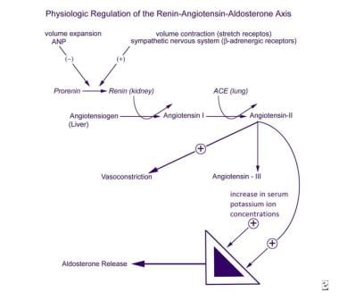
Zona glomerulosa is a region of the adrenal gland, specifically the outer portion of the adrenal cortex. It is responsible for producing mineralocorticoids, with the principal one being aldosterone. Aldosterone helps regulate electrolyte and fluid balance in the body by increasing the reabsorption of sodium ions and water in the distal nephron of the kidney while promoting the excretion of potassium ions. This process assists in maintaining blood pressure and volume within normal ranges. The zona glomerulosa's function is primarily under the control of the renin-angiotensin-aldosterone system (RAAS).
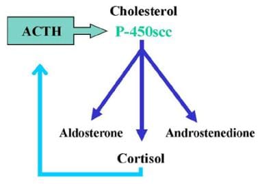
Adrenocorticotropic Hormone (ACTH) is a hormone produced and released by the anterior pituitary gland, a small endocrine gland located at the base of the brain. ACTH plays a crucial role in the regulation of the body's stress response and has significant effects on various physiological processes.
The primary function of ACTH is to stimulate the adrenal glands, which are triangular-shaped glands situated on top of the kidneys. The adrenal glands consist of two parts: the outer cortex and the inner medulla. ACTH specifically targets the adrenal cortex, where it binds to specific receptors and initiates a series of biochemical reactions leading to the production and release of steroid hormones, primarily cortisol (a glucocorticoid) and aldosterone (a mineralocorticoid).
Cortisol is involved in various metabolic processes, such as regulating blood sugar levels, modulating the immune response, and helping the body respond to stress. Aldosterone plays a vital role in maintaining electrolyte and fluid balance by promoting sodium reabsorption and potassium excretion in the kidneys.
ACTH release is controlled by the hypothalamus, another part of the brain, which produces corticotropin-releasing hormone (CRH). CRH stimulates the anterior pituitary gland to secrete ACTH, which in turn triggers cortisol production in the adrenal glands. This complex feedback system helps maintain homeostasis and ensures that appropriate amounts of cortisol are released in response to various physiological and psychological stressors.
Disorders related to ACTH can lead to hormonal imbalances, resulting in conditions such as Cushing's syndrome (excessive cortisol production) or Addison's disease (insufficient cortisol production). Proper diagnosis and management of these disorders typically involve assessing the function of the hypothalamic-pituitary-adrenal axis and addressing any underlying issues affecting ACTH secretion.
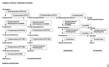
Steroid 11-beta-hydroxylase is a crucial enzyme involved in the steroidogenesis pathway, specifically in the synthesis of cortisol and aldosterone, which are vital hormones produced by the adrenal glands. This enzyme is encoded by the CYP11B1 gene in humans.
The enzyme's primary function is to catalyze the conversion of 11-deoxycortisol to cortisol and 11-deoxycorticosterone to aldosterone through the process of hydroxylation at the 11-beta position of the steroid molecule. Cortisol is a critical glucocorticoid hormone that helps regulate metabolism, immune response, and stress response, while aldosterone is a mineralocorticoid hormone responsible for maintaining electrolyte and fluid balance in the body.
Deficiencies or mutations in the CYP11B1 gene can lead to various disorders, such as congenital adrenal hyperplasia (CAH), which may result in impaired cortisol and aldosterone production, causing hormonal imbalances and associated symptoms.
Penfluridol is an antipsychotic medication that belongs to the class of diphenylbutylpiperidines. It is primarily used in the management of chronic schizophrenia and other related psychotic disorders. Penfluridol works by blocking dopamine receptors in the brain, which helps reduce the symptoms of psychosis such as hallucinations, delusions, and disordered thought processes.
The medication is available in oral form and is typically administered once daily due to its long half-life. Common side effects of penfluridol include sedation, dizziness, orthostatic hypotension, weight gain, and extrapyramidal symptoms (EPS), such as Parkinsonism, akathisia, and dystonia. Penfluridol has also been associated with tardive dyskinesia, a potentially irreversible movement disorder, with long-term use.
It is essential to monitor patients on penfluridol therapy for metabolic changes, cardiac function, and the emergence of extrapyramidal symptoms or other side effects. The medication should be used cautiously in elderly patients, those with a history of cardiovascular disease, and individuals with preexisting movement disorders.
Penfluridol is not approved for use in the United States but is available in some other countries as a treatment option for chronic schizophrenia and related psychotic disorders.
17-Hydroxycorticosteroids are a class of steroid hormones that are produced in the adrenal gland. They are formed from the metabolism of cortisol, which is a hormone that helps regulate metabolism, immune response, and stress response. 17-Hydroxycorticosteroids include compounds such as cortisone and corticosterone.
These hormones have various functions in the body, including:
* Regulation of carbohydrate, fat, and protein metabolism
* Suppression of the immune system
* Modulation of the stress response
* Influence on blood pressure and electrolyte balance
Abnormal levels of 17-hydroxycorticosteroids can indicate problems with the adrenal gland or pituitary gland, which regulates adrenal function. They are often measured in urine or blood tests to help diagnose conditions such as Cushing's syndrome (overproduction of cortisol) and Addison's disease (underproduction of cortisol).
Adrenal cortex diseases refer to a group of conditions that affect the adrenal glands, which are small glands located on top of the kidneys. The adrenal glands consist of two parts: the outer adrenal cortex and the inner medulla. The adrenal cortex is responsible for producing hormones such as cortisol, aldosterone, and androgens that regulate various bodily functions, including metabolism, blood pressure, and sexual development.
Diseases of the adrenal cortex can result from an overproduction or underproduction of these hormones. Some common adrenal cortex diseases include:
1. Addison's disease: a condition characterized by insufficient production of hormones by the adrenal glands, leading to symptoms such as fatigue, weight loss, low blood pressure, and darkening of the skin.
2. Cushing's syndrome: a condition caused by an excess of cortisol in the body, which can result from taking high doses of corticosteroid medications or from a tumor in the pituitary gland or adrenal glands. Symptoms include weight gain, particularly around the trunk and face, thinning of the skin, easy bruising, muscle weakness, and mood changes.
3. Congenital adrenal hyperplasia: a group of inherited disorders that affect the production of hormones by the adrenal glands. Depending on the specific type of congenital adrenal hyperplasia, symptoms can range from ambiguous genitalia in newborns to precocious puberty, short stature, and infertility in older children and adults.
4. Adrenal tumors: benign or cancerous growths that develop in the adrenal glands and can cause hormonal imbalances. Symptoms depend on the type of tumor and the hormones it produces.
Treatment for adrenal cortex diseases depends on the specific condition and its underlying cause. Treatment options may include medication, surgery, or radiation therapy.
Aldosterone synthase is a steroidogenic enzyme that is primarily responsible for the production of the hormone aldosterone in the adrenal gland. It is encoded by the CYP11B2 gene and is located within the mitochondria of the zona glomerulosa cells in the adrenal cortex.
Aldosterone synthase catalyzes two key reactions in the biosynthesis of aldosterone: the conversion of corticosterone to 18-hydroxycorticosterone and the subsequent conversion of 18-hydroxycorticosterone to aldosterone. These reactions involve the sequential addition of hydroxyl groups at the C18 position of the steroid molecule, which is a critical step in the synthesis of aldosterone.
Aldosterone plays an important role in regulating blood pressure and electrolyte balance by increasing the reabsorption of sodium and water in the distal nephron of the kidney, while promoting the excretion of potassium. Disorders of aldosterone synthase can lead to conditions such as primary hyperaldosteronism, which is characterized by excessive production of aldosterone and can result in hypertension and hypokalemia.
Tandem pore domain potassium (K2P) channels are a subfamily of potassium channels that contain two pore-forming domains in a single polypeptide chain. These channels are also known as "double-barreled" or "leak" potassium channels because they provide a background leak conductance for potassium ions across the cell membrane. They are involved in regulating the resting membrane potential and excitability of cells, and are targets for various therapeutic agents. Examples of K2P channels include TREK, TRAAK, TASK, TWIK, and THIK families.
Corticosterone is a hormone produced by the adrenal gland in many animals, including humans. It is a type of glucocorticoid steroid hormone that plays an important role in the body's response to stress, immune function, metabolism, and regulation of inflammation. Corticosterone helps to regulate the balance of sodium and potassium in the body and also plays a role in the development and functioning of the nervous system. It is the primary glucocorticoid hormone in rodents, while cortisol is the primary glucocorticoid hormone in humans and other primates.
The kinetoplast is a unique structure found in the single, mitochondrion of certain protozoan parasites, including those of the genera Trypanosoma and Leishmania. It consists of a network of circular DNA molecules that are highly concentrated and tightly packed. These DNA molecules contain genetic information necessary for the functioning of the unique mitochondrion in these organisms.
The kinetoplast DNA (kDNA) is organized into thousands of maxicircles and minicircles, which vary in size and number depending on the species. Maxicircles are similar to mammalian mitochondrial DNA and encode proteins involved in oxidative phosphorylation, while minicircles contain sequences that code for guide RNAs involved in the editing of maxicircle transcripts.
The kDNA undergoes dynamic rearrangements during the life cycle of these parasites, which involves different morphological and metabolic forms. The study of kDNA has provided valuable insights into the biology and evolution of these important pathogens and has contributed to the development of novel therapeutic strategies.
Aldosterone is a hormone produced by the adrenal gland. It plays a key role in regulating sodium and potassium balance and maintaining blood pressure through its effects on the kidneys. Aldosterone promotes the reabsorption of sodium ions and the excretion of potassium ions in the distal tubules and collecting ducts of the nephrons in the kidneys. This increases the osmotic pressure in the blood, which in turn leads to water retention and an increase in blood volume and blood pressure.
Aldosterone is released from the adrenal gland in response to a variety of stimuli, including angiotensin II (a peptide hormone produced as part of the renin-angiotensin-aldosterone system), potassium ions, and adrenocorticotropic hormone (ACTH) from the pituitary gland. The production of aldosterone is regulated by a negative feedback mechanism involving sodium levels in the blood. High sodium levels inhibit the release of aldosterone, while low sodium levels stimulate its release.
In addition to its role in maintaining fluid and electrolyte balance and blood pressure, aldosterone has been implicated in various pathological conditions, including hypertension, heart failure, and primary hyperaldosteronism (a condition characterized by excessive production of aldosterone).
Hydrocortisone is a synthetic glucocorticoid, which is a class of steroid hormones. It is identical to the naturally occurring cortisol, a hormone produced by the adrenal gland that helps regulate metabolism and helps your body respond to stress. Hydrocortisone has anti-inflammatory effects and is used to treat various inflammatory conditions such as allergies, skin disorders, and autoimmune diseases. It works by suppressing the immune system's response to reduce swelling, redness, itching, and other symptoms caused by inflammation.
Hydrocortisone is available in different forms, including oral tablets, topical creams, lotions, gels, and ointments, as well as injectable solutions. The specific use and dosage depend on the condition being treated and the individual patient's medical history and current health status.
As with any medication, hydrocortisone can have side effects, especially when used in high doses or for extended periods. Common side effects include increased appetite, weight gain, mood changes, insomnia, and skin thinning. Long-term use of hydrocortisone may also increase the risk of developing osteoporosis, diabetes, cataracts, and other health problems. Therefore, it is essential to follow your healthcare provider's instructions carefully when using this medication.
3-Hydroxysteroid dehydrogenases (3-HSDs) are a group of enzymes that play a crucial role in steroid hormone biosynthesis. These enzymes catalyze the conversion of 3-beta-hydroxy steroids to 3-keto steroids, which is an essential step in the production of various steroid hormones, including progesterone, cortisol, aldosterone, and sex hormones such as testosterone and estradiol.
There are several isoforms of 3-HSDs that are expressed in different tissues and have distinct substrate specificities. For instance, 3-HSD type I is primarily found in the ovary and adrenal gland, where it catalyzes the conversion of pregnenolone to progesterone and 17-hydroxyprogesterone to 17-hydroxycortisol. On the other hand, 3-HSD type II is mainly expressed in the testes, adrenal gland, and placenta, where it catalyzes the conversion of dehydroepiandrosterone (DHEA) to androstenedione and androstenedione to testosterone.
Defects in 3-HSDs can lead to various genetic disorders that affect steroid hormone production and metabolism, resulting in a range of clinical manifestations such as adrenal insufficiency, ambiguous genitalia, and sexual development disorders.
Steroids, also known as corticosteroids, are a type of hormone that the adrenal gland produces in your body. They have many functions, such as controlling the balance of salt and water in your body and helping to reduce inflammation. Steroids can also be synthetically produced and used as medications to treat a variety of conditions, including allergies, asthma, skin conditions, and autoimmune disorders.
Steroid medications are available in various forms, such as oral pills, injections, creams, and inhalers. They work by mimicking the effects of natural hormones produced by your body, reducing inflammation and suppressing the immune system's response to prevent or reduce symptoms. However, long-term use of steroids can have significant side effects, including weight gain, high blood pressure, osteoporosis, and increased risk of infections.
It is important to note that anabolic steroids are a different class of drugs that are sometimes abused for their muscle-building properties. These steroids are synthetic versions of the male hormone testosterone and can have serious health consequences when taken in large doses or without medical supervision.
"Cattle" is a term used in the agricultural and veterinary fields to refer to domesticated animals of the genus *Bos*, primarily *Bos taurus* (European cattle) and *Bos indicus* (Zebu). These animals are often raised for meat, milk, leather, and labor. They are also known as bovines or cows (for females), bulls (intact males), and steers/bullocks (castrated males). However, in a strict medical definition, "cattle" does not apply to humans or other animals.
Electron microscopy (EM) is a type of microscopy that uses a beam of electrons to create an image of the sample being examined, resulting in much higher magnification and resolution than light microscopy. There are several types of electron microscopy, including transmission electron microscopy (TEM), scanning electron microscopy (SEM), and reflection electron microscopy (REM).
In TEM, a beam of electrons is transmitted through a thin slice of the sample, and the electrons that pass through the sample are focused to form an image. This technique can provide detailed information about the internal structure of cells, viruses, and other biological specimens, as well as the composition and structure of materials at the atomic level.
In SEM, a beam of electrons is scanned across the surface of the sample, and the electrons that are scattered back from the surface are detected to create an image. This technique can provide information about the topography and composition of surfaces, as well as the structure of materials at the microscopic level.
REM is a variation of SEM in which the beam of electrons is reflected off the surface of the sample, rather than scattered back from it. This technique can provide information about the surface chemistry and composition of materials.
Electron microscopy has a wide range of applications in biology, medicine, and materials science, including the study of cellular structure and function, disease diagnosis, and the development of new materials and technologies.
Amide synthases are a class of enzymes that catalyze the formation of amide bonds between two molecules. Specifically, they facilitate the reaction between a carboxylic acid and an amine to produce an amide. This process is also known as amide bond formation or amide synthesis.
In the context of medical research and therapeutic development, amide synthases are important for understanding the biosynthesis of various endogenous compounds, such as peptides and proteins, as well as for developing methods to synthesize novel drugs and pharmaceutical agents.
There are several types of amide synthases, including:
1. Non-ribosomal peptide synthetases (NRPS): These enzymes catalyze the formation of complex peptides without the involvement of ribosomes. They typically consist of multiple modules, each of which is responsible for adding a single amino acid to the growing peptide chain.
2. Amidotransferases: These enzymes transfer an amino group from a donor molecule (usually glutamine) to a carboxylic acid, resulting in the formation of an amide bond. They are involved in various metabolic pathways, including the biosynthesis of amino acids, nucleotides, and other biomolecules.
3. Amide synthetases involved in lipid metabolism: These enzymes catalyze the formation of amide bonds between fatty acids and various amine-containing molecules, such as sphingosine or serine, during the biosynthesis of complex lipids like sphingolipids and glycerophospholipids.
Understanding the function and regulation of amide synthases is crucial for developing strategies to modulate their activity in various disease contexts, including infectious diseases, cancer, and neurodegenerative disorders.
Chamaecrista is a genus of flowering plants in the pea family (Fabaceae). It includes several species commonly known as "sensitive plants" because their leaves react to touch by folding in on themselves. These plants are native to warm temperate and tropical regions around the world, including North and South America, Africa, and Asia. Some Chamaecrista species have been used in traditional medicine to treat various ailments, such as skin conditions, inflammation, and fever. However, it is important to note that the medical uses of these plants are not well-studied, and they should not be used as a substitute for professional medical advice or treatment.
Angiotensin II is a potent vasoactive peptide hormone that plays a critical role in the renin-angiotensin-aldosterone system (RAAS), which is a crucial regulator of blood pressure and fluid balance in the body. It is formed from angiotensin I through the action of an enzyme called angiotensin-converting enzyme (ACE).
Angiotensin II has several physiological effects on various organs, including:
1. Vasoconstriction: Angiotensin II causes contraction of vascular smooth muscle, leading to an increase in peripheral vascular resistance and blood pressure.
2. Aldosterone release: Angiotensin II stimulates the adrenal glands to release aldosterone, a hormone that promotes sodium reabsorption and potassium excretion in the kidneys, thereby increasing water retention and blood volume.
3. Sympathetic nervous system activation: Angiotensin II activates the sympathetic nervous system, leading to increased heart rate and contractility, further contributing to an increase in blood pressure.
4. Thirst regulation: Angiotensin II stimulates the hypothalamus to increase thirst, promoting water intake and helping to maintain intravascular volume.
5. Cell growth and fibrosis: Angiotensin II has been implicated in various pathological processes, such as cell growth, proliferation, and fibrosis, which can contribute to the development of cardiovascular and renal diseases.
Angiotensin-converting enzyme inhibitors (ACEIs) and angiotensin receptor blockers (ARBs) are two classes of medications commonly used in clinical practice to target the RAAS by blocking the formation or action of angiotensin II, respectively. These drugs have been shown to be effective in managing hypertension, heart failure, and chronic kidney disease.
Organ size refers to the volume or physical measurement of an organ in the body of an individual. It can be described in terms of length, width, and height or by using specialized techniques such as imaging studies (like CT scans or MRIs) to determine the volume. The size of an organ can vary depending on factors such as age, sex, body size, and overall health status. Changes in organ size may indicate various medical conditions, including growths, inflammation, or atrophy.
Egg proteins, also known as egg white proteins or ovalbumin, refer to the proteins found in egg whites. There are several different types of proteins found in egg whites, including:
1. Ovalbumin (54%): This is the major protein found in egg whites and is responsible for their white color. It has various functions such as providing nutrition, maintaining the structural integrity of the egg, and protecting the egg from bacteria.
2. Conalbumin (13%): Also known as ovotransferrin, this protein plays a role in the defense against microorganisms by binding to iron and making it unavailable for bacterial growth.
3. Ovomucoid (11%): This protein is resistant to digestion and helps protect the egg from being broken down by enzymes in the digestive tract of predators.
4. Lysozyme (3.5%): This protein has antibacterial properties and helps protect the egg from bacterial infection.
5. Globulins (4%): These are a group of simple proteins found in egg whites that have various functions such as providing nutrition, maintaining the structural integrity of the egg, and protecting the egg from bacteria.
6. Avidin (0.05%): This protein binds to biotin, a vitamin, making it unavailable for use by the body. However, cooking denatures avidin and makes the biotin available again.
Egg proteins are highly nutritious and contain all nine essential amino acids, making them a complete source of protein. They are also low in fat and cholesterol, making them a popular choice for those following a healthy diet.
Trypanosomatina is not considered a medical term, but it is a taxonomic category in the field of biology. Trypanosomatina is a suborder that includes unicellular parasitic protozoans belonging to the order Kinetoplastida. Some notable members of this suborder include genera such as Trypanosoma and Leishmania, which are medically important parasites causing diseases in humans and animals.
Trypanosoma species are responsible for various trypanosomiases, including African sleeping sickness (caused by Trypanosoma brucei) and Chagas disease (caused by Trypanosoma cruzi). Leishmania species cause different forms of leishmaniasis, a group of diseases affecting the skin, mucous membranes, or internal organs.
In summary, while not a medical term itself, Trypanosomatina is a biology taxonomic category that includes several disease-causing parasites of medical importance.
Trypanosoma is a genus of flagellated protozoan parasites belonging to the family Trypanosomatidae. These microscopic single-celled organisms are known to cause various tropical diseases in humans and animals, including Chagas disease (caused by Trypanosoma cruzi) and African sleeping sickness (caused by Trypanosoma brucei).
The life cycle of Trypanosoma involves alternating between an insect vector (like a tsetse fly or kissing bug) and a mammalian host. The parasites undergo complex morphological changes as they move through the different hosts and developmental stages, often exhibiting distinct forms in the insect vector compared to the mammalian host.
Trypanosoma species have an undulating membrane and a single flagellum that helps them move through their environment. They can be transmitted through various routes, including insect vectors, contaminated food or water, or congenital transmission from mother to offspring. The diseases caused by these parasites can lead to severe health complications and may even be fatal if left untreated.
Sperm-ovum interactions, also known as sperm-egg interactions, refer to the specific series of events that occur between a spermatozoon (sperm) and an oocyte (egg or ovum) during fertilization in sexual reproduction.
The process begins with the sperm's attachment to the zona pellucida, a glycoprotein layer surrounding the oocyte. This interaction is mediated by specific proteins on the surface of both the sperm and the zona pellucida. Following attachment, the sperm undergoes the acrosome reaction, during which enzymes are released from the sperm's head to help digest and penetrate the zona pellucida.
Once the sperm has successfully traversed the zona pellucida, it makes contact with the oocyte's plasma membrane, triggering the fusion of the sperm and egg membranes. This results in the release of the sperm's genetic material into the oocyte's cytoplasm and the initiation of a series of intracellular signaling events within the oocyte that ultimately lead to its completion of meiosis II and formation of a zygote, marking the beginning of embryonic development.
Proper sperm-ovum interactions are crucial for successful fertilization and subsequent embryonic development, and any disruptions in these processes can result in infertility or early pregnancy loss.
I apologize for any confusion, but "Hyptis" is not a medical term. It is a genus of plants in the mint family, also known as "Brazilian mint" or " bushmint." Some Hyptis species have been used in traditional medicine, but there is limited scientific evidence supporting their effectiveness. If you have any questions about specific medical terms or concepts, I would be happy to help if I can!
Messenger RNA (mRNA) is a type of RNA (ribonucleic acid) that carries genetic information copied from DNA in the form of a series of three-base code "words," each of which specifies a particular amino acid. This information is used by the cell's machinery to construct proteins, a process known as translation. After being transcribed from DNA, mRNA travels out of the nucleus to the ribosomes in the cytoplasm where protein synthesis occurs. Once the protein has been synthesized, the mRNA may be degraded and recycled. Post-transcriptional modifications can also occur to mRNA, such as alternative splicing and addition of a 5' cap and a poly(A) tail, which can affect its stability, localization, and translation efficiency.
Trypanosoma brucei brucei is a species of protozoan flagellate parasite that causes African trypanosomiasis, also known as sleeping sickness in humans and Nagana in animals. This parasite is transmitted through the bite of an infected tsetse fly (Glossina spp.). The life cycle of T. b. brucei involves two main stages: the insect-dwelling procyclic trypomastigote stage and the mammalian-dwelling bloodstream trypomastigote stage.
The distinguishing feature of T. b. brucei is its ability to change its surface coat, which helps it evade the host's immune system. This allows the parasite to establish a long-term infection in the mammalian host. However, T. b. brucei is not infectious to humans; instead, two other subspecies, Trypanosoma brucei gambiense and Trypanosoma brucei rhodesiense, are responsible for human African trypanosomiasis.
In summary, Trypanosoma brucei brucei is a non-human-infective subspecies of the parasite that causes African trypanosomiasis in animals and serves as an essential model organism for understanding the biology and pathogenesis of related human-infective trypanosomes.
"Cells, cultured" is a medical term that refers to cells that have been removed from an organism and grown in controlled laboratory conditions outside of the body. This process is called cell culture and it allows scientists to study cells in a more controlled and accessible environment than they would have inside the body. Cultured cells can be derived from a variety of sources, including tissues, organs, or fluids from humans, animals, or cell lines that have been previously established in the laboratory.
Cell culture involves several steps, including isolation of the cells from the tissue, purification and characterization of the cells, and maintenance of the cells in appropriate growth conditions. The cells are typically grown in specialized media that contain nutrients, growth factors, and other components necessary for their survival and proliferation. Cultured cells can be used for a variety of purposes, including basic research, drug development and testing, and production of biological products such as vaccines and gene therapies.
It is important to note that cultured cells may behave differently than they do in the body, and results obtained from cell culture studies may not always translate directly to human physiology or disease. Therefore, it is essential to validate findings from cell culture experiments using additional models and ultimately in clinical trials involving human subjects.
I'd be happy to help! However, I believe there might be a slight confusion in your question. RNA stands for Ribonucleic Acid, which is a type of nucleic acid involved in various biological roles in the coding, decoding, regulation, and expression of genes. On the other hand, protozoan refers to a unicellular organism from the kingdom Protista, which includes a wide variety of simple eukaryotic organisms such as amoebas, paramecia, and plasmodium (the malaria-causing parasite).
There isn't a specific medical definition for "RNA, protozoan" since RNA is a molecule present in all living cells, including human cells, and protozoans are a group of organisms. However, I can tell you that RNA plays crucial roles in protozoan biology, such as acting as a messenger between DNA and ribosomes during protein synthesis or regulating gene expression.
If you have any further questions or need more specific information about RNA in protozoans, please let me know!
"Wistar rats" are a strain of albino rats that are widely used in laboratory research. They were developed at the Wistar Institute in Philadelphia, USA, and were first introduced in 1906. Wistar rats are outbred, which means that they are genetically diverse and do not have a fixed set of genetic characteristics like inbred strains.
Wistar rats are commonly used as animal models in biomedical research because of their size, ease of handling, and relatively low cost. They are used in a wide range of research areas, including toxicology, pharmacology, nutrition, cancer, cardiovascular disease, and behavioral studies. Wistar rats are also used in safety testing of drugs, medical devices, and other products.
Wistar rats are typically larger than many other rat strains, with males weighing between 500-700 grams and females weighing between 250-350 grams. They have a lifespan of approximately 2-3 years. Wistar rats are also known for their docile and friendly nature, making them easy to handle and work with in the laboratory setting.
Euglenozoa is a group of unicellular organisms that includes both free-living and parasitic species. Two major parasitic groups within Euglenozoa are the kinetoplastids, which include organisms such as Trypanosoma and Leishmania, and the diplonemids.
Trypanosoma infections can cause diseases such as African sleeping sickness (also known as human African trypanosomiasis) and Chagas disease (also known as American trypanosomiasis), while Leishmania infections can cause various forms of leishmaniasis, including cutaneous, mucocutaneous, and visceral leishmaniasis. These diseases are transmitted to humans through the bites of infected insect vectors, such as tsetse flies (in the case of African sleeping sickness) or sandflies (in the case of leishmaniasis and Chagas disease).
Diplonemid infections in humans have not been well-studied, and it is currently unclear whether these organisms are capable of causing disease in humans. However, diplonemids have been found to infect a wide range of marine and freshwater organisms, including fish, crustaceans, and other protists.
In general, euglenozoan infections can cause a variety of symptoms depending on the specific organism involved and the location of the infection within the body. Symptoms may include fever, swelling, skin lesions, anemia, and damage to various organs. Treatment for these infections typically involves the use of antiparasitic drugs, such as pentamidine, suramin, or benznidazole, although the specific treatment approach will depend on the organism involved and the severity of the infection.
 Zona fasciculata
Cortisol
Melanocortin 2 receptor accessory protein
Corticosteroid
ACTH receptor
Adrenal cortex
Aldosterone
Cortisone
Primary aldosteronism
Adrenocorticotropic hormone
Glucocorticoid
Corticosterone
Spongiocyte
Adrenal gland
Adrenocortical hormone
Cushing's syndrome
Developmental impact of child neglect in early childhood
Cholesterol side-chain cleavage enzyme
Glucocorticoid remediable aldosteronism
Proopiomelanocortin
Zona reticularis
Helen Wendler Deane
Hypoadrenocorticism in dogs
Congenital adrenal hyperplasia due to 17α-hydroxylase deficiency
Steroid 11β-hydroxylase
11-Deoxycorticosterone
Adrenocortical adenoma
Adrenal insufficiency
CYP17A1
Congenital adrenal hyperplasia due to 21-hydroxylase deficiency
Zona fasciculata - Wikipedia
Zona Fasciculata
Transcriptome analysis reveals differentially expressed transcripts in rat adrenal zona glomerulosa and zona fasciculata |...
 Pediatric Adrenal Gland Disorders: Anatomy, Embryology, Physiology
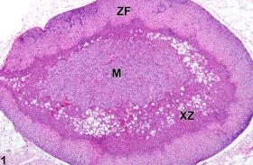
 Adrenal Gland, Cortex, X-Zone - Atrophy - Nonneoplastic Lesion Atlas
 BRS Endocrine Physiology Quiz: Trivia! - ProProfs Quiz
 Registration Dossier - ECHA
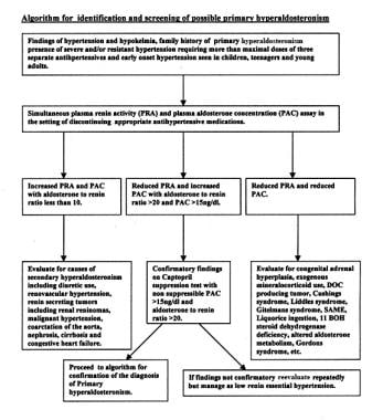
Primary Aldosteronism: Practice Essentials, Pathophysiology, Etiology
 Springbok (Antidorcas marsupialis) | IVIS
 Petasites for Pain and Inflammation - Naturopathic Doctor News and Review
 Acrylonitrile (EHC 28, 1983)
JCI - Partial MCM4 deficiency in patients with growth retardation, adrenal insufficiency, and natural killer cell deficiency
 What the Scriptures REALLY Teach about Health - Part 4 - EliYah Ministries
Registration Dossier - ECHA
 Cushing's disease/Cushing syndrome
 Adrenals - Oakton Primary Care Centers
 The Adrenal Glands - Biology of Aging
 What tissues make up the endocrine system? - My Medical Health Blog
SAMPLE QUESTIONS | PQJ2023
Pancreatic islets - Wikipedia
 Kate Shared 'Prenursing Smarter Module Three | Anatomy and Physiology' - 23 Picmonics
Adrenal - Endocrinology - Anatomy & Embryology - Picmonic for Physician Assistant
 Endocrine
 Plant and Animal Hormones Online Test 10th Science Lesson 16 Questions in English - WINMEEN
 Cushing's Syndrome - Signs, Symptoms, Diagnosis, Treatment
which layer of the epidermis is highlighted quizlet
 7.3: Notable Lipids - Introductory Biochemistry
VASCULAR SUPPLY TO THE KIDNEY - ANATOMY-LEXICON
17-Hydroxylase Deficiency Syndrome: Background, Pathophysiology, Epidemiology
Different isozymes of mouse 11/β-hydroxylase produce mineralocorticoids and glucocorticoids<...
Glomerulosa23
- The zona fasciculata (sometimes, fascicular or fasciculate zone) constitutes the middle and also the widest zone of the adrenal cortex, sitting directly beneath the zona glomerulosa. (wikipedia.org)
- Adult-type zona glomerulosa and fasciculata are detected in fetal life but make up only a small proportion of the gland, and the zona reticularis is not present at all. (medscape.com)
- The first layer of the cortex is the zona glomerulosa, followed by the zonas fasciculata and reticularis, the innermost zone. (aacc.org)
- Aldosterone, the most potent mineralocorticoid, is made exclusively in the zona glomerulosa. (aacc.org)
- It also stimulates the cells of the zona glomerulosa to produce the enzyme aldosterone synthase and, consequently, aldosterone. (aacc.org)
- The outer most portion of the cortex is called the Zona glomerulosa. (oaktonprimarycarecenters.com)
- A. zona fasciculata B. zona reticularis C. zona glomerulosa D. medulla Answer. (pqj2023.com)
- In the adrenal cortex, lesions are more frequent in the zona fasciculata and reticularis than in the zona glomerulosa. (qxmd.com)
- For convenience, think of the zona glomerulosa as the first endocrine organ and the zonae fasciculata and reticularis collectively as a second separate endocrine organ, as distinguished by distinct control systems. (medscape.com)
- In situ hybridizations of adrenal sections with gene-specific probes showed that the 11β-OHase gene was expressed in the zona fasciculata, whereas AS-derived transcripts were detected only in the outer zona glomerulosa. (elsevierpure.com)
- The zona glomerulosa is the outer part of the adrenal cortex. (anatomy.app)
- The cells within the zona glomerulosa are arranged in clusters and resemble balls of wool. (anatomy.app)
- The zona glomerulosa is only five to seven cell layers thick, but it depends on its functional state. (anatomy.app)
- For example, an individual with chronic sodium deprivation will have a larger zona glomerulosa than one with a normal sodium intake. (anatomy.app)
- The main cells of the zona glomerulosa have steroid-secreting cell characteristics with lipid droplets and large numbers of mitochondria. (anatomy.app)
- The zona glomerulosa is responsible for secreting mineralcorticoids , mainly aldosterone. (anatomy.app)
- Like the cells of zona glomerulosa, these cells also have steroid-secreting characteristics. (anatomy.app)
- The cells of the zona fasciculata are arranged in parallel radial cords that resemble long strings or bundles of sticks (fasciculi), stretching from the zona glomerulosa down in the direction of the adrenal medulla. (anatomy.app)
- This mineralocorticoid hormone produced by the zona glomerulosa plays a central role in regulating blood pressure and certain electrolytes (sodium and potassium). (ratingperson.com)
- The other two layers of the adrenal cortex beneath the glomerulosa are Zona fasciculata and Zona reticularis . (knowyourbody.net)
- Here are some assistive images of Zona glomerulosa that will help you get a visual idea about this physical structure. (knowyourbody.net)
- The suprarenal cortex is the largest part of the gland and is composed of 3 zones: the zona glomerulosa (outer zone), the zona fasciculata (middle zone), and the zona reticularis (inner zone). (medscape.com)
- The zona glomerulosa is responsible for the production of mineralocorticoids, mainly aldosterone, which regulates blood pressure and electrolyte balance. (medscape.com)
Reticularis8
- The main source of androgens will come from the zona reticularis region. (wikipedia.org)
- The zonas fasciculata and reticularis synthesize glucocorticoids and androgens. (aacc.org)
- The next zone in the adrenal cortex is the Zona reticularis. (oaktonprimarycarecenters.com)
- In contrast, cortisol synthesis and secretion is regulated by adrenocorticotropic hormone (ACTH), which stimulates the enzyme P-450scc (20, 22 desmolase) with subsequent increased production of all adrenal steroids in both the zona fasciculata and zona reticularis. (medscape.com)
- Zona reticularis - produces androgens, for example, androstenedione - the precursor of testosterone. (anatomy.app)
- 11β-hydroxylase is found in the zona fasciculata and reticularis. (wikidoc.org)
- These hormones produced by the zona reticularis are weak male hormones. (ratingperson.com)
- The zona reticularis produces gonadocorticoids and is responsible for administering these hormones to the reproductive regions of the body. (medscape.com)
Glucocorticoids2
- The zona fasciculata chiefly produces glucocorticoids (mainly cortisol in humans), which regulate the metabolism of glucose. (wikipedia.org)
- The zona fasciculata, is responsible for the production of glucocorticoids, predominantly cortisol, which increases blood sugar levels via gluconeogenesis, suppresses the immune system, and aids in metabolism. (medscape.com)
Androgens2
- The zona fasciculata also generates a small amount of weak androgens (e.g., dehydroepiandrosterone). (wikipedia.org)
- The third and innermost zone of the adrenal cortex is the Zona fasciculata and it secretes androgens. (oaktonprimarycarecenters.com)
Cortex5
- Adrenal sources of Cushing syndrome include unilateral, cortisol, producing adenomas, which are benign and originate in the Zona, fasciculata of the adrenal cortex. (standardofcare.com)
- The middle zone of the adrenal cortex is called zona fasciculata. (anatomy.app)
- It releases from zona fasciculata layer of adrenal cortex of the adrenal gland. (websparrow.org)
- Produced by the zona fasciculata of the adrenal cortex, cortisol is known as the "stress hormone. (tampanaturalcare.com)
- It is created from cholesterol by cytochrome P450 enzymes in the zona fasciculata of the adrenal cortex. (vitamonk.com)
Cortisol production2
- Steroid-producing adrenal tumours and hyperplasia of the zona fasciculata result in excess cortisol production and are the cause for adrenal Cushing's syndrome. (wikipedia.org)
- CRH (hypothalamus) stimulates ACTH release from the pituitary gland, causing cortisol production in adrenal zona fasciculata. (dailymeded.com)
Glucocorticoid1
- Cortisol is a glucocorticoid hormone produced by the zona fasciculata that plays several important roles in the body. (ratingperson.com)
Zones1
- The cells of the zona fasciculata have considerably more cytoplasm than those of the other cortical zones and stain palely because of their high concentration of lipid droplets. (medcell.org)
Aldosterone synthase1
- 1995, Zona fasciculata-like cells determine the response of plasma aldosterone to metoclopramide and aldosterone synthase messenger ribonucleic acid level in aldosterone-producing adenoma. (wustl.edu)
Cells2
- Although Y1 cells otherwise resemble zona fasciculata cells and express the 11β-OHase gene at high levels, transcripts encoded by the AS gene were detected at levels approximately 10-fold lower than the 11β-OHase transcripts. (elsevierpure.com)
- The zona fasciculata consists of vacuolated cells that contain lipid droplets, abundant mitochondria, and a smooth endoplasmic reticulum. (anatomy.app)
Responsible1
- The zona fasciculata is responsible for secreting glicocorticoids , including cortisol and corticosterone. (anatomy.app)
Cortisol7
- The zona fasciculata chiefly produces glucocorticoids (mainly cortisol in humans), which regulate the metabolism of glucose. (wikipedia.org)
- Steroid-producing adrenal tumours and hyperplasia of the zona fasciculata result in excess cortisol production and are the cause for adrenal Cushing's syndrome. (wikipedia.org)
- Esta zona produce una serie de enzimas que convierten la PREGNENOLONA en cortisol (HIDROCORTISONA) vía la 17-ALFA-HIDROXIPROGESTERONA. (bvsalud.org)
- Cortisol is secreted by the middle region (zona fasciculata) of adrenal cortex. (infinitylearn.com)
- Produced by the zona fasciculata of the adrenal cortex, cortisol is known as the "stress hormone. (littlemissacupuncture.com)
- 7 Wild-type mice exposed to stress were found to have significantly thinner zona fasciculata, which is where cortisol is produced. (tamhsc.edu)
- Cortisol is a glucocorticoid hormone produced with the help of the zona fasciculata that performs numerous vital roles in our body. (divinebeautytips.com)
Aldosterone6
- Aldosterone is a steroid hormone produced exclusively in the zona glomerulosa of the adrenal cortex. (medscape.com)
- Adrenal glands of the mice exhibited normal cortical zonation including a functionally undifferentiated cell-layer between the aldosterone-synthesizing zona glomerulosa cells and the corticosterone-synthesizing zona fasciculata cells. (elsevierpure.com)
- None of these clones express the zona glomerulosa-specific aldosterone synthase P450aldo gene under the conditions we tested. (elsevierpure.com)
- The zona glomerulosa produces aldosterone in response to angiotensin II. (topgradeapp.com)
- Microarray, qPCR, and KCNJ5 sequencing of aldosterone-producing adenomas reveal differences in genotype and phenotype between zona glomerulosa- and zona fasciculata-like tumors. (cdc.gov)
- Aldosterone is a steroid, released by the zona glomerulosa, that is responsible for regulating the salt and water in your body, having an effect on your blood pressure. (nesaz.com)
Adrenal fasciculata1
- We demonstrate that PDE8B is highly enriched in mouse adrenal fasciculata cells, and show that PDE8B knockout mice have elevated urinary corticosterone as a result of adrenal hypersensitivity toward adrenocorticotropin. (aspetjournals.org)
Necrosis2
- In mice, MeSO(2)-DDE induces mitochondrial degeneration and cellular necrosis in the adrenal zona fasciculata. (nih.gov)
- These criteria include nuclear grade III or IV, mitotic rate greater than five per fifty high power fields, presence of atypical mitotic figures, presence of less than or equal to twenty five percent of clear or vacuolated cells that resemble normal zona fasciculata, diffuse architecture, necrosis, venous invasion, sinusoid invasion, and invasion of tumor capsule. (nih.gov)
Glomerulosa cells3
- 3. Effects of C19 steroids on adrenal steroidogenic enzyme activities and their mRNA levels in guinea-pig fasciculata-glomerulosa cells in primary culture. (nih.gov)
- 4. Effect of ACTH on steroidogenic enzymes in guinea pig fasciculata-glomerulosa cells: changes in activity and mRNA levels. (nih.gov)
- [9] [12] However, together with other data on neuroendocrine properties of zona glomerulosa cells, NCAM expression may reflect a neuroendocrine differentiation of these cells. (wikipedia.org)
Fascicular1
- The zona fasciculata (sometimes, fascicular or fasciculate zone) constitutes the middle and also the widest zone of the adrenal cortex, sitting directly beneath the zona glomerulosa. (wikipedia.org)
Endocrinology1
- PREFACE TO THE THIRD EDITION The scepticism with which many medical men formerly held off from the study of the internal secretions has been gradually replaced by the opposite tendency which, in many students of endocrinology, is exhibited as an en- deavor to interpret every change in the living human organism as a disturbance of the endocrine balance. (nih.gov)
Steroidogenic3
- Interestingly, histological studies with Mcm4-depleted mice showed grossly abnormal adrenal morphology that was characterized by non-steroidogenic GATA4- and Gli1-positive cells within the steroidogenic cortex, which reduced the number of steroidogenic cells in the zona fasciculata of the adrenal cortex. (jci.org)
- These results show that AcE60 and AcA101 cells display a pattern of the steroidogenic gene expression similar to that of the undifferentiated cell-layer and are capable of differentiating into the zona fasciculata-like cells in vitro. (elsevierpure.com)
- These cells did not express either CYP11A1 or CYP11B1 and significantly reduced the number of steroidogenic cells in the zona fasciculata. (qmul.ac.uk)
Zone1
- [8] Pokazano je da tokom bolesti dojenčadi raste steroidogeni kapacitet zone fasciculata. (wikipedia.org)