Zona Glomerulosa
Adrenal Cortex
Aldosterone
Zona Fasciculata
Adrenal Glands
Zona Reticularis
Aldosterone Synthase
Steroid 11-beta-Hydroxylase
Adrenocorticotropic Hormone
Angiotensin II
Hyperaldosteronism
Interrenal Gland
Corticosterone
Mineralocorticoids
Cattle
Potassium
Receptors, Adrenomedullin
Renin
Receptors, Atrial Natriuretic Factor
Adrenomedullin
Receptors, Angiotensin
Cells, Cultured
Nitrendipine
Atrial Natriuretic Factor
Sperm-Ovum Interactions
Rats, Sprague-Dawley
Sugar Phosphates
Hydrocortisone
In Situ Hybridization
RNA, Messenger
Microscopy, Electron
Mitochondria
Immunohistochemistry
Actions of vasoactive intestinal peptide on the rat adrenal zona glomerulosa. (1/148)
Previous studies, by this group and others, have shown that vasoactive intestinal peptide (VIP) stimulates aldosterone secretion, and that the actions of VIP on aldosterone secretion by the rat adrenal cortex are blocked by beta adrenergic antagonists, suggesting that VIP may act by the local release of catecholamines. The present studies were designed to test this hypothesis further, by measuring catecholamine release by adrenal capsular tissue in response to VIP stimulation. Using intact capsular tissue it was found that VIP caused a dose-dependent increase in aldosterone secretion, with a concomitant increase in both adrenaline and noradrenaline release. The effects of VIP on aldosterone secretion were inhibited by atenolol, a beta1 adrenergic antagonist, but not by ICI-118,551, a beta2 adrenergic antagonist. Binding studies were carried out to investigate VIP receptors. It was found that adrenal zona glomerulosa tissue from control rats contained specific VIP binding sites (Bmax 853+/-101 fmol/mg protein; Kd 2.26+/-0.45 nmol/l). VIP binding was not displaced by ACTH, angiotensin II or by either of the beta adrenergic antagonists. The response to VIP in adrenals obtained from rats fed a low sodium diet was also investigated. Previous studies have found that adrenals from animals on a low sodium diet exhibit increased responsiveness to VIP. Specific VIP binding sites were identified, although the concentration or affinity of binding sites in the low sodium group was not significantly different from the controls. In the low sodium group VIP was found to increase catecholamine release to the same extent as in the control group, however, in contrast to the control group, the adrenal response to VIP was not altered by adrenergic antagonists in the low sodium group. These data provide strong support for the hypothesis that VIP acts by the local release of catecholamines in adrenal zona glomerulosa tissue in normal animals. It does not appear that VIP acts through the same mechanism in animals maintained on a low sodium diet. The mechanism by which VIP stimulates aldosterone in this group remains to be determined. (+info)Changes in the glomerulosa cell phenotype during adrenal regeneration in rats. (2/148)
In situ hybridization was used to examine cellular differentiation during rat adrenal regeneration, defining zona glomerulosa [cytochrome P-450 aldosterone synthase (P-450aldo) mRNA positive], zona fasciculata [cytochrome P-450 11beta-hydroxylase (P-45011beta) mRNA positive], or zona intermedia [negative for both but 3beta-hydroxysteroid dehydrogenase (3beta-HSD) mRNA positive]. After unilateral adrenal enucleation with contralateral adrenalectomy (ULE/ULA), the expression of all mRNA was reduced at 2 days. From 5 to 10 days, P-45011beta and 3beta-HSD mRNA increased while P-450aldo remained low; at 20 days, all mRNA were increased. From 2 to 10 days, cells adjacent to the capsule showed intermedia cell differentiation; by 20 days, the subcapsular glomerulosa cells reappeared. This suggests that after enucleation the glomerulosa dedifferentiates to zona intermedia. The experiment was repeated in rats where the postenucleation ACTH rise was prevented. Rats underwent ULE with sham ULA (ULE/SULA) or ULE/SULA with ACTH treatment. Adrenals from ULE/SULA rats expressed increased P-450aldo mRNA at 10 days and reduced P-45011beta mRNA and adrenal weight at 30 days. ACTH treatment reversed the pattern toward that seen in ULE/ULA. These findings show that the enucleation-induced dedifferentitation of the glomerulosa cell may result in part from elevated plasma ACTH and that prevention of dedifferentiation may result in impaired regeneration. (+info)A role for T-type Ca2+ channels in the synergistic control of aldosterone production by ANG II and K+. (3/148)
Independently, plasma K+ and ANG II stimulate aldosterone secretion from adrenal glomerulosa (AG) cells, but together they synergistically control production. We studied mechanisms to mediate this synergy using bovine AG cells studied under physiological conditions (in 1.25 mM Ca2+ at 37 degrees C). Increasing K+ from 2 to 5 mM caused a potentiation of ANG II-induced aldosterone secretion and a substantial membrane depolarization ( approximately 21 mV). ANG II inhibited a K+-selective conductance in both 2 and 5 mM K+ but caused only a slight depolarization because, under both conditions, membrane potential was close to the reversal potential of the ANG II-induced current. ANG II activated calcium/calmodulin-dependent protein kinase II (CaMKII) equivalently in 2 and 5 mM K+. However, CaMKII activation caused a hyperpolarizing shift in the activation of T-type Ca2+ channels, such that substantially more current was elicited at membrane potentials established by 5 mM K+. We propose that synergy in aldosterone secretion results from K+-induced depolarization and ANG II-induced modulation of T-type channel activation, such that together they promote enhanced steady-state Ca2+ flux. (+info)Angiotensin II negatively modulates L-type calcium channels through a pertussis toxin-sensitive G protein in adrenal glomerulosa cells. (4/148)
In bovine adrenal glomerulosa cells, angiotensin II and extracellular K+ stimulate aldosterone secretion in a calcium-dependent manner. In these cells, physiological concentrations of extracellular potassium activate both T-type (low threshold) and L-type (high threshold) voltage-operated calcium channels. Paradoxically, the cytosolic calcium response to 9 mM K+ is inhibited by angiotensin II. Because K+-induced calcium changes observed in the cytosol are almost exclusively due to L-type channel activity, we therefore studied the mechanisms of L-type channel regulation by angiotensin II. Using the patch-clamp method in its perforated patch configuration, we observed a marked inhibition (by 63%) of L-type barium currents in response to angiotensin II. This effect of the hormone was completely prevented by losartan, a specific antagonist of the AT1 receptor subtype. Moreover, this inhibition was strongly reduced when the cells were previously treated for 1 night with pertussis toxin. An effect of pertussis toxin was also observed on the modulation by angiotensin II of the K+ (9 mM)-induced cytosolic calcium response in fura-2-loaded cells, as well as on the angiotensin II-induced aldosterone secretion, at both low (3 mM) and high (9 mM) K+ concentrations. Finally, the expression of both Go and Gi proteins in bovine glomerulosa cells was detected by immunoblotting. Altogether, these results strongly suggest that in bovine glomerulosa cells, a pertussis toxin-sensitive G protein is involved in the inhibition of L-type channel activity induced by angiotensin II. (+info)Measurement of perimitochondrial Ca2+ concentration in bovine adrenal glomerulosa cells with aequorin targeted to the outer mitochondrial membrane. (5/148)
Microdomains of high cytosolic free Ca(2+) concentration in the proximity of mitochondria might have an important role in the stimulation of steroidogenesis in bovine adrenal glomerulosa cells. In the present study we have investigated local changes of free Ca(2+) concentration near the outer mitochondrial membrane ([Ca(2+)](om)) under stimulation with angiotensin II (Ang II) and K(+). Glomerulosa cells in primary culture were transfected with a recombinant cDNA encoding the N-terminal region of the human translocase protein 20 of the outer mitochondrial membrane, in frame with the Ca(2+)-sensitive photoprotein aequorin. This chimaeric aequorin (TomAeq) was associated with mitochondria-enriched subcellular fractions of transfected COS-7 cells and was susceptible to proteinase K, showing that it was targeted to the outer mitochondrial membrane, facing the cytosolic space. In bovine adrenal glomerulosa cells transfected with TomAeq cDNA, Ang II induced a transient [Ca(2+)](om) peak reaching 1.42+/-0.28 microM, which decreased immediately to the basal resting value. The peak response to Ang II was strikingly lower than the peak response of mitochondrial free Ca(2+) concentration, which increased to 5.4+/-1.2 microM. The smaller response of [Ca(2+)](om) to Ang II compared with the elevated matrix response did not result from buffering effects of the organelle, from altered mechanisms of intramitochondrial Ca(2+) transport or from differences in the affinity of the chimaeric aequorins for Ca(2+). This approach has allowed us to follow perimitochondrial Ca(2+) homeostasis in bovine glomerulosa cells under stimulation with Ca(2+)-mobilizing agonists and to reveal a strong gradient of Ca(2+) concentration between the mitochondrial matrix and the immediate environment of the organelle. (+info)The role of tyrosine kinases in capacitative calcium influx-mediated aldosterone production in bovine adrenal zona glomerulosa cells. (6/148)
In adrenal glomerulosa cells, the stimulation of aldosterone biosynthesis by angiotensin II (Ang II) involves the activation of a capacitative Ca(2+) influx through calcium release-activated calcium (CRAC) channels. In various mammalian cell systems, it has been shown that CRAC channel activation and Ca(2+) entry require tyrosine kinase activity. We have therefore examined in this work whether similar mechanisms contribute to Ang II-induced mineralocorticoid biosynthesis. In fluo-3-loaded isolated bovine glomerulosa cells, two inhibitors of tyrosine kinases, genistein and methyl-2, 5-dihydroxycinnamate (MDHC) (100 microM) prevented capacitative Ca(2+) entry elicited by Ang II (by 54 and 62% respectively), while the inhibitor of epidermal growth factor (EGF) receptor tyrosine kinase, lavendustin A, was without effect. Similar results were observed on Ca(2+) influx triggered by thapsigargin, an inhibitor of microsomal Ca(2+) pumps. The inhibitors blocked Ang II-stimulated pregnenolone and aldosterone production in the same rank order. In addition to its specific effect on capacitative Ca(2+) influx, genistein also affected the late steps of the steroidogenic pathway, as shown by experiments in which the rate-limiting step (intramitochondrial cholesterol transfer) was bypassed with 25-OH-cholesterol (25-OH-Chol), cytosolic calcium was clamped at stimulated levels or precursors of the late enzymatic steps were supplied. In contrast, genistin, a structural analogue of genistein devoid of tyrosine kinase inhibitory activity, was almost without effect on pregnenolone or 11-deoxycorticosterone (DOC) conversion to aldosterone. These results suggest that, in bovine adrenal glomerulosa cells, Ang II promotes capacitative Ca(2+) influx and aldosterone biosynthesis through tyrosine kinase activation. (+info)Inhibition of adrenal cell aldosterone synthesis by endogenous nitric oxide release. (7/148)
Adrenal zona glomerulosa (ZG) cells do not contain nitric oxide (NO) synthase (NOS). We conferred endothelial NOS activity onto adrenal ZG cells through transduction with a recombinant adenovirus encoding the endothelial NOS gene (AdeNOS) to determine the effect of endogenous NO on aldosterone synthesis. A 135-kDa protein band immunoreactive to anti-endothelial NOS antibody was observed in Western blots of AdeNOS-transduced ZG cells but not in control cells or cells transduced with adenovirus encoding the beta-galactosidase gene (AdbetaGal). Nitrate/nitrite production in AdeNOS-transduced ZG cells increased from 0.15+/-0.01 to 0.27+/-0.01 micromol/L after stimulation with 1 nmol/L angiotensin II. The treatment of AdeNOS-transduced cells with 30 micromol/L L-nitro-arginine decreased angiotensin II-stimulated nitrite production from 0.27+/-0. 01 to 0.17+/-0.01 micromol/L. Basal and angiotensin II-stimulated nitrite production was not increased in AdbetaGal-transduced or control cells. AdeNOS-transduced cells demonstrated diaminofluorescein-2 diacetate fluorescence, which was blocked by pretreatment with L-nitro-arginine. Angiotensin II-stimulated aldosterone synthesis decreased from 5123+/-177 pg/mL in AdbetaGal-transduced ZG cells to 72+/-27 pg/mL in AdeNOS-transduced cells. Treatment with the NOS inhibitor thiocitrulline (30 micromol/L) increased angiotensin II-stimulated aldosterone synthesis to 2158+/-45 pg/mL after AdeNOS transduction. These data demonstrate that adenovirus-mediated gene transfer of eNOS in ZG cells results in the expression of active endothelial NOS enzyme and that this endogenous NO production by ZG cells decreases aldosterone synthesis. (+info)Functional alteration of dihydropyridine-sensitive Ca(2+) channels in the adrenal glomerulosa of pregnant rats. (8/148)
Our previous work on aldosterone secretion suggested that dihydropyridine-sensitive calcium channels, one type of voltage-dependent calcium channels (VDCC), are functionally impaired in adrenal capsule preparations from the pregnant rat. The aim of this study was to determine whether, during pregnancy, the density and/or activity of these channels is altered in the adrenal zona glomerulosa. These VDCC measured with [(3)H]nitrendipine binding were not different between membrane preparations of nonpregnant and pregnant rats. Western blots were performed using two different antibodies, a polyclonal (PcAb) directed against the alpha(1)-subunit of VDCC and a monoclonal (McAb) that recognizes an intracellular domain of that protein. McAb immunoreactivity showed a significant decrease in preparations from pregnant rats, whereas no difference was observed with PcAb. VDCC activity was estimated by (45)Ca(2+) uptake in isolated adrenal cortex and by intracellular calcium concentration ([Ca(2+)](i)) in adrenal glomerulosa cells with the Ca(2+) probe fura PE3. These measurements revealed that KCl stimulation produced greater Ca(2+) influx in nonpregnant than in pregnant rats. Nifedipine (a blocker of VDCC) inhibited this stimulation only in nonpregnant rats, whereas BAY K 8644 (an activator of VDCC) increased Ca(2+) influx in pregnant rats only. These data suggest that, during pregnancy, the altered regulation of calcium homeostasis in adrenal glomerulosa is linked to a conformational alteration of VDCC. (+info)Zona glomerulosa is a region of the adrenal gland, specifically the outer portion of the adrenal cortex. It is responsible for producing mineralocorticoids, with the principal one being aldosterone. Aldosterone helps regulate electrolyte and fluid balance in the body by increasing the reabsorption of sodium ions and water in the distal nephron of the kidney while promoting the excretion of potassium ions. This process assists in maintaining blood pressure and volume within normal ranges. The zona glomerulosa's function is primarily under the control of the renin-angiotensin-aldosterone system (RAAS).
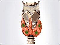
The adrenal cortex is the outer portion of the adrenal gland, which is located on top of the kidneys. It plays a crucial role in producing hormones that are essential for various bodily functions. The adrenal cortex is divided into three zones:
1. Zona glomerulosa: This outermost zone produces mineralocorticoids, primarily aldosterone. Aldosterone helps regulate sodium and potassium balance and thus influences blood pressure by controlling the amount of fluid in the body.
2. Zona fasciculata: The middle layer is responsible for producing glucocorticoids, with cortisol being the most important one. Cortisol regulates metabolism, helps manage stress responses, and has anti-inflammatory properties. It also plays a role in blood sugar regulation and maintaining the body's response to injury and illness.
3. Zona reticularis: The innermost zone produces androgens, primarily dehydroepiandrosterone (DHEA) and its sulfate form (DHEAS). These androgens are weak compared to those produced by the gonads (ovaries or testes), but they can be converted into more potent androgens or estrogens in peripheral tissues.
Disorders related to the adrenal cortex can lead to hormonal imbalances, affecting various bodily functions. Examples include Addison's disease (insufficient adrenal cortical hormone production) and Cushing's syndrome (excessive glucocorticoid levels).
Aldosterone is a hormone produced by the adrenal gland. It plays a key role in regulating sodium and potassium balance and maintaining blood pressure through its effects on the kidneys. Aldosterone promotes the reabsorption of sodium ions and the excretion of potassium ions in the distal tubules and collecting ducts of the nephrons in the kidneys. This increases the osmotic pressure in the blood, which in turn leads to water retention and an increase in blood volume and blood pressure.
Aldosterone is released from the adrenal gland in response to a variety of stimuli, including angiotensin II (a peptide hormone produced as part of the renin-angiotensin-aldosterone system), potassium ions, and adrenocorticotropic hormone (ACTH) from the pituitary gland. The production of aldosterone is regulated by a negative feedback mechanism involving sodium levels in the blood. High sodium levels inhibit the release of aldosterone, while low sodium levels stimulate its release.
In addition to its role in maintaining fluid and electrolyte balance and blood pressure, aldosterone has been implicated in various pathological conditions, including hypertension, heart failure, and primary hyperaldosteronism (a condition characterized by excessive production of aldosterone).
The Zona Fasciculata is a region within the adrenal gland, which is a small gland located on top of the kidneys. It plays an essential role in endocrine function. The adrenal gland is divided into two main parts: the outer cortex and the inner medulla. The cortex itself is further divided into three zones: the Zona Glomerulosa, the Zona Fasciculata, and the Zona Reticularis.
The Zona Fasciculata is the middle layer of the adrenal cortex. It is primarily responsible for producing and releasing steroid hormones, particularly glucocorticoids such as cortisol. Cortisol helps regulate metabolism, immune response, and stress response, among other functions. The Zona Fasciculata contains large, column-shaped cells called fasciculated cells that contain lipid droplets filled with cholesterol esters. These cells convert cholesterol into pregnenolone, which is then converted into cortisol through a series of enzymatic reactions.
In summary, the Zona Fasciculata is a crucial region within the adrenal gland that produces and releases cortisol, a vital glucocorticoid hormone involved in various physiological processes.
The adrenal glands are a pair of endocrine glands that are located on top of the kidneys. Each gland has two parts: the outer cortex and the inner medulla. The adrenal cortex produces hormones such as cortisol, aldosterone, and androgens, which regulate metabolism, blood pressure, and other vital functions. The adrenal medulla produces catecholamines, including epinephrine (adrenaline) and norepinephrine (noradrenaline), which help the body respond to stress by increasing heart rate, blood pressure, and alertness.
The zona reticularis is a layer of the adrenal cortex, which is the outer part of the adrenal gland. These glands are located on top of the kidneys and are responsible for producing several important hormones. The adrenal cortex itself has three distinct layers: the zona glomerulosa, the zona fasciculata, and the zona reticularis.
The zona reticularis is the innermost layer of the adrenal cortex. It is responsible for producing and releasing certain steroid hormones, particularly androgens such as dehydroepiandrosterone (DHEA) and its sulfate (DHEAS). These androgens are precursor hormones that can be converted into more potent androgens or estrogens in other parts of the body. The zona reticularis plays a crucial role in sexual development and function, as well as maintaining overall health and well-being.
Disorders related to the zona reticularis may result in abnormal hormone production, leading to conditions such as congenital adrenal hyperplasia, Cushing's syndrome, or Addison's disease. Proper diagnosis and treatment of these disorders typically involve endocrinologists, healthcare professionals specializing in hormonal and metabolic disorders.
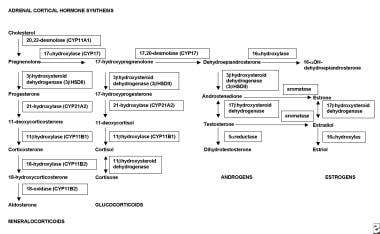
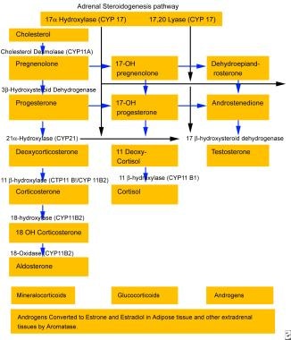
Aldosterone synthase is a steroidogenic enzyme that is primarily responsible for the production of the hormone aldosterone in the adrenal gland. It is encoded by the CYP11B2 gene and is located within the mitochondria of the zona glomerulosa cells in the adrenal cortex.
Aldosterone synthase catalyzes two key reactions in the biosynthesis of aldosterone: the conversion of corticosterone to 18-hydroxycorticosterone and the subsequent conversion of 18-hydroxycorticosterone to aldosterone. These reactions involve the sequential addition of hydroxyl groups at the C18 position of the steroid molecule, which is a critical step in the synthesis of aldosterone.
Aldosterone plays an important role in regulating blood pressure and electrolyte balance by increasing the reabsorption of sodium and water in the distal nephron of the kidney, while promoting the excretion of potassium. Disorders of aldosterone synthase can lead to conditions such as primary hyperaldosteronism, which is characterized by excessive production of aldosterone and can result in hypertension and hypokalemia.
Steroid 11-beta-hydroxylase is a crucial enzyme involved in the steroidogenesis pathway, specifically in the synthesis of cortisol and aldosterone, which are vital hormones produced by the adrenal glands. This enzyme is encoded by the CYP11B1 gene in humans.
The enzyme's primary function is to catalyze the conversion of 11-deoxycortisol to cortisol and 11-deoxycorticosterone to aldosterone through the process of hydroxylation at the 11-beta position of the steroid molecule. Cortisol is a critical glucocorticoid hormone that helps regulate metabolism, immune response, and stress response, while aldosterone is a mineralocorticoid hormone responsible for maintaining electrolyte and fluid balance in the body.
Deficiencies or mutations in the CYP11B1 gene can lead to various disorders, such as congenital adrenal hyperplasia (CAH), which may result in impaired cortisol and aldosterone production, causing hormonal imbalances and associated symptoms.
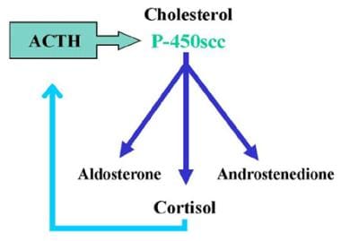
Adrenocorticotropic Hormone (ACTH) is a hormone produced and released by the anterior pituitary gland, a small endocrine gland located at the base of the brain. ACTH plays a crucial role in the regulation of the body's stress response and has significant effects on various physiological processes.
The primary function of ACTH is to stimulate the adrenal glands, which are triangular-shaped glands situated on top of the kidneys. The adrenal glands consist of two parts: the outer cortex and the inner medulla. ACTH specifically targets the adrenal cortex, where it binds to specific receptors and initiates a series of biochemical reactions leading to the production and release of steroid hormones, primarily cortisol (a glucocorticoid) and aldosterone (a mineralocorticoid).
Cortisol is involved in various metabolic processes, such as regulating blood sugar levels, modulating the immune response, and helping the body respond to stress. Aldosterone plays a vital role in maintaining electrolyte and fluid balance by promoting sodium reabsorption and potassium excretion in the kidneys.
ACTH release is controlled by the hypothalamus, another part of the brain, which produces corticotropin-releasing hormone (CRH). CRH stimulates the anterior pituitary gland to secrete ACTH, which in turn triggers cortisol production in the adrenal glands. This complex feedback system helps maintain homeostasis and ensures that appropriate amounts of cortisol are released in response to various physiological and psychological stressors.
Disorders related to ACTH can lead to hormonal imbalances, resulting in conditions such as Cushing's syndrome (excessive cortisol production) or Addison's disease (insufficient cortisol production). Proper diagnosis and management of these disorders typically involve assessing the function of the hypothalamic-pituitary-adrenal axis and addressing any underlying issues affecting ACTH secretion.
Angiotensin II is a potent vasoactive peptide hormone that plays a critical role in the renin-angiotensin-aldosterone system (RAAS), which is a crucial regulator of blood pressure and fluid balance in the body. It is formed from angiotensin I through the action of an enzyme called angiotensin-converting enzyme (ACE).
Angiotensin II has several physiological effects on various organs, including:
1. Vasoconstriction: Angiotensin II causes contraction of vascular smooth muscle, leading to an increase in peripheral vascular resistance and blood pressure.
2. Aldosterone release: Angiotensin II stimulates the adrenal glands to release aldosterone, a hormone that promotes sodium reabsorption and potassium excretion in the kidneys, thereby increasing water retention and blood volume.
3. Sympathetic nervous system activation: Angiotensin II activates the sympathetic nervous system, leading to increased heart rate and contractility, further contributing to an increase in blood pressure.
4. Thirst regulation: Angiotensin II stimulates the hypothalamus to increase thirst, promoting water intake and helping to maintain intravascular volume.
5. Cell growth and fibrosis: Angiotensin II has been implicated in various pathological processes, such as cell growth, proliferation, and fibrosis, which can contribute to the development of cardiovascular and renal diseases.
Angiotensin-converting enzyme inhibitors (ACEIs) and angiotensin receptor blockers (ARBs) are two classes of medications commonly used in clinical practice to target the RAAS by blocking the formation or action of angiotensin II, respectively. These drugs have been shown to be effective in managing hypertension, heart failure, and chronic kidney disease.
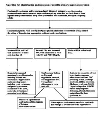
Hyperaldosteronism is a medical condition characterized by the overproduction of aldosterone, a hormone produced by the adrenal glands. Aldosterone helps regulate sodium and potassium balance and blood pressure by promoting sodium retention and potassium excretion in the kidneys.
There are two types of hyperaldosteronism: primary and secondary. Primary hyperaldosteronism is caused by an overproduction of aldosterone from an abnormality within the adrenal gland, such as a tumor (Conn's syndrome) or hyperplasia. Secondary hyperaldosteronism occurs when there is an excess production of renin, a hormone produced by the kidneys, which then stimulates the adrenal glands to produce more aldosterone. This can be caused by various conditions that affect kidney function, such as renal artery stenosis or heart failure.
Symptoms of hyperaldosteronism may include high blood pressure, low potassium levels (hypokalemia), muscle weakness, and frequent urination. Diagnosis typically involves measuring aldosterone and renin levels in the blood, as well as other tests to determine the underlying cause. Treatment depends on the type and cause of hyperaldosteronism but may include medications, surgery, or lifestyle changes.
I'm sorry for any confusion, but "Interrenal Gland" is not a recognized medical term in humans as we don't have this specific gland. However, in some animal species, particularly fish and amphibians, the interrenal gland is part of the adrenal gland equivalent, responsible for producing steroid hormones related to stress response and metabolism regulation. In humans and other mammals, these functions are carried out by the adrenal glands, specifically the adrenal cortex.
Corticosterone is a hormone produced by the adrenal gland in many animals, including humans. It is a type of glucocorticoid steroid hormone that plays an important role in the body's response to stress, immune function, metabolism, and regulation of inflammation. Corticosterone helps to regulate the balance of sodium and potassium in the body and also plays a role in the development and functioning of the nervous system. It is the primary glucocorticoid hormone in rodents, while cortisol is the primary glucocorticoid hormone in humans and other primates.
Mineralocorticoids are a class of steroid hormones that primarily regulate electrolyte and fluid balance in the body. The most important mineralocorticoid is aldosterone, which is produced by the adrenal gland in response to signals from the renin-angiotensin system. Aldosterone acts on the distal tubules and collecting ducts of the nephrons in the kidneys to increase the reabsorption of sodium ions (Na+) and water into the bloodstream, while promoting the excretion of potassium ions (K+) and hydrogen ions (H+) into the urine. This helps maintain blood pressure and volume, as well as ensuring a proper balance of electrolytes in the body. Other mineralocorticoids include cortisol and corticosterone, which have weak mineralocorticoid activity and play a more significant role as glucocorticoids, regulating metabolism and immune response.
"Cattle" is a term used in the agricultural and veterinary fields to refer to domesticated animals of the genus *Bos*, primarily *Bos taurus* (European cattle) and *Bos indicus* (Zebu). These animals are often raised for meat, milk, leather, and labor. They are also known as bovines or cows (for females), bulls (intact males), and steers/bullocks (castrated males). However, in a strict medical definition, "cattle" does not apply to humans or other animals.
Potassium is a essential mineral and an important electrolyte that is widely distributed in the human body. The majority of potassium in the body (approximately 98%) is found within cells, with the remaining 2% present in blood serum and other bodily fluids. Potassium plays a crucial role in various physiological processes, including:
1. Regulation of fluid balance and maintenance of normal blood pressure through its effects on vascular tone and sodium excretion.
2. Facilitation of nerve impulse transmission and muscle contraction by participating in the generation and propagation of action potentials.
3. Protein synthesis, enzyme activation, and glycogen metabolism.
4. Regulation of acid-base balance through its role in buffering systems.
The normal serum potassium concentration ranges from 3.5 to 5.0 mEq/L (milliequivalents per liter) or mmol/L (millimoles per liter). Potassium levels outside this range can have significant clinical consequences, with both hypokalemia (low potassium levels) and hyperkalemia (high potassium levels) potentially leading to serious complications such as cardiac arrhythmias, muscle weakness, and respiratory failure.
Potassium is primarily obtained through the diet, with rich sources including fruits (e.g., bananas, oranges, and apricots), vegetables (e.g., leafy greens, potatoes, and tomatoes), legumes, nuts, dairy products, and meat. In cases of deficiency or increased needs, potassium supplements may be recommended under the guidance of a healthcare professional.
Adrenomedullin receptors are a type of G protein-coupled receptor (GPCR) that bind to and are activated by the peptide hormone adrenomedullin. There are two main types of adrenomedullin receptors, identified as AM1 and AM2, which are formed by the combination of different subunits. The AM1 receptor is composed of the calcitonin receptor-like receptor (CLR) and receptor activity-modifying protein 2 (RAMP2), while the AM2 receptor is composed of CLR and RAMP3.
Adrenomedullin receptors play important roles in various physiological processes, including cardiovascular regulation, vasodilation, and inhibition of cell growth and proliferation. They are widely distributed throughout the body, particularly in the vascular system, kidneys, adrenal glands, and central nervous system. Activation of these receptors by adrenomedullin leads to a range of intracellular signaling events, including the activation of adenylyl cyclase, increased levels of cAMP, and activation of protein kinase A (PKA). These downstream effects contribute to the diverse biological activities of adrenomedullin.
In addition to adrenomedullin, related peptides such as adrenotensin and intermedin can also bind to and activate these receptors, albeit with lower affinity. Dysregulation of adrenomedullin receptor signaling has been implicated in several pathological conditions, including hypertension, heart failure, and cancer. As a result, targeting adrenomedullin receptors has emerged as a potential therapeutic strategy for the treatment of these diseases.
Adrenal cortex neoplasms refer to abnormal growths (tumors) in the adrenal gland's outer layer, known as the adrenal cortex. These neoplasms can be benign or malignant (cancerous). Benign tumors are called adrenal adenomas, while cancerous tumors are called adrenocortical carcinomas.
Adrenal cortex neoplasms can produce various hormones, leading to different clinical presentations. For instance, they may cause Cushing's syndrome (characterized by excessive cortisol production), Conn's syndrome (caused by aldosterone excess), or virilization (due to androgen excess). Some tumors may not produce any hormones and are discovered incidentally during imaging studies for unrelated conditions.
The diagnosis of adrenal cortex neoplasms typically involves a combination of imaging techniques, such as CT or MRI scans, and hormonal assessments to determine if the tumor is functional or non-functional. In some cases, a biopsy may be necessary to confirm the diagnosis and differentiate between benign and malignant tumors. Treatment options depend on the type, size, location, and hormonal activity of the neoplasm and may include surgical excision, radiation therapy, chemotherapy, or a combination of these approaches.
Renin is a medically recognized term and it is defined as:
"A protein (enzyme) that is produced and released by specialized cells (juxtaglomerular cells) in the kidney. Renin is a key component of the renin-angiotensin-aldosterone system (RAAS), which helps regulate blood pressure and fluid balance in the body.
When the kidney detects a decrease in blood pressure or a reduction in sodium levels, it releases renin into the bloodstream. Renin then acts on a protein called angiotensinogen, converting it to angiotensin I. Angiotensin-converting enzyme (ACE) subsequently converts angiotensin I to angiotensin II, which is a potent vasoconstrictor that narrows blood vessels and increases blood pressure.
Additionally, angiotensin II stimulates the adrenal glands to release aldosterone, a hormone that promotes sodium reabsorption in the kidneys and increases water retention, further raising blood pressure.
Therefore, renin plays a critical role in maintaining proper blood pressure and electrolyte balance in the body."
Atrial natriuretic factor (ANF) receptors are specialized proteins found on the surface of certain cells in the body, primarily in the kidneys, heart, and blood vessels. They play a crucial role in regulating blood pressure, volume, and electrolyte balance.
There are two main types of ANF receptors: type A and type B. Type A receptors, also known as guanylyl cyclase-A (GC-A) receptors, are found in the kidneys, heart, and blood vessels. When ANF binds to these receptors, it triggers a series of reactions that lead to an increase in the production of a molecule called cyclic GMP (cGMP). This, in turn, causes vasodilation (relaxation of blood vessels), increased urine production, and reduced sodium reabsorption in the kidneys, all of which help lower blood pressure.
Type B receptors, on the other hand, are found mainly in the brain and have been shown to modulate the release of ANF from the heart. When ANF binds to type B receptors, it inhibits the release of vasopressin, a hormone that helps regulate water balance in the body. This further contributes to the overall effects of ANF on blood pressure and fluid balance.
Overall, ANF receptors are essential components of the complex system that helps maintain homeostasis in the cardiovascular and renal systems.
Adrenomedullin is a hormone that is produced and released by the adrenal glands, specifically from the chromaffin cells in the adrenal medulla. It is a small peptide made up of 52 amino acids and has various physiological functions, including vasodilation, bronchodilation, and inhibition of cell growth.
Adrenomedullin acts as a potent vasodilator by binding to specific G protein-coupled receptors in the vascular smooth muscle cells, leading to relaxation of the blood vessels. It also has a role in regulating blood pressure and fluid balance in the body.
In addition to its effects on the cardiovascular system, adrenomedullin has been shown to have anti-inflammatory and neuroprotective properties. It is involved in various physiological processes such as wound healing, tissue repair, and angiogenesis (the formation of new blood vessels).
Abnormal levels of adrenomedullin have been implicated in several disease states, including hypertension, heart failure, sepsis, and cancer. Therefore, measuring adrenomedullin levels in the body can provide valuable diagnostic and prognostic information for these conditions.
Angiotensin receptors are a type of G protein-coupled receptor that binds the angiotensin peptides, which are important components of the renin-angiotensin-aldosterone system (RAAS). The RAAS is a hormonal system that regulates blood pressure and fluid balance.
There are two main types of angiotensin receptors: AT1 and AT2. Activation of AT1 receptors leads to vasoconstriction, increased sodium and water reabsorption in the kidneys, and cell growth and proliferation. On the other hand, activation of AT2 receptors has opposite effects, such as vasodilation, natriuresis (increased excretion of sodium in urine), and anti-proliferative actions.
Angiotensin II is a potent activator of AT1 receptors, while angiotensin IV has high affinity for AT2 receptors. Angiotensin-converting enzyme (ACE) inhibitors and angiotensin receptor blockers (ARBs) are two classes of drugs that target the RAAS by blocking the formation or action of angiotensin II, leading to decreased activation of AT1 receptors and improved cardiovascular outcomes.
Adrenal gland neoplasms refer to abnormal growths or tumors in the adrenal glands. These glands are located on top of each kidney and are responsible for producing hormones that regulate various bodily functions such as metabolism, blood pressure, and stress response. Adrenal gland neoplasms can be benign (non-cancerous) or malignant (cancerous).
Benign adrenal tumors are called adenomas and are usually small and asymptomatic. However, some adenomas may produce excessive amounts of hormones, leading to symptoms such as high blood pressure, weight gain, and mood changes.
Malignant adrenal tumors are called adrenocortical carcinomas and are rare but aggressive cancers that can spread to other parts of the body. Symptoms of adrenocortical carcinoma may include abdominal pain, weight loss, and hormonal imbalances.
It is important to diagnose and treat adrenal gland neoplasms early to prevent complications and improve outcomes. Diagnostic tests may include imaging studies such as CT scans or MRIs, as well as hormone level testing and biopsy. Treatment options may include surgery, radiation therapy, chemotherapy, or a combination of these approaches.
"Cells, cultured" is a medical term that refers to cells that have been removed from an organism and grown in controlled laboratory conditions outside of the body. This process is called cell culture and it allows scientists to study cells in a more controlled and accessible environment than they would have inside the body. Cultured cells can be derived from a variety of sources, including tissues, organs, or fluids from humans, animals, or cell lines that have been previously established in the laboratory.
Cell culture involves several steps, including isolation of the cells from the tissue, purification and characterization of the cells, and maintenance of the cells in appropriate growth conditions. The cells are typically grown in specialized media that contain nutrients, growth factors, and other components necessary for their survival and proliferation. Cultured cells can be used for a variety of purposes, including basic research, drug development and testing, and production of biological products such as vaccines and gene therapies.
It is important to note that cultured cells may behave differently than they do in the body, and results obtained from cell culture studies may not always translate directly to human physiology or disease. Therefore, it is essential to validate findings from cell culture experiments using additional models and ultimately in clinical trials involving human subjects.
Egg proteins, also known as egg white proteins or ovalbumin, refer to the proteins found in egg whites. There are several different types of proteins found in egg whites, including:
1. Ovalbumin (54%): This is the major protein found in egg whites and is responsible for their white color. It has various functions such as providing nutrition, maintaining the structural integrity of the egg, and protecting the egg from bacteria.
2. Conalbumin (13%): Also known as ovotransferrin, this protein plays a role in the defense against microorganisms by binding to iron and making it unavailable for bacterial growth.
3. Ovomucoid (11%): This protein is resistant to digestion and helps protect the egg from being broken down by enzymes in the digestive tract of predators.
4. Lysozyme (3.5%): This protein has antibacterial properties and helps protect the egg from bacterial infection.
5. Globulins (4%): These are a group of simple proteins found in egg whites that have various functions such as providing nutrition, maintaining the structural integrity of the egg, and protecting the egg from bacteria.
6. Avidin (0.05%): This protein binds to biotin, a vitamin, making it unavailable for use by the body. However, cooking denatures avidin and makes the biotin available again.
Egg proteins are highly nutritious and contain all nine essential amino acids, making them a complete source of protein. They are also low in fat and cholesterol, making them a popular choice for those following a healthy diet.
Nitrendipine is an antihypertensive drug, which belongs to the class of calcium channel blockers. It works by relaxing and widening the blood vessels, making it easier for the heart to pump blood and reducing the workload on the cardiovascular system. This helps to lower high blood pressure (hypertension) and improve overall cardiovascular health. Nitrendipine is available in oral tablet form and is typically prescribed by a healthcare professional for the treatment of hypertension.
It's important to note that this definition is intended to be a general overview of the medical use and properties of Nitrendipine, and it should not be used as a substitute for professional medical advice or treatment. Always consult with a qualified healthcare provider for information regarding any specific medical condition or treatment.
Atrial natriuretic factor (ANF), also known as atrial natriuretic peptide (ANP), is a hormone that is primarily produced and secreted by the atria of the heart in response to stretching of the cardiac muscle cells due to increased blood volume. ANF plays a crucial role in regulating body fluid homeostasis, blood pressure, and cardiovascular function.
The main physiological action of ANF is to promote sodium and water excretion by the kidneys, which helps lower blood volume and reduce blood pressure. ANF also relaxes vascular smooth muscle, dilates blood vessels, and inhibits the renin-angiotensin-aldosterone system (RAAS), further contributing to its blood pressure-lowering effects.
Defects in ANF production or action have been implicated in several cardiovascular disorders, including heart failure, hypertension, and kidney disease. Therefore, ANF and its analogs are being investigated as potential therapeutic agents for the treatment of these conditions.
Sperm-ovum interactions, also known as sperm-egg interactions, refer to the specific series of events that occur between a spermatozoon (sperm) and an oocyte (egg or ovum) during fertilization in sexual reproduction.
The process begins with the sperm's attachment to the zona pellucida, a glycoprotein layer surrounding the oocyte. This interaction is mediated by specific proteins on the surface of both the sperm and the zona pellucida. Following attachment, the sperm undergoes the acrosome reaction, during which enzymes are released from the sperm's head to help digest and penetrate the zona pellucida.
Once the sperm has successfully traversed the zona pellucida, it makes contact with the oocyte's plasma membrane, triggering the fusion of the sperm and egg membranes. This results in the release of the sperm's genetic material into the oocyte's cytoplasm and the initiation of a series of intracellular signaling events within the oocyte that ultimately lead to its completion of meiosis II and formation of a zygote, marking the beginning of embryonic development.
Proper sperm-ovum interactions are crucial for successful fertilization and subsequent embryonic development, and any disruptions in these processes can result in infertility or early pregnancy loss.
Sprague-Dawley rats are a strain of albino laboratory rats that are widely used in scientific research. They were first developed by researchers H.H. Sprague and R.C. Dawley in the early 20th century, and have since become one of the most commonly used rat strains in biomedical research due to their relatively large size, ease of handling, and consistent genetic background.
Sprague-Dawley rats are outbred, which means that they are genetically diverse and do not suffer from the same limitations as inbred strains, which can have reduced fertility and increased susceptibility to certain diseases. They are also characterized by their docile nature and low levels of aggression, making them easier to handle and study than some other rat strains.
These rats are used in a wide variety of research areas, including toxicology, pharmacology, nutrition, cancer, and behavioral studies. Because they are genetically diverse, Sprague-Dawley rats can be used to model a range of human diseases and conditions, making them an important tool in the development of new drugs and therapies.
Sugar phosphates are organic compounds that play crucial roles in various biological processes, particularly in the field of genetics and molecular biology. They are formed by the attachment of a phosphate group to a sugar molecule, most commonly to the 5-carbon sugar ribose or deoxyribose.
In genetics, sugar phosphates form the backbone of nucleic acids, such as DNA and RNA. In DNA, the sugar phosphate backbone consists of alternating deoxyribose (a sugar) and phosphate groups, linked together by covalent bonds between the 5' carbon atom of one sugar molecule and the 3' carbon atom of another sugar molecule. This forms a long, twisted ladder-like structure known as a double helix.
Similarly, in RNA, the sugar phosphate backbone is formed by ribose (a sugar) and phosphate groups, creating a single-stranded structure that can fold back on itself to form complex shapes. These sugar phosphate backbones provide structural support for the nucleic acids and help to protect the genetic information stored within them.
Sugar phosphates also play important roles in energy metabolism, as they are involved in the formation and breakdown of high-energy compounds such as ATP (adenosine triphosphate) and GTP (guanosine triphosphate). These molecules serve as energy currency for cells, storing and releasing energy as needed to power various cellular processes.
Hydrocortisone is a synthetic glucocorticoid, which is a class of steroid hormones. It is identical to the naturally occurring cortisol, a hormone produced by the adrenal gland that helps regulate metabolism and helps your body respond to stress. Hydrocortisone has anti-inflammatory effects and is used to treat various inflammatory conditions such as allergies, skin disorders, and autoimmune diseases. It works by suppressing the immune system's response to reduce swelling, redness, itching, and other symptoms caused by inflammation.
Hydrocortisone is available in different forms, including oral tablets, topical creams, lotions, gels, and ointments, as well as injectable solutions. The specific use and dosage depend on the condition being treated and the individual patient's medical history and current health status.
As with any medication, hydrocortisone can have side effects, especially when used in high doses or for extended periods. Common side effects include increased appetite, weight gain, mood changes, insomnia, and skin thinning. Long-term use of hydrocortisone may also increase the risk of developing osteoporosis, diabetes, cataracts, and other health problems. Therefore, it is essential to follow your healthcare provider's instructions carefully when using this medication.
In situ hybridization (ISH) is a molecular biology technique used to detect and localize specific nucleic acid sequences, such as DNA or RNA, within cells or tissues. This technique involves the use of a labeled probe that is complementary to the target nucleic acid sequence. The probe can be labeled with various types of markers, including radioisotopes, fluorescent dyes, or enzymes.
During the ISH procedure, the labeled probe is hybridized to the target nucleic acid sequence in situ, meaning that the hybridization occurs within the intact cells or tissues. After washing away unbound probe, the location of the labeled probe can be visualized using various methods depending on the type of label used.
In situ hybridization has a wide range of applications in both research and diagnostic settings, including the detection of gene expression patterns, identification of viral infections, and diagnosis of genetic disorders.
Messenger RNA (mRNA) is a type of RNA (ribonucleic acid) that carries genetic information copied from DNA in the form of a series of three-base code "words," each of which specifies a particular amino acid. This information is used by the cell's machinery to construct proteins, a process known as translation. After being transcribed from DNA, mRNA travels out of the nucleus to the ribosomes in the cytoplasm where protein synthesis occurs. Once the protein has been synthesized, the mRNA may be degraded and recycled. Post-transcriptional modifications can also occur to mRNA, such as alternative splicing and addition of a 5' cap and a poly(A) tail, which can affect its stability, localization, and translation efficiency.
Electron microscopy (EM) is a type of microscopy that uses a beam of electrons to create an image of the sample being examined, resulting in much higher magnification and resolution than light microscopy. There are several types of electron microscopy, including transmission electron microscopy (TEM), scanning electron microscopy (SEM), and reflection electron microscopy (REM).
In TEM, a beam of electrons is transmitted through a thin slice of the sample, and the electrons that pass through the sample are focused to form an image. This technique can provide detailed information about the internal structure of cells, viruses, and other biological specimens, as well as the composition and structure of materials at the atomic level.
In SEM, a beam of electrons is scanned across the surface of the sample, and the electrons that are scattered back from the surface are detected to create an image. This technique can provide information about the topography and composition of surfaces, as well as the structure of materials at the microscopic level.
REM is a variation of SEM in which the beam of electrons is reflected off the surface of the sample, rather than scattered back from it. This technique can provide information about the surface chemistry and composition of materials.
Electron microscopy has a wide range of applications in biology, medicine, and materials science, including the study of cellular structure and function, disease diagnosis, and the development of new materials and technologies.
Mitochondria are specialized structures located inside cells that convert the energy from food into ATP (adenosine triphosphate), which is the primary form of energy used by cells. They are often referred to as the "powerhouses" of the cell because they generate most of the cell's supply of chemical energy. Mitochondria are also involved in various other cellular processes, such as signaling, differentiation, and apoptosis (programmed cell death).
Mitochondria have their own DNA, known as mitochondrial DNA (mtDNA), which is inherited maternally. This means that mtDNA is passed down from the mother to her offspring through the egg cells. Mitochondrial dysfunction has been linked to a variety of diseases and conditions, including neurodegenerative disorders, diabetes, and aging.
Immunohistochemistry (IHC) is a technique used in pathology and laboratory medicine to identify specific proteins or antigens in tissue sections. It combines the principles of immunology and histology to detect the presence and location of these target molecules within cells and tissues. This technique utilizes antibodies that are specific to the protein or antigen of interest, which are then tagged with a detection system such as a chromogen or fluorophore. The stained tissue sections can be examined under a microscope, allowing for the visualization and analysis of the distribution and expression patterns of the target molecule in the context of the tissue architecture. Immunohistochemistry is widely used in diagnostic pathology to help identify various diseases, including cancer, infectious diseases, and immune-mediated disorders.
"Wistar rats" are a strain of albino rats that are widely used in laboratory research. They were developed at the Wistar Institute in Philadelphia, USA, and were first introduced in 1906. Wistar rats are outbred, which means that they are genetically diverse and do not have a fixed set of genetic characteristics like inbred strains.
Wistar rats are commonly used as animal models in biomedical research because of their size, ease of handling, and relatively low cost. They are used in a wide range of research areas, including toxicology, pharmacology, nutrition, cancer, cardiovascular disease, and behavioral studies. Wistar rats are also used in safety testing of drugs, medical devices, and other products.
Wistar rats are typically larger than many other rat strains, with males weighing between 500-700 grams and females weighing between 250-350 grams. They have a lifespan of approximately 2-3 years. Wistar rats are also known for their docile and friendly nature, making them easy to handle and work with in the laboratory setting.
 Zona glomerulosa
 "A strain difference in the adrenal zona glomerulosa determined by one " by J G. Shire
 Pediatric Adrenal Gland Disorders: Anatomy, Embryology, Physiology
 BRS Endocrine Physiology Quiz: Trivia! - ProProfs Quiz
 Nitrite (JECFA Food Additives Series 50)
 Primary Aldosteronism: Screening and Confirmatory Testing | myADLM.org
 Glass Slide images Flashcards by brittany gouse | Brainscape
 Medical Pharmacology: Vasoactive Peptides
 DACH1 Antibodies
 17.6 The Adrenal Glands - Anatomy and Physiology | OpenStax
 Mitaban for Dogs - Drugs.com
 Do kidneys secrete aldosterone?
 Registration Dossier - ECHA
 Adrenals - Oakton Primary Care Centers
 The Adrenal Glands - Biology of Aging
 What tissues make up the endocrine system? - My Medical Health Blog
 Synonyms
 Help cats with hyperaldosteronism hold their heads high
 Legit Internal Hacks | VAC Undetected, Legacy, Executor - Seminário Nacional de Planejamento e Desenvolvimento
 Kate Shared 'Prenursing Smarter Module Three | Anatomy and Physiology' - 23 Picmonics
 Civil Service Exam 2023 | UPSC | IAS | TNPSC |Mock Tests